drug resistance and the functional role of micrornas in
TRANSCRIPT

Drug resistance and the functional role of
microRNAs in neuroblastoma
Swapnil Parashram Bhavsar
A dissertation for the degree of Philosophiae Doctor - February 2019
Navn / stilling / tittel / 01.01.2014

Drug resistance and the functional role of
microRNAs in neuroblastoma
Swapnil Parashram Bhavsar
A dissertation for the degree of Philosophiae Doctor
UiT – The Arctic University of Norway
Faculty of Health Science
Department of Clinical Medicine
Pediatric Research Group
Tromsø, Norway
February
2019


Table of Contents Acknowledgements ................................................................................................................................. 1
List of abbreviations ................................................................................................................................ 2
Summary ................................................................................................................................................. 4
List of papers ........................................................................................................................................... 5
Introduction ............................................................................................................................................. 6
Neuroblastoma ........................................................................................................................................ 6
Origin and development ...................................................................................................................... 6
Biology and genomics .......................................................................................................................... 6
Clinical aspects .................................................................................................................................... 8
Drug resistance in human cancer .......................................................................................................... 10
Mechanisms of drug resistance ......................................................................................................... 10
Drug transport: Influx and efflux ....................................................................................................... 10
Drug metabolism: activation and inactivation .................................................................................. 13
Alterations in drug targets ................................................................................................................ 13
DNA damage repair mechanisms ...................................................................................................... 14
Downstream resistance mechanisms in cancer ................................................................................ 15
Role of tumor microenvironment in drug resistance ........................................................................ 16
Cancer stem cells and drug resistance .............................................................................................. 16
Small RNAs in human cancer ................................................................................................................. 17
MicroRNAs: Biogenesis and mode of action ..................................................................................... 18
MicroRNAs and their roles in human cancer .................................................................................... 21
MicroRNAs as oncogenes and tumor suppressors ............................................................................ 23
Mechanisms of microRNA deregulation in cancer ............................................................................ 25
MicroRNAs and drug resistance in human cancer ............................................................................ 29
MicroRNA as therapeutics ................................................................................................................. 31
The CCND1 and STAT3 oncogenes in human cancer ............................................................................. 34
CCND1 ................................................................................................................................................ 34
STAT3 ................................................................................................................................................. 36
Aims of the study ................................................................................................................................... 38
Summary of papers ............................................................................................................................... 39
Discussion (Paper I, II & III) .................................................................................................................... 42
References ............................................................................................................................................. 51
Appendix (Paper I, II & III) ..................................................................................................................... 65

1
Acknowledgements
I would like to express my sincere gratitude to the following creatures and institutions:
To my main supervisor, Prof.Christer Einvik who taught me through his questions, answers
and useful advices. Also for his continuous support, patience and motivation and for enabling
me to see things from other perspectives.
To my co-supervisor, Prof.Trond Flægstad who with his great sense of humour always kept
the pleasant environment at work and for his motivation, feedback and advices in the project.
And also to my second co-supervisor, Prof.Baldur Sveinbjørnsson for his guidance.
To my lab. engineer, Cecilie Løkke and Roy Lyså for technical assistance and expert advice
which helped me get a better understanding of laboratory techniques.
To my former and present lab. colleagues Sarah Roth, Lotte Olsen, Peter Utnes, Øyvind Hald,
Bjørn Helge Haug and Vera Susana for the interesting discussions and/or help in the
laboratory. Especially to Lotte Olsen for reading the thesis and manuscript thoroughly.
To my friends working on the 9th floor (MH-building): Pradip Bhujabal and Divya Borra for
their help and advice from their respective areas of research expertise.
To the Tromsø University, IKM, Northern-Norway Health Authorities, Simon Fougner
Hartmanns Familiefond and Barnekreftforeningen who funded my PhD project.
To my friends in the beautiful city of Tromsø and Bergen - Rahul & Priya Haware, Sujata
Bhujabal, Sagar & Sampada Darvekar, Chandra & Sunitha Ravuri, Shripati & Tejaswini Bhat,
Birendra & Neelam Shrestha, Rashmi Narawane, Ganesh Narawane, Kishore Kosuri, Diana
Canova, Jessin Janice & Sudhagar Balasundaram, Pearl, Samkumar Amos and Linn Evenseth
for all the fun and enjoyment other than work.
To Hemant Kedari and Shankar Darje who handled the important things in India in my
absence. And also to Anuja & Neeta Darje, Arun & Pramila Kedari, Rahul & Nitin Kedari,
Amar & Naritha Pukale, Kamlakar & Tara Pukale and Amol Pukale for all their important
help during my initial days in Sweden and Norway.
To my friends in India, Prasad Deshmane Sir, Monish & Apurva Babariya, Mayuri & Pratik
Nawalakha, Krishna Sangale, Rishikesh Autade and Ashutosh Toll for their warm friendship.
To all my family members, including Vasantrao & Leela Bhavsar, Dinesh & Sadhana
Bhavsar, Yamini and Abhishek Bhawsar, Mohit & Soham Kedari for their love and support.
To my parents, Parashram & Alka Bhavsar; sister, Aarti Bhavsar Kedari and wife, Nivedita
Bhavsar for their faith, encouragement and unconditional love and support.
February, 2019
Swapnil Parashram Bhavsar Tromsø, Norway

2
List of abbreviations
ABC ATP-Binding Cassette
ALK Anaplastic Lymphoma Kinase
BCRP Breast Cancer Resistance Protein
BCL-2 B-Cell Lymphoma 2
BCL2L1 (BCL-XL) BCL-2 Like 1
BAX BCL-2 Associated X
BAK BCL-2 Antagonist /Killer
CAR Constitutive Androstane Receptor
CSCs Cancer Stem Cells
CCND1 Cyclin D1
CDDP Cisplatin
CDK Cyclin-Dependent Kinase
c-MYC v-Myc Avian Myelocytomatosis Viral Oncogene Homolog
dsRNA Double-stranded RNA
DGCR8 DiGeorge Critical 8
DNMT DNA Methyl Transferase
DXR Doxorubicin
Exp5 Exportin 5
ETOP Etoposide
EGFR Epidermal Growth Factor Receptor
E2F3 E2F Transcription Factor 3
GSH Glutathione
HDAC Histone Deacetylase
HSRs Homogeneously Staining Regions
IGH Immunoglobulin Heavy Chain Locus
INSS International Neuroblastoma Staging System
INRG International Neuroblastoma Risk Group
INRGSS International Neuroblastoma Risk Group Staging System
IDRF Image-Defined Risk Factors
KRAS Kirsten Rat Sarcoma Viral Oncogene Homolog
LOH Loss of Heterozygosity
3
lncRNAs Long noncoding RNAs
MYCN v-Myc Avian Myelocytomatosis Viral Oncogene Neuroblastoma
derived Homolog
miRNA MicroRNA
miRISC MicroRNA Inducing Silencing Complex
MNA MYCN Amplified
mRNA Messenger RNA
MDR1 Multidrug Resistance Protein-1
MRP-1 Multidrug Resistance associated Protein-1
MRE MicroRNA Recognition Element
MMR Mismatch Repair
MCL-1 Myeloid Cell Leukemia-1
MAP3K9 Mitogen-Activated Protein Kinase Kinase Kinase 9
NAIP Neural Apoptosis Inhibitory Protein
NER Nucleotide Excision Repair
NHEJ Non-Homologous End Joining
NRAS Neuroblastoma RAS Viral Oncogene Homolog
NSCLC Non-Small Cell Lung Cancer
ORF Open Reading Frame
Pre-miRNA Precursor microRNA
Pri-mRNA Primary microRNA
PTEN Phosphatase and Tensin Homolog
PARP-1 Poly (ADP-Ribose)–Polymerase 1
RAS Rat Sarcoma
RIIID RNase III domain
SCID Severe Combined Immunodeficient Mouse
siRNA Small Interfering RNA
STAT3 Signal Transducer and Activator of Transcription 3
SLC Solute Carrier
TERF1 Telomeric Repeat Binding Factor 1
TP53 Tumor Protein P53
UTR Un-Translated Region

4
Summary
This thesis sheds light on the identification and functional role of microRNAs
(miRNAs) in neuroblastoma chemoresistance. Neuroblastoma is an embryonal malignancy of
childhood and the biology of neuroblastoma tumors is complex and dramatically heterogeneous
(Brodeur, 2003). Most of the children diagnosed above one year of age, shows metastatic
disease and poor prognosis (Brodeur, 2003). Chemotherapy is one of the principle mode of
treatment for the metastatic disease. However, resistance to drug represents the major clinical
obstacle for the effective treatment of cancer. MiRNAs are small, endogenous, non-coding
RNAs that reduce the translation of target mRNAs (Croce and Calin, 2005). Recently, miRNAs
have been shown to modulate drug resistance in multiple cancers. However, the role of miRNAs
in neuroblastoma chemoresistance is limited and poorly understood. In this study, we set out to
identify miRNAs (and their networks/pathways) along with their functional targets involved in
mediating drug resistance in neuroblastoma.
In paper I, we performed miRNA profiling studies, by employing next generation deep
sequencing technique to identify 34-downregulated and 8-upregulated miRNAs differentially
expressed in neuroblastoma cell lines isolated from six patients at diagnosis (before therapy)
and from the same patients at relapse (after therapy). Interestingly, 22 of the 34-downregulated
miRNAs were located on chromosome 14q32 locus. MiRNAs from this locus are previously
reported to be downregulated in multiple human cancers (Zehavi et al., 2012). Moreover, we
also demonstrated that reduced expression of certain chromosome 14q32 miRNAs correlates
with poor clinical outcome in a cohort consisting of 226-primary neuroblastomas. Furthermore,
Ingenuity pathway analysis (IPA) of differentially expressed miRNAs revealed important
biological pathways associated with the cancer progression and drug resistance. Hence, in this
study, we identified a unique set of miRNAs which may be involved in the development of drug
resistance in neuroblastoma.
In paper II and III, we have investigated the previously unknown functional role of
chromosome 14q32 located miRNAs (identified in Paper I), miR-376c-3p and miR-323a-3p
which are downregulated in resistant neuroblastoma cell lines. Using a set of molecular
methods, like flow cytometry, reverse transcriptase-polymerase chain reaction (RT-qPCR),
western blot and luciferase reporter assay we confirm and validate CCND1 as a direct target of
miR-376c-3p and STAT3 as a direct target of miR-323a-3p in neuroblastoma. We show that,
miR-376c-3p targets CCND1 and induce G1-cell cycle arrest whereas miR-323a-3p targets
STAT3 and induce G1-cell cycle arrest and apoptosis in neuroblastoma cells.

5
List of papers
Paper-I
Next generation sequencing of microRNAs from isogenic neuroblastoma cell lines
isolated before and after treatment
Roth S.A., Knutsen E., Fiskaa T., Utnes P., Bhavsar S.P., Hald O.H., Løkke C., Mestdagh P.,
Johansen S.D., Flægstad T., & Einvik C.
Cancer Letters (2016) 372(1):128-136
Paper-II
Hsa-miR-376c-3p targets Cyclin D1 and induces G1-cell cycle arrest in neuroblastoma
cells
Bhavsar S.P., Løkke C., Flægstad T., & Einvik C.
Oncology Letters (2018) 16(5):6786-6794
Paper-III
Hsa-miR-323a-3p targets STAT3 and induces G1-cell cycle arrest and apoptosis in
neuroblastoma cells
Bhavsar S.P., Olsen L., Løkke C., Flægstad T., & Einvik C.
Manuscript

6
Introduction
Neuroblastoma
Origin and development
Neuroblastoma is an embryonal malignancy, which arises from the sympathoadrenal
lineage of neural crest cells during development. Neural crest cells are the transient, migratory
population of cells found in the embryo during development of the neural tube. These cells
migrate and differentiates into diverse cell types including, peripheral neurons, enteric neurons,
glia, melanocytes, cartilage, schwann cells, cells of the craniofacial skeleton and adrenal
medulla (Knecht and Bronner-Fraser, 2002, Cheung and Dyer, 2013). The induction of neural
crest cells and subsequent specification of the neural crest-derived cells is dependent upon the
co-operative intrinsic and extrinsic functions of the regulatory gene network (Tsubota and
Kadomatsu, 2018).
Since neuroblastoma arises from the developing tissues, it generally occurs in infants
and young children and the median age of diagnosis is 17 months (Maris, 2010). In addition,
most of the children diagnosed above one year of age, shows metastatic disease and poor
prognosis (Brodeur, 2003). The incidence of neuroblastoma is 10.2 per million children under
15 years of age and it is necessary to improve outcome because 15% of all pediatric cancer
related deaths are due to neuroblastoma (Maris, 2010, Cheung and Dyer, 2013)
The biology of neuroblastoma tumors is complex and dramatically heterogeneous
(Brodeur, 2003). The tumors may contain different types of neuroblastoma cells, which include
cells resembling immature sympathetic neurons (N-type cells), non-neuronal schwann cell-like
(S-type cells) and stem-like cells (I-type) cells. Patients with S-type Schwann cells have better
outcome (Ratner et al., 2016).
Biology and genomics
Amplification of the MYC-family proto-oncogene, v-Myc Avian Myelocytomatosis
Viral Oncogene Neuroblastoma Derived Homolog (MYCN) is one of the most common genetic
abnormalities in neuroblastoma. MYCN amplification is found in approximately 25% of cases
and it is highly correlated with advanced stage of disease and poor prognosis (Brodeur et al.,
1984, Huang and Weiss, 2013). The MYCN gene was originally discovered as amplified DNA
sequences in the form of double minute chromosomes (DMs) and homogeneously staining
regions (HSRs) in cells of neuroblastoma (Schwab et al., 1983, Kohl et al., 1983).

7
Because of the MYCN amplification, high levels of N-myc protein is generated; this
protein is shown to act as a transcriptional regulator of certain genes involved in proliferation
and differentiation processes, thus leading to tumorigenesis (Negroni et al., 1991, Wenzel et al.,
1991, Wenzel and Schwab, 1995). In addition, MYCN has been shown to regulate oncogenic
miRNAs in neuroblastoma (Schulte et al., 2008). Moreover, MYCN is also shown to sensitize
the neuroblastoma cells for drug-induced apoptosis (Fulda et al., 1999). MYCN is therefore
considered a promising target for therapeutic intervention. Although direct targeting of MYCN
is not yet achieved, alternative indirect approaches of MYCN targeting will most certainly gain
success in future (Chen et al., 2018b).
Chromosomal gains and losses are the means of frequently occurring genetic variations
in human neuroblastoma. Caron and colleagues studied the allelic loss of certain chromosomes
in 89-neuroblastomas and they found that loss of chromosome 1p is a strong prognostic factor
in patients with neuroblastoma, independently of age and stage (Caron et al., 1996). Similarly,
chromosome 11q is also frequently deleted in neuroblastoma and that unbalanced 11q and 1p36
loss of heterozygosity (LOH) are independently associated with a worse outcome in patients
with neuroblastoma (Attiyeh et al., 2005). In contrast to deletions, gain of 17q is one of the
most frequent cytogenetic abnormalities in neuroblastoma cells. The 17q gain serves as an
important prognostic factor with negative outcome in children with neuroblastoma (Bown et
al., 1999). Additionally, gains of 1q, 2p, 7q, 11p and losses of 3p, 4p, 9p and 14q have also
been documented in neuroblastoma (Cheung and Dyer, 2013).
Mutations are observed in certain genes in neuroblastoma. Mutations in the paired like
homeobox 2b (PHOX2B) which is the regulator of normal autonomic nervous system
development was the first predisposing gene identified and linked to hereditary neuroblastoma
(Trochet et al., 2004). In addition, heritable mutations in the anaplastic lymphoma kinase (ALK)
gene, which encodes a receptor tyrosine kinase, lead to its constitutive activation and in vivo
tumor formation in mouse models (George et al., 2008, Janoueix-Lerosey et al., 2008, Mosse
et al., 2008, Chen et al., 2008). Moreover, somatic mutations were also observed in alpha
thalassemia/mental retardation syndrome X-linked (ATRX) gene in a cohort of 104 patients with
advanced stage neuroblastoma (Cheung et al., 2012). Albeit with low frequencies, mutations
have also been identified in protein tyrosine phosphatase-non receptor type 11 (PTPN11),
neuroblastoma RAS viral oncogene homolog (NRAS) and MYCN (Pugh et al., 2013).

8
Clinical aspects
Neuroblastoma may arise anywhere along the parasympathetic nervous system in the
neck, chest, abdomen or pelvis. However, tumors are mostly found in the adrenal medulla or
paraspinal ganglia (Maris, 2010) (Figure 1). The presence of tumor cells in the bone marrow or
tissue biopsies and the higher levels of urine catecholamines can be used to diagnose
neuroblastomas (Maris et al., 2007).
Figure 1. Clinical presentations in neuroblastoma. The primary distribution of neuroblastoma in children is mostly
in the neck, chest, abdomen and pelvis. Tumors are mainly observed in paraspnial nerve tissue and adrenal glands.
Illustration available from: https://ghr.nlm.nih.gov/condition/neuroblastoma with permission from the National
Cancer Institute © (2014) Terese Winslow LLC, U.S. Govt.

9
Staging of cancer helps to plan treatment and predict patient’s outcome. In 1988, an
internationally accepted staging system based on clinical, radiographic and surgical evaluation
was developed called International Neuroblastoma Staging System (INSS). However,
modifications of INSS were proposed later for the incorporation of both clinical and biological
features in the prediction of prognosis (Brodeur et al., 1993, Brodeur et al., 1988). Therefore,
in 2009, a new pre-treatment risk-stratification classification system called International
Neuroblastoma Risk Group (INRG) was established using 13 prognostic factors in a cohort of
8,800 children diagnosed with neuroblastoma to facilitate the comparison of risk based clinical
trials conducted in different parts of the world. The INRG task force later proposed a new
staging system based on clinical criteria and image-defined risk factors (IDRFs) called
International Neuroblastoma Risk Group Staging System (INRGSS) (Monclair et al., 2009,
Cohn et al., 2009). The currently followed INRG classification system broadly categorize
neuroblastoma tumors into four risk groups (very low risk, low risk, intermediate risk and high
risk) based on the analysis of age at diagnosis, INRG tumor stage, histologic category, grade of
tumor differentiation, DNA ploidy, MYCN oncogene status and chromosome 11q status (Maris,
2010).
Due to the clinical and biological heterogeneity of neuroblastoma tumors, the treatment
approaches are mainly based on INRG risk group system. Treatment of low and intermediate
risk patients rely on surgery with or without chemotherapy. However, subsets of infants with
localized tumors are cured with observation alone, without any cytotoxic treatment. Patients of
low and intermediate risk group show excellent prognosis as opposed to high-risk patients.
Treatment of high-risk neuroblastoma patients is mainly categorized into three phases:
Induction of remission phase (multiple rounds of chemotherapy and surgery), consolidation of
remission phase (high-dose chemotherapy with autologous stem-cell rescue and external-beam
radiotherapy) and the post-consolidation phase (where efforts are made to treat the therapy
resistant minimal residue disease by immunotherapy regimens, cytokines and isotretinoin)
(Pinto et al., 2015). Despite the multimodal treatment regimens, 50 to 60% of patients with
high-risk neuroblastoma will ultimately relapse with no curative treatment available for these
patients. The treatment failure has been associated with acquired drug resistance or the selection
of rare resistant clones from a heterogeneous tumor environment or both (Maris, 2010).
Therefore, recent research is focused around developing biological based therapies which
specifically target important genes, proteins or oncogenic pathways responsible for malignant
transformation and progression in neuroblastoma (Brodeur, 2003).

10
Drug resistance in human cancer
Mechanisms of drug resistance
Although there are significant advances in the cancer treatment approaches, no approach
is yet 100% effective against this deadly disease (Gottesman, 2002). Chemotherapy is one of
the principle mode of treatment for cancer. However, resistance to drug represents the major
clinical obstacle for effective treatment of cancer. Drug resistance accounts for 90% of
treatment failure in cancer patients (Longley and Johnston, 2005). The resistance can be
developed against every effective anticancer drugs and resistance to anticancer treatment can
be multifactorial. Owing to this complexity, resistance mechanisms are broadly categorized into
intrinsic and extrinsic mechanisms. Whereas intrinsic resistance mechanisms are inherent or
pre-existing in the tumor cells, the extrinsic or acquired resistance mechanisms are developed
during the course of treatment. Acquired resistance can be due to mutations or adaptive
responses which increases the expression of the therapeutic targets, the activation of alternative
compensatory signaling pathway not targeted by the treatment, selection of the resistant clones
from the subpopulation of tumor cells among others (Holohan et al., 2013, Buhagiar and Ayers,
2015, Kartal-Yandim et al., 2016).
A range of molecular mechanisms have been observed which lead to drug resistance
including alterations in drug transport and drug metabolism, alterations in drug targets, DNA-
damage repair mechanisms, downstream resistance mechanisms like dysfunctional apoptosis
and autophagy, local tumor microenvironment, cancer stem cells and adaptive off-target
responses (Figure 2) (Holohan et al., 2013, Kartal-Yandim et al., 2016). All these drug
resistance mechanisms can act independently or in combination leading to multidrug resistance
(MDR). MDR is a resistance mechanism in which malignant cells become resistant to
structurally and functionally unrelated chemotherapeutic agents (Gillet and Gottesman, 2010).
Drug transport: Influx and efflux
In order for the drug to exert its effect on the tumor cells, it must be efficiently
distributed along the body and reach the tumorous cells. However, pharmacokinetic effects such
as absorption, distribution, metabolism and elimination are important factors, which can limit
the amount of drug reaching the tumor cells (Holohan et al., 2013). After the drug reaches the
tumor cells, it must be transported into the cells in sufficient dosage to exert its effect. The drug
can enter the cells mainly through three different routes. Drug can get into cells through i)

11
passive diffusion across the plasma membrane (e.g. vinblastine, doxorubicin), ii) by energy
dependent transport
Figure 2. The drug resistance mechanisms in cancer cell. Resistance to drug can be acquired through various
mechanisms including alterations in DNA damage repair processes, alterations in drug targets, decreased drug
influx and increased drug efflux, resisting drug-induced apoptosis, drug compartmentalization and drug
metabolism.
through transmembrane transporter proteins (e.g. nucleoside analogs) and iii) by endocytosis
(e.g. immunotoxins) (Gottesman, 2002). The influx of drug into the cells is often altered leading
to decreased drug uptake. Therefore, drug accumulation deficiencies is one of the major cause
of drug resistance in cancer. The solute carrier (SLC) transporter superfamily is primarily
involved in uptake of small molecules into cells including chemotherapeutic drugs. Mutations

12
and/or reduced expression have been observed in the SLC family transporter proteins, limiting
the entry of drugs and causing cells resistant to drugs (Gillet and Gottesman, 2010). For
example, reduced expression of SLC22A4 was reported in a cohort of 251 primary
neuroblastomas (Fletcher J.I., 2012).
Contrary to influx, cell membrane transporter proteins also mediate removal or efflux
of drugs from the cells. Three efflux pumps have been extensively studied namely ABCB1,
ABCC1 and ABCG2. These transporter proteins belong to a large family of ATP-dependent
transporters known as ATP binding cassette (ABC) family (Higgins, 1992). ABC family
members are involved in transport of multiple structurally and mechanistically unrelated
chemotherapeutic agents like the removal of taxanes, topoisomerase inhibitors and
antimetabolites like 5-fluorouracil, methotrexate and pemetrexed. They are widely expressed
in tissues and human cancers and are not only involve in transport of drugs but also in the
transport of nutrients and necessary biological molecules across plasma and intracellular
membranes (Goldstein et al., 1989, Gottesman, 2002).
The multidrug transporter energy-dependent drug efflux pump ABCB1 (also called P-
gp or MDR1) is a product of the MDR1 gene (Chen et al., 1986). The MDR1 transporter was
the first ABC transporter identified. It is involved in transport of a large variety of hydrophobic
anticancer drugs like doxorubicin, etoposide, irinotecan, daunorubicin, vinblastine, vincristine
and taxol (Fletcher J.I., 2012, Gottesman, 2002). MDR1 is over-expressed in many tumors thus
causing intrinsic drug resistance. MDR1 is also shown induced by chemotherapy thus resulting
in acquired drug resistance (Thomas and Coley, 2003). Therefore, the obvious therapeutic
strategy would be to develop MDR1 inhibitors but the results from clinical trials for MDR1
inhibitors (e.g. Tariquidar in advanced breast carcinoma) are unsuccessful (Pusztai et al., 2005).
In neuroblastoma, MDR1 was shown highly expressed in drug resistant cell lines and in post-
treatment samples from relapsed patients, however, results from these studies were equivocal
due to small sample size and had no prognostic significance (Goldstein et al., 1990, Flahaut et
al., 2009).
Another ABC family transporter, ABCC1 (also called multidrug resistance associated
protein1; MRP1) transports negatively charged anticancer drugs and neutral drugs that have
been modified by conjugation with acidic ligands such as, sulfate, glutathione (GSH), and
glucuronate (Borst et al., 2000). This member is also widely expressed across tissues and human
cancers and have a broad spectrum of anticancer drug transport activity. MRP1 over-expression
is associated with drug resistance and treatment failure across cancer types (Zalcberg et al.,

13
2000, Triller et al., 2006). In neuroblastoma, MRP1 has been shown to efflux etoposide,
vincristine, doxorubicin and irinotecan. Interestingly, high expression of MRP1 is associated
with poor clinical outcome in primary neuroblastomas (Fletcher J.I., 2012, Haber et al., 2006,
Oberthuer et al., 2006). In addition, Manohar and colleagues demonstrated that MRP1 is a direct
transcriptional target of N-myc and it is highly expressed in MYCN-amplified neuroblastoma
(Manohar et al., 2004). Moreover, an inhibitor of MRP1, reversan was shown to mediate drug
resistance in a mouse model of neuroblastoma (Burkhart et al., 2009).
The ABCG2 transporter (also called breast cancer resistance protein; BCRP) has a
narrow range of drugs as compared to MDR1 and MRP1. BCRP is capable of transporting
doxorubicin, topotecan, mitoxantrone, methotrezate and it is over-expressed in multiple drug
resistant tumors (Alisi et al., 2013). Other members of ABC family are also involved in drug
transport in human cancer but they are not well studied.
Drug sequestration is another major factor in facilitating multidrug resistance in cancer
by limiting the amount of drug having access to intracellular targets. Whereas most commonly
used anticancer drugs have their targets located in the nucleus such as DNA or topoisomerases,
many new drugs have targets in the intracellular compartments like mitochondria, endosomes,
lysosomes, golgi apparatus and endoplasmic reticulum (ER). Therefore, the ability of anticancer
drugs to effectively concentrate in these cellular compartments will determine the drug’s
therapeutic efficiency. However, multiple resistance mechanisms can lead to altered
intracellular distribution of drugs resulting in drug resistance (Duvvuri and Krise, 2005).
Drug metabolism: activation and inactivation
After the drug enters the body, it is absorbed and systemically distributed throughout
the body. Most drugs undergo chemical transformation in the body. There are many
biochemical factors involved in metabolism and pharmacological activity of drugs. Drugs are
transformed in the body by a variety of drug metabolism enzymes (DMEs) to yield
pharmacologically active or inactive metabolites (Axelrod, 1960). DMEs are thus the second
line of cellular resistance. Different mechanisms exist for each different class of drugs as
reviewed in detail by Sheweita SA (Sheweita, 2000).
Alterations in drug targets
Alterations in drug or its targets have profound effect on the anti-cancer treatment
therapy. The genomic instability persistent in cancer cells can cause mutations or aberrant
expression of drug targets. The under or over-expression of drug targets could thus result in

14
loss of therapeutic potential, leading to drug resistance (Kartal-Yandim et al., 2016). For
example, topoisomerase inhibitors (e.g. anthracyclins) are widely used in anticancer treatment
and are highly dependent on the target for its effect. However, reduced drug target expression
reduces the effectiveness of inhibitors thus conferring resistance to the anticancer drugs (Beck
et al., 1999). In another example, germline and somatic activating mutations were identified in
ALK in neuroblastoma. These mutations led to autophosphorylation and constitutive activation
of the receptor tyrosine kinase (RTK). Inhibition of ALK by crizotinib or ceritinib (ALK
inhibitors) showed poor response rate and acquired secondary mutations were observed in the
ALK kinase domain of the treated patients (Wang et al., 2017a). Similarly, acquired resistance
was observed against the epidermal growth factor receptor (EGFR) tyrosine kinase inhibitor
(Gefinitib) in advanced non-small cell lung cancer (NSCLC) patient. The initially responsive
patient ultimately suffered relapse after 2 years of complete remission. The development of
secondary mutation was observed in the EGFR, which conferred resistance to gefitinib
(Kobayashi et al., 2005).
DNA damage repair mechanisms
Most anticancer drugs (e.g. platinum drugs or alkylating agents) are designed to target
actively dividing cells causing DNA damage and ultimately leading to cell cycle arrest and
programmed cell death (apoptosis). Depending upon the type and extent of DNA damage, a
complex of interacting pathways is activated which can detect the damage and activate a set of
proteins that can induce either cell cycle arrest or apoptosis. The cell cycle arrest induced upon
DNA damage allows cells to repair the damaged DNA (Bouwman and Jonkers, 2012, Gillet
and Gottesman, 2009). In order to fix the damaged DNA, cells have developed multiple DNA
repair processes including mismatch repair system (MMR), nucleotide excision repair (NER),
base excision repair system (BER), homologous recombination (HR), inter-strand crosslink
(ICL) repair and non-homologous end-joining (NHEJ). This ability of cells to repair the
damaged DNA by a multitude of repair mechanisms can reduce the efficiency of anticancer
drug treatments. But, mutations in the components involved in these DNA repair pathways can
lead to deficiencies in DNA damage repair systems, which are mainly associated with drug
resistance (Ciccia and Elledge, 2010, Bouwman and Jonkers, 2012). For example, the excision
repair cross complementing 1 (ERCC1), an important component of NER machinery has been
linked to drug resistance in gastric and non-small cell lung cancer (Lord et al., 2002, Kwon et
al., 2007) and loss of human mutL homolog (hMLH1) expression, a MMR repair system
component has been associated with drug resistance in ovarian cancer (Strathdee et al., 1999).

15
Therefore, alterations in DNA damage repair mechanisms will be crucial in determining the
drug resistant phenotype in cancer cells. Importantly, the obvious therapeutic strategy will be
to inhibit the DNA repair machinery in combination with increased cytotoxic therapy in cancer
cells.
Downstream resistance mechanisms in cancer
The principle aim of any chemotherapeutic drug treatment is to induce cell death.
However, several adaptive responses are initiated within cells, which help evade apoptosis and
promote cancer cell survival. These adaptive responses include deregulation of apoptosis,
activation of upstream pro-survival signaling pathways and stress induced autophagy (Holohan
et al., 2013).
The deregulation or dysfunction of apoptosis is one of the classic hallmarks of cancer
(Hanahan and Weinberg, 2000) and therefore resistance of cell to induce apoptosis can cause
resistance to cancer drug treatment. Apoptosis can be induced either by the mitochondrial
pathway (also called intrinsic pathway) or by the activation of death receptors (extrinsic
pathway). Both pathways ultimately lead to activation of specific proteases, the caspases. The
caspases mediate the biochemical and morphological activities in the cell responsible for the
DNA fragmentation and membrane blebbing, which are the characteristic features of apoptosis.
The BCL-2 family of genes mainly regulate apoptosis mediated by the mitochondrial pathway.
The anti-apoptotic BCL-2 family proteins (BCL-XL and MCL-1) and the pro-apoptotic family
proteins (BAX, BAD, and BAK) are important mediators of therapy response. They can
interplay to inhibit or facilitate cell death, which involves mitochondrial outer membrane
permeabilization (Elmore, 2007). Therefore, amplification, translocation, mutations or other
alterations in the BCL-2 family genes could ultimately lead to drug resistance.
Autophagy (here, macroautophagy) is the evolutionary conserved catabolic process
wherein targeted cytoplasmic components are sequestered and engulfed by double membrane
vesicles (called autophagosome) which eventually fuses with lysosome (called autolysosome)
for bulk degradation (Sui et al., 2013, Yang and Klionsky, 2010, Bhujabal, 2017). Autophagy
has a complex role in cellular biology in that it not only allows degradation of damaged
organelles or excessive proteins, but also it is an adaptive response to metabolic stresses like
nutrient deprivation, absence of growth factors, hypoxia, glucose deprivation, cytotoxic drugs,
etc. Autophagy can be upregulated or downregulated in response to various signaling pathways
and by chemotherapy. The induction of autophagy in response to chemotherapeutic drug
treatment can have either pro-death or pro-survival role depending on the cell type. Autophagy

16
can be activated to protect cells from the anti-cancer drug induced metabolic or therapeutic
stress thus allowing the resistant cells to survive, eventually promoting drug resistance. Here,
autophagy can be therapeutically inhibited to re-sensitize the resistant tumor cells and enhance
effectiveness of chemotherapeutic agents. Contrary to this, autophagy could also induce
autophagic cell death which is physiologically different from type I programmed cell death or
apoptosis. As mentioned earlier, dysfunctional apoptosis is a hallmark of cancer, thus
autophagic cell death is an alternative death mechanism, which can be activated to circumvent
drug resistance (Sui et al., 2013).
Role of tumor microenvironment in drug resistance
The malignant and non-transformed cells together create a tumor microenvironment
(TME), which may consist of extra cellular matrix (ECM), fibroblasts, pericytes, adipocytes,
immune cells, inflammatory cells and blood vessels. These TME cells communicate
intracellularly by a complex network of integrin, cytokines, chemokines, and growth factors
thus promoting the tumor growth and maturation (Balkwill et al., 2012). TME can thus protect
malignant cells from the toxic effects of drugs thus allowing them to evade apoptosis and to
develop cancer drug resistance (Holohan et al., 2013). For example, Challagundla and
colleagues discovered a complicated molecular mechanism wherein miR-155 is exchanged
between neuroblastoma tumors (via exosomes) and tumor associated macrophages (TAMs)
present in the tumor microenvironment. MiR-155 targets telomeric repeat binding factor 1
(TERF1), which functions as an inhibitor of telomerase. High telomerase activity is one of the
hallmarks of cancer. Interestingly, the TERF1-proteins levels were low in the cancer cells
resistant to chemotherapy (Challagundla et al., 2015). In another example, the growth factor
interlukin-6 (IL-6) produced by bone-marrow derived mesenchymal stem cells (BMMSC) and
TAMs in the bone-marrow microenvironment promotes the growth and survival of
neuroblastoma cells. IL-6 mediated drug resistance by activating the signal transducer and
activator of transcription 3 (STAT3), which is necessary for the upregulation of multiple survival
factors including survivin and anti-apoptotic BCL-2 family members (Ara et al., 2009, Ara et
al., 2013)
Cancer stem cells and drug resistance
Cancer stem cells (CSCs) are the rare immortal cells found within tumors that have stem
cell like capacity to self-renew. They exhibit the characteristics of both stem cells and cancer
cells and have the ability to produce tumors in transplanted host animals (Yu et al., 2012). In
1994, Lapidot and colleagues first proposed the evidence of cancer stem cells. They identified
17
a population of cells from acute myeloid leukemia (AML) patients which, when transplanted,
initiated AML in severe combined immune-deficient (SCID) mice (Lapidot et al., 1994).
Subsequently, cancer stem cells were identified in various cancer types including breast, brain,
colon, pancreas, lung, prostate, melanoma, glioblastoma (Yu et al., 2012) and neuroblastoma
(Ross et al., 1995).
CSCs are inherently drug resistant. They show high levels of drug efflux proteins,
amplified checkpoint activation, DNA damage repair, increased Wnt/β-catenin and Notch
signaling (Shervington and Lu, 2008, Eyler and Rich, 2008) and therefore they are one of the
major factors in cancer relapse and poor patient outcome. In one study, it was demonstrated that
a subpopulation of neuroblastoma cells termed ‘side population’ express higher levels of
ABCG2 transporter in mouse neuroblastoma and are enriched for neuroblastoma stem cells
(Stepanova, 2015).
The CD133 cell surface marker expression is a characteristic of the stem cells. The
treatment with variety of drugs have shown the enrichment of CD133 positive stem cells in vivo
in different chemotherapy resistant cancers (Alisi et al., 2013). Vangipuram and colleagues
demonstrated that CD133 positive neuroblastoma cells were more resistant to anticancer drugs
than the CD133 negative cells (Vangipuram et al., 2010).
Small RNAs in human cancer
The ‘small RNAs’ are generally referred to as small non-coding RNA molecules which
are less than 300 nucleotides in length (Hagemann-Jensen et al., 2018). There are different
classes of small RNAs which includes transfer RNAs (tRNAs), small nuclear RNAs (snRNAs),
small nucleolar RNAs (snoRNAs), miRNAs, PIWI-interacting RNAs (piRNAs), small
interfering RNAs (siRNAs), transcription initiation RNAs (tiRNAs) and splice site RNAs
(spliRNAs) (Morris and Mattick, 2014). These small RNAs participate in processes like RNA
translation, RNA splicing, RNA modifications, mRNA destabilization or degradation,
epigenetic processing, gene silencing, transcription initiation and splicing mechanisms
(Hagemann-Jensen et al., 2018, Morris and Mattick, 2014). In short, they are involved in the
regulation of the genome organization and gene expression. Therefore, the functional role of
these small RNAs needs to be investigated further. In this thesis, we will discuss in details about
the role of miRNAs in general and in relation to neuroblastoma.
18
MicroRNAs: Biogenesis and mode of action
MiRNAs are a large family of short (~22-25 nucleotides), endogenous, non-coding
RNAs, which binds the partial or perfect complementary sequences in the 3’-untranslated
region (UTR) of target messenger RNAs (mRNAs) leading to translational repression or mRNA
degradation (Croce and Calin, 2005). Mounting evidence have established the roles of miRNAs
in regulation of important cellular processes like survival, proliferation, metastasis,
development, apoptosis and stress response (Croce and Calin, 2005). In 1993, the first miRNA
was discovered while studying the development timing of the nematode Caenorhabditis
elegans (Lee et al., 1993). Since then thousands of miRNAs have been identified across
different species and the number is still increasing (Table 1).
Table 1: List of microRNA databases
The biogenesis of miRNAs takes place in a sequential manner which starts in the nucleus
and ends in the cytoplasm (Figure 3). The miRNA-genes are mostly transcribed in the nucleus
by RNA polymerase II (Pol II) enzyme into long primary miRNAs (pri-miRNAs) characterized
by unique hairpin structure with 5’-cap and polyadenylated tail (Lee et al., 2004). The
microprocessor complex (drosha ribonuclease III; DROSHA and its essential co-factor
DiGeorge critical 8; DGCR8) further crops these pri-miRNAs into ~70-100 nucleotides long
precursor miRNAs (pre-miRNAs) (Gregory et al., 2004, Denli et al., 2004). However, an
alternative ‘splicing machinery’ has been reported for intronic miRNAs (called Mirtrons) which
does not involve drosha-mediated cleavage (Ruby et al., 2007, Berezikov et al., 2007). Mirtrons
have been discovered in several species including mammals, fruit-fly, Drosophila
melanogaster and the nematode, Caenorhabditis elegans (Winter et al., 2009). After nuclear
processing, the pre-miRNAs produced are then exported to the cytoplasm by exportin-5
(XPO5) in complex with GTP-binding nuclear protein, RAN (Ran-GTP) (Yi et al., 2003).
Database Link Reference
deepBase http://deepbase.sysu.edu.cn/ (Yang et al., 2010)
miRGen2.0 http://www.microrna.gr/mirgen/ (Alexiou et al., 2010)
miRNAMap http://miRNAMap.mbc.nctu.edu.tw/ (Hsu et al., 2006)
miRBASE http://microrna.sanger.ac.uk/ (Griffiths-Jones, 2006)

19
Figure 3: The microRNA biogenesis pathway

20
In the cytoplasm, an RNase III enzyme-DICER1 along with transactivation-responsive RNA-
binding protein (TRBP) cleaves the pre-miRNA into an approximately 22 nucleotides long
double-stranded (ds) miRNA with 2-nucleotide 3’ overhangs (Chendrimada et al., 2005). For
some miRNAs, an additional endonuclease step by argonaute protein 2 (AGO2) cleaves the
pre-miRNA generating the nicked Ago2-cleaved-precursor-miRNA (ac-pre-miRNA), which
may facilitate the strand dissociation of mature miRNA (Diederichs and Haber, 2007). After
DICER1 mediated cleavage, the ds miRNA is unwinded by helicases (like p68, p72, RNA
helicase A, RCK/p54, TNRC6B, Gemin3/4 and Mov10) into single stranded mature miRNA
(the guide strand) and the complementary passenger strand is subsequently degraded (Winter
et al., 2009). The mature miRNA is then incorporated into the miRNA-induced silencing
complex (miRISC) containing the argonaute proteins (AGO1, AGO2, AGO3 or AGO4)
together with the members of GW182 family proteins and accessory factors. The mature
miRNA-miRISC complex recognizes the complementary sequences in the 3’-UTRs of target
mRNAs leading to mRNA degradation, destabilization or translational repression (Gregory et
al., 2005, Winter et al., 2009).
Studies have, however, shown that the mature miRNA can also bind the 5’-UTR or the
open reading frame (ORF) of the mRNA (Lytle et al., 2007, Moretti et al., 2010). In addition,
instead of its usual function of guiding argonaute protein complexes for target mRNA silencing,
miRNAs have been shown to act independently of argonaute proteins by interacting directly
with ribonuceloproteins (decoy activity) (Beitzinger and Meister, 2010). Moreover, miRNAs
have also been shown to directly interact with DNA and regulate the gene expression at
transcriptional level (Kim et al., 2008).
Given the nature of miRNA and its interaction with target mRNA, it is not surprising
that a single miRNA can target multiple genes. This regulatory function of miRNAs can thus
affect many cellular pathways controlling important developmental and oncogenic processes.
Scientists have developed various different bioinformatic tools to predict miRNA targets (Table
2). Some of the predicted miRNA targets have been experimentally validated in various cancer
types, which suggest a global role of miRNA regulation in cancer (Iorio and Croce, 2012).
21
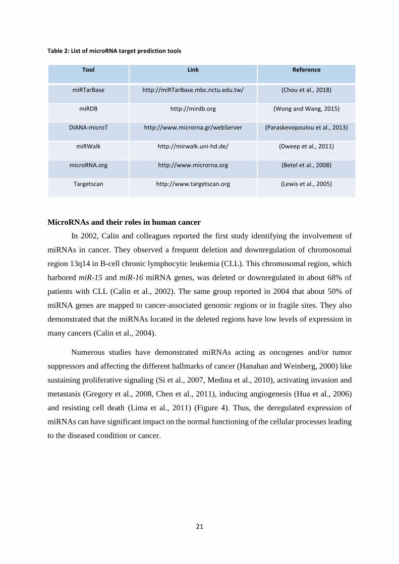
Table 2: List of microRNA target prediction tools
Tool Link Reference
miRTarBase http://miRTarBase.mbc.nctu.edu.tw/ (Chou et al., 2018)
miRDB http://mirdb.org (Wong and Wang, 2015)
DIANA-microT http://www.microrna.gr/webServer (Paraskevopoulou et al., 2013)
miRWalk http://mirwalk.uni-hd.de/ (Dweep et al., 2011)
microRNA.org http://www.microrna.org (Betel et al., 2008)
Targetscan http://www.targetscan.org (Lewis et al., 2005)
MicroRNAs and their roles in human cancer
In 2002, Calin and colleagues reported the first study identifying the involvement of
miRNAs in cancer. They observed a frequent deletion and downregulation of chromosomal
region 13q14 in B-cell chronic lymphocytic leukemia (CLL). This chromosomal region, which
harbored miR-15 and miR-16 miRNA genes, was deleted or downregulated in about 68% of
patients with CLL (Calin et al., 2002). The same group reported in 2004 that about 50% of
miRNA genes are mapped to cancer-associated genomic regions or in fragile sites. They also
demonstrated that the miRNAs located in the deleted regions have low levels of expression in
many cancers (Calin et al., 2004).
Numerous studies have demonstrated miRNAs acting as oncogenes and/or tumor
suppressors and affecting the different hallmarks of cancer (Hanahan and Weinberg, 2000) like
sustaining proliferative signaling (Si et al., 2007, Medina et al., 2010), activating invasion and
metastasis (Gregory et al., 2008, Chen et al., 2011), inducing angiogenesis (Hua et al., 2006)
and resisting cell death (Lima et al., 2011) (Figure 4). Thus, the deregulated expression of
miRNAs can have significant impact on the normal functioning of the cellular processes leading
to the diseased condition or cancer.

22
Figure 4: MicroRNAs targeting hallmarks of cancer
Dysregulation of miRNA expression is a common feature of cancer. MiRNAs are shown
to be over-expressed or under-expressed, and this aberrant expression has been associated with
cancer phenotype (Deng et al., 2008). Whole-genome miRNA expression profiling has been
used to detect the global expression of miRNAs in tumor specimens relative to normal tissues
(Lu et al., 2005). In addition, miRNA profiling not only distinguish between cancerous and
normal tumors but also between parental and resistant tumors. For instance, in our study, we
employed the next generation sequencing technique to identify differentially expressed
miRNAs from six different parental and resistant neuroblastoma cell lines isolated before and
after chemotherapy treatment. We observed a downregulated expression of several miRNAs in
the resistant cell lines, which is in concordance with the earlier studies demonstrating a general
downregulation of miRNAs in cancer (Roth et al., 2016, Williams et al., 2017)
Several methods are developed for detecting miRNAs like bead-based miRNA
profiling, miRNA microarrays, RT-qPCR, in situ hybridization techniques and the recent high-
throughput sequencing. Moreover, loss and gain-of-function studies have been established to
study the biology of miRNAs by overexpressing or silencing of particular miRNAs with
synthetic mimics or antagomirs, respectively (Iorio and Croce, 2012). Overall, these studies
have demonstrated the potential of miRNAs to be used as diagnostic and prognostic markers in
cancer.

23
Due to their small size, miRNAs are more stable and resistant to degradation. In
addition, extra-cellular miRNAs can be easily detected and extracted from body fluids such as
blood (total blood, plasma or serum), exosomes, and even from urine, saliva and sputum (Weber
et al., 2010). These so called circulating miRNAs are associated with various
pathophysiological conditions and can thus be used as prognostic biomarkers for early
diagnosis. For example, Lawrie et al. (2008), were the first to detect increased levels of tumor
associated miRNAs (miR-155, miR-210 and miR-21) in serum of patients with diffuse large B-
cell lymphoma and that increased levels of these miRNAs correlated with improved relapse-
free survival (Lawrie et al., 2008).
MiRNAs can also response to specific therapies. In cholangiocarcinoma cell lines,
targeted inhibition of miR-21 and miR-200b led to increased sensitivity to gemcitabine. This
was the first study demonstrating the involvement of miRNAs in modulating drug resistance in
cancer cells (Meng et al., 2006).
MicroRNAs as oncogenes and tumor suppressors
As mentioned earlier, dysregulation of miRNAs can affect one or several cellular
processes including survival, proliferation, invasion, migration, metastasis, differentiation and
apoptosis by acting as oncogenes or tumor suppressor genes (Babashah and Soleimani, 2011).
Cancer cells generally show the abundance of specific oncogenic miRNAs (also called
oncomiRs) and the loss of tumor-suppressor miRNAs (Figure 5) (Table 3) (Esquela-Kerscher
and Slack, 2006).
The oncomiRs repress the tumor suppressor genes, and/or genes that control cell
differentiation or apoptosis (Esquela-Kerscher and Slack, 2006, Lu et al., 2005, Babashah and
Soleimani, 2011). For instance, miR-155 is over-expressed and acts as an oncomiR by targeting
SH2 domain-containing inositol 5-phosphatase 1 (SHIP1) in acute myeloid leukemia (Xue et
al., 2014). In breast cancer, miR-21 was highly over-expressed compared to matched normal
breast tissues. Thus, knockdown of miR-21 by anti-miR-21 oligonucleotides, suppressed cell
growth in vitro and tumor growth in xenograft mouse model probably by indirect regulation of
BCL2 expression (Si et al., 2007).
The tumor-suppressor miRNAs negatively regulate protein-coding oncogenes and or
genes that inhibit cell differentiation or apoptosis (Esquela-Kerscher and Slack, 2006, Lu et al.,
2005, Babashah and Soleimani, 2011). For example, the tumor suppressor, miR-34a have been
shown to target MYCN (Wei et al., 2008) and E2F transcription factor 3 (E2F3) and induce

24
apoptosis in neuroblastoma (Welch et al., 2007). In chronic lymphocytic leukemia, miR-15a
and miR-16-1 are deleted or downregulated, however over-expression of these miRNAs in
leukemic cell line model negatively regulated the expression of anti-apoptotic BCL2 protein
(Cimmino et al., 2005).
Figure 5: MicroRNA as oncogenes and tumor suppressors
Depending on the cellular context, miRNAs could function either as oncomiRs or
tumor-suppressors. For instance, the polycistronic miR-17-92 cluster (includes miR-17-3p, miR-
17-5p, miR-18a, miR-19a, miR-19b-1, miR-20a and miR-92a-1) located at the genomic locus
13q31, was not only over-expressed in tumor-cell lines but also amplified in tumors in diffuse
large B-cell lymphoma. In addition, in vivo studies of miR-17-92 over-expression in transgenic
mouse model of human B-cell lymphoma resulted in aggressive tumors, indicating the
oncogenic role of miR-17-92 miRNA in cancer progression (Ota et al., 2004, He et al., 2005).
However, in another study, the c-Myc induced miR-17-92 cluster targeted and decreased the
expression of the E2F1, involved in transition of G1-S phase of cell cycle progression,
suggesting a tumor suppressor activity of this miRNA cluster (O'Donnell et al., 2005).
MiRNAs can function in complex regulatory circuits and feedback mechanisms. They
are shown to work together in groups and co-operate to regulate oncogenes necessary for tumor
progression. In human burkitt lymphomas, a group of miRNAs targeting the c-Myc oncogene

25
were silenced, which led to the over-expression of c-Myc and its targets involved in
proliferation and survival. Interestingly, over-expression of c-Myc led to repression of some c-
Myc targeting miRNAs, indicating a feedback mechanism in regulation of c-Myc expression
(Bueno et al., 2011).
Table 3: Key microRNAs with oncogenic and tumor suppressor roles in neuroblastoma
miRNA Regulation mRNA targets Function Reference
miR-17-92 Up-regulated DKK3 Oncogenic (De Brouwer et al., 2012)
miR-34a Down-regulated MAP3K9 Tumor suppressive (Tivnan et al., 2011)
miR-21 Up-regulated PTEN Oncogenic (Chen et al., 2012)
miR-376c Down-regulated CCND1 Tumor suppressive (Bhavsar et al., 2018)
miR-380 Up-regulated TP53 Oncogenic (Swarbrick et al., 2010)
miR-193b Down-regulated MCL1,CCND1,MYCN Tumor suppressive (Roth et al., 2018)
miR-15a Up-regulated RECK Oncogenic (Xin et al., 2013)
miR-323a Down-regulated STAT3 Tumor suppressive Manuscript I
The expression and function of oncomiRs can be increased or upregulated by multiple
mechanisms including genomic amplifications, activating mutations, transcriptional activation
and loss of epigenetic silencing. In contrast, loss of tumor-suppressor miRNAs can be due to
genetic deletions, in-activating mutations, transcriptional repression and epigenetic silencing
mechanisms (Lujambio and Lowe, 2012). Overall, the regulation of miRNA expression in
cancer is very complex and therefore the mechanisms pertaining to the deregulation of miRNAs
are discussed in the next section.
Mechanisms of microRNA deregulation in cancer
The deregulation or differential expression of miRNAs in cancer is undisputed. Not a
single universal mechanism but a combination of several different mechanisms operate to
modulate the expression profiles of individual or group of miRNAs in cancer setting (Deng et
al., 2008). The mechanisms of miRNA deregulation can be broadly categorized into structural
genetic variations, epigenetic modifications, transcriptional deregulation and defects in the
miRNA biogenesis machinery (Figure 6) (Lin and Gregory, 2015, Deng et al., 2008).

26
The structural genetic variations include the DNA-copy number alterations
(amplifications, deletions, and translocations) which are implicated in modulating the
expression of miRNAs in cancers (Lujambio and Lowe, 2012, Deng et al., 2008). For instance,
in chronic lymphocytic leukemia frequent deletions of chromosomal region 13q14 harboring
the miRNAs miR-15 and miR-16 were observed in more than 50% of patients (Calin et al.,
2002). In another study, an amplification of C13orf25 locus at 13q31-32 containing seven
miRNA polycistronic cluster was reported in lymphoma patients (Ota et al., 2004, Tagawa and
Seto, 2005).
In addition to genomic alterations, transcriptional regulators also play an important role
in modulating the expression of miRNAs in cancer. For example, the activation of tumor
suppressor gene, tumor protein P53 (TP53) led to the significant upregulation of 34-miRNAs
and downregulation of 16-miRNAs in a genome-wide screen for TP53-regulated miRNAs in
cancer. Among the deregulated miRNAs, miR-34 showed a marked upregulation, which is a
well-known tumor suppressor shown to target genes, involved in promoting cell growth and
proliferation. In the same study, other miRNAs like tumor suppressive, let-7a targeting the
oncogenes rat sarcoma (RAS), high mobility group AT-Hook 2 (HMGA2) and miR-15a/16
targeting the BCL-2 were also identified (Tarasov et al., 2007). In another example, the
transcriptional factor encoded by the proto-oncogene c-Myc, directly activates the expression
of oncogenic miR-17-92 cluster (O'Donnell et al., 2005). Interestingly, c-Myc has been shown
to repress a broader set of miRNA expression in mouse models of B cell lymphoma (Chang et
al., 2008).
Defects in the miRNA biogenesis machinery can also affect the miRNA expression. In
the first step of miRNA biogenesis, RNA polymerase II transcribes pri-miRNAs from miRNA
genes (Lee et al., 2004), which has been shown to be deregulated in several cancers. As
mentioned earlier, different genetic abnormalities like deletions, amplifications, and
translocations can alter the expression of miRNA genes (Lin and Gregory, 2015, Deng et al.,
2008).

27
Figure 6: The mechanisms of microRNA deregulation

28
In the second step of miRNA processing pathway, microprocessor complex, which
contains DROSHA and DGCR8 enzymes, cleaves the pri-miRNA to generate pre-miRNA
(Gregory et al., 2004, Denli et al., 2004). The microprocessor components DROSHA and
DGCR8 are often dysregulated in cancer. For instance, a study by Lin and colleagues identified
the downregulation of DROSHA in advanced stage neuroblastoma tumors, which correlated
with the global downregulation of the miRNAs and poor clinical outcome (Lin et al., 2010).
However, in cervical squamous cell carcinoma, the upregulation of DROSHA was observed to
link with altered miRNA expression (Muralidhar et al., 2011). In the third step, the pre-miRNAs
are exported to cytoplasm by XPO5 via RAN-GTP (Yi et al., 2003). A study by Melo and
colleagues demonstrated that, an inactivating mutation in XPO5 results in trapping of pre-
miRNAs in the nucleus, impairing miRNA biogenesis machinery (Melo et al., 2010). Defects
have also been observed in DICER and its essential co-factor TRBP, which are responsible for
further processing of pre-miRNAs into ~22 nucleotides mature miRNAs (Chendrimada et al.,
2005). For instance, DICER1 was shown downregulated in neuroblastoma and it correlated
with global downregulation of miRNAs and poor clinical outcome (Lin et al., 2010). However,
DICER was upregulated in metastatic prostate adenocarcinoma with global upregulation of
miRNA expression and correlated with the increase in clinical stages (Chiosea et al., 2006).
Frameshift mutations were identified in TRBP2, which caused decreased protein expression in
sporadic and hereditary colorectal carcinomas (Melo et al., 2009).
Epigenetic modifications like DNA methylations (mostly occurs in CpG sequences) and
histone covalent modifications (acetylation, methylation and phosphorylation) alter the
chromatin structure and regulate the pattern of gene expression (Portela and Esteller, 2010).
Weber and colleagues thoroughly analyzed miRNA genes and demonstrated that 50% of
miRNA genes are associated with CpG islands (short DNA sequences located at the gene
promoter). Moreover, the frequency of miRNA gene promotor methylation was higher as
compared to protein coding genes (Weber et al., 2007). Studies have reported that CpG islands
associated with miRNA gene promoter are frequently hypermethylated in cancer leading to
epigenetic silencing of tumor suppressor miRNAs. These miRNAs can be re-activated by the
treatment of chromatin-modifying drugs such as inhibitors of DNA methylation and/or histone
deacetylase (Lujambio et al., 2008). For example, miR-127, which targets the proto-oncogene
BCL6, is hypermethylated in human cancer cells. Upon treatment with chromatin modifying
agents, a strong upregulation of miR-127 was observed (Saito et al., 2006). In another study,
Parodi et al., (2016) analyzed the methylation status of a set of miRNAs in neuroblastoma cell

29
lines and identified a subset of hypermethylated and downregulated miRNAs (miR-34 and miR-
124) whose targets have important roles in fundamental cell processes like growth and
apoptosis (Parodi et al., 2016). Similar to DNA hypermethylation, DNA hypomethylation have
been shown to upregulate the expression of oncogenic miRNAs. The let-7a-3 gene was
hypomethylated in lung adenocarcinomas, leading to high expression of let-7a-3 having
oncogenic functions (Brueckner et al., 2007). In addition to DNA methylation, histone
modifications could also modulate the miRNA expression profiles in cancer (Guil and Esteller,
2009).
MicroRNAs and drug resistance in human cancer
The role of miRNAs in mediating drug resistance in neuroblastoma is poorly
understood. Very few studies have reported the direct involvement of miRNAs in modulating
the drug resistance mechanisms in neuroblastoma (Table 4).
In one of our miRNA profiling studies, we employed deep sequencing technique to
identify 34-downregulated and 8-upregulated miRNAs differentially expressed in
neuroblastoma cell lines isolated from six patients at diagnosis and at relapse following
intensive treatments (Roth et al., 2016). In another study, Ayers and colleagues identified
differential expression of miRNAs in chemoresistant cell line models (SH-SY5Y and UKF-
NB-3) of neuroblastoma (Ayers et al., 2015).
Chen and collaborators found miR-21 as the first miRNA associated with drug resistance
in neuroblastoma. They observed the increased expression of miR-21 in cisplatin-resistant SH-
SY5Y and BE(2)-M17 neuroblastoma cells as compared to parental cells. Therefore, by using
antagomir against miR-21, they knocked down the expression of miR-21, which sensitized the
cisplatin-resistant cells. Further, they ectopically expressed pre-miR-21 in parental cells, which
led to increased resistance to cisplatin treatment and enhanced proliferation by modulating the
phosphatase and tensin homolog (PTEN) protein levels (Chen et al., 2012).
In another study, miR-204 was shown to increase sensitivity of neuroblastoma cell lines
to cisplatin and etoposide. Surprisingly, miR-204 had no effect on neuroblastoma cell growth
in the absence of chemotherapeutic agents. The miR-204 has been shown to directly target the
3’UTR sequence of BCL2 and an oncogene, neurotrophic receptor tyrosine kinase 2 (NTRK2)
both of which are important mediators in facilitating resistance to several chemotherapeutic
agents. Moreover, BCL2 and NTRK2 are significantly associated with poor patient survival in
chemo-resistant neuroblastoma (Ryan et al., 2012).

30
Table 4: MiRNAs involved in modulating drug resistance in neuroblastoma
microRNA Drug/s Target/s Reference
miR-17-5p-92 Not determined p21 and BIM (Fontana et al., 2008)
miR-21 Cisplatin PTEN (Chen et al., 2012)
miR-204 Cisplatin, Etoposide BCL2 and NTRK2 (Ryan et al., 2012)
miR-137 Doxorubicin CAR (Takwi et al., 2014)
miR-520f Cisplatin, Etoposide NAIP (Harvey et al., 2015)
miR-155 Cisplatin TERF1 (Challagundla et al., 2015)
miR-497 Not determined CHEK1, AKT and VEGFA (Soriano et al., 2016)
miR-141 Cisplatin FUS (Wang et al., 2016)
miR-137 Doxorubicin CAR (Zhao et al., 2017)
Two independent reports highlighted the importance of miR-137 in modulating the
doxorubicin sensitivity of neuroblastoma cells. Takwi and colleagues observed downregulated
expression of miR-137 and an inverse high expression of constitutive androstane receptor
(CAR) and MDR1 in doxorubicin-resistant neuroblastoma cells as compared to parental cells.
Furthermore, miR-137 was shown to directly target CAR and that over-expression of miR-137
led to sensitization of resistant cells to doxorubicin and reduction of the resistant tumor growth
in vivo (Takwi et al., 2014). Zhao et al., (2017) proposed yet another mechanism to increase the
sensitivity of neuroblastoma cells to doxorubicin. In this study, short-interfering RNA (siRNA)
knockdown of histone deacetylase 8 (HDAC8) which is often upregulated and correlated with
advance stage disease, increased sensitivity of neuroblastoma cells to doxorubicin via
upregulation of miR-137 and inhibition of the MDR1 (Zhao et al., 2017).
Tumor microenvironment has been shown to play important role in mediating drug
resistance in cancers via release of exosomic miRNAs. The study was carried out to show how
exchange of exosomic miRNAs (miR-21 and miR-155) takes place between neuroblastoma cells
and neighboring human monocytes and how these exchanged miRNAs affect drug resistance
(Challagundla et al., 2015).

31
Harvey and colleagues showed that the expression of miR-520f was significantly
reduced in post-chemotherapy tumors as compared to matched pre-chemotherapy samples. In
the same study, a cisplatin and etoposide resistant SK-N-AS cell line was developed which
acquired resistance via increase in expression of neural apoptosis inhibitor protein (NAIP) and
the downregulation of miR-520f targeting 3’UTR of NAIP (Harvey et al., 2015).
In another study, the over-expression of miR-497 reduced proliferation of multiple
chemo-resistant neuroblastoma cell lines and induced apoptosis in MYCN-amplified cell lines.
In addition, increased miR-497 expression also reduced tumor growth and inhibited vascular
permeabilization in preclinical neuroblastoma mouse model. Moreover, low miR-497
expression correlated with poor patient outcome in a cohort of human neuroblastoma samples
(Soriano et al., 2016).
Recently, Wang and colleagues, found that miR-141 was downregulated in both MYCN-
amplified and non-amplified neuroblastoma cell lines. Over-expression of miR-141 inhibited
proliferation, migration and increased cisplatin chemo-sensitivity in neuroblastoma cells by
targeting fused in sarcoma (FUS) (Wang et al., 2016).
MicroRNA as therapeutics
Since the discovery of miRNAs in 1993, there have been significant advances in
deciphering the molecular mechanisms of cancer progression. Mounting studies have
documented the prominent role of miRNAs as the post-transcriptional regulators of
developmental and cellular processes (Croce and Calin, 2005). Indeed, they are shown to
function as oncogenes or tumor suppressors which make them promising targets for the
development of cancer therapeutics (Babashah and Soleimani, 2011). The most important
advantage of miRNA is that, single miRNAs can target multiple mRNAs, affecting different
oncogenic or tumor suppressive pathways (Croce and Calin, 2005). Moreover, miRNAs are
shown to differentially express not only across normal and cancerous tissues but also across
parental and drug resistant cells (Roth et al., 2016). Thus effective modulation of cancer
associated-miRNAs could be an interesting approach in the treatment of cancer.
Broadly, there are two main strategies to modulate the expression of miRNAs in cancer.
Direct strategy involves the use of either the chemically-modified synthetic ds oligonucleotides
(called mimics) or the viral based vector-constructs to re-express tumor suppressive miRNAs;
and/or chemically-modified single-stranded (ss) anti-miR oligonucleotides (called antagomirs)
to inhibit oncogenic miRNAs in cancer. Indirect strategy employs drugs to target the

32
components of miRNA biogenesis machinery or the epigenetic machinery, thus modulating
miRNA expression profile in cancerous cells (Iorio and Croce, 2012, Thorsen et al., 2012).
The stability and effective delivery of miRNAs into the target tissues are the challenges
mainly faced in the miRNA-based therapy. Hence, scientists have come up with different
designs and formulations for the efficient and robust delivery of miRNAs in vivo (Thorsen et
al., 2012).
For the success of miRNA-based therapy, efficient and safe delivery of miRNA mimics
or antagomirs in vivo is essential. MiRNA mimics or antagomirs can be administered via
intranasal, local and systemic routes through viruses (adeno associated viruses, AAVs or
lentivirus constructs), lipids (neutral lipid emulsion, polyethyleneimine, atelocollagen),
nanoparticles (iNOPs) or antimiR oligonucleotides (AMOs) (Iorio and Croce, 2012).
Table 5: List of microRNAs with therapeutic potential in human cancers
Micro-
RNA
Model AntimiRs or
mimics
Route of
admin.
Therapeutic
effect
Cancer type Reference
miR-380-
5p
Orthotopic
mouse model
LNA, 2’-
F/MOE
Intra-
peritoneal
Reduced
tumor
growth
Neuroblastoma (Swarbrick
et al., 2010)
miR-17-5p Orthotopic
LAN-5
neuroblasto
ma
xenografts
Cholesterol
-conjugated
2’-O-Me
antagomir
Intra-
tumoral
Reduced
tumor
growth
Neuroblastoma (Fontana et
al., 2008)
miR-34 Mouse K-Ras
G12D model
Lipid-
formulated
miR-34a
mimics
Intra-
vessicle
Reduced
tumor
growth
Lung cancer (Trang et
al., 2011)
let-7 Mouse K-Ras
G12D model
Lenti-viral
construct
expressing
let-7a
Intra-nasal Reduced
tumor
growth
Lung cancer (Trang et
al., 2010)

33
Several types of chemical modifications are done in order to confer nuclear resistance,
enhance binding affinity and facilitate cellular uptake of antagomirs so that relevant miRNAs
are exactly targeted. These antimiR chemistries of antagomirs include the use of high-affinity
2' sugar modifications such as locked nucleic acid (LNA), 2'-O-methyl (2'-O-Me); 2'-O-
methoxyethyl (2'-MOE) and 2'-fluoro (2'-F) as reviewed by Eva van Rooij (van Rooij and
Olson, 2012).
MRX34 is the first miRNA mimic to enter phase I clinical trial (ClinicalTrials.gov
Identifier: NCT01829971) in cancer. This ds synthetic miRNA mimic is delivered by liposome
technology. The tumor suppressor miRNA, miR-34 (MRX34) has been shown to target 24
known oncogenes having role in diverse cell processes including survival, proliferation,
metastasis, cell cycle and chemoresistance (Bouchie, 2013). Miravirsen (from Santris Pharma),
an antagomir targeting miR-122 is another therapeutic microRNA to enter clinical trial for the
treatment of hepatitis C virus (HCV). In addition, the follow up phase IIa clinical trial study
(ClinicalTrials.gov Identifier: NCT01200420) reported miravirsen to be safe, well tolerated and
provided prolonged antiviral activity without evidence of viral resistance (Janssen et al., 2013).
Recently, phase II clinical trial of a synthetic miR-29 miRNA mimic, MRG-201
(ClinicalTrials.gov Identifier: NCT02603224) from miRagen therapeutics was initiated for the
treatment of patients with fibrotic diseases (Rupaimoole and Slack, 2017) (Retrieved from:
http://www.miragen.com/press-release/miragen-therapeutics-announces-initiation-phase-2-
clinical-trial-mrg-201/)
All the above strategies mentioned take into account the modulation of single miRNA
or single miRNA family. However, the cancer phenotype may be as a result of aberrant
expression of multiple miRNAs in co-ordination with various mRNA targets. Therefore, an
indirect approach which employs drugs to target the epigenetic machinery is considered.
Epigenetic modifications like methylation and acetylation of genomic DNA affects global
miRNA expression levels. Thus, epigenetic drugs or modifiers like DNA methyltransferase
(DNMT) inhibitors and histone deacetylase (HDAC) inhibitors which modulate the methylation
and acetylation status, respectively, are used to revert the aberrant miRNA expression levels
with corresponding change in relevant genes. In other words, epigenetic treatment can be used
for the induction of specific miRNAs to serve as novel anticancer therapy (Monroig and Calin,
2013). For example, the miR-127 which is located within a CpG island and silenced in human
cancer cells was upregulated by the treatment of 5-aza-2'-deoxycytidine (inhibits DNA
methylation) and 4-phenylbutyric acid (inhibits histone deacetylase). In addition, this

34
upregulation of miR-127 lead to corresponding downregulation of its potential target, BCL6, in
human cancer cells (Saito et al., 2006).
Yet another alternative to chemically modified antisense oligonucleotides and
demethylating agents is the use of competitive inhibitors called miRNA sponges. These are
transcripts expressed from strong promoters containing multiple, tandem binding sites to a
miRNA of interest. Therefore, miRNA sponges can be used as an antagomir to decrease the
level of oncogenic miRNAs in the cultured cells. The miRNA sponge plasmids together with
luciferase constructs can be used for assessing transfection efficiency (by inclusion of GFP
reporter in sponge mRNA), validation of predicted miRNA targets and to assay miRNA loss of
function phenotype (Ebert et al., 2007).
Choi and colleagues developed another strategy in contrast to miRNA sponges. They
developed target protector morpholinos (ss chemically-modified oligonucleotides) to mask the
miRNA recognition element (MRE) or the complementary miRNA binding sites in the 3’UTR
of the target mRNA (Choi et al., 2007).
The CCND1 and STAT3 oncogenes in human cancer
CCND1
The CCND1 (cyclin D1) gene was first characterized in 1991 (Xiong et al., 1991). More
than two decades of research have established CCND1 as an important regulator of cell cycle
progression and cancer. It is one of the most extensively documented oncogenes in human
cancers. Given its prominent role in cell cycle regulation, alterations in levels of CCND1 have
had oncogenic effects. Multiple converging studies point out to CCND1 as the biomarker of
cancer phenotype and disease progression (Musgrove et al., 2011).
During the process of cell cycle, the regulatory catalytic subunits called “cyclins”
(especially D-type cyclins which includes cyclin D1, D2 and D3) are produced in a periodic,
cell cycle dependent manner. Biologically, in response to growth factors or mitogenic stimuli,
cyclin D1 gets associated with the serine-threonine protein kinases called cyclin dependent
kinases (CDKs) - CDK4 and CDK6. The cyclin D1-CDK4/6 complex thus phosphorylate the
tumor suppressor retinoblastoma (RB) protein which in turn releases the RB from E2F
transcription factors. The E2F therefore transactivate genes necessary for G1-S phase of cell
cycle progression. Consequently, the cell cycle enters DNA replication and cell division phase
leading to cellular proliferation (Motokura et al., 1991, Baldin et al., 1993, Bartek et al., 1996,
Musgrove et al., 2011). Moreover, other than its catalytic function in cell cycle regulation,

35
CCND1 plays a non-catalytic role in DNA repair mechanisms. Jirawatnotai and colleagues
reported that RAD51, a component of DNA-repair machinery, interacts directly with CCND1.
The RAD51 is an enzyme necessary for homologous recombination process, to seal DNA
breaks using the intact copy of the DNA sequence as templates (Jirawatnotai et al., 2011).
Furthermore, cyclin D-CDK4 also targets genes including SMADs (Matsuura et al., 2004),
RUNX family (Shen et al., 2006) and MEF2 (Lazaro et al., 2002) associated with proliferation,
degradation and differentiation respectively.
Knockdown of CCND1 and its partner CDK4 leads to inactivation of RB pathway, E2F
transcription and thus G1-cell cycle arrest in neuroblastoma (Molenaar et al., 2008) and
apoptosis in cutaneous melanoma (Sauter et al., 1999) and squamous carcinomas (Sauter et al.,
2002). Given the role of cyclin D/CDK4/CDK6/RB pathway members in numerous aspects of
cellular biology, the deregulation of these proteins is therefore associated with unlimited growth
or proliferation, hence tumorigenesis.
Extracellular signals like growth factors or mitogenic stimuli influence transcription,
translation, degradation and thus the abundance of cyclin D1 protein in the cell system.
Therefore, oncogenic activation of mitogenic signaling pathways may lead to abnormal increase
in cyclin D1 protein levels. Cyclin D1 is shown to interact with numerous proteins involved in
cell cycle regulation, transcriptional regulation and DNA repair mechanisms. Therefore,
deregulation of cyclin D1 will significantly affect different cellular processes directly or
indirectly and therefore have oncogenic effects (Fu et al., 2004).
CCND1 amplification and its subsequent over-expression is one of the most common
genetic aberrations in human cancers. CCND1 was shown to be amplified and over-expressed
in multiple cancers including breast cancer, lung cancer, oral squamous cell carcinoma and
malignant melanoma (Santarius et al., 2010). In mantle cell lymphoma and multiple myeloma,
the translocation event juxtaposes CCND1 with the immunoglobulin heavy chain locus (IGH),
leading to CCND1 over-expression (Li et al., 1999, Pratt, 2002). In addition, mutations that
result in deregulation of CCND1 expression have been shown in mantle cell lymphoma
(Wiestner et al., 2007), esophageal (Benzeno et al., 2006) and endometrial (Moreno-Bueno et
al., 2003) cancers. Furthermore, altered expression of miRNAs targeting CCND1 is another
mechanism deregulating CCND1 in human cancers. Both miR-15a and miR-16 are deleted or
downregulated and inversely associated with CCND1 expression in non-small cell lung cancer
(Bandi et al., 2009) and prostate cancer (Bonci et al., 2008).

36
In case of neuroblastoma, genetic aberrations and over-expression of CCND1 is
observed, similar to other cancers. Amplification of CCND1, CDK4 and mutation of CDK6
have been shown in a subset of neuroblastoma cell lines and tumors (Molenaar et al., 2008). In
addition, a rearrangement in the 3’UTR sequence of CCND1 has also been shown in
neuroblastoma tumor (Molenaar et al., 2003). Taken together, these findings suggest a crucial
role of cyclins and CDKs in promoting cellular proliferation in neuroblastoma and demonstrate
their potential for therapeutic intervention.
STAT3
The STAT3 gene was first described as a DNA binding protein activated in response to
growth factors and having a dual function of signal transduction and activation of transcription
(Zhong et al., 1994). STAT3 encodes one of the seven members (STAT1, STAT2, STAT3,
STAT4, STAT5A, STAT5B, and STAT6) of the STAT protein family (Copeland et al., 1995,
Darnell, 1997). This protein is activated in response to binding of cytokines and growth factors
to cellular receptors, which activates membrane-associated janus kinases (JAKs). JAK in turn
phosphorylates STAT3 at specific residues to form homo/heterodimers and translocate to cell
nucleus. In the nucleus, STAT3 acts as a transcription factor, which regulates the expression of
a wide range of genes involved in survival, proliferation, invasion, metastasis, angiogenesis,
and immunosuppression (Yu et al., 2014).
STAT3 has been shown to be activated by numerous activators (e.g. cytokines, growth
factors, toll-like receptors, etc.) and therefore it has a diverse range of biological functions
(Aggarwal et al., 2006). Hence, deregulation of STAT3 can lead to oncogenesis through various
different mechanisms. Yu et al., (2014) have reviewed the role of JAK-STAT3 signaling
pathway in multiple biological processes including obesity and metabolism, pre-metastatic
niche formation, cancer stem cells, and role in inflammation-mediated cancer (Yu et al., 2014).
STAT3 is constitutively activated in diverse human cancers and hence it functions as an
oncogene (Levy and Lee, 2002, Aggarwal et al., 2006). In a study by Ling and Arlinghaus,
mammary tumors were induced in mice by the 4T1 breast cancer cell line followed by effective
knockdown of STAT3 using a lentivirus small hairpin (shRNA) delivery system. This led to
efficient blockage of tumor formation. However, mice without shRNA for STAT3 efficiently
formed tumors. Thus, this study concluded that constitutively activated STAT3 plays an
important role in the induction of breast tumors by regulating the expression of c-Myc and
Twist (Ling and Arlinghaus, 2005). Contrary to this, a tumor suppressive role of STAT3 has
also been identified in some cancers (Zhang et al., 2016).

37
In addition, multiple studies have addressed the regulation of JAK-STAT3 signaling by
miRNAs in different cancers. MiRNAs like miR-135a (Navarro et al., 2009), miR-26a (Yang
et al., 2013), miR-337 (Du et al., 2012), miR-9 (Zhuang et al., 2012) and let-7 (Sugimura et al.,
2012) have shown reciprocal interactions with JAK-STAT3 signaling pathway, which have a
crucial role in regulating oncogenesis. Recent studies from Odate et al., (2017) have
demonstrated the specific targeting and inhibition of STAT3 by generation 2.5 antisense
oligonucleotide, AZD9150, which decreases neuroblastoma tumorigenicity and increases
chemosensitivity (Odate et al., 2017). Also, from our studies, we have demonstrated that miR-
323a-3p has been reduced in resistant neuroblastoma cell lines and advance stage primary
neuroblastoma tumors. Thus, over-expression of miR-323a-3p leads to reduced cell viability,
G1-cell cycle arrest and apoptosis by targeting STAT3 in neuroblastoma (Manuscript I).
Given the importance of JAK-STAT3 signaling in cancer, it is one of the most promising
targets for cancer therapy. STAT3 inhibitors are being developed. For instance, several tyrosine
kinase inhibitors are available in clinic, which could target JAK-STAT3 signaling indirectly,
ultimately leading to reduction of tumor growth and survival by promoting apoptosis (Yu et al.,
2009).

38
Aims of the study
The overall aim of this study is to identify and elucidate the functional role of
miRNAs involved in the drug resistance mechanisms in neuroblastoma.
The specific aims of the study are as follows:
i) To identify miRNAs and their pathways that are involved in the underlying drug
resistance mechanisms in neuroblastoma (Paper I)
ii) To evaluate the functional role of downregulated 14q32 locus miRNAs, miR-376c-3p
and miR-323a-3p in neuroblastoma (Paper II & III)

39
Summary of papers
Paper-I
Next generation sequencing of microRNAs from isogenic neuroblastoma cell lines isolated
before and after treatment
Roth S.A., Knutsen E., Fiskaa T., Utnes P., Bhavsar S.P., Hald O.H., Løkke C., Mestdagh P.,
Johansen S.D., Flægstad T., & Einvik C.
Cancer Letters (2016) 372(1):128-136
Recently, microRNAs (miRNAs) have been shown to modulate drug resistance in
multiple cancers. However, the role of miRNAs in neuroblastoma chemoresistance is limited
and poorly understood. In this study, we set out to identify miRNAs and their pathways
involved in mediating drug resistance in neuroblastoma. We hypothesize that differential
expression of miRNAs among parental and resistant cells could lead to development of drug
resistance in neuroblastoma. We used next generation deep sequencing technology to determine
the miRNA expression profiles from matched parental cell lines (isolated at diagnosis) and
resistant cell lines (isolated at relapse after intensive treatments) from six neuroblastoma
patients.
We observed a downregulation of 34 miRNAs and upregulation of 8 miRNAs in the cell
lines isolated post-treatment. RT-qPCR technique was used to confirm and validate the
deregulated miRNA expressions. Our results were in line with previous reports, where a general
downregulation of miRNAs is more common than upregulation of miRNAs in cancers.
Interestingly, 22 of the 34 downregulated miRNAs were located on chromosome 14q32 locus,
which have earlier been reported to be downregulated in multiple human cancers. Moreover,
we also demonstrated that the reduced expression of certain chromosome 14q32 miRNAs
correlates with poor clinical outcome in a cohort consisting of 226 primary neuroblastomas.
Furthermore, Ingenuity pathway analysis (IPA) of differentially expressed miRNAs revealed
important biological pathways associated with cancer progression and drug resistance. Hence,
in this study, we identified a unique set of miRNAs which may be involved in the development
of drug resistance in neuroblastoma. In addition, we showed that reduced expression of certain
miRNAs is correlated with poor prognostic factors in neuroblastoma.

40
Paper-II
Hsa-miR-376c-3p targets Cyclin D1 and induces G1-cell cycle arrest in neuroblastoma
cells
Bhavsar S.P., Løkke C., Flægstad T., & Einvik C.
Oncology Letters (2018) 16(5):6786-6794
In paper I, we showed that downregulation of chromosome 14q32 miRNAs is associated
with drug resistance and poor clinical outcome in neuroblastoma. In this paper, we investigated
the previously unknown functional role of chromosome 14q32 locus miRNA, miR-376c-3p that
is downregulated in post-therapy neuroblastoma cell line pairs. We hypothesize that restoration
of the downregulated miR-376c-3p could reverse the resistant phenotype caused due to reduced
expression of miR-376c-3p.
Therefore, we first over-expressed miR-376c-3p in multiple neuroblastoma cell lines
and demonstrated that upregulation of miR-376c-3p results in significant reduction of cell
growth and G1-cell cycle arrest in most of these cell lines. We then used miRNA target-
prediction software (TargetScan) to identify miR-376c-3p targets mainly related to cell cycle.
Dual luciferase reporter assay was performed to confirm and validate miR-376c-3p target.
Finally, RT-qPCR and western blot assays were performed to quantitate miR-376c-3p mediated
suppression of target mRNA and protein levels, respectively.
Interestingly, we observed cell cycle related CCND1 as one of the several genes
predicted to target by miR-376c-3p. Luciferase reporter assay further confirmed and validated
CCND1 as a direct target of miR-376c-3p. As expected, the mRNA and protein levels of
CCND1 were significantly reduced in miR-376c-3p transfected cells as compared to negative
control (NC) transfected cells. Hence, in this paper we proposed that miR-376c-3p could affect
cell growth and induce G1-cell cycle arrest by targeting CCND1 in neuroblastoma cells.

41
Paper-III
Hsa-miR-323a-3p targets STAT3 and induces G1-cell cycle arrest and apoptosis in
neuroblastoma cells
Bhavsar S.P., Olsen L., Løkke C., Flægstad T., & Einvik C.
Manuscript
Recently, the role of microRNAs (miRNAs) in modulating drug resistance in multiple
cancers has emerged as a promising therapeutic approach in the treatment of therapy-resistant
tumors. However, the role of miRNAs in neuroblastoma drug resistance is poorly understood.
In our previous research, we have demonstrated that miR-323a-3p is downregulated in
post-chemotherapy neuroblastoma cells as compared to matched pre-chemotherapy cells.
Moreover, miR-323a-3p is downregulated in stage 4 vs stage 1-2 in a cohort of 226 primary
neuroblastoma tumors. Therefore, in this study, we set out to investigate the functional role of
miR-323a-3p in neuroblastoma.
We used RT-qPCR to quantitate the basic expression of miR-323a-3p in BE(2)-C, Kelly
and SHSY-5Y neuroblastoma cell lines. The functional role of miR-323a-3p in these cell lines
was evaluated by alamar blue-cell viability, flow cytometry and western blot assays. Luciferase
reporter assay, RT-qPCR and western blotting techniques were used for miR-323a-3p target
identification and quantification in neuroblastoma cell lines.
Over-expression of miR-323a-3p resulted in significant reduction of cell viability in
BE(2)-C, Kelly and SHSY-5Y cell lines. By flow cytometric cell cycle analysis we found G1-
cell cycle arrest in Kelly and SHSY-5Y but not in BE(2)-C cells. By western blot analysis, we
found that miR-323a-3p induces apoptosis in BE(2)-C, Kelly and SHSY-5Y cell lines.
Bioinformatic miRNA target prediction software (miRDB) predicted 793 potential
target genes for miR-323a-3p. Subsequent literature search, target analysis and luciferase
reporter assay identified and confirmed STAT3 as a direct target of miR-323a-3p. Furthermore,
RT-qPCR and western blot analysis revealed that the mRNA and protein levels of STAT3 were
significantly reduced in miR-323a-3p transfected cells as compared to negative control (NC)
transfected cells.
Thus, the study showed that over-expression of miR-323a-3p led to significant
inhibition of cell viability, G1 cell cycle arrest and apoptosis by targeting STAT3 in
neuroblastoma cells.

42
Discussion (Paper I, II & III)
Chemotherapy is the primary choice of treatment for patients with high-risk
neuroblastoma. However, resistance to chemotherapy has emerged as an important clinical
obstacle. These high-risk patients usually relapse after prolonged treatment and eventually die
(Habib et al., 2012). Therefore, instead of the conventional chemotherapy approaches,
alternative strategies need to be employed which may target specific genes to combat this lethal
disease. Mounting studies have demonstrated the ability of miRNAs to regulate the expression
of multiple genes affecting the growth and survival of multiple cancer types (Gurtan and Sharp,
2013). Thus, the known functionality of miRNAs to act as oncogenes or tumor suppressors
establish that aberrant expression of miRNAs may represent a general feature of human cancer
and provides an opportunity for therapeutic intervention.
In this study, by deep sequencing analysis, we observed reduced expression of miR-
376c-3p and miR-323a-3p in resistant neuroblastoma cell lines as compared to matched parental
neuroblastoma cell lines. In addition, these miRNAs were also downregulated in a cohort
consisting of 226 primary neuroblastoma tumors. We thus propose that downregulation of miR-
376c-3p and miR-323a-3p miRNAs may be associated with underlying resistance mechanisms
observed in resistant neuroblastoma cell lines. Over-expression of these downregulated
miRNAs correlated with decreased survival in neuroblastoma cells by promoting G1-cell cycle
arrest and apoptosis. Therefore, this study has implications for understanding the functional
roles of miRNAs in neuroblastoma tumorigenesis and drug resistance.
MicroRNAs are differentially expressed in parental and resistant neuroblastoma cell lines
Drug resistance is one of the most important clinical obstacles in treatment of cancer.
Multiple factors are involved in mediating cells resistant to drugs, which are described in detail
in the introduction section. Growing evidence have established the roles of miRNAs in
mediating drug resistance in human cancers (Ayers et al., 2015, Chen et al., 2012). In this study,
we have characterized the roles of certain miRNAs in drug resistant neuroblastoma.
Generally, researchers create resistant cell lines by exposing cancer cells to high
concentrations of drugs. These in vitro-selected resistant cell lines are then subjected for the
analysis of underlying drug resistance mechanisms. However, this process of making cells
resistant to drug may create a significant impact on the cellular system as a whole causing high
expression of a myriad of genes and proteins. In short, the cell system might be restructured
drastically due to high concentration of drugs. This scenario is far from what we can imagine
43
about resistant cancerous cells present in the tumor microenvironment in the body. Owing to
the numerous possibilities or complexities offered by the tumor microenvironment as described
in the introduction section and the drugs pharmacokinetic and pharmacodynamic properties. It
is very difficult to correlate the results obtained from such resistant cell lines with the naturally
acquired resistance of the cancerous cells present in the patient’s body. Therefore, we opt for
another strategy, which included cells isolated from neuroblastoma patients at diagnosis and
from the same patients at relapse following chemotherapy. This strategy provided us with the
parental cells at diagnosis which were not treated with any kind of drugs and we also got the
resistant cells from the same patient at relapse which were treated with chemotherapeutic drugs.
Therefore, these resistant cells obtained via naturally acquired resistance in the body at relapse
would mimic the underlying drug resistance mechanisms more closely than the cells, which are
created by exposure to high concentrations of drugs.
Very few studies have demonstrated the deregulation of miRNAs and their association
with neuroblastoma pathogenesis and drug resistance. Bray et al., (2009) have shown the
dysregulation of miRNAs in neuroblastoma tumors caused by large-scale chromosomal
imbalances and the over-expression of MYCN. In addition, this study concluded that patterns
of miRNA expression might be predictive of clinical outcome (Bray et al., 2009). In another
study by Chen and Stallings, expression profiling of 157 miRNA loci in 35 primary
neuroblastoma tumors resulted in differential expression of 32 loci in favorable and unfavorable
tumor subtypes. Interestingly, several miRNAs were downregulated in tumors with MYCN
amplification. They further assessed the functional role of single locus (miR-184), and found
that miR-184 had significant role in apoptosis. Therefore, this study concludes that differential
patterns of miRNA expression are correlated with clinical and biological factors (Chen and
Stallings, 2007). Similarly, Schulte and colleagues, used next-generation sequencing
technology to analyze five favorable and five unfavorable neuroblastoma tumor samples. This
study also revealed the differential pattern of miRNA expression in favorable versus
unfavorable neuroblastoma (Schulte et al., 2010). Taken together all these studies, points
towards the clinical and functional relevance of differentially expressed miRNAs in the
pathogenesis of neuroblastoma.

44
Figure 7. Schematic representation summarizing the workflow of the study. Total RNA extracted from
neuroblastoma cell lines at diagnosis and after chemotherapeutic treatments at relapse are reverse-transcribed to
produce microRNA-cDNA-libraries, which are further subjected to next generation sequencing. The data obtained
after sequencing is systematically analyzed to study the functional role of miRNAs in neuroblastoma pathogenesis
and drug resistance.
Isolation of total RNA from neuroblastoma cell line pairs (at diagnosis/relapse)
Analysis of sequencing data
Differential expression of 42 microRNAs
Functional in vitro studies of selected microRNAs (miR-376c-3p and miR-323a-3p)
Next Generation Sequencing of microRNAs
Preparation of microRNA library
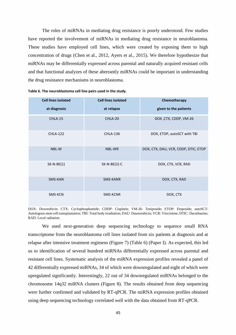
45
The roles of miRNAs in mediating drug resistance is poorly understood. Few studies
have reported the involvement of miRNAs in mediating drug resistance in neuroblastoma.
These studies have employed cell lines, which were created by exposing them to high
concentration of drugs (Chen et al., 2012, Ayers et al., 2015). We therefore hypothesize that
miRNAs may be differentially expressed across parental and naturally acquired resistant cells
and that functional analyzes of these aberrantly miRNAs could be important in understanding
the drug resistance mechanisms in neuroblastoma.
Table 6. The neuroblastoma cell line pairs used in the study.
Cell lines isolated
at diagnosis
Cell lines isolated
at relapse
Chemotherapy
given to the patients
CHLA-15 CHLA-20 DOX ,CTX, CDDP, VM-26
CHLA-122 CHLA-136 DOX, ETOP, autoSCT with TBI
NBL-W NBL-WR DOX, CTX, DAU, VCR, CDDP, DTIC, ETOP
SK-N-BE(1) SK-N-BE(2)-C DOX, CTX, VCR, RAD
SMS-KAN SMS-KANR DOX, CTX, RAD
SMS-KCN SMS-KCNR DOX, CTX
DOX: Doxorubicin; CTX: Cyclophosphamide; CDDP: Cisplatin; VM-26: Teniposide; ETOP: Etoposide; autoSCT:
Autologous stem cell transplantation; TBI: Total body irradiation; DAU: Daunorubicin; VCR: Vincristine; DTIC: Dacarbazine;
RAD: Local radiation.
We used next-generation deep sequencing technology to sequence small RNA
transcriptome from the neuroblastoma cell lines isolated from six patients at diagnosis and at
relapse after intensive treatment regimens (Figure 7) (Table 6) (Paper I). As expected, this led
us to identification of several hundred miRNAs differentially expressed across parental and
resistant cell lines. Systematic analysis of the miRNA expression profiles revealed a panel of
42 differentially expressed miRNAs, 34 of which were downregulated and eight of which were
upregulated significantly. Interestingly, 22 out of 34 downregulated miRNAs belonged to the
chromosome 14q32 miRNA clusters (Figure 8). The results obtained from deep sequencing
were further confirmed and validated by RT-qPCR. The miRNA expression profiles obtained
using deep sequencing technology correlated well with the data obtained from RT-qPCR.

46
Figure 8. Schematic representation of the miRNA-clusters at 14q32 chromosomal region. Multiple miRNAs
located at 14q32 chromosomal region are downregulated (bold type) and associated with poor prognosis factors
in neuroblastoma. The miRNAs of interest, miR-376c and miR-323a are highlighted in bold and rectangular box.
DLK1, delta like non-canonical notch ligand 1; MEG, maternally expressed; snoRNA, small nucleolar RNA;
DIO3, iodothyronine deiodinase 3; miRNA, microRNA.
The reduced expression of chromosome 14q32 miRNAs in tumors associated with poor
prognosis factors was confirmed in a cohort consisting of 226 primary neuroblastomas.
Moreover, we also applied Ingenuity Pathway Analysis (IPA), which identifies new targets,
pathways and candidate biomarkers within the context of biological systems to gain insights
into the nature of genes and pathways affected by the deregulated miRNAs in neuroblastoma.
The results of this analysis revealed several biological pathways associated with cancer and
drug resistance (Paper I).
MiR-376c-3p and miR-323a-3p are novel microRNAs with tumor suppressive activity in
neuroblastoma
Since the chromosome 14q32 miRNA clusters are shown downregulated in multiple
cancers (Zehavi et al., 2012), we focused on miRNAs from this region to study their functional
roles in neuroblastoma. We selected miR-376c-3p and miR-323a-3p, to assess their role in the
pathogenesis of neuroblastoma. Both miRNAs were downregulated in most of the cell lines
isolated from patients after chemotherapeutic treatments relative to matched cell lines isolated
47
at diagnosis. In addition, the expression of miR-376c-3p and miR-323a-3p, was reduced in INSS
stage 4 compared to stage 1-2 in a cohort of neuroblastoma tumors. Interestingly, literature
search on miR-376c-3p and miR-323a-3p showed a dual nature of these miRNAs to act as either
oncogenic or the tumor suppressive miRNA depending on cellular context. Taken together, all
these important properties of miR-376c-3p and miR-323a-3p, prompted us to evaluate the
unknown functional role of these miRNAs in neuroblastoma pathogenesis.
The miR-376c-3p is found downregulated in multiple human cancers, including prostate
cancer (Formosa et al., 2014), cervical cancer (Deng et al., 2016), oral squamous cell carcinoma
(Wang et al., 2017b), intrahepatic cholangiocarcinoma (Iwaki et al., 2013), melanoma (Zehavi
et al., 2012), osteosarcoma (Jin et al., 2013) and gliomas (Huang et al., 2017). However, it was
upregulated in acute myeloid leukemia (Dixon-McIver et al., 2008) and gastric cancer (Shiotani
et al., 2013). From these studies, miR-376c-3p was shown to target a set of genes including B‐
cell‐specific moloney murine leukemia virus insertion site 1 (BMI1), homeobox B7 (HOXB7),
growth factor receptor-bound protein 2 (GRB2), transforming growth factor-alpha (TGFA),
liver receptor homolog-1 (LRH-1), insulin growth factor 1 receptor (IGF1R) and activin
receptor-like kinase 7 (ALK7) involved in cell growth, cell invasion (Deng et al., 2016, Wang
et al., 2017b, Jin et al., 2013, Jiang et al., 2016), migration (Formosa et al., 2014, Wang et al.,
2017b, Iwaki et al., 2013, Zehavi et al., 2012), cell cycle arrest (Deng et al., 2016), apoptosis
(Wang et al., 2017b) and drug resistance (Ye et al., 2011).
We therefore performed functional studies to evaluate the effect of miR-376c-3p over-
expression on the neuroblastoma cells (Paper II). We demonstrated that over-expression of miR-
376c-3p led to suppression of cell viability and G1-cell cycle arrest in neuroblastoma cell lines.
These results suggested that the cell cycle genes might be affected and could serve as miR-
376c-3p targets. To test this hypothesis, we used bioinformatics algorithms to predict miR-376c-
3p targets related to cell cycle regulation. TargetScan algorithm predicted CCNDl as a target of
miR-376c-3p, which was further confirmed and validated by dual-luciferase reporter assay. Our
experimental data showed that the expression of CCND1 mRNA and protein levels were
significantly reduced after transfection of miR-376c-3p as compared to NC transfected cells. In
addition, we performed a rescue experiment to see if CCND1 counteracts the effect of miR-
376c-3p induced G1-cell cycle arrest upon CCND1 over-expression. However, a modest and
statistically insignificant effect of CCND1 over-expression on reducing the effect of miR-376c-
3p over-expression (i.e. G1-cell cycle arrest) was observed in neuroblsatoma cells. This is
because CCND1 might be targeted not only by miR-376c-3p but also by some other miRNAs

48
simultaneously. Many studies have demonstrated that several miRNAs can target the same
mRNA to effectively repress the target gene expression (Shukla et al., 2011). This result thus
point out towards the role of additional miRNAs in the regulation of CCND1 expression in
neuroblastoma cell lines. The CCND1 has been proposed as an important oncogene in a variety
of cancers (Musgrove et al., 2011). Multiple studies have demonstrated increased expression of
CCND1 and that knockdown of CCND1 induced G1-cell cycle arrest and inhibited Cyclin D-
Rb pathway in neuroblastoma cells (Molenaar et al., 2008, Molenaar et al., 2003, Rihani et al.,
2015). Moreover, CCND1 was also shown to be targeted by miR-34 and induce G1-cell cycle
arrest in NSCLC (Sun et al., 2008). Taken together, our findings suggest that miR-376c-3p has
the ability of arresting growth in neuroblastoma cells by targeting CCND1 oncogene. Further
studies are necessitated to understand the role of additional miRNAs in the regulation of
CCND1 in neuroblastoma.
A similar study was performed to evaluate the functional role of miR-323a-3p in
neuroblastoma (Manuscript I). Previous studies have demonstrated that miR-323a-3p is
downregulated in multiple cancers including glioblastoma (Shahar et al., 2016), osteosarcoma
(Chen et al., 2018a), bladder cancer (Li et al., 2017) and pancreatic ductal adenocarcinoma
(PDAC) (Wang et al., 2016) whereas upregulated in prostate cancer (Gao et al., 2015, Gao and
Zheng, 2018). Thus, miR-323a-3p functions as both tumor suppressive or oncogenic miRNA
by regulating growth, apoptosis, invasion and migration depending on the cellular context.
We demonstrated that over-expression of miR-323a-3p led to suppression of cell
viability, G1-cell cycle arrest and apoptosis in neuroblastoma cell lines. Given the tumor
suppressive nature of this miRNA, we searched the literature for probable targets of miR-323a-
3p validated in other cancer types. RT-qPCR was performed on such selected targets. In
addition, we used online bioinformatic tool (miRDB) to predict miR-323a-3p targets. We thus
identified STAT3 as a target of miR-323a-3p, which was further confirmed and validated by
dual-luciferase reporter assay. Next, we over-expressed miR-323a-3p in neuroblastoma cell
lines and quantitated the expression of STAT3 mRNA and protein levels. Interestingly, the
expression of STAT3 mRNA and protein levels was significantly reduced after transfection with
miR-323a-3p as compared to NC transfected cells. Thus, we propose that miR-323a-3p
decreases cell growth and induces G1-cell cycle arrest and apoptosis by targeting STAT3 in
neuroblastoma.

49
Multiple studies have demonstrated the oncogenic role of STAT3 in diverse cancers.
And, given the importance of JAK-STAT3 signaling in cancer, it is one of the most promising
targets for cancer therapy (Yu et al., 2014). STAT3 inhibitors are being developed, which show
decreased neuroblastoma tumorigenicity and increases chemosensitivity (Odate et al., 2017).
Furthermore, several tyrosine kinase inhibitors are also developed and now available in clinic,
which could target JAK-STAT3 signaling indirectly, ultimately leading to reduction of tumor
growth and survival by promoting apoptosis (Yu et al., 2009).
In a recent study, Ara et al., (2013) identified an IL-6/sIL-6R/STAT3 interactive
pathway functioning between neuroblastoma cells and its microenvironment, which led to drug
resistance. This study showed that STAT3 is not constitutively active in neuroblastoma cells
and that treatment of IL-6 alone or in combination with soluble IL-6 receptor (sIL-6R) results
in high activation of STAT3. The study demonstrated that drug-induced apoptosis of
neuroblastoma cells was rescued with the treatment of IL-6 alone and/or in combination with
sIL-6R. The rescue effect is observed due to the high activation of STAT3 in the cells since
STAT3 inhibition (with STAT3 inhibitor, Stattic) was not able to protect cells from drug-
induced apoptosis (Ara et al., 2013).
Given the important role of IL-6/sIL-6R activated STAT3 in drug resistance and our
recent results showing that STAT3 is a direct target of miR-323a-3p in neuroblastoma. We
hypothesize that miR-323a-3p could target STAT3 and rescue the drug-induced apoptosis in
neuroblastoma cells. In addition, it would be interesting to find the expression of STAT3
downstream factors such as survivin, BCL-2, etc. affected by STAT3 repression.
To summarize, in this thesis, we were not able to address the direct role of miRNA in
modulating drug resistance in neuroblastoma. However, we propose that miR-323a-3p could
play a potential role in drug resistance by targeting STAT3 in neuroblastoma cells. Further
experimental studies with chemotherapeutic drugs are necessitated to address the role of IL-
6/sIL-6R/STAT3/miR-323a pathway in modulating drug resistance in neuroblastoma.
Lastly, our findings suggest that the reduced expression of miRNAs may contribute to
a resistant phenotype in neuroblastoma cell lines, and therefore ectopic over-expression of these
miRNAs should be considered for overcoming resistance. The reduced expression of multiple
miRNAs in post-chemotherapy neuroblastoma cell lines and primary tumors indicate a strong
functional connection between miRNAs and underlying drug resistance mechanisms in the
worst prognostic class of neuroblastoma. In addition, our study establishes that the restoration
of certain miRNAs could be key therapeutic goal in neuroblastoma. According to our

50
knowledge, there are very few studies demonstrating the importance of chromosome 14q32
miRNA downregulation in neuroblastoma. Hence, additional studies are warranted to establish
the role of all other chromosome 14q32 miRNAs in neuroblastoma pathogenesis and drug
resistance.

51
References
AGGARWAL, B. B., SETHI, G., AHN, K. S., SANDUR, S. K., PANDEY, M. K., KUNNUMAKKARA, A. B., SUNG, B. & ICHIKAWA, H. 2006. Targeting signal-transducer-and-activator-of-transcription-3 for prevention and therapy of cancer: modern target but ancient solution. Ann N Y Acad Sci, 1091, 151-69.
ALEXIOU, P., VERGOULIS, T., GLEDITZSCH, M., PREKAS, G., DALAMAGAS, T., MEGRAW, M., GROSSE, I., SELLIS, T. & HATZIGEORGIOU, A. G. 2010. miRGen 2.0: a database of microRNA genomic information and regulation. Nucleic Acids Res, 38, D137-41.
ALISI, A., CHO, W. C., LOCATELLI, F. & FRUCI, D. 2013. Multidrug resistance and cancer stem cells in neuroblastoma and hepatoblastoma. Int J Mol Sci, 14, 24706-25.
ARA, T., NAKATA, R., SHEARD, M. A., SHIMADA, H., BUETTNER, R., GROSHEN, S. G., JI, L., YU, H., JOVE, R., SEEGER, R. C. & DECLERCK, Y. A. 2013. Critical role of STAT3 in IL-6-mediated drug resistance in human neuroblastoma. Cancer Res, 73, 3852-64.
ARA, T., SONG, L., SHIMADA, H., KESHELAVA, N., RUSSELL, H. V., METELITSA, L. S., GROSHEN, S. G., SEEGER, R. C. & DECLERCK, Y. A. 2009. Interleukin-6 in the bone marrow microenvironment promotes the growth and survival of neuroblastoma cells. Cancer Res, 69, 329-37.
ATTIYEH, E. F., LONDON, W. B., MOSSE, Y. P., WANG, Q., WINTER, C., KHAZI, D., MCGRADY, P. W., SEEGER, R. C., LOOK, A. T., SHIMADA, H., BRODEUR, G. M., COHN, S. L., MATTHAY, K. K., MARIS, J. M. & CHILDREN'S ONCOLOGY, G. 2005. Chromosome 1p and 11q deletions and outcome in neuroblastoma. N Engl J Med, 353, 2243-53.
AXELROD, J. 1960. Biochemical factors in the activation and inactivation of drugs. Naunyn Schmiedebergs Arch Exp Pathol Pharmakol, 238, 24-35.
AYERS, D., MESTDAGH, P., VAN MAERKEN, T. & VANDESOMPELE, J. 2015. Identification of miRNAs contributing to neuroblastoma chemoresistance. Comput Struct Biotechnol J, 13, 307-19.
BABASHAH, S. & SOLEIMANI, M. 2011. The oncogenic and tumour suppressive roles of microRNAs in cancer and apoptosis. Eur J Cancer, 47, 1127-37.
BALDIN, V., LUKAS, J., MARCOTE, M. J., PAGANO, M. & DRAETTA, G. 1993. Cyclin D1 is a nuclear protein required for cell cycle progression in G1. Genes Dev, 7, 812-21.
BALKWILL, F. R., CAPASSO, M. & HAGEMANN, T. 2012. The tumor microenvironment at a glance. J Cell Sci, 125, 5591-6.
BANDI, N., ZBINDEN, S., GUGGER, M., ARNOLD, M., KOCHER, V., HASAN, L., KAPPELER, A., BRUNNER, T. & VASSELLA, E. 2009. miR-15a and miR-16 are implicated in cell cycle regulation in a Rb-dependent manner and are frequently deleted or down-regulated in non-small cell lung cancer. Cancer Res, 69, 5553-9.
BARTEK, J., BARTKOVA, J. & LUKAS, J. 1996. The retinoblastoma protein pathway and the restriction point. Curr Opin Cell Biol, 8, 805-14.
BECK, W. T., MORGAN, S. E., MO, Y. Y. & BHAT, U. G. 1999. Tumor cell resistance to DNA topoisomerase II inhibitors: new developments. Drug Resist Updat, 2, 382-389.
BEITZINGER, M. & MEISTER, G. 2010. Preview. MicroRNAs: from decay to decoy. Cell, 140, 612-4. BENZENO, S., LU, F., GUO, M., BARBASH, O., ZHANG, F., HERMAN, J. G., KLEIN, P. S., RUSTGI, A. &
DIEHL, J. A. 2006. Identification of mutations that disrupt phosphorylation-dependent nuclear export of cyclin D1. Oncogene, 25, 6291-303.
BEREZIKOV, E., CHUNG, W. J., WILLIS, J., CUPPEN, E. & LAI, E. C. 2007. Mammalian mirtron genes. Mol Cell, 28, 328-36.
BETEL, D., WILSON, M., GABOW, A., MARKS, D. S. & SANDER, C. 2008. The microRNA.org resource: targets and expression. Nucleic Acids Res, 36, D149-53.
BHAVSAR, S. P., LOKKE, C., FLAEGSTAD, T. & EINVIK, C. 2018. Hsa-miR-376c-3p targets Cyclin D1 and induces G1-cell cycle arrest in neuroblastoma cells. Oncol Lett, 16, 6786-6794.
BHUJABAL, Z. B. 2017. FKBP8 and the autophagy inducing Class-III PI3K Complex. PhD PhD thesis, University of Tromsø.

52
BONCI, D., COPPOLA, V., MUSUMECI, M., ADDARIO, A., GIUFFRIDA, R., MEMEO, L., D'URSO, L., PAGLIUCA, A., BIFFONI, M., LABBAYE, C., BARTUCCI, M., MUTO, G., PESCHLE, C. & DE MARIA, R. 2008. The miR-15a-miR-16-1 cluster controls prostate cancer by targeting multiple oncogenic activities. Nat Med, 14, 1271-7.
BORST, P., EVERS, R., KOOL, M. & WIJNHOLDS, J. 2000. A family of drug transporters: the multidrug resistance-associated proteins. J Natl Cancer Inst, 92, 1295-302.
BOUCHIE, A. 2013. First microRNA mimic enters clinic. Nat Biotechnol, 31, 577. BOUWMAN, P. & JONKERS, J. 2012. The effects of deregulated DNA damage signalling on cancer
chemotherapy response and resistance. Nat Rev Cancer, 12, 587-98. BOWN, N., COTTERILL, S., LASTOWSKA, M., O'NEILL, S., PEARSON, A. D., PLANTAZ, D., MEDDEB, M.,
DANGLOT, G., BRINKSCHMIDT, C., CHRISTIANSEN, H., LAUREYS, G., SPELEMAN, F., NICHOLSON, J., BERNHEIM, A., BETTS, D. R., VANDESOMPELE, J. & VAN ROY, N. 1999. Gain of chromosome arm 17q and adverse outcome in patients with neuroblastoma. N Engl J Med, 340, 1954-61.
BRAY, I., BRYAN, K., PRENTER, S., BUCKLEY, P. G., FOLEY, N. H., MURPHY, D. M., ALCOCK, L., MESTDAGH, P., VANDESOMPELE, J., SPELEMAN, F., LONDON, W. B., MCGRADY, P. W., HIGGINS, D. G., O'MEARA, A., O'SULLIVAN, M. & STALLINGS, R. L. 2009. Widespread dysregulation of MiRNAs by MYCN amplification and chromosomal imbalances in neuroblastoma: association of miRNA expression with survival. PLoS One, 4, e7850.
BRODEUR, G. M. 2003. Neuroblastoma: biological insights into a clinical enigma. Nat Rev Cancer, 3, 203-16.
BRODEUR, G. M., PRITCHARD, J., BERTHOLD, F., CARLSEN, N. L., CASTEL, V., CASTELBERRY, R. P., DE BERNARDI, B., EVANS, A. E., FAVROT, M., HEDBORG, F. & ET AL. 1993. Revisions of the international criteria for neuroblastoma diagnosis, staging, and response to treatment. J Clin Oncol, 11, 1466-77.
BRODEUR, G. M., SEEGER, R. C., BARRETT, A., BERTHOLD, F., CASTLEBERRY, R. P., D'ANGIO, G., DE BERNARDI, B., EVANS, A. E., FAVROT, M., FREEMAN, A. I. & ET AL. 1988. International criteria for diagnosis, staging, and response to treatment in patients with neuroblastoma. J Clin Oncol, 6, 1874-81.
BRODEUR, G. M., SEEGER, R. C., SCHWAB, M., VARMUS, H. E. & BISHOP, J. M. 1984. Amplification of N-myc in untreated human neuroblastomas correlates with advanced disease stage. Science, 224, 1121-4.
BRUECKNER, B., STRESEMANN, C., KUNER, R., MUND, C., MUSCH, T., MEISTER, M., SULTMANN, H. & LYKO, F. 2007. The human let-7a-3 locus contains an epigenetically regulated microRNA gene with oncogenic function. Cancer Res, 67, 1419-23.
BUENO, M. J., GOMEZ DE CEDRON, M., GOMEZ-LOPEZ, G., PEREZ DE CASTRO, I., DI LISIO, L., MONTES-MORENO, S., MARTINEZ, N., GUERRERO, M., SANCHEZ-MARTINEZ, R., SANTOS, J., PISANO, D. G., PIRIS, M. A., FERNANDEZ-PIQUERAS, J. & MALUMBRES, M. 2011. Combinatorial effects of microRNAs to suppress the Myc oncogenic pathway. Blood, 117, 6255-66.
BUHAGIAR, A. & AYERS, D. 2015. Chemoresistance, cancer stem cells, and miRNA influences: the case for neuroblastoma. Anal Cell Pathol (Amst), 2015, 150634.
BURKHART, C. A., WATT, F., MURRAY, J., PAJIC, M., PROKVOLIT, A., XUE, C., FLEMMING, C., SMITH, J., PURMAL, A., ISACHENKO, N., KOMAROV, P. G., GUROVA, K. V., SARTORELLI, A. C., MARSHALL, G. M., NORRIS, M. D., GUDKOV, A. V. & HABER, M. 2009. Small-molecule multidrug resistance-associated protein 1 inhibitor reversan increases the therapeutic index of chemotherapy in mouse models of neuroblastoma. Cancer Res, 69, 6573-80.
CALIN, G. A., DUMITRU, C. D., SHIMIZU, M., BICHI, R., ZUPO, S., NOCH, E., ALDLER, H., RATTAN, S., KEATING, M., RAI, K., RASSENTI, L., KIPPS, T., NEGRINI, M., BULLRICH, F. & CROCE, C. M. 2002. Frequent deletions and down-regulation of micro- RNA genes miR15 and miR16 at 13q14 in chronic lymphocytic leukemia. Proc Natl Acad Sci U S A, 99, 15524-9.

53
CALIN, G. A., SEVIGNANI, C., DUMITRU, C. D., HYSLOP, T., NOCH, E., YENDAMURI, S., SHIMIZU, M., RATTAN, S., BULLRICH, F., NEGRINI, M. & CROCE, C. M. 2004. Human microRNA genes are frequently located at fragile sites and genomic regions involved in cancers. Proc Natl Acad Sci U S A, 101, 2999-3004.
CARON, H., VAN SLUIS, P., DE KRAKER, J., BOKKERINK, J., EGELER, M., LAUREYS, G., SLATER, R., WESTERVELD, A., VOUTE, P. A. & VERSTEEG, R. 1996. Allelic loss of chromosome 1p as a predictor of unfavorable outcome in patients with neuroblastoma. N Engl J Med, 334, 225-30.
CHALLAGUNDLA, K. B., WISE, P. M., NEVIANI, P., CHAVA, H., MURTADHA, M., XU, T., KENNEDY, R., IVAN, C., ZHANG, X., VANNINI, I., FANINI, F., AMADORI, D., CALIN, G. A., HADJIDANIEL, M., SHIMADA, H., JONG, A., SEEGER, R. C., ASGHARZADEH, S., GOLDKORN, A. & FABBRI, M. 2015. Exosome-mediated transfer of microRNAs within the tumor microenvironment and neuroblastoma resistance to chemotherapy. J Natl Cancer Inst, 107.
CHANG, T. C., YU, D., LEE, Y. S., WENTZEL, E. A., ARKING, D. E., WEST, K. M., DANG, C. V., THOMAS-TIKHONENKO, A. & MENDELL, J. T. 2008. Widespread microRNA repression by Myc contributes to tumorigenesis. Nat Genet, 40, 43-50.
CHEN, C. J., CHIN, J. E., UEDA, K., CLARK, D. P., PASTAN, I., GOTTESMAN, M. M. & RONINSON, I. B. 1986. Internal duplication and homology with bacterial transport proteins in the mdr1 (P-glycoprotein) gene from multidrug-resistant human cells. Cell, 47, 381-9.
CHEN, H., GAO, S. & CHENG, C. 2018a. MiR-323a-3p suppressed the glycolysis of osteosarcoma via targeting LDHA. Hum Cell, 31, 300-309.
CHEN, H., LIU, H. & QING, G. 2018b. Targeting oncogenic Myc as a strategy for cancer treatment. Signal Transduct Target Ther, 3, 5.
CHEN, J., WANG, L., MATYUNINA, L. V., HILL, C. G. & MCDONALD, J. F. 2011. Overexpression of miR-429 induces mesenchymal-to-epithelial transition (MET) in metastatic ovarian cancer cells. Gynecol Oncol, 121, 200-5.
CHEN, Y. & STALLINGS, R. L. 2007. Differential patterns of microRNA expression in neuroblastoma are correlated with prognosis, differentiation, and apoptosis. Cancer Res, 67, 976-83.
CHEN, Y., TAKITA, J., CHOI, Y. L., KATO, M., OHIRA, M., SANADA, M., WANG, L., SODA, M., KIKUCHI, A., IGARASHI, T., NAKAGAWARA, A., HAYASHI, Y., MANO, H. & OGAWA, S. 2008. Oncogenic mutations of ALK kinase in neuroblastoma. Nature, 455, 971-4.
CHEN, Y., TSAI, Y. H., FANG, Y. & TSENG, S. H. 2012. Micro-RNA-21 regulates the sensitivity to cisplatin in human neuroblastoma cells. J Pediatr Surg, 47, 1797-805.
CHENDRIMADA, T. P., GREGORY, R. I., KUMARASWAMY, E., NORMAN, J., COOCH, N., NISHIKURA, K. & SHIEKHATTAR, R. 2005. TRBP recruits the Dicer complex to Ago2 for microRNA processing and gene silencing. Nature, 436, 740-4.
CHEUNG, N. K. & DYER, M. A. 2013. Neuroblastoma: developmental biology, cancer genomics and immunotherapy. Nat Rev Cancer, 13, 397-411.
CHEUNG, N. K., ZHANG, J., LU, C., PARKER, M., BAHRAMI, A., TICKOO, S. K., HEGUY, A., PAPPO, A. S., FEDERICO, S., DALTON, J., CHEUNG, I. Y., DING, L., FULTON, R., WANG, J., CHEN, X., BECKSFORT, J., WU, J., BILLUPS, C. A., ELLISON, D., MARDIS, E. R., WILSON, R. K., DOWNING, J. R., DYER, M. A. & ST JUDE CHILDREN'S RESEARCH HOSPITAL-WASHINGTON UNIVERSITY PEDIATRIC CANCER GENOME, P. 2012. Association of age at diagnosis and genetic mutations in patients with neuroblastoma. JAMA, 307, 1062-71.
CHIOSEA, S., JELEZCOVA, E., CHANDRAN, U., ACQUAFONDATA, M., MCHALE, T., SOBOL, R. W. & DHIR, R. 2006. Up-regulation of dicer, a component of the MicroRNA machinery, in prostate adenocarcinoma. Am J Pathol, 169, 1812-20.
CHOI, W. Y., GIRALDEZ, A. J. & SCHIER, A. F. 2007. Target protectors reveal dampening and balancing of Nodal agonist and antagonist by miR-430. Science, 318, 271-4.
CHOU, C. H., SHRESTHA, S., YANG, C. D., CHANG, N. W., LIN, Y. L., LIAO, K. W., HUANG, W. C., SUN, T. H., TU, S. J., LEE, W. H., CHIEW, M. Y., TAI, C. S., WEI, T. Y., TSAI, T. R., HUANG, H. T., WANG, C. Y., WU, H. Y., HO, S. Y., CHEN, P. R., CHUANG, C. H., HSIEH, P. J., WU, Y. S., CHEN, W. L., LI,

54
M. J., WU, Y. C., HUANG, X. Y., NG, F. L., BUDDHAKOSAI, W., HUANG, P. C., LAN, K. C., HUANG, C. Y., WENG, S. L., CHENG, Y. N., LIANG, C., HSU, W. L. & HUANG, H. D. 2018. miRTarBase update 2018: a resource for experimentally validated microRNA-target interactions. Nucleic Acids Res, 46, D296-D302.
CICCIA, A. & ELLEDGE, S. J. 2010. The DNA damage response: making it safe to play with knives. Mol Cell, 40, 179-204.
CIMMINO, A., CALIN, G. A., FABBRI, M., IORIO, M. V., FERRACIN, M., SHIMIZU, M., WOJCIK, S. E., AQEILAN, R. I., ZUPO, S., DONO, M., RASSENTI, L., ALDER, H., VOLINIA, S., LIU, C. G., KIPPS, T. J., NEGRINI, M. & CROCE, C. M. 2005. miR-15 and miR-16 induce apoptosis by targeting BCL2. Proc Natl Acad Sci U S A, 102, 13944-9.
COHN, S. L., PEARSON, A. D., LONDON, W. B., MONCLAIR, T., AMBROS, P. F., BRODEUR, G. M., FALDUM, A., HERO, B., IEHARA, T., MACHIN, D., MOSSERI, V., SIMON, T., GARAVENTA, A., CASTEL, V., MATTHAY, K. K. & FORCE, I. T. 2009. The International Neuroblastoma Risk Group (INRG) classification system: an INRG Task Force report. J Clin Oncol, 27, 289-97.
COPELAND, N. G., GILBERT, D. J., SCHINDLER, C., ZHONG, Z., WEN, Z., DARNELL, J. E., JR., MUI, A. L., MIYAJIMA, A., QUELLE, F. W., IHLE, J. N. & ET AL. 1995. Distribution of the mammalian Stat gene family in mouse chromosomes. Genomics, 29, 225-8.
CROCE, C. M. & CALIN, G. A. 2005. miRNAs, cancer, and stem cell division. Cell, 122, 6-7. DARNELL, J. E., JR. 1997. STATs and gene regulation. Science, 277, 1630-5. DE BROUWER, S., MESTDAGH, P., LAMBERTZ, I., PATTYN, F., DE PAEPE, A., WESTERMANN, F.,
SCHROEDER, C., SCHULTE, J. H., SCHRAMM, A., DE PRETER, K., VANDESOMPELE, J. & SPELEMAN, F. 2012. Dickkopf-3 is regulated by the MYCN-induced miR-17-92 cluster in neuroblastoma. Int J Cancer, 130, 2591-8.
DENG, S., CALIN, G. A., CROCE, C. M., COUKOS, G. & ZHANG, L. 2008. Mechanisms of microRNA deregulation in human cancer. Cell Cycle, 7, 2643-6.
DENG, Y., XIONG, Y. & LIU, Y. 2016. miR-376c inhibits cervical cancer cell proliferation and invasion by targeting BMI1. Int J Exp Pathol, 97, 257-65.
DENLI, A. M., TOPS, B. B., PLASTERK, R. H., KETTING, R. F. & HANNON, G. J. 2004. Processing of primary microRNAs by the Microprocessor complex. Nature, 432, 231-5.
DIEDERICHS, S. & HABER, D. A. 2007. Dual role for argonautes in microRNA processing and posttranscriptional regulation of microRNA expression. Cell, 131, 1097-108.
DIXON-MCIVER, A., EAST, P., MEIN, C. A., CAZIER, J. B., MOLLOY, G., CHAPLIN, T., ANDREW LISTER, T., YOUNG, B. D. & DEBERNARDI, S. 2008. Distinctive patterns of microRNA expression associated with karyotype in acute myeloid leukaemia. PLoS One, 3, e2141.
DU, L., SUBAUSTE, M. C., DESEVO, C., ZHAO, Z., BAKER, M., BORKOWSKI, R., SCHAGEMAN, J. J., GREER, R., YANG, C. R., SURAOKAR, M., WISTUBA, II, GAZDAR, A. F., MINNA, J. D. & PERTSEMLIDIS, A. 2012. miR-337-3p and its targets STAT3 and RAP1A modulate taxane sensitivity in non-small cell lung cancers. PLoS One, 7, e39167.
DUVVURI, M. & KRISE, J. P. 2005. Intracellular drug sequestration events associated with the emergence of multidrug resistance: a mechanistic review. Front Biosci, 10, 1499-509.
DWEEP, H., STICHT, C., PANDEY, P. & GRETZ, N. 2011. miRWalk--database: prediction of possible miRNA binding sites by "walking" the genes of three genomes. J Biomed Inform, 44, 839-47.
EBERT, M. S., NEILSON, J. R. & SHARP, P. A. 2007. MicroRNA sponges: competitive inhibitors of small RNAs in mammalian cells. Nat Methods, 4, 721-6.
ELMORE, S. 2007. Apoptosis: a review of programmed cell death. Toxicol Pathol, 35, 495-516. ESQUELA-KERSCHER, A. & SLACK, F. J. 2006. Oncomirs - microRNAs with a role in cancer. Nat Rev
Cancer, 6, 259-69. EYLER, C. E. & RICH, J. N. 2008. Survival of the fittest: cancer stem cells in therapeutic resistance and
angiogenesis. J Clin Oncol, 26, 2839-45. FLAHAUT, M., MEIER, R., COULON, A., NARDOU, K. A., NIGGLI, F. K., MARTINET, D., BECKMANN, J. S.,
JOSEPH, J. M., MUHLETHALER-MOTTET, A. & GROSS, N. 2009. The Wnt receptor FZD1

55
mediates chemoresistance in neuroblastoma through activation of the Wnt/beta-catenin pathway. Oncogene, 28, 2245-56.
FLETCHER J.I., H. M., HENDERSON M.J. AND NORRIS M.D. 2012. Targeting Multidrug Resistance in Neuroblastoma, Netherlands, Springer.
FONTANA, L., FIORI, M. E., ALBINI, S., CIFALDI, L., GIOVINAZZI, S., FORLONI, M., BOLDRINI, R., DONFRANCESCO, A., FEDERICI, V., GIACOMINI, P., PESCHLE, C. & FRUCI, D. 2008. Antagomir-17-5p abolishes the growth of therapy-resistant neuroblastoma through p21 and BIM. PLoS One, 3, e2236.
FORMOSA, A., MARKERT, E. K., LENA, A. M., ITALIANO, D., FINAZZI-AGRO, E., LEVINE, A. J., BERNARDINI, S., GARABADGIU, A. V., MELINO, G. & CANDI, E. 2014. MicroRNAs, miR-154, miR-299-5p, miR-376a, miR-376c, miR-377, miR-381, miR-487b, miR-485-3p, miR-495 and miR-654-3p, mapped to the 14q32.31 locus, regulate proliferation, apoptosis, migration and invasion in metastatic prostate cancer cells. Oncogene, 33, 5173-82.
FU, M., WANG, C., LI, Z., SAKAMAKI, T. & PESTELL, R. G. 2004. Minireview: Cyclin D1: normal and abnormal functions. Endocrinology, 145, 5439-47.
FULDA, S., LUTZ, W., SCHWAB, M. & DEBATIN, K. M. 1999. MycN sensitizes neuroblastoma cells for drug-induced apoptosis. Oncogene, 18, 1479-86.
GAO, Q., YAO, X. & ZHENG, J. 2015. MiR-323 Inhibits Prostate Cancer Vascularization Through Adiponectin Receptor. Cell Physiol Biochem, 36, 1491-8.
GAO, Q. & ZHENG, J. 2018. microRNA-323 upregulation promotes prostate cancer growth and docetaxel resistance by repressing p73. Biomed Pharmacother, 97, 528-534.
GEORGE, R. E., SANDA, T., HANNA, M., FROHLING, S., LUTHER, W., 2ND, ZHANG, J., AHN, Y., ZHOU, W., LONDON, W. B., MCGRADY, P., XUE, L., ZOZULYA, S., GREGOR, V. E., WEBB, T. R., GRAY, N. S., GILLILAND, D. G., DILLER, L., GREULICH, H., MORRIS, S. W., MEYERSON, M. & LOOK, A. T. 2008. Activating mutations in ALK provide a therapeutic target in neuroblastoma. Nature, 455, 975-8.
GILLET, J. P. & GOTTESMAN, M. M. 2009. Identification of gene signatures involved in the mechanisms of multidrug resistance. Per Med, 6, 133-134.
GILLET, J. P. & GOTTESMAN, M. M. 2010. Mechanisms of multidrug resistance in cancer. Methods Mol Biol, 596, 47-76.
GOLDSTEIN, L. J., FOJO, A. T., UEDA, K., CRIST, W., GREEN, A., BRODEUR, G., PASTAN, I. & GOTTESMAN, M. M. 1990. Expression of the multidrug resistance, MDR1, gene in neuroblastomas. J Clin Oncol, 8, 128-36.
GOLDSTEIN, L. J., GALSKI, H., FOJO, A., WILLINGHAM, M., LAI, S. L., GAZDAR, A., PIRKER, R., GREEN, A., CRIST, W., BRODEUR, G. M. & ET AL. 1989. Expression of a multidrug resistance gene in human cancers. J Natl Cancer Inst, 81, 116-24.
GOTTESMAN, M. M. 2002. Mechanisms of cancer drug resistance. Annu Rev Med, 53, 615-27. GREGORY, P. A., BRACKEN, C. P., BERT, A. G. & GOODALL, G. J. 2008. MicroRNAs as regulators of
epithelial-mesenchymal transition. Cell Cycle, 7, 3112-8. GREGORY, R. I., CHENDRIMADA, T. P., COOCH, N. & SHIEKHATTAR, R. 2005. Human RISC couples
microRNA biogenesis and posttranscriptional gene silencing. Cell, 123, 631-40. GREGORY, R. I., YAN, K. P., AMUTHAN, G., CHENDRIMADA, T., DORATOTAJ, B., COOCH, N. &
SHIEKHATTAR, R. 2004. The Microprocessor complex mediates the genesis of microRNAs. Nature, 432, 235-40.
GRIFFITHS-JONES, S. 2006. miRBase: the microRNA sequence database. Methods Mol Biol, 342, 129-38.
GUIL, S. & ESTELLER, M. 2009. DNA methylomes, histone codes and miRNAs: tying it all together. Int J Biochem Cell Biol, 41, 87-95.
GURTAN, A. M. & SHARP, P. A. 2013. The role of miRNAs in regulating gene expression networks. J Mol Biol, 425, 3582-600.

56
HABER, M., SMITH, J., BORDOW, S. B., FLEMMING, C., COHN, S. L., LONDON, W. B., MARSHALL, G. M. & NORRIS, M. D. 2006. Association of high-level MRP1 expression with poor clinical outcome in a large prospective study of primary neuroblastoma. J Clin Oncol, 24, 1546-53.
HABIB, E. E., EL-KASHEF, A. T. & FAHMY, E. S. 2012. Management of neuroblastoma: a study of first- and second-line chemotherapy responses, a single institution experience. Oncol Rev, 6, e3.
HAGEMANN-JENSEN, M., ABDULLAYEV, I., SANDBERG, R. & FARIDANI, O. R. 2018. Small-seq for single-cell small-RNA sequencing. Nat Protoc, 13, 2407-2424.
HANAHAN, D. & WEINBERG, R. A. 2000. The hallmarks of cancer. Cell, 100, 57-70. HARVEY, H., PISKAREVA, O., CREEVEY, L., ALCOCK, L. C., BUCKLEY, P. G., O'SULLIVAN, M. J., SEGURA,
M. F., GALLEGO, S., STALLINGS, R. L. & BRAY, I. M. 2015. Modulation of chemotherapeutic drug resistance in neuroblastoma SK-N-AS cells by the neural apoptosis inhibitory protein and miR-520f. Int J Cancer, 136, 1579-88.
HE, L., THOMSON, J. M., HEMANN, M. T., HERNANDO-MONGE, E., MU, D., GOODSON, S., POWERS, S., CORDON-CARDO, C., LOWE, S. W., HANNON, G. J. & HAMMOND, S. M. 2005. A microRNA polycistron as a potential human oncogene. Nature, 435, 828-33.
HIGGINS, C. F. 1992. ABC transporters: from microorganisms to man. Annu Rev Cell Biol, 8, 67-113. HOLOHAN, C., VAN SCHAEYBROECK, S., LONGLEY, D. B. & JOHNSTON, P. G. 2013. Cancer drug
resistance: an evolving paradigm. Nat Rev Cancer, 13, 714-26. HSU, P. W., HUANG, H. D., HSU, S. D., LIN, L. Z., TSOU, A. P., TSENG, C. P., STADLER, P. F., WASHIETL,
S. & HOFACKER, I. L. 2006. miRNAMap: genomic maps of microRNA genes and their target genes in mammalian genomes. Nucleic Acids Res, 34, D135-9.
HUA, Z., LV, Q., YE, W., WONG, C. K., CAI, G., GU, D., JI, Y., ZHAO, C., WANG, J., YANG, B. B. & ZHANG, Y. 2006. MiRNA-directed regulation of VEGF and other angiogenic factors under hypoxia. PLoS One, 1, e116.
HUANG, M. & WEISS, W. A. 2013. Neuroblastoma and MYCN. Cold Spring Harb Perspect Med, 3, a014415.
HUANG, Q., WANG, C., HOU, Z., WANG, G., LV, J., WANG, H., YANG, J., ZHANG, Z. & ZHANG, H. 2017. Serum microRNA-376 family as diagnostic and prognostic markers in human gliomas. Cancer Biomark, 19, 137-144.
IORIO, M. V. & CROCE, C. M. 2012. MicroRNA dysregulation in cancer: diagnostics, monitoring and therapeutics. A comprehensive review. EMBO Mol Med, 4, 143-59.
IWAKI, J., KIKUCHI, K., MIZUGUCHI, Y., KAWAHIGASHI, Y., YOSHIDA, H., UCHIDA, E. & TAKIZAWA, T. 2013. MiR-376c down-regulation accelerates EGF-dependent migration by targeting GRB2 in the HuCCT1 human intrahepatic cholangiocarcinoma cell line. PLoS One, 8, e69496.
JANOUEIX-LEROSEY, I., LEQUIN, D., BRUGIERES, L., RIBEIRO, A., DE PONTUAL, L., COMBARET, V., RAYNAL, V., PUISIEUX, A., SCHLEIERMACHER, G., PIERRON, G., VALTEAU-COUANET, D., FREBOURG, T., MICHON, J., LYONNET, S., AMIEL, J. & DELATTRE, O. 2008. Somatic and germline activating mutations of the ALK kinase receptor in neuroblastoma. Nature, 455, 967-70.
JANSSEN, H. L., REESINK, H. W., LAWITZ, E. J., ZEUZEM, S., RODRIGUEZ-TORRES, M., PATEL, K., VAN DER MEER, A. J., PATICK, A. K., CHEN, A., ZHOU, Y., PERSSON, R., KING, B. D., KAUPPINEN, S., LEVIN, A. A. & HODGES, M. R. 2013. Treatment of HCV infection by targeting microRNA. N Engl J Med, 368, 1685-94.
JIANG, W., TIAN, Y., JIANG, S., LIU, S., ZHAO, X. & TIAN, D. 2016. MicroRNA-376c suppresses non-small-cell lung cancer cell growth and invasion by targeting LRH-1-mediated Wnt signaling pathway. Biochem Biophys Res Commun, 473, 980-986.
JIN, Y., PENG, D., SHEN, Y., XU, M., LIANG, Y., XIAO, B. & LU, J. 2013. MicroRNA-376c inhibits cell proliferation and invasion in osteosarcoma by targeting to transforming growth factor-alpha. DNA Cell Biol, 32, 302-9.
JIRAWATNOTAI, S., HU, Y., MICHOWSKI, W., ELIAS, J. E., BECKS, L., BIENVENU, F., ZAGOZDZON, A., GOSWAMI, T., WANG, Y. E., CLARK, A. B., KUNKEL, T. A., VAN HARN, T., XIA, B., CORRELL, M., QUACKENBUSH, J., LIVINGSTON, D. M., GYGI, S. P. & SICINSKI, P. 2011. A function for cyclin

57
D1 in DNA repair uncovered by protein interactome analyses in human cancers. Nature, 474, 230-4.
KARTAL-YANDIM, M., ADAN-GOKBULUT, A. & BARAN, Y. 2016. Molecular mechanisms of drug resistance and its reversal in cancer. Crit Rev Biotechnol, 36, 716-26.
KIM, D. H., SAETROM, P., SNOVE, O., JR. & ROSSI, J. J. 2008. MicroRNA-directed transcriptional gene silencing in mammalian cells. Proc Natl Acad Sci U S A, 105, 16230-5.
KNECHT, A. K. & BRONNER-FRASER, M. 2002. Induction of the neural crest: a multigene process. Nat Rev Genet, 3, 453-61.
KOBAYASHI, S., BOGGON, T. J., DAYARAM, T., JANNE, P. A., KOCHER, O., MEYERSON, M., JOHNSON, B. E., ECK, M. J., TENEN, D. G. & HALMOS, B. 2005. EGFR mutation and resistance of non-small-cell lung cancer to gefitinib. N Engl J Med, 352, 786-92.
KOHL, N. E., KANDA, N., SCHRECK, R. R., BRUNS, G., LATT, S. A., GILBERT, F. & ALT, F. W. 1983. Transposition and amplification of oncogene-related sequences in human neuroblastomas. Cell, 35, 359-67.
KWON, H. C., ROH, M. S., OH, S. Y., KIM, S. H., KIM, M. C., KIM, J. S. & KIM, H. J. 2007. Prognostic value of expression of ERCC1, thymidylate synthase, and glutathione S-transferase P1 for 5-fluorouracil/oxaliplatin chemotherapy in advanced gastric cancer. Ann Oncol, 18, 504-9.
LAPIDOT, T., SIRARD, C., VORMOOR, J., MURDOCH, B., HOANG, T., CACERES-CORTES, J., MINDEN, M., PATERSON, B., CALIGIURI, M. A. & DICK, J. E. 1994. A cell initiating human acute myeloid leukaemia after transplantation into SCID mice. Nature, 367, 645-8.
LAWRIE, C. H., GAL, S., DUNLOP, H. M., PUSHKARAN, B., LIGGINS, A. P., PULFORD, K., BANHAM, A. H., PEZZELLA, F., BOULTWOOD, J., WAINSCOAT, J. S., HATTON, C. S. & HARRIS, A. L. 2008. Detection of elevated levels of tumour-associated microRNAs in serum of patients with diffuse large B-cell lymphoma. Br J Haematol, 141, 672-5.
LAZARO, J. B., BAILEY, P. J. & LASSAR, A. B. 2002. Cyclin D-cdk4 activity modulates the subnuclear localization and interaction of MEF2 with SRC-family coactivators during skeletal muscle differentiation. Genes Dev, 16, 1792-805.
LEE, R. C., FEINBAUM, R. L. & AMBROS, V. 1993. The C. elegans heterochronic gene lin-4 encodes small RNAs with antisense complementarity to lin-14. Cell, 75, 843-54.
LEE, Y., KIM, M., HAN, J., YEOM, K. H., LEE, S., BAEK, S. H. & KIM, V. N. 2004. MicroRNA genes are transcribed by RNA polymerase II. EMBO J, 23, 4051-60.
LEVY, D. E. & LEE, C. K. 2002. What does Stat3 do? J Clin Invest, 109, 1143-8. LEWIS, B. P., BURGE, C. B. & BARTEL, D. P. 2005. Conserved seed pairing, often flanked by
adenosines, indicates that thousands of human genes are microRNA targets. Cell, 120, 15-20. LI, J., XU, X., MENG, S., LIANG, Z., WANG, X., XU, M., WANG, S., LI, S., ZHU, Y., XIE, B., LIN, Y., ZHENG,
X., LIU, B. & XIE, L. 2017. MET/SMAD3/SNAIL circuit mediated by miR-323a-3p is involved in regulating epithelial-mesenchymal transition progression in bladder cancer. Cell Death Dis, 8, e3010.
LI, J. Y., GAILLARD, F., MOREAU, A., HAROUSSEAU, J. L., LABOISSE, C., MILPIED, N., BATAILLE, R. & AVET-LOISEAU, H. 1999. Detection of translocation t(11;14)(q13;q32) in mantle cell lymphoma by fluorescence in situ hybridization. Am J Pathol, 154, 1449-52.
LIMA, R. T., BUSACCA, S., ALMEIDA, G. M., GAUDINO, G., FENNELL, D. A. & VASCONCELOS, M. H. 2011. MicroRNA regulation of core apoptosis pathways in cancer. Eur J Cancer, 47, 163-74.
LIN, R. J., LIN, Y. C., CHEN, J., KUO, H. H., CHEN, Y. Y., DICCIANNI, M. B., LONDON, W. B., CHANG, C. H. & YU, A. L. 2010. microRNA signature and expression of Dicer and Drosha can predict prognosis and delineate risk groups in neuroblastoma. Cancer Res, 70, 7841-50.
LIN, S. & GREGORY, R. I. 2015. MicroRNA biogenesis pathways in cancer. Nat Rev Cancer, 15, 321-33. LING, X. & ARLINGHAUS, R. B. 2005. Knockdown of STAT3 expression by RNA interference inhibits the
induction of breast tumors in immunocompetent mice. Cancer Res, 65, 2532-6. LONGLEY, D. B. & JOHNSTON, P. G. 2005. Molecular mechanisms of drug resistance. J Pathol, 205,
275-92.

58
LORD, R. V., BRABENDER, J., GANDARA, D., ALBEROLA, V., CAMPS, C., DOMINE, M., CARDENAL, F., SANCHEZ, J. M., GUMERLOCK, P. H., TARON, M., SANCHEZ, J. J., DANENBERG, K. D., DANENBERG, P. V. & ROSELL, R. 2002. Low ERCC1 expression correlates with prolonged survival after cisplatin plus gemcitabine chemotherapy in non-small cell lung cancer. Clin Cancer Res, 8, 2286-91.
LU, J., GETZ, G., MISKA, E. A., ALVAREZ-SAAVEDRA, E., LAMB, J., PECK, D., SWEET-CORDERO, A., EBERT, B. L., MAK, R. H., FERRANDO, A. A., DOWNING, J. R., JACKS, T., HORVITZ, H. R. & GOLUB, T. R. 2005. MicroRNA expression profiles classify human cancers. Nature, 435, 834-8.
LUJAMBIO, A., CALIN, G. A., VILLANUEVA, A., ROPERO, S., SANCHEZ-CESPEDES, M., BLANCO, D., MONTUENGA, L. M., ROSSI, S., NICOLOSO, M. S., FALLER, W. J., GALLAGHER, W. M., ECCLES, S. A., CROCE, C. M. & ESTELLER, M. 2008. A microRNA DNA methylation signature for human cancer metastasis. Proc Natl Acad Sci U S A, 105, 13556-61.
LUJAMBIO, A. & LOWE, S. W. 2012. The microcosmos of cancer. Nature, 482, 347-55. LYTLE, J. R., YARIO, T. A. & STEITZ, J. A. 2007. Target mRNAs are repressed as efficiently by microRNA-
binding sites in the 5' UTR as in the 3' UTR. Proc Natl Acad Sci U S A, 104, 9667-72. MANOHAR, C. F., BRAY, J. A., SALWEN, H. R., MADAFIGLIO, J., CHENG, A., FLEMMING, C., MARSHALL,
G. M., NORRIS, M. D., HABER, M. & COHN, S. L. 2004. MYCN-mediated regulation of the MRP1 promoter in human neuroblastoma. Oncogene, 23, 753-62.
MARIS, J. M. 2010. Recent advances in neuroblastoma. N Engl J Med, 362, 2202-11. MARIS, J. M., HOGARTY, M. D., BAGATELL, R. & COHN, S. L. 2007. Neuroblastoma. Lancet, 369, 2106-
20. MATSUURA, I., DENISSOVA, N. G., WANG, G., HE, D., LONG, J. & LIU, F. 2004. Cyclin-dependent
kinases regulate the antiproliferative function of Smads. Nature, 430, 226-31. MEDINA, P. P., NOLDE, M. & SLACK, F. J. 2010. OncomiR addiction in an in vivo model of microRNA-
21-induced pre-B-cell lymphoma. Nature, 467, 86-90. MELO, S. A., MOUTINHO, C., ROPERO, S., CALIN, G. A., ROSSI, S., SPIZZO, R., FERNANDEZ, A. F.,
DAVALOS, V., VILLANUEVA, A., MONTOYA, G., YAMAMOTO, H., SCHWARTZ, S., JR. & ESTELLER, M. 2010. A genetic defect in exportin-5 traps precursor microRNAs in the nucleus of cancer cells. Cancer Cell, 18, 303-15.
MELO, S. A., ROPERO, S., MOUTINHO, C., AALTONEN, L. A., YAMAMOTO, H., CALIN, G. A., ROSSI, S., FERNANDEZ, A. F., CARNEIRO, F., OLIVEIRA, C., FERREIRA, B., LIU, C. G., VILLANUEVA, A., CAPELLA, G., SCHWARTZ, S., JR., SHIEKHATTAR, R. & ESTELLER, M. 2009. A TARBP2 mutation in human cancer impairs microRNA processing and DICER1 function. Nat Genet, 41, 365-70.
MENG, F., HENSON, R., LANG, M., WEHBE, H., MAHESHWARI, S., MENDELL, J. T., JIANG, J., SCHMITTGEN, T. D. & PATEL, T. 2006. Involvement of human micro-RNA in growth and response to chemotherapy in human cholangiocarcinoma cell lines. Gastroenterology, 130, 2113-29.
MOLENAAR, J. J., EBUS, M. E., KOSTER, J., VAN SLUIS, P., VAN NOESEL, C. J., VERSTEEG, R. & CARON, H. N. 2008. Cyclin D1 and CDK4 activity contribute to the undifferentiated phenotype in neuroblastoma. Cancer Res, 68, 2599-609.
MOLENAAR, J. J., VAN SLUIS, P., BOON, K., VERSTEEG, R. & CARON, H. N. 2003. Rearrangements and increased expression of cyclin D1 (CCND1) in neuroblastoma. Genes Chromosomes Cancer, 36, 242-9.
MONCLAIR, T., BRODEUR, G. M., AMBROS, P. F., BRISSE, H. J., CECCHETTO, G., HOLMES, K., KANEKO, M., LONDON, W. B., MATTHAY, K. K., NUCHTERN, J. G., VON SCHWEINITZ, D., SIMON, T., COHN, S. L., PEARSON, A. D. & FORCE, I. T. 2009. The International Neuroblastoma Risk Group (INRG) staging system: an INRG Task Force report. J Clin Oncol, 27, 298-303.
MONROIG, P. D. & CALIN, G. A. 2013. MicroRNA and Epigenetics: Diagnostic and Therapeutic Opportunities. Curr Pathobiol Rep, 1, 43-52.
MORENO-BUENO, G., RODRIGUEZ-PERALES, S., SANCHEZ-ESTEVEZ, C., HARDISSON, D., SARRIO, D., PRAT, J., CIGUDOSA, J. C., MATIAS-GUIU, X. & PALACIOS, J. 2003. Cyclin D1 gene (CCND1) mutations in endometrial cancer. Oncogene, 22, 6115-8.

59
MORETTI, F., THERMANN, R. & HENTZE, M. W. 2010. Mechanism of translational regulation by miR-2 from sites in the 5' untranslated region or the open reading frame. RNA, 16, 2493-502.
MORRIS, K. V. & MATTICK, J. S. 2014. The rise of regulatory RNA. Nat Rev Genet, 15, 423-37. MOSSE, Y. P., LAUDENSLAGER, M., LONGO, L., COLE, K. A., WOOD, A., ATTIYEH, E. F., LAQUAGLIA, M.
J., SENNETT, R., LYNCH, J. E., PERRI, P., LAUREYS, G., SPELEMAN, F., KIM, C., HOU, C., HAKONARSON, H., TORKAMANI, A., SCHORK, N. J., BRODEUR, G. M., TONINI, G. P., RAPPAPORT, E., DEVOTO, M. & MARIS, J. M. 2008. Identification of ALK as a major familial neuroblastoma predisposition gene. Nature, 455, 930-5.
MOTOKURA, T., BLOOM, T., KIM, H. G., JUPPNER, H., RUDERMAN, J. V., KRONENBERG, H. M. & ARNOLD, A. 1991. A novel cyclin encoded by a bcl1-linked candidate oncogene. Nature, 350, 512-5.
MURALIDHAR, B., WINDER, D., MURRAY, M., PALMER, R., BARBOSA-MORAIS, N., SAINI, H., ROBERTS, I., PETT, M. & COLEMAN, N. 2011. Functional evidence that Drosha overexpression in cervical squamous cell carcinoma affects cell phenotype and microRNA profiles. J Pathol, 224, 496-507.
MUSGROVE, E. A., CALDON, C. E., BARRACLOUGH, J., STONE, A. & SUTHERLAND, R. L. 2011. Cyclin D as a therapeutic target in cancer. Nat Rev Cancer, 11, 558-72.
NAVARRO, A., DIAZ, T., MARTINEZ, A., GAYA, A., PONS, A., GEL, B., CODONY, C., FERRER, G., MARTINEZ, C., MONTSERRAT, E. & MONZO, M. 2009. Regulation of JAK2 by miR-135a: prognostic impact in classic Hodgkin lymphoma. Blood, 114, 2945-51.
NEGRONI, A., SCARPA, S., ROMEO, A., FERRARI, S., MODESTI, A. & RASCHELLA, G. 1991. Decrease of proliferation rate and induction of differentiation by a MYCN antisense DNA oligomer in a human neuroblastoma cell line. Cell Growth Differ, 2, 511-8.
O'DONNELL, K. A., WENTZEL, E. A., ZELLER, K. I., DANG, C. V. & MENDELL, J. T. 2005. c-Myc-regulated microRNAs modulate E2F1 expression. Nature, 435, 839-43.
OBERTHUER, A., BERTHOLD, F., WARNAT, P., HERO, B., KAHLERT, Y., SPITZ, R., ERNESTUS, K., KONIG, R., HAAS, S., EILS, R., SCHWAB, M., BRORS, B., WESTERMANN, F. & FISCHER, M. 2006. Customized oligonucleotide microarray gene expression-based classification of neuroblastoma patients outperforms current clinical risk stratification. J Clin Oncol, 24, 5070-8.
ODATE, S., VESCHI, V., YAN, S., LAM, N., WOESSNER, R. & THIELE, C. J. 2017. Inhibition of STAT3 with the Generation 2.5 Antisense Oligonucleotide, AZD9150, Decreases Neuroblastoma Tumorigenicity and Increases Chemosensitivity. Clin Cancer Res, 23, 1771-1784.
OTA, A., TAGAWA, H., KARNAN, S., TSUZUKI, S., KARPAS, A., KIRA, S., YOSHIDA, Y. & SETO, M. 2004. Identification and characterization of a novel gene, C13orf25, as a target for 13q31-q32 amplification in malignant lymphoma. Cancer Res, 64, 3087-95.
PARASKEVOPOULOU, M. D., GEORGAKILAS, G., KOSTOULAS, N., VLACHOS, I. S., VERGOULIS, T., RECZKO, M., FILIPPIDIS, C., DALAMAGAS, T. & HATZIGEORGIOU, A. G. 2013. DIANA-microT web server v5.0: service integration into miRNA functional analysis workflows. Nucleic Acids Res, 41, W169-73.
PARODI, F., CAROSIO, R., RAGUSA, M., DI PIETRO, C., MAUGERI, M., BARBAGALLO, D., SALLUSTIO, F., ALLEMANNI, G., PISTILLO, M. P., CASCIANO, I., FORLANI, A., SCHENA, F. P., PURRELLO, M., ROMANI, M. & BANELLI, B. 2016. Epigenetic dysregulation in neuroblastoma: A tale of miRNAs and DNA methylation. Biochim Biophys Acta, 1859, 1502-1514.
PINTO, N. R., APPLEBAUM, M. A., VOLCHENBOUM, S. L., MATTHAY, K. K., LONDON, W. B., AMBROS, P. F., NAKAGAWARA, A., BERTHOLD, F., SCHLEIERMACHER, G., PARK, J. R., VALTEAU-COUANET, D., PEARSON, A. D. & COHN, S. L. 2015. Advances in Risk Classification and Treatment Strategies for Neuroblastoma. J Clin Oncol, 33, 3008-17.
PORTELA, A. & ESTELLER, M. 2010. Epigenetic modifications and human disease. Nat Biotechnol, 28, 1057-68.
PRATT, G. 2002. Molecular aspects of multiple myeloma. Mol Pathol, 55, 273-83.

60
PUGH, T. J., MOROZOVA, O., ATTIYEH, E. F., ASGHARZADEH, S., WEI, J. S., AUCLAIR, D., CARTER, S. L., CIBULSKIS, K., HANNA, M., KIEZUN, A., KIM, J., LAWRENCE, M. S., LICHENSTEIN, L., MCKENNA, A., PEDAMALLU, C. S., RAMOS, A. H., SHEFLER, E., SIVACHENKO, A., SOUGNEZ, C., STEWART, C., ALLY, A., BIROL, I., CHIU, R., CORBETT, R. D., HIRST, M., JACKMAN, S. D., KAMOH, B., KHODABAKSHI, A. H., KRZYWINSKI, M., LO, A., MOORE, R. A., MUNGALL, K. L., QIAN, J., TAM, A., THIESSEN, N., ZHAO, Y., COLE, K. A., DIAMOND, M., DISKIN, S. J., MOSSE, Y. P., WOOD, A. C., JI, L., SPOSTO, R., BADGETT, T., LONDON, W. B., MOYER, Y., GASTIER-FOSTER, J. M., SMITH, M. A., GUIDRY AUVIL, J. M., GERHARD, D. S., HOGARTY, M. D., JONES, S. J., LANDER, E. S., GABRIEL, S. B., GETZ, G., SEEGER, R. C., KHAN, J., MARRA, M. A., MEYERSON, M. & MARIS, J. M. 2013. The genetic landscape of high-risk neuroblastoma. Nat Genet, 45, 279-84.
PUSZTAI, L., WAGNER, P., IBRAHIM, N., RIVERA, E., THERIAULT, R., BOOSER, D., SYMMANS, F. W., WONG, F., BLUMENSCHEIN, G., FLEMING, D. R., ROUZIER, R., BONIFACE, G. & HORTOBAGYI, G. N. 2005. Phase II study of tariquidar, a selective P-glycoprotein inhibitor, in patients with chemotherapy-resistant, advanced breast carcinoma. Cancer, 104, 682-91.
RATNER, N., BRODEUR, G. M., DALE, R. C. & SCHOR, N. F. 2016. The "neuro" of neuroblastoma: Neuroblastoma as a neurodevelopmental disorder. Ann Neurol, 80, 13-23.
RIHANI, A., VANDESOMPELE, J., SPELEMAN, F. & VAN MAERKEN, T. 2015. Inhibition of CDK4/6 as a novel therapeutic option for neuroblastoma. Cancer Cell Int, 15, 76.
ROSS, R. A., SPENGLER, B. A., DOMENECH, C., PORUBCIN, M., RETTIG, W. J. & BIEDLER, J. L. 1995. Human neuroblastoma I-type cells are malignant neural crest stem cells. Cell Growth Differ, 6, 449-56.
ROTH, S. A., HALD, O. H., FUCHS, S., LOKKE, C., MIKKOLA, I., FLAEGSTAD, T., SCHULTE, J. & EINVIK, C. 2018. MicroRNA-193b-3p represses neuroblastoma cell growth via downregulation of Cyclin D1, MCL-1 and MYCN. Oncotarget, 9, 18160-18179.
ROTH, S. A., KNUTSEN, E., FISKAA, T., UTNES, P., BHAVSAR, S., HALD, O. H., LOKKE, C., MESTDAGH, P., JOHANSEN, S. D., FLAEGSTAD, T. & EINVIK, C. 2016. Next generation sequencing of microRNAs from isogenic neuroblastoma cell lines isolated before and after treatment. Cancer Lett, 372, 128-36.
RUBY, J. G., JAN, C. H. & BARTEL, D. P. 2007. Intronic microRNA precursors that bypass Drosha processing. Nature, 448, 83-6.
RUPAIMOOLE, R. & SLACK, F. J. 2017. MicroRNA therapeutics: towards a new era for the management of cancer and other diseases. Nat Rev Drug Discov, 16, 203-222.
RYAN, J., TIVNAN, A., FAY, J., BRYAN, K., MEEHAN, M., CREEVEY, L., LYNCH, J., BRAY, I. M., O'MEARA, A., TRACEY, L., DAVIDOFF, A. M. & STALLINGS, R. L. 2012. MicroRNA-204 increases sensitivity of neuroblastoma cells to cisplatin and is associated with a favourable clinical outcome. Br J Cancer, 107, 967-76.
SAITO, Y., LIANG, G., EGGER, G., FRIEDMAN, J. M., CHUANG, J. C., COETZEE, G. A. & JONES, P. A. 2006. Specific activation of microRNA-127 with downregulation of the proto-oncogene BCL6 by chromatin-modifying drugs in human cancer cells. Cancer Cell, 9, 435-43.
SANTARIUS, T., SHIPLEY, J., BREWER, D., STRATTON, M. R. & COOPER, C. S. 2010. A census of amplified and overexpressed human cancer genes. Nat Rev Cancer, 10, 59-64.
SAUTER, E. R., NESBIT, M., LITWIN, S., KLEIN-SZANTO, A. J., CHEFFETZ, S. & HERLYN, M. 1999. Antisense cyclin D1 induces apoptosis and tumor shrinkage in human squamous carcinomas. Cancer Res, 59, 4876-81.
SAUTER, E. R., TAKEMOTO, R., LITWIN, S. & HERLYN, M. 2002. p53 alone or in combination with antisense cyclin D1 induces apoptosis and reduces tumor size in human melanoma. Cancer Gene Ther, 9, 807-12.
SCHULTE, J. H., HORN, S., OTTO, T., SAMANS, B., HEUKAMP, L. C., EILERS, U. C., KRAUSE, M., ASTRAHANTSEFF, K., KLEIN-HITPASS, L., BUETTNER, R., SCHRAMM, A., CHRISTIANSEN, H., EILERS, M., EGGERT, A. & BERWANGER, B. 2008. MYCN regulates oncogenic MicroRNAs in neuroblastoma. Int J Cancer, 122, 699-704.

61
SCHULTE, J. H., MARSCHALL, T., MARTIN, M., ROSENSTIEL, P., MESTDAGH, P., SCHLIERF, S., THOR, T., VANDESOMPELE, J., EGGERT, A., SCHREIBER, S., RAHMANN, S. & SCHRAMM, A. 2010. Deep sequencing reveals differential expression of microRNAs in favorable versus unfavorable neuroblastoma. Nucleic Acids Res, 38, 5919-28.
SCHWAB, M., ALITALO, K., KLEMPNAUER, K. H., VARMUS, H. E., BISHOP, J. M., GILBERT, F., BRODEUR, G., GOLDSTEIN, M. & TRENT, J. 1983. Amplified DNA with limited homology to myc cellular oncogene is shared by human neuroblastoma cell lines and a neuroblastoma tumour. Nature, 305, 245-8.
SHAHAR, T., GRANIT, A., ZRIHAN, D., CANELLO, T., CHARBIT, H., EINSTEIN, O., ROZOVSKI, U., ELGAVISH, S., RAM, Z., SIEGAL, T. & LAVON, I. 2016. Expression level of miRNAs on chromosome 14q32.31 region correlates with tumor aggressiveness and survival of glioblastoma patients. J Neurooncol, 130, 413-422.
SHEN, R., WANG, X., DRISSI, H., LIU, F., O'KEEFE, R. J. & CHEN, D. 2006. Cyclin D1-cdk4 induce runx2 ubiquitination and degradation. J Biol Chem, 281, 16347-53.
SHERVINGTON, A. & LU, C. 2008. Expression of multidrug resistance genes in normal and cancer stem cells. Cancer Invest, 26, 535-42.
SHEWEITA, S. A. 2000. Drug-metabolizing enzymes: mechanisms and functions. Curr Drug Metab, 1, 107-32.
SHIOTANI, A., MURAO, T., KIMURA, Y., MATSUMOTO, H., KAMADA, T., KUSUNOKI, H., INOUE, K., UEDO, N., IISHI, H. & HARUMA, K. 2013. Identification of serum miRNAs as novel non-invasive biomarkers for detection of high risk for early gastric cancer. Br J Cancer, 109, 2323-30.
SHUKLA, G. C., SINGH, J. & BARIK, S. 2011. MicroRNAs: Processing, Maturation, Target Recognition and Regulatory Functions. Mol Cell Pharmacol, 3, 83-92.
SI, M. L., ZHU, S., WU, H., LU, Z., WU, F. & MO, Y. Y. 2007. miR-21-mediated tumor growth. Oncogene, 26, 2799-803.
SORIANO, A., PARIS-CODERCH, L., JUBIERRE, L., MARTINEZ, A., ZHOU, X., PISKAREVA, O., BRAY, I., VIDAL, I., ALMAZAN-MOGA, A., MOLIST, C., ROMA, J., BAYASCAS, J. R., CASANOVAS, O., STALLINGS, R. L., SANCHEZ DE TOLEDO, J., GALLEGO, S. & SEGURA, M. F. 2016. MicroRNA-497 impairs the growth of chemoresistant neuroblastoma cells by targeting cell cycle, survival and vascular permeability genes. Oncotarget, 7, 9271-87.
STEPANOVA, L. A. S., B. 2015. ''Side Population'' Cells in Mouse Neuroblastoma Express Abcg2 and Are Enriched for Neuroblastoma Stem Cells. Blood, 108, 4268.
STRATHDEE, G., MACKEAN, M. J., ILLAND, M. & BROWN, R. 1999. A role for methylation of the hMLH1 promoter in loss of hMLH1 expression and drug resistance in ovarian cancer. Oncogene, 18, 2335-41.
SUGIMURA, K., MIYATA, H., TANAKA, K., HAMANO, R., TAKAHASHI, T., KUROKAWA, Y., YAMASAKI, M., NAKAJIMA, K., TAKIGUCHI, S., MORI, M. & DOKI, Y. 2012. Let-7 expression is a significant determinant of response to chemotherapy through the regulation of IL-6/STAT3 pathway in esophageal squamous cell carcinoma. Clin Cancer Res, 18, 5144-53.
SUI, X., CHEN, R., WANG, Z., HUANG, Z., KONG, N., ZHANG, M., HAN, W., LOU, F., YANG, J., ZHANG, Q., WANG, X., HE, C. & PAN, H. 2013. Autophagy and chemotherapy resistance: a promising therapeutic target for cancer treatment. Cell Death Dis, 4, e838.
SUN, F., FU, H., LIU, Q., TIE, Y., ZHU, J., XING, R., SUN, Z. & ZHENG, X. 2008. Downregulation of CCND1 and CDK6 by miR-34a induces cell cycle arrest. FEBS Lett, 582, 1564-8.
SWARBRICK, A., WOODS, S. L., SHAW, A., BALAKRISHNAN, A., PHUA, Y., NGUYEN, A., CHANTHERY, Y., LIM, L., ASHTON, L. J., JUDSON, R. L., HUSKEY, N., BLELLOCH, R., HABER, M., NORRIS, M. D., LENGYEL, P., HACKETT, C. S., PREISS, T., CHETCUTI, A., SULLIVAN, C. S., MARCUSSON, E. G., WEISS, W., L'ETOILE, N. & GOGA, A. 2010. miR-380-5p represses p53 to control cellular survival and is associated with poor outcome in MYCN-amplified neuroblastoma. Nat Med, 16, 1134-40.

62
TAGAWA, H. & SETO, M. 2005. A microRNA cluster as a target of genomic amplification in malignant lymphoma. Leukemia, 19, 2013-6.
TAKWI, A. A., WANG, Y. M., WU, J., MICHAELIS, M., CINATL, J. & CHEN, T. 2014. miR-137 regulates the constitutive androstane receptor and modulates doxorubicin sensitivity in parental and doxorubicin-resistant neuroblastoma cells. Oncogene, 33, 3717-29.
TARASOV, V., JUNG, P., VERDOODT, B., LODYGIN, D., EPANCHINTSEV, A., MENSSEN, A., MEISTER, G. & HERMEKING, H. 2007. Differential regulation of microRNAs by p53 revealed by massively parallel sequencing: miR-34a is a p53 target that induces apoptosis and G1-arrest. Cell Cycle, 6, 1586-93.
THOMAS, H. & COLEY, H. M. 2003. Overcoming multidrug resistance in cancer: an update on the clinical strategy of inhibiting p-glycoprotein. Cancer Control, 10, 159-65.
THORSEN, S. B., OBAD, S., JENSEN, N. F., STENVANG, J. & KAUPPINEN, S. 2012. The therapeutic potential of microRNAs in cancer. Cancer J, 18, 275-84.
TIVNAN, A., TRACEY, L., BUCKLEY, P. G., ALCOCK, L. C., DAVIDOFF, A. M. & STALLINGS, R. L. 2011. MicroRNA-34a is a potent tumor suppressor molecule in vivo in neuroblastoma. BMC Cancer, 11, 33.
TRANG, P., MEDINA, P. P., WIGGINS, J. F., RUFFINO, L., KELNAR, K., OMOTOLA, M., HOMER, R., BROWN, D., BADER, A. G., WEIDHAAS, J. B. & SLACK, F. J. 2010. Regression of murine lung tumors by the let-7 microRNA. Oncogene, 29, 1580-7.
TRANG, P., WIGGINS, J. F., DAIGE, C. L., CHO, C., OMOTOLA, M., BROWN, D., WEIDHAAS, J. B., BADER, A. G. & SLACK, F. J. 2011. Systemic delivery of tumor suppressor microRNA mimics using a neutral lipid emulsion inhibits lung tumors in mice. Mol Ther, 19, 1116-22.
TRILLER, N., KOROSEC, P., KERN, I., KOSNIK, M. & DEBELJAK, A. 2006. Multidrug resistance in small cell lung cancer: expression of P-glycoprotein, multidrug resistance protein 1 and lung resistance protein in chemo-naive patients and in relapsed disease. Lung Cancer, 54, 235-40.
TROCHET, D., BOURDEAUT, F., JANOUEIX-LEROSEY, I., DEVILLE, A., DE PONTUAL, L., SCHLEIERMACHER, G., COZE, C., PHILIP, N., FREBOURG, T., MUNNICH, A., LYONNET, S., DELATTRE, O. & AMIEL, J. 2004. Germline mutations of the paired-like homeobox 2B (PHOX2B) gene in neuroblastoma. Am J Hum Genet, 74, 761-4.
TSUBOTA, S. & KADOMATSU, K. 2018. Origin and initiation mechanisms of neuroblastoma. Cell Tissue Res, 372, 211-221.
VAN ROOIJ, E. & OLSON, E. N. 2012. MicroRNA therapeutics for cardiovascular disease: opportunities and obstacles. Nat Rev Drug Discov, 11, 860-72.
VANGIPURAM, S. D., WANG, Z. J. & LYMAN, W. D. 2010. Resistance of stem-like cells from neuroblastoma cell lines to commonly used chemotherapeutic agents. Pediatr Blood Cancer, 54, 361-8.
WANG, H. Q., HALILOVIC, E., LI, X., LIANG, J., CAO, Y., RAKIEC, D. P., RUDDY, D. A., JEAY, S., WUERTHNER, J. U., TIMPLE, N., KASIBHATLA, S., LI, N., WILLIAMS, J. A., SELLERS, W. R., HUANG, A. & LI, F. 2017a. Combined ALK and MDM2 inhibition increases antitumor activity and overcomes resistance in human ALK mutant neuroblastoma cell lines and xenograft models. Elife, 6.
WANG, K., JIN, J., MA, T. & ZHAI, H. 2017b. MiR-376c-3p regulates the proliferation, invasion, migration, cell cycle and apoptosis of human oral squamous cancer cells by suppressing HOXB7. Biomed Pharmacother, 91, 517-525.
WANG, Z., LEI, H. & SUN, Q. 2016. MicroRNA-141 and its associated gene FUS modulate proliferation, migration and cisplatin chemosensitivity in neuroblastoma cell lines. Oncol Rep, 35, 2943-51.
WEBER, B., STRESEMANN, C., BRUECKNER, B. & LYKO, F. 2007. Methylation of human microRNA genes in normal and neoplastic cells. Cell Cycle, 6, 1001-5.
WEBER, J. A., BAXTER, D. H., ZHANG, S., HUANG, D. Y., HUANG, K. H., LEE, M. J., GALAS, D. J. & WANG, K. 2010. The microRNA spectrum in 12 body fluids. Clin Chem, 56, 1733-41.
63
WEI, J. S., SONG, Y. K., DURINCK, S., CHEN, Q. R., CHEUK, A. T., TSANG, P., ZHANG, Q., THIELE, C. J., SLACK, A., SHOHET, J. & KHAN, J. 2008. The MYCN oncogene is a direct target of miR-34a. Oncogene, 27, 5204-13.
WELCH, C., CHEN, Y. & STALLINGS, R. L. 2007. MicroRNA-34a functions as a potential tumor suppressor by inducing apoptosis in neuroblastoma cells. Oncogene, 26, 5017-22.
WENZEL, A., CZIEPLUCH, C., HAMANN, U., SCHURMANN, J. & SCHWAB, M. 1991. The N-Myc oncoprotein is associated in vivo with the phosphoprotein Max(p20/22) in human neuroblastoma cells. EMBO J, 10, 3703-12.
WENZEL, A. & SCHWAB, M. 1995. The mycN/max protein complex in neuroblastoma. Short review. Eur J Cancer, 31A, 516-9.
WIESTNER, A., TEHRANI, M., CHIORAZZI, M., WRIGHT, G., GIBELLINI, F., NAKAYAMA, K., LIU, H., ROSENWALD, A., MULLER-HERMELINK, H. K., OTT, G., CHAN, W. C., GREINER, T. C., WEISENBURGER, D. D., VOSE, J., ARMITAGE, J. O., GASCOYNE, R. D., CONNORS, J. M., CAMPO, E., MONTSERRAT, E., BOSCH, F., SMELAND, E. B., KVALOY, S., HOLTE, H., DELABIE, J., FISHER, R. I., GROGAN, T. M., MILLER, T. P., WILSON, W. H., JAFFE, E. S. & STAUDT, L. M. 2007. Point mutations and genomic deletions in CCND1 create stable truncated cyclin D1 mRNAs that are associated with increased proliferation rate and shorter survival. Blood, 109, 4599-606.
WILLIAMS, M., CHENG, Y. Y., BLENKIRON, C. & REID, G. 2017. Exploring Mechanisms of MicroRNA Downregulation in Cancer. Microrna, 6, 2-16.
WINTER, J., JUNG, S., KELLER, S., GREGORY, R. I. & DIEDERICHS, S. 2009. Many roads to maturity: microRNA biogenesis pathways and their regulation. Nat Cell Biol, 11, 228-34.
WONG, N. & WANG, X. 2015. miRDB: an online resource for microRNA target prediction and functional annotations. Nucleic Acids Res, 43, D146-52.
XIN, C., BUHE, B., HONGTING, L., CHUANMIN, Y., XIWEI, H., HONG, Z., LULU, H., QIAN, D. & RENJIE, W. 2013. MicroRNA-15a promotes neuroblastoma migration by targeting reversion-inducing cysteine-rich protein with Kazal motifs (RECK) and regulating matrix metalloproteinase-9 expression. FEBS J, 280, 855-66.
XIONG, Y., CONNOLLY, T., FUTCHER, B. & BEACH, D. 1991. Human D-type cyclin. Cell, 65, 691-9. XUE, H., HUA, L. M., GUO, M. & LUO, J. M. 2014. SHIP1 is targeted by miR-155 in acute myeloid
leukemia. Oncol Rep, 32, 2253-9. YANG, J. H., SHAO, P., ZHOU, H., CHEN, Y. Q. & QU, L. H. 2010. deepBase: a database for deeply
annotating and mining deep sequencing data. Nucleic Acids Res, 38, D123-30. YANG, X., LIANG, L., ZHANG, X. F., JIA, H. L., QIN, Y., ZHU, X. C., GAO, X. M., QIAO, P., ZHENG, Y.,
SHENG, Y. Y., WEI, J. W., ZHOU, H. J., REN, N., YE, Q. H., DONG, Q. Z. & QIN, L. X. 2013. MicroRNA-26a suppresses tumor growth and metastasis of human hepatocellular carcinoma by targeting interleukin-6-Stat3 pathway. Hepatology, 58, 158-70.
YANG, Z. & KLIONSKY, D. J. 2010. Eaten alive: a history of macroautophagy. Nat Cell Biol, 12, 814-22. YE, G., FU, G., CUI, S., ZHAO, S., BERNAUDO, S., BAI, Y., DING, Y., ZHANG, Y., YANG, B. B. & PENG, C.
2011. MicroRNA 376c enhances ovarian cancer cell survival by targeting activin receptor-like kinase 7: implications for chemoresistance. J Cell Sci, 124, 359-68.
YI, R., QIN, Y., MACARA, I. G. & CULLEN, B. R. 2003. Exportin-5 mediates the nuclear export of pre-microRNAs and short hairpin RNAs. Genes Dev, 17, 3011-6.
YU, H., LEE, H., HERRMANN, A., BUETTNER, R. & JOVE, R. 2014. Revisiting STAT3 signalling in cancer: new and unexpected biological functions. Nat Rev Cancer, 14, 736-46.
YU, H., PARDOLL, D. & JOVE, R. 2009. STATs in cancer inflammation and immunity: a leading role for STAT3. Nat Rev Cancer, 9, 798-809.
YU, Z., PESTELL, T. G., LISANTI, M. P. & PESTELL, R. G. 2012. Cancer stem cells. Int J Biochem Cell Biol, 44, 2144-51.
ZALCBERG, J., HU, X. F., SLATER, A., PARISOT, J., EL-OSTA, S., KANTHARIDIS, P., CHOU, S. T. & PARKIN, J. D. 2000. MRP1 not MDR1 gene expression is the predominant mechanism of acquired

64
multidrug resistance in two prostate carcinoma cell lines. Prostate Cancer Prostatic Dis, 3, 66-75.
ZEHAVI, L., AVRAHAM, R., BARZILAI, A., BAR-ILAN, D., NAVON, R., SIDI, Y., AVNI, D. & LEIBOWITZ-AMIT, R. 2012. Silencing of a large microRNA cluster on human chromosome 14q32 in melanoma: biological effects of mir-376a and mir-376c on insulin growth factor 1 receptor. Mol Cancer, 11, 44.
ZHANG, H. F., CHEN, Y., WU, C., WU, Z. Y., TWEARDY, D. J., ALSHAREEF, A., LIAO, L. D., XUE, Y. J., WU, J. Y., CHEN, B., XU, X. E., GOPAL, K., GUPTA, N., LI, E. M., XU, L. Y. & LAI, R. 2016. The Opposing Function of STAT3 as an Oncoprotein and Tumor Suppressor Is Dictated by the Expression Status of STAT3beta in Esophageal Squamous Cell Carcinoma. Clin Cancer Res, 22, 691-703.
ZHAO, G., WANG, G., BAI, H., LI, T., GONG, F., YANG, H., WEN, J. & WANG, W. 2017. Targeted inhibition of HDAC8 increases the doxorubicin sensitivity of neuroblastoma cells via up regulation of miR-137. Eur J Pharmacol, 802, 20-26.
ZHONG, Z., WEN, Z. & DARNELL, J. E., JR. 1994. Stat3: a STAT family member activated by tyrosine phosphorylation in response to epidermal growth factor and interleukin-6. Science, 264, 95-8.
ZHUANG, G., WU, X., JIANG, Z., KASMAN, I., YAO, J., GUAN, Y., OEH, J., MODRUSAN, Z., BAIS, C., SAMPATH, D. & FERRARA, N. 2012. Tumour-secreted miR-9 promotes endothelial cell migration and angiogenesis by activating the JAK-STAT pathway. EMBO J, 31, 3513-23.

65
Appendix (Paper I, II & III)

PAPER-I

Original Articles
Next generation sequencing of microRNAs from isogenicneuroblastoma cell lines isolated before and after treatmentSarah Andrea Roth a, Erik Knutsen b, Tonje Fiskaa b, Peter Utnes c, Swapnil Bhavsar a,Øyvind H. Hald c, Cecilie Løkke a, Pieter Mestdagh d, Steinar D. Johansen b,e,Trond Flægstad a,c, Christer Einvik a,c,*a Pediatric Research Group, Department of Clinical Medicine, Faculty of Health Science, The Arctic University of Norway – UiT, NO-9037 Tromsø, Norwayb RNA and Molecular Pathology (RAMP), Department of Medical Biology, Faculty of Health Sciences, The Arctic University of Norway – UiT, NO-9037Tromsø, Norwayc Department of Pediatrics, Division of Child and Adolescent Health, UNN – University Hospital of North-Norway, NO-9038 Tromsø, Norwayd Center for Medical Genetics Ghent, Ghent University, Ghent, Belgiume Marine Genomics Group, Faculty of Biosciences and Aquaculture, University of Nordland, Bodø, Norway
A R T I C L E I N F O
Article history:Received 10 September 2015Received in revised form 16 November2015Accepted 17 November 2015
Keywords:NeuroblastomamicroRNAmiRNA14q32 miRNA clusterDrug resistanceNext generation sequencingSOLiD
A B S T R A C T
Neuroblastoma is a pediatric cancer of the developing sympathetic nervous system. High risk neuro-blastoma patients typically undergo an initial remission in response to treatment, followed by recurrenceof aggressive tumors that have become refractory to further treatment. Recent works have underlinedthe involvement of microRNAs (miRNAs) in neuroblastoma development and evolution of drug resistance.
In this study we have used deep sequencing technology to identify miRNAs differentially expressedin neuroblastoma cell lines isolated from 6 patients at diagnosis and at relapse after intensive treat-ments. This approach revealed a panel of 42 differentially expressed miRNAs, 8 of which were upregulatedand 34 were downregulated. Most strikingly, the 14q32 miRNA clusters encode 22 of the downregulatedmiRNAs. Reduced expression of 14q32 miRNAs in tumors associated with poor prognosis factors was con-firmed in a cohort consisting of 226 primary neuroblastomas. In order to gain insight into the nature ofthe genes that may be affected by the differentially expressed miRNAs we utilized Ingenuity PathwayAnalysis (IPA). This analysis revealed several biological functions and canonical pathways associated withcancer progression and drug resistance.
The results of this study contribute to the identification of miRNAs involved in the complex pro-cesses of surviving therapeutic treatment and developing drug resistance in neuroblastoma.
© 2015 Elsevier Ireland Ltd. All rights reserved.
Introduction
Neuroblastoma is a neoplasm of the sympathetic nervous systemrepresenting the most frequently diagnosed solid tumor in infants[1]. As neuroblastomas originate from neuroblasts of thesympathoadrenal lineage of the neural crest, the tumor may belocated anywhere along the sympathetic ganglia and the adrenalmedulla [2]. One hallmark of neuroblastoma is its extremely het-erogeneous behavior ranging from spontaneous regression ordifferentiation into benign histological variants to aggressive meta-static behavior with poor prognosis in children older than 18 monthsat the time of diagnosis [3,4]. Despite continued improvements incancer treatment, the overall survival of patients with high risk neu-roblastoma is still only 40–50% [5].
Low risk neuroblastomas, characterized by potential spontane-ous regression, often require no treatment or may be treated bysurgical resection. Nevertheless, even in low risk neuroblastoma,chemotherapy cannot be avoided in all cases [6]. Irrespective of riskfactors, neuroblastomas generally respond well to initial therapy.However, the majority of high risk patients relapse with tumors re-fractory to standard chemotherapeutic agents [7]. Treatment failureshave been associated with acquired multidrug resistance in re-sponse to initial therapy but also with selection and expansion ofintrinsic resistant tumor clones [7].
Beside genetic (mutation, amplification) and epigenetic changes(DNA hypermethylation, histone-modification), recent evidence hasrevealed a substantial role of microRNAs (miRNAs) in multidrug re-sistance in various cancers [8]. MiRNAs are small (~22 nucleotides)non-coding RNA molecules that regulate the expression of genesat the post-transcriptional level by either direct cleavage of targetmRNAs or repression of translation [8,9]. It has been estimated that20–30% of all human encoding genes are regulated by miRNAs. Thus,miRNAs have been implicated to be involved in the regulation of a
* Corresponding author. Tel.: +47 77 66 97 26; fax: +47 77 64 53 50.E-mail address: [email protected] (C. Einvik).
http://dx.doi.org/10.1016/j.canlet.2015.11.0260304-3835/© 2015 Elsevier Ireland Ltd. All rights reserved.
Cancer Letters 372 (2016) 128–136
Contents lists available at ScienceDirect
Cancer Letters
journal homepage: www.elsevier.com/ locate /canlet

wide variety of cellular processes such as proliferation, differenti-ation, apoptosis, and survival [9].
Whole-genome miRNA profiling revealed aberrant expression ofmiRNAs in most human cancers indicating that miRNAs have crit-ical functions in carcinogenesis and tumor progression through actingas either oncogenes or tumor suppressors [10,11]. Furthermore, thereare increasing number of publications providing new candidatemiRNAs whose deviant expressions have been shown to influencecellular responses to anticancer agents through modulation of sur-vival pathways and apoptotic responses. Moreover, aberrantexpression of miRNAs targeting drug transporters and drug me-tabolizing enzymes may also modulate drug efficiency [8].
In recent years, miRNA-based anticancer therapy has emergedas a promising approach. The aim of miRNA-based therapy is torestore the activity of tumor-suppressive miRNAs or to inhibit on-cogenic miRNAs [12].
Several studies indicate that deviant expression of certain miRNAscorrelates with poor clinical outcome in neuroblastoma [13].However, the role of miRNAs in neuroblastoma cell resistance to che-motherapeutic drugs is limited and poorly understood.
In this study, we used next generation sequencing technology(SOLiD ligation sequencing) to determine miRNA expression pro-files in neuroblastoma cell lines established from patients at diagnosisand at relapse after treatment.
Materials and methods
Neuroblastoma cell lines
We used a unique panel of twelve neuroblastoma cell lines (Table 1) isolatedfrom six neuroblastoma patients before (SK-N-BE(1), SMS-KAN, SMS-KCN, CHLA-15, CHLA-122, NBL-W) and after (SK-N-BE(2), SMS-KANR, SMS-KCNR, CHLA-20, CHLA-136, NBL-WR) treatment with combination chemotherapy regimens.
CHLA cells were grown in Iscove’s modified Dulbecco’s medium supple-mented with 20% fetal bovine serum, 4 mM l-Glutamine and 1× ITS (5 μg/ml insulin,5 μg/ml transferrin, 5 ng/ml selenous acid). The other cell lines were grown in RPMI-1640 supplemented with 10% fetal bovine serum and 2 mM l-Glutamine (finalconcentrations).
All cell lines were cultured at 37 °C in a humidified incubator containing a 95%air/5% CO2 atmosphere without antibiotics. The identity of all cell lines was authen-ticated by short tandem repeat analysis at the Center of Forensic Genetics, The ArcticUniversity of Norway – UiT, Norway. Cells were tested and confirmed negative formycoplasma contamination.
Total RNA isolation and small RNA enrichment
Total RNA was isolated from 8 × 10⁶ cells using TRIzol (Invitrogen, Life Tech-nologies, Carlsbad, USA), with prolonged precipitation and centrifugation steps inorder to preserve the small RNA fraction. Total RNA quantification and integrity as-sessment were performed using Quant-iT assay (Life Technologies) and Agilent 2100Bioanalyzer (Agilent Technologies, Santa Clara, CA, USA), respectively. All RNA usedin this study had RNA integrity number (RIN value) above 9.5. The small RNA fraction
was enriched from total RNA samples by PureLink™ miRNA Isolation Kit (Life Tech-nologies) according to the manufacturer instructions.
Reverse transcription of miRNAs
Purity and concentration of TRIzol-isolated RNA used for real-time quantita-tive PCR was estimated photometrically by measuring the absorbance at 260 nm(A260) using a NanoDrop™ 1000 spectrophotometer (Thermo Scientific, Wilming-ton, DE, USA). cDNA was synthesized from isolated total RNA using the miScript IIRT Kit (Qiagen, Crawley, UK). In short, 1 μg of isolated total RNA was brought to atotal volume of 12 μl with deionized water. 2 μl of miScript Reverse TranscriptaseMix and 2 μl of miScript Nucleics Mix diluted in 4 μl of 5× miScript HiSpec bufferwere added to each sample. The reverse transcription was performed at 37 °C for60 minutes followed by 95 °C for 5 minutes to terminate the reaction. The result-ing cDNA was diluted with deionized water to achieve a concentration of 1 ng/μl.The cDNA was stored at −20 °C.
Real-time quantitative PCR
miScript SYBR® Green PCR Kit (Qiagen) was used for quantitative real-time PCR.The PCR was performed in a 10 μl reaction volume according to the following pro-tocol: Each 10 μl reaction was composed of 1 μl of cDNA equivalent to 1 ng of cDNA,2 μl of water, 5 μl of QuantiTect SYBR Green, 1 μl of 10× Universal primers and 1 μlof 10× specific miScript primers. The following miScript Primer Assays (Qiagen) wereused: Hs_miR-337-3p_1 (MS00007651), Hs_miR-381_1 (MS00004116), Hs_miR-375_2 (MS00031829), Hs_miR-21_2 (MS00009079).
Hs_RNU6-2_11 (MS00033740), Hs_SNORD38B_11 (MS00007490), Hs_miR-149_1 (MS00003570) and Hs_miR-4286_1 (MS00021371) were used as internalcontrols for normalization.
Amplifications were carried out according to manufacturer’s recommenda-tions using the ABI 7300 Real-Time PCR System (Applied Biosystems, Carlsbad, CA,USA). To confirm amplification specificity, a melt curve was generated after the com-pletion of the amplification reaction. Expression levels of mature miRNAs wereevaluated using the ΔΔCT comparative cycle threshold method. To determine dif-ferential expression of miRNAs between matched cell line pairs, miRNA expressionlevels of cell lines isolated after therapy were calculated relative to miRNA expres-sion levels of cell lines isolated before therapy whose mean miRNA concentrationwas set to 1.
SOLiD4 next generation sequencing
Small RNA sequencing was performed according to Ref. 14 using the SOLiD4 plat-form. In short, 100 ng of small RNA enriched samples was subjected to adaptor ligationand subsequently cDNA synthesis. Size selected cDNAs were then barcoded, puri-fied, and analyzed for size and concentration, before pooled into a single library thatwas used in emulsion PCR. Approximately 700 million enriched beads were depos-ited on a full glass slide for SOLiD4 sequencing.
Data analyses
The obtained raw color-space data from SOLiD4 sequencing were analyzed inCLC Genomic Workbench (CLCbio, Aarhus, Denmark). Adaptors were trimmed, se-quences were grouped, counted and selected according to length. Unique small RNAsequences were mapped to the human genome (Ensembl GRCh37.74) and anno-tated using miRBase V21. Mapping and annotation were done in color space andonly a single mismatch was allowed. Total linear count scaling was used for nor-malization of the unique small RNA sequences, where the read numbers are givenas reads per million (RPM).
miRNA expression data from 226 primary neuroblastoma tumors were ob-tained through the Neuroblastoma Research Consortium (NRC). Differential miRNAexpression was analyzed using a Mann–Whitney test; p-values were corrected formultiple testing using the Benjamini–Hochberg procedure.
Ingenuity Pathway Analysis
MiRNAs with a range of at least 100 in four of the five cell line pairs and a foldchange of at least two or minus two in three of the five cell line pairs (adjusted p-val-ue < 0.01) were analyzed with Ingenuity Pathway Analyses (IPA) (Ingenuity Systems,Inc., Redwood City, CA, USA). Using IPA’s microRNA target filter, a list of experimen-tally validated mRNA targets for the most significant upregulated and downregulatedmiRNAs was generated. This mRNA target list was then uploaded into the ingenu-ity knowledge base to determine affected molecular and cellular functions and theenrichment of canonical pathways.
The p-value associated with a function or a pathway is a measure of the like-lihood that the association between a set of focus genes (IPA Global Molecular Networkeligible molecules) in an experiment and a given process or pathway is due to randomchance. The p-values were calculated using the right-tailed Fisher’s exact test.
Table 1Overview of neuroblastoma cell lines used in this study.
Cell line isolatedat diagnosis
Cell line isolatedat relapse followingtherapy
Therapy given to thepatients
SMS-KAN SMS-KANR DOX, CTX, RADCHLA-15 CHLA-20 DOX, CTX, CDDP, VM-26SK-N-BE (1) SK-N-BE (2) DOX, CTX, VCR, RADCHLA-122 CHLA-136 DOX, ETOP, autoSCT with TBINBL-W NBL-WR DOX, CTX, DAU, VCR, CDDP,
DTIC, ETOPSMS-KCN SMS-KCNR DOX, CTX
Abbreviations: DOX: Doxorubicin; CTX: Cyclophosphamide; VCR: Vincristine; CDDP:Cisplatin; ETOP: Etoposide; DAU: Daunorubicin; DTIC: Dacarbazine; VM-26:Teniposide; RAD: local radiation; TBI: total body irradiation; autoSCT: autologousstem cell transplantation.
129S.A. Roth et al./Cancer Letters 372 (2016) 128–136

Results
Deep sequencing of miRNAs from neuroblastoma cell lines
To investigate the role of miRNAs in drug resistance in neuro-blastoma, we used twelve neuroblastoma cell lines, consisting ofsix isogenic matched pairs isolated from neuroblastoma patients atthe time of initial diagnosis and at the time of relapse following treat-ment (Table 1).
Small RNA libraries were generated from all twelve cell lines andsubsequently sequenced by SOLiD (Sequencing by Oligonucle-otide Ligation and Detection) on the SOLiD4 platform.
An average of 27.3 million reads were generated from the twelvelibraries, ranging from 11.5 to 45.0 million reads (SupplementaryTable S1). Raw reads were trimmed for adaptor sequence and uniquesequences reads (tags) with less than 10 read counts were dis-carded as noise. In addition, reads with read length above 29nucleotides were removed in order to prevent inclusion of un-trimmed reads. This gave an average of 9.7 million reads per sample,ranging from 5.0 to 14.0 million reads (Supplementary Table S1).Processed sequencing reads were mapped to the human genomeas a quality control for the library preparation. Out of 116 millionreads, 81 million were mapped to either genomic or intergenomicregions of the human genome (69.4%), and 51% of the uniquelymapped reads corresponded to miRNAs (Fig. 1).
To determine the relationship between all small RNA profilesacross all cell lines, we performed a multivariate correlation (Pear-son’s) analysis on the processed sequence reads and representedthe data with a correlation matrix heat map (SupplementaryTable S2). Mapping data in this fashion clearly revealed that the smallRNA expression profile from SMS-KCN correlated poorly to all othercell lines (average r = 0.238/SD = 0.052, average between all othercell lines r = 0.774/SD = 0.123), including the isogenic paired SMS-KCNR cell line (r = 0.176, average between other isogenic pairs:r = 0.843/SD = 0.152). The reason for this difference is most likelydue to selection of a substrate-adherent variant of SMS-KCN duringcultivation in our laboratory. This variant, similar to previously re-ported SMS-KCNs cells [15], showed complete loss of N-myc proteinexpression (Supplementary Fig. S1, lane 2). The loss of N-myc
expression, in this otherwise “MYCN-addicted” cell line, could explainthe extensive difference in small RNA expression. Therefore, the SMS-KCN/SMS-KCNR pair was excluded from further analysis.
Differentially expressed miRNAs between neuroblastoma cell linesderiving from patients before and after therapy
In order to identify differentially expressed miRNAs, unique smallRNA sequencing tags were compared between all cell lines. 157.899unique tags were identified by at least one cell line. To avoid theproblem of high fold change values generated from low expressedtags, only tags with a difference in expression of at least 100 RPM(reads per million) between the highest and lowest expressed celllines were included. This reduced the number of unique small RNAsequence tags to 2.449. Out of these, 908 were annotated as miRNAsequences. However only about 1/3 of sequence tags (37.1%) wereannotated as miRNAs; this corresponded to close to 3/4 of se-quence reads (74.2%).
Both upregulation and downregulation of miRNAs were evidentwhen the expression level in cell lines isolated at diagnosis was com-pared to the corresponding matched cell line isolated after treatment.To restrict our analysis to the most robustly expressed miRNAs, weconsidered only miRNAs with a range of at least 100 in four of thefive matched cell line pairs, and a fold change of at least ± two inthree of the five cell line pairs (adjusted p-value < 0.01). Using thesestrict criteria, we identified 34 downregulated and 8 upregulatedmiRNAs (Table 2). Interestingly, 22 of the 34 downregulated miRNAsare located on chromosome 14q32. However, only four miRNAs werefound to be downregulated (miR-136-3p, miR-337-3p, miR-34a-5pand miR-381-3p) in all five neuroblastoma cell lines isolated aftertherapy, three of which are located on chromosome 14q32 (miR-136-3p, miR-337-3p and miR-381-3p).
RT-qPCR validation analysis
To independently validate the miRNA deep sequencing data, weused RT-qPCR to examine the expression of selected miRNAs (miR-337-3p, miR-381-3p, miR-375 and miR-21-5p) that were both
Fig. 1. Pie chart with average percentage distribution of the uniquely mapped reads. The percentage was calculated by dividing uniquely mapped read from each sub-category by the total uniquely mapped reads from all sequence cell lines.
130 S.A. Roth et al./Cancer Letters 372 (2016) 128–136

up- and downregulated according to the SOLiD sequencing data. Atotal of four small RNAs (RNU6 and SNORD38B), including twomiRNAs (miR-149 and miR-4286) that showed constant expressionbetween cell lines, were used as internal controls for normalization.
As illustrated in Table 3, the normalized expression values ob-tained from RT-qPCR data are comparable to the sequencing data,verifying the reliability of the SOLiD sequencing based expressionanalysis.
Table 2MiRNAs differentially expressed in neuroblastoma cell lines isolated before and after treatment.
Fold change (range)
miRNA Change Location SMS-KAN vs.SMS-KANR
CHLA-15 vs.CHLA-20
SK-N-BE1 vs.SK-N-BE2
CHLA-122 vs.CHLA-136
NBL-W vs.NBL-WR
mir-181a-1-5pmir-181a-2-5p
↑ 1q32.19q33.3
3.0 (2,410.2) 3.1 (2,848.6) −1.4 (754.1) 4.4 (1,173.6) 1.4 (1,287.3)
mir-181b-1-5pmir-181b-2-5p
↑ 1q32.19q33.3
1.7 (177.8) 4.5 (1,321.0) 7.1 (602.6) 13.9 (430.5) −3.3 (815.5)
mir-193b-3p ↑ 16p13.12 3.8 (577.9) 1.1 (19.0) 4.7 (103.1) 2.9 (247.3) 2.7 (347.6)mir-21-5p ↑ 17q23.1 11.7 (3,129.3) 2.0 (1,676.9) −2.8 (3,098.1) 15.8 (2,646.3) 4.2 (208.5)mir-30a-5p ↑ 6q13 14.0 (1,347.0) 3.9 (217.6) −2.3 (50.9) −8.3 (332.4) 4.6 (128.1)miR-375 ↑ 2q35 3.0 (181.7) 9.3 (252.8) 1.3 (5.4) −1.4 (107.2) 95.7 (204.8)mir-424-5p ↑ Xq26.3 −4.1 (2,135.4) 2.1 (3,544.5) 33.7 (3,763.3) 2.7 (2,751.8) −1.8 (2,266.1)mir-99b-5p ↑ 19q13.41 2.7 (31,388.9) 1.3 (4,789.8) 14.6 (11,368.3) 48.8 (28,895.5) 1.1 (2,915.4)mir-100-5p ↓ 11q24.1 −21.3 (1,759.8) −3.1 (3,653.3) 4.4 (167.8) 3.3 (49.5) −4.9 (391.6)mir-10b-5p ↓ 2q31.1 −11.8 (6,303.5) −1.3 (1,696.8) −4.4 (3,597.3) −10.0 (1,952.7) 1.3 (1,801.3)mir-136-5p ↓ 14q32.2 −5.7 (154.8) −7.3 (284.8) −70.9 (3,319.2) 2.0 (731.6) −220.4 (538.7)mir-136-3p ↓ 14q32.2 −5.0 (131.3) −7.5 (111.6) −3.9 (469.9) −2.4 (521.6) −4.8 (67.0)mir-138-1-5p mir-138-2-5p ↓ 3p21.32
16q13−20.8 (2,671.1) 1.0 (52.5) −5.5 (249.3) −13.1 (2127) 1.7 (484.7)
mir-154-3p ↓ 14q32.31 −10.5 (293.3) −6.4 (236.7) 3.8 (324.0) −4.9 (370.6) −6.0 (28.1)mir-16-2-3p ↓ 3q25.33 6.2 (1,290.3) 3.8 (289.1) −19.8 (547.3) −3.5 (781.9) −65.6 (411.7)mir-181a-1-3p ↓ 1q32.1 −38.3 (724.2) 1.1 (91.4) 1.6 (583.6) −71.5 (417.2) −5.0 (2,049.6)mir-199a-1-3p mir-199a-2-3p
mir-199b-3p↓ 19p13.2
1q24.39q34.11
−68.5 (6,139.3) −25.4 (7,230.1) 8.0 (3,106.5) 3.3 (194.4) −2.3 (5,723.0)
mir-29b-1-3p mir-29b-2-3p ↓ 7q32.31q32.2
−2.0 (806.7) 1.7 (1,154.1) 1.2 (324.9) −2.5 (756.9) −3.1 (1,903.6)
mir-30e-5p ↓ 1p34.2 1.4 (480.3) 1.2 (230.0) −4.0 (769.2) −2.2 (1,519.2) −2.8 (694.4)mir-323a-3p ↓ 14q32.31 −34.3 (627.4) −5.9 (440.1) −1.3 (89.2) 1.3 (339.1) −3.0 (372.9)mir-323b-3p ↓ 14q32.31 −49.5 (713.1) −7.2 (443.6) −1.2 (54.5) −2.0 (753.1) −3.9 (88.4)mir-329-3p ↓ 14q32.31 −12.7 (644.5) −5.6 (412.0) 1.3 (157.5) −1.5 (571.9) −6.2 (317.4)mir-337-3p ↓ 14q32.2 −10.6 (795.1) −5.9 (783.6) −1.4 (436.0) −2.5 (1505.1) −15.1 (423.6)mir-33a-5p ↓ 22q13.2 2.1 (199.6) 1.0 (2.2) −3.7 (216.4) −3.1 (388.7) −6.0 (117.3)mir-34a-5p ↓ 1p36.22 −44.0 (1245.8) −5.3 (491.5) −5.3 (40.1) −4.2 (728.5) −4.5 (1684.0)mir-369-3p ↓ 14q32.31 −7.8 (101.0) −7.7 (192.3) −1.7 (70.5) −6.2 (182.5) −4.0 (14.3)mir-376a-3p ↓ 14q32.31 −19.9 (12,039.2) −6.9 (8,317.8) 1.3 (1,776.5) 53.6 (4,975.9) −4.0 (1,617.0)mir-376b-3p ↓ 14q32.31 −10.3 (866.4) −5.4 (416.2) −1.2 (176.3) 211.8 (783.8) −11.7 (221.3)mir-376c-3p ↓ 14q32.31 −13.9 (19,313.9) −4.9 (9,416.1) 2.8 (7,216.9) 57.3 (11,853.6) −2.4 (1,195.9)mir-379-5p ↓ 14q32.31 −16.0 (385.6) −9.6 (389.7) −3.3 (975.8) −1.4 (179.5) 9.0 (33.3)mir-380-3p ↓ 14q32.31 −8.1 (202.0) −6.0 (165.0) 3.0 (145.8) −2.3 (177.4) −5.5 (43.6)mir-381-3p ↓ 14q32.31 −2.8 (146.0) −8.0 (149.1) −5.4 (781.9) −6.3 (729.5) −3.2 (16.4)mir-409-3p ↓ 14q32.31 −29.3 (3,316.0) −9.6 (2,853.3) 16.8 (2,853.6) −1.6 (323.8) −32.8 (195.5)mir-410-3p ↓ 14q32.31 −9.1 (173.0) −6.4 (109.7) 6.2 (142.1) −8.5 (232.3) −2.9 (3.4)mir-4454-5p ↓ 4q32.2 1.0 (39.2) −2.6 (576.8) −2.4 (2,019.0) −2.1 (1,938.4) −3.8 (2,440.5)mir-487a-3p ↓ 14q32.31 −19.5 (424.9) −5.7 (335.9) 6.9 (489.3) −3.3 (283.8) −3.7 (25.2)mir-487b-3p ↓ 14q32.31 −13.7 (2,097.6) −7.1 (1,957.0) 2.1 (1,472.5) −1.7 (1,377.1) −3.4 (478.7)mir-494-3p ↓ 14q32.31 −9.8 (903.2) −5.9 (715.8) 4.4 (655.8) −2.3 (310.8) −5.4 (134.0)mir-495-3p ↓ 14q32.31 −12.4 (5,963.3) −3.6 (3,744.1) 16.5 (5,721.3) −3.5 (1,566.7) −6.5 (284.2)mir-543-3p ↓ 14q32.31 −13.3 (819.5) −4.3 (578.7) 11.1 (589.8) −2.7 (173.1) −5.3 (29.1)mir-654-3p ↓ 14q32.31 −6.0 (1,695.6) −469.9 (2,033.0) 6.6 (1,727.3) −1.6 (567.1) −39.0 (607.0)mir-7-2-3p ↓ 15q26.1 −1.6 (119.7) −35.8 (393.6) −7.5 (129.1) −2.9 (219.5) 7.9 (48.8)
Considered are miRNAs with a range of at least 100 in four of the five cell line pairs and a fold change of at least minus two or two in three of the five cell line pairs (ad-justed p-value < 0.01).
Table 3Validation of the SOLiD sequencing data by qRT-PCR.
miRNA SMS-KAN vs. SMS-KANR CHLA-15 vs. CHLA-20 SK-N-BE1 vs. SK-N-BE2 NBL-W vs. NBL-WR CHLA-122 vs. CHLA-136
SOLiD RT-PCR SOLiD RT-PCR SOLiD RT-PCR SOLiD RT-PCR SOLiD RT-PCR
hsa-miR-381-3p 0.36 0.5547 0.12 0.3008 0.19 0.4285 0.31 0.3241 0.16 1.3104hsa-miR-375 3.0 3.8966 9.3 2.6631 1.3 1.4702 95.7 60.1543 0.7 0.8936hsa-miR-21-5p 11.7 3.2547 2.0 3.2340 0.3618 0.4620 4.2 1.7716 15.8 2.0768hsa-miR-337-3p 0.0940 0.6791 0.1683 0.5139 0.6947 1.1014 0.0663 0.1987 0.4038 1.2062
Expression levels of mature miRNAs were evaluated using the ΔΔCT comparative cycle threshold method. MiRNA expression levels of cell lines isolated after treatmentwere calculated relative to miRNA expression levels of cell lines isolated before therapy whose mean miRNA concentration was set to 1. RNU6, SNORD38B, miR-149 andmiR-4286 were used as housekeeping genes. Results are given as mean from three independent reverse transcription reactions and triplicate qPCR reactions.
131S.A. Roth et al./Cancer Letters 372 (2016) 128–136

Reduced expression of 14q32 miRNAs correlates with poor prognosisfactors in neuroblastoma tumors
To investigate whether the expression of 14q32 miRNAs is as-sociated with prognostic markers of poor outcome in neuroblastomapatients, the expression levels of 14q32 miRNAs were evaluated ina cohort of 226 primary neuroblastoma tumors with known MYCNstatus and International Neuroblastoma Staging System (INSS). Ascan be seen from Fig. 2, the mean expression levels of 14 of the 22mature 14q32 miRNAs from Table 2 were lower in MYCN-amplified
(MNA) tumors compared to MYCN single-copy (MNSC) tumors.Similarly, these miRNAs were also lower expressed in INSS stage 4tumors compared to the tumors belonging to stage 1 and 2(Supplementary Fig. S2).
Functional enrichment analysis on predicted miRNA targets
To analyze miRNA expression data in the context of known bi-ological functions, a functional enrichment analysis using IngenuityPathway Analysis (IPA) was performed.
Fig. 2. Reduced expression of 14q32-miRNAs in MNA neuroblastoma tumors. Box plot presentations showing relative expression levels of mature 14q32-miRNAs in MYCN-amplified (MNA, n = 40) and MYCN single-copy (MNSC, n = 160) primary neuroblastoma tumors. The boxes represent the 25%–75% quartiles. The horizontal line in the boxesrepresents the median level. Whiskers represent the non-outlier range. Open circles represent the outliers. Adjusted p-values (adj p) were calculated using the Benjamini–Hochberg procedure.
132 S.A. Roth et al./Cancer Letters 372 (2016) 128–136
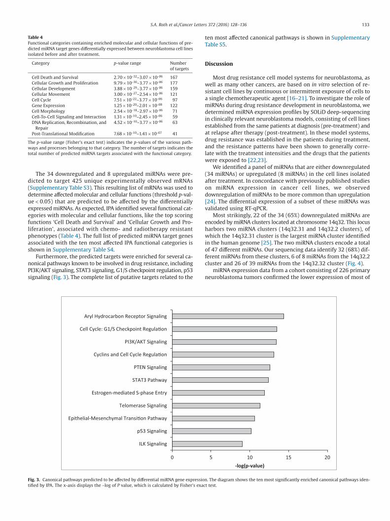
The 34 downregulated and 8 upregulated miRNAs were pre-dicted to target 425 unique experimentally observed mRNAs(Supplementary Table S3). This resulting list of mRNAs was used todetermine affected molecular and cellular functions (threshold p-val-ue < 0.05) that are predicted to be affected by the differentiallyexpressed miRNAs. As expected, IPA identified several functional cat-egories with molecular and cellular functions, like the top scoringfunctions ‘Cell Death and Survival’ and ‘Cellular Growth and Pro-liferation’, associated with chemo- and radiotherapy resistantphenotypes (Table 4). The full list of predicted miRNA target genesassociated with the ten most affected IPA functional categories isshown in Supplementary Table S4.
Furthermore, the predicted targets were enriched for several ca-nonical pathways known to be involved in drug resistance, includingPI3K/AKT signaling, STAT3 signaling, G1/S checkpoint regulation, p53signaling (Fig. 3). The complete list of putative targets related to the
ten most affected canonical pathways is shown in SupplementaryTable S5.
Discussion
Most drug resistance cell model systems for neuroblastoma, aswell as many other cancers, are based on in vitro selection of re-sistant cell lines by continuous or intermittent exposure of cells toa single chemotherapeutic agent [16–21]. To investigate the role ofmiRNAs during drug resistance development in neuroblastoma, wedetermined miRNA expression profiles by SOLiD deep-sequencingin clinically relevant neuroblastoma models, consisting of cell linesestablished from the same patients at diagnosis (pre-treatment) andat relapse after therapy (post-treatment). In these model systems,drug resistance was established in the patients during treatment,and the resistance patterns have been shown to generally corre-late with the treatment intensities and the drugs that the patientswere exposed to [22,23].
We identified a panel of miRNAs that are either downregulated(34 miRNAs) or upregulated (8 miRNAs) in the cell lines isolatedafter treatment. In concordance with previously published studieson miRNA expression in cancer cell lines, we observeddownregulation of miRNAs to be more common than upregulation[24]. The differential expression of a subset of these miRNAs wasvalidated using RT-qPCR.
Most strikingly, 22 of the 34 (65%) downregulated miRNAs areencoded by miRNA clusters located at chromosome 14q32. This locusharbors two miRNA clusters (14q32.31 and 14q32.2 clusters), ofwhich the 14q32.31 cluster is the largest miRNA cluster identifiedin the human genome [25]. The two miRNA clusters encode a totalof 47 different miRNAs. Our sequencing data identify 32 (68%) dif-ferent miRNAs from these clusters, 6 of 8 miRNAs from the 14q32.2cluster and 26 of 39 miRNAs from the 14q32.32 cluster (Fig. 4).
miRNA expression data from a cohort consisting of 226 primaryneuroblastoma tumors confirmed the lower expression of most of
Table 4Functional categories containing enriched molecular and cellular functions of pre-dicted miRNA target genes differentially expressed between neuroblastoma cell linesisolated before and after treatment.
Category p-value range Numberof targets
Cell Death and Survival 2.70 × 10⁻³²–3.07 × 10−06 167Cellular Growth and Proliferation 9.79 × 10⁻³⁰–3.77 × 10−06 177Cellular Development 3.88 × 10⁻²⁹–3.77 × 10−06 159Cellular Movement 3.00 × 10⁻²⁷–2.54 × 10−06 121Cell Cycle 7.51 × 10⁻²²–3.77 × 10−06 97Gene Expression 1.25 × 10⁻²⁰–2.01 × 10−08 122Cell Morphology 2.54 × 10⁻¹⁸–2.97 × 10−06 71Cell-To-Cell Signaling and Interaction 1.31 × 10⁻¹⁰–2.45 × 10−06 59DNA Replication, Recombination, and
Repair4.52 × 10−10–3.77 × 10−06 63
Post-Translational Modification 7.68 × 10⁻¹⁰–1.41 × 10−07 41
The p-value range (Fisher’s exact test) indicates the p-values of the various path-ways and processes belonging to that category. The number of targets indicates thetotal number of predicted miRNA targets associated with the functional category.
0 5 10 15 20
ILK Signaling
p53 Signaling
Epithelial-Mesenchymal Transi on Pathway
Telomerase Signaling
Estrogen-mediated S-phase Entry
STAT3 Pathway
PTEN Signaling
Cyclins and Cell Cycle Regula on
PI3K/AKT Signaling
Cell Cycle: G1/S Checkpoint Regula on
Aryl Hydrocarbon Receptor Signaling
-log(p-value)
Fig. 3. Canonical pathways predicted to be affected by differential miRNA gene expression. The diagram shows the ten most significantly enriched canonical pathways iden-tified by IPA. The x-axis displays the −log of P value, which is calculated by Fisher’s exact test.
133S.A. Roth et al./Cancer Letters 372 (2016) 128–136

our identified 14q32 miRNAs in tumors exhibiting poor prognosisfactors like MNA and INSS stage 4. We also identified several ad-ditional 14q32 miRNAs (all observed to be repressed in cell linesisolated after treatment but not following our strict criteria) thatwere lower expressed in the tumors associated with poor progno-sis (Supplementary Fig. S3).
The 14q32.31 miRNA cluster has previously been shown to bedownregulated in multiple human cancers, including glioblas-toma multiforme, ovarian cancer, breast invasive carcinoma, kidneyrenal clear cell carcinoma, stomach adenocarcinoma, prostate ad-enocarcinoma and bladder urothelial cancer [26]. Using genome-wide miRNA expression profiling, Xu et al. found strongdownregulation of all miRNAs coded by the 14q32.31 cluster indoxorubicin-resistant K562 leukemia cells when compared to theirdrug-sensitive counterparts [27]. Furthermore, miR-381 and miR-495, which also in our study were significantly downregulated inthe post-treatment cell lines, were previously shown to specifical-ly repress expression of MDR1/p-gp through direct targeting of the3’UTR. Similar to our results, most aberrantly expressed miRNAs weredownregulated in drug resistant cells, while miR-375 wasupregulated.
miR-337-3p, encoded by the 14q32.2 miRNA cluster, is one of themiRNAs that was downregulated in all cell lines isolated from pa-tients after treatment. miR-337-3p has previously been reported tosensitize lung cancer cells to taxanes by direct targeting and re-pression of signal transducer and activator of transcription 3 (STAT3)and Ras-related Protein 1A (RAP1A) expression [28]. STAT3 is wellknown for its positive regulation of several survival proteins in-cluding cyclin D1, survivin, c-myc, Bcl-XL and Bcl-2 [29]. Unlike manyother cancers, STAT3 is not constitutively activated in neuroblas-toma; however, in vitro studies demonstrate that bone marrow-derived IL-6 increases the proliferation and decreases etoposide-induced apoptosis through activation of STAT3 in neuroblastoma cells[30].
Very recently, Xiang et al. showed that miR-337-3p isdownregulated in neuroblastoma tumor samples and that miR-337-3p expression is inversely correlated with advanced neuroblastomastages, tumor progression and MYCN amplification [31]. Exoge-nous overexpression of miR-337-3p inhibited growth, invasion,metastasis and angiogenesis in neuroblastoma cell lines. miR-337-3p was shown to directly bind the matrix metalloproteinase 14(MMP-14) promoter to repress its transcription. These studies in-dicate that miR-337-3p could be a critical regulator of severalprocesses involved in survival and progression of aggressiveneuroblastomas.
Allelic loss of the long arm of chromosome 14 (14q) has beenreported to be quite common (20–30%) in neuroblastoma patientsdespite no correlation between 14q loss of heterozygosity (LOH) andclinicopathologic features [32,33]. However, thirteen miRNAs be-longing to the 14q32.31 cluster were significantly downregulatedin high-risk neuroblastoma tumors and two of these (miR-410 andmiR-487) were proposed as prognostic markers since they were ableto discriminate high-risk from low-risk neuroblastoma [34].
Several non-14q32 differentially expressed miRNAs from thisstudy have previously been associated with poor clinical outcomein neuroblastoma and chemoresistance in various other cancers. Forinstance, we found that the tumor suppressor miRNA miR-34a isdownregulated in cell lines isolated from patients after treatment.Loss or downregulation of miR-34a has been reported in severalcancers including neuroblastoma, breast and bladder cancer [35–37].Furthermore, miR-34a overexpression sensitizes various cancer celllines to a number of chemotherapeutic drugs and to radiotherapy[37–47].
Although only few miRNAs were found to be upregulated in ourstudy, several of these have previously been reported to be asso-ciated with clinical outcome in neuroblastoma.
miR-21 is an established oncomir that promotes growth, me-tastasis, chemo-/radioresistance and angiogenesis in several cancers
miR-127miR-136miR-337miR-431miR-432miR-433miR-493miR-665
miR-134miR-154miR-299miR-300miR-323amiR-323bmiR-329-1/2miR-369miR-376a-1/2miR-376bmiR-376cmiR-377miR-379
miR-380miR-381miR-382miR-409miR-410miR-411miR-412miR-485miR-487amiR-487bmiR-494miR-495miR-496
miR-539miR-541miR-543miR-544amiR-654miR-655miR-656miR-668miR-758miR-889miR-1185-1/2miR-1193miR-1197
MEG3 antiRTL1
RTL1
DLK1
paternally expressed
9GEM8GEM DIO3snoRNAcluster
miRNA cluster B(14q32.31)
miRNA cluster A(14q32.2)
ANRcnlANRcnlANRcnl mRNAmRNA
maternally expressed
Fig. 4. Schematic overview of the DLK1-DIO3 imprinted domain located on chromosome 14q32. Green and blue represent paternally and maternally expressed genes, re-spectively. Boxed miRNA names are listed according to the miRNA IDs and not chromosomal location. MiRNAs shown in red and black are differentially expressed, but onlymiRNAs labeled in red meet our strict criteria to represent the panel of differentially expressed miRNAs. MiRNAs shown in gray were not detected. DLK1: delta-like 1 homolog(Drosophila); MEG3: maternally expressed 3; RTL1: retrotransposon-like 1; MEG8: maternally expressed 8; MEG9: maternally expressed 9; DIO3: Type III iodothyroninedeiodinase.
134 S.A. Roth et al./Cancer Letters 372 (2016) 128–136

by direct targeting known tumor suppressor genes [48]. We foundmiR-21 to be upregulated in all cell line pairs except SK-N-BE(1)/(2). SK-N-BE(2) has acquired a p53 mutation that we speculate isresponsible for the major difference in several differentially ex-pressed miRNA in this cell line compared to the other post-treatment isolated cell lines. No previous reports have shown miR-21 to be transcriptionally modulated by p53. Increased miR-21expression has been linked to in vitro selected cisplatin-resistantneuroblastoma cell lines. Treatment with antagomirs of miR-21 sen-sitized resistant cells to cisplatin treatment [18]. Furthermore, a studyusing deep sequencing of neuroblastoma tumors identified miR-181 family members (miR-181a-5p and miR-181b-5p) to beoverexpressed in unfavorable neuroblastomas [49]. In concor-dance with this, we observed upregulation of both miR-181a-5p andmiR-181b-5p in cell lines isolated from patients after treatment. Arecent report including in vitro and in vivo experiments revealed thatmiR-181a-5p confers chemoresistance in cervical squamous cell car-cinoma by directly targeting and decreasing PRKCD expression [50].PRKCD is a protein kinase C isoform critical for DNA damage-induced apoptosis and chemotherapeutic drug resistance in severalcancers, including neuroblastoma [51–53]. A search through thepublic available R2 database (http://r2.amc.nl) using the two largestneuroblastoma datasets (Kocak; n = 649 and SEQC; n = 498) showeda highly significant correlation between low expression levels ofPRKCD and poor survival of neuroblastoma patients (SupplementaryFig. S4).
Increased expression of miR-375 has also been associated withneuroblastoma patients with unfavorable outcome and metastaticdissemination [54] and miR-375 was one of ten miRNAs whose in-creased expression was associated with advanced stage disease [55].Recently, high miR-375 expression was also associated with tumor-igenic undifferentiated neuroblastoma cell lines and miR-375 wasshown to target HuD, a gene involved in neuronal differentiation[56].
In order to gain insight into biological functions and pathwaysthat may be affected by the miRNAs differentially expressed betweenthe pre- and post-treatment isolated cell line pairs, we utilized In-genuity Pathway Analysis (IPA). Predicted targets belong to severalmolecular and cellular functions well known to be implicated in drugresistant cancer cells. The top scoring functions were ‘cell death andsurvival’ and ‘cellular growth and proliferation’, both of which wererecently identified as the top scoring functions affected in severalcisplatin-resistant neuroblastoma cell lines [21]. For the canonicalpathway analysis of the predicted miRNA targets, all pathways areknown to play important functions during cancer progression anddrug resistance. The roles of cancer-associated PI3K/Akt-, PTEN-,STAT3- and p53 pathways have recently been extensively re-viewed [57–60]. Furthermore, the aryl hydrocarbon receptor (AHR)was recently suggested to be an important upstream regulator ofMYCN in neuroblastoma implying the involvement of AHR signal-ing in many aspects of neuroblastoma tumorigenesis [61]. Epithelial–mesenchymal transition (EMT) and telomerase signaling have beenimplicated as important processes during development of chemo-resistance in neuroblastoma [21,62,63]. Functional enrichmentanalysis using IPA-predicted up- and downregulated miRNA targetgenes separately (separate-DE), or the strong-evidence target genes(all-DE and separate-DE) from miRTarBase, revealed similar results(Supplementary Table S6).
Our functional enrichment analysis of differentially expressedmiRNAs has several limitations and must be interpreted with somecaution. First, IPA is a threshold-dependent tool where a user-preselected list of interesting genes (here the criteria-selecteddifferentially expressed miRNAs) is used as input data. Second, themiRNA target gene prediction is based on predetermined data-base knowledge and was not functionally validated by experimentaltesting. Third, the functional enrichment analysis considers that each
individual gene is of equal importance, which is often not the sit-uation in biological systems. Finally, the enrichment analysis is basedon the current state of knowledge. Since new relationships betweengene products and biological functions/pathways are expected tobe unveiled in the future, the presented results capture only a subsetof the biological processes affected in the analyzed model systems.
Our work may serve as a model on which to base future inves-tigations on the role of miRNAs in the complex processes of survivingtherapeutic treatment and developing drug resistance in neuro-blastoma cell lines and also in clinical patient samples. However,the applicability of the study to a clinical setting remains to bedetermined.
Conflict of interest
None.
Acknowledgments
The NBL-W and NBL-WR cell lines were kindly provided by Pro-fessor Susan Cohn, University of Chicago, Chicago, USA. SK-N-BE(1), SK-N-BE(2), SMS-KAN, SMS-KANR, CHLA-15, CHLA-20, CHLA-122 and CHLA-136 were provided by Children’s Oncology Group(COG) Cell Culture and Xenograft Repository, Texas Tech Universi-ty Health Sciences Center, Lubbock, Texas, USA. miRNA expressiondata from primary tumors were obtained through the Neuroblas-toma Research Consortium (NRC) (grant no SFP1096-13). We thankTor Erik Jørgensen, University of Nordland, for technical assis-tance in SOLiD4 sequencing.
This work was supported by grants from the Northern-NorwegianHealth Authorities and the Norwegian Cancer Society.
Appendix: Supplementary material
Supplementary data to this article can be found online atdoi:10.1016/j.canlet.2015.11.026.
References
[1] J.R. Park, A. Eggert, H. Caron, Neuroblastoma: biology, prognosis, and treatment,Hematol. Oncol. Clin. North Am. 24 (1) (2010) 65–86.
[2] T.A. Ishola, D.H. Chung, Neuroblastoma, Surg. Oncol. 16 (3) (2007) 149–156.[3] S. Modak, N.K. Cheung, Neuroblastoma: therapeutic strategies for a clinical
enigma, Cancer Treat. Rev. 36 (4) (2010) 307–317.[4] C. Owens, M. Irwin, Neuroblastoma: the impact of biology and cooperation
leading to personalized treatments, Crit. Rev. Clin. Lab. Sci. 49 (3) (2012) 85–115.[5] J.M. Maris, Recent advances in neuroblastoma, N. Engl. J. Med. 362 (23) (2010)
2202–2211.[6] G.P. Tonini, A. Nakagawara, F. Berthold, Towards a turning point of
neuroblastoma therapy, Cancer Lett. 326 (2) (2012) 128–134.[7] J.I. Fletcher, et al., Targeting multidrug resistance in neuroblastoma, in: M.A.
Hayat (Ed.), Pediatric cancer, vol. 1: Neuroblastoma, Springer, New York, USA,2012, pp. 115–123.
[8] E. Giovannetti, et al., Molecular mechanisms underlying the role of microRNAs(miRNAs) in anticancer drug resistance and implications for clinical practice,Crit. Rev. Oncol. Hematol. 81 (2) (2012) 103–122.
[9] F. Chen, S.J. Hu, Effect of microRNA-34a in cell cycle, differentiation, andapoptosis: a review, J. Biochem. Mol. Toxicol. 26 (2) (2012) 79–86.
[10] W.C. Cho, MicroRNAs as therapeutic targets and their potential applications incancer therapy, Expert Opin. Ther. Targets 16 (8) (2012) 747–759.
[11] Y.W. Kong, et al., microRNAs in cancer management, Lancet Oncol. 13 (6) (2012)e249–e258.
[12] S.B. Thorsen, et al., The therapeutic potential of microRNAs in cancer, CancerJ. 18 (3) (2012) 275–284.
[13] J. Buechner, C. Einvik, N-myc and noncoding RNAs in neuroblastoma, Mol. CancerRes. 10 (10) (2012) 1243–1253.
[14] E. Knutsen, et al., Performance comparison of digital microRNA profilingtechnologies applied on human breast cancer cell lines, PLoS ONE 8 (10) (2013)e75813.
[15] B.A. Spengler, et al., Cell lineage and differentiation state are primarydeterminants of MYCN gene expression and malignant potential in humanneuroblastoma cells, Oncol. Res. 9 (9) (1997) 467–476.
135S.A. Roth et al./Cancer Letters 372 (2016) 128–136

[16] R. Kotchetkov, et al., Development of resistance to vincristine and doxorubicinin neuroblastoma alters malignant properties and induces additional karyotypechanges: a preclinical model, Int. J. Cancer 104 (1) (2003) 36–43.
[17] J. Bedrnicek, et al., Characterization of drug-resistant neuroblastoma cell linesby comparative genomic hybridization, Neoplasma 52 (5) (2005) 415–419.
[18] Y. Chen, et al., Micro-RNA-21 regulates the sensitivity to cisplatin in humanneuroblastoma cells, J. Pediatr. Surg. 47 (10) (2012) 1797–1805.
[19] S.V. Govindan, et al., Establishment and characterization of triple drug resistanthead and neck squamous cell carcinoma cell lines, Mol. Med. Rep. 12 (2) (2015)3025–3032.
[20] D. Ayers, et al., Identification of miRNAs contributing to neuroblastomachemoresistance, Comput. Struct. Biotechnol. J. 13 (2015) 307–319.
[21] O. Piskareva, et al., The development of cisplatin resistance in neuroblastomais accompanied by epithelial to mesenchymal transition in vitro, Cancer Lett.364 (2) (2015) 142–155.
[22] N. Keshelava, R.C. Seeger, C.P. Reynolds, Drug resistance in human neuroblastomacell lines correlates with clinical therapy, Eur. J. Cancer 33 (12) (1997) 2002–2006.
[23] N. Keshelava, et al., Loss of p53 function confers high-level multidrug resistancein neuroblastoma cell lines, Cancer Res. 61 (16) (2001) 6185–6193.
[24] N. Dahiya, et al., MicroRNA expression and identification of putative miRNAtargets in ovarian cancer, PLoS ONE 3 (6) (2008) e2436.
[25] E.A. Glazov, et al., Origin, evolution, and biological role of miRNA cluster inDLK-DIO3 genomic region in placental mammals, Mol. Biol. Evol. 25 (5) (2008)939–948.
[26] S.V. Laddha, et al., Genome-wide analysis reveals downregulation of miR-379/miR-656 cluster in human cancers, Biol. Direct 8 (2013) 10.
[27] Y. Xu, et al., Changes in the expression of miR-381 and miR-495 are inverselyassociated with the expression of the MDR1 gene and development of multi-drug resistance, PLoS ONE 8 (11) (2013) e82062.
[28] L. Du, et al., miR-337-3p and its targets STAT3 and RAP1A modulate taxanesensitivity in non-small cell lung cancers, PLoS ONE 7 (6) (2012) e39167.
[29] A. Xiong, et al., Transcription factor STAT3 as a novel molecular target for cancerprevention, Cancers (Basel) 6 (2) (2014) 926–957.
[30] T. Ara, et al., Interleukin-6 in the bone marrow microenvironment promotesthe growth and survival of neuroblastoma cells, Cancer Res. 69 (1) (2009)329–337.
[31] X. Xiang, et al., miRNA-337-3p suppresses neuroblastoma progression byrepressing the transcription of matrix metalloproteinase 14, Oncotarget 6 (26)(2015) 22452–22466.
[32] M. Hoshi, et al., Detailed deletion mapping of chromosome band 14q32 inhuman neuroblastoma defines a 1.1-Mb region of common allelic loss, Br. J.Cancer 82 (11) (2000) 1801–1807.
[33] P.M. Thompson, et al., Loss of heterozygosity for chromosome 14q inneuroblastoma, Med. Pediatr. Oncol. 36 (1) (2001) 28–31.
[34] C.H. Gattolliat, et al., Expression of miR-487b and miR-410 encoded by 14q32.31locus is a prognostic marker in neuroblastoma, Br. J. Cancer 105 (9) (2011)1352–1361.
[35] G. Feinberg-Gorenshtein, et al., Reduced levels of miR-34a in neuroblastomaare not caused by mutations in the TP53 binding site, Genes ChromosomesCancer 48 (7) (2009) 539–543.
[36] A. Javeri, et al., Downregulation of miR-34a in breast tumors is not associatedwith either p53 mutations or promoter hypermethylation while it correlateswith metastasis, Med. Oncol. 30 (1) (2013).
[37] X. Wang, et al., microRNA-34a sensitizes lung cancer cell lines to DDP treatmentindependent of p53 status, Cancer Biother. Radiopharm. 28 (1) (2013) 45–50.
[38] Y. Akao, et al., Dysregulation of microRNA-34a expression causes drug-resistanceto 5-FU in human colon cancer DLD-1 cells, Cancer Lett. 300 (2) (2011) 197–204.
[39] W. Cao, et al., miR-34a regulates cisplatin-induce gastric cancer cell death bymodulating PI3K/AKT/survivin pathway, Tumour Biol. (2013). doi:10.1007/s13277-013-1171-7.
[40] W. Duan, et al., Ectopic expression of miR-34a enhances radiosensitivity ofnon-small cell lung cancer cells, partly by suppressing the LyGDI signalingpathway, J Radiat Res (2013). doi: 10.1093/jrr/rrs136.
[41] K. Kojima, et al., MiR-34a attenuates paclitaxel-resistance of hormone-refractoryprostate cancer PC3 cells through direct and indirect mechanisms, Prostate 70(14) (2010) 1501–1512.
[42] X.J. Li, et al., MicroRNA-34a modulates chemosensitivity of breast cancercells to adriamycin by targeting notch1, Arch. Med. Res. 43 (7) (2012) 514–521.
[43] F. Nakatani, et al., miR-34a predicts survival of Ewing’s sarcoma patients anddirectly influences cell chemo-sensitivity and malignancy, J. Pathol. 226 (5)(2012) 796–805.
[44] P. Neri, et al., Mir-34a sensitizes multiple myeloma (MM) cells to the proteasomeinhibitor bortezomib, Blood 118 (21) (2011) 66–67.
[45] R.L. Vinall, et al., MiR-34a chemosensitizes bladder cancer cells to cisplatintreatment regardless of p53-Rb pathway status, Int. J. Cancer 130 (11) (2012)2526–2538.
[46] S.D. Weeraratne, et al., miR-34a confers chemosensitivity through modulationof MAGE-A and p53 in medulloblastoma, Neuro Oncol. 13 (2) (2011) 165–175.
[47] F. Yang, et al., MicroRNA-34a targets bcl-2 and sensitizes human hepatocellularcarcinoma cells to sorafenib treatment, Technol. Cancer Res. Treat. (2013).doi:10.7785/tcrt.2012.500364.
[48] S.R. Pfeffer, C.H. Yang, L.M. Pfeffer, The role of miR-21 in cancer, Drug Dev. Res.(2015). doi:10.1002/ddr.21257.
[49] J.H. Schulte, et al., Deep sequencing reveals differential expression of microRNAsin favorable versus unfavorable neuroblastoma, Nucleic Acids Res. 38 (17) (2010)5919–5928.
[50] Y. Chen, et al., MicroRNA-181a enhances the chemoresistance of human cervicalsquamous cell carcinoma to cisplatin by targeting PRKCD, Exp. Cell Res. 320(1) (2014) 12–20.
[51] A. Basu, Involvement of protein kinase C-delta in DNA damage-inducedapoptosis, J. Cell. Mol. Med. 7 (4) (2003) 341–350.
[52] T.W. Day, C.H. Wu, A.R. Safa, Etoposide induces protein kinase Cdelta- andcaspase-3-dependent apoptosis in neuroblastoma cancer cells, Mol. Pharmacol.76 (3) (2009) 632–640.
[53] B. Marengo, et al., PKCdelta sensitizes neuroblastoma cells to L-buthionine-sulfoximine and etoposide inducing reactive oxygen species overproductionand DNA damage, PLoS ONE 6 (2) (2011) e14661.
[54] J. Guo, et al., Identification of miRNAs that are associated with tumor metastasisin neuroblastoma, Cancer Biol. Ther. 9 (6) (2010) 446–452.
[55] V.R. Chu, I. Lee, MicroRNA target signatures in advanced stage neuroblastoma,in: Neuroblastoma – present and future, Intech, 2012, pp. 271–286.
[56] L. Samaraweera, et al., MicroRNAs define distinct human neuroblastoma cellphenotypes and regulate their differentiation and tumorigenicity, BMC Cancer14 (2014) 309.
[57] M.C. Hollander, G.M. Blumenthal, P.A. Dennis, PTEN loss in the continuum ofcommon cancers, rare syndromes and mouse models, Nat. Rev. Cancer 11 (4)(2011) 289–301.
[58] T. Soussi, K.G. Wiman, TP53: an oncogene in disguise, Cell Death Differ. 22 (8)(2015) 1239–1249.
[59] H. Yu, et al., Revisiting STAT3 signalling in cancer: new and unexpected biologicalfunctions, Nat. Rev. Cancer 14 (11) (2014) 736–746.
[60] D.A. Fruman, C. Rommel, PI3K and cancer: lessons, challenges and opportunities,Nat. Rev. Drug Discov. 13 (2) (2014) 140–156.
[61] P.Y. Wu, et al., Aryl hydrocarbon receptor downregulates MYCN expression andpromotes cell differentiation of neuroblastoma, PLoS ONE 9 (2) (2014) e88795.
[62] J.A. Naiditch, et al., Mesenchymal change and drug resistance in neuroblastoma,J. Surg. Res. 193 (1) (2015) 279–288.
[63] S. Wesbuer, et al., Association of telomerase activity with radio- andchemosensitivity of neuroblastomas, Radiat. Oncol. 5 (2010) 66.
136 S.A. Roth et al./Cancer Letters 372 (2016) 128–136

PAPER-II

ONCOLOGY LETTERS 16: 6786-6794, 20186786
Abstract. High-risk neuroblastoma is the most aggressive form of cancer in children. The estimated survival of children with high-risk neuroblastoma is 40-50% compared with low and intermediate risk neuroblastoma, which is >98 and 90-95%, respectively. In addition, patients with high-risk neuroblas-toma often experience relapse following intensive treatments with standard chemotherapeutic drugs. Therefore alternative strategies are required to address this problem. MicroRNAs (miRNAs/miRs) are small, endogenously expressed non-coding RNAs, which when deregulated have been demonstrated to serve significant roles in the tumorigenesis of a number of different types of cancer. Results from a previous deep sequencing study identified 22 downregulated miRNAs from the 14q32 miRNA cluster differentially expressed in neuroblastoma cell lines isolated from 6 patients at diagnosis and at relapse following intensive treatments. miR-376c-3p is one of the 22 miRNAs that was downregulated in the majority of the cell lines isolated from patients post treatment. The present study employed reverse transcription-quantitative polymerase chain reaction (RT-qPCR) to quantify the basic expression of miR-376c-3p in 6 neuroblastoma cell lines. The functional role of miR-376c-3p in the neuroblastoma cell lines was evaluated by alamar blue-cell viability and propidium iodide‑flow cytometric assays. In addition, luciferase reporter assays, RT-qPCR and western blotting were performed to identify and quantify the targets of miR-376c-3p in neuro-blastoma cell lines. Ectopic expression of miR-376c-3p led to
significant inhibition of cell viability and G1‑cell cycle arrest in multiple neuroblastoma cell lines by reducing the expres-sion of cyclin D1, an oncogene critical for neuroblastoma pathogenesis. The results of the present study provide novel insights into the functional role of miR-376c-3p and suggest new approaches for the treatment of neuroblastoma.
Introduction
Neuroblastoma is the most common childhood malignancy derived from the primitive cells of the sympathetic nervous system (1). It accounts for ~15% of all pediatric oncology deaths (2). The treatment of patients with advanced neuro-blastoma or high-risk neuroblastoma is still a challenge due to several factors including, dose-limiting toxicity to standard chemotherapeutic agents, disease heterogeneity and tumor regression (3). Moreover, these patients have a very poor prognosis with the overall ten-year survival rates of less than 10% (4). Therefore, the development of innovate alternate treatment strategies is necessary to increase the treatment effectiveness and lower toxicity.
MicroRNAs (miRNAs/miRs) are a large family of small (~22-25 nucleotides), endogenous, non-coding RNAs, which binds the partial or perfect complementary sequences in the 3'untranslated region (3'UTR) of target messenger RNAs (mRNAs) leading to translational repression or mRNA degra-dation (5). They regulate the expression of genes involved in proliferation, apoptosis, development and stress response. Thus, miRNAs have shown to play an important role in the initiation and progression of cancer (5). Depending on their respective target, miRNAs can act as oncogenes and or tumor suppres-sors (6). miRNAs are differentially expressed across cancer types and microRNA‑profiling studies have revealed a general downregulation in tumors as compared with normal tissues (7). Interestingly, growing evidence have now shown the potential of aberrant expression of miRNAs to use as the prognostic and diagnostic biomarkers of human malignancies (6).
miR-376 family of miRNAs comprises of miR‑376a2, miR‑376b, miR‑376c (earlier known as miR‑368), miR‑376b1 and miR‑376b2 and their sequence identity is highly similar to mouse miR‑376a‑c miRNAs (8). miR-376 family members are located on chromosome 14 in humans and at the distal end of mouse chromosome 12 (9). They are expressed in
Hsa‑miR‑376c‑3p targets Cyclin D1 and induces G1‑cell cycle arrest in neuroblastoma cells
SWAPNIL PARASHRAM BHAVSAR1, CECILIE LØKKE1, TROND FLÆGSTAD1,2 and CHRISTER EINVIK1,2
1Pediatric Research Group, Department of Clinical Medicine, Faculty of Health Science, The Arctic University of Norway-UiT, NO-9037 Tromsø; 2Department of Pediatrics,
Division of Child and Adolescent Health, University Hospital of North-Norway, NO-9038 Tromsø, Norway
Received November 2, 2017; Accepted July 5, 2018
DOI: 10.3892/ol.2018.9431
Correspondence to: Professor Christer Einvik, Pediatric Research Group, Department of Clinical Medicine, Faculty of Health Science, The Arctic University of Norway-UiT, Hansine Hansens veg 18, NO-9037 Tromsø, NorwayE-mail: [email protected]
Abbreviations: miRNA, microRNA; NC, negative control; miR-376c-3p, microRNA-376c-3p; CCND1, Cyclin D1
Key words: high-risk neuroblastoma, microRNA, deep sequencing, 14q32 microRNA cluster, microRNA-376c-3p, Cyclin D1

BHAVSAR et al: Hsa-miR-376c-3p IN NEUROBLASTOMA CELLS 6787
placenta, developing embryos, and adult tissues (8). The expression of miR‑376c is downregulated in many human malignancies including cervical cancer (10), prostate cancer (11), oral squamous cell carcinoma (12), intrahepatic cholangiocarcinoma (13), melanoma (14), osteosarcoma (15) and gliomas (16). However, it has been shown that miR‑376c is upregulated and it act as oncogenic in acute myeloid leukemia (17) and gastric cancer (18). In addition, forced expression of miR‑376c enhance ovarian cancer cell survival by targeting activing-receptor like kinase 7 (19). Whereas, forced overexpression of miR‑376c suppressed growth and invasion of non-small cell lung cancer (20). According to our knowledge, there is no report regarding the role of miR‑376c in neuroblastoma. Thus uncovering the mechanisms of miR‑376c function is critical to both the fundamental understanding of neuroblastoma pathogenesis and novel therapeutic treatments.
The Cyclin D1 (CCND1) is one of the extensively docu-mented oncogene in human cancers. Functionally, CCND1 binds with cyclin-dependent kinases (CDK 4/6) which phos-phorylate pRB family proteins, which in turn transactivates genes necessary for cell cycle progression (21). Dysregulated expression of CCND1 is a common feature in various human cancers (22,23). Inhibitors targeting CCND1 are thoroughly studied but no results have yet been proven effective (22,23). The CCND1 gene has one of the longest 3'UTR (~3.1 kb), suggesting a strong functional relevance (24). To date, many experimentally validated miRNAs targeting CCND1 in different cancers are identified. For example, let‑7e, miR‑9‑5p, miR‑15a‑5p, miR‑16, miR‑17, miR‑20a, miR‑106b, miR‑34a and miR‑206 (25-31). However, no miRNA directly targeting CCND1 3'UTR is yet identified in neuroblastoma.
In this study, we examined the relationship between miR‑376c‑3p expression and neuroblastoma tumorigenesis. In our previous deep sequencing study, we have analyzed miR‑376c‑3p expression in neuroblastoma cell lines with different genetic characteristics (32). miR‑376c‑3p was down-regulated in most of the cell lines tested and therefore we overexpressed miR‑376c‑3p, which have had significant effects on cell growth and survival of neuroblastoma cells.
In addition, we demonstrated that CCND1 is a direct target of miR‑376c‑3p in neuroblastoma and overexpression of miR‑376c‑3p might have a significant influence on inhibition of neuroblastoma tumorigenesis.
Materials and methods
Cell culture. Human neuroblastoma cell lines, CHLA-15 and CHLA‑20 were cultivated in Iscove's Modified Dulbecco's Medium (Sigma-Aldrich; Merck KGaA, Darmstadt, Germany) supplemented with 20% fetal bovine serum, 4 mM L-Glutamine and 1X ITS (5 µg/ml insulin, 5 µg/ml trans-ferrin, and 5 ng/ml selenous acid). CHLA-15 and CHLA-20 were obtained from the same neuroblastoma patient prior to and following treatment with combination chemotherapy regimens, respectively. SKNAS, BE(2)-C, Kelly and SHSY-5Y cells were grown in RPMI-1640 medium (Sigma-Aldrich; Merck KGaA) supplemented with 10% fetal bovine serum and 2 mM L‑Glutamine (final concentrations). All cells were split before confluence and maintained at 37˚C in a humidified incubator with 4.5 to 5% CO2 atmosphere. The cell lines were
authenticated by short tandem repeat profiling at the Center of Forensic Genetics, The Arctic University of Norway-UiT, Norway and tested negative for mycoplasma contamination. CHLA-15 and CHLA-20 cell lines were kindly provided by Children's Oncology Group, Cell Culture and Xenograft Repository, Texas Tech University Health Science Centre (Lubbock, TX, USA). BE(2)-C, SKNAS, Kelly and SHSY-5Y were provided by Dr. John Inge Johnsen (Childhood Cancer Research Unit, Department of Women's and Children's Health, Karolinska Institutet, Stockholm, Sweden).
Transfections. MicroRNA miR‑376c‑3p mimics or negative control (NC) mimics were purchased from GenePharma Co., Ltd. (Shanghai, China) and Ambion (Thermo Fisher Scientific, Inc., Waltham, MA, USA). Transfections of miRNA and NC mimics (25-40 nM) were carried out using Lipofectamine® 2000 reagent (Thermo Fisher Scientific, Inc.) according to manufacturer's instructions.
Cell viability assay. CHLA-15, CHLA-20, BE(2)-C, Kelly and SHSY-5Y cells were seeded in 24-well plates and reverse transfected with 25 nM miR‑376c‑3p or NC mimics using Lipofectamine® 2000 reagent (Thermo Fisher Scientific, Inc.). Every 24 h post-transfection, alamarBlue® (Thermo Fisher Scientific, Inc.) cell viability assay was performed according to manufacturer's instructions. Ten percent of the alamarBlue reagent was added to the cultured cells, mixed gently and incubated at 37˚C for three hours. 100 µl of medium was then transferred to black-walled 96-well plate and fluorescence was monitored at 540 nm excitation wavelength and 590 nm emission wavelength in a microplate reader (CLARIOstar; BMG Labtech GmbH, Offenburg, Germany). Cell viability was calculated as the percentage of NC transfected cells set to 100 percent.
Flow cytometric analysis of cell cycle distribution. The BE(2)-C, Kelly and SHSY-5Y cells were seeded in 25 cm2 culture flasks and reverse transfected with 25 nM mimics as described previously. 24 h post-transfection cells were tryp-sinized and washed with 1X phosphate-buffered saline (PBS). The cells were then fixed in ice‑cold 70% ethanol and incu-bated overnight at -20˚C. Next day, the ethanol fixed cells were centrifuged for 10 min at 0.8 x g and washed twice with 1X PBS and resuspended in the propidium iodide (PI) (Thermo Fisher Scientific, Inc.) staining solution (PBS with 100 µg/ml RNase, 50 µg/ml PI). The cells were then incubated for 30 min protected from light and stored on ice until analyzed. Fluorescence emitted from the PI-DNA complex was analyzed by flow cytometry using BD LSRFortessa™ cell analyzer (BD Biosciences, Franklin Lakes, NJ, USA). FlowJo 7.6.5 software was used to analyze the cell cycle data using the Dean-Jett-Fox model for cell cycle evaluation.
Bioinformatics target prediction. TargetScan (version 6.2; www.targetscan.org/) target prediction software was used to predict miR-376c-3p targets related to cell cycle.
Reverse transcription‑quantitative polymerase chain reaction (RT‑qPCR). The Kelly, SHSY-5Y and BE(2)-C cells were seeded in 6-well plates and transfected with 40 nM of miR‑376c‑3p or

ONCOLOGY LETTERS 16: 6786-6794, 20186788
NC mimics. Cells were harvested 48 h post-transfection and total RNA was isolated with QIAzol®Lysis reagent (Qiagen Sciences, Inc., Gaithersburg, MD, USA) according to manu-facturer's instructions and quantified by NanoDrop™ 1000 spectrophotometer (Thermo Fisher Scientific, Inc.).
For miRNA expression analysis, complementary DNA (cDNA) was synthesized from isolated total RNAs using the miScript II RT kit (Qiagen Sciences, Inc.) according to manufacturer's instructions. The reaction mixture (1X) (20 µl reaction volume) for reverse transcription was as follows: Total RNA, 1 µg (diluted in RNase Free Water up to 12 µl); 5X miScript HiSpec Buffer, 4 µl; 10X miScript Nucleics Mix, 2 µl; miScript Reverse Transcriptase Mix, 2 µl; The cycling conditions were 37˚C for 60 min followed by 95˚C for 5 min. The cDNA obtained was diluted with 80 µl RNase Free Water to achieve 10 ng/µl concentration and stored at ‑20˚C until use. To quantitate miR‑376c‑3p levels, quantitative poly-merase chain reaction was performed with miScript primer assay for miR-376c (cat. no MS00004046) using miScript SYBR®Green PCR kit (Qiagen Sciences, Inc.,). The miScript primer assay for miR-4286 (cat. no MS00021371) was used as an internal control for normalization. The reaction mixture (1X) (20 µl reaction volume) for real time PCR was as follows: cDNA (1 ng/5 µl), 5 µl; QuantiTect SYBR-Green PCR Master Mix, 10 µl; Specific miScript primer assay, 2 µl; 10X miScript Universal Primer, 2 µl; RNase Free Water, 1 µl.
For basic miRNA expression levels in neuroblastoma cell lines, the following method was used for calculations. Raw fluorescence values (non‑baseline corrected) gener-ated from RT-qPCR reactions were used to calculate mean PCR efficiencies in the LinRegPCR software (Version 11.0; http://LinRegPCR.HFRC.nl.). N0 values (starting concen-trations calculated by LinRegPCR software, N0=threshold/ (mean amplicon efficiencyCq)) were used to calculate the expression of miR‑376c‑3p relative to the stably expressed miR‑4286 (32). qPCR reactions were performed in trip-licates on 2 independent biological replicates. Standard deviations were calculated taking into account the principle of error propagation (including technical and biological replicates).
For mRNA expression analysis, complementary DNA (cDNA) was synthesized from isolated total RNAs using the maxima reverse transcriptase (Thermo Fisher Scientific, Inc.) according to manufacturer's instructions. The reaction mixture (1X) (20 µl reaction volume) for reverse transcription of mRNAs was as follows: Oligo DT primer (20 µM), 1 µl; dNTP (10 mM each), 1 µl; Total RNA, 1 µg (diluted in RNase Free Water up to 13.75 µl); Incubate 65˚C for 5 min followed by addition of 5x RT Buffer, 4 µl; Maxima Reverse Transcriptase, 0.25 µl; The cycling conditions were 60˚C for 30 min followed by 85˚C for 5 min. The cDNA obtained was diluted with 80 µl RNase Free Water to achieve 10 ng/µl concentration and stored at ‑20˚C until use. To quantitate CCND1 levels, quan-titative polymerase chain reaction was performed with Power SYBR-Green PCR Master Mix (Thermo Fisher Scientific, Inc.). SDHA housekeeping gene was used as an internal control for normalization. The reaction mixture (1X) (20 µl reaction volume) for real time PCR was as follows: cDNA (1 ng/1 µl), 10 µl; Power SYBR-Green PCR Master Mix, 5 µl; Forward
Primer (10 µM), 0.4 µl; Reverse Primer (10 µM), 0.4 µl; RNase Free Water 4.2 µl.
Amplifications were carried out using Light Cycler 96 SW 1.1 (Roche Diagnostics GmbH, Mannheim, Germany) and expression levels of miRNAs and mRNAs were evaluated using the comparative ΔΔCq comparative cycle threshold method (33). The primers used were CCND1 (forward: 5'-CCG TCC ATG CGG AAG ATC-3'; reverse: 5'-ATG GCC AGC GGG AAG AC-3') and SDHA (forward: 5'-CTG ATG AGA CAA GAT GTG GTG-3'; reverse: 5'-CAA TCT CCC TTC AAT GTA CTC C-3').
Western blot analysis. Cells were trypsinized and lysed in 40 µl RIPA buffer (50 mM Tris-HCL pH 8, 150 mM NaCl, 1% NP-40, 0.5% sodium deoxycholate, 0.1% SDS) supplemented with 1X Protein Inhibitor Cocktail (Roche Diagnostics GmbH) and 1 mM dithiothreitol (DTT). Lysate was cleared with centrifugation (21.1 x g) and the total protein concentra-tions were determined using DCTMProtein Assay kit (Bio-Rad Laboratories, Inc., Hercules, CA, USA) according to manu-facturer's instructions. 40 µg protein was then loaded in each well and separated on a NuPAGE® Novex 4-12% Bis-Tris precast polyacrylamide gel (Thermo Fisher Scientific, Inc.). The separated proteins were transferred on Immobilon-FL PVDF membrane (EMD Millipore, Billerica, MA, USA) and blocked for 1 h at room temperature in 5 ml Odyssey Blocking Buffer (LI-COR Biosciences, Lincoln, NE, USA). The PVDF membrane was then incubated overnight at 4˚C with primary antibodies anti-Cyclin D1-(H-295)-Human Cyclin D1 Rabbit, polyclonal; (1:1,000; Santa Cruz Biotechnology, Inc., Dallas, TX, USA) and anti-actin-(ab3280)-Human Actin Mouse, monoclonal (1:1,000; Abcam, Cambridge, UK). The secondary antibodies used were goat anti-rabbit-IRDye800CW, (1:5,000) (Rockland Immunochemicals, Inc., Gilbertsville, PA, USA) and goat anti-mouse-Alexa Fluor 680, (1:5,000; Thermo Fisher Scientific, Inc.). Antibody binding was detected using the Odyssey CLx Infrared Imaging System (LI-COR Biosciences). ImageJ software was used to quantify western blot results.
Luciferase reporter assay. The pMIR-Report-Cyclin D1-UTR-WT construct was a generous gif t f rom Dr. Laura Barkley (30) and pMIR-Report-Cyclin D1-UTR-MUT construct with a mutation in the putative miR‑376c‑3p seed sequence (mutant) was generated using QuikChange® Multi Site-Directed Mutagenesis kit (Agilent Technologies, Inc., Santa Clara, CA, USA). The primers used for mutagenesis were CCND1_3'UTR_miR-376c-3p (forward: 5'-CAC ATC TTG GCA TAC TAA TTC TTG-3'; reverse: 5'-CAA GAA TTA GTA TGC CAA GAT GTG-3'). To validate for mutation in seed sequence the mutant plasmid was sequenced using sequencing primer 5'-CAT CTG ATT GGA CAG GCA TG-3'. The cells seeded at a density of 5x10⁴cells/well in a 12-well plate were co-transfected with 40 nM NC or miR‑376c‑3p mimics, 20 ng pRL-SV40 construct (Promega Corporation, Madison, WI, USA) and 100 ng wild type or mutant luciferase constructs using Lipofectamine® 2000 reagent (Thermo Fisher Scientific, Inc.). 24 h post‑transfection, firefly and renilla luciferase activities analyzed using the Dual-Luciferase Reporter Assay (Promega Corporation), according to manufacturer's instructions. Firefly luciferase activity was normalized against renilla luciferase

BHAVSAR et al: Hsa-miR-376c-3p IN NEUROBLASTOMA CELLS 6789
activity and luciferase activities of miR‑376c‑3p transfected cells were calculated relative to NC transfected cells set to 100 %.
MicroRNA expression data from neuroblastoma tumors. miRNA expression data from 226 primary neuroblastoma tumors were obtained through the Neuroblastoma Research Consortium (NRC). Differential miRNA expression was analyzed using a Kruskal-Wallis test.
Statistical analysis. The data was expressed as mean ± standard deviation (SD). Unless otherwise stated, all experiments were performed at least three times independently. Statistical analysis was performed using the software GraphPad Prism version 5.00 for Windows (GraphPad Software, Inc., La Jolla, CA, USA; available at www.graphpad.com). Statistical differ-ences between means were determined using Student's t-test. P<0.05 was considered to indicate a statistically significant difference.
Results
Multiple miRNAs located at 14q32 chromosomal region are downregulated and their lower levels are associated with poor prognosis factors in neuroblastoma. Results from our previous study, where we used next generation sequencing technology (SOLiD ligation sequencing) to determine miRNA expression profiles in neuroblastoma cell lines established from patients at diagnosis and at relapse after treatment, identified 22 downregulated microRNAs from 14q32 miRNA cluster. The expression of these downregulated miRNAs was confirmed in a cohort consisting of 226 primary neuroblas-tomas (32). miR‑376c‑3p, was one of the 22 miRNAs that was downregulated in most of the cell lines isolated from patients after the treatment (Fig. 1A). When miR‑376c expression was compared in neuroblastoma primary tumors of different stages from the 226-cohort, we observed a trend towards lower expression in advanced stage disease compared to tumors in stage 1 (Fig. 1B). Thus, we sought out to focus on the func-tional role of miR‑376c‑3p in neuroblastoma.
miR‑376c‑3p reduces cell viability in neuroblastoma cell lines. Even though miR‑376c‑3p has been shown to play either the oncogenic or the tumor suppressive role in different cancers (10-20) the functional role of miR‑376c‑3p is not yet determined in neuroblastoma. Therefore, we first used RT-qPCR to measure miR‑376c‑3p basic expression levels relative to miR‑4286 in 6 neuroblastoma cell lines. miR‑4286 was previously showed to be stably expressed in neuroblastoma cell lines (32). Among the cell lines, SKNAS and BE(2)-C cells have the highest expression level of miR‑376c‑3p, whereas SHSY-5Y, Kelly, CHLA-15 and CHLA-20 cells showed barely detectable levels of expression (Fig. 2A). In order to find out the potential role of miR‑376c‑3p in neuroblastoma, cell viability assay was performed on several neuroblastoma cell lines by overexpressing NC or miR‑376c‑3p mimics. The expression of miR‑376c‑3p was significantly increased in miR‑376c‑3p transfected cell lines compared with NC transfected cells, as validated and confirmed by RT‑qPCR (Fig. 2B). Cell viability alamarBlue assay was performed at 24, 48, 72 and 96 h post-transfection, which showed that neuroblastoma cell
lines transfected with miR‑376c‑3p mimics, had significantly reduced cell viability as compared to NC transfected cells. Thus, ectopic expression of miR‑376c‑3p reduced the growth of SKNAS, CHLA-15, CHLA-20, SHSY-5Y, Kelly and BE(2)-C cells as compared to NC transfected cells (Fig. 2C). These results indicate that cell growth of neuroblastoma cell lines is significantly affected by overexpression of miR‑376c‑3p.
miR‑376c‑3p induces a G1‑cell cycle arrest in neuroblastoma cells. We did not detect significant apoptosis in miR‑376c‑3p transfected neuroblastoma cells using Annexin V and PARP cleavage assay (data not shown). Thus, we investigated the effects of miR‑376c‑3p overexpression on cell cycle distribu-tion of representative neuroblastoma cell lines BE(2)-C, Kelly and SHSY‑5Y by flow cytometric assay. Ectopic expression of miR‑376c‑3p in BE(2)-C, Kelly and SHSY-5Y resulted in increased percentage of cells in G1-phase of cell cycle as compared to NC transfected cells by 13% (**P=0.0023),
Figure 1. (A) Schematic representation of the miRNA-cluster at 14q32 chro-mosomal region. Multiple miRNAs located at 14q32 chromosomal region are downregulated (bold type) and associated with poor prognosis factors in neuroblastoma. The miRNA of interest, miR-376c-3p is shown in bold and a rectangular box. (B) Box-plots of miR-376c expression in tumors from neuroblastoma patients at different stages. The boxes represent the 25-75% quartiles. The horizontal line in the boxes represents the median level. Whiskers represent the non-outlier range. Open circles represent the outliers. P=0.011, comparing the median expression level of miR376c-3p across all pathological stages. DLK1, Delta Like Non-canonical Notch Ligand 1; MEG, maternally expressed; snoRNA, small nucleolar RNAs; DIO3, iodothyronine deiodinase 3; miR/miRNA, microRNA.

ONCOLOGY LETTERS 16: 6786-6794, 20186790
16% (***P=0.0001) and 9% (*P=0.0106), respectively with a corresponding reduction in the percentage of cells in the S and G2/M-phase (Fig. 3). Thus, this observation led us to the conclusion that decrease in cell viability might be due to induction of G1-cell cycle arrest in neuroblastoma cells and not apoptosis.
CCND1 is a direct target of miR‑376c‑3p in neuroblastoma. In order to investigate the underlying molecular mechanisms of miR‑376c‑3p induced suppression of the cell viability and G1-cell cycle arrest, a bioinformatics analysis was performed using miRNA target prediction algorithm TargetScan (Release 6.2; www.targetscan.org/) to predict the target genes of miR‑376c‑3p mainly associated with the cell cycle progres-sion. TargetScan revealed 254 potential downstream targets with the conserved sites for miR‑376c‑3p (data not shown). It was theoretically demonstrated that miR‑376c‑3p had single binding site in the 3'UTR of CCND1 oncogene. Thus, to determine whether miR‑376c‑3p could directly target the predicted 3'UTR of CCND1, a dual-luciferase reporter assay was performed in BE(2)-C and SHSY-5Y cells. Here, we used a luciferase reporter containing full-length (~3.1 kb), wild type CCND1 3'UTR (wt) or mutant CCND1 3'UTR (MUT) construct having a mutation of the putative miR‑376c‑3p target site shown in bold and italics (Fig. 4A).
Transient co-transfection of BE(2)-C and SHSY-5Y cells with miR‑376c‑3p mimics and the wild type CCND1 3'UTR (wt) reporter construct suppressed the luciferase activity as compared to NC transfected cells by 35% (**P=0.0091) and 38% (*P=0.0135), respectively. However, the activity of the reporter construct mutated at the specific miR‑376c‑3p target site is unaffected (Fig. 4B). These data indicated that miR‑376c‑3p represses CCND1 expression by directly binding to the 3'UTR sequence of CCND1 mRNA.
miR‑376c‑3p reduces mRNA and protein levels of CCND1 in neuroblastoma cells. To further confirm whether miR‑376c‑3p directly targets CCND1 gene, neuroblastoma cells were transfected with miR‑376c‑3p or NC mimics and the expres-sion levels of CCND1 was analyzed by quantitative RT-qPCR analysis. The levels of CCND1 mRNA was markedly decreased by miR‑376c‑3p overexpression in BE2-(C), Kelly and SHSY-5Y cells by 57% (*P=0.0110), 57% (**P=0.0017), and 53% (*P=0.0134), respectively as compared to NC trans-fected cells (Fig. 5A). Moreover, we also performed western blot analysis and observed significant decrease in levels of cyclin D1 proteins upon miR‑376c‑3p overexpression in BE2-(C), Kelly and SHSY-5Y cells by 41% (**P=0.0089), 67% (**P=0.0032), and 69% (**P=0.0069), respectively as compared to NC transfected cells (Fig. 5B and C). Taken together, these results demonstrates that endogenous expression of CCND1 gene is directly regulated by miR‑376c‑3p and suggest that overexpression of CCND1 gene could be reduced by enforced expression of miR‑376c‑3p in neuroblastoma cells.
In addition, to test whether CCND1 could also counteract the effect of miR‑376c‑3p induced G1 cell cycle arrest, we overexpressed CCND1 in SHSY-5Y cells. However, we only saw a modest and statistically insignificant effect of CCND1 overexpression on reducing the effect of miR-376c-3p (Fig. 6).
Discussion
Current treatment strategies for high-risk neuroblastoma patients have limitations due to the refractory nature of the disease (2-4). Hence, alternative strategies are necessary for diagnosis and treatment of this disease. Mounting evidence have shown the potential of miRNAs as key regulators of
Figure 2. miR-376c-3p reduces cell viability in neuroblastoma cell lines. (A) RT-qPCR analysis was performed to quantitate the basic expression of miR‑376c‑3p in 6-neuroblastoma cell lines. miR‑4286 served as an endog-enous control for miRNAs. Error bars indicate mean ± SD of two independent experiments, each repeated in triplicate. (B) RT‑qPCR analysis for confirma-tion of miR‑376c‑3p overexpression in 6-neuroblastoma cell lines transfected with NC or miR‑376c‑3p mimics. Expression of miR‑376c‑3p in SHSY-5Y cell line was set to 1 and miR‑4286 served as an endogenous control for miRNAs. Error bars indicate mean ± SD. of two independent experiments, each repeated in triplicates. (C) Cell growth analysis of neuroblastoma cells transfected with NC or miR‑376c‑3p mimics. BE(2)-C, Kelly and SHSY-5Y were more sensitive compared with SKNAS, CHLA-15 and CHLA-20. Error bars indi-cate mean ± SD of three independent experiments repeated in triplicate. The results were statistically significant at 48, 72 and 96 h for all cell lines. *P<0.05. RT-qPCR, reverse transcription-quantitative polymerase chain reaction; NC, negative control; SD, standard deviation; miR, microRNA.

BHAVSAR et al: Hsa-miR-376c-3p IN NEUROBLASTOMA CELLS 6791
cancer pathogenesis by acting as oncogenes or tumor suppres-sors (6). Researchers have thus exploited these properties for the development of the novel anticancer therapies. miRNAs act by inhibiting the expression of its one or more target genes. Hence, identification of specific miRNAs and their targets is very important for the diagnosis and therapy of cancer (34).
Results from our previous deep sequencing analysis study identified 22 downregulated microRNAs from 14q32 miRNA
cluster differentially expressed in neuroblastoma cell lines isolated from six patients at diagnosis and at relapse after intensive treatments. miR‑376c‑3p, is one of the 22 miRNAs that was downregulated in most of the cell lines isolated from patients after treatment (32). Moreover, the expression of miR‑376c‑3p was reduced in International Neuroblastoma Staging System (INSS) stage 4 compared to stage 1-2 in a cohort of 226 primary neuroblastoma tumors (32). However,
Figure 3. miR‑376c‑3p induces G1-cell cycle arrest in neuroblastoma cells. BE(2)-C, Kelly and SHSY-5Y cells were transfected with NC or miR‑376c‑3p mimics and the cell cycle analysis was evaluated by a propidium iodide‑flow cytometric assay. The percentage of G1‑arrested cells increased after miR‑376c‑3p overexpression. Error bars indicate mean ± standard deviation of three independent experiments repeated in triplicates. NC, negative control; miR, microRNA *P<0.05, **P<0.01 and ***P<0.001 vs. NC.
Figure 4. CCND1 is a direct target of miR‑376c‑3p in neuroblastoma. (A) The CCND1 3'UTR contain one predicted miR‑376c‑3p binding site (nucleotides 1,792 to 1,798). In the figure, the alignment of the seed region of miR‑376c‑3p with CCND1 and the site of target mutagenesis are shown in bold and italics. (B) pMIR-Report-CCND1 luciferase constructs containing a full-length wt or mutated CCND1 3'UTR and miR‑376c‑3p or NC mimics, were co-transfected into BE(2)‑C and SHSY‑5Y cells. Luciferase activity of wt construct was significantly reduced compared with mutated constructs. Relative repression of firefly luciferase activity is normalized with renilla luciferase activity. Error bars indicate mean ± standard devaition of three independent experiments repeated in triplicates. *P<0.05, **P<0.01 vs. the adjacent NC. miR, microRNA; NC, negative control; CCND1, cyclin D1; ORF, open reading frame; UTR, untranslated region; wt, wild type; MUT, mutated.

ONCOLOGY LETTERS 16: 6786-6794, 20186792
the functional role of miR‑376c‑3p in neuroblastoma is not yet established.
Earlier reports from various cancers have demonstrated the dual nature of miR‑376c‑3p to act as either the oncogenic or the tumor suppressive miRNA depending on the cellular contexts (10-20). miR‑376c‑3p is downregulated in multiple human cancers, including prostate cancer (11), cervical cancer (10), oral squamous cell carcinoma (12), intrahepatic cholangiocarcinoma (13), melanoma (14), osteosarcoma (15) and gliomas (16). In contrast, miR‑376c‑3p was upregulated in acute myeloid leukemia (17) and gastric cancer (18). In these cancers, miR‑376‑3p has been shown to target a set of genes including B-cell-specific moloney murine leukemia virus insertion site 1 (BMI1), homeobox B7 (HOXB7),
growth factor receptor-bound protein 2 (GRB2), transforming growth factor-alpha (TGFA), liver receptor homolog-1 (LRH-1), insulin growth factor 1 receptor (IGF1R) and activin receptor-like kinase 7 (ALK7). Other than affecting cell growth and proliferation, these genes act as important media-tors of cell invasion (10,12,15, 20), migration (11-14), cell cycle arrest (10), apoptosis (12) and chemoresistance (19).
In order to find out how miR‑376c‑3p affects the growth and viability of neuroblastoma cells we first performed alamarBlue cell viability assay and demonstrated that tran-sient overexpression of miR‑376c‑3p resulted in inhibition of cell viability in neuroblastoma cell lines. Consistent with this finding, we also observed that miR‑376c‑3p induced G1-cell cycle arrest suggesting that cell cycle genes may be affected and could serve as miR‑376c‑3p targets. To test this hypothesis, we used miRNA bioinformatics algorithms to predict miR‑376c‑3p targets related to the cell cycle regulation. TargetScan algorithm predicted the CCND1 gene as a prob-able target of miR‑376c‑3p. CCND1 is an important cell cycle regulator whose mRNA contains a conserved miR‑376c‑3p binding site on the 3'UTR. By dual-luciferase reporter assay, we further demonstrated that miR‑376c‑3p could signifi-cantly reduce luciferase activity of wild type constructs but not mutated CCND1 3'UTR construct confirming the direct regulation on CCND1 by miR‑376c‑3p. Our experimental data further showed that the expression of CCND1 mRNA and protein levels were significantly reduced after transfection with miR‑376c‑3p mimics as compared to NC mimics transfected cells. Additionally, we also performed flow cytometric rescue experiment by overexpression of CCND1 in neuroblastoma cells to see if CCND1 counteracts the effect of miR‑376c‑3p induced G1 cell cycle arrest. However, we only saw a modest and statistically insignificant effect of CCND1 overexpression on reducing the effect of miR‑376c‑3p overexpression.
Cyclin D1, Cyclin D2 and Cyclin D3 belongs to the class of cyclins, which activates the cyclin-dependent kinases (CDKs). These cyclins through the phosphorylation of the substrates at specific cell stages co-ordinates the sequential comple-tion of DNA replication and cell division (23). Unlike other cyclins, CCND1 is induced by extracellular signals, including
Figure 5. miR‑376c‑3p reduces mRNA and protein levels of CCND1 in neuroblastoma cells. (A) miR‑376c‑3p overexpression significantly decreases CCND1 mRNA levels in BE(2)-C, Kelly and SHSY-5Y. These cells were transfected with NC or miR‑376c‑3p mimics for 48 h and CCND1 levels were assessed by reverse transcription-quantitative polymerase chain reac-tion. (B) miR‑376c‑3p overexpression significantly decreases endogenous levels of Cyclin D1 proteins in BE(2)-C, Kelly and SHSY-5Y. These cells were transfected with NC or miR‑376c‑3p mimics for 96 h and Cyclin D1 expression assessed by western blot analysis. The β-actin antibody used as a loading control. (C) Quantitative analysis of Cyclin D1 protein expression on the western blots (n=3). Error bars indicate mean ± standard deviation of three independent experiments. *P<0.05 and **P<0.01 vs. the adjacent NC. miR, microRNA; NC, negative control; CCND1, cyclin D1.
Figure 6. CCND1 overexpression partially rescues neuroblastoma cells from miR‑376c‑3p induced G1 cell cycle arrest. SHSY-5Y cells were co-transfected with NC or miR‑376c‑3p mimics and an empty or cyclin D1 overexpression plasmid. 96 h after the transfection, cells were ethanol fixed and cell cycle analysis was evaluated by the PI‑flow cytometric assay. The results of the cell cycle analysis are depicted as percentage of cells in particular phases of cell cycle. Error bars indicate mean ± standard deviation of at least three repeats. miR, microRNA; NC, negative control; CCND1, cyclin D1.

BHAVSAR et al: Hsa-miR-376c-3p IN NEUROBLASTOMA CELLS 6793
growth factor receptor activation and integrin-derived adhesion signaling (23,35). Functionally, CCND1 activates CDK4/6, which phosphorylates pRB family proteins causing the release of E2F transcription factors, which are essential for transcription of genes necessary for G1-S transition of cell cycle. Hence, the expression of cyclins are tightly regulated by variety of signaling pathways at the transcriptional as well as post-transcriptional levels (21).
The CCND1 gene has been proposed as an important onco-gene in various cancers. Numerous studies have documented the relationship between deregulation of CCND1 and onset of tumorigenesis in wide variety of cancers. For instance, Molenaar et al (36,37) found that CCND1 levels were increased in neuroblastoma tumors and high expression of CCND1 led to tumorigenesis in neuroblastoma. In addition, Rihani et al (22) found that knockdown of CCND1 reduced cell proliferation, induced G1-cell cycle arrest and inhibited the cyclin D1-Rb pathway in neuroblastoma cells. Similarly, Sun and colleagues also found that inhibition of CCND1 and CDK6 by miR‑34a resulted in G1-cell cycle arrest in non-small cell lung cancer (38). In line with these reports, our data indicate that ectopic expression of miR‑376c‑3p leads to the suppression of endogenous CCND1 gene resulting in the reduced cell growth and G1-cell cycle arrest in the neuroblastoma cells.
Taken together, our study for the first time demonstrates that miR‑376c‑3p directly target CCND1 gene leading to reduced cell growth and G1-cell cycle arrest in neuroblastoma. Therefore, we suggest that ‘miR‑376c‑CCND1’ axis could be a potential molecular target for preventing neuroblastoma tumorigenesis.
Acknowledgements
The authors would like to thank the Children's Oncology Group Cell Culture and Xenograft Repository (Texas Tech University Health Science Center, Lubbock, TX, USA) for providing the BE(2)-C, CHLA-15 and CHLA-20 cell lines, and Dr. Laura Barkley (National Centre for Biomedical Engineering Science, National University of Ireland Galway, Galway, Ireland) for providing the full-length CCND1 3'UTR construct (pMIR-Report-Cyclin D1-UTR-WT). The authors would also like to thank Professor Pieter Mestdagh (Ghent University, Ghent, Belgium) for providing the miRNA expres-sion data from primary neuroblastoma tumors obtained through the Neuroblastoma Research Consortium initiative.
Funding
This work was supported by grants from the Northern-Norway Health Authorities (grant no. SFP 1278-16).
Availability of data and materials
All data generated or analyzed during this study are included in this published article.
Author's contributions
SPB, TF and CE designed the research. SPB performed the experiments. CL assisted with the experiments. CE, CL and
TF supervised the experimental work. SPB wrote the manu-script. CE, CL and TF critically amended the manuscript. The final manuscript was read and approved by all authors.
Ethics approval and consent to participate
Not applicable.
Patient consent for publication
Not applicable.
Competing interests
The authors declare that they have no competing interests.
References
1. Brodeur GM: Neuroblastoma: Biological insights into a clinical enigma. Nat Rev Cancer 3: 203-216, 2003.
2. Maris JM, Hogarty MD, Bagatell R and Cohn SL: Neuroblastoma. Lancet 369: 2106-2120, 2007.
3. Garaventa A: High risk neuroblastoma: Small steps towards cure. Pediatr Blood Cancer 61: 964-965, 2014.
4. Garaventa A, Parodi S, De Bernardi B, Dau D, Manzitti C, Conte M, Casale F, Viscardi E, Bianchi M, D'Angelo P, et al: Outcome of children with neuroblastoma after progression or relapse. A retrospective study of the Italian neuroblastoma registry. Eur J Cancer 45: 2835-2842, 2009.
5. Croce CM and Calin GA: miRNAs, cancer, and stem cell divi-sion. Cell 122: 6-7, 2005.
6. Berindan-Neagoe I, Monroig Pdel C, Pasculli B and Calin GA: MicroRNAome genome: A treasure for cancer diagnosis and therapy. CA Cancer J Clin 64: 311-336, 2014.
7. Lu J, Getz G, Miska EA, Alvarez-Saavedra E, Lamb J, Peck D, Sweet-Cordero A, Ebert BL, Mak RH, Ferrando AA, et al: MicroRNA expression profiles classify human cancers. Nature 435: 834-838, 2005.
8. Kawahara Y, Zinshteyn B, Sethupathy P, I izasa H, Hatzigeorgiou AG and Nishikura K: Redirection of silencing targets by adenosine-to-inosine editing of miRNAs. Science 315: 1137-1140, 2007.
9. Seitz H, Royo H, Bortolin ML, Lin SP, Ferguson-Smith AC and Cavaillé J: A large imprinted microRNA gene cluster at the mouse Dlk1-Gtl2 domain. Genome Res 14: 1741-1748, 2004.
10. Deng Y, Xiong Y and Liu Y: miR-376c inhibits cervical cancer cell proliferation and invasion by targeting BMI1. Int J Exp Pathol 97: 257-265, 2016.
11. Formosa A, Markert EK, Lena AM, Italiano D, Finazzi-Agro' E, Levine AJ, Bernardini S, Garabadgiu AV, Melino G and Candi E: MicroRNAs, miR-154, miR-299-5p, miR-376a, miR-376c, miR-377, miR-381, miR-487b, miR-485-3p, miR-495 and miR-654-3p, mapped to the 14q32.31 locus, regulate prolif-eration, apoptosis, migration and invasion in metastatic prostate cancer cells. Oncogene 33: 5173-5182, 2014.
12. Wang K, Jin J, Ma T and Zhai H: miR-376c-3p regulates the proliferation, invasion, migration, cell cycle and apoptosis of human oral squamous cancer cells by suppressing HOXB7. Biomed Pharmacother 91: 517-525, 2017.
13. Iwaki J, Kikuchi K, Mizuguchi Y, Kawahigashi Y, Yoshida H, Uchida E and Takizawa T: miR-376c down-regulation accelerates EGF-dependent migration by targeting GRB2 in the HuCCT1 human intrahepatic cholangiocarcinoma cell line. PLoS One 8: e69496, 2013.
14. Zehavi L, Avraham R, Barzilai A, Bar-Ilan D, Navon R, Sidi Y, Avni D and Leibowitz-Amit R: Silencing of a large microRNA cluster on human chromosome 14q32 in melanoma: Biological effects of mir-376a and mir-376c on insulin growth factor 1 receptor. Mol Cancer 11: 44, 2012.
15. Jin Y, Peng D, Shen Y, Xu M, Liang Y, Xiao B and Lu J: MicroRNA-376c inhibits cell proliferation and invasion in osteo-sarcoma by targeting to transforming growth factor-alpha. DNA Cell Biol 32: 302-309, 2013.
ONCOLOGY LETTERS 16: 6786-6794, 20186794
16. Huang Q, Wang C, Hou Z, Wang G, Lv J, Wang H, Yang J, Zhang Z and Zhang H: Serum microRNA-376 family as diagnostic and prognostic markers in human gliomas. Cancer Biomark 19: 137-144, 2017.
17. Dixon-McIver A, East P, Mein CA, Cazier JB, Molloy G, Chaplin T, Andrew Lister T, Young BD and Debernardi S: Distinctive patterns of microRNA expression associated with karyotype in acute myeloid leukaemia. PLoS One 3: e2141, 2008.
18. Shiotani A, Murao T, Kimura Y, Matsumoto H, Kamada T, Kusunoki H, Inoue K, Uedo N, Iishi H and Haruma K: Identification of serum miRNAs as novel non-invasive biomarkers for detection of high risk for early gastric cancer. Br J Cancer 109: 2323-2330, 2013.
19. Ye G, Fu G, Cui S, Zhao S, Bernaudo S, Bai Y, Ding Y, Zhang Y, Yang BB and Peng C: MicroRNA 376c enhances ovarian cancer cell survival by targeting activin receptor-like kinase 7: Implications for chemoresistance. J Cell Sci 124: 359-368, 2011.
20. Jiang W, Tian Y, Jiang S, Liu S, Zhao X and Tian D: MicroRNA-376c suppresses non-small-cell lung cancer cell growth and invasion by targeting LRH-1-mediated Wnt signaling pathway. Biochem Biophys Res Commun 473: 980-986, 2016.
21. Arand J and Sage J: G1 cyclins protect pluripotency. Nat Cell Biol 19: 149-150, 2017.
22. Rihani A, Vandesompele J, Speleman F and Van Maerken T: Inhibition of CDK4/6 as a novel therapeutic option for neuroblas-toma. Cancer Cell Int 15: 76, 2015.
23. Musgrove EA, Caldon CE, Barraclough J, Stone A and Sutherland RL: Cyclin D as a therapeutic target in cancer. Nat Rev Cancer 11: 558-572, 2011.
24. Deshpande A, Pastore A, Deshpande AJ, Zimmermann Y, Hutter G, Weinkauf M, Buske C, Hiddemann W and Dreyling M: 3'UTR mediated regulation of the cyclin D1 proto-oncogene. Cell Cycle 8: 3592-3600, 2009.
25. Mitra D, Das PM, Huynh FC and Jones FE: Jumonji/ARID1 B (JARID1B) protein promotes breast tumor cell cycle progres-sion through epigenetic repression of microRNA let-7e. J Biol Chem 286: 40531-40535, 2011.
26. Zheng L, Qi T, Yang D, Qi M, Li D, Xiang X, Huang K and Tong Q: microRNA-9 suppresses the proliferation, invasion and metastasis of gastric cancer cells through targeting cyclin D1 and Ets1. PLoS One 8: e55719, 2013.
27. Bonci D, Coppola V, Musumeci M, Addario A, Giuffrida R, Memeo L, D'Urso L, Pagliuca A, Biffoni M, Labbaye C, et al: The miR-15a-miR-16-1 cluster controls prostate cancer by targeting multiple oncogenic activities. Nat Med 14: 1271-1277, 2008.
28. Trompeter HI, Abbad H, Iwaniuk KM, Hafner M, Renwick N, Tuschl T, Schira J, Müller HW and Wernet P: MicroRNAs miR-17, miR-20a, and miR-106b act in concert to modulate E2F activity on cell cycle arrest during neuronal lineage differentia-tion of USSC. PLoS One 6: e16138, 2011.
29. Hermeking H: The miR-34 family in cancer and apoptosis. Cell Death Differ 17: 193-199, 2010.
30. Elliman SJ, Howley BV, Mehta DS, Fearnhead HO, Kemp DM and Barkley LR: Selective repression of the oncogene cyclin D1 by the tumor suppressor miR-206 in cancers. Oncogenesis 3: e113, 2014.
31. Buechner J, Tømte E, Haug BH, Henriksen JR, Løkke C, Flægstad T and Einvik C: Tumour-suppressor microRNAs let-7 and mir-101 target the proto-oncogene MYCN and inhibit cell proliferation in MYCN-amplified neuroblastoma. Br J Cancer 105: 296-303, 2011.
32. Roth SA, Knutsen E, Fiskaa T, Utnes P, Bhavsar S, Hald ØH, Løkke C, Mestdagh P, Johansen SD, Flægstad T and Einvik C: Next generation sequencing of microRNAs from isogenic neuro-blastoma cell lines isolated before and after treatment. Cancer Lett 372: 128-136, 2016.
33. Livak KJ and Schmittgen TD: Analysis of relative gene expres-sion data using real-time quantitative PCR and the 2(-Delta Delta C(T)) method. Methods 25: 402-408, 2001.
34. Thorsen SB, Obad S, Jensen NF, Stenvang J and Kauppinen S: The therapeutic potential of microRNAs in cancer. Cancer J 18: 275-284, 2012.
35. Witzel II, Koh LF and Perkins ND: Regulation of cyclin D1 gene expression. Biochem Soc Trans 38: 217-222, 2010.
36. Molenaar JJ, Ebus ME, Koster J, van Sluis P, van Noesel CJ, Versteeg R and Caron HN: Cyclin D1 and CDK4 activity contribute to the undifferentiated phenotype in neuroblastoma. Cancer Res 68: 2599-2609, 2008.
37. Molenaar JJ, van Sluis P, Boon K, Versteeg R and Caron HN: Rearrangements and increased expression of cyclin D1 (CCND1) in neuroblastoma. Genes Chromosomes Cancer 36: 242-249, 2003.
38. Sun F, Fu H, Liu Q, Tie Y, Zhu J, Xing R, Sun Z and Zheng X: Downregulation of CCND1 and CDK6 by miR-34a induces cell cycle arrest. FEBS Lett 582: 1564-1568, 2008.

PAPER-III