drosophila rho-kinase (drok) is required for tissue ... · drosophila rho-kinase (drok) is required...
TRANSCRIPT

97 (2006) 417–432www.elsevier.com/locate/ydbio
Developmental Biology 2
Drosophila Rho-kinase (DRok) is required for tissue morphogenesis indiverse compartments of the egg chamber during oogenesis
Valerie Verdier a, James E. Johndrow b, Martha Betson, Guang-Chao Chen a, David A. Hughes c,Susan M. Parkhurst b, Jeffrey Settleman a,⁎
a Massachusetts General Hospital Cancer Center and Harvard Medical School, 149 13th Street, Charlestown, MA 02129, USAb Division of Basic Sciences, Fred Hutchinson Cancer Research Center, 1100 Fairview Avenue North, A1-162, PO Box 19024, Seattle, WA 98109-1024, USA
c The Faculty of Life Sciences, The University of Manchester, Sackville Street, Manchester, UK
Received for publication 21 October 2005; revised 11 May 2006; accepted 15 May 2006Available online 19 May 2006
Abstract
The Rho-kinases are widely utilized downstream targets of the activated Rho GTPase that have been directly implicated in many aspects ofRho-dependent effects on F-actin assembly, acto-myosin contractility, and microtubule stability, and consequently play an essential role inregulating cell shape, migration, polarity, and division. We have determined that the single closely related Drosophila Rho-kinase ortholog, DRok,is required for several aspects of oogenesis, including maintaining the integrity of the oocyte cortex, actin-mediated tethering of nurse cell nuclei,“dumping” of nurse cell contents into the oocyte, establishment of oocyte polarity, and the trafficking of oocyte yolk granules. These defects areassociated with abnormalities in DRok-dependent actin dynamics and appear to be mediated by multiple downstream effectors of activated DRokthat have previously been implicated in oogenesis. DRok regulates at least one of these targets, the membrane cytoskeletal cross-linker DMoesin,via a direct phosphorylation that is required to promote localization of DMoesin to the oocyte cortex. The collective oogenesis defects associatedwith DRok deficiency reveal its essential role in multiple aspects of proper oocyte formation and suggest that DRok defines a novel class ofoogenesis determinants that function as key regulators of several distinct actin-dependent processes required for proper tissue morphogenesis.© 2006 Elsevier Inc. All rights reserved.
Keywords: Oogenesis; Rho-kinase; Actin; Moesin; Cytoskeleton; Drosophila
Introduction
The molecular pathways underlying the complex morpho-genesis of tissues in developing multicellular organisms arebeginning to be elucidated. At the cellular level, it is clear thatnumerous highly coordinated shape changes and movements areamong the key regulatory processes required for proper tissuedevelopment, and these depend on dynamic cytoskeletalrearrangements that are stringently regulated by the activitiesof the Ras-like GTPases of the Rho subfamily (Hall, 1998;Nobes and Hall, 1995b; Ridley, 1996). These evolutionarilyconserved proteins, which include the prototypical familymembers, Rho, Rac and Cdc42, have been implicated in avariety of cellular processes associated with cytoskeletal
⁎ Corresponding author. Fax: +1 617 726 7808.E-mail address: [email protected] (J. Settleman).
0012-1606/$ - see front matter © 2006 Elsevier Inc. All rights reserved.doi:10.1016/j.ydbio.2006.05.016
rearrangements, including cell shape change, cell migration,cell adhesion (Nobes and Hall, 1995a, 1999; Raftopoulou andHall, 2004), gene transcription (Hill et al., 1995; Sahai et al.,1998) and protein trafficking (Qualmann and Mellor, 2003;Symons and Rusk, 2003), among others.
The Rho proteins function as molecular switches, cyclingbetween an inactive GDP-bound state and an active GTP-boundstate (Symons and Settleman, 2000). In their active form, RhoGTPases transduce signals by binding various downstream effectortargets to trigger cellular responses to extracellular stimuli (Burridgeand Wennerberg, 2004; Van Aelst and D'Souza-Schorey, 1997).Among the key identified Rho effectors is Rho-kinase, a serine/threonine kinase that has been found to mediate Rho-directedcellular responses through direct phosphorylation of various proteinsubstrates that participate in diverse biological processes, includingsmooth muscle contraction, cytokinesis, axon outgrowth, cellmigration, and cell adhesion (Riento and Ridley, 2003). Extensive

418 V. Verdier et al. / Developmental Biology 297 (2006) 417–432
studies of Rho-kinase function in mammalian smooth muscle aswell as nonmuscle cells have implicatedRho-kinase in acto-myosincontractility: Upon stimulus-induced activation, GTP-bound Rhobinds to and activates Rho-kinase which in turn phosphorylatesmyosin regulatory light chain and the myosin-binding subunit ofmyosin phosphatase. This dual phosphorylation results in aconformational change that allows myosin II to form filamentsand increases its actin-activated ATPase activity, leading to theformation of actin stress fibers and focal adhesions in nonmusclecells (Kawano et al., 1999; Kimura et al., 1996), to smooth musclecontraction (Uehata et al., 1997), and to neurite retraction (Amanoet al., 1998; Hirose et al., 1998).
Rho-kinase has also been shown to mediate Rho-inducedeffects on the actin cytoskeleton through phosphorylation ofother actin-binding proteins, such as adducin, to regulate theorganization of the subcortical spectrin-F-actin meshwork inMDCK epithelial cells (Fukata et al., 1999b), and the ERM(Ezrin–Radixin–Moesin) family proteins, to ensure the anchor-ing of F-actin to the plasma membrane and subsequentmicrovilli-like structure formation (Fukata et al., 1999a; Oshiroet al., 1998). In addition, Rho-kinase directly phosphorylatesLIM kinase, which regulates the turnover of actin filamentsthrough an inactivating phosphorylation of the actin-cappingprotein, cofilin (Maekawa et al., 1999).
In light of its prominent role in mediating Rho-dependentcytoskeletal dynamics, Rho-kinase function has also beenstudied in the context of tissue morphogenesis in severalmulticellular model organisms where it has been implicated invarious developmental processes, including organogenesis inhigher vertebrates such as chicken and mouse (Wei et al., 2001),embryo elongation and cytokinesis in Caenorhabditis elegans(Piekny and Mains, 2002; Piekny et al., 2000; Wissmann et al.,1997), and gastrulation in zebrafish (Lai et al., 2005). Rho-kinase has also been shown to function downstream of the Wnt/planar cell polarity pathway to ensure convergent extension cellmovements during vertebrate gastrulation in the Xenopusembryo (Kim and Han, 2005). In Drosophila, analysis ofsomatic clones of Drok2, a loss-of-function mutation of thesingle closely related Rho-kinase ortholog, Drok (Mizuno et al.,1999), revealed a role for DRok in the evolutionarily conservedFrizzled-Dishevelled pathway that controls planar cell polarity.Thus, Drok2 mutant clones exhibit tissue polarity defectsresulting in an abnormal number of wing hairs and improperorientation of photoreceptor clusters in the eye (Winter et al.,2001). In this developmental context, DRok's ability to regulateacto-myosin contractility appears to account largely for itsbiological function.
ZygoticDrok2 mutant animals die just prior to the third instarlarval stage (Winter et al., 2001), and maternal contribution ofDrok mRNA or DRok protein to the egg may preclude theidentification of roles for DRok in earlier fly development.Therefore, to further explore DRok biological functions in earlyDrosophila development, we generated germline clones (GLCs)of DRok, using a null allele. We observed that DRok is requiredfor proper oogenesis, and that DRok deficiency results in loss ofintegrity of the oocyte cortex and in defective cytoplasmictransport, two developmental processes that depend on actin
cytoskeletal organization (Polesello et al., 2002; Theurkauf andHazelrigg, 1998). Many of the previously reported Rho-kinase-mediated developmental functions have been found to involveRho-kinase signaling to nonmuscle myosin light chain andconsequent acto-myosin contractility. Here, we show that DRokis likely to control the oocyte cortex integrity through itsregulation of the phosphorylation and localization of thecytoskeletal-membrane anchoring protein, DMoesin, at theoocyte cortex. We also demonstrate that DRok is involved in theestablishment of egg polarity and the trafficking of yolkgranules. We conclude that Drosophila oogenesis provides acomplex developmental setting in which DRok functions as aspatio-temporal regulator of tissue morphogenesis in diversecompartments of the egg chamber through the engagement ofmultiple downstream effectors.
Materials and methods
Drosophila strains
Drosophila stocks were maintained at 25°C. To generate germline clones ofDrok2, the following stocks were used: the y, w, rok2 P[neoFRT19A]/Fm7i Act-GFP mutant line, kindly provided by L. Luo, from which the mutation wassegregated from the FRT19A site and consequently recombined with P[neoFRT18E] to generate the y, w, rok2 P[neoFR18E]/Fm7i, Act-GFP line;ovoD2P[neoFRT18E]/Fm6;P{ hs-Flp}38 flies. Third instar larval progeny of thegenotype y, w, rok2 P[neoFR18E]/ovoD2P[neoFRT18E];P{ hs-Flp}38/+ wereincubated at 37°C for 2 h each day for 3 days (Chou and Perrimon, 1996).Females with Drok2 homozygous GLCs (non-FM6 females) were allowed to layeggs and tested for sterility. Germline clones of Drok1 (Bloomington StockCenter) were generated similarly. Other stocks utilized include w1118 andw1118; P[nosVP16-GAL4] P[UAS-αTub84B-GFP] (Bloomington StockCenter).
RNA in situ hybridization
In situ mRNA hybridization to adult ovaries of the wild-type genotype wasperformed using a full-length Drok cDNA. oskar and bicoid mRNA in situhybridizations to adult ovaries of the wild-type and Drok2 germline clonegenotypes were carried out using PCR fragments from wild-type fly genomicDNA, corresponding to amplified sequences from oskar or bicoid mRNAregions. 5′-ACGTTCTAGACAAAAATGCCAGTACCCATCA-3′ and 5′-CATGGGATCCCCCTTTCGTTGATTAGACAGGA-3′, and 5′-ACGTTC-TAGACACCACTTTTACCAGCTCTCAA-3′ and 5′-CATGGGATCCCTG-TAGCGTCGTCTTCTTGCT-3′ were used as left and right primers for oskarand bicoid, respectively, with the introduction of the Xba1 and BamH1restriction sites (in bold type) for subsequent subcloning into the Xba1 andBamH1 linearized pBSK vector. Ovaries from 2- to 4-day old well-fed femaleflies were dissected in EBR (13 mM NaCl, 0.47 mM KCl, 0.19 mM CaCl2,1 mM HEPES, pH 6.9), fixed in 1.6 ml of fixing solution (0.1 M HEPES, pH6.9, 2 mMMgSO4, 1 mM EGTA), 0.4 ml of 20% paraformaldehyde and 8 ml ofHeptane, for 29′ with vigorous shaking. The following steps were performedaccording to a standard protocol for in situ hybridization, with the indicatedmodifications (O'Neill and Bier, 1994). Proteinase K treatment was performedfor 10 min. Hybridization with a digoxygenin-labeled RNA probe, prepared assuggested by the manufacturer (Boehringer Mannheim), was performed at 50°Covernight and followed by washes in hybridization solution, 1:1 mixture ofhybridization solution and PBT (PBS, 0.1% Tween-20), and PBT at 50°C. Thestained ovaries were mounted in 50% glycerol in PBS.
Immunohistochemistry
Ovaries were dissected and fixed as described (Cant et al., 1994). Thefollowing antibodies were used: rabbit anti-Phospho-ERM (1:50, Cell Signaling

419V. Verdier et al. / Developmental Biology 297 (2006) 417–432
Technology), mouse anti-Hu-li-tai-shao-RC (1:10, anti-Hts-RC), kindly provid-ed by Lynn Cooley, anti-Gurken (1D12, 1:10, Developmental StudiesHybridoma Bank), mouse anti-β tubulin (1:500, mouse ascites E7, Develop-mental Studies Hybridoma Bank). Immunostaining using rabbit anti-Moesinantiserum (1:5000, D. Kiehart, Duke University) was performed as described.The secondary antibodies Cy3-conjugated or Cy2-conjugated goat anti-rabbit(Jackson Labs), Cy3-conjugated or Alexa 488-conjugated goat anti-mouse(Jackson ImmunoResearch Laboratories), were used at a dilution of 1:200. F-actin was visualized after staining with TRITC-Phalloidin (Sigma, 1:200).Nuclei were stained with Alexa 488-conjugated Wheat Germ Agglutinin(WGA) (1:1000, Molecular Probes). Lectin staining was performed according toJankovics et al. (2002). Ovaries were mounted in anti-fade solution (50%glycerol containing 0.05% propyl gallate), and fluorescent images wererecorded using a Zeiss LSM510 confocal microscope.
In vitro kinase assays
Reactions of 30 μl containing 50 mU of purified recombinant mRok(Upstate), 2 μg of GST fusion protein, 0.5 μl of 32P-ATP (10 mM HEPES [pH7.5], 150 mM NaCl, 10 mMMgCl2, 10 mMMnCl2, 1 mM DTT and 25 mM β-
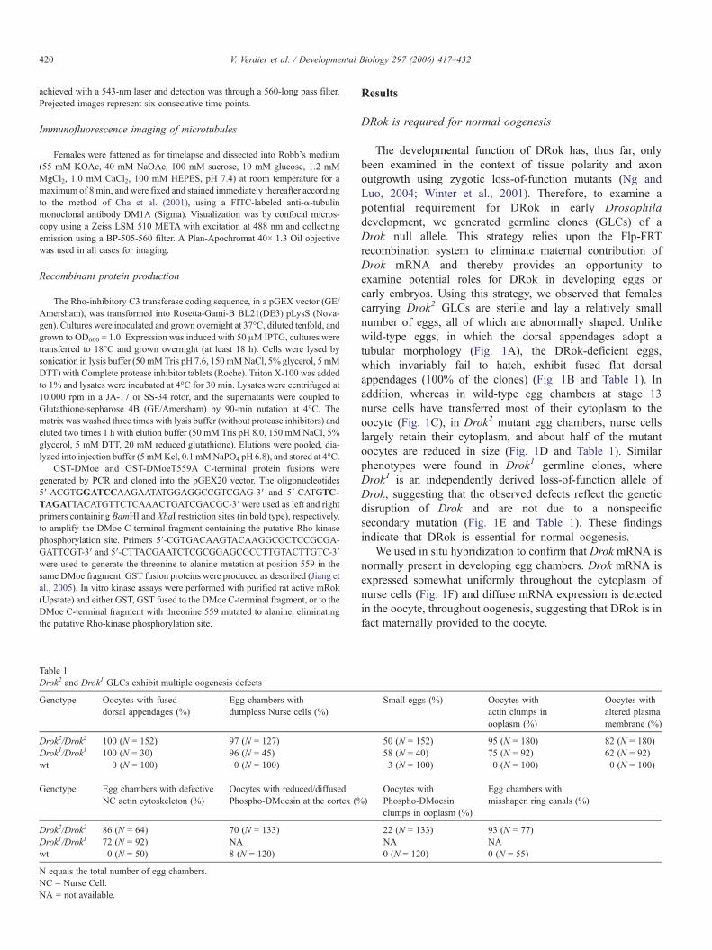
Fig. 1. Drok2 and Drok1 germline clones produce abnormal eggs with dorsal appendaegg chamber (A) and a Drok2 germline mutant stage 14 egg chamber (B). Drok2 germdorsal appendages. (C-E) Phase contrast images of a wild type (C), a Drok2 mutant (Dchambers exhibit dumpless-like nurse cells, resulting in eggs smaller than their wildwhich marks the developmental stage of these eggs. (F) In situ hybridization to Drochambers. Drok is mainly expressed in nurse cells, along with a diffuse expression inAnterior is to the left and dorsal is to the top.
glycerol phosphate) were incubated for 20 min at 30°C and the reaction productswere subjected to SDS-PAGE electrophoresis. Gels were stained withCoomassie solution to reveal protein content, dried, and subjected toautoradiography.
Timelapse confocal imaging
Female flies were fattened for 2 days on yeast–cornmeal–molasses withsupplemental dried active baker's yeast at 25°C. Females were injected in theabdomen with 0.4% trypan blue in normal saline. Two hours after injection,ovaries were dissected into ovarioles in halocarbon 700 oil on glass-bottomculture dishes (Bioptechs). For injection of C3 transferase, wild-type eggchambers were prepared as above and were injected with C3 transferase(approximately 100 nM) following dissection into halocarbon oil using amicroinjection apparatus. As controls, egg chambers were injected with GSTprotein alone (in the same buffer) or buffer alone. In both cases, there was noeffect on ovaries (no premature swirling)— even with higher concentrations ofGST (up to 500 nM) injected compared to GST-C3. Timelapse movies wererecorded by taking images of a single 1- to 2-μm central section of the oocyteevery 10 s on a Zeiss LSM 510 META confocal microscope. Excitation was
ge defects. (A, B) Phase contrast images of dorsal views of a wild-type stage 14line clones produce eggs with chorion patterning defects such as fused and flat), and a Drok1 mutant (E) stage 13 egg chambers. Drok2 and Drok1 mutant egg-type counterparts. The arrows point to the newly forming dorsal appendages,k mRNA in wild-type stage 6 (st 6), stage 10a (st 10a) and stage 13 (st 13) eggthe oocyte throughout oogenesis. Sense mRNAwas used as a negative control.
420 V. Verdier et al. / Developmental Biology 297 (2006) 417–432
achieved with a 543-nm laser and detection was through a 560-long pass filter.Projected images represent six consecutive time points.
Immunofluorescence imaging of microtubules
Females were fattened as for timelapse and dissected into Robb's medium(55 mM KOAc, 40 mM NaOAc, 100 mM sucrose, 10 mM glucose, 1.2 mMMgCl2, 1.0 mM CaCl2, 100 mM HEPES, pH 7.4) at room temperature for amaximum of 8 min, and were fixed and stained immediately thereafter accordingto the method of Cha et al. (2001), using a FITC-labeled anti-α-tubulinmonoclonal antibody DM1A (Sigma). Visualization was by confocal micros-copy using a Zeiss LSM 510 META with excitation at 488 nm and collectingemission using a BP-505-560 filter. A Plan-Apochromat 40× 1.3 Oil objectivewas used in all cases for imaging.
Recombinant protein production
The Rho-inhibitory C3 transferase coding sequence, in a pGEX vector (GE/Amersham), was transformed into Rosetta-Gami-B BL21(DE3) pLysS (Nova-gen). Cultures were inoculated and grown overnight at 37°C, diluted tenfold, andgrown to OD600 = 1.0. Expression was induced with 50 μM IPTG, cultures weretransferred to 18°C and grown overnight (at least 18 h). Cells were lysed bysonication in lysis buffer (50 mMTris pH 7.6, 150mMNaCl, 5% glycerol, 5 mMDTT) with Complete protease inhibitor tablets (Roche). Triton X-100 was addedto 1% and lysates were incubated at 4°C for 30 min. Lysates were centrifuged at10,000 rpm in a JA-17 or SS-34 rotor, and the supernatants were coupled toGlutathione-sepharose 4B (GE/Amersham) by 90-min nutation at 4°C. Thematrix was washed three times with lysis buffer (without protease inhibitors) andeluted two times 1 h with elution buffer (50 mM Tris pH 8.0, 150 mMNaCl, 5%glycerol, 5 mM DTT, 20 mM reduced glutathione). Elutions were pooled, dia-lyzed into injection buffer (5 mMKcl, 0.1 mMNaPO4 pH 6.8), and stored at 4°C.
GST-DMoe and GST-DMoeT559A C-terminal protein fusions weregenerated by PCR and cloned into the pGEX20 vector. The oligonucleotides5′-ACGTGGATCCAAGAATATGGAGGCCGTCGAG-3′ and 5′-CATGTC-TAGATTACATGTTCTCAAACTGATCGACGC-3′ were used as left and rightprimers containing BamHI and XbaI restriction sites (in bold type), respectively,to amplify the DMoe C-terminal fragment containing the putative Rho-kinasephosphorylation site. Primers 5′-CGTGACAAGTACAAGGCGCTCCGCGA-GATTCGT-3′ and 5′-CTTACGAATCTCGCGGAGCGCCTTGTACTTGTC-3′were used to generate the threonine to alanine mutation at position 559 in thesame DMoe fragment. GST fusion proteins were produced as described (Jiang etal., 2005). In vitro kinase assays were performed with purified rat active mRok(Upstate) and either GST, GST fused to the DMoe C-terminal fragment, or to theDMoe C-terminal fragment with threonine 559 mutated to alanine, eliminatingthe putative Rho-kinase phosphorylation site.
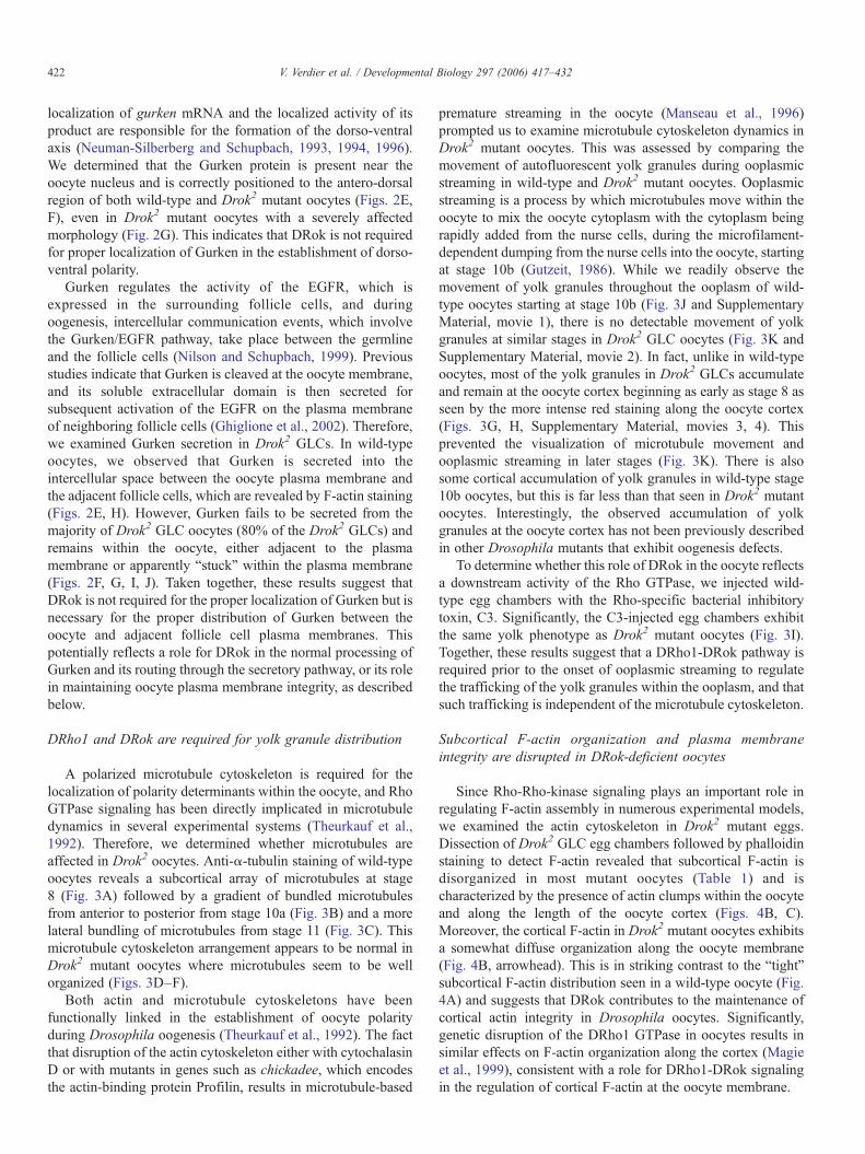
Table 1Drok2 and Drok1 GLCs exhibit multiple oogenesis defects
Genotype Oocytes with fuseddorsal appendages (%)
Egg chambers withdumpless Nurse cells (%)
Drok2/Drok2 100 (N = 152) 97 (N = 127)Drok1/Drok1 100 (N = 30) 96 (N = 45)wt 0 (N = 100) 0 (N = 100)
Genotype Egg chambers with defectiveNC actin cytoskeleton (%)
Oocytes with reduced/diffusedPhospho-DMoesin at the cortex (%
Drok2/Drok2 86 (N = 64) 70 (N = 133)Drok1/Drok1 72 (N = 92) NAwt 0 (N = 50) 8 (N = 120)
N equals the total number of egg chambers.NC = Nurse Cell.NA = not available.
Results
DRok is required for normal oogenesis
The developmental function of DRok has, thus far, onlybeen examined in the context of tissue polarity and axonoutgrowth using zygotic loss-of-function mutants (Ng andLuo, 2004; Winter et al., 2001). Therefore, to examine apotential requirement for DRok in early Drosophiladevelopment, we generated germline clones (GLCs) of aDrok null allele. This strategy relies upon the Flp-FRTrecombination system to eliminate maternal contribution ofDrok mRNA and thereby provides an opportunity toexamine potential roles for DRok in developing eggs orearly embryos. Using this strategy, we observed that femalescarrying Drok2 GLCs are sterile and lay a relatively smallnumber of eggs, all of which are abnormally shaped. Unlikewild-type eggs, in which the dorsal appendages adopt atubular morphology (Fig. 1A), the DRok-deficient eggs,which invariably fail to hatch, exhibit fused flat dorsalappendages (100% of the clones) (Fig. 1B and Table 1). Inaddition, whereas in wild-type egg chambers at stage 13nurse cells have transferred most of their cytoplasm to theoocyte (Fig. 1C), in Drok2 mutant egg chambers, nurse cellslargely retain their cytoplasm, and about half of the mutantoocytes are reduced in size (Fig. 1D and Table 1). Similarphenotypes were found in Drok1 germline clones, whereDrok1 is an independently derived loss-of-function allele ofDrok, suggesting that the observed defects reflect the geneticdisruption of Drok and are not due to a nonspecificsecondary mutation (Fig. 1E and Table 1). These findingsindicate that DRok is essential for normal oogenesis.
We used in situ hybridization to confirm that Drok mRNA isnormally present in developing egg chambers. Drok mRNA isexpressed somewhat uniformly throughout the cytoplasm ofnurse cells (Fig. 1F) and diffuse mRNA expression is detectedin the oocyte, throughout oogenesis, suggesting that DRok is infact maternally provided to the oocyte.
Small eggs (%) Oocytes withactin clumps inooplasm (%)
Oocytes withaltered plasmamembrane (%)
50 (N = 152) 95 (N = 180) 82 (N = 180)58 (N = 40) 75 (N = 92) 62 (N = 92)3 (N = 100) 0 (N = 100) 0 (N = 100)
)Oocytes withPhospho-DMoesinclumps in ooplasm (%)
Egg chambers withmisshapen ring canals (%)
22 (N = 133) 93 (N = 77)NA NA0 (N = 120) 0 (N = 55)

421V. Verdier et al. / Developmental Biology 297 (2006) 417–432
DRok is required for establishing normal oocyte polarity
Our observation that stages 13–14 Drok2 mutant oocytesharbor incompletely formed or fused dorsal appendages, adefect that is often associated with abnormal egg polarity(Neuman-Silberberg and Schupbach, 1994, 1996), prompted usto investigate the polarity of Drok2 mutant oocytes. Byexamining oskar mRNA localization as a marker of antero-posterior polarity (Kim-Ha et al., 1991) and the localization ofbicoidmRNA as a marker of the anterior pole (van Eeden and StJohnston, 1999), we observed that, whereas oskar mRNA isrestricted to the posterior tip of wild-type oocytes (Fig. 2A), itslocalization is altered in Drok2 mutant oocytes, where oskarmRNA is detected throughout the ooplasm (Fig. 2B). The lossof posterior localization of oskar mRNA in Drok2 GLCs is also
Fig. 2. Drok2 GLCs exhibit polarity defects. (A–D) In situ hybridizations to oskarmRD) egg chambers. oskar mRNA is normally restricted to the posterior side of wild-typarrow denotes some oskarmRNA still anchored at the posterior tip of mutant oocytes(C, D). (E–G) Triple anti-Gurken, TRITC-phalloidin and nuclear staining of a wildoocytes. In both the wild-type and mutant oocytes, Gurken accumulates in close proxithe oocyte. However, unlike in wild-type oocytes, Gurken is not being secreted in thcell apical membranes, in Drok2 mutant oocytes (arrows in H, I and J).
observed in early oocytes (stage 8/9) (data not shown). We notethat, as has been reported in GLCs of Dmoesin (Dmoe GLCs), aputative effector target of DRok (Polesello et al., 2002), a smallfraction of correctly positioned oskar mRNA can be detected inDrok2 GLCs (Fig. 2B), indicating that the absence of eitherDRok or DMoesin does not entirely abolish the localization ofoskarmRNA. On the other hand, similar to Dmoe GLCs, bicoidmRNA localizes properly at the anterior margin of wild-typeand Drok2 mutant oocytes (Figs. 2C, D). Together, these resultsindicate that DRok, like DMoesin, is not required for theformation of the anterior pole, but is required for formation ofthe posterior pole.
We also investigated dorso-ventral polarity of oocytes byexamining the subcellular localization of the Gurken product, aTGFα-like ligand for the Drosophila EGFR. The antero-dorsal
NA (A, B) or bicoidmRNA (C, D) in wild-type (A,C) or Drok2 GLC mutant (B,e oocytes (A) but this localization is disrupted in Drok2 mutant oocytes (B). The. bicoidmRNA anterior margin distribution is unaltered in Drok2 mutant oocytes-type (E and close-up H) or Drok2 mutant (F and close-up I, G and close-up J)mity to the oocyte nucleus and distributes normally to the antero-dorsal region ofe extracellular space between the oocyte membrane and the neighboring follicle
422 V. Verdier et al. / Developmental Biology 297 (2006) 417–432
localization of gurken mRNA and the localized activity of itsproduct are responsible for the formation of the dorso-ventralaxis (Neuman-Silberberg and Schupbach, 1993, 1994, 1996).We determined that the Gurken protein is present near theoocyte nucleus and is correctly positioned to the antero-dorsalregion of both wild-type and Drok2 mutant oocytes (Figs. 2E,F), even in Drok2 mutant oocytes with a severely affectedmorphology (Fig. 2G). This indicates that DRok is not requiredfor proper localization of Gurken in the establishment of dorso-ventral polarity.
Gurken regulates the activity of the EGFR, which isexpressed in the surrounding follicle cells, and duringoogenesis, intercellular communication events, which involvethe Gurken/EGFR pathway, take place between the germlineand the follicle cells (Nilson and Schupbach, 1999). Previousstudies indicate that Gurken is cleaved at the oocyte membrane,and its soluble extracellular domain is then secreted forsubsequent activation of the EGFR on the plasma membraneof neighboring follicle cells (Ghiglione et al., 2002). Therefore,we examined Gurken secretion in Drok2 GLCs. In wild-typeoocytes, we observed that Gurken is secreted into theintercellular space between the oocyte plasma membrane andthe adjacent follicle cells, which are revealed by F-actin staining(Figs. 2E, H). However, Gurken fails to be secreted from themajority of Drok2 GLC oocytes (80% of the Drok2 GLCs) andremains within the oocyte, either adjacent to the plasmamembrane or apparently “stuck” within the plasma membrane(Figs. 2F, G, I, J). Taken together, these results suggest thatDRok is not required for the proper localization of Gurken but isnecessary for the proper distribution of Gurken between theoocyte and adjacent follicle cell plasma membranes. Thispotentially reflects a role for DRok in the normal processing ofGurken and its routing through the secretory pathway, or its rolein maintaining oocyte plasma membrane integrity, as describedbelow.
DRho1 and DRok are required for yolk granule distribution
A polarized microtubule cytoskeleton is required for thelocalization of polarity determinants within the oocyte, and RhoGTPase signaling has been directly implicated in microtubuledynamics in several experimental systems (Theurkauf et al.,1992). Therefore, we determined whether microtubules areaffected in Drok2 oocytes. Anti-α-tubulin staining of wild-typeoocytes reveals a subcortical array of microtubules at stage8 (Fig. 3A) followed by a gradient of bundled microtubulesfrom anterior to posterior from stage 10a (Fig. 3B) and a morelateral bundling of microtubules from stage 11 (Fig. 3C). Thismicrotubule cytoskeleton arrangement appears to be normal inDrok2 mutant oocytes where microtubules seem to be wellorganized (Figs. 3D–F).
Both actin and microtubule cytoskeletons have beenfunctionally linked in the establishment of oocyte polarityduring Drosophila oogenesis (Theurkauf et al., 1992). The factthat disruption of the actin cytoskeleton either with cytochalasinD or with mutants in genes such as chickadee, which encodesthe actin-binding protein Profilin, results in microtubule-based
premature streaming in the oocyte (Manseau et al., 1996)prompted us to examine microtubule cytoskeleton dynamics inDrok2 mutant oocytes. This was assessed by comparing themovement of autofluorescent yolk granules during ooplasmicstreaming in wild-type and Drok2 mutant oocytes. Ooplasmicstreaming is a process by which microtubules move within theoocyte to mix the oocyte cytoplasm with the cytoplasm beingrapidly added from the nurse cells, during the microfilament-dependent dumping from the nurse cells into the oocyte, startingat stage 10b (Gutzeit, 1986). While we readily observe themovement of yolk granules throughout the ooplasm of wild-type oocytes starting at stage 10b (Fig. 3J and SupplementaryMaterial, movie 1), there is no detectable movement of yolkgranules at similar stages in Drok2 GLC oocytes (Fig. 3K andSupplementary Material, movie 2). In fact, unlike in wild-typeoocytes, most of the yolk granules in Drok2 GLCs accumulateand remain at the oocyte cortex beginning as early as stage 8 asseen by the more intense red staining along the oocyte cortex(Figs. 3G, H, Supplementary Material, movies 3, 4). Thisprevented the visualization of microtubule movement andooplasmic streaming in later stages (Fig. 3K). There is alsosome cortical accumulation of yolk granules in wild-type stage10b oocytes, but this is far less than that seen in Drok2 mutantoocytes. Interestingly, the observed accumulation of yolkgranules at the oocyte cortex has not been previously describedin other Drosophila mutants that exhibit oogenesis defects.
To determine whether this role of DRok in the oocyte reflectsa downstream activity of the Rho GTPase, we injected wild-type egg chambers with the Rho-specific bacterial inhibitorytoxin, C3. Significantly, the C3-injected egg chambers exhibitthe same yolk phenotype as Drok2 mutant oocytes (Fig. 3I).Together, these results suggest that a DRho1-DRok pathway isrequired prior to the onset of ooplasmic streaming to regulatethe trafficking of the yolk granules within the ooplasm, and thatsuch trafficking is independent of the microtubule cytoskeleton.
Subcortical F-actin organization and plasma membraneintegrity are disrupted in DRok-deficient oocytes
Since Rho-Rho-kinase signaling plays an important role inregulating F-actin assembly in numerous experimental models,we examined the actin cytoskeleton in Drok2 mutant eggs.Dissection of Drok2 GLC egg chambers followed by phalloidinstaining to detect F-actin revealed that subcortical F-actin isdisorganized in most mutant oocytes (Table 1) and ischaracterized by the presence of actin clumps within the oocyteand along the length of the oocyte cortex (Figs. 4B, C).Moreover, the cortical F-actin in Drok2 mutant oocytes exhibitsa somewhat diffuse organization along the oocyte membrane(Fig. 4B, arrowhead). This is in striking contrast to the “tight”subcortical F-actin distribution seen in a wild-type oocyte (Fig.4A) and suggests that DRok contributes to the maintenance ofcortical actin integrity in Drosophila oocytes. Significantly,genetic disruption of the DRho1 GTPase in oocytes results insimilar effects on F-actin organization along the cortex (Magieet al., 1999), consistent with a role for DRho1-DRok signalingin the regulation of cortical F-actin at the oocyte membrane.

Fig. 3. DRok does not perturb the bundling of microtubules but is involved in yolk granule trafficking. (A–F) Visualization of wild-type (A–C) and Drok2 mutantoocytes (D–F), all expressing the Tubulin-GFP fusion protein to reveal microtubules. As expected in wild-type oocytes, early stages (stage 8/9) are marked by an arrayof subcortical microtubules around the oocyte (A). Starting from stage 10a, microtubules are bundling from the anterior margin of the oocyte, and this creates ananterior to posterior gradient of microtubules in the oocyte (B). Later stages are characterized by anterior and more lateral bundling of microtubules that extend more inthe ooplasm, as they are ready for cytoplasmic streaming (C). This microtubule distribution is not perturbed in Drok2 mutant oocytes and microtubules bundle quitenormally from early to late stages (D–F). (G–I) Visualization of auto-fluorescent yolk granules by time-lapse microscopy in wild-type (G, J), Drok2 GLCs (H, K) andC3-treated wild-type egg chambers (I). While yolk granules are uniformly present throughout stage 8 wild-type oocytes (G), they aggregate and accumulate at thecortex of same stageDrok2mutant oocytes (H). This phenotype has also been detected in C3-treated oocytes (I). While cytoplasmic streaming can be observed in stage10b wild-type oocytes as swirling arrays corresponding to temporal projections of yolk granules movements (J), visualization of streaming is not possible in stage 10bDrok2 mutant oocytes as most yolk granules have accumulated at the cortex (K).
423V. Verdier et al. / Developmental Biology 297 (2006) 417–432

Fig. 4. Subcortical F-actin organization and plasma membrane integrity are disrupted in DRok-deficient oocytes. (A–C) Phalloidin staining of a wild type (A), a Drok2
mutant (B) and a Drok1 mutant (C) stage 10a oocytes. Drok2 and Drok1 mutants display disorganized F-actin at the cortex of the oocyte (B and C, arrowheads pointingto discontinuous F-actin staining), along with the presence of actin clumps near the cortex or within the ooplasm (B, C, arrows). (D–L) Visualization of the F-actinnetwork (D, G and J), using phalloidin, and cell membranes (E, H and K), using a fluorescein-conjugated lectin. (F, I and L) are the merged images of actin andmembrane stainings. In the wild-type stage 9 oocyte, the lectin co-localizes with F-actin and outlines juxtaposed membranes between the oocyte and the surroundingfollicle cells (F). Mutant stage 9 or 10 oocytes display co-localization of F-actin and lectin (I) but exhibit an abnormal lectin distribution, associated with an apparentdetachment of the oocyte membrane from the follicle cell layer (H, arrows), as well as a defect in the oocyte shape itself. This lectin defect was also observed in Drok1
mutant oocytes (K, arrow). Anterior is to the left and dorsal is to the top.
424 V. Verdier et al. / Developmental Biology 297 (2006) 417–432
In addition to the actin defects described above, wefrequently observe a significant deformation along localizedregions of the cortical membrane in Drok2 GLC oocytes,resulting in protrusions into the oocyte cytoplasm in a majorityof stage 9 and later stage oocytes (Table 1). To determinewhether these deformations result from an abnormally shaped
oocyte or reflect a detachment of subcortical actin from anotherwise normal cell membrane, we used a cell membrane-specific marker (fluorescently labeled lectin) that co-localizeswith F-actin and outlines juxtaposed membranes between theoocyte and the surrounding follicle cell layer in a wild-type eggchamber (Figs. 4D–F). In Drok2 GLCs, the lectin marker co-

425V. Verdier et al. / Developmental Biology 297 (2006) 417–432
localizes normally with F-actin, revealing an abnormal oocytecell shape and a significant detachment of the oocyte membranefrom the apical membrane of follicle cells (Figs. 4G–I). Theexpressivity of that phenotype ranged from mild (stage 9 eggchambers) to very severe (stage 10 and 11 egg chambers) (datanot shown), suggesting that the defect progresses during oocytedevelopment. In severe cases, the oocyte membrane seems tolose its integrity, as indicated by the appearance of diffuse lectinstaining between the oocyte and the follicle cell layer (data notshown). Moreover, strong phenotypes are associated withdiscontinuous lectin staining of the oocyte membrane (Fig.4K, arrowheads inDrok1 mutant oocytes). These results suggestthat DRok is required for the integrity of the oocyte plasmamembrane and is consequently required to maintain the propershape of the oocyte. Similarly, Drok1 germline clones arecharacterized by a disorganized subcortical F-actin networkassociated with the presence of actin clumps and a reduction inplasma membrane integrity (Figs. 4C, J–L and Table 1),confirming that Drok2 germline clone-generated defects are duespecifically to the genetic disruption of the Drok gene. Theseresults suggest that DRok is required for the integrity of theoocyte plasma membrane and is consequently required tomaintain the proper shape of the oocyte.
Drok2 mutant nurse cells exhibit a “dumpless-like” phenotype
During oogenesis, the 15 polyploid nurse cells of the eggchamber continually provide the oocyte with nutrients,proteins, and maternal RNAs to support its growth throughactin-rich structures called rings canals. At stage 11 ofoogenesis, the entire cytoplasmic content of the nurse cellsis rapidly transferred to the oocyte within 30 min, in aprocess known as “dumping” (see Spradling, 1993 for areview of oogenesis). Nurse cell dumping was previouslyshown to depend upon an intact actin cytoskeleton (Cant etal., 1994; Verheyen and Cooley, 1994; Xue and Cooley,1993). In wild-type stage 13 egg chambers, the nurse cellsretain a cluster of nuclei after they have dumped all of theircytoplasm into the oocyte. In contrast, in similarly stagedDrok2 (or Drok1) GLC egg chambers, the nurse cells haveretained most of their cytoplasm (Figs. 5A–C). Thisdumpless-like nurse cell phenotype was observed in mostlate stage Drok2 (or Drok1) mutant egg chambers and resultsin the generation of either small eggs (50%) (Fig. 5B, Table1) or normal size eggs (50%) that appear “deflated” andpresumably lack much of their normal cytoplasmic content(Fig. 5C).
As has been previously observed in other dumplessmutants, including chickadee, quail or singed, failure ofcytoplasmic transport is often due to the obstruction of thering canals by nurse cell nuclei that fail to be properlytethered by actin filaments (Cant et al., 1994; Mahajan-Miklos and Cooley, 1994; Verheyen and Cooley, 1994).Therefore, we determined whether Drok2 egg chambersexhibit an abnormal actin cytoskeleton and whether ringcanal obstruction potentially accounts for the dumpless-likephenotype. The Anti-Hts-RC (Hu li tai shao-Ring Canals)
antibody was used to detect the adducin cytoskeletal protein,a specific actin-binding component of the ring canals (Yueand Spradling, 1992). In contrast to anti-Hts-RC staining inwild-type, Drok2 GLCs at stage 10b or 11 revealed asomewhat irregular and square shape of the ring canals inmost GLCs (93%) with between one and four abnormallyshaped canals per egg chamber, but no major difference inthe overall adducin distribution and number of ring canals(Figs. 5J, K, Table 1). Phalloidin staining to reveal F-actinin ring canals yielded similar findings (Figs. 5D, E). Thesefindings indicate that DRok is not required for the formationof ring canals but appears to play some role in eitherestablishing or maintaining their normal morphology.
Additional analysis of F-actin organization within the nursecells revealed that while wild-type egg chambers typicallyexhibit thick F-actin filaments extending from the plasmamembranes towards the nurse cell nuclei in order to anchorthem, the F-actin network, although present in some clones, isperturbed in nurse cells within the vast majority of Drok2 orDrok1 GLCs (86% and 72%, respectively) (Table 1). Specif-ically F-actin filaments are generally much thinner and shorterin those rare cells where they do form (Figs. 5D, E) and do notspan the nurse cell cytoplasm from the plasma membrane to thenucleus (Figs. 5F, G). This suggests two potential defects. Onthe one hand, the acto-myosin contractile apparatus may beaffected, which would prevent nurse cells from contractingproperly, thereby altering cytoplasmic transport. On the otherhand, actin-mediated anchoring of the nuclei may be defective,which could obstruct the ring canals, consequently preventingcytoplasmic transport. To address the latter possibility, we useddouble staining of F-actin and nuclear membranes to reveal theF-actin network which normally tethers nuclei to the center ofeach nurse cell (Fig. 5H). In Drok2 GLCs, staining clearlyreveals the lack of functional F-actin filaments within nursecells, as well as the presence of nuclei of increased size (about30% increased volume compared to wild-type nuclei) that arenot properly localized near the center of the nurse cells.Significantly, while these enlarged nuclei are in close proximityto the ring canals, they do not appear to obstruct them (Fig. 5I).DRok-deficient nurse cells (in Drok2 or Drok1 GLCs) alsoexhibit a less structured and “looser” subcortical F-actinorganization (Fig. 5I) when compared to wild-type nurse cells(Fig. 5H). Taken together, these results indicate that while DRokis not required for the formation of ring canals, it is essential forcytoplasmic transport. Moreover, lack of cytoplasmic transportin Drok2 GLCs does not result from obstruction of the ringcanals by untethered nurse cell nuclei, which clearly distin-guishes Drok from the “chickadee, singed or quail” class ofdumpless mutants. In addition, DRok is required for maintain-ing the plasma membrane integrity of nurse cells.
DMoesin membrane localization is disrupted in DRok-deficientoocytes
In mammalian cells, Rho-kinase has been shown todirectly phosphorylate the ERM (Ezrin–Radixin–Moesin)family protein Moesin in vitro (Matsui et al., 1998; Oshiro

Fig. 5. DRok is required for cytoplasmic transport during oogenesis. (A–C) Phase contrast images of wild-type (A) andDrok2 GLC (B, C) stage 13 egg chambers. Thearrows denote the newly forming dorsal appendages, a marker of stage 13 egg chambers. In panel (A), the nurse cells have dumped their entire cytoplasmic contentsinto the oocyte and are no longer visible at the anterior region of the egg chamber, whereas DRok-deficient nurse cells retain most of their cytoplasm, resulting in amuch smaller egg (B). (C) An example of a less severe dumpless-like phenotype where the mutant oocyte is about the size of a wild-type oocyte but appears somewhat“deflated”. (D–G) Phalloidin staining of wild-type stage 10b (D) and stage 11 (F), and Drok2 mutant stage 10b (E) and stage 11 (G) nurse cells. The rapid phase ofcytoplasmic transport takes place from stage 10b until the end of stage 11. In wild-type nurse cells, thick transverse F-actin filaments initially form around the nurse cellcortex at stage 10b (D, arrow) and assemble in bundles all around the nucleus, extending into the nurse cell cytoplasm between the nucleus and the plasma membrane atstage 11 (F, arrow). By contrast, in DRok-deficient nurse cells, F-actin filaments are much thinner and shorter when they do form, and they fail to extend from theplasma membrane to the center of the cells (E, G, arrows). (H, I) Double F-actin and nuclear staining of wild-type and Drok2 mutant stage11 nurse cells. In wild-typenurse cells, the nuclei remain anchored at the center of each cell by the surrounding F-actin filament bundles. In mutant nurse cells, the nuclei, which appear larger, areclearly displaced from their normal centered localization within the nurse cells (I). The arrowheads point to the subcortical F-actin network which appears more diffuseand disorganized in the mutant nurse cells. (J, K) High magnification images of a ring canal from a wild-type (J) or a Drok2 mutant (K) nurse cell stained with an anti-adducin antibody (anti-Hts-RC). These doughnut-shaped actin and actin-binding protein-rich structures of wild-type nurse cells are somewhat abnormally shaped andare more “squared” in Drok2 mutant nurse cells, but their size and number do not vary between the two genotypes. Anterior is to the left and dorsal to the top.
426 V. Verdier et al. / Developmental Biology 297 (2006) 417–432
et al., 1998). In vivo, Rho-kinase-dependent phosphorylationof ERM proteins has been reported to vary according to celltype, and it is presently unclear as to whether Rho-kinaseacts directly or indirectly to modify ERM phosphorylation(Ivetic and Ridley, 2004; Jeon et al., 2002; Matsui et al.,1999; Oshiro et al., 1998). In Drosophila, genetic disruptionof the closely related ortholog, Dmoesin, results in aphenotype in developing oocytes that resembles the F-actin
defects seen in Drok2 GLCs, including the accumulation ofF-actin clumps within the oocyte cytoplasm (Polesello et al.,2002). ERM proteins have been implicated in severalbiological processes, including cell–cell adhesion, mainte-nance of cell shape, and cell motility. Upon carboxy-terminal threonine phosphorylation, they are recruited to theplasma membrane, where they function as cross-linkersbetween the cell membrane and the actin cytoskeleton

427V. Verdier et al. / Developmental Biology 297 (2006) 417–432
(Bretscher, 1999; Mangeat et al., 1999). DMoesin isrequired for anchoring F-actin filaments to the plasmamembrane in the oocyte, and this function requiresphosphorylation of T559 in DMoesin, which is analogousto a Thr site in mammalian Moesin (Fig. 6D) that can bedirectly phosphorylated by Rho-kinases in vitro (Jankovicset al., 2002; Matsui et al., 1998; Oshiro et al., 1998;Polesello et al., 2002). Therefore, we tested the possibilitythat membrane localization of DMoesin is disrupted in theoocytes of Drok2 GLCs.
Using an antibody directed against the conserved T558phospho-peptide site in Moesin, we observe that phospho-DMoesin localizes to the oocyte plasma membrane in a wild-type egg chamber (Fig. 6A). However, in Drok2 GLCs, there isa significant reduction in phospho-DMoesin at the oocyte cortex(Fig. 6B, Table 1). In most clones, the cortex still retains somephospho-DMoesin staining which appears more diffuse relativeto the staining seen in wild-type oocytes (Fig. 6C). In addition,in ∼20% of the GLCs, phospho-DMoesin is also foundmislocalized in patches within the ooplasm (Fig. 6C). FiguresB and C represent relatively strong and weak phenotypes,respectively, with regard to decreased phospho-DMoesin at theoocyte plasma membrane. However, the overall distribution andexpression level of total DMoesin is unchanged inDrok2 mutantoocytes, exhibiting a diffuse staining pattern throughout theooplasm that is indistinguishable from the staining seen in wild-type oocytes (Figs. 6D, E). Taken together, these observationssuggest that proper membrane localization of DMoesin requiresDRok activity.
We then determined whether Rho-kinase can directlyphosphorylate DMoesin on the T559 residue using an in vitrokinase assay with purified recombinant proteins. In thisexperiment, we used the commercially available purified kinasedomain corresponding to mammalian Rho-kinase (mRok),which is highly conserved with the kinase domain of DRok(Fig. 6E). In the in vitro assay, mRok undergoes autopho-sphorylation, as expected, and additionally, is able to directlyphosphorylate DMoesin (Fig. 6F). The observed phosphoryla-tion of DMoesin is virtually abolished when using a GST-DMoesin-TA mutant in which the putative phosphorylation sitewas substituted with an alanine residue (Fig. 6F). Takentogether, these findings suggest that direct phosphorylation ofDMoesin by DRok on a single site facilitates the association ofDMoesin with the cortical oocyte membrane during oogenesisto maintain its proper integrity.
Overall, the similarity of antero-posterior polarity, microtu-bule cytoskeleton integrity and F-actin distribution phenotypesbetween Drok2 and Dmoe GLCs, along with the cell biologyand biochemical phosphorylation data suggests that DRok andits putative effector protein DMoesin interact in the developingoocyte and that DRok mediates some, but not all, of itsbiological effects through DMoesin. However,Drok2 andDmoeGLCs are not identical. For example, Drok2 GLCs exhibit ayolk granule transport phenotype, whereas Dmoe GLCs do not.Moreover, the oocyte plasma membrane integrity is moreseverely affected in Drok2 GLCs than in Dmoe GLCs. Thesedifferences imply that DRok must signal through effector
proteins other than DMoesin to exert additional distinct effectsduring oogenesis.
Discussion
In this study, we have determined that the single closelyrelated Drosophila ortholog of the mammalian Rho-kinases,DRok, is required for oogenesis and participates in severaldistinct aspects of this complex developmental processes,including organization of the oocyte cortex, cytoplasmictransport from nurse cells, and germline-soma cross-signalingnecessary for establishment of the antero-posterior and dorso-ventral axis of the resulting mature egg. In addition, we foundthat DRok is required for a developmental process aroundstages 7–8 of oogenesis, which enables the yolk granules tomove freely in the ooplasm after they have been internalized ortransferred to the oocyte and before cytoplasmic streaming cantake place (stage 10b and higher). These findings, whenconsidered in the context of a variety of other previouslyreported mutants that exhibit oogenesis phenotypes, suggestthat DRok represents a novel class of oogenesis regulators.
DRok has been previously implicated as an effector of theDRho1 GTPase in the regulation of planar cell polarity in theeye and in the wing, downstream of Frizzled-Dishevelledsignals (Winter et al., 2001). In germline cells, DRok andDRho1 mutants exhibit some overlapping actin defects; e.g., theoocyte cortex exhibits a more diffuse F-actin distribution in bothDrok2 GLCs and Rho1 loss-of-function egg chambers in whichRho1 levels have been reduced in a heterozygous mutant Rho1and wimp background (Rho1GLCs are not viable) (Magie et al.,1999). In addition, wild-type oocytes injected with the Rho-inhibitory C3 toxin exhibit the same ooplasmic streamingdefects as Drok2 mutant oocytes, as discussed below, stronglysuggesting that the germline clone phenotypes reflect thedisruption of DRho1-DRok signaling in germ cells.
DRok regulates oocyte polarity
Previous analysis of the polarity of Dmoe GLC oocytesindicated that DMoesin is specifically required for thelocalization of posterior determinants such as oskar mRNAand Oskar protein but not the formation of the dorso-ventralaxis nor the anterior pole. DMoesin appears to function inmaintaining posterior polarity by anchoring actin to themembrane cortex which in turn anchors microtubule-delivered oskar mRNA and its protein product Oskar tothe posterior pole (Polesello et al., 2002). Similarly, inDrok2 GLCs, the localization of anterior (bicoid) or dorso-ventral determinants (gurken) is not altered although mostoskar mRNA is found mislocalized within the ooplasmstarting at stage 9. While the establishment of oocytepolarity generally depends upon microtubule cytoskeletonorganization (Theurkauf et al., 1992), it has been reportedthat Dmoe null mutations do not disrupt the microtubulecytoskeleton and do not perturb its polarity (Polesello et al.,2002). Similarly, we observed that microtubules in Drok2
mutant oocytes appear normal. Taken together with the fact

428 V. Verdier et al. / Developmental Biology 297 (2006) 417–432
that some oskar mRNA remains anchored at the posteriortip of the oocyte plasma membrane in Drok2 GLCs, as isseen in Dmoe GLCs, this indicates that oskar mislocaliza-tion, and consequently, the alteration of posterior polarity inDrok2 GLCs is not due to an abnormally organizedmicrotubule cytoskeleton. Moreover, unlike other germlineclone mutants with oocyte polarity defects, such as chic orcapu, the Drok2 and Dmoe polarity defects most likelyreflect the incapacity of the disorganized subcortical actincytoskeleton to properly anchor oskar at the posteriormembrane of the oocyte.
The similarity between the oskar polarity phenotype ofDrok2 and Dmoe GLCs is also consistent with a likely rolefor DMoesin as an essential DRok substrate that mediates itseffects on the formation of posterior polarity and furthersupports the functional significance of a signaling pathwayfrom DRok to DMoesin to the actin cytoskeleton in oocytedevelopment. Moreover, the proper localization of Gurken,as defined by the position of the oocyte nucleus, whichmigrates in a microtubule-dependent manner from theposterior to the anterior and then to the antero-dorsal sideof the oocyte starting at stage 8, is consistent with the

429V. Verdier et al. / Developmental Biology 297 (2006) 417–432
presence of a grossly normal appearing microtubulecytoskeleton in DRok-deficient oocytes.
DRok plays a role in germline-soma cross-signaling
A majority of mutations resulting in egg chambers withdorso-ventral axis patterning defects, such as dorsalappendages aberrations, have been associated with genesencoding components of the Gurken-EGFR signalingpathway (Neuman-Silberberg and Schupbach, 1994, 1996).Cross-signaling between the oocyte and the surroundingfollicle cells at the antero-dorsal side of the oocyte has beenextensively studied and involves the binding of the secretedGurken ligand to the EGFR present on the apical membraneof follicle cells and subsequent activation of downstreamsignaling to control the formation of follicle cell-deriveddorsal structures (Nilson and Schupbach, 1999). AlthoughGurken localization is correct in Drok2 mutant oocytes, lossof Gurken secretion (in 80% of the Drok2 mutant oocytes)in the intercellular space between the oocyte membrane andthe follicle apical membranes indicates the likelihood ofaltered communication between the oocyte and surroundingfollicle cells, possibly resulting in a disruption of the EGFRsignaling pathway leading to dorsal appendage defects. Theobserved requirement for DRok in Gurken secretion mayreflect a well established role of Rho signaling in thecontrol of vesicular trafficking and secretion (Symons andRusk, 2003). However, it remains possible that the apparentabsence of Gurken secretion into the intercellular spacereflects a consequence of the observed disruption of oocyteplasma membrane integrity.
DRok and the trafficking of yolk granules in the early oocyte
The unexpected observation in our time-lapse confocalmicroscopy studies that most autofluorescent yolk granulesin Drok2 mutant oocytes or C3-treated wild-type eggchambers accumulate at the oocyte membrane suggests arole for DRho1 and DRok in early vitellogenesis. Vitello-genesis is a process that begins around stage 8 and isdefined by the co-secretion of vitelline membrane and yolkmaterial by the surrounding follicle cells leading to the
Fig. 6. DMoesin membrane localization is disrupted in DRok-deficient oocytes. (A–(B, C), using an antibody specific for phospho-T558 mammalian Moesin and anmembrane in the wild-type oocyte (A). In Drok2 GLCs, phospho-DMoesin is signifiretains some phospho-DMoesin staining, but it appears more diffuse (C, arrowhead).in their ooplasm (C, arrow). (D, E) Double immunostaining of wild-type (D) and Dphalloidin. Both wild-type and Drok2 mutant oocytes exhibit a similar diffuse patteDMoesin expression levels between the two genotypes. Anterior is to the left and dspecific threonine residue (in bold type) defined at position 558 in human and rat Moeall three sequences (identical residues are shaded in red). (G) Schematic representatiomRok1 and mRok2 (mRok1-CAT, mRok2-CAT) with the numbered start and endingcorrespond to the percentage identity and similarity (in parentheses) between DRok-Ccommercially available purified kinase domain of mammalian Rho-kinase (mRokindicated bands correspond to radiolabeled proteins visualized by autoradiography fomRok is detected in all reactions. In the presence of GST-DMoesin, mRok directly phDMoesin-T559Amutant (threonine 559 is replaced by an alanine residue) (upper paneCoomassie Blue staining to confirm that equal amounts of protein were used in the
eventual formation of chorionic structures of the egg andnormal oocyte growth, respectively. After their secretion,yolk proteins are internalized into the oocyte throughendocytosis and are swirled around the ooplasm at laterstages, when microtubule-dependent streaming occurs(Spradling, 1993). The high concentration of yolk granulesat the oocyte membrane from early vitellogenesis underlies apossible defect in endocytosis of the yolk granules. Togetherwith the fact that C3-treated egg chambers and Drok2 GLCsexhibit an identical yolk granule phenotype, this suggeststhat DRok mediates Rho1's role in the trafficking of yolkgranules at the oocyte plasma membrane. In addition, nursecells also normally accumulate yolk material and transfer itto the oocyte. The detection of yolk granules moving to theplasma membrane of Drok2 mutant oocytes or oocytes inC3-treated egg chambers after they are deposited by thenurse cells is an intriguing phenotype that has not beenpreviously reported and may reflect a trafficking defect inthe ooplasm. Further studies to examine molecular compo-nents of the endocytic machinery will be required todevelop a better understanding of the roles of Rho1 andDRok in yolk granule trafficking within the ooplasm.Notably, it is also conceivable that alteration of oocyteplasma membrane integrity through disruption of actincytoskeleton organization in most Drok2 GLCs, as wehave observed, could exert a secondary effect on theendocytosis of yolk granules.
Because of the yolk granule phenotype in Drok2 GLCs inearly oogenesis, it is not possible to visualize microtubulecytoskeleton dynamics at later stages in time-lapse confocalmicroscopy. Thus, it is difficult to determine whether Drok2
mutant oocytes would undergo normal or premature ooplasmicstreaming at stages 10b–11. As a functional relationshipbetween actin and microtubule cytoskeletons has been sug-gested based on findings with several mutants with oogenesisdefects, it is quite conceivable that the abnormalities of the actincytoskeleton in Drok2 mutant oocytes could affect microtubulecytoskeleton dynamics. Indeed, it has been demonstrated thatsome aspect of the actin cytoskeleton normally repressesmicrotubule-based streaming within the oocyte (Manseau etal., 1996). Thus, it is possible that the accumulation of yolkgranules near the plasma membrane of Drok2 mutant oocytes
C) Double immunostaining of wild-type (A) and Drok2 mutant stage 10 oocytesantibody against β-tubulin. Phospho-DMoesin localizes to the oocyte plasmacantly decreased at the oocyte cortex (B, arrow). In most clones, the cortex stillAbout 20% of the Drok2 mutant oocytes also exhibit phospho-DMoesin patchesrok2 mutant stage 10 oocytes (E), using an antibody against total DMoesin andrn of total DMoesin throughout the ooplasm with no significant difference inorsal is to the top. (F) Alignment of the amino acid sequences surrounding thesin and position 559 inDrosophilaMoesin. Note the significant identity betweenn of the catalytic domains of DRok (DRok-CAT) and mammalian Rho-kinases,amino acids for each domain. The bold type numbers within the kinase domainsAT and either mRok1-CAT or mRok2-CAT. (H) In vitro kinase assay, using the
) and GST recombinant proteins. Kinase reactions included 32P-ATP, and thellowing protein resolution by SDS-PAGE. As expected, autophosphorylation ofosphorylates DMoesin, and the phosphorylation is abolished when using a GST-l). Recombinant proteins were separately assessed after separate SDS-PAGE andassays (lower panel).
430 V. Verdier et al. / Developmental Biology 297 (2006) 417–432
reflects a combination of trafficking/endocytosis defects andactin cytoskeleton perturbation-induced alteration of microtu-bule cytoskeleton dynamics in the ooplasm during earlyoogenesis.
DRok is required in the nurse cells to ensure their contractilityduring nurse cell dumping
The oocyte volume inDrok2 GLCs is frequently smaller thanthat seen in wild-type oocytes, before the rapid phase ofcytoplasmic transport takes place. This suggests a possibledefect in the slow phase of cytoplasmic transport. It has beenpreviously reported that transport of some particles towards theoocyte during stages 7–10A depends upon a proper acto-myosin network. In addition, sqhAX3 GLCs exhibit a similaroocyte size defect. sqhAX3 is a loss-of-function mutation in thesqh locus which codes for the Drosophila ortholog of myosinlight chain of myosin II (Jordan and Karess, 1997). Takentogether with the fact that DRok has been shown tophosphorylate Sqh in vivo (Amano et al., 1996; Winter et al.,2001), these data suggest that DRok mediates, via regulation ofSqh, some aspects of the acto-myosin contractility involved incytoplasmic transport from early stages of oogenesis.
The observation of dumpless-like oversized nurse cells inmost of Drok2 GLCs also supports a role for DRok in the rapidphase of cytoplasmic transport at stages 10B–11 of oogenesis.Unlike other classes of dumpless mutants including chickadee,singed or quail, failure of rapid cytoplasmic transport from theDrok2 mutant nurse cells to the oocyte does not result from theobstruction of the ring canals by unanchored nurse cell nuclei,suggesting that Drok constitutes a distinct class of dumpless-like mutants. In addition, in sqhAX3 GLCs, dumpless nurse cellsare associated with a lack of acto-myosin contractility by nursecells, as revealed by mislocalization of myosin II and byabsence of the perinuclear organization of actin filamentsbundles in the nurse cells. Therefore, sqhAX3 mutant nurse cellscannot contract properly to expulse their cytoplasm throughotherwise weakly damaged ring canals. Drok2 and sqhAX3
mutant nurse cells do not share the same actin filamentphenotype, as Drok2 mutant nurse cells exhibit a more dramaticphenotype associated with absence of radial filaments anddisorganization of cortical actin. It is, however, likely that DRokand Sqh are part of the same signaling pathway that regulatesacto-myosin contractility in nurse cells, as it has already beenshown that DRok phosphorylates Sqh in Drosophila develop-ment. Moreover, the severity of the Drok2 mutant F-actinphenotypes may reflect DRok's potential to engage multipledistinct downstream substrates, of which Sqh is only one.Significantly, the actin-binding protein, adducin, is alsoreportedly a direct substrate for mammalian Rho-kinases(Fukata et al., 1999b), and the Drosophila Adducin ortholog,Hts, is a major component of ring canals (Yue and Spradling,1992). Thus, it is possible that the observed defects in ring canalmorphology in Drok2 GLCs involve abnormal regulation ofadducin by DRok. However, it is difficult to determine whetherthis ring canal phenotype contributes to the dumpless-like nursecell phenotype observed in Drok2 GLCs.
The observation that nurse cell nuclei are substantiallyincreased in size in Drok2 GLCs suggests a possibleinvolvement of DRok in increased endoreplication of thenurse cells. The Rho-related Rac and Cdc42 GTPases havepreviously been associated with endoreplication in porcineaortic endothelial (PAE) cells, although Rho has not beenimplicated thus far (Muris et al., 2002). Interestingly, this nursecell nuclei phenotype has not been observed in other previouslydescribed GLC mutants of other actin cytoskeleton-regulatingsignaling components that exhibit oogenesis defects. Thus, chicas well as sqhAX3 GLCs reveal cytokinesis defects associatedwith the presence of multinucleated nurse cells. In addition, themajority of sqhAX3 mutant egg chambers harbor less than 15nurse cells (64% of sqhAX3 mutant egg chambers have less than7 nurse cells), a phenotype that is not shared by Drok2 mutantnurse cells (Jordan and Karess, 1997; Manseau et al., 1996).These findings also suggest that Drok2 defines a new categoryof oogenesis mutants that affect the actin cytoskeleton.
DRok is required for oocyte plasma membrane integrity
Both Dmoe and Drok2 GLCs exhibit similar actin defects inthe oocyte, associated with a loose uneven cortical actindistribution and the presence of actin clumps in the ooplasm andnear the cortex. Moreover, phospho-DMoesin levels aredecreased at the cortex or mislocalized within the ooplasm ofDrok2 GLCs and the conserved kinase domain of Rho-kinasephosphorylates DMoesin on threonine 559 in vitro. A potentialmechanism for the DRok-DMoesin signal in this setting is thatDRok controls actin reorganization through phosphorylation ofDMoesin, which has been previously shown to cross-link actinto the plasma membrane when phosphorylated on T559 at theoocyte cortex (Polesello et al., 2002). However, the detection ofsome phospho-DMoesin in the Drok2 GLCs indicates that thecritical T559 residue can be phosphorylated by other kinases inthe oocyte. Indeed, direct phosphorylation of T559 ofmammalian Moesin by protein kinase C (PKC)-θ has beenshown in vitro (Pietromonaco et al., 1998). In addition,mammalian Rho-kinase and PAK have been reported to bothphosphorylate the very conserved T508 residue of LIM-kinasein vitro (Edwards et al., 1999; Ohashi et al., 2000). Therefore,phosphorylation of the conserved T559 residue of Moesin byadditional kinases might also occur in Drosophila, highlightingthe complexity of cross-talk within developmental signalingpathways.
The observation that Drok2 mutant oocytes are morpho-logically more affected than Dmoe mutant oocytes withregard to the deformed plasma membrane (Fig. 4; 82% of theGLCs) also suggests that to exert its functions at the oocytecortex, DRok is not only signaling to DMoesin but probablyalso to additional downstream targets that cooperate withDMoesin in the maintenance of the cortical actin cytoskel-eton. The strong phenotype associated with the deformedoocyte plasma membrane, which separates dramatically fromthe apical plasma membranes of the follicle cell layer in mostDrok2 GLCs (82%, Table 1), raises an intriguing questionabout DRok's apparent role in an adhesive process. That
431V. Verdier et al. / Developmental Biology 297 (2006) 417–432
specific phenotype has not been previously reported in studiesof other oogenesis mutants associated with defective adhesionbetween the oocyte and the surrounding follicle cells.Previous reports regarding such adhesion largely addresscross-signaling between the apical Notch receptor and thegermline-derived putative secreted and transmembrane pro-teins, Brainiac and Egghead, respectively, in which germlineloss of either Brainiac or Egghead results in loss ofepithelial apico-basal polarity and accumulation of follicularepithelial cells in multiple layers around the oocyte, butdoes not lead to a physical separation between the oocyteand the follicle cells membranes (Goode et al., 1996). Theunique phenotype of Drok2 GLCs could reflect a role forDRok in mediating a distinct signaling pathway from theoocyte to regulate its shape and its adherence to thesurrounding follicle cells. Alternatively, the aberrant mor-phology of the nurse cells, which appear to “push” againstthe oocyte without contracting, might produce a mechanicalstress on the oocyte itself that prevents it from remainingapposed to the follicle cell layer. Notably, we have alsofound that the follicle cells themselves also appear to requireDRok function for the maintenance of their shape, and it ispossible that their ability to signal to the oocyte is alsoaffected by DRok deficiency.
In summary, we have determined that the single closelyrelated Drosophila Rho-kinase ortholog, DRok, is required forseveral aspects of oogenesis, including maintaining the integrityof the oocyte cortex, actin-dependent tethering of nurse cellnuclei, “dumping” of nurse cell contents into the oocyte,establishment of oocyte polarity, and the trafficking of oocyteyolk granules. It is likely that several previously identified directphosphorylation targets of DRok, including DMoesin, Sqh(myosin light chain), and Hts (adducin), which have each beenimplicated in various aspects of oogenesis, mediate at leastsome of the functions of DRok in developing egg chambers.These findings indicate an essential role for Rho-DRoksignaling via multiple DRok effectors in several distinct aspectsof oogenesis.
Acknowledgments
We are grateful to Laurel Raftery, Andi McClatchey,Lynn Cooley, and members of the Cooley and Settlemanlaboratories for their helpful discussions. We thank AlexeyVeraksa for the assistance with confocal microscopy andMartha Betson for the critical comments on the manuscript.Reagents were generously provided by Liqun Luo, DanielKiehart and Lynn Cooley. Some Drosophila stocks andantibodies were obtained from the Bloomington StockCenter and Developmental Studies Hybridoma Bank,respectively. This work was supported by NIH grants RO1GM60466 to J.S. and RO1 GM066847 to S.M.P.
Appendix A. Supplementary data
Supplementary data associated with this article can be found,in the online version, at doi:10.1016/j.ydbio.2006.05.016.
References
Amano, M., Ito, M., Kimura, K., Fukata, Y., Chihara, K., Nakano, T., Matsuura,Y., Kaibuchi, K., 1996. Phosphorylation and activation of myosin by Rho-associated kinase (Rho-kinase). J. Biol. Chem. 271, 20246–20249.
Amano, M., Chihara, K., Nakamura, N., Fukata, Y., Yano, T., Shibata, M.,Ikebe, M., Kaibuchi, K., 1998. Myosin II activation promotes neuriteretraction during the action of Rho and Rho-kinase. Genes Cells 3, 177–188.
Bretscher, A., 1999. Regulation of cortical structure by the Ezrin–Radixin–Moesin protein family. Curr. Opin. Cell Biol. 11, 109–116.
Burridge, K., Wennerberg, K., 2004. Rho and Rac take center stage. Cell 116,167–179.
Cant, K., Knowles, B.A., Mooseker, M.S., Cooley, L., 1994. Drosophila singed,a fascin homolog, is required for actin bundle formation during oogenesisand bristle extension. J. Cell Biol. 125, 369–380.
Cha, B.J., Koppetsch, B.S., Theurkauf, W.E., 2001. In vivo analysis ofDrosophila bicoid mRNA localization reveals a novel microtubule-dependent axis specification pathway. Cell 106, 35–46.
Chou, T.B., Perrimon, N., 1996. The autosomal FLP-DFS technique forgenerating germline mosaics in Drosophila melanogaster. Genetics 144,1673–1679.
Edwards, D.C., Sanders, L.C., Bokoch, G.M., Gill, G.N., 1999. Activation ofLIM-kinase by Pak1 couples Rac/Cdc42 GTPase signalling to actincytoskeletal dynamics. Nat. Cell Biol. 1, 253–259.
Fukata, Y., Oshiro, N., Kaibuchi, K., 1999a. Activation of moesin and adducinby Rho-kinase downstream of Rho. Biophys. Chem. 82, 139–147.
Fukata, Y., Oshiro, N., Kinoshita, N., Kawano, Y., Matsuoka, Y., Bennett,V., Matsuura, Y., Kaibuchi, K., 1999b. Phosphorylation of adducin byRho-kinase plays a crucial role in cell motility. J. Cell Biol. 145,347–361.
Ghiglione, C., Bach, E.A., Paraiso, Y., Carraway III, K.L., Noselli, S., Perrimon,N., 2002. Mechanism of activation of the Drosophila EGF Receptor by theTGFalpha ligand Gurken during oogenesis. Development 129, 175–186.
Goode, S., Melnick, M., Chou, T.B., Perrimon, N., 1996. The neurogenic genesegghead and brainiac define a novel signaling pathway essential forepithelial morphogenesis during Drosophila oogenesis. Development 122,3863–3879.
Gutzeit, H.O., 1986. The role of microfilaments in cytoplasmic streaming inDrosophila follicles. J. Cell Sci. 80, 159–169.
Hall, A., 1998. Rho GTPases and the actin cytoskeleton. Science 279, 509–514.Hill, C.S., Wynne, J., Treisman, R., 1995. The Rho family GTPases RhoA,
Rac1, and CDC42Hs regulate transcriptional activation by SRF. Cell 81,1159–1170.
Hirose, M., Ishizaki, T., Watanabe, N., Uehata, M., Kranenburg, O., Moolenaar,W.H., Matsumura, F., Maekawa, M., Bito, H., Narumiya, S., 1998.Molecular dissection of the Rho-associated protein kinase (p160ROCK)-regulated neurite remodeling in neuroblastoma N1E-115 cells. J. Cell Biol.141, 1625–1636.
Ivetic, A., Ridley, A.J., 2004. Ezrin/radixin/moesin proteins and Rho GTPasesignalling in leucocytes. Immunology 112, 165–176.
Jankovics, F., Sinka, R., Lukacsovich, T., Erdelyi, M., 2002. MOESINcrosslinks actin and cell membrane in Drosophila oocytes and is requiredfor OSKAR anchoring. Curr. Biol. 12, 2060–2065.
Jeon, S., Kim, S., Park, J.B., Suh, P.G., Kim, Y.S., Bae, C.D., Park, J.,2002. RhoA and Rho kinase-dependent phosphorylation of moesin atThr-558 in hippocampal neuronal cells by glutamate. J. Biol. Chem. 277,16576–16584.
Jiang, W., Sordella, R., Chen, G.C., Hakre, S., Roy, A.L., Settleman, J., 2005.An FF domain-dependent protein interaction mediates a signaling pathwayfor growth factor-induced gene expression. Mol. Cell 17, 23–35.
Jordan, P., Karess, R., 1997. Myosin light chain-activating phosphorylationsites are required for oogenesis in Drosophila. J. Cell Biol. 139,1805–1819.
Kawano, Y., Fukata, Y., Oshiro, N., Amano, M., Nakamura, T., Ito, M.,Matsumura, F., Inagaki, M., Kaibuchi, K., 1999. Phosphorylation ofmyosin-binding subunit (MBS) of myosin phosphatase by Rho-kinase invivo. J. Cell Biol. 147, 1023–1038.
Kim, G.H., Han, J.K., 2005. JNK and ROKalpha function in the noncanonical

432 V. Verdier et al. / Developmental Biology 297 (2006) 417–432
Wnt/RhoA signaling pathway to regulate Xenopus convergent extensionmovements. Dev. Dyn. 232, 958–968.
Kim-Ha, J., Smith, J.L., Macdonald, P.M., 1991. oskar mRNA is localized to theposterior pole of the Drosophila oocyte. Cell 66, 23–35.
Kimura, K., Ito, M., Amano, M., Chihara, K., Fukata, Y., Nakafuku, M.,Yamamori, B., Feng, J., Nakano, T., Okawa, K., Iwamatsu, A., Kaibuchi, K.,1996. Regulation of myosin phosphatase by Rho and Rho-associated kinase(Rho-kinase). Science 273, 245–248.
Lai, S.L., Chang, C.N., Wang, P.J., Lee, S.J., 2005. Rho mediates cytokinesisand epiboly via ROCK in zebrafish. Mol. Reprod. Dev. 71, 186–196.
Maekawa, M., Ishizaki, T., Boku, S., Watanabe, N., Fujita, A., Iwamatsu, A.,Obinata, T., Ohashi, K., Mizuno, K., Narumiya, S., 1999. Signaling fromRho to the actin cytoskeleton through protein kinases ROCK and LIM-kinase. Science 285, 895–898.
Magie, C.R., Meyer, M.R., Gorsuch, M.S., Parkhurst, S.M., 1999. Mutations inthe Rho1 small GTPase disrupt morphogenesis and segmentation duringearly Drosophila development. Development 126, 5353–5364.
Mahajan-Miklos, S., Cooley, L., 1994. The villin-like protein encoded by theDrosophila quail gene is required for actin bundle assembly duringoogenesis. Cell 78, 291–301.
Mangeat, P., Roy, C., Martin, M., 1999. ERM proteins in cell adhesion andmembrane dynamics. Trends Cell Biol. 9, 187–192.
Manseau, L., Calley, J., Phan, H., 1996. Profilin is required for posteriorpatterning of the Drosophila oocyte. Development 122, 2109–2116.
Matsui, T., Maeda, M., Doi, Y., Yonemura, S., Amano, M., Kaibuchi, K.,Tsukita, S., 1998. Rho-kinase phosphorylates COOH-terminal threonines ofezrin/radixin/moesin (ERM) proteins and regulates their head-to-tailassociation. J. Cell Biol. 140, 647–657.
Matsui, T., Yonemura, S., Tsukita, S., 1999. Activation of ERM proteins in vivoby Rho involves phosphatidyl-inositol 4-phosphate 5-kinase and not ROCKkinases. Curr. Biol. 9, 1259–1262.
Mizuno, T., Amano, M., Kaibuchi, K., Nishida, Y., 1999. Identification andcharacterization ofDrosophila homolog of Rho-kinase. Gene 238, 437–444.
Muris, D.F., Verschoor, T., Divecha, N., Michalides, R.J., 2002. Constitutiveactive GTPases Rac and Cdc42 are associated with endoreplication in PAEcells. Eur. J. Cancer 38, 1775–1782.
Neuman-Silberberg, F.S., Schupbach, T., 1993. The Drosophila dorsoventralpatterning gene gurken produces a dorsally localized RNA and encodes aTGF alpha-like protein. Cell 75, 165–174.
Neuman-Silberberg, F.S., Schupbach, T., 1994. Dorsoventral axis formation inDrosophila depends on the correct dosage of the gene gurken. Development120, 2457–2463.
Neuman-Silberberg, F.S., Schupbach, T., 1996. The Drosophila TGF-alpha-likeprotein Gurken: expression and cellular localization during Drosophilaoogenesis. Mech. Dev. 59, 105–113.
Ng, J., Luo, L., 2004. Rho GTPases regulate axon growth through convergentand divergent signaling pathways. Neuron 44, 779–793.
Nilson, L.A., Schupbach, T., 1999. EGF receptor signaling in Drosophilaoogenesis. Curr. Top. Dev. Biol. 44, 203–243.
Nobes, C.D., Hall, A., 1995a. Rho, rac and cdc42 GTPases: regulators of actinstructures, cell adhesion and motility. Biochem. Soc. Trans. 23, 456–459.
Nobes, C.D., Hall, A., 1995b. Rho, rac, and cdc42 GTPases regulate theassembly of multimolecular focal complexes associated with actin stressfibers, lamellipodia, and filopodia. Cell 81, 53–62.
Nobes, C.D., Hall, A., 1999. Rho GTPases control polarity, protrusion, andadhesion during cell movement. J. Cell Biol. 144, 1235–1244.
Ohashi, K., Nagata, K., Maekawa, M., Ishizaki, T., Narumiya, S., Mizuno, K.,2000. Rho-associated kinase ROCK activates LIM-kinase 1 by phosphor-ylation at threonine 508 within the activation loop. J. Biol. Chem. 275,3577–3582.
O'Neill, J.W., Bier, E., 1994. Double-label in situ hybridization using biotin anddigoxigenin-tagged RNA probes. BioTechniques 17 (870), 874–875.
Oshiro, N., Fukata, Y., Kaibuchi, K., 1998. Phosphorylation of moesin by rho-associated kinase (Rho-kinase) plays a crucial role in the formation ofmicrovilli-like structures. J. Biol. Chem. 273, 34663–34666.
Piekny, A.J., Mains, P.E., 2002. Rho-binding kinase (LET-502) and myosinphosphatase (MEL-11) regulate cytokinesis in the early Caenorhabditiselegans embryo. J. Cell Sci. 115, 2271–2282.
Piekny, A.J., Wissmann, A., Mains, P.E., 2000. Embryonic morphogenesis inCaenorhabditis elegans integrates the activity of LET-502 Rho-bindingkinase, MEL-11 myosin phosphatase, DAF-2 insulin receptor and FEM-2PP2c phosphatase. Genetics 156, 1671–1689.
Pietromonaco, S.F., Simons, P.C., Altman, A., Elias, L., 1998. Protein kinase C-theta phosphorylation of moesin in the actin-binding sequence. J. Biol.Chem. 273, 7594–7603.
Polesello, C., Delon, I., Valenti, P., Ferrer, P., Payre, F., 2002. Dmoesin controlsactin-based cell shape and polarity during Drosophila melanogasteroogenesis. Nat. Cell Biol. 4, 782–789.
Qualmann, B., Mellor, H., 2003. Regulation of endocytic traffic by RhoGTPases. Biochem. J. 371, 233–241.
Raftopoulou, M., Hall, A., 2004. Cell migration: Rho GTPases lead the way.Dev. Biol. 265, 23–32.
Ridley, A.J., 1996. Rho: theme and variations. Curr. Biol. 6, 1256–1264.Riento, K., Ridley, A.J., 2003. Rocks: multifunctional kinases in cell behaviour.
Nat. Rev., Mol. Cell Biol. 4, 446–456.Sahai, E., Alberts, A.S., Treisman, R., 1998. RhoA effector mutants reveal
distinct effector pathways for cytoskeletal reorganization, SRF activationand transformation. EMBO J. 17, 1350–1361.
Spradling, A.C., 1993. Germline cysts: communes that work. Cell 72, 649–651.Symons, M., Rusk, N., 2003. Control of vesicular trafficking by Rho GTPases.
Curr. Biol. 13, R409–R418.Symons, M., Settleman, J., 2000. Rho family GTPases: more than simple
switches. Trends Cell Biol. 10, 415–419.Theurkauf, W.E., Hazelrigg, T.I., 1998. In vivo analyses of cytoplasmic
transport and cytoskeletal organization during Drosophila oogenesis:characterization of a multi-step anterior localization pathway. Development125, 3655–3666.
Theurkauf, W.E., Smiley, S., Wong, M.L., Alberts, B.M., 1992. Reorganizationof the cytoskeleton during Drosophila oogenesis: implications for axisspecification and intercellular transport. Development 115, 923–936.
Uehata, M., Ishizaki, T., Satoh, H., Ono, T., Kawahara, T., Morishita, T.,Tamakawa, H., Yamagami, K., Inui, J., Maekawa, M., Narumiya, S., 1997.Calcium sensitization of smooth muscle mediated by a Rho-associatedprotein kinase in hypertension. Nature 389, 990–994.
Van Aelst, L., D'Souza-Schorey, C., 1997. Rho GTPases and signalingnetworks. Genes Dev. 11, 2295–2322.
van Eeden, F., St Johnston, D., 1999. The polarisation of the anterior-posteriorand dorsal-ventral axes during Drosophila oogenesis. Curr. Opin. Genet.Dev. 9, 396–404.
Verheyen, E.M., Cooley, L., 1994. Profilin mutations disrupt multiple actin-dependent processes during Drosophila development. Development 120,717–728.
Wei, L., Roberts, W., Wang, L., Yamada, M., Zhang, S., Zhao, Z., Rivkees,S.A., Schwartz, R.J., Imanaka-Yoshida, K., 2001. Rho kinases play anobligatory role in vertebrate embryonic organogenesis. Development128, 2953–2962.
Winter, C.G., Wang, B., Ballew, A., Royou, A., Karess, R., Axelrod, J.D.,Luo, L., 2001. Drosophila Rho-associated kinase (Drok) links Frizzled-mediated planar cell polarity signaling to the actin cytoskeleton. Cell105, 81–91.
Wissmann, A., Ingles, J., McGhee, J.D., Mains, P.E., 1997. Caenorhabditiselegans LET-502 is related to Rho-binding kinases and human myotonicdystrophy kinase and interacts genetically with a homolog of the regulatorysubunit of smooth muscle myosin phosphatase to affect cell shape. GenesDev. 11, 409–422.
Xue, F., Cooley, L., 1993. kelch encodes a component of intercellular bridges inDrosophila egg chambers. Cell 72, 681–693.
Yue, L., Spradling, A.C., 1992. hu-li tai shao, a gene required for ring canalformation during Drosophila oogenesis, encodes a homolog of adducin.Genes Dev. 6, 2443–2454.