drosophila melanogaster as a model for studying ehrlichia
TRANSCRIPT

DROSOPHILA MELANOGASTER AS A MODEL FOR STUDYING EHRLICHIA
CHAFFEENSIS INFECTIONS
by
ALISON LUCE-FEDROW
B.S., University of Pittsburgh, 2001 M.S. Shippensburg University, 2004
AN ABSTRACT OF A DISSERTATION
submitted in partial fulfillment of the requirements for the degree
DOCTOR OF PHILOSOPHY
Division of Biology College of Arts and Sciences
KANSAS STATE UNIVERSITY Manhattan, Kansas
2010
Abstract
Ehrlichia chaffeensis is an obligate, intracellular bacterium that causes human monocytic
ehrlichiosis (HME). The bacteria are vectored by the Lone Star tick (Amblyomma americanum),
which is found primarily in the Midwestern and Southeastern United States E. chaffeensis was
first reported in 1986 and HME was designated a nationally reportable disease by the United
States Centers for Disease Control in 1999. Ehrlichia grows in several mammalian cell lines, but
NO consensus model for pathogenesis exists for arthropods or vertebrates. Moreover, the host
genes required for intracellular growth of this bacteria are unknown. We first established that the
bacteria could infect and replicate both in vitro and in vivo in Drosophila melanogaster S2 cells
and adult flies, respectively. We performed microarrays on S2 cells, comparing host gene
expression between permissive or non-permissive conditions for E. chaffeensis growth. A total
of 210 permissive, exclusive and 83 non-permissive, exclusive genes were up-regulated greater
than 1.5-fold above uninfected cells. We screened flies mutant for genes identified in our
microarrays for their ability to support Ehrlichia replication. Five mutant stocks were resistant
to infection with Ehrlichia (genes CG6479, separation anxiety, CG3044, CG6364, and
CG6543). qRT-PCR confirmed that bacterial load was decreased in mutant flies compared to
wild-type controls. In particular, gene CG6364 is predicted to have uridine kinase activity.
Thus, the in vivo mutation of this gene putatively disrupts the nucleotide salvage pathway,
causing a decrease in bacterial replication. To further test the function of gene CG6364 in
bacterial replication, we obtained cyclopentenyl cytosine (CPEC) from the National Cancer
Institute. CPEC is a cytidine triphosphate (CTP) inhibitor known to deplete CTP pools in
various cancers and to exhibit antiviral activity. Consequently, it inhibits de novo nucleotide
synthesis, but doesn’t affect the nucleotide salvage pathway. When S2 cells were treated with
CPEC and infected with Ehrlichia, an increase in bacterial replication was confirmed by qRT-
PCR. Furthermore, addition of cytosine to S2 cells also resulted in increased bacterial
replication. Therefore the nucleotide salvage pathway through cytidine appears necessary for
bacterial replication. Our approach has successfully identified host genes that contribute to the
pathogenicity of E. chaffeensis in Drosophila.

DROSOPHILA MELANOGASTER AS A MODEL FOR STUDYING EHRLICHIA
CHAFFEENSIS INFECTIONS
by
ALISON LUCE-FEDROW
B.S., University of Pittsburgh, 2001 M.S., Shippensburg University, 2004
A DISSERTATION
submitted in partial fulfillment of the requirements for the degree
DOCTOR OF PHILOSOPHY
Division of Biology College of Arts and Sciences
KANSAS STATE UNIVERSITY Manhattan, Kansas
2010
Approved by:
Major Professor Dr. Stephen Keith Chapes

Abstract
Ehrlichia chaffeensis is an obligate, intracellular bacterium that causes human monocytic
ehrlichiosis (HME). The bacteria are vectored by the Lone Star tick (Amblyomma americanum),
which is found primarily in the Midwestern and Southeastern United States E. chaffeensis was
first reported in 1986 and HME was designated a nationally reportable disease by the United
States Centers for Disease Control in 1999. Ehrlichia grows in several mammalian cell lines, but
NO consensus model for pathogenesis exists for arthropods or vertebrates. Moreover, the host
genes required for intracellular growth of this bacteria are unknown. We first established that the
bacteria could infect and replicate both in vitro and in vivo in Drosophila melanogaster S2 cells
and adult flies, respectively. We performed microarrays on S2 cells, comparing host gene
expression between permissive or non-permissive conditions for E. chaffeensis growth. A total
of 210 permissive, exclusive and 83 non-permissive, exclusive genes were up-regulated greater
than 1.5-fold above uninfected cells. We screened flies mutant for genes identified in our
microarrays for their ability to support Ehrlichia replication. Five mutant stocks were resistant
to infection with Ehrlichia (genes CG6479, separation anxiety, CG3044, CG6364, and
CG6543). qRT-PCR confirmed that bacterial load was decreased in mutant flies compared to
wild-type controls. In particular, gene CG6364 is predicted to have uridine kinase activity.
Thus, the in vivo mutation of this gene putatively disrupts the nucleotide salvage pathway,
causing a decrease in bacterial replication. To further test the function of gene CG6364 in
bacterial replication, we obtained cyclopentenyl cytosine (CPEC) from the National Cancer
Institute. CPEC is a cytidine triphosphate (CTP) inhibitor known to deplete CTP pools in
various cancers and to exhibit antiviral activity. Consequently, it inhibits de novo nucleotide
synthesis, but doesn’t affect the nucleotide salvage pathway. When S2 cells were treated with
CPEC and infected with Ehrlichia, an increase in bacterial replication was confirmed by qRT-
PCR. Furthermore, addition of cytosine to S2 cells also resulted in increased bacterial
replication. Therefore the nucleotide salvage pathway through cytidine appears necessary for
bacterial replication. Our approach has successfully identified host genes that contribute to the
pathogenicity of E. chaffeensis in Drosophila.

Table of Contents
List of Figures ................................................................................................................................ ix
List of Tables ............................................................................................................................... xiv
Acknowledgements....................................................................................................................... xv
Dedication ................................................................................................................................... xvii
CHAPTER 1 - Literature Review................................................................................................... 1
Rickettsia .................................................................................................................................... 1
Ehrlichia chaffeensis................................................................................................................... 4
The host response to Ehrlichia chaffeensis................................................................................. 8
Significance of studying Ehrlichia chaffeensis ........................................................................ 15
Drosophila melanogaster ......................................................................................................... 16
Drosophila melanogaster immunity......................................................................................... 16
Drosophila as an experimental system ..................................................................................... 22
Host-pathogen studies using Drosophila .................................................................................. 23
Screening in Drosophila ........................................................................................................... 27
Conclusion ................................................................................................................................ 33
References................................................................................................................................. 34
CHAPTER 2 - Use of Drosophila S2 cells as a model for studying Ehrlichia chaffeensis
infections................................................................................................................................ 46
ABSTRACT.............................................................................................................................. 46
INTRODUCTION .................................................................................................................... 46
MATERIALS & METHODS ................................................................................................... 49
Maintenance of cell lines and Ehrlichia chaffeensis infections........................................ 49
Time Course Infections of DH82 and Drosophila S2 cells. ............................................. 49
Assessment of bacterial numbers for infection................................................................. 50
Determination of infection by RT-PCR............................................................................ 51
Immunocytochemistry. ..................................................................................................... 52
Activation of S2 cells with Lipopolysaccharide. .............................................................. 53
Transmission electron microscopy and sample preparation. ............................................ 53
vi

Statistics. ........................................................................................................................... 54
RESULTS ................................................................................................................................. 54
DISCUSSION........................................................................................................................... 62
REFERENCES ......................................................................................................................... 65
CHAPTER 3 - Ehrlichia chaffeensis infections in Drosophila melanogaster ............................. 67
ABSTRACT.............................................................................................................................. 67
INTRODUCTION .................................................................................................................... 67
MATERIALS AND METHODS.............................................................................................. 69
Maintenance of cell lines and Ehrlichia chaffeensis infections........................................ 69
Assessment of bacterial numbers for infection................................................................. 70
Fluorescent labeling of bacteria for injection. .................................................................. 71
Salmonella enterica serovar typhimurium . ...................................................................... 72
Flies................................................................................................................................... 72
Fly Infections. ................................................................................................................... 73
Bead injection. .................................................................................................................. 73
Assessment of fluorescence in antimicrobial peptide promoter-EGFP expressing
Drosophila. ....................................................................................................................... 74
RNA extraction and quantitative real time reverse transcription – PCR (qRT-PCR). ..... 74
Transfection of S2 cells. ................................................................................................... 75
Statistics. ........................................................................................................................... 76
RESULTS ................................................................................................................................. 76
Growth and pathogenicity of E. chaffeensis in adult Drosophila. .................................... 76
Injection of beads affects fly survival. .............................................................................. 81
Drosophila hemocytes phagocytose E. chaffeensis. ......................................................... 83
Antimicrobial peptides are induced in response to E. chaffeensis infection in D.
melanogaster..................................................................................................................... 85
relish and dredd mutants are more susceptible to E. chaffeensis infection. ..................... 88
pelle, but not Toll and cactus mutants are more susceptible to E. chaffeensis infections. 90
E. chaffeensis membrane components activate the Imd pathway..................................... 93
DISCUSSION........................................................................................................................... 95
REFERENCES ....................................................................................................................... 101
vii
CHAPTER 4 - Identification of host genes that contribute to Ehrlichia chaffeensis infections in
Drosophila melanogaster .................................................................................................... 106
ABSTRACT............................................................................................................................ 106
INTRODUCTION .................................................................................................................. 107
MATERIALS & METHODS ................................................................................................. 108
Maintenance of cell lines and Ehrlichia chaffeensis infections...................................... 108
Incubation of S2 cells with Lipopolysaccharide (LPS). ................................................. 109
Infections in Permissive and Non-Permissive Drosophila S2 cells................................ 109
Determination of infection by RT-PCR and quantitative real time reverse transcription –
PCR (qRT-PCR). ............................................................................................................ 110
Microarray Analysis........................................................................................................ 113
Drosophila melanogaster................................................................................................ 114
Adult Drosophila Infections. .......................................................................................... 114
Cyclopentenylcytosine (CPEC) treatment of S2 cells. ................................................... 115
Addition of exogenous cytosine to Drosophila S2 cells................................................. 115
Methyl-β-Cyclodextrin (MβCD) treatment of Drosophila S2 cells. .............................. 116
Statistics. ......................................................................................................................... 116
RESULTS ............................................................................................................................... 117
Microarray data analysis reveals “permissive-exclusive” genes. ................................... 117
Ehrlichia chaffeensis infection of selected mutant Drosophila. ..................................... 118
Uridine/Cytidine Kinase mutations affect fly survival and bacterial replication. .......... 122
CPEC treatment increases E. chaffeensis infection. ....................................................... 124
Cytosine treatment increases E. chaffeensis infection. ................................................... 126
MβCD treatment does not affect Ehrlichia replication. ................................................. 127
DISCUSSION......................................................................................................................... 128
REFERENCES ....................................................................................................................... 133
CHAPTER 5 - Conclusion.......................................................................................................... 137
REFERENCES ....................................................................................................................... 139
Appendix A - American Society for Microbiology License for Chapter 2 ................................ 140
Appendix B - American Society for Microbiology License for Chapter 3................................. 144
viii

List of Figures
Figure 2.1 Percentage of cells containing morulae in S2 and DH82 cells after infection with E.
chaffeensis. Cells were considered positive when one or more morulae were present.
Results are the averages of three separate infection experiments (mean ± S.d., n=3).
Statistical significance is represented by * (P<0.06) or ** (P<0.02). .................................. 56
Figure 2.2 A, RT-PCR of E. chaffeensis 16S ribosomal RNA gene (band at 430 bp) after
infection of S2 cells at 12-120 hpi. Time course infection experiments and RT-PCR of 16S
ribosomal RNA gene were repeated greater than 6 times with the same outcome. One
representative experiment is shown. B, RT-PCR of D. melanogaster rp49 (165 bp) and dog
GAPDH (308 bp) housekeeping genes, performed on samples shown in A. ....................... 57
Figure 2.3 Growth of E. chaffeensis (originally grown in S2 cells) in DH82 cells. Bacteria
grown in S2 cells for the indicated length of time were used to infect DH82 cells. The
percentage of DH82 cells containing morulae was assessed 72 hours later. Results are the
averages of three separate time course experiments (mean ± S.d., n=3). ............................. 58
Figure 2.4 Reinfection of DH82 cells with S2 cell-grown bacteria (A). RT-PCR of E.
chaffeensis 16S ribosomal RNA gene (band at 430 bp) after DH82 cells were infected with
bacteria grown in S2 cells for the indicated times. RT-PCR of D. melanogaster rp49 (165
bp) and dog GAPDH (308 bp) housekeeping genes, performed on samples shown in A (B).
............................................................................................................................................... 59
Figure 2.5 Immunocytochemical detection of Ehrlichia in S2 and DH82 cells. A,B,C Infected
S2 cells incubated with E. chaffeensis-specific mouse serum and secondary antibody; D,
Infected DH82 cells incubated with E. chaffeensis-specific mouse serum and secondary
antibody; E, Infected S2 cells incubated with normal mouse serum (NMS) and secondary
antibody; F, Infected S2 cells incubated with secondary antibody only; G, Uninfected S2
cells incubated with E. chaffeensis-specific mouse serum and secondary antibody; H,
Uninfected DH82 cells incubated with E. chaffeensis-specific mouse serum and secondary
antibody. Each image captured at 20X magnification. ........................................................ 60
Figure 2.6 Transmission electron micrographs of S2 and DH82 cells infected with E. chaffeensis
or uninfected. A, uninfected S2; B, infected S2; C, infected S2 (black arrowheads indicate
ix

dense core form; diamond with black arrowhead indicates reticulate form); D, uninfected
DH82; E, infected DH82....................................................................................................... 61
Figure 2.7 RT-PCR results of LPS activation of S2 cells. E. chaffeensis 16S ribosomal RNA
(band at 430 bp) present only in cells not treated with LPS. S2 cells were infected with
increasing numbers of bacteria with 1.4 x 10⁷ bacteria/200 µl. Housekeeping gene is
Drosophila melanogaster ribosomal protein 49 (rp49). ....................................................... 62
Figure 3.1 Comparison of survival of adult, wild-type (WT)( w;Hemese-Gal4, UASGFP) and
yellow white (yw) flies infected with E. chaffeensis. Flies were injected with PBS or cell-
free E. chaffeensis. Data presented represent the mean ± SEM of 3 independent
experiments. 20-25 flies were injected per treatment group per experiment....................... 77
Figure 3.2 Dose-dependent survival after E. chaffeensis challenge. Wild-type flies were
challenged with an mean, initial dose of ~26 bacteria or 1:2, 1:5, and 1:10 dilutions of the
initial dose. Data represent the mean ± SEM of 3 independent experiments, 30 flies per
treatment group challenged per experiment.......................................................................... 79
Figure 3.3 Quantitation of bacterial load in flies infected with E. chaffeensis. Bacterial load was
estimated by qRT-PCR for Ehrlichial 16S rRNA as described in the materials and methods.
Three independent experiments are illustrated. Each point represents 4-7 flies per RNA
preparation. ........................................................................................................................... 80
Figure 3.4 Bacterial growth in adult Drosophila. Wild-type flies were infected with the dose of
E. chaffeensis indicated on the abscissa and the number of bacteria present at 96 hpi was
quantitated (ordinate). Bacterial load was estimated based on copies of 16S rRNA as
described in the materials and methods. Each point represents 13-26 flies per RNA
preparation. ........................................................................................................................... 81
Figure 3.5 Effects of polystyrene bead injection on E. chaffeensis challenge. A, beads or PBS
injected 4 hours prior to Ehrlichia challenge; B, beads or PBS injected 24 hours prior to
Ehrlichia challenge; C, beads or PBS injected 4 hours prior to Ehrlichia challenge. For C
and D, the numbers in parantheses indicate the bacterial load per fly at the corresponding
time point. For panels A and B, the data represent the mean ± SEM of 3-5 independent
experiments (20 flies per treatment group per experiment); absence of error bars indicates
error smaller than the size of the marker. For panels C and D, one representative
experiment is shown. ............................................................................................................ 83
x

Figure 3.6 Hemocytes participate in E. chaffeensis infection. Flies were injected with a mixture
of pHrodo-labelled and FITC-labelled E. chaffeensis. Panels A-H are flies that received a
mixture of pHrodo and FITC-labelled bacteria; panels I-L are flies that did not receive an
injection. The same fly was photographed in panel A-B; C-D; E-F; G-H; I-J; and K-L.
Hemocytes (pH-rodo) are designated by boxes and FITC-labeled bacteria are designated by
circles in panels A-H. Panels A-H are dorsal views of hemocytes and bacteria and panels E-
F show presence of hemocytes and bacteria in the thoracic region. The high intensity foci
on the image represent the hemocytes. Images in panels A-H were photographed at 4-24
hpi, at 25-30X magnification. ............................................................................................... 85
Figure 3.7 E. chaffeensis induces antimicrobial peptide transcription. Transgenic flies
expressing GFP under the control of the attacin, diptericin, or drosomycin promoter were
injected with E. chaffeensis, PBS, or received no injection. At 24 hpi, flies were viewed
under a fluorescence, dissecting microscope at 25-30X magnification................................ 87
Figure 3.8 E. chaffeensis induces antimicrobial peptides. Transgenic flies expressing GFP under
the control of the attacin, diptericin, or drosomycin promoter were injected with E.
chaffeensis, PBS, S. enterica serovar typhimurium, or received no injection. At 6 and 24
hpi, individual flies were homogenized and GFP expression was measured on a
spectrophotometer. Data shown are the mean of 3 independent experiments ± SEM, ≥ 10
flies per treatment group per experiment. ............................................................................. 88
Figure 3.9 Survival of Imd pathway mutants after E. chaffeensis challenge. dredd and relish
mutants and wild-type flies were infected with E. chaffeensis and monitored for survival for
120 hours. Data presented are the mean of 3 independent experiments ± SEM, 30 flies per
treatment group per experiment. dredd and relish mutant fly survival was significantly
different from wild-type fly survival (P<0.05, log-rank test). ............................................. 90
Figure 3.10 The Toll-constitutive mutant flies are less susceptible to E. chaffeensis infections.
Mutants for the Toll protein and wild-type flies were infected with E. chaffeensis and
monitored for survival for 120 hours. Numbers represent mean ± SEM of ≥ 3 independent
experiments, 20 flies per experiment. Survival of wild-type flies was significantly different
from Toll mutants (P<0.05, log-rank test)............................................................................ 91
Figure 3.11 Effect of E. chaffeensis infection on cactD11 and cactD13 mutants. cactus mutants and
wild-type flies were infected with E. chaffeensis and monitored for survival for 120 hours.
xi
Data represents the mean of 3 independent experiments ± SEM, 20 flies per treatment group
per experiment. Absence of error bars indicates error smaller than the size of the marker. 92
Figure 3.12 Effects of E. chaffeensis infection on pelle (3111) mutants. pelle mutants and wild-
type flies were infected with E. chaffeensis and monitored for survival for 120 hours. Data
represent the mean of 3 independent experiments ± SEM , 20 flies per treatment group per
experiment. Significant decreases in survival were observed between wild-type and mutant
flies (P<0.05, log-rank test). Absence of error bars indicates error smaller than the size of
the marker. ............................................................................................................................ 93
Figure 3.13 Antimicrobial peptides are activated in response to E. chaffeensis in S2 cells. S2
cells were transfected with attacin, diptericin, or drosomycin promoter-GFP constructs.
The S2 cells were dosed with live or boiled E. chaffeensis, or with sterile S2 medium
(negative). GFP output was measured on a spectrophotometer. Numbers represent the
mean of two independent experiments. ................................................................................ 95
Figure 4.1 Comparison of survival of adult wild-type to mutant flies challenged with E.
chaffeensis. Flies were injected with PBS or cell free E. chaffeensis. Data presented
represent the mean ± SEM of 3 independent experiments. 20 flies were injected per
treatment group per experiment. ......................................................................................... 121
Figure 4.2 Average number of Ehrlichiae present in mutant flies (CG3044, CG6479, san,
CG6364, and CG6543) compared to wild-type flies at 120 (CG3044, CG6479, san,
CG6364) or 96 hours post infection (CG6543). Gene name on the X-axis designates flies
with a mutation in that gene................................................................................................ 122
Figure 4.3 Comparison of survival of adult wild-type (WT) to mutant flies (dmy=dumpy; tt=tilt;
whd=withered) challenged with E. chaffeensis. Flies were injected with PBS or cell free
E. chaffeensis. Data presented represent the mean ± SEM of 4-5 independent experiments.
20 flies were injected per treatment group per experiment................................................. 123
Figure 4.4 Average number of Ehrlichiae present in mutant flies compared to wild-type flies at
96 hours post infection. Data presented represent the mean of 3 independent experiments.
............................................................................................................................................. 124
Figure 4.5 De novo and salvage pathways for production of dCTP (deoxycytidine triphosphate).
De novo synthesis through glutamine and UMP (uridine monophosphate) is represented by
bold arrows. The salvage pathways through uridine or cytidine are represented by
xii
thin/hatched arrows. The star (*) represents the enzyme CTP synthetase; and the black
triangles represent uridine/cytidine kinase.......................................................................... 125
Figure 4.6 Percent change in Ehrlichia 16S rRNA copies in S2 cells treated with CPEC
compared to S2 cells treated with carrier only, then infected with Ehrlichia. Two treatment
schemes are respresented in the figure. For the samples labeled “Day 1: CPEC”, S2 cells
were first treated with CPEC (at indicated concentration) for 48 hours, and then infected
with E. chaffeensis for an additional 48 hours. For the samples labeled “Day 1: Ec”, S2
cells were first infected with E. chaffeensis for 48 hours, and then treated with CPEC (at
indicated concentration) for 48 hours. RNA extraction was performed following both
treatment schemes. Data presented represent the mean ± SEM of 3 independent
experiments. ........................................................................................................................ 126
Figure 4.7 Percent change in Ehrlichia 16S rRNA copies in S2 cells treated with cytosine and
infected with Ehrlichia compared to S2 cells treated with carrier only and infected with
Ehrlichia. Data presented represent the mean of 3 independent experiments. .................. 127
Figure 4.8 Percent change in Ehrlichia 16S rRNA copies in S2 cells treated with MβCD and
infected with Ehrlichia compared to S2 cells treated with carrier only and infected with
Ehrlichia. Data presented represent the mean ± SD of 2 independent experiments.......... 128
Figure 4.9 Role of host chitinase in E. chaffeensis infections. MAPK levels increase as a result
of activation of macrophages by chitin, which is enhanced by cholesterol exposure.
Chitinase putatively stops the activation of macrophages by degrading the chitin agonist,
making them more permissible to infection. MβCD sequesters cholesterol, putatively
lowering MAPK activation and making the environment less hostile to bacteria.............. 132
xiii

List of Tables
Table 4.1 Stock numbers (Bloomington Stock Center; Bloomington, IN), genotypes, and
associated genes of viable, fertile, adult Drosophila screened by microinjection.............. 120
xiv

Acknowledgements
To the faculty and staff of the Division of Biology – thank you for being kind and helpful
from beginning to end.
To my committee Dr. Chapes, Dr. Von Ohlen, Dr. Ganta, and Dr. McKown – Thank you
for all your suggestions, ideas, and general help. My degree is the result of your years of
expertise that I feel thankful to have been bestowed upon me.
To Dr. Tonia Von Ohlen – Thank you for teaching me about the world of Drosophila.
You exposed me to an area of research that I will always hold dear. Without your expertise and
development of the project, my dissertation work would not have happened. I couldn’t have
asked for a better Co-PI.
To Dr. Stephen K. Chapes – Thank you for being the best PI. You are my friend for life.
I hope to achieve the same level of scientific knowledge and skills that you have obtained. Your
guidance and mentoring has made me a better scientist and I will use the knowledge that you
have instilled in me throughout my career. My successes as a graduate student are ultimately
yours too.
To my labmates, past & present , particularly Teresa Ortega, Lea Dib, Ryan Gallagher,
Alejandro Estrada, Akshay Moharir, and Rishi Drolia - Thank you for making the lab a second
home. I am thankful that we shared so many good times together and look forward to meeting
again in the future.
To BenJohn Patton – Thank you for being my best friend throughout grad school. I
couldn’t be more thankful that we met that fateful morning in Biochemistry. The rest is history.
To my cousin, Lt. Christopher “Pete” Donohoe – Thank you for adventuring with me in
the Midwest. It’s been just a big, little thing for us. I love you.
To my sisters, Abbey and Roxanne Luce-Fedrow – Thank you for being my best friends
always. You are my constant companions even though we are so far apart. I love you. SOLID.
To my husband, Capt. Frank E. Kostik, Jr. – Thank you for taking care of me. You have
provided me with a life better than I could have ever imagined. Your constant support and
understanding has been my comfort through the years. I love you and look forward to years and
years of great times together.
xv

To my parents, Gary and Linda Fedrow – Thank you for everything. You have made me
the person I am today. All of my achievements are the result of the ideals and work ethic that
you instilled in me. My success is yours. I love you.
To all of my family and friends – Thank you for being so good to me always. Our times
together are the heartbeat of my existence.
xvi

Dedication
Dedicated to my loved ones who have long passed on, but would have undoubtedly
celebrated with me if they were still here --- my grandparents Bunny & Evelyn Luce and Steinie
& Ella Fedrow; my nanny Martha Miller; and my cousins “Uncle” Jeff and Rachel Ann Abbott.
I think of you often….
xvii

xviii
CHAPTER 1 - Literature Review
Rickettsia The order Rickettsiales includes the families of Anaplasmataceae and Rickettsiaceae
(48). The organisms included in these families are Gram-negative, obligate, intracellular
bacteria that grow in the cytoplasm or vacuoles within the host cell, and are vectored by
arthropods and trematodes (125, 139). Among the diseases caused by bacteria in the order
Rickettsiales are: (1) rickettsioses caused by bacteria of the genus Rickettsia; (2) ehrlichioses
and anaplamoses caused by the members of the Anaplasmataceae family; and (3) scrub typhus
resulting from infection with Orientia tsutsugamushi (125). The first rickettsiosis reported in the
United States was Rickettsia rickettsii in 1906, which is the causative agent of Rocky Mountain
Spotted Fever (145, 184). Since then, diseases resulting from infection with bacteria in the order
Rickettsiales have been reported in Europe, Asia, Africa, and Australia.
Bacteria of the genus Rickettsia are responsible for diseases classified as spotted fever
and typhus group rickettsioses. Several members of the Rickettsia genus are considered to be
emerging pathogens across the world and include bacteria such as R. japonica (6), R. africae
(91), R. honei (9), and R. slovaca (28). The spotted fever rickettsia are transmitted by hard ticks
(Ixodids), via the biting of humans/mammals. Upon transmission to humans, the bacteria grow
and multiply in the cytoplasm of endothelial cells (125). Symptoms associated with rickettsial
infection are flu-like in nature and include the hallmark inoculation eschar at the site of the tick
bite (125). Disease resulting from infection with spotted fever group rickettsia can range from
mild to lethal.
1

Members of Rickettsiaceae can also cause typhus in humans. R. felis is the causative
agent of cat-flea typhus and O. tsutsugamushi causes scrub typhus. The details concerning
infection with R. felis are not well described. Suspected infections in humans have been detected
in patients from several countries (140, 144, 192). In addition, R. felis has also been isolated
from fleas in several countries (95, 96, 110, 121, 128, 140).
Among the ehrlichioses and anaplasmoses that are found in the family Anaplasmataceae,
four have been shown to cause disease in humans: (1) Ehrlichia chaffeensis; (2) Anaplasma
phagocytophilum; (3) Neorickettsia sennetsu; and (4) E. ewingii (47). All of these bacteria are
transmitted to humans by the bite of an infected tick or through the consumption of contaminated
snails/fish. The bacteria are maintained in the tick by transtadial transmission (from one life
stage to another) and are maintained in nature by transmission from the tick(s) to mammals, or
flukes to mammals via snails or fish, which serve as reservoirs for the bacteria. Humans are an
accidental host for the bacteria, which is transmitted through the bite of an infected tick.
E. chaffeensis is the causative agent of human monocytic ehrlichiosis (HME) and was
first identified in 1986 in a man bitten by ticks at Fort Chaffee, Arkansas (47). Earlier reports of
illness resulting from the bite of Lonestar ticks were reported in troops in 1943 at Camp Bullis
(71, 185). It is suspected that E. chaffeensis may have been the causative bacteria in these early
reports. From 1986-2006, approximately 3600 cases of HME were reported to the CDC; among
which 338 were confirmed by a United States national surveillance program (64, 123, 187). At
commercial labs in the United States, approximately 1200 cases of HME were reported between
2
1992-1998 (123). 828 cases were reported for 2007, which represent a 44% increase in over
those reported for 2006, and an overall 159% increase since 2003 (76). In addition to being
reported in the United States, the occurrence of E. chaffeensis has also been documented in
Africa, Europe, China, and Brazil (23, 36, 108, 180).
A. phagocytophilum causes human granulocytic ehrlichiosis (which is now regarded as
human granulocytic anaplasmosis) and was first reported in the United States in 1994 (30). Like
E. chaffeensis, cases of A. phagocytophilum have also been recorded in the United States. Since
1994, approximately 2963 cases have been recognized (29). Moreover, A. phagocytophilum has
been documented in humans and animals across Europe (19, 125, 130).
N. sennetsu is an obligate, intracellular bacterium believed to be harbored by trematodes
found in fish, and is transferred to humans by ingestion of raw or undercooked fish (118, 177).
The bacteria was first reported in Japan in 1954 and then seemed to disappear until 1985, when
antibodies were isolated from patients in Malaysia (118). The body of knowledge surrounding
N. sennetsu is limited and information concerning its incidence and geographic associations
require additional research. Some concern over its prevalence has recently emerged due to the
spreading trends of consuming raw fish, particularly in the United States and East Asia (118).
E. ewingii is vectored by the same tick species as E. chaffeensis, Amblyomma
americanum. While the first infections of E. ewingii were reported in dogs in the United States
in 1971, the first cases in human were not reported until 1999 (47, 56). An additional four
human infections in HIV patients were reported in 2001, which reflects the opportunistic nature
3
of these organisms (124). Diagnosis of E. ewingii is difficult because the bacteria are
uncultivable in the lab and have high antibody cross-reactivity with E. chaffeensis. The most
confirmatory and recently used technique for verifying infections is polymerase chain reaction
(PCR) (7, 105, 167). Hence, statistics concerning the prevalence of the bacteria are lacking.
Other old and new members of Anaplasmataceae have recently been suspected of having
the potential to cause infection in humans. E. canis was isolated from a man in Venezuela and E.
ruminantium from patients in South Africa (107, 129). Two new members of the E. canis group
were identified from ticks in Mali and Niger; and Ixodes ovatus ehrlichiae (thought to be closely
related to E. chaffeensis) was isolated from ticks in Japan. It is not known if this newly isolated
species is capable of infecting humans (126, 159).
Ehrlichia chaffeensis
Ehrlichia chaffeensis is an obligate, intracellular bacterium, and the causative agent of
human monocytic ehrlichiosis (HME). Its genome sequence was completed in 2006 and
contains 1,176,248 base pairs (81). E. chaffeensis belongs to the family Anaplasmetaceae and
the Ehrlichiosis group of diseases which includes Neorickettisa sennetsu (causative agent of
Sennetsu fever), Anaplasma phagocytophilum (causative agent of human granulocytic
ehrlichiosis), E. ewingii (causative agent of granulocytic ehrlichiosis in dogs), and E. chaffeensis.
Symptoms of HME usually begin 1-21 days post infection and include fever, headache, chills,
muscle aches, fatigue, nausea/vomiting, swollen glands, diarrhea, delirium, and coma (174). In
addition, a rash also occurs in approximately 40% of HME cases, and central nervous system
4

involvement and gastrointestinal disorders may also be observed (174). The drug of choice for
treatment of HME is tetracycline or doxycycline, which inhibits protein synthesis of bacteria by
reversible binding to the 30S ribosomal subunit, thereby preventing formation of peptide chains
(10, 123). Case fatality rates are approximately 3% for HME, but without appropriate
treatment(s) of the disease, irreversible neurological damage may result due to acute
inflammatory responses (10, 123, 174). Diagnostic procedures for detection of the bacteria in
patient samples include PCR amplification, antibody-based immunofluorescence assay or
Western blot, and/or cultivation of the bacteria in cell monolayers (125).
Ehrlichiae are gram-negative, cocci (round) shaped bacteria, measuring 0.5-2 μm in
diameter (133, 134) that, upon infection, form vacuole bound colonies (called morulae) mostly in
leukocytes (123). Intracellularly, the bacteria are observed as either an infectious dense-cored
form or a dividing reticulate form within intracellular vacuoles (193). Although E. chaffeensis is
classified as a Gram-negative organism, this organism does not have genes for the synthesis of
lipopolysaccharide (LPS) or peptidoglycan (106), which may ultimately affect the host immune
response to infection with this organism.
E. chaffeensis is primarily vectored by the Ixodes tick Amblyomma americanum (also
known as the lone star tick) and white-tailed deer (Odocoileus virginianus) are considered to be
the major reservoir for the organism. More recently, E. chaffeensis was found to also be present
in Dermacentor variabilis and Ixodes pacificus ticks (178). The bacteria are transmitted
transtadially (from one life form to the next), but not transovarially among the ticks. The
distribution of A. americanum occurs from western Texas across the southeastern and
5

midwestern United States. Correspondingly, the majority of cases of HME are reported from
these regions of the United States. However, cases of HME have been reported in the
northeastern United States and A. americanum ticks from Connecticut and Rhode Island have
tested PCR-positive for E. chaffeensis (123). A. americanum ticks follow a three-host life cycle
that takes 2-4 years for completion (127) and all stages of A. americanum may bite humans. In
order to molt from one life stage to the next (e.g. egg to larvae to nymph to adult), the tick must
take a blood meal from a vertebrate host. Immature feeding states (larvae and nymphs) can
usually be found taking a blood meal on smaller mammalian hosts such as Peromyscus leucopus
(white-footed mouse), Tamais striatus (chipmunk), Microtus pennsylvanicus (short-tailed
shrews), Sylvilagus floridanus (cottontail rabbits), and Turdus migratorius (American robin)
(79). Feeding adults are most often found on medium/larger-sized mammals. The white-tailed
deer is the most commonly parasitized species, but the ticks have also been found on Ursus
americanus (black bears), Didelphis virginiana (opossums), Procyon lotor (raccoons), Sciurus
carolinensis (gray squirrels), and Vulpes vulpes (red fox) (79). Each feeding stage takes its
blood meal, detaches from the host, drops from the host and molts to the next stage. Adults feed
to repletion on their final host, the males die on the host, females fall to the ground, lay their
eggs, and die. The three-host lifecycle is in contrast to the one-host lifecycle followed by other
Ixodes ticks, such as D. albipictus (winter tick) (4). In the case of a one-host lifecycle, all life
stages and mating take place on one vertebrate host. Thus transmission of bacteria occurs only
between the tick and its one host. During the three-host life cycle, the tick has the potential to
infect or be infected by multiple different hosts.
E. chaffeensis has been defined as containing immunodominant 29-kilodalton outer
membrane proteins (119, 142, 161). The genes encoding the p28-Omp are contained in a
6
multigene locus containing 22 tandem genes (119, 142, 161). Other members of Rickettsiales
such as E. canis, E. ruminantium, and Anaplasma species also have homologous multigene loci
(11, 27, 31). It has been hypothesized that differential expression of the proteins from these
genetic loci may contribute to the lifecycle and persistence of E. chaffeensis in invertebrate and
vertebrate hosts. To better define protein expression from the p28-Omp locus, Singu et al. (161,
162) performed a series of proteomic studies to determine the expression of these proteins in
vertebrate (canine DH82 macrophage cell line) and invertebrate (Ixodes scapularis tick cells)
hosts. In addition, the group also demonstrated the presence of post-translations modifications of
p28-Omp locus proteins. It was found that 50% of the proteins were expressed differentially
between the E. chaffeensis grown in canine macrophage cells and the bacteria grown in the
Ixodes tick cells. Accordingly, the immunodominant proteins expressed by E. chaffeensis grown
in the canine macrophages were p28-Omp19 and -20; and p28-Omp14 for the E. chaffeensis
grown in the tick cells.
Singu et al. (2006) also detailed the gene expression from the p28-Omp locus of E.
chaffeensis isolates of Group I (Arkansas), II (St. Vincent), and III (Jax) (31). In addition, they
monitored gene expression of E. chaffeensis grown in an Amblyomma americanum cell line,
which was derived from the natural tick vector for the bacteria. They confirmed that the
expressed 28-30 kilodalton proteins were orthologues of the p28-Omp19 and -20 genes. They
also found that protein expression in the A. americanum cells was similar to that in the I.
scapularis cells – p28-Omp gene 14 expression.
7

The host response to Ehrlichia chaffeensis The differential gene expression from the p28-Omp locus in vertebrate and invertebrate
systems may be an important factor for bacterial survival/persistence in its tick and mammalian
hosts. There were significant differences in clearance of bacteria, antibody responses,
macrophage activation, generation of memory cells, and cytokine production in mice that were
infected with the bacteria that had been grown in DH82 compared to bacteria grown in tick cells
(61). In particular, mice infected with bacteria grown in the tick cells took approximately 1.5
weeks longer to completely clear the infection, and bacterial loads in the spleen, peritoneum, and
liver were higher than in the mice that were challenged with DH82-grown Ehrlichia chaffeensis
(61). Similarly, the IgG levels in mice infected with tick cell-derived bacteria were also higher
for a longer period of time, which reflected the prolonged infection (61). While nitric oxide
production and effector cell memory were similar between the two groups of mice, IL-6 and
spleen cytokine production was higher in mice infected with the E. chaffeensis that was grown in
DH82 cells (61). This suggested that the innate responses may be critical to the success of the
bacterial infection.
Various types of mice have been used extensively to study the host immune response to
Ehrlichia species. E. chaffeensis, E. muris, and Ixodes ovatue ehrlichiae (IOE) all infect mice,
and each pathogen presents differently in the host. E. chaffeensis is cleared by
immunocompetent mice; E. muris causes persistent infection; and IOE causes fatal infection.
Among the three models of HME, E. muris and IOE most closely resemble the
symptoms/pathogenesis observed during acute human monocytic ehrlichiosis (120). The role(s)
of antibodies and T-cells have been investigated for each type of bacterial infection.
8

One of the first studies to detail the murine immune response to E. chaffeensis was
performed by Telford and Dawson (1996) (171). In their study, C3H/HeJ mice were infected
with E. chaffeensis and monitored for their antibody response and ehrlichial invasion of the
blood, cells, and organs. C3H/HeJ mice have macrophages that do not respond to endotoxin and
display reduced Fc-binding (175). In the C3H/HeJ mice, seroconversion was observed in 92% of
the mice by 15 days following infection and the mice developed persistent infections for at least
one month. The persistent infection was marked by a high IgG antibody titer specific for E.
chaffeensis. Blood and spleen samples were also monitored for the presence of ehrlichiae.
Visual inspections of blood smears did not show the presence of morulae within the cells, but
polymerase chain reaction (PCR) demonstrated the presence of ehrlichial DNA for up to one
month following infection. Furthermore, lesions were not detected in organs taken from the
C3H/HeJ mice at 14 days following infection. They concluded that the C3H/HeJ mice may not
serve as an appropriate model for studying the pathogenesis of human HME, but they were a
good model for studying the development of protective immunity against E. chaffeensis.
Another animal model for HME was introduced by Winslow et al. (1998) (182). These
investigators compared immunocompetent and immunocompromised (SCID) mice to
demonstrate that T- and B-cells were necessary for adaptive immunity during E. chaffeensis
infections. C.B-17, C.B-17-scid, C.B-17-scid/bg, C57BL/6, and C57BL/6-scid mice were
monitored for infection, morbidity, and pathology. Bacteria were detected in immunocompetent
mice, but the infection was cleared by 17 days post infection. In contrast, immunodeficient mice
were unable to resolve the infection and by 17 days post infection, bacteria were detected in the
liver, spleen, lymph nodes, peritoneal exudate cells, lung, brain, bone marrow, and blood
9
samples. By 24 days post infection, the immunodeficient mice were moribund. Splenomegaly
was observed in both immunocompetent and immunodeficient mice. This condition was
resolved in immunocompetent mice, but continued to worsen in the immunodeficient mice.
Tissue damage was also more severe in the SCID mice. Extensive liver necrosis,
lymphoadenopathy, pericarditis, bone marrow hypercellularity, and granulomatous infiltration of
the brain were observed. The immunocompetent mice displayed some granulomatous infiltration
of the liver during early infection, but it did not progress into the later stages of infection.
Consequently, SCID mice were unable to clear infections with E. chaffeensis. The lack of B-
cells and T-cells in these mice effectively demonstrates the necessity of the adaptive immune
response for modulation of E. chaffeensis infections in mice.
The humoral immune response contributes to the host resistance to Ehrlichia (104, 183).
SCID mice were protected from E. chaffeensis by immune serum adoptively transferred from
wildtype mice. There was a reduction in bacteremia in the tissues and a lack of morbidity and
pathology. Monoclonal antibodies specific for E. chaffeensis also protected SCID mice from
infection (104). The protective antibodies were IgG2a or IgG3 isotypes and specifically
recognized the outer membrane protein (OMP)-1g. A similar antibody response was also
reported by Ganta et al. (62, 63), with IgG2a, IgG2b, and IgG3 being the predominant isotypes
following infection. Comparatively, studies on E. muris infections showed that passive transfer
of E. muris-specific antibodies protected SCID mice (59); and that priming of mice with E. muris
followed by challenge with IOE produced IgG2a isotypes (88). Moreover, adoptive transfer of
the E. muris-specific antibodies increased the survival of naïve mice that received a high dose
challenge of IOE (88). The importance of the B-cell to host resistance was confirmed by studies
10
that demonstrated that B-cell-deficient mice could not be protected from IOE challenge with or
without priming by E. muris (18, 189). In fact, antibody alone may provide host resistance.
Antibodies specific for OMPs of E. muris could be detected in CD4-deficient and MHC II-
deficient mice following infection (18). Likewise, serum from IOE-infected mice contained
antibodies specific for OMPs and upon adoptive transfer it decreased bacterial load in infected
mice (189). It should not be a surprise that antibodies are protective. The induced OMP-specific
isotypes are known to be involved in complement fixation and FcR binding (68, 141, 163, 164).
These observations are important for better understanding the relevance of humoral immunity
during infection and translating the results to cases of human Ehrlichiosis.
Although humoral immunity can provide host resistance to Ehrlichia infection, both
innate immunity and T-cell immunity contribute to optimal host responses. Ganta et al (2002,
2004) found that gene disruptions in mouse T-cell genes or in tlr4 could alter the course of an E.
chaffeensis infection (62, 63). In mice that were mutant for tlr4, clearance of the bacteria was
delayed approximately two weeks compared to wildtype mice (63). This was accompanied by
decreased macrophage secretion of nitric oxide and IL-6 (63). Decreases in these cytokines may
have impacted macrophage stimulation, reflected by the impaired response.
The impact of helper T-cells on Ehrlichia infections has also been monitored using mice.
MHC II-deficient mice were unable to clear E. chaffeensis infections, but did not exhibit
differences in nitric oxide levels compared to wildtype animals (62, 63). The unchanged nitric
oxide levels along with decreased liver inflammation in these persistently infected mice indicated
that other immune responses are active even when CD4+ T-cells are absent. The T-cell
11

contribution to Ehrlichia host defense is exemplified by the fact that mice lacking CD4+ T-cells
cleared Ehrlichia infections approximately two weeks later than wildtype animals (62). This is
an interesting contrast to MHCII-deficient mice which also lack CD4+ T-cells. It is known that
CD4-deficient mice also have a population of CD4-CD8- T-cells (135), so help from those cells
or activation of those helper cells may contribute to clearance of the infection. The T-cell
requirement was also demonstrated by secondary challenge experiments. Clearance of the
bacteria was not enhanced in CD4-deficient mice by a second challenge of E. chaffeensis as was
observed in wildtype mice. The participation of CD4+ T-cells was confirmed in other model
systems (24, 26, 115). There was an increase in INFγ-secreting CD4+ cells during E. muris and
low dose IOE infections but not in lethal/high dose IOE infections (17, 88). Decreased survival
to E. muris infections occurs in CD4-depleted, MHCII-deficient, and IFNγ/TNFα-depleted mice
(59). These data suggest that one of the primary roles of CD4+ TCs during E. muris and low
dose IOE is the secretion of IFNγ which would lead to the development of a strong Th1 response
(17, 88). This hypothesis is supported by the fact that priming of mice with E. muris can protect
against high dose IOE challenge (88), suggesting that the generation of Ehrlichia-specific T-cells
protects against a lethal IOE challenge only if the appropriate idiotype(s) have had ample time to
increase in frequency. The decrease in CD4+ T-cells observed during lethal IOE challenge has
been attributed to increased apoptosis, because an increased percentage of apoptotic CD4+ T-
cells was observed in the spleen following IOE infection (87). Hence, CD4-dependent immunity
also contributes to the host defense against different Ehrlichia species.
Although IFNγ-producing CD4+ T-cells may contribute to host defense against
Ehrlichia, cytotoxic T-cells were not critical to host resistance (62). Cytolytic activity was
12
detected in MHCII-deficient mice after a second challenge of bacteria, yet the mice were unable
to clear the bacteria. Wildtype mice cleared the bacteria after a single challenge without
generation of a strong cytotoxic T-lymphocyte (CTL) response. Although, the cytotoxic
response to E. chaffeensis does not appear to be required for clearance, CD8+ T-cells may still
have other roles in the immune response. When MHC I-deficient mice were infected with E.
muris, 81% fatalities were reported, indicating that CD8+ T-cells were necessary (59).
Interestingly, infection of β2m-deficient or TNFR-deficient mice with IOE resulted in decreased
bacterial burden and liver injury (87, 89). TNFR-deficient mice developed increased bacterial
burdens during early infection, suggesting that the lack of TNFα production led to impaired
bacterial clearance. Since IL-10 and TNFα levels decreased in infected β2m-deficient mice, but
IL-10 increased in TNFR-deficient and wildtype mice during IOE infection (87, 89), the data
suggested that CD8+ T-cells are responsible for the overproduction of TNFα in some types of
infections. The current hypothesis is that the dysregulation of these cytokines influenced
processes such as macrophage activation, apoptosis/necrosis, and inflammation. The toxic-shock
like syndrome that is associated with fatal IOE infections is dependent on antigen specific CD8+
T-cells and the production of TNFα.
The route of administration of Ehrlichia can also affect the immune response(s) (165).
Whereas intradermal (ID) IOE infection caused mild disease, intraperitoneal (IP) infection
caused fatalities with as little as 100 bacteria. Compared to IP infection, ID administration of
IOE caused minimal liver damage, lower bacterial burden and an increase in CD4+CD8+ T-cells
and a Th1 response. The Th1 response was defined by increased production of IFNγ and TNFα
in the spleen. Consequently, the survival of ID-infected mice may be attributed to better initial
13

containment of the bacteria and/or an accelerated priming of T-cells, resulting in better initial
control of the infection. A notable difference in mice that were inoculated IP was an increase in
the numbers of apoptotic cells, which correlated with increased levels of TNFα and IL10 in the
serum and decreased numbers of CD4+ and CD8+ splenic T-cells. Therefore, increased levels of
pro-inflammatory/apoptotic cytokines and decreased levels of T-cells most likely contributed to
the fatal responses of the IP-infected mice. These differences are important observations because
the natural transmission of Ehrlichioses is via tick bites and therefore better compares to ID
inoculation of bacteria.
To understand the impact of E. chaffeensis at the cellular level, microarrays were done on
THP-1 monocyte cells up to 24 hours following infection (194). THP-1 cells were chosen
because of the macrophage tropism of E. chaffeensis. Gene expression levels were monitored
following infection of the cells with E. chaffeensis at 1, 7, 11, and 24 hours post infection.
Beside down-regulating the innate immune response and alternately regulating cell cycle genes,
E. chaffeensis altered transcriptional activity of genes that were involved in
biosynthesis/metabolism, ion channel transport, cell differentiation, signal
transduction/transcription, inflammation, and membrane trafficking. In particular, the authors
concentrated on genes that affected innate immunity and cell cycling that were down-regulated
during infection(s). Innate immune response genes responsible for cytokine production,
apoptosis, and phagosome-lysosome fusion were all down-regulated in THP-1 cells in response
to infection with E. chaffeensis (194). It was hypothesized that down-regulation of IL-12, 15,
and 18 may decrease IFN-γ production and the subsequent activation of macrophages, natural
killer cells and CTLs. This was thought to lead to impaired killing of infected cells - a condition
14
that favored bacterial replication. In addition to down-regulating the expression of distinct
cytokine genes, E. chaffeensis infections also down-regulated the JAK-STAT pathway. This
pathway is involved in activation of cytokine signaling and its down-regulation could
compromise the host’s ability to kill E. chaffeensis-infected cells. Regulation of apoptotic genes
was also affected by E. chaffeensis infection. Expression of apoptosis-related genes was also
reduced early in infection and returned to normal levels at later times. During later stages of
infection, pro-apoptotic genes were induced. This pattern of gene expression favors the
survival/growth of the E. chaffeensis in the cell during early infection and release of bacteria out
of the cell during late infection. Lastly, E. chaffeensis infection down-regulated a number of
proteins that are necessary for fusion of bacteria-containing phagosomes with lysosomes (3).
This also promotes survival by allowing the E. chaffeensis to avoid destruction in
phagolysosomes. Therefore, there is a clear pattern that indicates that THP-1 monocytes are
hijacked after infection to promote E. chaffeensis survival and subsequent replication in the host.
Significance of studying Ehrlichia chaffeensis Much remains to be learned about the bacterial pathogenesis and host-response to E.
chaffeensis. Since its discovery in 1986, infections have been reported in Africa (22), Europe
(113), China (180), and South America (108). Moreover, the expansion of the white-tailed deer
population and increased tick populations in the United States may cause increased human
exposure to the bacterial vector. The increase in susceptible populations, such as the elderly and
immunocompromised people in the United States, may also contribute to the emergence of new
HME cases. A better understanding of the bacteria will facilitate optimum development of rapid
diagnostics and the possibility of a vaccine(s).
15

Drosophila melanogaster
Drosophila melanogaster belongs to the order Diptera, the family Drosophilidae, and is
commonly known as the fruit fly. It was popularized by Thomas Hunt Morgan (114) and it has
been one of the most frequently used model organisms among researchers. The advantages of D.
melanogaster as a model organism include: ease of rearing and maintenance of stocks (116),
high fecundity and short generation time (116), ease of genetic manipulation (117), accessibility
to mutants (16), and a completed genome (15). The entire life cycle of Drosophila lasts
approximately 12-14 days and includes the life forms of egg, first instar larva, second instar
larva, third instar larva, pupa, and adult. The egg stage lasts approximately one day and the
larvae hatch, which is followed by approximately five days of eating by the larvae (116). The
larvae crawl out of the food source and then molt to non-motile pupa for approximately five days
before eclosing as adults (116). Adults are fully developed upon hatching and begin mating
within twelve hours of eclosure (116).
Drosophila melanogaster immunity Drosophila melanogaster constantly comes in contact with various pathogens and
microbes because of their lifestyle. They congregate on decaying fruit/food and/or yeast.
Therefore, Drosophila have a well-developed innate immune system (99). Acquired immunity
in the Drosophila system has not yet been described, although some primordial genes
reminiscent of T- or B-cell receptor genes have recently been discovered (82). The innate
immune responses of Drosophila include epithelial barriers for protection from microbes (99),
production of antimicrobial peptides (humoral/systemic response) (99), and phagocytosis of
16
invading pathogens by cells found in the hemolymph (cellular response) (99). The relationship
between the humoral and cellular response has not yet been clearly defined.
Epithelial immunity begins at the cuticle of Drosophila and also extends to the
reproductive system and the respiratory and digestive tracts (99). These epithelia are exposed to
microbes via food, respiration, and mating. It has been shown that they can all serve as routes of
infection for microbes (122, 173). Perturbations to the epithelia lead to formation of clots at the
site of disturbance that act to surround invading microbes and stop hemolymph from being lost
(152). Hemolectin and fondue are two of the proteins involved in clot formation, and both are
expressed by plasmatocytes (72, 153). Drosophila RNAi mutants for these proteins did not die
after bacterial challenge with a needle, but larger scabs/clots were produced than observed in
wildtype flies (72, 152, 153). Another innate epithelial response to pathogens involves the
production of reactive oxygen species (ROS), particularly in the gut, controlled by the Duox and
immune responsive catalase (IRC) genes (74, 75). Duox RNAi flies had no ROS production and
were unable to control microorganism growth when infected orally (74). RNAi of IRC in flies
caused an increase in ROS and subsequent death of the flies (74, 75). Thus there appears to be a
balance between the Duox and IRC gene products in the control of the ROS response to
microbes. A third mechanism of epithelial immunity in Drosophila involves the local production
of antimicrobial peptides (AMPs) either in a constitutive or inducible manner. Drosomycin is
constitutively expressed (regardless of infection) in salivary glands and in the female
spermatheca (173). Cecropin is produced in the male ejaculatory duct (173). Inducible
expression of Drosomycin and Diptercin has been observed in response to Gram-negative
bacteria, but not to Gram-positive bacteria or Fungi (173). The induction is controlled by the
Imd pathway and has been observed in the trachea and gut of flies (13, 122). Therefore, the
17
defenses associated with epithelia are complex and serve as part of the first responses during
bacterial challenge(s) to the fly.
The humoral/systemic immune response in Drosophila involves the production of AMPs
which are controlled by the Toll and Imd pathways. Approximately 20 genes encoding AMPs
are present in Drosophila (99). The AMPs can be grouped according to their activity for
combating either fungi (83), Gram-negative (83), or Gram-positive bacteria (83). Antifungal
AMPs include Drosomycin, Attacin, Cecropin and Metchnikowin (8, 25, 53, 58, 98, 103). Anti-
Gram-negative AMPs include Diptericin, Drosocin, Cecropin and Attacin (8, 25, 98, 181). An
anti-Gram-positive AMP is Defensin (44). AMPs are produced by the fat body of the fly in
response to the presence of pathogens within the hemolymph (20) and the activity of the AMPs
has been observed to last for several days at a time (20, 99). AMP production is achieved
through the binding of transcription factors to promoter sequences, and this transcriptional
regulation is the result of activation of either the Toll or Imd immune pathways. The Toll and
Imd pathways are the only known immune-regulating pathways in Drosophila, and studies using
Toll/imd double mutants demonstrated that 80% of genes associated with septic injury are
regulated by either one or both of the pathways (40). To date, natural deletion mutants for AMPs
have not been identified. This probably reflects the essential nature of these genes.
The Toll pathway was first described for its role in dorso-ventral developmental
patterning processes in Drosophila (5), and was subsequently found to participate in antifungal
and anti-Gram-positive responses in the fly (101, 149). The pathway contains components that
are homologous to Toll-like receptor, interleukin-1 receptor, and tumor necrosis factor receptor
18

signaling molecules (78, 147). The main components of the Toll pathway include Spatzle, Toll1
receptor, Tube, MyD88 (homolog of mammalian MyD88), Pelle (homolog of mammalian
IRAK), Cactus (homolog of mammalian IκB), and Dorsal and Dif (homologs of mammalian
NF-κB) (77) . Activation of the pathway begins when Gram-positive bacteria are recognized by
peptidoglycan recognition proteins (PGRPs) PGRP-SA, PGRP-SD, or GNBP1 (Gram-negative
binding protein 1) (70, 111, 132); or when fungi are recognized by glucan binding protein 3
(GNBP3) (73). Binding induces the activation of Spatzle processing enzyme (SPE), which
cleaves and activates Spatzle so that is can bind to the Toll1 receptor (179). This leads to
recruitment of the intracellular death domain proteins MyD88 and Tube, which in turn recruit
Pelle kinase (168, 170, 172). The phosphorylation of Cactus by Pelle results in the degradation
of Cactus, releasing Dif and Dorsal to translocate to the nucleus of the cells, leading to the
transcriptional activation of the AMP genes Defensin, Drosomycin, Cecropin, and Metchnikowin
(40, 41, 60, 69, 86). Dif and Dorsal are described as NF-κB-related proteins that have been
shown to bind to κB sites, thereby activating transcription of AMPs (84, 143). Flies mutant-
deficient for components of the Toll pathway such as Spatzle, Toll, and Pelle render flies
susceptible to fungal and Gram-positive infections and disrupt the production of AMPs (101,
149).
The Drosophila Imd pathway is activated in response to the presence of Gram-negative
pathogens. The recognition of the Imd pathway was preceded by the discovery of the immune
deficiency (imd) mutation that cause decreased survival in flies infected with Gram-negative
bacteria and impaired production of the anti-Gram-negative AMPs (100). Components of the
Imd pathway include PGRP-LC, TAK1, TAB2, DIAP2, IKKβ/ird5, IKKγ/Kenny, dFADD,
19

Dredd, and Relish (99). The exact roles of all components of the Imd pathway have not been
deduced. In general and in contrast to the Toll pathway, the Imd pathway is directely activated
by binding of Gram-negative bacteria to the PGRP-LC receptor (94), which results in the
recruitment of the Imd protein and interaction with dFADD (191). The Imd protein is a homolog
of the mammalian tumor necrosis factor receptor interacting protein (RIP) (67). dFADD
associates with Dredd (191), which is believed to cleave the phosphorylated form of the
transcription factor Relish (102), thereby releasing the Rel domain to the nucleus of the cell for
subsequent activation of immune genes (50). Similar to the Dif and Dorsal proteins of the Toll
pathway, Relish has also been described as an NF-κB-related protein binds to κB sites (50). It is
believed that the phosphorylation of Relish is performed by the IKKβ/ird5/ IKKγ/Kenny
complex (160). TAK1, TAB2, and DIAP2 have not been ascribed distinct roles in the Imd
pathway, but are thought to act as adaptor and/or activator molecules for the IKK complex and
possibly each other (99). Drosophila that are mutant-deficient for various components of the
Imd pathway are viable and fertile, but succumb quickly to infections with Gram-negative
bacteria.
The similarity of the Dif, Dorsal and Relish proteins and their NF-κB-relatedness has led
to the question of whether or not crosstalk/cooperation exists between the Toll and Imd
pathways, especially at the level of transcription factors. Tanji et al. (2007) (169), demonstrated
that the pathways can act in concert to activate AMP genes and that the activation is dependent
on Dif, Dorsal, and Relish. They showed that direct stimulation in vitro and in vivo with ligands
of the Toll and Imd pathways induced the greatest expression of AMPs, compared to when the
ligands were assayed separately. Moreover, it was shown that the synergy between the pathways
20
was not the result of Imd ligands binding to extracellular Toll, but resulted from the activation of
a separate, intracellular pathway. The proposed mechanism for the synergistic activation of the
pathways was determined by mutation of κB binding sites of the Drosomycin promoter. The
model proposed that different κB binding sites are only permissive for the binding of Dorsal or
Dif homodimers or Relish homodimers. Once the homodimers have bound to their respective
sites, cooperation between them activates transcription of the AMP gene(s). In an alternate
situation, Relish may form a heterodimer with Dorsal or Dif, bind to its respective site, and then
interact with the Dorsal/Dif homodimer that is bound to a separate site. Overall, the binding of
κB sites at the promoters of AMP genes by the different transcriptional activators of the Toll and
Imd pathways may be the determining factor for the synergistic expression of distinct sets of
AMP genes.
The cellular immune response in Drosophila involves the phagocytosis and encapsulation
of microorganisms and foreign invaders. Three types of cells contribute to the Drosophila
cellular immune response: (1) plasmatocytes; (2) lamellocytes; and (3) crystal cells (99). The
major immune functions of these cells are phagocytosis, encapsulation, and melanization,
respectively. Plasmatocytes are responsible for phagocytosis of invading pathogens and foreign
substances, and are likened to antigen presenting cells (APCs) of mammalian systems (166).
These cells account for 90-95% of mature larval hemocytes (99) and are also present in embryos
and adult flies. However, the plasmatocytes in the adult are sessile unless an immune stimulus is
present; they circulate freely within embryos and larvae (80). Encapsulation is mediated by
lamellocytes and occurs when the foreign substance/object is too large to be phagocytosed.
Lamellocytes have only been reported in larvae, specifically larvae that are infected with
21

parasitoid wasp eggs - which necessitate encapsulation (99). A Drosophila mutant that lacks
lamellocytes was unable to encapsulate parasitic wasp eggs after experimental infection (55).
Crystal cells are also found in larvae, and make up approximately 5% of the total cell population
(99). These cells store a crystallized form of pro-phenoloxidase (pro-PO), which is released
when melanization of wounds or invading organisms is required (146).
Drosophila as an experimental system There have been over one hundred Drosophila cell lines established from embryos and
larvae in the past 40 years (14). Among these are the Drosophila S2 (148), Kc (34), mbn
(malignant blood neoplasm) (66), and S2R+ (190) lines. The S2 and Kc lines were established
in the late 1960’s and early 1970’s and were made from spontaneously immortalized cells taken
from embryos (51, 93, 155). The mbn lines were established approximately 10 years later from
primary embryo cultures that harbored blood cell tumors (65). The most recently established cell
line is the Drosophila S2R+, which was isolated from the S2 cell line and has qualities
associated with hemocytes taken from larvae (190). All of the above cells lines display
functional immune signaling and mirror the characteristics of hemocytes observed in vivo and in
primary larval cultures (14), including phagocytosis. Additionally, the S2 and mbn lines are
known to express AMP genes in response to appropriate stimuli (150). The hemocytic properties
and immune responsiveness of these cells lines make them appropriate and attractive tools for
studying Drosophila host-pathogen relationships.
Isolates of Ehrlichia chaffeensis are most often cultured in the canine macrophage cell
line DH82. The bacteria have also been shown to grow in various cell lines, including human
22

microvascular endothelial cells, African green monkey kidney cells, human cervical epithelioid
carcinoma cells, human monocytic leukemia cells, mouse embryo cells, buffalo green monkey
cells, and murine fibroblasts (123). Although these cells lines are helpful for exploring host-
pathogen interactions of E. chaffeensis, Drosophila melanogaster would be more useful as a
model system to study infections. Drosophila offer advantages that include ease of
manipulation, amenability to large screens, a completed genome, availability of a large number
of mutants, and ease of RNAi-mediated silencing of genes in vitro. Moreover, the genetic
similarities between Drosophila and mammalian systems are numerous and easily translated.
This is advantageous because many bacteria that are infectious to humans can also be used to
infect Drosophila. Genes discovered to play a role in Drosophila-pathogen interactions can
often be used to identify the corresponding mammalian homologs and their function(s). The
advantages of Drosophila have been successfully manipulated in several in vitro and in vivo
studies of pathogens including Listeria monocytogenes (2, 3), Chlamydia trachomatis (54),
Mycobacterium marinum(2, 45, 97, 131), Francisella tularensis (151, 176), and the protozoan
parasite Plasmodium gallinaceum (21, 154). Ultimately, these types of studies contribute to
broadening the knowledge base of different host-pathogen relationships in invertebrate and
vertebrate systems.
Host-pathogen studies using Drosophila Listeria monocytogenes is a facultative, intracellular pathogen which grows and
replicates in Drosophila S2 cells, larvae, and adults (32, 109). When an actin-nucleating mutant
strain of Listeria was used for infections, bacteria spread less efficiently to surrounding cells,
both in vitro and in vivo (109). The bacteria acquired actin tails during infections in the S2 cells
23

and in larvae (109). These observations implicated actin as a key factor during infections, which
is similar to findings in mammalian systems (38, 85).
In addition to identifying bacterial components that contributed to pathogenesis, the host
response to L. monocytogenes was dissected by infecting Drosophila Toll and Imd mutant flies.
Increased susceptibility to infection occurred in both types of mutants (109), which indicated the
participation of both immune pathways in host defense. Traditionally, the Drosophila Toll
pathway is activated in response to Gram-positive bacteria. Therefore, some undefined
component of Listeria is eliciting an immune response through the Imd pathway.
Studies of the human pathogen Chlamydia trachomatis are particularly important because
C. trachomatis infections are one of the most widespread and commonly reported sexually
transmitted diseases in the United States (186). Elwell & Engel (2005) (54) demonstrated that
early infection processes of Chlamydia are similar between Drosophila S2 and mammalian cells.
Both S2 and mammalian cells required heparin sulphate-decorated molecules for attachment to
host cells and actin rearrangements. Following entry into the S2 cells, the Chlamydia-containing
phagosome avoided fusion with lysosomes, and the bacterial elementary bodies differentiated
into metabolically active reticulate bodies. The best demonstration that infections in Drosophila
parallel events in human cells was accomplished using RNAi techniques in the S2 cells.
Knockdown of the Rho family GTPase gene Rac ½ with dsRNA resulted in decreased infection
in the cells (54), which similarly occurs in mammalian cells (157, 158).
24
Dionne et al. (2003) found that Mycobacterium marinum caused lethal infection in adult
D. melanogaster (45). It proliferated in the hemocytes, disseminated as infection progressed,
and caused tissue damage. To compare infections in the fly to those in mammals, M. marinum
carrying the promoter gene mag24::GFP was used for infections. Mag24 encodes a gene from
the member of PE-PGRS family (137). The function(s) of mag24 are not completely defined,
but are hypothesized to be specific to pathogenic mycobacteria and to contribute to bacterial/host
interactions (42, 137). In mouse macrophages, mag24 is activated only upon phagocytosis and
its deletion caused decreased virulence of the bacteria (137). The same results were observed in
Drosophila hemocytes when the mag24::GFP Mycobacteria was used for in vivo infections.
Likewise, M. marinum could not be found in the acidic vacuoles of the fly hemocytes, suggesting
that the bacteria may have a mechanism for blocking vesicle acidification, thereby promoting
their own survival. The ability of the bacteria to stop acidification has also been observed in
vertebrate macrophages (12). Therefore, the function of fly hemocytes clearly does parallel
those of macrophages and can provide valuable insights for better understanding M. tuberculosis,
which infects approximately one third of the world’s population (188).
Multiple studies have utilized Drosophila S2 or mbn-2 cells to detail infections of the
facultative, intracellular bacteria Francisella tularensis (151, 176). F. tularensis is classified as a
category A bioterrorism agent (57) and little is known about the pathogenesis of F. tularensis in
its arthropod vectors. F. tularensis caused a lethal infection in adult, wild-type flies, was widely
disseminated, and infected larval hemocytes. Adult flies that were mutant-deficient for
components of the Imd signaling pathway succumbed to infection faster than wild-type flies,
indicating that the Imd pathway serves as one of the primary host defenses. Most importantly,
25

fly experiments were used to demonstrate that the intracellular growth locus (igl) virulence
factors of Francisella contributed to pathogenesis. Although the igl mutant bacteria were lethal
to the flies, death occurred at a slower rate than when wild-type bacteria were used for infections.
It was hypothesized that the impact of igl virulence factors in S2 cells may be involved in
fusion/non-fusion of the phagosome(s) with the lysosome(s). Further investigations using
Drosophila cells will be necessary to ultimately define the role of igl virulence factors in the
pathogenesis of F. tularensis.
Drosophila models of host-pathogen interactions have not only been used to learn more
about mammalian systems but have also been used to identify mosquito genes important to
Plasmodium gallinaceum pathogenesis (21). P. gallinaceum developed in adult D. melanogaster
(154), and the sporozoites isolated from the fly could be used to infect white leghorn chickens
(154). In addition, when Aedes aegypti mosquitoes fed on the infected chickens, they became
infected with Plasmodium. This demonstrated that the Plasmodium was completing part of its
lifecycle in the fly and maintained its virulence throughout. Following this work, Brandt et al.
(2008) screened 1,452 mutant Drosophila lines and identified 18 mutants that displayed
decreased growth of P. gallinaceum. Of those, 18 genes were found to have strong homologs in
An. gambiae. RNAi was subsequently used for in vivo silencing of the genes in An. gambiae.
Specifically, silencing of the mosquito oxr1 (oxidation resistance) and argk (arginine kinase)
genes led to decreased Plasmodium infection in the mosquitoes. Thus, the homologs identified
in Drosophila were used to successfully identify novel genes that contribute to Plasmodium
infection in its natural host and that had not been previously implicated in Plasmodium infections
in the mosquito.
26

Screening in Drosophila Large scale RNAi screens in Drosophila melanogaster can be based on the use of double-
stranded (dsRNA), short-interfering (siRNA), or short-hairpin (shRNA) RNA’s to silence target
genes via destruction of the gene message/mRNA. The use of RNAi in Drosophila is
advantageous because: (1) long dsRNAs are taken up by Drosophila cells without the use of a
transfection reagent (37); and (2) full-genome RNAi screens make it possible to test the function
of all genes in the Drosophila genome. Several types of reporters are available for RNAi screens
depending on the gene, model system, and antibody reagents that are available. Transcriptional
reporters are based on fluorescence that is activated in response to gene expression (39).
Antibody-based screens use specific antibodies and FACS-based reporting to monitor
phenotypes (52). Microscopy-based screens are useful when visual imaging is adequate to
confirm phenotypes; however, these screens are labor intensive (136). Nevertheless, RNAi
screens have been used to define components of imd signaling, JAK/STAT signaling,
phagocytosis, actin remodeling, and genes involved in the survival/replication of intracellular
bacteria (35). Moreover, RNAi has been used extensively in Drosophila cell lines to study
host/pathogen interactions of organisms including: Escherichia coli (40, 138), Staphylococcus
aureus (132), various Mycobacterium species (131), Legionella pneumophila (46), different
Chlamydia species (43), and Listeria monocytogenes (33). The use of RNAi has provided
extensive contributions to the underlying body of knowledge concerning host/pathogen
interactions in Drosophila, and most importantly has been translated into mammalian systems in
several instances.
Gene expression profiling via microarray has also been used to better understand host-
pathogen relationships of Drosophila (40, 86, 92). In general, microarrays have been used to
27

better understand the genes involved in cellular function(s) and biochemical pathways involved
in normal and diseased conditions. Microarrays have become popular tools for large scale gene
expression studies because of their cost efficiency, accessibility, and the universality of their
standard protocol (90). Affymetrix was the first to develop Drosophila oligonucleotide
arrays/gene chips (112). The most current Affymetrix Drosophila gene chip is the Drosophila
Genome 2.0 Array, which measures 18,500 different transcripts and variants (1). The design of
the 2.0 Array was based off of the Berkeley Drosophila Genome Project annotation (release 3.1)
and includes fourteen pairs of oligonucleotide probes to measure transcription levels for each
sequence on the chip (1). Briefly, the protocol for chip preparation involves the isolation of
RNA from cells and/or tissues of interest, converting it to DNA, labeling the product, and
hybridizing to the gene chip. Hybridization can be detected by phosphor-imaging and/or
fluorescence scanning (156). Drosophila genes involved in the recognition, phagocytosis,
melanization, and coagulation following pathogen challenge (40); pathogen-specific immune-
related protease and apoptotic genes (86); and those genes dependent on hemocyte function (92)
have all been defined using microarray analysis.
Philips et al. (2005) (131) used a genomewide RNAi screen to determine host factors
required for non-specific and specific phagocytosis of M. fortuitum. The screen employed GFP
reporter constructs under the control of Map24 and Map49 promoters that were induced by
phagocytosis. Visual inspections and automated image analyses were used to confirm the ability
of M. fortuitum to infect dsRNA-treated S2 cells. 86 genes were identified in this screen. These
genes were involved in processes that included vesicle trafficking and actin-mediated
rearrangements. These host genes were labeled as non-specific because their disruption also
28

impacted uptake of fluorescently labeled E. coli. However one host gene, CG7228, was found to
be specifically necessary for mycobacterial uptake. CG7228 is a scavenger receptor and member
of the CD36 family. dsRNA treatment blocked S2 cell phagocytosis of M. fortuitum. To
confirm that CG7228 was necessary for M. fortuitum infections, it was expressed in human
embryonic kidney 293 (HEK293) cells. These cells are normally resistant to M. fortuitum
infection. Expression of CG7228 in the HEK293 cells allowed for M. fortuitum infection in the
cells. In follow-up experiments, human and mouse CD36 family scavenger receptors were
expressed in HEK293 cells. They found that the mammalian class B scavenger receptors
increased M. fortuitum infection in HEK293 cells (131).
This same group used Drosophila SL2 cells in an RNAi screen to identify host factors
required for Listeria monocytogenes infections (2). They identified host genes that contributed
to entry, intracellular replication, and vacuole escape. When genes involved in vesicular
trafficking, signal transduction, and cytoskeletal organization were silenced, a decrease in
infection was observed and believed to be the result of defects in the entry of the bacteria into the
cells. Conversely, the genes involved in cell cycle and RNA processing contributed to increased
growth of bacteria. This screen also confirmed that CG7228 decreased the entry/uptake of L.
monocytogenes, and identified an additional 59 host genes that specifically decreased L.
monocytogenes infections. Among these were a predicted glucose and lysosomal transporter.
The comparative results of these two studies (2, 131) will be valuable in further identifying the
mammalian homologs that may contribute to infection by these types of intracellular bacteria.
29

Cheng et al. (2005) (33) used three different L. monocytogenes strains in a high-
throughput RNAi screen to identify genes involved in listeriolysin O (LLO)-dependent entry,
vacuolar escape, growth, and LLO toxicity. The genes CG3573, myotubularin, and sbf were
found to be involved in entry, vacuolar escape, and intracellular growth processes. Knockdown
of the genes MESR4, string, CG5451, and CG5505 all led to increased growth of Listeria.
Therefore, these genes probably control bacteria replication. In their screen looking at LLO-
dependent processes, these authors identified the genes SNF7, Vps4, Bro1, and Vps16, which
control vesicular trafficking. The final RNAi screen used a Listeria mutant that expresses LLO
in the cytosol of the host cell, which causes cellular toxicity. The enzyme SPT, which is involved
in the biosynthesis of sphingolipids, was crucial to controlling toxicity (33). Pharmacological
inhibitors of SPT confirmed its role in pathogenesis in murine bone marrow derived
macrophages (33). Therefore, this genomewide RNAi screen revealed a myriad of host genes
that contribute to several aspects of Listeria infections. The availability of the bacterial mutants
was invaluable in this screen for shedding light on the more distinct and specific host processes
involved in Listeria infections.
RNAi screening has also been used to determine host genes that contribute to Legionella
pneumophila pathogenesis (46). In contrast to other studies utilizing Drosophila for RNAi
screening, this study did not employ a whole genome screen approach. Instead, a set of 73
dsRNAs for genes in the categories of secretory pathways, endoplasmic reticulum (ER)
dynamics, and endocytic transport were used to determine their impact on Legionella infection in
Drosophila Kc167 cells. Silencing of genes involved in the transport protein particle (TRAPP)
complex slightly affected replication of the bacteria (46). The greatest decrease in bacterial
30

replication was observed only when knockdown of multiple genes involved in the TRAPP
complex was performed. The authors concluded that multiple components of the TRAPP
complex were involved in the L. pneumophila replication cycle. Among the dsRNAs that singly
produced a significant phenotype was Cdc48/p97 (Drosophila Ter94). Cdc48/p97 functions to
remove ubiquinated proteins from the ER before they are destroyed by the proteosome. To
confirm Cdc48/p97’s role in infection, it was targeted by hairpin siRNA in HEK293 cells.
Silencing caused a decrease in bacterial replication in the HEK293 cells. In addition, dsRNA
treatment of genes that associate with Cdc48/p97 was also performed simultaneously with the
silencing of Cdc48/p97. The dual silencing of Cdc48/p97 with the Pac10 gene or CG32566
caused a significant decrease in Legionella replication, indicating a possible role for these genes
as cofactors of Cdc48/p97. Therefore, this RNAi screen was important because it was the first to
demonstrate the involvement of Cdc48/p97 in the intracellular replication of L. pneumophila.
Derre et al. (2007) (43) used an RNAi screen to identify 162 candidate genes that
decreased Chlamydia caviae infection in Drosophila SL2 cells upon silencing. 54 of the genes
were confirmed for their ability to control growth of C. caviae, but only four genes, classified as
members of the Drosophila mitochondrial membrane translocase, were found to be specific to C.
caviae infections. To test if these genes had similar impacts in infection in a mammalian system,
Tom40 and Tom22 were silenced using RNAi in HeLa229 cells. Derre et al. (2007) found that
silencing Tom40 and Tom22 reduced the overall size of C. caviae inclusions in the cells,
interfered with reticulate/elementary body formation in the cells, and reduced the number of
infectious cells produced during C. caviae infections. The Tom40 and Tom22 requirement was
specific to C. caviae infections because Tom40 or Tom22 silencing had no effect on infection
31
when the same experiments were performed using C. trachomatis (43). The results of this screen
present another example in which whole genome Drosophila RNAi screens were successful in
identifying host genes that contribute to the infection cycle of an obligate, intracellular bacterium
in both specific and non-specific fashions.
Mycobacterium marinum is an intracellular bacterium that normally infects fish and
amphibians (45). Because of its genetic and pathogenic similarities to M. tuberculosis, it is
considered to be a model for infections of M. tuberculosis. In addition, it will complete its
growth cycle at temperatures of 25-28°C. Therefore, it can grow at temperatures at which
Drosophila S2 cells grow. Since M. tuberculosis completes its replication cycle at such low
temperatures, Koo et al. (2008) (97) used M. marinum infection of Drosophila S2 cells to
identify host genes critical to replication (97). Among approximately 1000 Drosophila genes
screened, Koo et al. (2008) found that knockdown of the gene Hexo2 increased bacterial growth.
Hexo2 encodes the Drosophila homolog of the β-subunit of mammalian lysosomal enzyme β-
hexosaminidase. The discovery of the contribution of Hexo2 to M. marinum infections was
translated to a mammalian system using bone marrow derived macrophages from HexB (Hexo2
homolog) knockout mice. Infected cells from the knockout mice had increased bacteria levels
compared to infected cells from wildtype mice. It was discovered that β-hexosaminidase is
secreted from infected cells and that it is bactericidal to M. marinum. The bactericidal activity
was measured in the absence of phagocytosis, and had similar cytotoxicity when phagocytosis
was not blocked. These data suggest that killing of M. marinum by β-hexosaminidase occurred
intra- and extra-cellularly. Since M. marinum is a model of the pathogenesis of the human
32
disease tuberculosis, the results of this study will contribute to the knowledge surrounding an
important and worldwide human pathogen.
Conclusion The use of Drosophila melanogaster for deciphering host-pathogen interactions and
identifying mammalian homologs has been successfully executed for a wide range of bacterial
pathogens. Using the knowledge gained from these studies, new and old biochemical pathways
have been redefined and previously undefined host genes have been assigned function(s). This
has contributed to a refinement and overall better understanding of the pathogenesis by different
microorganisms. Therefore, D. melanogaster was tested in this project to determine if it could
serve as an appropriate model system to better understand the pathogenesis of E. chaffeensis. E.
chaffeensis causes human monocytic ehrlichiosis, which is considered an emerging, infectious
disease. It emergence can be attributed to increases in the population density and geographic
distribution of A. americanum, in vertebrate host populations, in human contact with natural foci
of infection, and in the size of the aging and/or immunocompromised population (123). No
vaccine is yet available for controlling E. chaffeensis infections. Continued studies of the
pathogenesis of the organism will contribute to the development of possible vaccine targets,
immunodiagnostics, and to keep the general public well-informed of the risks associated with
HME.
33

References 1. Affymetrix 2004-2005, posting date. Data Sheet GeneChip Drosophila Genome 2.0
Array. [Online.] 2. Agaisse, H., L. S. Burrack, J. A. Philips, E. J. Rubin, N. Perrimon, and D. E.
Higgins. 2005. Genome-wide RNAi screen for host factors required for intracellular bacterial infection. Science 309:1248-51.
3. Alvarez-Dominguez, C., A. M. Barbieri, W. Beron, A. Wandinger-Ness, and P. D. Stahl. 1996. Phagocytosed live Listeria monocytogenes influences Rab5-regulated in vitro phagosome-endosome fusion. J Biol Chem 271:13834-43.
4. Anderson, J. F., and L. A. Magnarelli. 1980. Vertebrate host relationships and distribution of ixodid ticks (Acari: Ixodidae) in Connecticut, USA. J Med Entomol 17:314-23.
5. Anderson, K. V., G. Jurgens, and C. Nusslein-Volhard. 1985. Establishment of dorsal-ventral polarity in the Drosophila embryo. Genetic studies on the role of the Toll gene product. Cell 42:779-789.
6. Araki, M., K. Takatsuka, J. Kawamura, and Y. Kanno. 2002. Japanese spotted fever involving the central nervous system: two case reports and a literature review. J Clin Microbiol 40:3874-6.
7. Arens, M. Q., A. M. Liddell, G. Buening, M. Gaudreault-Keener, J. W. Sumner, J. A. Comer, R. S. Buller, and G. A. Storch. 2003. Detection of Ehrlichia spp. in the blood of wild white-tailed deer in Missouri by PCR assay and serologic analysis. J Clin Microbiol 41:1263-5.
8. Asling, B., M. S. Dushay, and D. Hultmark. 1995. Identification of early genes in the Drosophila immune response by PCR-based differential display: the Attacin A gene and the evolution of attacin-like proteins. Insect Biochem Mol Biol 25:511-8.
9. Baird, R. W., J. Stenos, R. Stewart, B. Hudson, M. Lloyd, S. Aiuto, and B. Dwyer. 1996. Genetic variation in Australian spotted fever group rickettsiae. J Clin Microbiol 34:1526-30.
10. Bakken, J. S., and J. S. Dumler. 2000. Human granulocytic ehrlichiosis. Clin Infect Dis 31:554-60.
11. Barbet, A. F., A. Lundgren, J. Yi, F. R. Rurangirwa, and G. H. Palmer. 2000. Antigenic variation of Anaplasma marginale by expression of MSP2 mosaics. Infect Immun 68:6133-8.
12. Barker, L. P., K. M. George, S. Falkow, and P. L. Small. 1997. Differential trafficking of live and dead Mycobacterium marinum organisms in macrophages. Infect Immun 65:1497-504.
13. Basset, A., R. S. Khush, A. Braun, L. Gardan, F. Boccard, J. A. Hoffmann, and B. Lemaitre. 2000. The phytopathogenic bacteria Erwinia carotovora infects Drosophila and activates an immune response. Proc Natl Acad Sci U S A 97:3376-81.
14. Baum, B., and L. Cherbas. 2008. Drosophila cell lines as model systems and as an experimental tool. Methods Mol Biol 420:391-424.
15. BDGP 02-Feb-2010, posting date. Berkeley Drosophila Genome Project. The Berkeley Drosophila Genome Project (BDGP) is a consortium of the Drosophila Genome Center funded by the National Human Genome Research Institute and the National Institute of General Medical Sciences through its support of work in the Susan Celniker, Roger Hoskins, Suzanna Lewis and Gary Karpen laboratories. [Online.]
34

16. BDSC, posting date. Bloomington Drosophila Stock Center at Indiana University. [Online.]
17. Bitsaktsis, C., J. Huntington, and G. Winslow. 2004. Production of IFN-gamma by CD4 T cells is essential for resolving ehrlichia infection. J Immunol 172:6894-901.
18. Bitsaktsis, C., B. Nandi, R. Racine, K. C. MacNamara, and G. Winslow. 2007. T-Cell-independent humoral immunity is sufficient for protection against fatal intracellular ehrlichia infection. Infect Immun 75:4933-41.
19. Blanco, J., and J. Oteo. 2002. Human granulocytic ehrlichiosis in Europe. Microbiol Infect 8:763-772.
20. Boman, H. G., I. Nilsson, and B. Rasmuson. 1972. Inducible antibacterial defence system in Drosophila. Nature 237:232-5.
21. Brandt, S. M., G. Jaramillo-Gutierrez, S. Kumar, C. Barillas-Mury, and D. S. Schneider. 2008. Use of a Drosophila model to identify genes regulating Plasmodium growth in the mosquito. Genetics 180:1671-8.
22. Brouqui P, Le Cam C, Kelly PJ, Laurens R, Tounkara A, Sawadogo S, G. L. Velo-Marcel, Faugere B, Delmont J, and e. al. 1994. Serologic evidence for human ehrlichiosis in Africa. Eur J Epidemiol 10:695-98.
23. Brouqui, P., C. Lecam, J. Olson, and D. Raoult. 1994. Serologic diagnosis of human monocytic ehrlichiosis by immunoblot analysis. Clin Diagn Lab Immunol 1:645-9.
24. Brown, W. C., T. C. McGuire, W. Mwangi, K. A. Kegerreis, H. Macmillan, H. A. Lewin, and G. H. Palmer. 2002. Major histocompatibility complex class II DR-restricted memory CD4(+) T lymphocytes recognize conserved immunodominant epitopes of Anaplasma marginale major surface protein 1a. Infect Immun 70:5521-32.
25. Bulet, P., J. L. Dimarcq, C. Hetru, M. Lagueux, M. Charlet, G. Hegy, A. Van Dorsselaer, and J. A. Hoffmann. 1993. A novel inducible antibacterial peptide of Drosophila carries an O-glycosylated substitution. J Biol Chem 268:14893-7.
26. Byrom, B., A. F. Barbet, M. Obwolo, and S. M. Mahan. 2000. CD8(+) T cell knockout mice are less susceptible to Cowdria ruminantium infection than athymic, CD4(+) T cell knockout, and normal C57BL/6 mice. Vet Parasitol 93:159-72.
27. Caspersen, K., J. H. Park, S. Patil, and J. S. Dumler. 2002. Genetic variability and stability of Anaplasma phagocytophila msp2 (p44). Infect Immun 70:1230-4.
28. Cazorla, C., M. Enea, F. Lucht, and D. Raoult. 2003. First isolation of Rickettsia slovaca from a patient, France. Emerg Infect Dis 9:135.
29. CfDCa, P. 2006. Notifiable diseases/deaths in selected cities weekly information. MMWR 54:1320-1330.
30. Chen, S. M., J. S. Dumler, J. S. Bakken, and D. H. Walker. 1994. Identification of a granulocytotropic Ehrlichia species as the etiologic agent of human disease. J Clin Microbiol 32:589-95.
31. Cheng, C., C. D. Paddock, and R. R. Ganta. 2003. Molecular heterogeneity of Ehrlichia chaffeensis isolates determined by sequence analysis of the 28-kilodalton outer membrane protein genes and other regions of the genome. Infect Immun 71:187-195.
32. Cheng, L. W., and D. A. Portnoy. 2003. Drosophila S2 cells: an alternative infection model for Listeria monocytogenes. Cell Microbiol 5:875-85.
33. Cheng, L. W., J. P. Viala, N. Stuurman, U. Wiedemann, R. D. Vale, and D. A. Portnoy. 2005. Use of RNA interference in Drosophila S2 cells to identify host pathways
35

controlling compartmentalization of an intracellular pathogen. Proc Natl Acad Sci U S A 102:13646-51.
34. Cherbas, P., L. Cherbas, and C. M. Williams. 1977. Induction of acetylcholinesterase activity by -ecdysone in a Drosophila cell line. Science 197:275-277.
35. Cherry, S. 2008. Genomic RNAi screening in Drosophila S2 cells: what have we learned about host-pathogen interactions? Curr Opin Microbiol 11:262-70.
36. Cinco, M., F. Barbone, M. Grazia Ciufolini, M. Mascioli, M. Anguero Rosenfeld, P. Stefanel, and R. Luzzati. 2004. Seroprevalence of tick-borne infections in forestry rangers from northeastern Italy. Clin Microbiol Infect 10:1056-61.
37. Clemens, J. C., C. A. Worby, N. Simonson-Leff, M. Muda, T. Maehama, B. A. Hemmings, and J. E. Dixon. 2000. Use of double-stranded RNA interference in Drosophila cell lines to dissect signal transduction pathways. Proc Natl Acad Sci U S A 97:6499-503.
38. Cossart, P., and M. Lecuit. 1998. Interactions of Listeria monocytogenes with mammalian cells during entry and actin-based movement: bacterial factors, cellular ligands and signaling. Embo J 17:3797-806.
39. DasGupta, R., K. Nybakken, M. Booker, B. Mathey-Prevot, F. Gonsalves, B. Changkakoty, and N. Perrimon. 2007. A case study of the reproducibility of transcriptional reporter cell-based RNAi screens in Drosophila. Genome Biol 8:R203.
40. De Gregorio, E., P. T. Spellman, G. M. Rubin, and B. Lemaitre. 2001. Genome-wide analysis of the Drosophila immune response by using oligonucleotide microarrays. Proc Natl Acad Sci U S A 98:12590-5.
41. DeGregorio, E., S. Han, W. J. Lee, M. Baek, T. Osaki, S. Kawabata, B. Lee, S. Iwanaga, B. Lemaitre, and P. T. Brey. 2002. An immune-responsive Serpin regulates tha melanization cascase in Drosophila. Dev Cell 3:581-592.
42. Delogu, G., M. Sanguinetti, C. Pusceddu, A. Bua, M. J. Brennan, S. Zanetti, and G. Fadda. 2006. PE_PGRS proteins are differentially expressed by Mycobacterium tuberculosis in host tissues. Microbes Infect 8:2061-7.
43. Derre, I., M. Pypaert, A. Dautry-Varsat, and H. Agaisse. 2007. RNAi screen in Drosophila cells reveals the involvement of the Tom complex in Chlamydia infection. PLoS Pathog 3:1446-58.
44. Dimarcq, J. L., D. Hoffmann, M. Meister, P. Bulet, R. Lanot, J. M. Reichhart, and J. A. Hoffmann. 1994. Characterization and transcriptional profiles of a Drosophila gene encoding an insect defensin. A study in insect immunity. Eur J Biochem 221:201-9.
45. Dionne, M. S., N. Ghori, and D. S. Schneider. 2003. Drosophila melanogaster is a genetically tractable model host for Mycobacterium marinum. Infect Immun 71:3540-50.
46. Dorer, M. S., D. Kirton, J. S. Bader, and R. R. Isberg. 2006. RNA interference analysis of Legionella in Drosophila cells: exploitation of early secretory apparatus dynamics. PLoS Pathog 2:e34.
47. Doudier, B., J. Olano, P. Parola, and P. Brouqui. 2009. Factors contributing to emergence of Ehrlichia and Anaplasma spp. as human pathogens. Vet Parasitol.
48. Dumler, J. S., A. F. Barbet, C. P. Bekker, G. A. Dasch, G. H. Palmer, S. C. Ray, Y. Rikhisa, and F. R. Rurangirwa. 2001. Reorganization of genera in the families Rickettsiaceae and Anaplasmataceae in the order Rickettsiales: unification of some species of Ehrlichia with Anaplasma, Cowdria with Ehrlichia and Ehrlichia with Neorickettsia, descriptions of six new species combinations and designation of Ehrlichia
36
equi and "HGE" agent as subjective synonyms of Ehrlichia phagocytophila. J. Syst. Evol. Microbiol. 51:2145-2165.
49. Dumler, J. S., K. S. Choi, J. C. Garcia-Garcia, N. S. Barat, D. G. Scorpio, J. W. Garyu, D. J. Grab, and J. S. Bakken. 2005. Human granulocytic anaplasmosis and Anaplasma phagocytophilum. Emerg Infect Dis 11:1828-34.
50. Dushay, M. S., B. Asling, and D. Hultmark. 1996. Origins of immunity: Relish, a compound Rel-like gene in the antibacterial defense of Drosophila. Proc Natl Acad Sci U S A 93:10343-7.
51. Echalier, G., and A. Ohanessian. 1969. [Isolation, in tissue culture, of Drosophila melangaster cell lines]. C R Acad Sci Hebd Seances Acad Sci D 268:1771-3.
52. Echeverri, C. J., and N. Perrimon. 2006. High-throughput RNAi screening in cultured cells: a user's guide. Nat Rev Genet 7:373-84.
53. Ekengren, S., and D. Hultmark. 1999. Drosophila cecropin as an antifungal agent. Insect Biochem Mol Biol 29:965-72.
54. Elwell, C., and J. N. Engel. 2005. Drosophila melanogaster S2 cells: a model system to study Chlamydia interaction with host cells. Cell Microbiol 7:725-39.
55. Eslin, P., and G. Doury. 2006. The fly Drosophila subobscura: a natural case of innate immunity deficiency. Dev Comp Immunol 30:977-83.
56. Ewing, S. A., W. R. Robertson, R. G. Buckner, and C. S. Hayat. 1971. A new strain of Ehrlichia canis. J. Am. Vet. Med. Assoc. 159:1771-1774.
57. Farlow, J., D. M. Wagner, M. Dukerich, M. Stanley, M. Chu, K. Kubota, J. Petersen, and P. Keim. 2005. Francisella tularensis in the United States. Emerg Infect Dis 11:1835-41.
58. Fehlbaum, P., P. Bulet, L. Michaut, M. Lagueux, W. Broeckaert, and e. al. 1994. Insect immunity: Septic injury of Drosophila induces the synthesis of a potent antifungal peptide with sequence homology to plant antifungal peptides. J Biol Chem 269:33159-63.
59. Feng, H. M., and D. H. Walker. 2004. Mechanisms of immunity to Ehrlichia muris: a model of monocytotropic ehrlichiosis. Infect Immun 72:966-71.
60. Fernandez, N. Q., J. Grosshans, J. S. Goltz, and D. Stein. 2001. Separable and redundant regulatory determinants in Cactus mediate its dorsal group dependent degradation. Development 128:2963-74.
61. Ganta, R. R., C. Cheng, E. C. Miller, B. L. McGuire, L. Peddireddi, K. R. Sirigireddy, and S. K. Chapes. 2007. Differential clearance and immune responses to tick cell-derived versus macrophage culture-derived Ehrlichia chaffeensis in mice. Infect Immun 75:135-45.
62. Ganta, R. R., C. Cheng, M. J. Wilkerson, and S. K. Chapes. 2004. Delayed clearance of Ehrlichia chaffeensis infection in CD4+ T-cell knockout mice. Infect Immun 72:159-67.
63. Ganta, R. R., M. J. Wilkerson, C. Cheng, A. M. Rokey, and S. K. Chapes. 2002. Persistent Ehrlichia chaffeensis infection occurs in the absence of functional major histocompatibility complex class II genes. Infect Immun 70:380-8.
64. Gardner, S. L., R. C. Holman, J. W. Krebs, R. Berkelman, and J. E. Childs. 2003. National surveillance for the human ehrlichioses in the United States, 1997-2001, and proposed methods for evaluation of data quality. Ann N Y Acad Sci 990:80-9.
37
65. Gateff, E., L. Gissmann, R. Shrestha, and e. al. 1980. p. 517-533. In E. Kurstak, K. Maramorosch, and A. Duvendorfer (ed.), Invertebrate Systems In Vitro. Elseview/North Holland Biomedical Press, Amsterdam.
66. Gateff, E., L. Gissmann, R. Shrestha, N. Plus, H. Pfister, J. Schroder, and H. Z. Hausen. 1980. Invertebrate Systems in Vitro. Fifth International Conference on Invertebrate Tissue Culture, Rigi-Kaltbad, Switzerland, 1979.
67. Georgel, P., S. Naitza, C. Kappler, D. Ferrandon, D. Zachary, C. Swimmer, C. Kopczynski, G. Duyk, J. M. Reichhart, and J. A. Hoffmann. 2001. Drosophila immune deficiency (IMD) is a death domain protein that activates antibacterial defense and can promote apoptosis. Dev Cell 1:503-14.
68. Germann, T., M. Bongartz, H. Dlugonska, H. Hess, E. Schmitt, L. Kolbe, E. Kolsch, F. J. Podlaski, M. K. Gately, and E. Rude. 1995. Interleukin-12 profoundly up-regulates the synthesis of antigen-specific complement-fixing IgG2a, IgG2b and IgG3 antibody subclasses in vivo. Eur J Immunol 25:823-9.
69. Gillespie, S. K., and S. A. Wasserman. 1994. Dorsal, a Drosophila Rel-like protein, is phosphorylated upon activation of the transmembrane protein Toll. Mol Cell Biol 14:3559-68.
70. Gobert, V., M. Gottar, A. A. Matskevich, S. Rutschmann, J. Royet, M. Belvin, J. A. Hoffmann, and D. Ferrandon. 2003. Dual activation of the Drosophila toll pathway by two pattern recognition receptors. Science 302:2126-30.
71. Goddard, J. 1998. Was Bullis fever actually ehrlichiosis? JAMA 260:3006-3007. 72. Goto, A., T. Kadowaki, and Y. Kitagawa. 2003. Drosophila hemolectin gene is
expressed in embryonic and larval hemocytes and its knock down causes bleeding defects. Dev Biol 264:582-91.
73. Gottar, M., V. Gobert, A. A. Matskevich, J. M. Reichhart, C. Wang, T. M. Butt, M. Belvin, J. A. Hoffmann, and D. Ferrandon. 2006. Dual detection of fungal infections in Drosophila via recognition of glucans and sensing of virulence factors. Cell 127:1425-37.
74. Ha, E. M., C. T. Oh, Y. S. Bae, and W. J. Lee. 2005. A direct role for dual oxidase in Drosophila gut immunity. Science 310:847-50.
75. Ha, E. M., C. T. Oh, J. H. Ryu, Y. S. Bae, S. W. Kang, I. H. Jang, P. T. Brey, and W. J. Lee. 2005. An antioxidant system required for host protection against gut infection in Drosophila. Dev Cell 8:125-32.
76. Hall-Baker, P. A., J. Enrique Nieves, D. Ruth Ann Jajosky, D. A. Adams, P. Sharp, W. J. Anderson, J. J. Aponte, G. F. Jones, M. Aaron E. Aranas, MBA, M. Araceli Rey, M. Bernetta Lane, and M. S. Wodajo. 2009. Summary of Notifiable Diseases --- United States, 2007. MMWR 56:1-94.
77. Hargreaves, D. C., and R. Medzhitov. 2005. Innate sensors of microbial infection. J Clin Immunol 25:503-10.
78. Hoffmann, J. A. 2003. The immune response of Drosophila. Nature 426:33-38. 79. Hofmeister, E. K., B. A. Ellis, G. E. Glass, and J. E. Childs. 1999. Longitudinal study
of infection with Borrelia burgdorferi in a population of Peromyscus leucopus at a Lyme disease-enzootic site in Maryland. Am J Trop Med Hyg 60:598-609.
80. Holz, A., B. Bossinger, T. Strasser, W. Janning, and R. Klapper. 2003. The two origins of hemocytes in Drosophila. Development 130:4955-62.
81. Hotopp, J. C., M. Lin, R. Madupu, J. Crabtree, S. V. Angiuoli, J. A. Eisen, R. Seshadri, Q. Ren, M. Wu, T. R. Utterback, S. Smith, M. Lewis, H. Khouri, C.
38
Zhang, H. Niu, Q. Lin, N. Ohashi, N. Zhi, W. Nelson, L. M. Brinkac, R. J. Dodson, M. J. Rosovitz, J. Sundaram, S. C. Daugherty, T. Davidsen, A. S. Durkin, M. Gwinn, D. H. Haft, J. D. Selengut, S. A. Sullivan, N. Zafar, L. Zhou, F. Benahmed, H. Forberger, R. Halpin, S. Mulligan, J. Robinson, O. White, Y. Rikihisa, and H. Tettelin. 2006. Comparative genomics of emerging human ehrlichiosis agents. PLoS Genet 2:e21.
82. Hultmark, D., and K. Borge-Renberg. 2007. Drosophila immunity: is antigen processing the first step? Curr Biol 17:R22-4.
83. Imler, J. L., and P. Bulet. 2005. Antimicrobial peptides in Drosophila: structures, activities and gene regulation. Chem Immunol Allergy 86:1-21.
84. Ip, Y. T., M. Reach, Y. Engstrom, L. Kadalayil, H. Cai, S. Gonzalez-Crespo, K. Tatei, and M. Levine. 1993. Dif, a dorsal-related gene that mediates an immune response in Drosophila. Cell 75:753-63.
85. Ireton, K., and P. Cossart. 1997. Host-pathogen interactions during entry and actin-based movement of Listeria monocytogenes. Annu Rev Genet 31:113-38.
86. Irving, P., L. Troxler, T. S. Heuer, M. Belvin, C. Kopczynski, J. M. Reichhart, J. A. Hoffmann, and C. Hetru. 2001. A genome-wide analysis of immune responses in Drosophila. Proc Natl Acad Sci U S A 98:15119-24.
87. Ismail, N., E. C. Crossley, H. L. Stevenson, and D. H. Walker. 2007. Relative importance of T-cell subsets in monocytotropic ehrlichiosis: a novel effector mechanism involved in Ehrlichia-induced immunopathology in murine ehrlichiosis. Infect Immun 75:4608-20.
88. Ismail, N., L. Soong, J. W. McBride, G. Valbuena, J. P. Olano, H. M. Feng, and D. H. Walker. 2004. Overproduction of TNF-alpha by CD8+ type 1 cells and down-regulation of IFN-gamma production by CD4+ Th1 cells contribute to toxic shock-like syndrome in an animal model of fatal monocytotropic ehrlichiosis. J Immunol 172:1786-800.
89. Ismail, N., H. L. Stevenson, and D. H. Walker. 2006. Role of tumor necrosis factor alpha (TNF-alpha) and interleukin-10 in the pathogenesis of severe murine monocytotropic ehrlichiosis: increased resistance of TNF receptor p55- and p75-deficient mice to fatal ehrlichial infection. Infect Immun 74:1846-56.
90. Jares, P. 2006. DNA microarray applications in functional genomics. Ultrastruct Pathol 30:209-19.
91. Jensenius, M., P. E. Fournier, S. Vene, T. Hoel, G. Hasle, A. Z. Henriksen, K. B. Hellum, D. Raoult, and B. Myrvang. 2003. African tick bite fever in travelers to rural sub-Equatorial Africa. Clin Infect Dis 36:1411-7.
92. Johansson, K. C., C. Metzendorf, and K. Soderhall. 2005. Microarray analysis of immune challenged Drosophila hemocytes. Exp Cell Res 305:145-55.
93. Kakpakov, V., V. Gvozdev, T. Platova, and L. Polukarova. 1969. In vitro establishment of embryonic cell lines of Drosophila melanogaster. Genetika Moscow 4:67-75.
94. Kaneko, T., T. Yano, K. Aggarwal, J. H. Lim, K. Ueda, Y. Oshima, C. Peach, D. Erturk-Hasdemir, W. E. Goldman, B. H. Oh, S. Kurata, and N. Silverman. 2006. PGRP-LC and PGRP-LE have essential yet distinct functions in the drosophila immune response to monomeric DAP-type peptidoglycan. Nat Immunol 7:715-23.
39

95. Kelly, P. J., N. Meads, A. Theobald, P. E. Fournier, and D. Raoult. 2004. Rickettsia felis, Bartonella henselae, and B. clarridgeiae, New Zealand. Emerg Infect Dis 10:967-8.
96. Kenny, M. J., R. J. Birtles, M. J. Day, and S. E. Shaw. 2003. Rickettsia felis in the United Kingdom. Emerg Infect Dis 9:1023-4.
97. Koo, I. C., Y. M. Ohol, P. Wu, J. H. Morisaki, J. S. Cox, and E. J. Brown. 2008. Role for lysosomal enzyme beta-hexosaminidase in the control of mycobacteria infection. Proc Natl Acad Sci U S A 105:710-5.
98. Kylsten, P., C. Samakovlis, and D. Hultmark. 1990. The cecropin locus in Drosophila: a compact gene cluster involved in the response to infection. Embo J 9:217-24.
99. Lemaitre, B., and J. Hoffmann. 2007. The host defense of Drosophila melanogaster. Annu Rev Immunol 25:697-743.
100. Lemaitre, B., E. Kromer-Metzger, L. Michaut, E. Nicolas, M. Meister, P. Georgel, J. M. Reichhart, and J. A. Hoffmann. 1995. A recessive mutation, immune deficiency (imd), defines two distinct control pathways in the Drosophila host defense. Proc Natl Acad Sci U S A 92:9465-9.
101. Lemaitre, B., E. Nicolas, L. Michaut, J. M. Reichhart, and J. A. Hoffmann. 1996. The dorsoventral regulatory gene cassette spatzle/Toll/cactus controls the potent antifungal response in Drosophila adults. Cell 86:973-83.
102. Leulier, F., A. Rodriguez, R. S. Khush, J. M. Abrams, and B. Lemaitre. 2000. The Drosophila caspase Dredd is required to resist gram-negative bacterial infection. EMBO Rep 1:353-8.
103. Levashina, E., S. Ohresser, P. Bulet, J. Reichart, C. Hetru, and J. Hoffmann. 1995. Metchnikowin, a novel immune-inducible proline-rich peptide from Drosophila with antibacterial and antifungal properties. Eur J Biochem 233:694-700.
104. Li, J. S., E. Yager, M. Reilly, C. Freeman, G. R. Reddy, A. A. Reilly, F. K. Chu, and G. M. Winslow. 2001. Outer membrane protein-specific monoclonal antibodies protect SCID mice from fatal infection by the obligate intracellular bacterial pathogen Ehrlichia chaffeensis. J Immunol 166:1855-62.
105. Liddell, A. M., S. L. Stockham, M. A. Scott, J. W. Sumner, C. D. Paddock, M. Gaudreault-Keener, M. Q. Arens, and G. A. Storch. 2003. Predominance of Ehrlichia ewingii in Missouri dogs. J Clin Microbiol 41:4617-22.
106. Lin, M., and Y. Rikihisa. 2003. Ehrlichia chaffeensis and Anaplasma phagocytophilum lack genes for lipid A biosynthesis and incorporate cholesterol for their survival. Infect Immun 71:5324-31.
107. Louw, M., M. T. Allsopp, and E. C. Meyer. 2005. Ehrlichia ruminantium, an emerging human pathogen--a further report. S Afr Med J 95:948, 950.
108. Machado, R. Z., J. M. Duartes, A. S. Dagnone, and M. P. Szabo. 2006. Detection of Ehrlichia chaffeensis in Brazilain marsh deer (Blastocerus dichotomus. Vet Parasitol 139:262-266.
109. Mansfield, B. E., M. S. Dionne, D. S. Schneider, and N. E. Freitag. 2003. Exploration of host-pathogen interactions using Listeria monocytogenes and Drosophila melanogaster. Cell Microbiol 5:901-11.
110. Marquez, F. J., M. A. Muniain, J. M. Perez, and J. Pachon. 2002. Presence of Rickettsia felis in the cat flea from southwestern Europe. Emerg Infect Dis 8:89-91.
40
111. Michel, T., J. M. Reichhart, J. A. Hoffmann, and J. Royet. 2001. Drosophila Toll is activated by Gram-positive bacteria through a circulating peptidoglycan recognition protein. Nature 414:756-9.
112. Montalta-He, H., R. Leemans, T. Loop, M. Strahm, U. Certa, M. Primig, D. Acampora, A. Simeone, and H. Reichert. 2002. Evolutionary conservation of otd/Otx2 transcription factor action: a genome-wide microarray analysis in Drosophila. Genome Biol 3:RESEARCH0015.
113. Morais, J. D., J. E. Dawson, C. Greene, A. R. Filipe, L. C. Galhardas, and F. Bacellar. 1991. First European case of ehrlichiosis. Lancet 338:633-4.
114. Morgan, T. H. 1919. The physical basis of heredity. J. B. Lippincott Company. 115. Mwangi, D. M., D. J. McKeever, J. K. Nyanjui, A. F. Barbet, and S. M. Mahan.
2002. Immunisation of cattle against heartwater by infection with Cowdria ruminantium elicits T lymphocytes that recognise major antigenic proteins 1 and 2 of the agent. Vet Immunol Immunopathol 85:23-32.
116. NASA September 2006 2006, posting date. Flies in Space. [Online.] 117. NCBI, posting date. Drosophila Genome Resources. NCBI. [Online.] 118. Newton, P. N., J. M. Rolain, B. Rasachak, M. Mayxay, K. Vathanatham, P. Seng, R.
Phetsouvanh, T. Thammavong, J. Zahidi, Y. Suputtamongkol, B. Syhavong, and D. Raoult. 2009. Sennetsu neorickettsiosis: a probable fish-borne cause of fever rediscovered in Laos. Am J Trop Med Hyg 81:190-4.
119. Ohashi, N., N. Zhi, Y. Zhang, and Y. Rikihisa. 1998. Immunodominant major outer membrane proteins of Ehrlichia chaffeensis are encoded by a polymorphic multigene family. Infect Immun 66:132-9.
120. Olano, J. P., G. Wen, H. M. Feng, J. W. McBride, and D. H. Walker. 2004. Histologic, serologic, and molecular analysis of persistent ehrlichiosis in a murine model. Am J Pathol 165:997-1006.
121. Oliveira, R. P., M. A. Galvao, C. L. Mafra, C. B. Chamone, S. B. Calic, S. U. Silva, and D. H. Walker. 2002. Rickettsia felis in Ctenocephalides spp. fleas, Brazil. Emerg Infect Dis 8:317-9.
122. Onfelt Tingvall, T., E. Roos, and Y. Engstrom. 2001. The imd gene is required for local Cecropin expression in Drosophila barrier epithelia. EMBo Reports 2:239-243.
123. Paddock, C. D., and J. E. Childs. 2003. Ehrlichia chaffeensis: a prototypical emerging pathogen. Clin Microbiol Rev 16:37-64.
124. Paddock, C. D., M. F. Scott, G. M. Shore, L. J. Machado, M. M. Huycke, L. N. Slater, A. M. Liddell, P. Parola, L. Beati, M. Cambon, P. Brouqui, and D. Raoult. 2001. Ehrlichial DNA amplified from Ixodes ricinus (Acari: Ixodidae) in France. J Med Entomol 35:180-183.
125. Parola, P., B. Davoust, and D. Raoult. 2005. Tick- and flea-borne rickettsial emerging zoonoses. Vet Res 36:469-92.
126. Parola, P., H. Inokuma, J. L. Camicas, P. Brouqui, and D. Raoult. 2001. Detection and identification of spotted fever group Rickettsiae and Ehrlichiae in African ticks. Emerg Infect Dis 7:1014-7.
127. Parola, P., and D. Raoult. 2001. Ticks and tickborne bacterial diseases in humans: an emerging infectious threat. Clin Infect Dis 32:897-928.
128. Parola, P., O. Y. Sanogo, K. Lerdthusnee, Z. Zeaiter, G. Chauvancy, J. P. Gonzalez, R. S. Miller, S. R. Telford, 3rd, C. Wongsrichanalai, and D. Raoult. 2003.
41

Identification of Rickettsia spp. and Bartonella spp. in ffrom the Thai-Myanmar border. Ann N Y Acad Sci 990:173-81.
129. Perez, M., Y. Rikihisa, and B. Wen. 1996. Ehrlichia canis-like agent isolated from a man in Venezuela: antigenic and genetic characterization. J Clin Microbiol 34:2133-9.
130. Petrovec, M., F. S. Lotric, and T. A. Zupanc. 1997. Human diseases in Europe caused by a granulocytic Ehrlichia species. J Clin Microbiol 35:1556-1559.
131. Philips, J. A., E. J. Rubin, and N. Perrimon. 2005. Drosophila RNAi screen reveals CD36 family member required for mycobacterial infection. Science 309:1251-3.
132. Pili-Floury, S., F. Leulier, K. Takahashi, K. Saigo, E. Samain, R. Ueda, and B. Lemaitre. 2004. In vivo RNA interference analysis reveals an unexpected role for GNBP1 in the defense against Gram-positive bacterial infection in Drosophila adults. J Biol Chem 279:12848-53.
133. Popov, V. L., S. M. Chen, H. M. Feng, and D. H. Walker. 1995. Ultrastructural variation of cultured Ehrlichia chaffeensis. J Med Microbiol 43:411-21.
134. Popov, V. L., V. C. Han, S. M. Chen, J. S. Dumler, H. M. Feng, T. G. Andreadis, R. B. Tesh, and D. H. Walker. 1998. Ultrastructural differentiation of the genogroups in the genus Ehrlichia. J Med Microbiol 47:235-51.
135. Rahemtulla, A., T. M. Kundig, A. Narendran, M. F. Bachmann, M. Julius, C. J. Paige, P. S. Ohashi, R. M. Zinkernagel, and T. W. Mak. 1994. Class II major histocompatibility complex-restricted T cell function in CD4-deficient mice. Eur J Immunol 24:2213-8.
136. Ramadan, N., I. Flockhart, M. Booker, N. Perrimon, and B. Mathey-Prevot. 2007. Design and implementation of high-throughput RNAi screens in cultured Drosophila cells. Nat Protoc 2:2245-64.
137. Ramakrishnan, L., N. A. Federspiel, and S. Falkow. 2000. Granuloma-specific expression of Mycobacterium virulence proteins from the glycine-rich PE-PGRS family. Science 288:1436-9.
138. Ramet, M., P. Manfruelli, A. Pearson, B. Mathey-Prevot, and R. A. Ezekowitz. 2002. Functional genomic analysis of phagocytosis and identification of a Drosophila receptor for E. coli. Nature 416:644-8.
139. Raoult, D., P. E. Fournier, M. Eremeeva, S. Graves, P. J. Kelly, J. Oteo, Z. Sekeyova, A. Tamura, I. Tarasevich, and L. Zhang. 2005. Naming of Rickettsiae and Rickettsial Diseases. Ann N Y Acad Sci 1063:1-12.
140. Raoult, D., B. La Scola, M. Enea, P. E. Fournier, V. Roux, F. Fenollar, M. A. Galvao, and X. de Lamballerie. 2001. A flea-associated Rickettsia pathogenic for humans. Emerg Infect Dis 7:73-81.
141. Ravetch, J. V., and J. P. Kinet. 1991. Fc receptors. Annu Rev Immunol 9:457-92. 142. Reddy, G. R., C. R. Sulsona, A. F. Barbet, S. M. Mahan, M. J. Burridge, and A. R.
Alleman. 1998. Molecular characterization of a 28 kDa surface antigen gene family of the tribe Ehrlichiae. Biochem Biophys Res Commun 247:636-43.
143. Reichhart, J. M., P. Georgel, M. Meister, B. Lemaitre, C. Kappler, and J. A. Hoffmann. 1993. Expression and nuclear translocation of the rel/NF-kappa B-related morphogen dorsal during the immune response of Drosophila. C R Acad Sci III 316:1218-24.
42

144. Richter, J., P. E. Fournier, J. Petridou, D. Haussinger, and D. Raoult. 2002. Rickettsia felis infection acquired in Europe and documented by polymerase chain reaction. Emerg Infect Dis 8:207-8.
145. Ricketts, H. T. 1906. The transmission of Rocky Mountain spotted fever by the bite of the wood tick (Dermacentor occidentalis). JAMA 47:358.
146. Rizki, T., R. Rizki, and E. Grell. 1980. A mutant affecting the crystal cells in Drosophila melanogaster. Roux's Arch. Dev. Biol. 188:91-99.
147. Rock, F. L., G. Hardiman, J. C. Timans, R. A. Kastelein, and J. F. Bazan. 1998. A family of human receptors structurally related to Drosophila Toll. Proc Natl Acad Sci U S A 95:588-93.
148. Rogers, S., and G. Rogers. 2008. Culture of Drosophila S2 cells and their use for RNAi-mediated loss-of-function studies and immunofluorescence microscopy. Nature Protocols 3:606-611.
149. Rutschmann, S., A. Kilinc, and D. Ferrandon. 2002. Cutting edge: the toll pathway is required for resistance to gram-positive bacterial infections in Drosophila. J Immunol 168:1542-6.
150. Samakovlis, C., B. Asling, H. G. Boman, E. Gateff, and D. Hultmark. 1992. In vitro induction of cecropin genes--an immune response in a Drosophila blood cell line. Biochem Biophys Res Commun 188:1169-75.
151. Santic, M., C. Akimana, R. Asare, J. C. Kouokam, S. Atay, and Y. A. Kwaik. 2009. Intracellular fate of Francisella tularensis within arthropod-derived cells. Environ Microbiol 11:1473-81.
152. Scherfer, C., C. Karlsson, O. Loseva, G. Bidla, A. Goto, J. Havemann, M. S. Dushay, and U. Theopold. 2004. Isolation and characterization of hemolymph clotting factors in Drosophila melanogaster by a pullout method. Curr Biol 14:625-9.
153. Scherfer, C., M. R. Qazi, K. Takahashi, R. Ueda, M. S. Dushay, U. Theopold, and B. Lemaitre. 2006. The Toll immune-regulated Drosophila protein Fondue is involved in hemolymph clotting and puparium formation. Dev Biol 295:156-63.
154. Schneider, D., and M. Shahabuddin. 2000. Malaria parasite development in a Drosophila model. Science 288:2376-9.
155. Schneider, I. 1972. Cell lines derived from late embryonic stages of Drosophila melanogaster. J Embryol Exp Morphol 27:353-65.
156. Schulze, A., and J. Downward. 2001. Navigating gene expression using microarrays--a technology review. Nat Cell Biol 3:E190-5.
157. Scidmore, M. A., E. R. Fischer, and T. Hackstadt. 2003. Restricted fusion of Chlamydia trachomatis vesicles with endocytic compartments during the initial stages of infection. Infect Immun 71:973-84.
158. Scidmore, M. A., and T. Hackstadt. 2001. Mammalian 14-3-3beta associates with the Chlamydia trachomatis inclusion membrane via its interaction with IncG. Mol Microbiol 39:1638-50.
159. Shibata, S., M. Kawahara, Y. Rikihisa, H. Fujita, Y. Watanabe, C. Suto, and T. Ito. 2000. New Ehrlichia species closely related to Ehrlichia chaffeensis isolated from Ixodes ovatus ticks in Japan. J Clin Microbiol 38:1331-8.
160. Silverman, N., R. Zhou, S. Stoven, N. Pandey, D. Hultmark, and T. Maniatis. 2000. A Drosophila IkappaB kinase complex required for Relish cleavage and antibacterial immunity. Genes Dev 14:2461-71.
43

161. Singu, V., H. Liu, C. Cheng, and R. R. Ganta. 2005. Ehrlichia chaffeensis expresses macrophage- and tick cell-specific 28-kilodalton outer membrane proteins. Infect Immun 73:79-87.
162. Singu, V., L. Peddireddi, K. R. Sirigireddy, C. Cheng, U. Munderloh, and R. R. Ganta. 2006. Unique macrophage and tick cell-specific protein expression from the p28/p30-outer membrane protein multigene locus in Ehrlichia chaffeensis and Ehrlichia canis. Cell Microbiol 8:1475-87.
163. Snapper, C. M., and W. E. Paul. 1987. Interferon-gamma and B cell stimulatory factor-1 reciprocally regulate Ig isotype production. Science 236:944-7.
164. Stevens, T. L., A. Bossie, V. M. Sanders, R. Fernandez-Botran, R. L. Coffman, T. R. Mosmann, and E. S. Vitetta. 1988. Regulation of antibody isotype secretion by subsets of antigen-specific helper T cells. Nature 334:255-8.
165. Stevenson, H. L., J. M. Jordan, Z. Peerwani, H. Q. Wang, D. H. Walker, and N. Ismail. 2006. An intradermal environment promotes a protective type-1 response against lethal systemic monocytotropic ehrlichial infection. Infect Immun 74:4856-64.
166. Stuart, L. M., and R. A. Ezekowitz. 2008. Phagocytosis and comparative innate immunity: learning on the fly. Nat Rev Immunol 8:131-41.
167. Sumner, J. W., G. A. Storch, R. S. Buller, A. M. Liddell, S. L. Stockham, Y. Rikihisa, S. Messenger, and C. D. Paddock. 2000. PCR amplification and phylogenetic analysis of groESL operon sequences from Ehrlichia ewingii and Ehrlichia muris. J Clin Microbiol 38:2746-9.
168. Sun, H., B. N. Bristow, G. Qu, and S. A. Wasserman. 2002. A heterotrimeric death domain complex in Toll signaling. Proc Natl Acad Sci U S A 99:12871-6.
169. Tanji, T., X. Hu, A. N. Weber, and Y. T. Ip. 2007. Toll and IMD pathways synergistically activate an innate immune response in Drosophila melanogaster. Mol Cell Biol 27:4578-88.
170. Tauszig-Delamasure, S., H. Bilak, M. Capovilla, J. A. Hoffmann, and J. L. Imler. 2002. Drosophila MyD88 is required for the response to fungal and Gram-positive bacterial infections. Nat Immunol 3:91-7.
171. Telford, S. R., 3rd, and J. E. Dawson. 1996. Persistent infection of C3H/HeJ mice by Ehrlichia chaffeensis. Vet Microbiol 52:103-12.
172. Towb, P., R. L. Galindo, and S. A. Wasserman. 1998. Recruitment of Tube and Pelle to signaling sites at the surface of the Drosophila embryo. Development 125:2443-50.
173. Tzou, P., S. Ohresser, D. Ferrandon, M. Capovilla, and J. Reichart. 2000. Tissue-specific inducible expression of antimicrobial peptide genes in Drosophila surface epithelia. Immunity 13:737-748.
174. USACHPPM April 2006 2006, posting date. Human Ehrlichiosis. USACHPPM. [Online.]
175. Vogel, S. N., and D. L. Rosenstreich. 1979. Defective Fc receptor-mediated phagocytosis in C3H/HeJ macrophages. I. Correction by lymphokine-induced stimulation. J Immunol 123:2842-50.
176. Vonkavaara, M., M. V. Telepnev, P. Ryden, A. Sjostedt, and S. Stoven. 2008. Drosophila melanogaster as a model for elucidating the pathogenicity of Francisella tularensis. Cell Microbiol 10:1327-38.
177. Walker, D. H. 2009. Sennetsu neorickettsiosis: a potentially prevalent, treatable, acutely incapacitating tropical infectious disease. Am J Trop Med Hyg 81:187-8.
44
178. Walker, D. H., N. Ismail, J. P. Olano, J. W. McBride, X. J. Yu, and H. M. Feng. 2004. Ehrlichia chaffeensis: a prevalent, life-threatening, emerging pathogen. Trans Am Clin Climatol Assoc 115:375-82; discussion 382-4.
179. Weber, A. N., S. Tauszig-Delamasure, J. A. Hoffmann, E. Lelievre, H. Gascan, K. P. Ray, M. A. Morse, J. L. Imler, and N. J. Gay. 2003. Binding of the Drosophila cytokine Spatzle to Toll is direct and establishes signaling. Nat Immunol 4:794-800.
180. Wen, B., W. Cao, and H. Pan. 2003. Ehrlichiae and ehrlichial diseases in china. Ann N Y Acad Sci 990:45-53.
181. Wicker, C., J. M. Reichhart, D. Hoffmann, D. Hultmark, C. Samakovlis, and J. A. Hoffmann. 1990. Insect immunity. Characterization of a Drosophila cDNA encoding a novel member of the diptericin family of immune peptides. J Biol Chem 265:22493-8.
182. Winslow, G. M., E. Yager, K. Shilo, D. N. Collins, and F. K. Chu. 1998. Infection of the laboratory mouse with the intracellular pathogen Ehrlichia chaffeensis. Infect Immun 66:3892-9.
183. Winslow, G. M., E. Yager, K. Shilo, E. Volk, A. Reilly, and F. K. Chu. 2000. Antibody-mediated elimination of the obligate intracellular bacterial pathogen Ehrlichia chaffeensis during active infection. Infect Immun 68:2187-95.
184. Wolbach, S. B. 1919. Studies on rocky mountain spotted fever. J. Med. Res. 41:1-70. 185. Woodland, J. C., M. M. McDowell, and J. T. Richards. 1943. Bullis fever (Lone star
tick fever-tick fever). JAMA 122:1156-1273. 186. www.cdc.gov 2008, posting date. Sexually Transmitted Diseases Surveillance, 2008.
[Online.] 187. www.cdc.gov 2008 2008, posting date. Tickborne Rickettsial Diseases. [Online.] 188. www.cdc.gov 2009, posting date. Tuberculosis Data and Statistics. [Online.] 189. Yager, E., C. Bitsaktsis, B. Nandi, J. W. McBride, and G. Winslow. 2005. Essential
role for humoral immunity during Ehrlichia infection in immunocompetent mice. Infect Immun 73:8009-16.
190. Yanagawa, S., J. S. Lee, and A. Ishimoto. 1998. Identification and characterization of a novel line of Drosophila Schneider S2 cells that respond to wingless signaling. J Biol Chem 273:32353-9.
191. Yu, S., and X. Yang. 2000. dFADD, a novel death domain-containing adapter protein for the Drosophila caspase DREDD. J Biol Chem 275:30761-64.
192. Zavala-Velazquez, J. E., J. A. Ruiz-Sosa, R. A. Sanchez-Elias, G. Becerra-Carmona, and D. H. Walker. 2000. Rickettsia felis rickettsiosis in Yucatan. Lancet 356:1079-80.
193. Zhang, J. Z., V. L. Popov, S. Gao, D. H. Walker, and X. J. Yu. 2007. The developmental cycle of Ehrlichia chaffeensis in vertebrate cells. Cell Microbiol 9:610-8.
194. Zhang, J. Z., M. Sinha, B. A. Luxon, and X. J. Yu. 2004. Survival strategy of obligately intracellular Ehrlichia chaffeensis: novel modulation of immune response and host cell cycles. Infect Immun 72:498-507.
45
CHAPTER 2 - Use of Drosophila S2 cells as a model for
studying Ehrlichia chaffeensis infections
ABSTRACT Ehrlichia chaffeensis is an obligate intracellular bacterium and the causative agent of
human monocytic ehrlichiosis (HME). Although this pathogen grows in several mammalian cell
lines, no general model for eukaryotic cellular requirements for bacterial replication has yet been
proposed. We found that Drosophila S2 cells are permissive for the growth of E. chaffeensis.
We saw morulae (aggregates of bacteria) by microscopy, detected E. chaffeensis 16S ribosomal
RNA gene by RT-PCR, and used immunocytochemistry to detect E. chaffeensis in S2 and
mammalian cells. Bacteria grown in S2 cells reinfected mammalian macrophages. S2 cells
were made non-permissive for E. chaffeensis through incubation with lipopolysaccharide. Our
results demonstrate that S2 cells are an appropriate system for studying the pathogenesis of E.
chaffeensis. The use of a Drosophila system has the potential to serve as a model system for
studying Ehrlichia due to its completed genome, ease of genetic manipulation, and availability of
mutants.
INTRODUCTION Ehrlichia chaffeensis, the causative agent of human monocytic ehrlichiosis (HME), was
first reported in 1987. It has subsequently been reported in 30 UNITED STATES states, and was
designated a nationally reportable disease by the UNITED STATES Centers for Disease Control
in 1999 (3). The bacteria are vectored by Amblyomma americanum (lone star tick) and white-
tailed deer are considered to be the major reservoir for the bacteria. Although some details are
46
known about the host response(s) of vertebrates to E. chaffeensis, little is known about the
invertebrate response to the bacteria in the tick (7). Ehrlichiae are Gram-negative, cocci
(round) shaped bacteria, measuring 0.5-2 μm in diameter that, upon infection, form vacuole
bound colonies (called morulae) in leukocytes (2). In particular, E. chaffeensis infects
mononuclear leukocytes (monocytes and macrophages). Clinical symptoms of the disease
include fever, headache, chills, muscle aches, fatigue, nausea/vomiting, swollen glands, and/or
delirium. Drugs such as tetracycline/doxycycline, which inhibit protein synthesis of the bacteria
by binding to the 30S ribosomal subunit, are often used during treatment(s) (2, 15). In
approximately 40-60% of cases, hospitalization is necessary (20). Although case fatality rates
are approximately 3% (15), inappropriate treatment of the disease may lead to irreversible
neurological damage due to the result of acute inflammatory responses.
Isolates of E. chaffeensis are most often cultured in the canine, macrophage-like cell line
DH82. The bacteria also grow in other cell lines, including human microvascular endothelial
cells, African green monkey kidney cells, human cervical epithelioid carcinoma cells, human
monocytic leukemia cells, mouse embryo cells, buffalo green monkey cells, and murine
fibroblasts (15). However, no clear requirements for cell tropism have been defined. The host
genes that are required for intracellular growth of E. chaffeensis are not known. Some efforts to
characterize gene expression in mammalian macrophages have been initiated (21), but those
studies have not revealed significant details about host requirements. The Drosophila
melanogaster system offers several advantages over mammalian macrophages for these studies.
For example, the D. melanogaster genome is well defined, with many of the innate immune
response genes having homologues in mammals. S2 cells were chosen because E. chaffeensis is
macrophage-tropic and S2 cells have characteristics of hemocytes, the insect equivalent of
47
macrophages. S2 cells are more easily transfected than mammalian macrophages. This
characteristic is advantageous for experiments utilizing siRNA techniques. Moreover, while the
tick is the natural host for the bacteria, the tick system is less defined than D. melanogaster.
Thus, the D. melanogaster system offers a closer parallel as an insect system versus a
mammalian systems for studying the tick-derived bacteria, E. chaffeensis. Most recently, it has
been recognized that insect systems are an extremely useful tool for studying the innate immune
responses elicited by various pathogens in comparison to the response(s) observed in vertebrate
systems (11). The D. melanogaster derived cell line, S2 (Schneider line-2 cells), were isolated
from primary cultures of 20-24 hour old embryos over 20 years ago (10). These cells are
classified as hemocytes, which are known to circulate freely in the hemolymph of Drosophila,
and are phagocytic in nature. In addition, these cells are responsible for the synthesis/secretion
of antimicrobial peptides (19). Indeed, Drosophila S2 cells have served as an in vitro model to
study another obligate intracellular pathogen, Chlamydia trachomatis (8). The early steps of C.
trachomatis infection in S2 cells mirror those seen in mammalian cells. Conservation of
infection was observed between steps of entry, inclusion formation, inhibition of
phagolysosomal fusion, and acquisition of golgi-derived sphingolipids. Other intracellular
bacteria, including Listeria monocytogenes and Mycobacteria marinum have also been grown in
S2 cells (14, 16, 18). Consequently, the use of S2 cells for the study of intracellular bacteria is
helping to contribute to the elucidation of bacterial and cellular mechanisms that are important to
these infections. To date, there are no reports of successful E. chaffeensis growth in Drosophila
S2 cells. Therefore, we tested the hypothesis that E. chaffeensis would grow in D. melanogaster-
derived S2 cells to determine if it could serve as a model system. The data in this manuscript
document conditions that allow E. chaffeensis replication in S2 cells.
48

MATERIALS & METHODS
Maintenance of cell lines and Ehrlichia chaffeensis infections.
The canine macrophage cell line, DH82, was maintained at 37°C in Dulbeccos Modified Eagle’s
Medium with 3.5% fetal bovine serum and 3.5% Nu Serum (DMEM-7). The E. chaffeensis
Arkansas isolate was continuously cultivated in the DH82 cell line at 37°C, 8% CO2 in DMEM-7
medium. Bacteria were passaged when infectivity reached 80-90% as visualized using cytospin
prepared slides (stained with Hema3 fixative and Dif-Quik stain) to monitor formation of
morulae in the cells. Infected cells were removed by scraping each plate with a cell scraper,
transferring the culture to a conical tube, and vortexing the suspension with glass beads. The
freed bacteria were purified by centrifuging the suspension at 600 x g for twenty minutes to
remove cell debris. The bacteria-containing supernatant was removed, transferred to a sterile
conical tube, and was centrifuged at 15,000 x g for twenty minutes to pellet the free bacteria. The
final supernatant was removed/discarded and the pellet was re-suspended in an appropriate
amount of sterile PBS. Purified bacteria were used to re-infect fresh DH82 cells. Drosophila S2
cells were cultivated at 28°C in Schneider’s Drosophila medium (Gibco, Grand Island, NY,
#11720) supplemented with 10% fetal bovine serum (Atlanta Biologicals, #S11150).
Time Course Infections of DH82 and Drosophila S2 cells.
Drosophila S2 cells were plated in 6-well tissue culture plates (60-mm, Techno Plastic Products
AG, #92406) at a concentration of 1x10⁶ cells per ml. The cells were allowed to adhere for at
least 30 minutes but no longer than 24 hours. Bacteria purified from infected DH82 (85-100%)
cells, approximately 3 x 10⁷ bacteria, were added to S2 cell cultures and then were monitored for
infection at 12, 24, 48, 72, 96, and 120 hours post-infection (hpi). At each time point, RNA was
49
isolated using TriReagent (Molecular Research Center, Inc., Cincinnati, OH, #TR118). S2 cells
were removed by pipetting the cells off the dish and pelletted by centrifugation at 300 x g for 5
minutes. The supernatants were discarded and 2 ml of TriReagent was used to lyse the pellet.
For the DH82 cells, spent medium was aspirated from the culture dishes and cells were removed
from the dish using 2 ml of TriReagent and repeated pipetting. The TriReagent plus cells was
then transferred to polypropylene tubes, 200 µl of chloroform was added, mixtures were
incubated on ice for 15 minutes, and centrifuged for 15 minutes at 10,000 rpm. 750 µl of the
aqueous phase was mixed with 750 µl of isopropanol and incubated overnight at 4°C. Samples
were then centrifuged for 15 minutes at 13,000 rpm, 4°C. The pellets were washed with 75%
ethanol, centrifuged for 10 minutes at 8,000 rpm (4°C), and resuspended in 50 µl of nuclease free
water. RNA concentrations were determined spectrophotometrically (NanoDrop Technologies,
Wilmington, DE). Infections were quantitated by assessing morulae formation on cytospins of
infected cells. Cells were randomly scored for the presence or absence of morulae. Lastly, the
bacteria from infected S2 cells were isolated at each time point as described above. Purified
bacteria (from each time point of the S2 time course) were used to re-infect fresh DH82 cells to
determine if S2-grown bacteria would be infectious. These DH82 cells were set up as a time
course: cells were plated in 6-well tissue culture plates at a concentration of 1x10⁶ cells per ml
and were subsequently infected with a range of purified bacteria concentrations derived from S2
cells that had been infected for various lengths of time. The DH82 cells were assessed for
infection 72 hours later as described above. RNA was also isolated from the DH82 cells.
Assessment of bacterial numbers for infection.
The amount of bacteria used for infection experiments of DH82 and Drosophila S2 cells was
estimated using a TaqMan-based real-time reverse transcription-PCR as previously described (9,
50
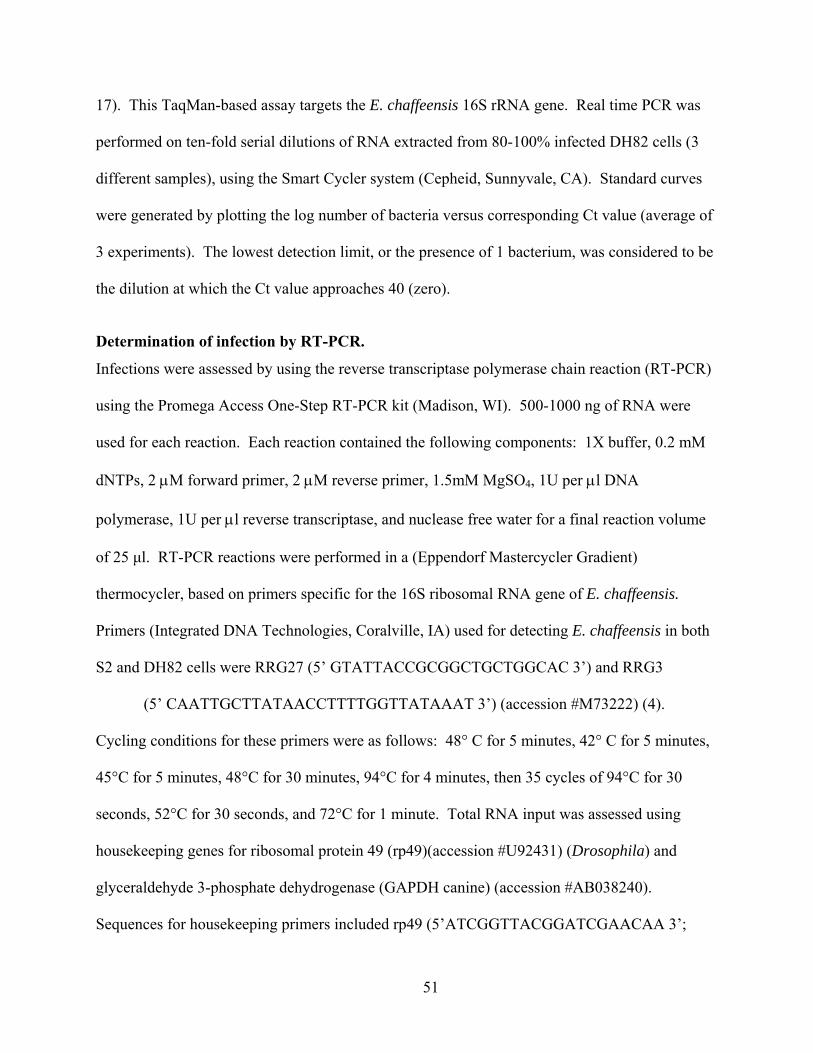
17). This TaqMan-based assay targets the E. chaffeensis 16S rRNA gene. Real time PCR was
performed on ten-fold serial dilutions of RNA extracted from 80-100% infected DH82 cells (3
different samples), using the Smart Cycler system (Cepheid, Sunnyvale, CA). Standard curves
were generated by plotting the log number of bacteria versus corresponding Ct value (average of
3 experiments). The lowest detection limit, or the presence of 1 bacterium, was considered to be
the dilution at which the Ct value approaches 40 (zero).
Determination of infection by RT-PCR.
Infections were assessed by using the reverse transcriptase polymerase chain reaction (RT-PCR)
using the Promega Access One-Step RT-PCR kit (Madison, WI). 500-1000 ng of RNA were
used for each reaction. Each reaction contained the following components: 1X buffer, 0.2 mM
dNTPs, 2 μM forward primer, 2 μM reverse primer, 1.5mM MgSO4, 1U per μl DNA
polymerase, 1U per μl reverse transcriptase, and nuclease free water for a final reaction volume
of 25 μl. RT-PCR reactions were performed in a (Eppendorf Mastercycler Gradient)
thermocycler, based on primers specific for the 16S ribosomal RNA gene of E. chaffeensis.
Primers (Integrated DNA Technologies, Coralville, IA) used for detecting E. chaffeensis in both
S2 and DH82 cells were RRG27 (5’ GTATTACCGCGGCTGCTGGCAC 3’) and RRG3
(5’ CAATTGCTTATAACCTTTTGGTTATAAAT 3’) (accession #M73222) (4).
Cycling conditions for these primers were as follows: 48° C for 5 minutes, 42° C for 5 minutes,
45°C for 5 minutes, 48°C for 30 minutes, 94°C for 4 minutes, then 35 cycles of 94°C for 30
seconds, 52°C for 30 seconds, and 72°C for 1 minute. Total RNA input was assessed using
housekeeping genes for ribosomal protein 49 (rp49)(accession #U92431) (Drosophila) and
glyceraldehyde 3-phosphate dehydrogenase (GAPDH canine) (accession #AB038240).
Sequences for housekeeping primers included rp49 (5’ATCGGTTACGGATCGAACAA 3’;
51

5’GACAATCTCCTTGCGCTTCT 3’) for Drosophila S2 cells and GAPDH
(5’GATTGTCAGCAATGCCTCCT 3’; 5’GGCAGGTCAGATCCACAACT 3’) for the DH82
cells. Cycling conditions for rp49 primers were as follows: 48°C for 45 minutes, 94°C for 2
minutes, and then 35 cycles of 94°C for 45 seconds, 50°C for 1 minute, and 72°C for 1.5
minutes. Cycling conditions for GAPDH primers were as follows: 48°C for 45 minutes, 94°C
for 2 minutes, and then 35 cycles of 94°C for 30 seconds, 55°C for 1 minute, and 68°C for 1
minute. RT-PCR reactions were also performed without reverse transcriptase in order to assure
that DNA was absent from the sample(s). RT-PCR products were identified on a ChemiImager
after electrophoresis in 2% agarose gels and staining with ethidium bromide. The sized
amplicons for each primer set were: RRG 27 & 3 (430 bp), rp49 (165 bp), and GAPDH (308
bp).
Immunocytochemistry.
Specific E. chaffeensis infections were confirmed by immunocytochemical techniques.
Immunocytochemistry was performed on E. chaffeensis-infected S2 cells, E. chaffeensis-infected
DH82 cells, uninfected S2 cells, and uninfected DH82 cells. Cells were prepared using cytospin
slides and dried for 24 hours at room temperature. Cells were fixed with acetone, dried ten
minutes, and outlined using a Pap Pen (The Binding Site, Inc., San Diego, CA). Samples were
placed in PBS containing 0.01% Tween for five minutes. The samples were blocked in 10%
blocking solution (PBS containing 50% normal goat serum) for 30 minutes at 37°C, washed in
PBS-Tween for five minutes, and incubated in primary antibody (mouse-anti-E. chaffeensis;
1:100 dilution) for 24 hours in the dark at 4°C. Primary antibody was either normal mouse
serum or serum taken from mice infected with E. chaffeensis. After incubation, the slides were
washed with PBS-Tween for five minutes and were incubated for one hour in the dark at room
52

temperature with a 1:50 dilution of goat-anti-mouse IgG conjugated with Rhodamine (Organon
Teknika Corp., West Chester, PA). Samples were washed in PBS-Tween for five minutes, then
in distilled water for five minutes before viewing.
Activation of S2 cells with Lipopolysaccharide.
To determine if lipopolysaccharide (LPS) activation would inhibit the growth of E. chaffeensis,
S2 cells were plated at a concentration of 1 x 10⁶ cells per plate in 6-well plates and were allowed
to adhere for at least 30 minutes. LPS (from Salmonella minnesota; Sigma ) was sonicated for
one hour and then added to each well at a concentration of 10 μg per ml. The cells plus LPS
were incubated for five hours. S2 cells were infected with E. chaffeensis purified from DH82
cells. RNA was extracted (using Tri-reagent) from each sample at 48 hpi, and assessed for the
presence of Ehrlichia using RT-PCR, as previously described.
Transmission electron microscopy and sample preparation.
Infected S2 (72 hpi (hpi)), uninfected S2, infected DH82 (72 hpi), and uninfected DH82 cells
were pelletted (5-10 ul pellet) and immersion fixed in fixative containing 2% paraformaldehyde
and 2% glutaraldehyde in 0.1 sodium cacodylate buffer (pH 7.4) for 16 hours at room
temperature with constant rotation. Each sample was washed 3 times for 5 minutes in 0.1M
sodium cacodylate buffer at room temperature (RT) with constant rotation. The samples were
post-fixed with 2% Osmiun tetroxide in 0.1M sodium cacodylate buffer with constant rotation
for 1-2 hours and then washed 3 times for 5 minutes in 0.1M sodium cacodylate buffer at RT
with constant rotation. Samples were pre-embedded/stained with 2% uranyl acetate in 0.2M
sodium acetate buffer (pH 5.2) for 1 hour at RT, light-protected, with constant rotation, and
subsequently washed 3 times for 5 minutes in 0.2M sodium acetate buffer at room temperature
with constant rotation. The samples were dehydrated in an ascending acetone series (50, 60, 70,
53

80, 90, 95, and 100%) with constant rotation at RT and then in 100% propylene oxide for 10
minutes. Infiltration of samples was accomplished using EMBED 812/Araldite 502 resin at RT
with constant rotation and consisted of 10 minutes in 1:1 propylene oxide:resin, 20 minutes in
1:2 propylene oxide:resin, 10 minutes in 100% resin. New 100% resin was finally added to the
sample, allowed to infiltrate for 16 hours, and then polymerize in a drying oven at 60º C for 24-
48 hours. The images were collected on a CM 100 (FEI company) transmission electron
microscope at 100kV using a Hamamatsu C8484 digital camera and Advanced Microscopy
Techniques Corp. software version 5.4.2.22B.
Statistics.
Statistical values were determined using the Student’s t-test (two-tailed, general). P values of
<0.02 (as indicated) were considered highly significant. Data are presented as mean ± SD.
Differences were determined using the StatMost Statistical Package (Data XIOM, Los Angeles,
CA, USA).
RESULTS To test the hypothesis that E. chaffeensis can be propagated in macrophage-like S2 cells,
we infected the S2 cells and assessed the infection via morphological, molecular, and
immunological techniques. In our initial experiments, bacteria isolated from heavily infected
DH82 cells (~1.5 x 10⁷ bacteria) was used in an attempt to infect the S2 cells. The S2 cells were
tested for the presence of bacteria at time points ranging from 24 hpi to as late as 120 hpi. At 24
hpi, 18.8% of the S2 cells contained one or more morulae. At 120 hpi, 91.7% of the S2 cells
contained one or more morulae (Figure 2.1). We observed an increase in the percentage of cells
infected with each subsequent time point. At the 120 hour time-point, many of the S2 cells
54
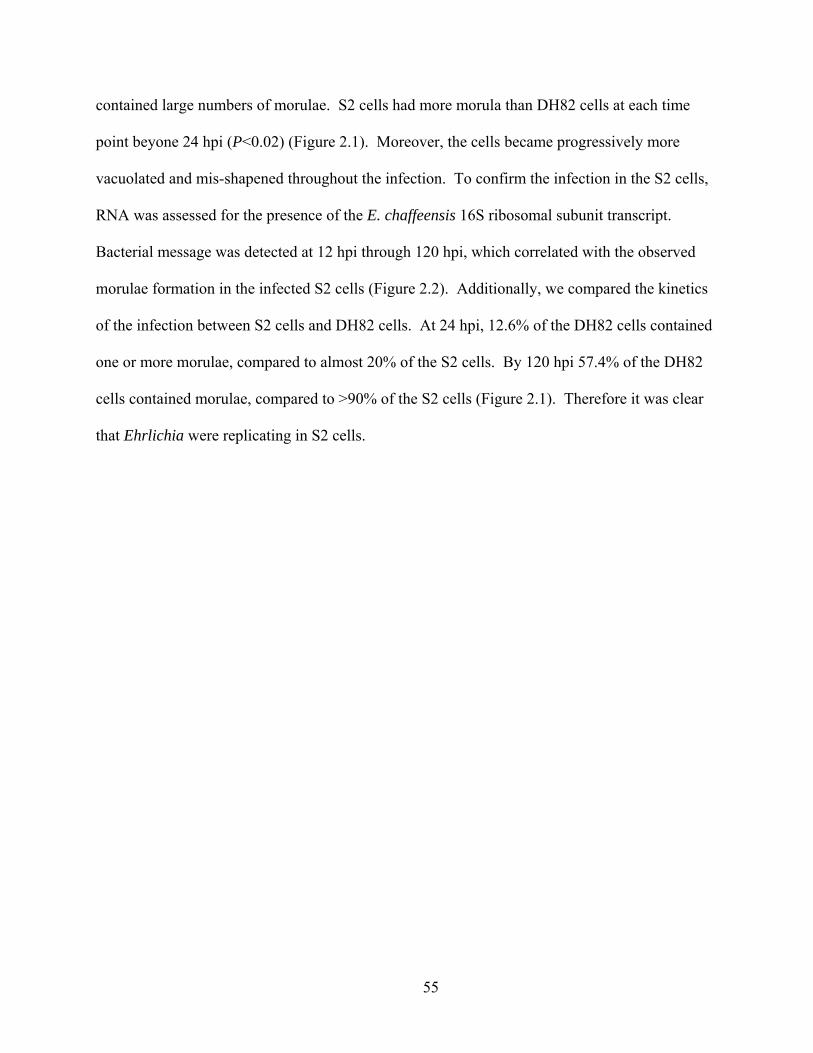
contained large numbers of morulae. S2 cells had more morula than DH82 cells at each time
point beyone 24 hpi (P<0.02) (Figure 2.1). Moreover, the cells became progressively more
vacuolated and mis-shapened throughout the infection. To confirm the infection in the S2 cells,
RNA was assessed for the presence of the E. chaffeensis 16S ribosomal subunit transcript.
Bacterial message was detected at 12 hpi through 120 hpi, which correlated with the observed
morulae formation in the infected S2 cells (Figure 2.2). Additionally, we compared the kinetics
of the infection between S2 cells and DH82 cells. At 24 hpi, 12.6% of the DH82 cells contained
one or more morulae, compared to almost 20% of the S2 cells. By 120 hpi 57.4% of the DH82
cells contained morulae, compared to >90% of the S2 cells (Figure 2.1). Therefore it was clear
that Ehrlichia were replicating in S2 cells.
55
Figure 2.1 Percentage of cells containing morulae in S2 and DH82 cells after infection with
E. chaffeensis. Cells were considered positive when one or more morulae were present.
Results are the averages of three separate infection experiments (mean ± S.d., n=3).
Statistical significance is represented by * (P<0.06) or ** (P<0.02).
56

Figure 2.2 A, RT-PCR of E. chaffeensis 16S ribosomal RNA gene (band at 430 bp) after
infection of S2 cells at 12-120 hpi. Time course infection experiments and RT-PCR of 16S
ribosomal RNA gene were repeated greater than 6 times with the same outcome. One
representative experiment is shown. B, RT-PCR of D. melanogaster rp49 (165 bp) and dog
GAPDH (308 bp) housekeeping genes, performed on samples shown in A.
To confirm that the bacteria grown in S2 cells were completing replication and were
infectious to vertebrate cells, DH82 cells were infected with bacteria purified from S2 cells at 24,
48, 72, 96, and 120 hpi of the S2 cells. The purified bacteria were used to infect the DH82 cells
and the amount of bacteria present in DH82 cells was assessed 72 hours later by quantitation of
morulae. When S2 cells were only infected for 24 hours, the bacteria isolated from those cells
infected 8% of the DH82 cells (Figure 2.3). 67% of the DH82 cells were infected with bacteria
isolated from S2 cells infected for 120 hours (Figure 2.3). We confirmed these infections by RT-
PCR of the 16S E. chaffeensis rRNA (Figure 2.4). Consequently, E. chaffeensis is capable of
57
completing its lifecycle in S2 cells and also maintains its ability to reinfect the mammalian
macrophage cell line, DH82.
Figure 2.3 Growth of E. chaffeensis (originally grown in S2 cells) in DH82 cells. Bacteria
grown in S2 cells for the indicated length of time were used to infect DH82 cells. The
percentage of DH82 cells containing morulae was assessed 72 hours later. Results are the
averages of three separate time course experiments (mean ± S.d., n=3).
58

Figure 2.4 Reinfection of DH82 cells with S2 cell-grown bacteria (A). RT-PCR of E.
chaffeensis 16S ribosomal RNA gene (band at 430 bp) after DH82 cells were infected with
bacteria grown in S2 cells for the indicated times. RT-PCR of D. melanogaster rp49 (165
bp) and dog GAPDH (308 bp) housekeeping genes, performed on samples shown in A (B).
To further demonstrate that the inclusions seen in the S2 cells after infection with E.
chaffeensis, we utilized immunocytochemistry to detect bacteria. Slides were made with infected
S2 and DH82 cells or with uninfected cells. Sera collected from E. chaffeensis-infected mice or
from normal mice were used as primary antibodies. Only E. chaffeensis-specific antiserum
reacted with the infected DH82 (Figure 2.5D) and S2 cells (Figure 2.5A-C). No bacteria were
detected with the normal mouse serum incubated with infected or uninfected cells, or by
secondary antibody alone (Figure 2.5E-H).
59
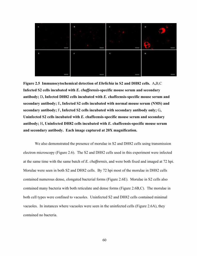
Figure 2.5 Immunocytochemical detection of Ehrlichia in S2 and DH82 cells. A,B,C
Infected S2 cells incubated with E. chaffeensis-specific mouse serum and secondary
antibody; D, Infected DH82 cells incubated with E. chaffeensis-specific mouse serum and
secondary antibody; E, Infected S2 cells incubated with normal mouse serum (NMS) and
secondary antibody; F, Infected S2 cells incubated with secondary antibody only; G,
Uninfected S2 cells incubated with E. chaffeensis-specific mouse serum and secondary
antibody; H, Uninfected DH82 cells incubated with E. chaffeensis-specific mouse serum
and secondary antibody. Each image captured at 20X magnification.
We also demonstrated the presence of morulae in S2 and DH82 cells using transmission
electron microscopy (Figure 2.6). The S2 and DH82 cells used in this experiment were infected
at the same time with the same batch of E. chaffeensis, and were both fixed and imaged at 72 hpi.
Morulae were seen in both S2 and DH82 cells. By 72 hpi most of the morulae in DH82 cells
contained numerous dense, elongated bacterial forms (Figure 2.6E). Morulae in S2 cells also
contained many bacteria with both reticulate and dense forms (Figure 2.6B,C). The morulae in
both cell types were confined to vacuoles. Uninfected S2 and DH82 cells contained minimal
vacuoles. In instances where vacuoles were seen in the uninfected cells (Figure 2.6A), they
contained no bacteria.
60

Figure 2.6 Transmission electron micrographs of S2 and DH82 cells infected with E.
chaffeensis or uninfected. A, uninfected S2; B, infected S2; C, infected S2 (black
arrowheads indicate dense core form; diamond with black arrowhead indicates reticulate
form); D, uninfected DH82; E, infected DH82.
Lastly, it is known that mammalian macrophages are non-permissive for E. chaffeensis
growth after LPS activation (13). Therefore, we wanted to determine if S2 cells could be
resistant to E. chaffeensis infection. To test this hypothesis, we incubated S2 cells with LPS (10
μg per ml) for 4 hours, and then we challenged the cells with increasing doses of purified E.
chaffeensis bacteria. RNA was extracted from the cells 48 hpi, and the RNA samples were
analyzed by RT-PCR for Ehrlichia infection. S2 cells activated with LPS (10 μg per ml) were
non-permissive for E. chaffeensis regardless of the number of bacteria used for infection (Figure
2.7). As a control, unstimulated S2 cells were infected at the same time using the same number
61

of bacteria (Figure 2.7). Thus, activated S2 cells become non-permissive to infection by E.
chaffeensis, similar to results seen in activated human monocytes (13).
Figure 2.7 RT-PCR results of LPS activation of S2 cells. E. chaffeensis 16S ribosomal
RNA (band at 430 bp) present only in cells not treated with LPS. S2 cells were infected
with increasing numbers of bacteria with 1.4 x 10⁷ bacteria/200 µl. Housekeeping gene is
Drosophila melanogaster ribosomal protein 49 (rp49).
DISCUSSION We have presented novel data showing that Drosophila S2 cells can be successfully
infected with the obligate, intracellular bacterium, E. chaffeensis. Infection was confirmed using
three distinct criteria: (1) presence of morulae in the S2 cells; (2) the detection of 16S ribosomal
RNA of E. chaffeensis in infected cells; and (3) the specific detection of E. chaffeensis in S2
62
cells by bacteria-specific antibody. Morulae in mammalian cells are known to have two forms:
dense core and reticulate cells (15). We found the presence of both dense and reticulate forms of
bacteria in S2 cells, particularly a preponderance of reticulate forms in infected S2 cells.
Considering the differences observed between bacterial growth in the S2 and DH82 cells (Figure
1), this is not surprising. The reticulate form is the dividing form of the the bacteria, and thus an
increased number of dividing bacteria coincides with the increase bacterial kinetics observed in
the S2 cells when compared to the DH82 cells. Zhang et al. (2007) found that the dense core
form of bacteria were present exclusively during early infection (0-1 hpi). Between 24 and 48
hpi, the reticulate form predominated and binary fission was often observed. Subsequent to that,
dense core forms reappeared. It is possible that the reticulate forms predominate in insect cells
grown at lower temperatures or that the reappearance of dense core forms occurs later.
Alternatively, the presence of reticulate and dense core forms may be cell-line and/or
temperature-dependent. Additional electron microscopy work will be necessary to determine
this. Nevertheless, these data confirm that E. chaffeensis can be propagated in S2 cells and its
developmental patterns are similar to those seen in mammalian macrophages. In addition,
vacuoles containing appropriately sized inclusions (presumably bacteria) were observed by light
microscopy/live video imaging in both S2 cells and DH82 cells that had been infected with E.
chaffensis (data not shown). The bacteria were confined to the vacuoles in the infected cells;
conversely, no bacteria were observed in the uninfected S2 or DH82 cells.
The kinetics of bacterial infection of S2 cells was comparable to the growth of bacteria in
DH82 cells, which are one of the most commonly used cell lines to propagate infections of E.
chaffeensis. Moreover, bacteria isolated from infected S2 cells were infectious and could be
used to infect DH82 cells. Therefore, the growth of E. chaffeensis in dipteran insect cells does
63

not compromise the viability of the bacteria. This is consistent with the natural life cycle of
Ehrlichia that includes an arachnid host, the tick. The host requirements for E. chaffeensis to
grow in ticks are not clear. The D. melanogaster system offers the advantage of having a well
defined genome. Using the S2 cells to identify which host genes are necessary to support E.
chaffeensis growth will eventually allow us to study homologous genes in both arachnids and
vertebrates. Our data are also consistent with observations that S2 cells are capable of
supporting other macrophage-tropic intracellular bacteria. For example, Chlamydia trachomatis,
Listeria monocytogenes and Mycobacterium fortuitum all infect S2 cells (1, 5, 6, 8, 14, 16).
Studies on these bacteria in S2 cells have provided valuable information about early and late
infection processes of C. trachomatis (8). S2 cells have also been critical to the identification of
key components involved in the pathogenesis of L. monocytogenes (6), and the involvement of
CD36 in M. fortuitum infections (16). Nevertheless, our findings open the molecular and genetic
tool box of the Drosophila genome for the study of E. chaffeensis.
In addition to the observation that E. chaffeensis is capable of infecting the Drosophila
S2 cells, we also were able to make the S2 cells non-permissive for infection by E. chaffeensis.
Although E. chaffeensis does not make LPS or peptidoglycan (12), we wanted to determine of
LPS could activate the S2 cells. Activation with LPS prevented bacterial replication even at high
MOIs. More importantly, the option of a system made non-permissive by the addition of LPS
increases the utility of S2 cells and makes them comparable to vertebrate macrophages in this
respect (13). In particular, comparing gene expression in activated versus infected S2 cells will
allow us to pinpoint genes specific to E. chaffeensis infections as well as genes that are simply
activation-specific. It is our goal to define the mechanisms that allow for maintenance of E.
chaffeensis in its invertebrate and vertebrate hosts.
64
REFERENCES 1. Agaisse, H., L. S. Burrack, J. A. Philips, E. J. Rubin, N. Perrimon, and D. E.
Higgins. 2005. Genome-wide RNAi screen for host factors required for intracellular bacterial infection. Science 309:1248-51.
2. Bakken, J. S., and J. S. Dumler. 2000. Human granulocytic ehrlichiosis. Clin Infect Dis 31:554-60.
3. Cao, W. C., Y. M. Gao, P. H. Zhang, X. T. Zhang, Q. H. Dai, J. S. Dumler, L. Q. Fang, and H. Yang. 2000. Identification of Ehrlichia chaffeensis by nested PCR in ticks from Southern China. J Clin Microbiol 38:2778-80.
4. Cheng, C., C. D. Paddock, and R. Reddy Ganta. 2003. Molecular heterogeneity of Ehrlichia chaffeensis isolates determined by sequence analysis of the 28-kilodalton outer membrane protein genes and other regions of the genome. Infect Immun 71:187-95.
5. Cheng, L. W., and D. A. Portnoy. 2003. Drosophila S2 cells: an alternative infection model for Listeria monocytogenes. Cell Microbiol 5:875-85.
6. Cheng, L. W., J. P. Viala, N. Stuurman, U. Wiedemann, R. D. Vale, and D. A. Portnoy. 2005. Use of RNA interference in Drosophila S2 cells to identify host pathways controlling compartmentalization of an intracellular pathogen. Proc Natl Acad Sci U S A 102:13646-51.
7. Childs, J. E., and C. D. Paddock. 2003. The ascendancy of Amblyomma americanum as a vector of pathogens affecting humans in the United States. Annu Rev Entomol 48:307-37.
8. Elwell, C., and J. N. Engel. 2005. Drosophila melanogaster S2 cells: a model system to study Chlamydia interaction with host cells. Cell Microbiol 7:725-39.
9. Ganta, R. R., C. Cheng, E. C. Miller, B. L. McGuire, L. Peddireddi, K. R. Sirigireddy, and S. K. Chapes. 2007. Differential clearance and immune responses to tick cell-derived versus macrophage culture-derived Ehrlichia chaffeensis in mice. Infect Immun 75:135-45.
10. Han, K. 1996. An efficient DDAB-mediated transfection of Drosophila S2 cells. Nucleic Acids Res 24:4362-3.
11. Kavanagh, K., and E. P. Reeves. 2007. Insect and Mammalian Innate Immune Responses Are Much Alike. Microbe 2:596-599.
12. Lin, M., and Y. Rikihisa. 2003. Ehrlichia chaffeensis and Anaplasma phagocytophilum lack genes for lipid A biosynthesis and incorporate cholesterol for their survival. Infect Immun 71:5324-31.
13. Lin, M., and Y. Rikihisa. 2004. Ehrlichia chaffeensis downregulates surface Toll-like receptors 2/4, CD14 and transcription factors PU.1 and inhibits lipopolysaccharide activation of NF-kappa B, ERK 1/2 and p38 MAPK in host monocytes. Cell Microbiol 6:175-86.
14. Mansfield, B. E., M. S. Dionne, D. S. Schneider, and N. E. Freitag. 2003. Exploration of host-pathogen interactions using Listeria monocytogenes and Drosophila melanogaster. Cell Microbiol 5:901-11.
15. Paddock, C. D., and J. E. Childs. 2003. Ehrlichia chaffeensis: a prototypical emerging pathogen. Clin Microbiol Rev 16:37-64.
16. Philips, J. A., E. J. Rubin, and N. Perrimon. 2005. Drosophila RNAi screen reveals CD36 family member required for mycobacterial infection. Science 309:1251-3.
65

17. Sirigireddy, K. R., and R. R. Ganta. 2005. Multiplex detection of Ehrlichia and Anaplasma species pathogens in peripheral blood by real-time reverse transcriptase-polymerase chain reaction. J Mol Diagn 7:308-16.
18. Stroschein-Stevenson, S. L., E. Foley, P. H. O'Farrell, and A. D. Johnson. 2006. Identification of Drosophila gene products required for phagocytosis of Candida albicans. PLoS Biol 4:e4.
19. Vilmos, P., and E. Kurucz. 1998. Insect immunity: evolutionary roots of the mammalian innate immune system. Immunol Lett 62:59-66.
20. Zhang, J. Z., V. L. Popov, S. Gao, D. H. Walker, and X. J. Yu. 2007. The developmental cycle of Ehrlichia chaffeensis in vertebrate cells. Cell Microbiol 9:610-8.
21. Zhang, J. Z., M. Sinha, B. A. Luxon, and X. J. Yu. 2004. Survival strategy of obligately intracellular Ehrlichia chaffeensis: novel modulation of immune response and host cell cycles. Infect Immun 72:498-507.
66
CHAPTER 3 - Ehrlichia chaffeensis infections in Drosophila
melanogaster
ABSTRACT Ehrlichia chaffeensis is an obligate, intracellular bacterium, transmitted by the tick
Amblyomma americanum, and is the causative agent of human monocytic ehrlichiosis infections.
We previously demonstrated that E. chaffeensis is capable of growing in Drosophila S2 cells.
Therefore, we tested the hypothesis that E. chaffeensis can infect adult D. melanogaster. Adult
Drosophila were experimentally challenged with intra-abdominal injections of bacteria.
Ehrlichia-infected flies showed decreased survival compared to wild-type flies and bacteria
isolated from flies could reinfect mammalian macrophages. Ehrlichia infections activated both
the cellular and humoral immune responses in the fly. Hemocytes phagocytosed bacteria after
injection and antimicrobial peptide pathways were induced following infection. Increased
pathogenicity in flies carrying mutation for genes in both the Toll and Imd pathways suggests
that both immune defense pathways participate in host defense. Induction of Drosophila cellular
and humoral responses and the in vivo replication of E. chaffeensis suggests that D.
melanogaster is a suitable host for E. chaffeensis. In the future, it will be a useful tool to unlock
some of the in vivo mysteries of this arthropod-borne bacteria.
INTRODUCTION Drosophila melanogaster is a valuable tool for studies focused on innate immune
responses. It is especially attractive because of the ability to study innate host defense without
the complicating variables of acquired immunity (28, 58). Drosophila innate immunity involves
67
both cellular and humoral components. The cellular immune response involves phagocytosis,
encapsulation, and/or melanization of pathogens via hemocytes, plasmatocytes, or crystal cells,
respectively (18, 31, 63). Humoral immunity involves the production of antimicrobial peptides
through either the Toll or immune deficiency (Imd) pathways (18, 31, 63). The Toll pathway is
activated by Gram-positive bacteria or fungi, and elicits production of the antimicrobial peptide
Drosomycin (18, 31, 63). The Imd pathway is activated by Gram-negative bacteria and is
characterized by the production of antimicrobial peptides such as Attacin and Diptericin (18, 31,
63). Additionally, D. melanogaster’s completed genome, ease of manipulation, availability of
mutants, and homology to vertebrate systems make it an attractive tool as a model system for
detailing the innate immune responses to various pathogens. In particular, it has been used to
characterize immune reactions elicited in response to Erwinia carotovora, Mycobacterium
marinum, Plasmodium gallinaceum, Francisella tularensis, Serratia marcesans, Listeria
monocytogenes, and Salmonella enterica serovar typhimurium (3, 7, 13, 40, 42, 52, 64).
Ehrlichia chaffeensis is an obligate, intracellular bacterium and is vectored by
Amblyomma americanum (lone star tick). It is the causative agent of human monocytic
ehrlichiosis (HME), which can be particularly life-threatening in young, elderly, and/or
immunocompromised patients. In 2006, the CDC reported an infection rate of 0.2/100,000
persons in the United States (41). E. chaffeensis is classified as a Gram-negative bacterium, but
lacks the genes necessary for the synthesis of peptidoglycan (PGN) or lipopolysaccharide (LPS)
(37). Therefore, many questions exist about early host resistance to Ehrlichia as well as
questions about the host genetic requirements for bacterial growth. Drosophila melanogaster
could be a useful tool to address these questions. We have established that E. chaffeensis can
infect and replicate in the hemocytic, macrophage-like Drosophila S2 cell line (38). We
68
hypothesized that E. chaffeensis would infect adult flies and activate host defenses. We present
evidence indicating that Ehrlichia can infect and replicate in adult Drosophila, that hemocytes
respond to the infection, and Drosophila humoral immune pathways are activated.
MATERIALS AND METHODS
Maintenance of cell lines and Ehrlichia chaffeensis infections.
The canine macrophage cell line DH82 was maintained at 37°C in Dulbecco's modified Eagle's
medium with 3.5% fetal bovine serum and 3.5% Nu serum (DMEM-7). The E. chaffeensis
Arkansas isolate was continuously cultivated in the DH82 cell line at 37°C, 8% CO2 in DMEM-7
medium. Bacteria were passaged when infectivity reached 80 to 90% as visualized by using
cytospin-prepared slides (stained with Hema3 fixative and Dif-Quik stain) to monitor the
formation of morulae in the cells. Infected cells were removed by scraping each plate with a cell
scraper, transferring the culture to a conical tube, and shaking the suspension with glass beads.
The liberated bacteria were purified by centrifuging the suspension at 600 x g for 20 minutes to
remove cell debris. The bacteria-containing supernatant was removed, transferred to a sterile
conical tube, and centrifuged at 15,000 x g for 20 minutes to pellet the free bacteria. The final
supernatant was removed and discarded, and the pellet was resuspended in sterile phosphate-
buffered saline (PBS).
For preparation of dead bacteria cultures, the final pellet was resuspended in sterile PBS
and the tube was subsequently placed in a boiling water bath for 15 minutes. The boiled bacteria
was then centrifuged at 15,000 x g for 20 minutes to re-pellet the bacteria. The supernatant was
removed and the dead bacterial pellet was resuspended in 5 ml sterile PBS.
69

Infected flies were used to re-infect DH82 cells. Ten flies that had been infected for 168
hours were anesthetized using CO2 and transferred to a sterile, 1.5 ml conical tube (Kimble
Kontes #749510-1500). Flies were homogenized with a disposable pestle (Kimble Kontes
#749521-1590) in 1 ml of sterile PBS. The homogenate was spun at 10,000 x g for 10 seconds
in a microcentrifuge and the resulting supernatant was added to a 150-mm plate of 40%
confluent DH82 cells (treated with 0.13 μg per ml of Fungizone). Cells were observed for the
formation of morulae, using cytospin slide preparations. At two weeks after infection, morulae
formation was observed in 90-100% of the cells. RNA extraction and RT-PCR analysis for
Ehrlichia 16S ribosomal gene was performed on the infected and on uninfected DH82 cells as
previously described (38).
Assessment of bacterial numbers for infection.
The number of bacteria used for infection experiments of DH82 cells and flies was estimated
using TaqMan-based real-time reverse transcriptase PCR (RT-PCR) as previously described (55).
This TaqMan-based assay targets the E. chaffeensis 16S rRNA gene transcripts. Detection of the
16S rRNA is 100 times more sensitive than rDNA (20, 55). We have confirmed that there is no
difference in the relative levels of 16S rRNA and rDNA during the course of an infection using
qRT-PCR and qPCR. Real-time quantitative PCR (qRT-PCR) was performed on 10-fold serial
dilutions of RNA extracted from 80- to 100%-infected DH82 cells (three different samples) using
a Smart Cycler system (Cepheid, Sunnyvale, CA). Standard curves were generated by plotting
the log number of bacteria versus the corresponding threshold cycle value (mean of the results of
three experiments). The lowest detection limit, or the presence of 1 bacterium (100 rRNA copies
= 1 bacterium), was considered to be the dilution at which the threshold cycle value approaches
40 (zero).
70
Fluorescent labeling of bacteria for injection.
Cell-free E. chaffeensis was purified as described above and was fluoresceinated as previously
described (10) with modifications described as follows. Fluorescein isothocyanate (FITC) was
dissolved into 0.2M Na2CO3 (pH 9.5) buffer to a final concentration of 5 mg per ml. One ml of
the FITC solution was added to the cell-free E. chaffeensis pellet, mixed gently, and incubated
with rocking for 15 minutes at room temperature (protected from light). The FITC-bacteria
mixture was then added to 4 ml of 135 mM NaCl/10 mM phosphate buffer (pH 7.4) and
incubated for 5 minutes at room temperature. Following incubation, the mixture was centrifuged
at 10,000 x g for 5 minutes, the supernatant was removed, the pellet was resuspended in 2.83%
Na2HPO4 (pH 8.5), and the mixture was centrifuged again. The resulting pellet was resuspended
in sterile PBS and subsequently washed three times with sterile PBS. The bacterial pellet was
resuspended in sterile PBS for fly injections.
Bacteria were also labeled following the protocol of the pHrodo Phagocytosis
Particle Labeling Kit for Flow Cytometry (Invitrogen, Carlsbad, CA; #A10026). Briefly, cell-
free E. chaffeensis was obtained as previously described. The bacterial pellet was resuspended in
3 ml of 100 mM sodium bicarbonate buffer (Component F) and centrifuged at 16,000 x g in a
microcentrifuge for 60 seconds. The pellet was then suspended in 750 μl of Component F, 18.25
μl of the pHrodo dye (Component D) was added, and the bacteria were incubated with rocking
for 45 minutes at room temperature (protected from light). 750 μl of wash buffer (Component C)
was added to the bacteria mixture, centrifuged at 16,000 x g for 60 seconds, and the supernatant
was removed. The bacterial pellet was resuspended in 1.5 ml of 100% methanol, vortexed for 30
seconds, centrifuged at 16,000 x g for 60 seconds, and the supernatant was removed. The
bacteria were resuspended in 1.5 ml of Component C, vortexed for 30 seconds, and centrifuged
71

at 16,000 x g for 60 seconds. The wash step was repeated and the final bacterial pellet was
resuspended in sterile PBS for injection.
For fly injection, equal volumes of the FITC-labeled and pHrodo-labeled E. chaffeensis
were mixed and simultaneously injected into yellow white flies. Flies were viewed on an
Olympus SZX12 fluorescent, dissecting microcope with emission filters of 535/50 for FITC
detection and 620/60 for pHrodo detection.
Salmonella enterica serovar typhimurium .
S. enterica serovar typhimurium (strain KSU-7)(15) was grown and streaked for isolation on
MacKonkey agar plates. Nutrient broth was inoculated with a single colony and grown
overnight at 37°C. The cultures were centrifuged at 10,000 rpm for 10 minutes and rinsed with
PBS. Bacterial pellets were resuspended in PBS, absorbance was measured on a NanoDrop
Spectrophotometer (Thermo Scientific; Wilmington, DE), and bacterial concentration was
estimated as previously described (9).
Flies.
Flies were maintained on standard dextrose/molasses/yeast medium at 18-29°C. For initial E.
chaffeensis infections and dose response infections, w;Hemese-Gal4, UASGFP flies were used as
wild-type (WT) in these experiments (from Dr. Michael J. Williams; Umea Centre for Molecular
Pathogenesis; Umea University; Umea, Sweden) (66). yellow white (yw) flies are continuously
maintained at Kansas State University for use as a wild-type stock. Transgenic flies expressing
green fluorescent protein (GPF) under the control of the Attacin (Attacin-GFP(II))(attacin-GFP),
Diptericin (Pw+/Pw+ III (Dipt-GFP-Drom))(diptericin-GFP), or Drosomycin (ywP(w+,Drom-
GFP)D4)(drosomycin-GFP) promoters; and mutants for dredd (ywD44)(dreddD44) and for relish
(relE20, e+) (from Dr. Bruno Lemaitre; Ecole Polytechnique Federale de Lausanne; Lausanne,
72
France)(35, 62) were used to study the Imd and Toll pathways. Toll-constitutive flies (Tl3,ri+e-
/TM3,Ser)(Tl3) were received from Drs. Claudia Mizutani and Ethan Bier (University of
California at San Diego)(1). pelle mutant (pll2) stocks were obtained from the Bloomington
Drosophila Stock Center at Indiana University, Bloomington, Indiana, e1 pll2 ca1/TM3, Sb1
(#3111)(2). cactus deficient flies (cactD11/CyO; cactD13/CyO)(cactD11 and cactD13 ) have been
previously described (51).
Fly Infections.
Flies were transferred to fresh food at least 24 hours priors to injection/infection. For
injection/infection, adult male and female flies were anesthetized with CO2 (for no longer than 15
minutes at a time). Flies were injected with approximately 50 nl of sterile PBS with or without
bacteria, using pulled, glass capillary needles. Injections were made in the abdomen of the fly,
close to the junction of the thorax, and ventral to the junction between the dorsal and ventral
cuticles. Following injection, flies were maintained in clean bottles with molasses caps that
were changed every other day throughout the course of the experiments. Survival was monitored
daily.
Bead injection.
In order to inhibit phagocytosis by hemocytes (13, 16, 47, 52), flies were injected with a 2%
solution of PBS mixed with FluoSpheres carboxylate-modified microspheres (0.2 μm, red
fluorescent – 580/605)(Invitrogen #F8810) or PBS alone. Injections were performed four hours
or 24 hours prior to injection with E. chaffeensis. Survival was monitored for 96 hours post
infection.
73

Assessment of fluorescence in antimicrobial peptide promoter-EGFP expressing
Drosophila.
Live, adult flies were assayed for eGFP expression by placing each fly into a 0.5 ml microfuge
tube containing 120 μl of sterile PBS. The flies were pulverized using a Dremel 100 Series
rotary tool with a conical shaped grinding bit. The bit was sterilized by flaming the bit after
dipping it in 70% ethanol. The bit was cooled between each sample. After pulverization, the
tubes were briefly microfuged at 12,000 x g and 50 μl of each supernatant was transferred to one
well of a 96-well plate. The fluorescence was quantitated using a Perkin Elmer Wallac Victor3
Multilabel Counter.
RNA extraction and quantitative real time reverse transcription – PCR (qRT-PCR).
To quantitate transcript levels, flies were anesthetized, placed in 1.5 ml tubes (Kimble Kontes
#749510-1500), and homogenized with disposable pestles in 1 ml of TriReagent (Molecular
Research Center) (Kimble Kontes #749521-1590). Homogenates were transferred to 2.0 ml,
Heavy Phase Lock Gel tubes (5 Prime/Eppendorf; Westbury, New York; #2302830). 300 μl of
chloroform was added and the mixture was shaken (not vortexed) for 15 seconds. The samples
were then centrifuged at 12,000 x g for 10 minutes at 4°C and the aqueous phase was transferred
to clean 1.5 ml tubes. 500 μl of isopropanol was added and RNA was precipitated at -20°C for
24 hours. Samples were subsequently centrifuged at 12,000 x g for 10 minutes. The RNA pellet
was washed with 1 ml of 70% ethanol and samples were centrifuged at 7.4 x g for 5 minutes.
The 70% ethanol was decanted from the pellet, the pellet was allowed to slightly air dry and was
resuspended in 50 μl of nuclease-free water.
qRT-PCR was performed using the Invitrogen’s One-Step Platinum qRT-PCR kit
(#11732) or Invitrogen’s Superscript III Platinum SYBR Green One-Step qRT-PCR kit (#11732)
in a Cepheid Smart Cycler. E. chaffeensis was detected as described above. Drosophila
74
ribosomal protein 15a (Accession #NM_136772) was detected using left primer
TGGACCACGAGGAGGCTAGG, right primer GTTGGTGCATGGTCGGTGA, and Taqman
probe TGGGAGGCAAAATTCTCGGCTTC (13). Antimicrobial peptides were detected using
the following primer sets: diptericin (Accession #NM_057460), (5')
ACCGCAGTACCCACTCAATC, (5') CCCAAGTGCTGTCCATATCC; and drosomycin
(Accession #NM_079177), (5') GTACTTGTTCGCCCTCTTCG, (5')
CTTGCACACACGACGACAG (52).
Transfection of S2 cells.
Plasmids carrying GFP reporter-AMP promoter constructs for Attacin, Diptercin, and
Drosomycin were obtained from the lab of Bruno Lemaitre (62). Competent E. coli cells
(Invitrogen #C404003) were transformed and plated on nutrient agar containing 50 μg per ml of
ampicillin. The prescence of the attacin, diptericin, or drosomycin plasmid was confirmed using
colony PCR. For detection of diptericin and drosomycin, the previously mentioned primer sets
were used. For the detection of attacin (Accession #NM_079021), the following primer set was
used: left primer-CAATGGCAGACACAATCTGG and right primer-
ATTCCTGGGAAGTTGCTGTG (13). PCR reactions were performed using the Platinum Taq
Polymerase kit (Invitrogen #10966-034). Upon confirmation of the plasmid of interest, nutrient
broth cultures containing 50 μg per ml of carbenacillin were inoculated and grown overnight at
37°C. Plasmid DNA was extracted using the Qiagen Midi-Prep Kit (#12145). For transfection,
5 x 10⁵ S2 cells were plated in 60 mm2 tissue culture wells of 6-well plates and incubated
overnight. One μg of plasmid DNA was mixed with 15 μl of Cellfectin Reagent (Invitrogen
#10362), and subsequently mixed in incomplete S2 medium (no serum) in a total volume of 200
μl. The mixture was incubated at room temperature for 30 minutes. The S2 cells were rinsed
75
with incomplete S2 medium and a final volume of 800 μl of incomplete medium was added to
the cells. Plasmid/CellFectin complexes were added to the cells, the cells were briefly shaken,
and incubated with the complexes for 5.5-6 hours. Cells were then washed with complete S2
medium and resuspended in a final volume of 2 ml. To test for activation of the transgene,
viable or heat-killed cell-free E. chaffeensis was added to the cells 48 hours post-transfection and
fluorescence was measured at 24 hours later using a Perkin Elmer Wallac Victor3 Multilabel
Counter.
Statistics.
Data are presented as the mean ± standard error (SE) of independent experiments. Differences in
mean were determined by using Student's t test (two-tailed, general) (StatMost statistical package
(Data XIOM, Los Angeles, CA)). Survival data were analyzed for significance using the log-
rank test of Kaplan Meier plots using Prism Graphpad software (La Jolla, CA). P values of
<0.05 were considered highly significant.
RESULTS
Growth and pathogenicity of E. chaffeensis in adult Drosophila.
We previously demonstrated that E. chaffeensis replicated in the Drosophila hemocyte-like S2
cells (38). These data raised the issue of whether E. chaffeensis could grow in vivo. To test the
hypothesis that adult D. melanogaster can be infected with E. chaffeensis, we injected cell-free
E. chaffeensis into the abdomens of WT adult male and female D. melanogaster. Negative
control male and female flies were injected with sterile PBS. Survival was monitored in a total
of 20-25 flies per experiment for 120 hours. There was a significant amount of death in the
infected flies compared to those injected with PBS (Figure 3.1). For example, at 24 hpi, a mean
76
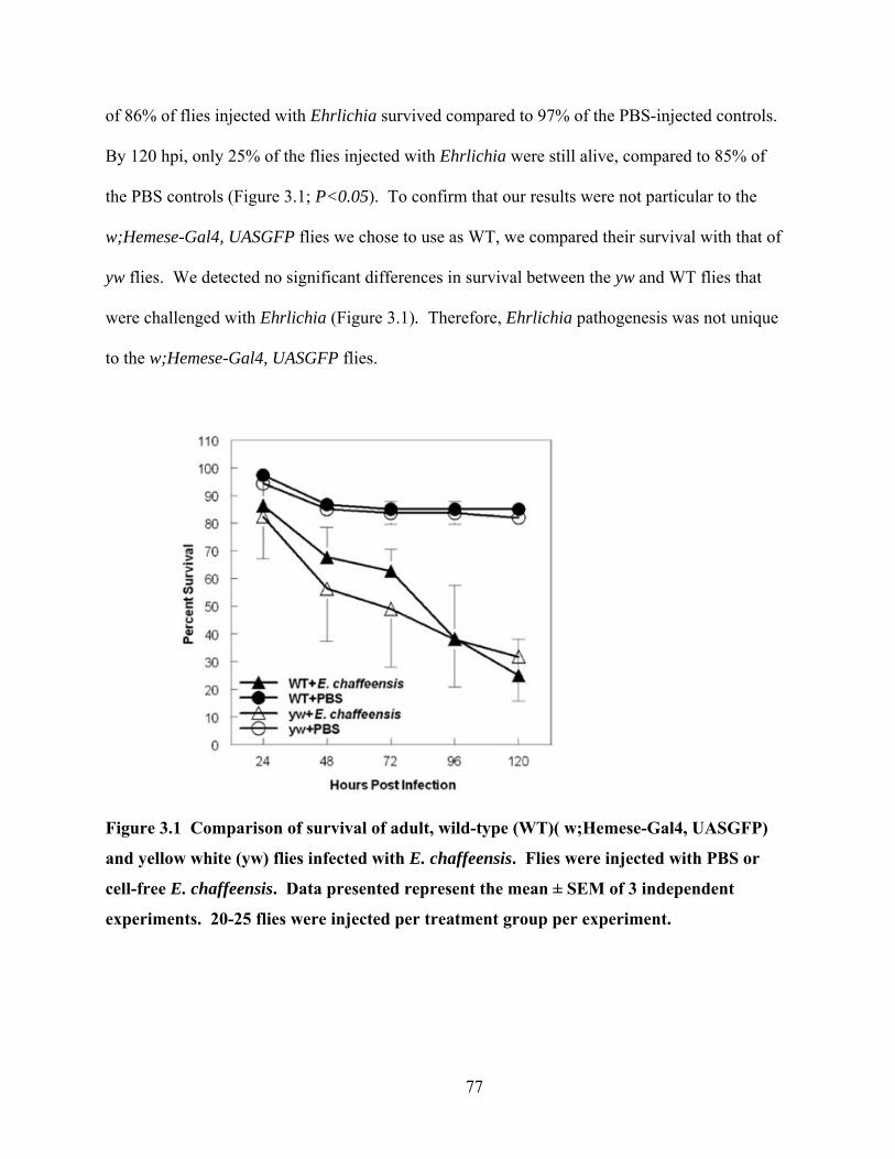
of 86% of flies injected with Ehrlichia survived compared to 97% of the PBS-injected controls.
By 120 hpi, only 25% of the flies injected with Ehrlichia were still alive, compared to 85% of
the PBS controls (Figure 3.1; P<0.05). To confirm that our results were not particular to the
w;Hemese-Gal4, UASGFP flies we chose to use as WT, we compared their survival with that of
yw flies. We detected no significant differences in survival between the yw and WT flies that
were challenged with Ehrlichia (Figure 3.1). Therefore, Ehrlichia pathogenesis was not unique
to the w;Hemese-Gal4, UASGFP flies.
Figure 3.1 Comparison of survival of adult, wild-type (WT)( w;Hemese-Gal4, UASGFP)
and yellow white (yw) flies infected with E. chaffeensis. Flies were injected with PBS or
cell-free E. chaffeensis. Data presented represent the mean ± SEM of 3 independent
experiments. 20-25 flies were injected per treatment group per experiment.
77

The number of flies that survived was dependent on bacterial dose (Figure 3.2). At 96
hours post infection, a mean of 47% of flies survived at the highest challenge dose, with 68%,
72%, and 77% survival when challenged with decreasing doses (Figure 3.2). Bacteria replicated
in the flies over time. RNA was collected from live flies at several time points for subsequent
qRT-PCR to measure 16S rRNA E. chaffeensis and Drosophila ribosomal protein 15a. By 24
hours, there were usually more bacteria detected than were originally injected and the number of
bacteria increased and decreased in a cyclical fashion over a period of 120 hours (Figure 3.3).
By 96 hours post infection there were higher bacterial loads in the flies than in the original
injections in 9 of the 11 measurements made (Figure 3.4). Therefore, these data indicate that E.
chaffeensis is capable of infecting D. melanogaster in a dose-dependent fashion and the bacteria
were actively replicating in adult flies. Furthermore, bacteria isolated from adult flies were used
to reinfect DH82 cells in vitro, as determined by RT-PCR and by the identification of morulae in
the infected cell after infection (data not shown). Therefore, the bacteria that replicate in adult
flies are capable of re-infecting mammalian cells.
78
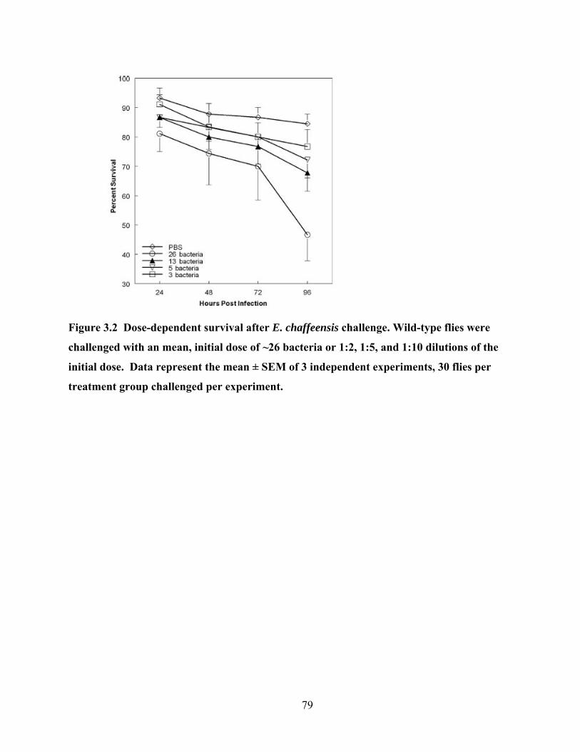
Figure 3.2 Dose-dependent survival after E. chaffeensis challenge. Wild-type flies were
challenged with an mean, initial dose of ~26 bacteria or 1:2, 1:5, and 1:10 dilutions of the
initial dose. Data represent the mean ± SEM of 3 independent experiments, 30 flies per
treatment group challenged per experiment.
79
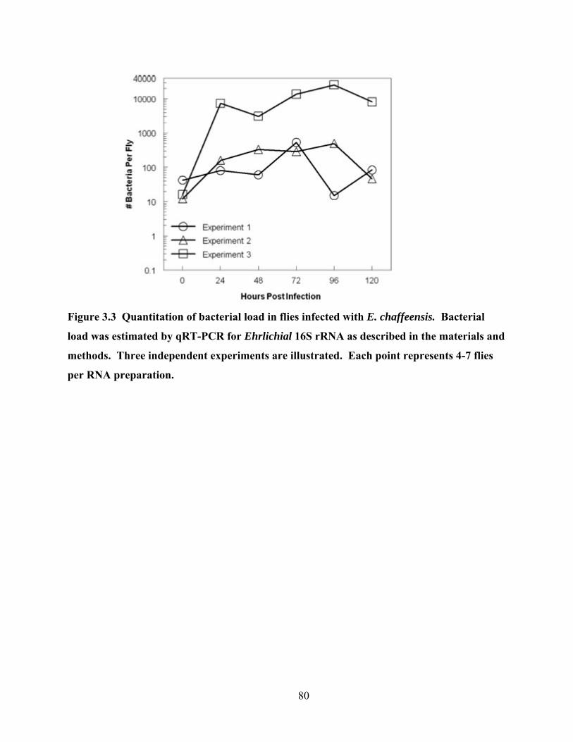
Figure 3.3 Quantitation of bacterial load in flies infected with E. chaffeensis. Bacterial
load was estimated by qRT-PCR for Ehrlichial 16S rRNA as described in the materials and
methods. Three independent experiments are illustrated. Each point represents 4-7 flies
per RNA preparation.
80
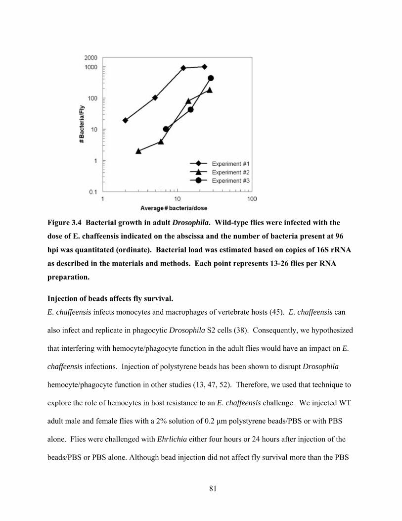
Figure 3.4 Bacterial growth in adult Drosophila. Wild-type flies were infected with the
dose of E. chaffeensis indicated on the abscissa and the number of bacteria present at 96
hpi was quantitated (ordinate). Bacterial load was estimated based on copies of 16S rRNA
as described in the materials and methods. Each point represents 13-26 flies per RNA
preparation.
Injection of beads affects fly survival.
E. chaffeensis infects monocytes and macrophages of vertebrate hosts (45). E. chaffeensis can
also infect and replicate in phagocytic Drosophila S2 cells (38). Consequently, we hypothesized
that interfering with hemocyte/phagocyte function in the adult flies would have an impact on E.
chaffeensis infections. Injection of polystyrene beads has been shown to disrupt Drosophila
hemocyte/phagocyte function in other studies (13, 47, 52). Therefore, we used that technique to
explore the role of hemocytes in host resistance to an E. chaffeensis challenge. We injected WT
adult male and female flies with a 2% solution of 0.2 μm polystyrene beads/PBS or with PBS
alone. Flies were challenged with Ehrlichia either four hours or 24 hours after injection of the
beads/PBS or PBS alone. Although bead injection did not affect fly survival more than the PBS
81

injection (Figure 3.5), the bead injection had an immediate impact on flies challenged with
Ehrlichia (P<0.05, log rank test). When flies were challenged four hours after bead injection,
60% of the flies died compared to 30% of the flies challenged with Ehrlichia alone, at 24 hours
post infection. The impact of the beads diminished over time. By 96 hours post infection, there
were no differences in survival (P>0.05, t-test) between bead-injected and non-bead-injected
flies that were challenged with Ehrlichia. Flies challenged with Ehrlichia 24 hours after bead
injection had a similar pattern compared to the flies that were challenged 4 hours after bead
injection (Figures 3.5A, B). In addition, we performed qRT-PCR to determine if bead injection
had an impact on the number of bacteria present in surviving flies. We observed an increase in
the number of bacteria present per fly in those flies that were injected with the beads prior to
Ehrlichia challenge (Figures 3.5C, D). For example, flies injected with beads four hours prior to
challenge had an average of 40 bacteria per surviving fly compared to 23 bacteria per control
flies at 24 hours after Ehrlichia challenge. The increased bacterial load was observed when the
beads were injected four or twenty-four hours before Ehrlichia challenge (Figure 3.5).
82
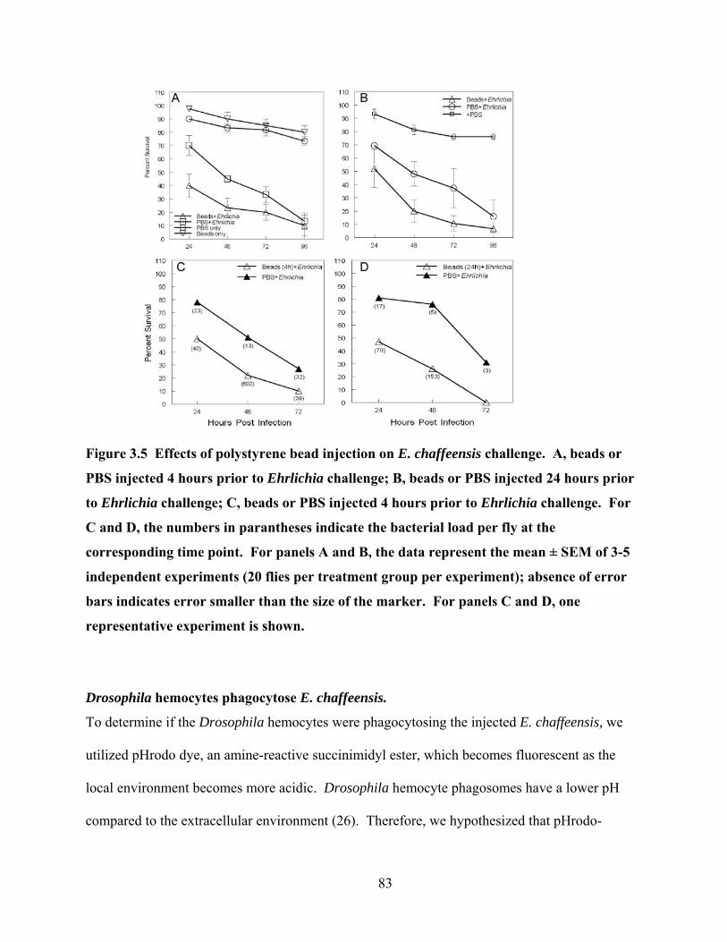
Figure 3.5 Effects of polystyrene bead injection on E. chaffeensis challenge. A, beads or
PBS injected 4 hours prior to Ehrlichia challenge; B, beads or PBS injected 24 hours prior
to Ehrlichia challenge; C, beads or PBS injected 4 hours prior to Ehrlichia challenge. For
C and D, the numbers in parantheses indicate the bacterial load per fly at the
corresponding time point. For panels A and B, the data represent the mean ± SEM of 3-5
independent experiments (20 flies per treatment group per experiment); absence of error
bars indicates error smaller than the size of the marker. For panels C and D, one
representative experiment is shown.
Drosophila hemocytes phagocytose E. chaffeensis.
To determine if the Drosophila hemocytes were phagocytosing the injected E. chaffeensis, we
utilized pHrodo dye, an amine-reactive succinimidyl ester, which becomes fluorescent as the
local environment becomes more acidic. Drosophila hemocyte phagosomes have a lower pH
compared to the extracellular environment (26). Therefore, we hypothesized that pHrodo-
83

labelled E. chaffeensis would fluoresce only if it was phagocytosed by hemocytes. Additionally,
we FITC-labeled E. chaffeensis to visualize bacterial trafficking and localization. FITC-labeled
bacteria were identified in 98% of the flies (3 experiments, 20 flies/experiment) examined 4
hours after injection and in 93% of the flies examined at 24 hours after injection. We observed
pHrodo-labeled bacteria in 82% of the flies at 4 hours and 88% of the flies at 24 hours after
injection. Therefore, it appeared that the lower pH environment of the hemocytes caused the
bacteria to fluoresce red. Hemocytes and extracellular bacteria were distinctly observed in the
central, dorsal abdomen at four and 24 hours post injection (Figure 3.6), and was the most
frequent location where hemocytes and bacteria were observed. We also observed dissemination
of the hemocytes and bacteria throughout the fly (Figure 3.6).
84
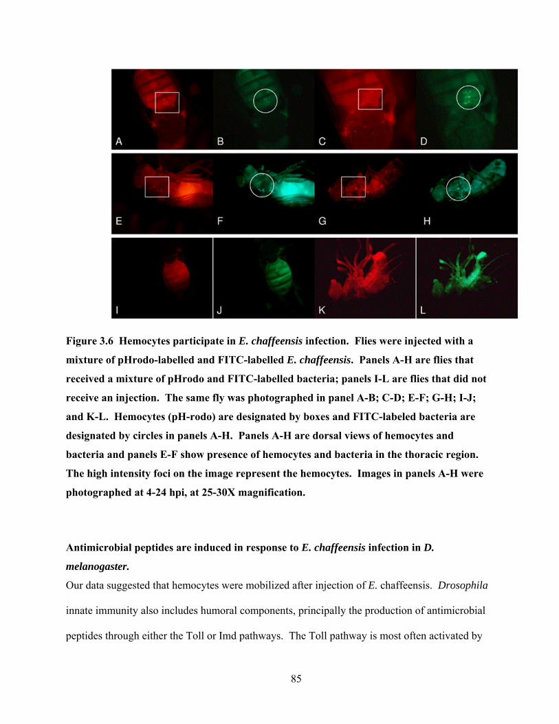
Figure 3.6 Hemocytes participate in E. chaffeensis infection. Flies were injected with a
mixture of pHrodo-labelled and FITC-labelled E. chaffeensis. Panels A-H are flies that
received a mixture of pHrodo and FITC-labelled bacteria; panels I-L are flies that did not
receive an injection. The same fly was photographed in panel A-B; C-D; E-F; G-H; I-J;
and K-L. Hemocytes (pH-rodo) are designated by boxes and FITC-labeled bacteria are
designated by circles in panels A-H. Panels A-H are dorsal views of hemocytes and
bacteria and panels E-F show presence of hemocytes and bacteria in the thoracic region.
The high intensity foci on the image represent the hemocytes. Images in panels A-H were
photographed at 4-24 hpi, at 25-30X magnification.
Antimicrobial peptides are induced in response to E. chaffeensis infection in D.
melanogaster.
Our data suggested that hemocytes were mobilized after injection of E. chaffeensis. Drosophila
innate immunity also includes humoral components, principally the production of antimicrobial
peptides through either the Toll or Imd pathways. The Toll pathway is most often activated by
85

Gram-positive bacteria and fungi, which elicits production of the antimicrobial peptide
Drosomycin (18, 31, 63). The Imd pathway is most often activated by Gram-negative bacteria
and is characterized by production of antimicrobial peptides such as Attacin and Diptericin (18,
31, 63). Although E. chaffeensis is Gram-negative, it lacks the genes for synthesis of both
lipopolysaccharide and peptidoglycan (37). Because E. chaffeensis has this atypical outer
membrane, we were particularly interested in determining if the host defense pathways (Imd
and/or Toll) were activated in response to E. chaffeensis challenge.
To address this question, we challenged transgenic flies expressing green fluorescent
protein (GPF) under the control of the attacin, diptericin, or drosomycin promoters (62) with E.
chaffeensis. After injection with bacteria or sterile PBS, GFP-expressing flies were examined
under a fluorescent, dissecting microscope at 24 hpi as described in the methods. There were
distinct differences in attacin-GFP and diptericin-GFP expression between the flies that
received bacteria compared to those that received PBS or no injection at all (Figure 3.7).
Fluorescence was also quantitated in the transgenic flies that were injected with Salmonella
enterica serovar typhimurium, as described in the methods. Salmonella is a Gram-negative
bacterium that is lethal to flies (47). No significant differences in antimicrobial peptide
induction were detected between flies that were challenged with Ehrlichia and Salmonella
(Figure 3.8). For the Ehrlichia-injected flies, the attacin-GFP flies had a significant increase
(P<0.05, n = 3 independent experiments) compared to control flies (injected with PBS only) at
both 6 and 24 hours post infection (Figure 3.8). For example, at 6 hours post infection, attacin-
GFP expression in the Ehrlichia-injected flies had a mean of 95,009 arbitrary fluorescent units
(AFU) compared to 54,894 AFU in the PBS-injected flies. Significant increases in GFP
expression were also observed in the diptericin-GFP flies that were injected with Ehrlichia
86
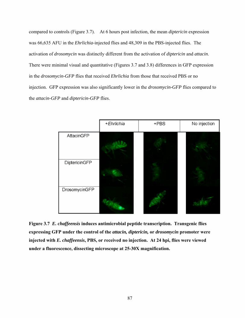
compared to controls (Figure 3.7). At 6 hours post infection, the mean diptericin expression
was 66,635 AFU in the Ehrlichia-injected flies and 48,309 in the PBS-injected flies. The
activation of drosomycin was distinctly different from the activation of diptericin and attacin.
There were minimal visual and quantitative (Figures 3.7 and 3.8) differences in GFP expression
in the drosomycin-GFP flies that received Ehrlichia from those that received PBS or no
injection. GFP expression was also significantly lower in the drosomycin-GFP flies compared to
the attacin-GFP and diptericin-GFP flies.
Figure 3.7 E. chaffeensis induces antimicrobial peptide transcription. Transgenic flies
expressing GFP under the control of the attacin, diptericin, or drosomycin promoter were
injected with E. chaffeensis, PBS, or received no injection. At 24 hpi, flies were viewed
under a fluorescence, dissecting microscope at 25-30X magnification.
87
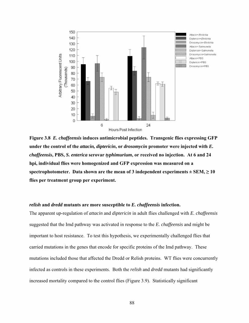
Figure 3.8 E. chaffeensis induces antimicrobial peptides. Transgenic flies expressing GFP
under the control of the attacin, diptericin, or drosomycin promoter were injected with E.
chaffeensis, PBS, S. enterica serovar typhimurium, or received no injection. At 6 and 24
hpi, individual flies were homogenized and GFP expression was measured on a
spectrophotometer. Data shown are the mean of 3 independent experiments ± SEM, ≥ 10
flies per treatment group per experiment.
relish and dredd mutants are more susceptible to E. chaffeensis infection.
The apparent up-regulation of attacin and diptericin in adult flies challenged with E. chaffeensis
suggested that the Imd pathway was activated in response to the E. chaffeensis and might be
important to host resistance. To test this hypothesis, we experimentally challenged flies that
carried mutations in the genes that encode for specific proteins of the Imd pathway. These
mutations included those that affected the Dredd or Relish proteins. WT flies were concurrently
infected as controls in these experiments. Both the relish and dredd mutants had significantly
increased mortality compared to the control flies (Figure 3.9). Statistically significant
88

differences in survival between relish mutants and wild-type flies were observed at 24, 48, 72, 96
and 120 hours post infection (P<0.05, t-test and log-rank test). For the dredd mutants,
significant differences in survival compared to the wildtype flies were confirmed at 24 and 96
hours post infections (t-test P<0.05); and significant difference in the survival curves was
confirmed by the log-rank test. We confirmed that the dredd and relish mutants maintained their
phenotypes. When we used qRT-PCR to measure the AMP transcript levels in the mutants, there
were decreased transcript levels for diptericin in the dredd and relish mutants (compared to wild-
type flies); drosomycin transcript levels were not altered in either of these mutants.
We also used qRT-PCR to measure the bacteriemia in the mutant flies after infection to
give us a second measure of pathogenesis. Infection was detected in all mutant flies, and was
higher in the relish mutants compared to the wild-type flies. These results suggested that Relish
was a key component of the Imd pathway necessary for Drosophila to combat E. chaffeensis
infections.
89

Figure 3.9 Survival of Imd pathway mutants after E. chaffeensis challenge. dredd and
relish mutants and wild-type flies were infected with E. chaffeensis and monitored for
survival for 120 hours. Data presented are the mean of 3 independent experiments ± SEM,
30 flies per treatment group per experiment. dredd and relish mutant fly survival was
significantly different from wild-type fly survival (P<0.05, log-rank test).
pelle, but not Toll and cactus mutants are more susceptible to E. chaffeensis infections.
The data from the drosomycin-GFP transgenic flies suggested that the Toll pathway was
activated to a lesser extent after Ehrlichia infection compared to attacin and diptercin. To
directly assess the role of the Toll defense pathway in host resistance to E. chaffeensis, we also
challenged adult, pll2, Tl3, cactD11 and cactD13 flies. Tl3 mutant flies have constitutively active
Toll and enhanced Drosomycin activity. pll2 flies are mutants lacking the Pelle kinase and have
an impaired ability to make Drosomycin. cactD11 and cactD13 flies are mutants lacking the Cactus
protein, which is a negative inhibitor of the Toll pathway. Therefore, these flies have enhanced
Drosomycin activity. We observed decreased mortality (P<0.05, log-rank test) in the Tl3 flies
90

compared to wild-type flies. (Figure 3.10). We observed no significant difference in death at any
time point in the cactus mutants (Figure 3.11). In contrast to the Toll and cactus mutants, the
pelle mutants displayed significantly decreased survival (P<0.05) compared to wildtype flies at
all time points (Figure 3.12). At 24 hpi, a mean of 75% of the pelle mutants were alive
compared to 92% of wild-type flies. By 120 hpi, a mean of 25% of pelle mutants survived
compared to 53% of wild-type flies.
Figure 3.10 The Toll-constitutive mutant flies are less susceptible to E. chaffeensis
infections. Mutants for the Toll protein and wild-type flies were infected with E.
chaffeensis and monitored for survival for 120 hours. Numbers represent mean ± SEM of
≥ 3 independent experiments, 20 flies per experiment. Survival of wild-type flies was
significantly different from Toll mutants (P<0.05, log-rank test).
91

Figure 3.11 Effect of E. chaffeensis infection on cactD11 and cactD13 mutants. cactus mutants
and wild-type flies were infected with E. chaffeensis and monitored for survival for 120
hours. Data represents the mean of 3 independent experiments ± SEM, 20 flies per
treatment group per experiment. Absence of error bars indicates error smaller than the
size of the marker.
92
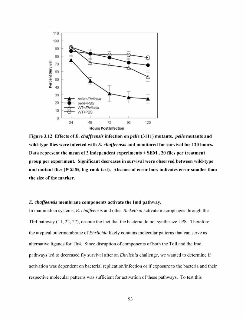
Figure 3.12 Effects of E. chaffeensis infection on pelle (3111) mutants. pelle mutants and
wild-type flies were infected with E. chaffeensis and monitored for survival for 120 hours.
Data represent the mean of 3 independent experiments ± SEM , 20 flies per treatment
group per experiment. Significant decreases in survival were observed between wild-type
and mutant flies (P<0.05, log-rank test). Absence of error bars indicates error smaller than
the size of the marker.
E. chaffeensis membrane components activate the Imd pathway.
In mammalian systems, E. chaffeensis and other Rickettsia activate macrophages through the
Tlr4 pathway (11, 22, 27), despite the fact that the bacteria do not synthesize LPS. Therefore,
the atypical outermembrane of Ehrlichia likely contains molecular patterns that can serve as
alternative ligands for Tlr4. Since disruption of components of both the Toll and the Imd
pathways led to decreased fly survival after an Ehrlichia challenge, we wanted to determine if
activation was dependent on bacterial replication/infection or if exposure to the bacteria and their
respective molecular patterns was sufficient for activation of these pathways. To test this
93
hypothesis, we used the AMP-GFP reporters used in the fly experiments above as an in vitro
readout system using Drosophila S2 cells as outlined in the methods. Results from a microarray
analysis (data not shown) revealed that several antimicrobial peptide genes were up-regulated in
S2 cells after infection with E. chaffeensis. attacin, diptericin, and drosomycin were among
those genes. Therefore, we anticipated that the up-regulation of GFP in S2 cells would occur in
response to the appropriate signals either delivered by exposure to bacteria alone, or by infection
by live bacteria. attacin-GFP and diptericin-GFP expression were both up-regulated in the
transfected S2 cells in response to E. chaffeensis infection by viable bacteria (Figure 3.13).
drosomycin was also up-regulated in response to infection (Figure 3.13). Interestingly, we also
observed induction of the antimicrobial peptides when heat-killed E. chaffeensis was added to
the cells (Figure 3.13). However, the intensity of the activation was lower compared to viable
bacteria. These results suggest that some component of the Ehrlichia membrane is activating the
AMP pathways and that replication is able to augment that response.
94

Figure 3.13 Antimicrobial peptides are activated in response to E. chaffeensis in S2 cells.
S2 cells were transfected with attacin, diptericin, or drosomycin promoter-GFP constructs.
The S2 cells were dosed with live or boiled E. chaffeensis, or with sterile S2 medium
(negative). GFP output was measured on a spectrophotometer. Numbers represent the
mean of two independent experiments.
DISCUSSION E. chaffeensis can infect and complete its replication cycle in adult D. melanogaster.
This is the first demonstration that Ehrlichia chaffeensis is able to replicate in an arthropod other
than the tick. We previously demonstrated that that Ehrlichia could grow in Drosophila S2 cells
(38). Therefore, establishing infections at the organismic level represents an important step
forward in our ability to work in an alternative arthropod system. We concede that the mode of
Drosophila infection was not similar to the natural route of infection in a tick, which acquires the
infection by a blood meal. However, the ability to grow the bacteria in the fly opens the
95

biological and genetic tool box of Drosophila melanogaster to address questions about Ehrlichia
that cannot be addressed in the tick.
E. chaffeensis infection was pathogenic to adult flies. It is not completely clear how this
outcome differs when bacteria infects ticks. Little is known about tick survival after Rickettsia
infection. Our results are consistent with the observation of a decrease in larval molting and
overall survival among Ixodes scapularis ticks when allowed to feed on mice infected with
various isolates of Anaplasma phagocytophilum (50). Increased death during molting to adult
stage or before feeding of Dermacentor andersoni larvae and nymphs after experimental
infection with Rickettsia rickettsii has also been reported (44). In the same study, fewer larvae
developed from infected ticks. Decreases in molting success and increased death were also
observed in a study in which Rhipicephalus sanguineus ticks were experimentally infected with
Rickettsia conorii (36).
Although we observed a dose-dependent survival when flies were injected at any one
particular time (Figure 3.2), there was some experiment-to-experiment variability in the number
of bacteria that caused death in the challenged flies. This variability probably resulted from a
combination of factors including differences in bacterial preparations (ratio of dense core to
reticulate forms (67)), variability in the custom made glass needles used to inject the bacteria, the
ages and sexes of the flies (14, 19, 46), and the skill of the individual doing the injections.
Therefore, the experiments we presented were always reported as the outcome of multiple
independent experiments. Controls and direct comparisons were done at the same time.
Hemocytes appear to participate in the Drosophila cellular immune response to E.
chaffeensis. This is supported by three different experiments. There was significantly decreased
survival when the flies received an injection of polystyrene beads prior to bacterial challenge.
96
We also detected phagocytosis of the Ehrlichia using the pHrodo dye and an increase in the
number of bacteria in flies receiving the beads compared to the bead-free controls. It is possible
that the beads inhibited hemocyte phagocytosis, rendering the fly less capable of controlling the
initial infection of the bacteria. This hypothesis is supported by previous studies where
polystyrene beads inhibited phagocytosis of Streptococcus pneumoniae, Plasmodium
gallinaceum, Mycobacterium marinum, and Escherichia coli (13, 16, 47, 52). Alternatively, the
beads may inhibit Ehrlichia-specific receptors on the hemocytes. This could lead to an
overgrowth of bacteria in the haemocoel, resulting in tissue damage that may be fatal.
Our attempts to isolate hemocytes from infected or uninfected adult flies were
unsuccessful, even after using several different techniques that were successful in other dipterans
or Drosophila (8, 29). Therefore we visualized hemocyte location within the fly with the
pHrodo dye. Since not all the FITC-labelled bacteria were localized to hemocytes (identified
with pHrodo-labelled bacteria), it appears that some of the bacteria (~25%) were not
phagocytosed (extracellular bacteria). We observed an abundance of hemocytes as well as
extracellular bacteria as early as one hour after and as late as 120 hours after injection of
bacteria. Hemocytes and extracellular bacteria were most often observed at the dorsal midline at
the anterior end of the abdomen of the fly. There were also hemocytes and free bacteria
distributed throughout the body of the fly. We suspect that Ehrlichia are capable of growing in
hemocytes since bacteria can grow in S2 cells (38). However, we have yet to determine whether
the pathogenesis we see is due to bacteria growing inside the hemocyte, extracellularly, or by
infecting other tissues. It has been reported in Ixodid ticks that Rickettsiae infect hemocytes and
thus disseminate to all tissues and organs and can survive in the tissues and the body cavity for
long periods of time (56). Additional experiments will be needed to determine if this dynamic is
97

also the case in Drosophila and were beyond the goals of this investigation. However, based on
the survival kinetics after bead injections, it does appear that the early plasmatocyte/hemocyte
response does work to control the infection.
Ehrlichia infection in flies appears to activate both the Toll and Imd host defense
pathways. This conclusion is supported by several different experiments. For the Toll pathway,
we saw increased survival in the Tl3 flies, and decreased survival in the pelle mutant flies. The
Tl3 flies were created with the mutagen ethyl methanesulfonate and have a mutation that renders
Toll constitutively active (1, 32, 33, 49, 53). The pelle mutant flies were created with the
mutagen ethyl methanesulfonate and have a mutation that ablates that function of the pelle kinase
(54), which is believed to play a role in the degradation of Cactus (31, 61). The continuous
expression of drosomycin in the Tl3 flies and the loss of drosomycin in the pelle-mutant flies
would support some role for this pathway after Ehrlichia challenge. Spaetzle is the ligand for
Toll and the interaction initiates Toll signaling (24, 33, 65), followed by interaction of
MyD88/Pelle/Tube, which causes the degradation of Cactus (4, 59). Cactus is an inhibitor of the
Toll pathway (43). Therefore, the loss of function Cactus mutants have a constitutively active
Toll pathway (5, 51). Toll pathway involvement was also supported by activation of the
drosomycin promoter when S2 cells were infected in vitro. It is not clear why activation of
drosomycin was lower than that of attacin and diptericin in vivo. drosomycin expression has
been shown to be up-regulated later than diptericin (6, 34). Therefore, it is possible that our data
reflect those kinetics.
The Imd pathway also appears to be activated after an Ehrlichia challenge. We observed
activation of the promoters of the Imd-dependent antimicrobial peptides attacin and diptericin
both in vivo and in vitro, and increased transcript levels of diptericin in wild-type flies after
98

infection (data not shown). We also observed significantly diminished survival of relish and
dredd mutant flies after experimental challenge. Relish is a key component of the Imd pathway,
which is activated by Gram-negative bacteria (31, 48, 64). It is a homologous component to the
p100 and p105 precursors in the NF-κB family of transcription factors and is essential for the
transcription of antimicrobial peptides in D. melanogaster (57). The activation of the Imd
pathway by the obligate intracellular pathogen E. chaffeensis is similar to observations made
with another Gram-negative, intracellular bacterium, Francisella tularensis (64). We also
observed a significant decrease in the survival of dredd mutants. Dredd is the upstream caspase
activator of Relish (17) (39) and it has been previously demonstrated that dredd mutants are
more susceptible to Gram-negative bacterial infections (35). Therefore, several different
observations demonstrate that Imd plays a role in the host defense of E. chaffeensis.
Our data show that bacteria are growing in adult flies and the oscillatory nature of the
bacterial load over time suggest that there is an active host response against the bacteria just as
there is in mice (21). Interestingly, the activation of AMPs did not appear to require bacterial
replication. Exposure to boiled E. chaffeensis (dead) was sufficient for activation of AMPs in
the S2 cell reporter system. The activation of AMP production is consistent with the control of
these innate responses by pattern recognition receptors after they engage their ligands (31). In
flies, the Toll and Imd pathways are activated by different forms of peptidoglycan, but not by
LPS (31). In rodents, E chaffeensis and other Rickettsia appear to engage Toll-like receptors
even in the absence of LPS (22, 27, 60). Therefore, the Ehrlichia outer membrane components
responsible for activation of antimicrobial peptides may be lipoproteins (25). The isolation of
several immunogenic lipoproteins from the outer membrane of E. chaffeensis would support this
hypothesis (25). Moreover, the activation of both the Imd and Toll pathways would be
99

consistent with the atypical outer membrane of E. chaffeensis compared to the unique activation
of Imd by classical, pyogenic Gram-negative, LPS-containing bacteria (12, 23, 30).
In conclusion, we have demonstrated that E. chaffeensis is able to grow and replicate in
adult D. melanogaster. Infection induces innate cellular and humoral responses in the fly that
contribute to host resistance. These findings are significant because the Drosophila system will
allow us to dissect the role of host genes in bacterial replication and elucidate which bacterial
components contribute to the generation of innate resistance in arthropods.
100
REFERENCES 1. Anderson, K. V., G. Jurgens, and C. Nusslein-Volhard. 1985. Establishment of dorsal-
ventral polarity in the Drosophila embryo: genetic studies on the role of the Toll gene product. Cell 42:779-89.
2. Anderson, K. V., and C. Nusslein-Volhard. 1984. Information for the dorsal-ventral pattern of the Drosophila embryo is stored as maternal mRNA. Nature 311:223-227.
3. Basset, A., R. S. Khush, A. Braun, L. Gardan, F. Boccard, J. A. Hoffmann, and B. Lemaitre. 2000. The phytopathogenic bacteria Erwinia carotovora infects Drosophila and activates an immune response. Proc Natl Acad Sci U S A 97:3376-81.
4. Belvin, M. P., and K. V. Anderson. 1996. A conserved signaling pathway: the Drosophila toll-dorsal pathway. Annu Rev Cell Dev Biol 12:393-416.
5. Bergmann, A., D. Stein, R. Geisler, S. Hagenmaier, B. Schmid, N. Fernandez, B. Schnell, and C. Nusslein-Volhard. 1996. A gradient of cytoplasmic Cactus degradation establishes the nuclear localization gradient of the dorsal morphogen in Drosophila. Mech Dev 60:109-23.
6. Boutros, M., H. Agaisse, and N. Perrimon. 2002. Sequential activation of signaling pathways during innate immune responses in Drosophila. Dev Cell 3:711-22.
7. Brandt, S. M., M. S. Dionne, R. S. Khush, L. N. Pham, T. J. Vigdal, and D. S. Schneider. 2004. Secreted Bacterial Effectors and Host-Produced Eiger/TNF Drive Death in aSalmonella-Infected Fruit Fly. PLoS Biol 2:e418.
8. Castillo, J. C., A. E. Robertson, and M. R. Strand. 2006. Characterization of hemocytes from the mosquitoes Anopheles gambiae and Aedes aegypti. Insect Biochem Mol Biol 36:891-903.
9. Chapes, S. K., and A. A. Beharka. 1998. Salmonella infections in the absence of the major histocompatibility complex II. J Leukoc Biol 63:297-304.
10. Chapes, S. K., and S. Haskill. 1982. Role of Corynebacterium parvum in the activation of peritoneal macrophages. 1. Association between intracellular C. parvum and cytotoxic macrophages. Cell Immunol 70:65-75.
11. Damas, J. K., M. Jensenius, T. Ueland, K. Otterdal, A. Yndestad, S. S. Froland, J. M. Rolain, B. Myrvang, D. Raoult, and P. Aukrust. 2006. Increased levels of soluble CD40L in African tick bite fever: possible involvement of TLRs in the pathogenic interaction between Rickettsia africae, endothelial cells, and platelets. J Immunol 177:2699-706.
12. De Gregorio, E., P. T. Spellman, P. Tzou, G. M. Rubin, and B. Lemaitre. 2002. The Toll and Imd pathways are the major regulators of the immune response in Drosophila. Embo J 21:2568-79.
13. Dionne, M. S., N. Ghori, and D. S. Schneider. 2003. Drosophila melanogaster is a genetically tractable model host for Mycobacterium marinum. Infect Immun 71:3540-50.
14. Domanitskaya, E. V., H. Liu, S. Chen, and E. Kubli. 2007. The hydroxyproline motif of male sex peptide elicits the innate immune response in Drosophila females. Febs J 274:5659-68.
15. Eicher, S. D., and D. S. McVey. 1995. Dietary modulation of Kupffer cell and splenocyte function during a Salmonella typhimurium challenge in mice. J Leukoc Biol 58:32-9.
16. Elrod-Erickson, M., S. Mishra, and D. Schneider. 2000. Interactions between the cellular and humoral immune responses in Drosophila. Curr Biol 10:781-4.
101
17. Erturk-Hasdemir, D., M. Broemer, F. Leulier, W. S. Lane, N. Paquette, D. Hwang, C. H. Kim, S. Stoven, P. Meier, and N. Silverman. 2009. Two roles for the Drosophila IKK complex in the activation of Relish and the induction of antimicrobial peptide genes. Proc Natl Acad Sci U S A 106:9779-84.
18. Erturk-Hasdemir, D., N. Paquette, K. Aggarwal, and N. Silverman. 2008. Bug Versus Bug: Humoral Immune Responses in Drosophila melanogaster, p. 43-63. In H. Heine (ed.), Innate Immunity of Plants, Animal, and Humans. Springer-Verlag, Heidelberg.
19. Fedorka, K. M., J. E. Linder, W. Winterhalter, and D. Promislow. 2007. Post-mating disparity between potential and realized immune response in Drosophila melanogaster. Proc Biol Sci 274:1211-7.
20. Felek, S., A. Unver, R. W. Stich, and Y. Rikihisa. 2001. Sensitive detection of Ehrlichia chaffeensis in cell culture, blood, and tick specimens by reverse transcription-PCR. J Clin Microbiol 39:460-3.
21. Ganta, R. R., C. Cheng, E. C. Miller, B. L. McGuire, L. Peddireddi, K. R. Sirigireddy, and S. K. Chapes. 2007. Differential clearance and immune responses to tick cell-derived versus macrophage culture-derived Ehrlichia chaffeensis in mice. Infect Immun 75:135-45.
22. Ganta, R. R., M. J. Wilkerson, C. Cheng, A. M. Rokey, and S. K. Chapes. 2002. Persistent Ehrlichia chaffeensis infection occurs in the absence of functional major histocompatibility complex class II genes. Infect Immun 70:380-8.
23. Hedengren-Olcott, M., M. C. Olcott, D. T. Mooney, S. Ekengren, B. L. Geller, and B. J. Taylor. 2004. Differential activation of the NF-kappaB-like factors Relish and Dif in Drosophila melanogaster by fungi and Gram-positive bacteria. J Biol Chem 279:21121-7.
24. Hu, X., Y. Yagi, T. Tanji, S. Zhou, and Y. T. Ip. 2004. Multimerization and interaction of Toll and Spatzle in Drosophila. Proc Natl Acad Sci U S A 101:9369-74.
25. Huang, H., M. Lin, X. Wang, T. Kikuchi, H. Mottaz, A. Norbeck, and Y. Rikihisa. 2008. Proteomic analysis of and immune responses to Ehrlichia chaffeensis lipoproteins. Infect Immun 76:3405-14.
26. Ivory, A., K. Randle, and L. Wu. 2008. Chapter 4 - Cellular Immune Responses in Drosophila melanogaster, p. 73-91. In H. Heine (ed.), Innate Immunity of Plants, Animals, and Humans, 21 ed. Springer-Verlag Berlin.
27. Jordan, J. M., M. E. Woods, J. Olano, and D. H. Walker. 2008. The absence of Toll-like receptor 4 signaling in C3H/HeJ mice predisposes them to overwhelming rickettsial infection and decreased protective Th1 responses. Infect Immun 76:3717-24.
28. Kavanagh, K., and E. Reeves. 2007. Insect and mammalian innate immune responses are much alike. Microbe 2:596-599.
29. Kurucz, E., B. Vaczi, R. Markus, B. Laurinyecz, P. Vilmos, J. Zsamboki, K. Csorba, E. Gateff, D. Hultmark, and I. Ando. 2007. Definition of Drosophila hemocyte subsets by cell-type specific antigens. Acta Biol Hung 58 Suppl:95-111.
30. Lau, G. W., B. C. Goumnerov, C. L. Walendziewicz, J. Hewitson, W. Xiao, S. Mahajan-Miklos, R. G. Tompkins, L. A. Perkins, and L. G. Rahme. 2003. The Drosophila melanogaster toll pathway participates in resistance to infection by the gram-negative human pathogen Pseudomonas aeruginosa. Infect Immun 71:4059-66.
102

31. Lemaitre, B., and J. Hoffmann. 2007. The host defense of Drosophila melanogaster. Annu Rev Immunol 25:697-743.
32. Lemaitre, B., M. Meister, S. Govind, P. Georgel, R. Steward, J. M. Reichhart, and J. A. Hoffmann. 1995. Functional analysis and regulation of nuclear import of dorsal during the immune response in Drosophila. Embo J 14:536-45.
33. Lemaitre, B., E. Nicolas, L. Michaut, J. M. Reichhart, and J. A. Hoffmann. 1996. The dorsoventral regulatory gene cassette spatzle/Toll/cactus controls the potent antifungal response in Drosophila adults. Cell 86:973-83.
34. Lemaitre, B., J. M. Reichhart, and J. A. Hoffmann. 1997. Drosophila host defense: differential induction of antimicrobial peptide genes after infection by various classes of microorganisms. Proc Natl Acad Sci U S A 94:14614-9.
35. Leulier, F., A. Rodriguez, R. S. Khush, J. M. Abrams, and B. Lemaitre. 2000. The Drosophila caspase Dredd is required to resist gram-negative bacterial infection. EMBO Rep 1:353-8.
36. Levin, M. L., L. Killmaster, G. Zemtsova, D. Grant, K. Y. Mumcuoglu, M. E. Eremeeva, and G. A. Dasch. 2009. Incongruent effects of two isolates of Rickettsia conorii on the survival of Rhipicephalus sanguineus ticks. Exp Appl Acarol.
37. Lin, M., and Y. Rikihisa. 2003. Ehrlichia chaffeensis and Anaplasma phagocytophilum lack genes for lipid A biosynthesis and incorporate cholesterol for their survival. Infect Immun 71:5324-31.
38. Luce-Fedrow, A., T. Von Ohlen, D. Boyle, R. R. Ganta, and S. K. Chapes. 2008. Use of Drosophila S2 cells as a model for studying Ehrlichia chaffeensis infections. Appl Environ Microbiol 74:1886-91.
39. Maillet, F., V. Bischoff, C. Vignal, J. Hoffmann, and J. Royet. 2008. The Drosophila peptidoglycan recognition protein PGRP-LF blocks PGRP-LC and IMD/JNK pathway activation. Cell Host Microbe 3:293-303.
40. Mansfield, B. E., M. S. Dionne, D. S. Schneider, and N. E. Freitag. 2003. Exploration of host-pathogen interactions using Listeria monocytogenes and Drosophila melanogaster. Cell Microbiol 5:901-11.
41. McNabb, S. J., R. A. Jajosky, P. A. Hall-Baker, D. A. Adams, P. Sharp, C. Worshams, W. J. Anderson, A. J. Javier, G. J. Jones, D. A. Nitschke, A. Rey, and M. S. Wodajo. 2008. Summary of notifiable diseases--United States, 2006. MMWR Morb Mortal Wkly Rep 55:1-92.
42. Nehme, N. T., S. Liégeois, B. Kele, P. Giammarinaro, E. Pradel, J. A. Hoffmann, J. J. Ewbank, and D. Ferrandon. 2007. A Model of Bacterial Intestinal Infections in Drosophila melanogaster. PLoS Pathogens 2:1694-1709.
43. Nicolas, E., J. M. Reichhart, J. A. Hoffmann, and B. Lemaitre. 1998. In vivo regulation of the IkappaB homologue cactus during the immune response of Drosophila. J Biol Chem 273:10463-9.
44. Niebylski, M. L., M. G. Peacock, and T. G. Schwan. 1999. Lethal effect of Rickettsia rickettsii on its tick vector (Dermacentor andersoni). Appl Environ Microbiol 65:773-8.
45. Paddock, C. D., and J. E. Childs. 2003. Ehrlichia chaffeensis: a prototypical emerging pathogen. Clin Microbiol Rev 16:37-64.
46. Peng, J., P. Zipperlen, and E. Kubli. 2005. Drosophila sex-peptide stimulates female innate immune system after mating via the Toll and Imd pathways. Curr Biol 15:1690-4.
103
47. Pham, L. N., M. S. Dionne, M. Shirasu-Hiza, and D. S. Schneider. 2007. A specific primed immune response in Drosophila is dependent on phagocytes. PLoS Pathog 3:e26.
48. Pili-Floury, S., F. Leulier, K. Takahashi, K. Saigo, E. Samain, R. Ueda, and B. Lemaitre. 2004. In vivo RNA interference analysis reveals an unexpected role for GNBP1 in the defense against Gram-positive bacterial infection in Drosophila adults. J Biol Chem 279:12848-53.
49. Ray, R. P., K. Arora, C. Nusslein-Volhard, and W. M. Gelbart. 1991. The control of cell fate along the dorsal-ventral axis of the Drosophila embryo. Development 113:35-54.
50. Ross, D. E., and M. L. Levin. 2004. Effects of Anaplasma phagocytophilum infection on the molting success of Ixodes scapularis (Acari: Ixodidae) larvae. J Med Entomol 41:476-83.
51. Roth, S., Y. Hiromi, D. Godt, and C. Nusslein-Volhard. 1991. Cactus, a maternal gene required for proper formation of the dorsoventralmorphogen gradient in Drosophila embryos. Development 112:371-388.
52. Schneider, D., and M. Shahabuddin. 2000. Malaria parasite development in a Drosophila model. Science 288:2376-9.
53. Schneider, D. S., K. L. Hudson, T. Y. Lin, and K. V. Anderson. 1991. Dominant and recessive mutations define functional domains of Toll, a transmembrane protein required for dorsal-ventral polarity in the Drosophila embryo. Genes Dev 5:797-807.
54. Shelton, C. A., and S. A. Wasserman. 1993. pelle encodes a protein kinase required to establish dorsoventral polarity in the Drosophila embryo. Cell 72:515-25.
55. Sirigireddy, K. R., and R. R. Ganta. 2005. Multiplex detection of Ehrlichia and Anaplasma species pathogens in peripheral blood by real-time reverse transcriptase-polymerase chain reaction. J Mol Diagn 7:308-16.
56. Socolovschi, C., O. Mediannikov, D. Raoult, and P. Parola. 2009. The relationship between spotted fever group Rickettsiae and Ixodid ticks. Vet Res 40:34.
57. Stoven, S., N. Silverman, A. Junell, M. Hedengren-Olcott, D. Erturk, Y. Engstrom, T. Maniatis, and D. Hultmark. 2003. Caspase-mediated processing of the Drosophila NF-kappaB factor Relish. Proc Natl Acad Sci U S A 100:5991-6.
58. Tanji, T., X. Hu, A. N. Weber, and Y. T. Ip. 2007. Toll and IMD pathways synergistically activate an innate immune response in Drosophila melanogaster. Mol Cell Biol 27:4578-88.
59. Tauszig-Delamasure, S., H. Bilak, M. Capovilla, J. A. Hoffmann, and J. L. Imler. 2002. Drosophila MyD88 is required for the response to fungal and Gram-positive bacterial infections. Nat Immunol 3:91-7.
60. Telford, S. R., 3rd, and J. E. Dawson. 1996. Persistent infection of C3H/HeJ mice by Ehrlichia chaffeensis. Vet Microbiol 52:103-12.
61. Towb, P., A. Bergmann, and S. A. Wasserman. 2001. The protein kinase Pelle mediates feedback regulation in the Drosophila Toll signaling pathway. Development 128:4729-4736.
62. Tzou, P., S. Ohresser, D. Ferrandon, M. Capovilla, J. M. Reichhart, B. Lemaitre, J. A. Hoffmann, and J. L. Imler. 2000. Tissue-specific inducible expression of antimicrobial peptide genes in Drosophila surface epithelia. Immunity 13:737-48.
63. Vodovar, N., C. Acosta, B. Lemaitre, and F. Boccard. 2004. Drosophila: a polyvalent model to decipher host-pathogen interactions. Trends Microbiol 12:235-42.
104

64. Vonkavaara, M., M. V. Telepnev, P. Rydén, A. Sjöstedt, and S. Stöven. 2008. Drosophila melanogaster as a model for elucidating the pathogenicity of Francisella tularensis. Cellular Microbiology 10:1327-1338.
65. Weber, A. N., S. Tauszig-Delamasure, J. A. Hoffmann, E. Lelievre, H. Gascan, K. P. Ray, M. A. Morse, J. L. Imler, and N. J. Gay. 2003. Binding of the Drosophila cytokine Spatzle to Toll is direct and establishes signaling. Nat Immunol 4:794-800.
66. Williams, M. J., M. L. Wiklund, S. Wikman, and D. Hultmark. 2006. Rac1 signalling in the Drosophila larval cellular immune response. J Cell Sci 119:2015-24.
67. Zhang, J. Z., M. Sinha, B. A. Luxon, and X. J. Yu. 2004. Survival strategy of obligately intracellular Ehrlichia chaffeensis: novel modulation of immune response and host cell cycles. Infect Immun 72:498-507.
105
CHAPTER 4 - Identification of host genes that contribute to
Ehrlichia chaffeensis infections in Drosophila
melanogaster
ABSTRACT Ehrlichia chaffeensis is an obligate, intracellular bacterium that causes human monocytic
ehrlichiosis (HME) and the bacteria are vectored by the Lone Star tick (Amblyomma
americanum). Ehrlichia grows in several mammalian cell lines, but NO consensus model of
pathogenesis exists for arthropods or vertebrates and the host genes required for intracellular
growth of the bacteria are unknown. We performed microarrays on S2 cells, comparing host
gene expression between permissive or non-permissive conditions for E. chaffeensis growth. A
total of 517 genes were identified that had increased transcript levels only under permissive
growth conditions at 24 hours post infection. We screened adult flies which were mutant for
some of the permissive genes identified in our microarrays for their ability to support Ehrlichia
replication. Five mutant stocks were resistant to infection with Ehrlichia (genes CG6479,
separation anxiety, CG3044 (Cht11), CG6364, and CG6543). qRT-PCR confirmed that the
bacterial load was decreased in these mutant flies compared to wildtype controls. Gene CG6364
is described as potentially having uridine kinase activity and the in vivo mutation of this gene is
predicted to disrupt the nucleotide salvage pathway, which caused a decrease in bacterial
replication in vivo. Disruption of cytidine triphosphate synthesis with cyclopentenyl cytosine
(CPEC), a CTP synthetase inhibitor, demonstrated that the de novo synthesis of CTP from
glutamine was unnecessary. S2 cells treated with CPEC and infected with Ehrlichia had higher
bacterial loads than untreated controls. Furthermore, addition of cytosine to S2 cells also
106

resulted in increased bacterial replication. Therefore the nucleotide salvage pathway via cytidine
may be essential for bacterial replication. We also investigated what role CG3044 (Cht11) had
in the replication of Ehrlichia. We hypothesized that it may be involved in cholesterol regulation
or have a regulatory role in cellular activation. Our approach has successfully identified host
genes that contribute to E. chaffeensis replication in Drosophila.
INTRODUCTION Drosophila melanogaster has been used to study a variety of intracellular pathogens and
has been successfully manipulated for successful identification of the genes involved in host-
pathogen interactions in several in vitro and in vivo studies of pathogens. Some of these studies
have focused on Drosophila interactions with Listeria monocytogenes (1, 2), Chlamydia
trachomatis (10), Mycobacterium marinum (1, 9, 30, 43), Francisella tularensis (45, 54), and the
protozoan parasite Plasmodium gallinaceum (5, 47).
We previously demonstrated that the obligate, intracellular bacterium Ehrlichia
chaffeensis is capable of infecting, completing its lifecycle, and maintaining its pathogenicity in
both Drosophila S2 cells and in adult flies (34, 35). E. chaffeensis is the causative agent of
human monocytic ehrlichiosis, of which 828 cases of were reported for 2007 (25). This
represents a 44% increase in over those reported for 2006, and an overall 159% increase since
2003 (25). In addition to being reported in the United States, the occurrence of E. chaffeensis
has also been documented in Africa, Europe, China, and Brazil (7, 8, 36, 56). Therefore,
establishing a system to determine the host genes that contribute to the replication and
pathogenicity of E. chaffeensis is important. To date, no such model system is yet available.
We previously identified growth conditions that were “non-permissive” for the growth of
E. chaffeensis infection in Drosophila S2 cells. Incubating S2 cells with lipopolysaccharide
107

(LPS) effectively stopped bacterial replication (34). We hypothesized that microarray analysis
of permissively-infected and non-permissively-infected S2 cells would reveal host genes that
contribute to the replication of Ehrlichia. We used the Affymetrix Drosophila 2.0 array to
identify a subset of genes that were exclusively expressed during Ehrlichia-permissive
conditions in the infected S2 cells. A search of the Drosophila Flybase (www.flybase.org) for
viable, fertile adult flies that contained mutations in genes from the narrowed data set revealed
over 100 possible targets for investigation. In this paper we describe our initial screening
outcomes. In particular, we screened a total of 15 genetic mutants and identified five in which
survival was not impacted by Ehrlichia challenge and bacterial replication in the fly was
hindered. We describe those genes and some follow up experiments that attempt to elucidate
how these genes contribute to replication. To our knowledge this is the first description of host
genes essential for replication of E. chaffeensis.
MATERIALS & METHODS
Maintenance of cell lines and Ehrlichia chaffeensis infections.
The canine macrophage cell line, DH82, was maintained at 37°C in Dulbeccos Modified Eagle’s
Medium with 3.5% fetal bovine serum and 3.5% Nu Serum (DMEM-7). The E. chaffeensis
Arkansas isolate was continuously cultivated in the DH82 cell line at 37°C, 8% CO2 in DMEM-7
medium. Bacteria were passaged when infectivity reached 80-90% as visualized using cytospin
prepared slides (stained with Hema3 fixative and Dif-Quik stain) to monitor formation of
morulae in the cells. Infected cells were removed by scraping each plate with a cell scraper,
transferring the culture to a conical tube, and vortexing the suspension with glass beads. The
freed bacteria were purified by centrifuging the suspension at 600 x g for twenty minutes to
108
remove cell debris. The bacteria-containing supernatant was removed, transferred to a sterile
conical tube, and was centrifuged at 15,000 x g for twenty minutes to pellet the free bacteria. The
final supernatant was removed and the pellet was re-suspended in an appropriate amount of
sterile PBS. Purified bacteria were used to re-infect DH82 and S2 cells. Drosophila S2 cells
were cultivated at 28°C in Schneider’s Drosophila medium (Gibco, Grand Island, NY, #11720)
supplemented with 10% fetal bovine serum (Atlanta Biologicals, #S11150).
Incubation of S2 cells with Lipopolysaccharide (LPS).
To determine if LPS incubation would inhibit the growth of E. chaffeensis, S2 cells were plated
at a concentration of 1 x 10⁶ cells per plate in 6-well plates and were allowed to adhere for at
least 30 minutes. LPS (from Salmonella minnesota; Sigma #L-6261) was sonicated for one hour
and then added to each well at a concentration of 10 μg per ml. The cells plus LPS were
incubated for five hours. S2 cells were then infected with E. chaffeensis purified from DH82
cells.
Infections in Permissive and Non-Permissive Drosophila S2 cells.
Drosophila S2 cells were plated in 6-well tissue culture plates (60-mm, Techno Plastic Products
AG, #92406) at a concentration of 1x10⁶ cells per ml. The cells were allowed to adhere for at
least 30 minutes but no longer than 24 hours. To create non-permissive samples, cells were
treated with LPS as described above. Bacteria purified from infected DH82 (85-100% of the
cells infected) cells were added to S2 cell cultures and then were monitored for infection at 24
and 96 hours post-infection (hpi). Uninfected S2 cells and S2 cells activated with LPS (no
Ehrlichia added) were used as a control for each infection experiment. At each time point, RNA
was isolated using TriReagent (Molecular Research Center, Inc., Cincinnati, OH, #TR118). S2
cells were removed by pipetting the cells off the dish and pelletted by centrifugation at 300 x g
109

for 5 minutes. The supernatants were discarded and 1 ml of TriReagent was used to lyse the
pellet. The TriReagent/cell mix was transferred to 2.0 ml, Heavy Phase Lock Gel tubes (5
Prime/Eppendorf; Westbury, New York; #2302830). 300 μl of chloroform was added and the
mixture was shaken (not vortexed) for 15 seconds. The samples were then centrifuged at 12,000
x g for 10 minutes at 4°C and the aqueous phase was transferred to clean 1.5 ml tubes. 500 μl of
isopropanol was added and RNA was precipitated at -20°C for 24 hours. Samples were
subsequently centrifuged at 12,000 x g for 10 minutes. The RNA pellet was washed with 1 ml of
70% ethanol and samples were centrifuged at 7.4 x g for 5 minutes. The 70% ethanol was
decanted from the pellet, the pellet was allowed to slightly air dry and was resuspended in 50 μl
of nuclease-free water. RNA concentrations were determined spectrophotometrically
(NanoDrop Technologies, Wilmington, DE).
Determination of infection by RT-PCR and quantitative real time reverse transcription –
PCR (qRT-PCR).
Infections were assessed by using the reverse transcriptase polymerase chain reaction (RT-PCR)
using the Promega Access One-Step RT-PCR kit (Madison, WI). 500-1000 ng of RNA were
used for each reaction. Each reaction contained the following components: 1X buffer, 0.2 mM
dNTPs, 2 μM forward primer, 2 μM reverse primer, 1.5mM MgSO4, 1U per μl DNA
polymerase, 1U per μl reverse transcriptase, and nuclease free water for a final reaction volume
of 25 μl. RT-PCR reactions were performed in a (Eppendorf Mastercycler Gradient)
thermocycler, based on primers specific for the 16S ribosomal RNA gene of E. chaffeensis.
Primers (Integrated DNA Technologies, Coralville, IA) used for detecting E. chaffeensis were
RRG27 (5’ GTATTACCGCGGCTGCTGGCAC 3’) and RRG3
110

(5’ CAATTGCTTATAACCTTTTGGTTATAAAT 3’) (accession #M73222) (23). Cycling
conditions for these primers were as follows: 48° C for 5 minutes, 42° C for 5 minutes, 45°C for
5 minutes, 48°C for 30 minutes, 94°C for 4 minutes, then 35 cycles of 94°C for 30 seconds, 52°C
for 30 seconds, and 72°C for 1 minute. Total RNA input was assessed using housekeeping genes
for Drosophila ribosomal protein 49 (rp49)(accession #U92431). Sequences for housekeeping
primers rp49 were (5’ATCGGTTACGGATCGAACAA 3’; 5’GACAATCTCCTTGCGCTTCT
3’) for Drosophila S2 cells. Cycling conditions for rp49 primers were as follows: 48°C for 45
minutes, 94°C for 2 minutes, and then 35 cycles of 94°C for 45 seconds, 50°C for 1 minute, and
72°C for 1.5 minutes. RT-PCR reactions were also performed without reverse transcriptase in
order to ensure that DNA was absent from the sample(s). RT-PCR products were identified on a
ChemiImager after electrophoresis in 2% agarose gels and staining with ethidium bromide. The
sized amplicons for each primer set were: RRG 27 & 3 (430 bp) and rp49 (165 bp).
To quantitate bacterial and housekeeping transcript levels, flies were anesthetized using
CO2, placed in 1.5 ml tubes (Kimble Kontes #749510-1500), and homogenized with disposable
pestles in 1 ml of TriReagent (Molecular Research Center) (Kimble Kontes #749521-1590).
Homogenates were transferred to 2.0 ml, Heavy Phase Lock Gel tubes and processed as
described above.
The number of bacteria used for infection experiments of cells and flies was estimated
using TaqMan-based real-time reverse transcriptase PCR (RT-PCR) as previously described (49).
This TaqMan-based assay targets the E. chaffeensis 16S rRNA gene. Real-time PCR was
performed on 10-fold serial dilutions of RNA extracted from 80- to 100%-infected DH82 cells
(three different samples) using a Smart Cycler system (Cepheid, Sunnyvale, CA). Standard
curves were generated by plotting the log number of bacteria versus the corresponding threshold
111
cycle value (mean of the results of three experiments). The lowest detection limit, or the
presence of 1 bacterium, was considered to be the dilution at which the threshold cycle value
approaches 40 (zero). In addition, we compared the ratio of Ehrlichial 16S rRNA and
Drosophila housekeeping gene ribosomal protein 15a, using qPCR and qRT-PCR and DNA and
RNA made from a single infection experiment. We found no differences in the ratio of 16S to
15a.
qRT-PCR was performed using the Invitrogen’s One-Step Platinum qRT-PCR kit (#11732) or
Invitrogen’s Superscript III Platinum SYBR Green One-Step qRT-PCR kit (#11732) in a
Cepheid Smart Cycler. E. chaffeensis was detected as described above. Sequences for
Drosophila ribosomal protein 15a (Accession #NM_136772) were 5’
TGGACCACGAGGAGGCTAGG 3’, 5’GTTGGTGCATGGTCGGTGA 3’, and Taqman probe
5’TGGGAGGCAAAATTCTCGGCTTC3’ (9).
Analysis of gene expression based on qRT-PCR results was performed using the method
described by Pfaffl (2001) (42). In short, primer efficiencies were calculated by performing
serial dilutions of RNA, using the corresponding Ct values to generate standard curves for each
unique set of primers, and using the equation:
Efficiency = 10(-1/slope of standard curve)
The change in Ct values for both genes of interest and housekeeping controls were calculated by
subtracting the Ct of the “treated” sample from that of the “control/untreated” sample:
Δ Ct interest = Ct treated – Ct control/untreated
The differences were then applied back to the calculated efficiencies to determine the percent
change in gene expression from control samples using the following equation:
112
(Efficiency of gene interest Gene interest:ΔCt control – treated) _________________________________________________________ (Efficiency of housekeeping gene Housekeeping gene:ΔCt control – treated)
Control samples were set at 100% expression of the gene of interested and all calculated values
from treated samples were compared to 100%.
Microarray Analysis.
Microarray analysis was performed at the University of Kansas Medical Center Microarray
Facility (Kansas City, KS) using Affymetrix (Santa Clara) Drosophila 2.0 Gene chips according
to manufacturer’s specifications. Analysis was performed on four treatment groups (each
submitted in triplicate at 24 and 96 hpi): (1) S2 cells infected with E. chaffeensis; (2) S2 cells
incubated with LPS and then infected with E. chaffeensis; (3) S2 cells incubated with LPS; and
(4) untreated/uninfected S2 cells. CHP files were rececived from the University of Kansas
Medical Center Microarray Facility and were analyzed using GeneSpring 7.3 and normalized by
“Per Gene: Normalize to median”. The “Filter on volcano plot” was applied fold change at 1.5
and one-way ANOVA at significant level α = 0.05. Gene expression levels of permissive and
non-permissive cells at 24 hpi were compared to uninfected controls at 24 hpi. Genes up-
regulated 1.5 fold or higher above basal expression levels were identified in both the permissive
and non-permissive conditions. These gene sets were then compared and those exclusively up-
regulated in either permissive or non-permissive conditions at 24hpi were identified. Microarray
data and the MIAMI compliant data are publicly accessible by creating an account and logging
into bioinformatics.kumc.edu/mdms/login.php, and accessing the experiment titled “Differential
Gene Expression in Ehrlichia chaffeensis-infected S2 cells”.
113

Drosophila melanogaster.
Flies were maintained on standard dextrose/molasses/yeast medium at 18-29°C. For all
experiments, flies of the appropriate background were used as wild-type (WT) controls.
w;Hemese-Gal4, UASGFP flies (GFPHeme) (from Dr. Michael J. Williams; Umea Centre for
Molecular Pathogenesis; Umea University; Umea, Sweden), yellow-white (yw) (maintained in
our stock collection at Kansas State University), and/or white ocelli (wo [1]) (Stock #634, from
Bloomington Drosophila Stock Center at Indiana University; Bloomington, IN) were used as
WT in these experiments. Withered (stock #441, whd[1]), dumpy (stock #276, dp[ov1]), and tilt
(stock #623, tt[1]wo[1]) mutants are all the result of spontaneous mutations (33, 37, 55)and were
obtained from Bloomington Drosophila Stock Center at Indiana University, Bloomington, IN.
The stock numbers, genotypes, and associated genes of adult Drosophila (obtained from
Bloomington Stock Center; Bloomington, IN) screened by microinjection can be found in Table
4.1.
Adult Drosophila Infections.
Flies were transferred to fresh food at least 24 hours priors to injection/infection. For
injection/infection, adult male and female flies were anesthetized with CO2 (for no longer than 15
minutes at a time). Flies were injected with approximately 50 nl of sterile PBS with or without
an average of 50-80 bacteria, using pulled, glass capillary needles. Injections were made in the
abdomen of the fly, close to the junction of the thorax, and ventral to the junction between the
dorsal and ventral cuticles. Following injection, flies were maintained in clean bottles with
molasses caps that were changed every other day throughout the course of the experiments.
Survival was monitored daily.
114
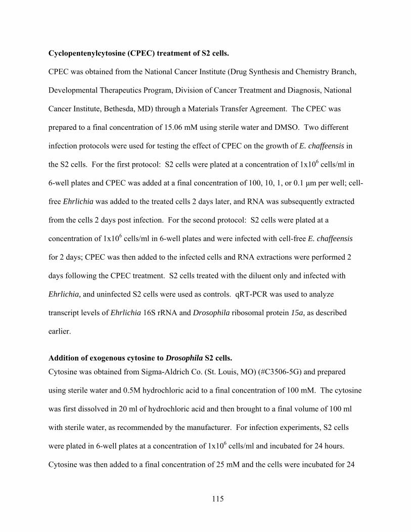
Cyclopentenylcytosine (CPEC) treatment of S2 cells.
CPEC was obtained from the National Cancer Institute (Drug Synthesis and Chemistry Branch,
Developmental Therapeutics Program, Division of Cancer Treatment and Diagnosis, National
Cancer Institute, Bethesda, MD) through a Materials Transfer Agreement. The CPEC was
prepared to a final concentration of 15.06 mM using sterile water and DMSO. Two different
infection protocols were used for testing the effect of CPEC on the growth of E. chaffeensis in
the S2 cells. For the first protocol: S2 cells were plated at a concentration of 1x10⁶ cells/ml in
6-well plates and CPEC was added at a final concentration of 100, 10, 1, or 0.1 μm per well; cell-
free Ehrlichia was added to the treated cells 2 days later, and RNA was subsequently extracted
from the cells 2 days post infection. For the second protocol: S2 cells were plated at a
concentration of 1x10⁶ cells/ml in 6-well plates and were infected with cell-free E. chaffeensis
for 2 days; CPEC was then added to the infected cells and RNA extractions were performed 2
days following the CPEC treatment. S2 cells treated with the diluent only and infected with
Ehrlichia, and uninfected S2 cells were used as controls. qRT-PCR was used to analyze
transcript levels of Ehrlichia 16S rRNA and Drosophila ribosomal protein 15a, as described
earlier.
Addition of exogenous cytosine to Drosophila S2 cells.
Cytosine was obtained from Sigma-Aldrich Co. (St. Louis, MO) (#C3506-5G) and prepared
using sterile water and 0.5M hydrochloric acid to a final concentration of 100 mM. The cytosine
was first dissolved in 20 ml of hydrochloric acid and then brought to a final volume of 100 ml
with sterile water, as recommended by the manufacturer. For infection experiments, S2 cells
were plated in 6-well plates at a concentration of 1x10⁶ cells/ml and incubated for 24 hours.
Cytosine was then added to a final concentration of 25 mM and the cells were incubated for 24
115

hours. Cell-free E. chaffeensis was added to the cytosine-treated cells, to untreated S2 cells, and
to S2 cells treated with the cytosine diluent. Untreated, uninfected S2 cells were used as a
negative control. Following addition of cell-free E. chaffeensis, RNA was extracted from cells at
24, 48, 72, and 96 hpi. qRT-PCR was used to measure transcript levels of Ehrlichia 16S rRNA
and Drosophila ribosomal protein 15a, as described earlier.
Methyl-β-Cyclodextrin (MβCD) treatment of Drosophila S2 cells.
MβCD was obtained from Sigma-Aldrich Co. (St. Louis, MO) (#C4555-5G) and prepared using
sterile water. S2 cells were plated at a concentration of 1x10⁶ cells per ml in 6-well plates.
MβCD was added to the cells at a final concentration of 2.5 mM for approximately 1.5 hours
prior to infection with E. chaffeensis. Following infection, RNA was extracted from the cells
at 24, 48, and 72 hpi and qRT-PCR was used to measure transcript levels of Ehrlichia 16S rRNA
and Drosophila ribosomal protein 15a, as described earlier. Infections were compared between
S2 cells that received the MβCD treatment and were then infected with Ehrlichia to those that
were treated with sterile water and then infected with Ehrlichia. Uninfected S2 cells were used
as negative controls.
Statistics.
Data are presented as the means ± standard errors of the means (SEM) or standard deviation
(SD) of independent experiments. SEM was used when the means of 3 or more independent
experiments were being compared. SD was used when less than 3 independent replicate
experimental values were being compared. Differences in means and replicates were determined
by using Students’s t test (two tailed, general) (StatMost statistical package; Data XIOM, Los
Angeles, CA). Survival data were analyzed for significance using the log rank test of Kaplan-
116
Meier plots using Prism Graphpad software (La Jolla, CA). P values of <0.05 were considered
highly significant.
RESULTS
Microarray data analysis reveals “permissive-exclusive” genes.
We chose to use microarrays to identify Drosophila host genes that have a role during E.
chaffeensis infections. We tested four different conditions of S2 cells using the microarray at
both 24 and 96 hpi: (1) uninfected S2 cells; (2) S2 cells activated with LPS; (3) S2 cells
activated with LPS and then infected with E. chaffeensis; and (4) S2 cells infected with E.
chaffeensis. Uninfected cells were used to discern what the basal transcript levels were in the S2
cells and comparisons across the different conditions were based on this basal level of
expression. In order to understand which gene transcripts were specific to our “non-permissive”
S2 cells, we used the S2 cells that were treated only with LPS as a comparison. Cells that
received only LPS treatment revealed activation specific gene transcripts. Comparing those
genes to the genes that had increased transcript levels in our cells infected under conditions that
were “non-permissive” for bacterial growth allowed us to deduce which genes up-regulated
transcripts exclusive to non-permissive conditions. Finally, by utilizing the “permissive” and
“non-permissive” infection conditions generated in S2 cells, we were able to determine which
gene transcripts were exclusively up-regulated during conditions that were “permissive” for the
growth of Ehrlichia. The genes in which transcript levels were up-regulated 1.5 fold or higher
compared to uninfected cells in both “permissive” and “non-permissive” conditions were
compared. We then focused on the genes that had transcript levels up-regulated only in the
“permissive” subset of genes. We hypothesized that these genes would have some type of role in
117
the ability of E. chaffeensis to replicate in Drosophila S2 cells. Although we assessed transcript
levels 24 and 96 hours post infection, we limited this analysis to just the 24 hour time point. As
described in the material and methods, we focused on genes that had transcript levels 1.5 fold
higher or above compared to uninfected cells. 2227 gene transcripts were up-regulated in S2
cells infected under “permissive” conditions and 1811 gene transcripts were up-regulated in S2
cells infected under “non-permissive” conditions. No genes were identified that had lower
transcript levels when S2 cells were infected under either permissive or non-permissive
conditions. Among the 2,227 genes with up-regulated transcript levels in permissive conditions,
2,128 of the genes were 1.5 fold higher than uninfected controls. Of the 1,811 genes that had
increased transcript levels, 1,742 of the genes were 1.5 fold higher than uninfected S2 cells.
When we compared genes that were up-regulated during permissive growth conditions compared
to genes that were up-regulated under non-permissive growth conditions, we identified 517
genes that had higher transcript levels exclusively during permissive conditions. Of these 517
genes, 210 of them had previously been ascribed some function and had some characterization.
However, 317 of the genes had yet to be defined and had “CG” gene designations. Among the
517 genes, we determined that viable, fertile, adult mutant stocks were available for 118 of the
genes (37 stocks for defined genes and 81 stocks for undefined genes). These flies had
appropriate mutations in coding exons in the genes of interest, and allowed us to test whether the
absence of a functional gene would affect fly survival and/or bacterial replication in vivo.
Ehrlichia chaffeensis infection of selected mutant Drosophila.
We screened 15 Drosophila lines with mutations in the gene of interest (Table 4.1) for fly
survival and bacterial replication. Flies (WT or mutant) were injected with cell-free E.
chaffeensis or sterile PBS and monitored for survival for 96-120 hpi (20 flies per treatment group
118

per experiment; results averaged from 3 independent experiments). We were interested in the
mutants that displayed increased survival after bacterial challenge compared to the wild-type
flies. These mutants included stocks 20231 (gene CG6364, FBgn0039179), 18540 (gene
CG3044 (CHT11), FBgn0029913), 13981 (gene separation anxiety, FBgn0024188), 15228 (gene
CG6479, FBgn0036710), and 20026 (gene CG6543, FBgn0033879) (Figure 1). Stocks 20231,
18540, 13981 and 20026 were all created by transposable element insertion. The sequence
location of the insertions for each stock was: (1) 20231 (3R:20,115,939); (2) 18540
(X:6,742,691); (3) 13981 (2R:7,248,253); and (4) 20026 (2R:9,741,819). Stock 15228 has been
discontinued since the start of our experiments and our own stock of these flies became ill and
died. Our hypothesis is that the gene affected in the mutants allowed increased survival because
its expression was needed for successful replication of Ehrlichia. We found that CG3044, san,
CG6364, CG6479, and CG6543 all displayed significantly increased survival compared to WT
flies after infection with E. chaffeensis (Figure 4.1; panels A-E respectively). Moreover, knock-
out/down-regulation of the gene in these mutants did not allow for bacterial replication as
measured by qRT-PCR (Figure 4.2).
119

Stock Number Stock Genotype Associated Gene
1124 al[1] Gpdh[n1-4]/SM1 Glycerol 3 phosphate dehydrogenase
1125 al[1] Gpdh[n0]/SM1 Glycerol 3 phosphate dehydrogenase
2402 al[1] Gpdh[n5-4]/SM1 Glycerol 3 phosphate dehydrogenase
14560 y[1] w[67c23]; P{y[+mDint2] w[BR.E.BR]=SUPor-P}CG10672[KG07864] ry[506]
CG10672
18615 w[1118]; PBac{w[+mC]=WH}CG4743[f03065]
CG4743
20231 y[1] w[67c23]; P{w[+mC] y[+mDint2]=EPgy2}CG6364[EY10942]
CG6364
18540 w[1118] PBac{w[+mC]=WH}Cht11[f02328]
Cht11
14880 y[1]; P{y[+mDint2] w[BR.E.BR]=SUPor-P}Map60[KG00506]/SM6a; ry[506]
Map60
13981 y[1]; P{y[+mDint2] w[BR.E.BR]=SUPor-P}san[KG04816]/CyO; ry[506]
san
24723 w[1118]; Mi{ET1}CG9300[MB04640] CG9300 15228 NO LONGER AVAILABLE NO LONGER
AVAILABLE 18141 PBac{w[+mC]=RB}Tsp3A[e03287]
w[1118] Tsp3A
15434 y[1] w[67c23] P{w[+mC] y[+mDint2]=EPgy2}CG10992[EY03339]
CG10992
15676 y[1] w[67c23]; P{w[+mC] y[+mDint2]=EPgy2}Gap69C[EY03771]
Gap69C
20026 y[1] w[67c23]; P{w[+mC] y[+mDint2]=EPgy2}CG6543[EY08499]
CG6543
Table 4.1 Stock numbers (Bloomington Stock Center; Bloomington, IN), genotypes, and
associated genes of viable, fertile, adult Drosophila screened by microinjection.
120
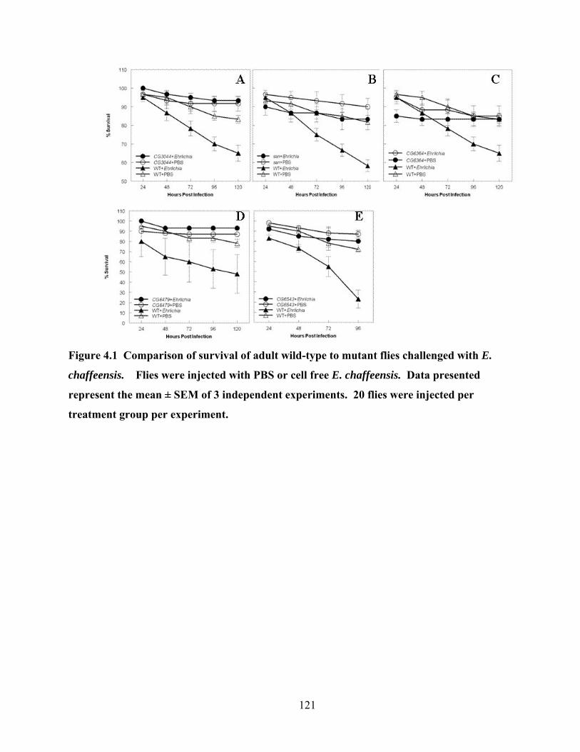
Figure 4.1 Comparison of survival of adult wild-type to mutant flies challenged with E.
chaffeensis. Flies were injected with PBS or cell free E. chaffeensis. Data presented
represent the mean ± SEM of 3 independent experiments. 20 flies were injected per
treatment group per experiment.
121

Figure 4.2 Average number of Ehrlichiae present in mutant flies (CG3044, CG6479, san,
CG6364, and CG6543) compared to wild-type flies at 120 (CG3044, CG6479, san, CG6364)
or 96 hours post infection (CG6543). Gene name on the X-axis designates flies with a
mutation in that gene.
Uridine/Cytidine Kinase mutations affect fly survival and bacterial replication.
To better understand the role of the genes identified in our screen, we examined the gene
CG6364. The molecular function of this gene is predicted to be uridine/cytidine kinase activity
(putatively uridine 5’-phosphotransferase, E.c. 2.7.1.48) (15). Disruption of CG6364 from an
insertion at the sequence location 3R:20,115,939 resulted in poor bacterial replication in vivo
(Figure 4.1; panel C). To confirm the impact of the uridine/cytidine kinase on E. chaffeensis
growth, we obtained the mutant Drosophila stocks dumpy (FBgn0053196), tilt (FBgn0003868),
and withered (FBgn0004012). All of these flies have been previously reported to carry
mutation(s) which affect uridine/cytidine kinase function (50). Each stock was infected with E.
chaffeensis and assessed for bacterial replication as described in the materials and methods. All
122
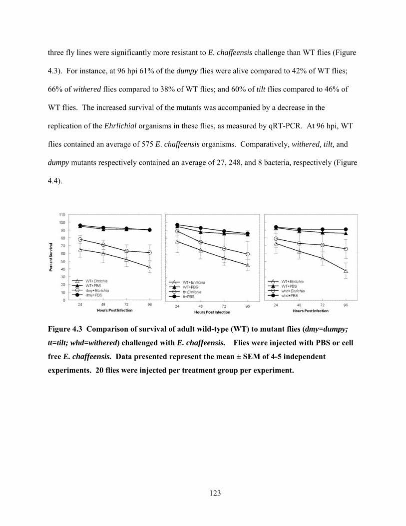
three fly lines were significantly more resistant to E. chaffeensis challenge than WT flies (Figure
4.3). For instance, at 96 hpi 61% of the dumpy flies were alive compared to 42% of WT flies;
66% of withered flies compared to 38% of WT flies; and 60% of tilt flies compared to 46% of
WT flies. The increased survival of the mutants was accompanied by a decrease in the
replication of the Ehrlichial organisms in these flies, as measured by qRT-PCR. At 96 hpi, WT
flies contained an average of 575 E. chaffeensis organisms. Comparatively, withered, tilt, and
dumpy mutants respectively contained an average of 27, 248, and 8 bacteria, respectively (Figure
4.4).
Figure 4.3 Comparison of survival of adult wild-type (WT) to mutant flies (dmy=dumpy;
tt=tilt; whd=withered) challenged with E. chaffeensis. Flies were injected with PBS or cell
free E. chaffeensis. Data presented represent the mean ± SEM of 4-5 independent
experiments. 20 flies were injected per treatment group per experiment.
123

Figure 4.4 Average number of Ehrlichiae present in mutant flies compared to wild-type
flies at 96 hours post infection. Data presented represent the mean of 3 independent
experiments.
CPEC treatment increases E. chaffeensis infection.
The uridine/cytidine kinase enzyme functions in pyrimidine synthesis pathways, being
specifically involved in the conversion of uridine to uridine monophosphate and/or cytidine to
cytidine monophosphate and vice versa (46, 52, 53). Deoxycytidine triphosphate can be
synthesized from glutamine through the de novo pathway (Figure 4.5). Alternatively,
deoxycytidine triphosphate can be synthesized through the salvage pathway, which is dependent
upon the uridine/cytidine kinase to convert cytidine or uridine to cytidine monophosphate or
uridine monophosphate, respectively (Figure 4.5) (46, 52, 53). We obtained the drug CPEC,
which inhibits the conversion of [3H]-UTP to [3H]-CTP (26). Therefore it is an inhibitor of
cytidine triphosphate synthetase (CTP) (38, 46, 52) (Figure 4.5). Consequently this drug
effectively inhibits de novo synthesis of pyrimidines, leaving only the salvage pathway with
124
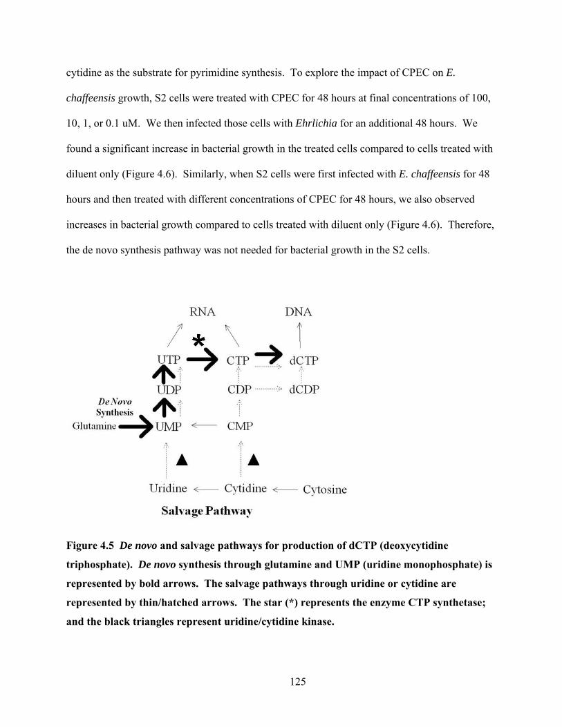
cytidine as the substrate for pyrimidine synthesis. To explore the impact of CPEC on E.
chaffeensis growth, S2 cells were treated with CPEC for 48 hours at final concentrations of 100,
10, 1, or 0.1 uM. We then infected those cells with Ehrlichia for an additional 48 hours. We
found a significant increase in bacterial growth in the treated cells compared to cells treated with
diluent only (Figure 4.6). Similarly, when S2 cells were first infected with E. chaffeensis for 48
hours and then treated with different concentrations of CPEC for 48 hours, we also observed
increases in bacterial growth compared to cells treated with diluent only (Figure 4.6). Therefore,
the de novo synthesis pathway was not needed for bacterial growth in the S2 cells.
Figure 4.5 De novo and salvage pathways for production of dCTP (deoxycytidine
triphosphate). De novo synthesis through glutamine and UMP (uridine monophosphate) is
represented by bold arrows. The salvage pathways through uridine or cytidine are
represented by thin/hatched arrows. The star (*) represents the enzyme CTP synthetase;
and the black triangles represent uridine/cytidine kinase.
125
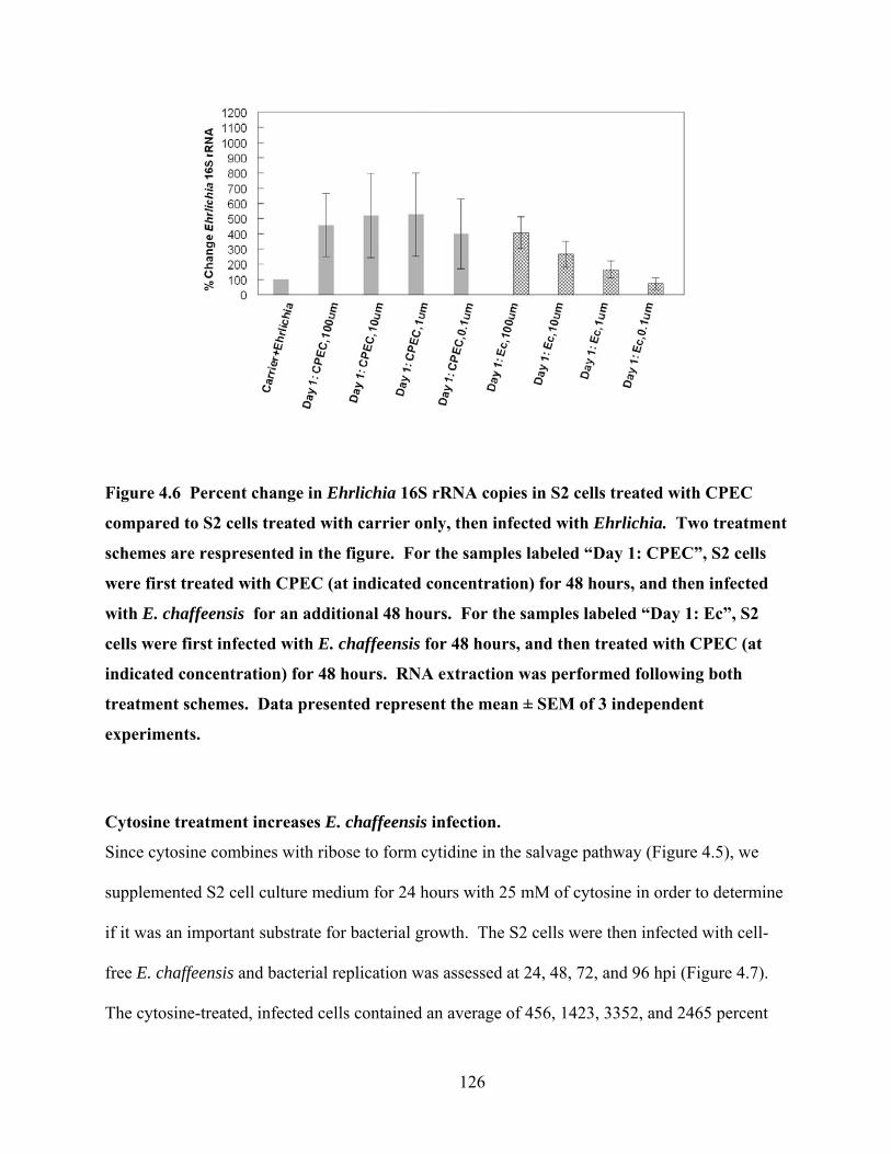
Figure 4.6 Percent change in Ehrlichia 16S rRNA copies in S2 cells treated with CPEC
compared to S2 cells treated with carrier only, then infected with Ehrlichia. Two treatment
schemes are respresented in the figure. For the samples labeled “Day 1: CPEC”, S2 cells
were first treated with CPEC (at indicated concentration) for 48 hours, and then infected
with E. chaffeensis for an additional 48 hours. For the samples labeled “Day 1: Ec”, S2
cells were first infected with E. chaffeensis for 48 hours, and then treated with CPEC (at
indicated concentration) for 48 hours. RNA extraction was performed following both
treatment schemes. Data presented represent the mean ± SEM of 3 independent
experiments.
Cytosine treatment increases E. chaffeensis infection.
Since cytosine combines with ribose to form cytidine in the salvage pathway (Figure 4.5), we
supplemented S2 cell culture medium for 24 hours with 25 mM of cytosine in order to determine
if it was an important substrate for bacterial growth. The S2 cells were then infected with cell-
free E. chaffeensis and bacterial replication was assessed at 24, 48, 72, and 96 hpi (Figure 4.7).
The cytosine-treated, infected cells contained an average of 456, 1423, 3352, and 2465 percent
126

more copies of Ehrlichia 16S rRNA at 24, 48, 72, and 96 hpi compared to infected cells treated
with diluent only. Taken together with the CPEC experiments, these data support the hypothesis
that utilization of cytidine through the salvage pathway occurs during the replication of E.
chaffeensis.
Figure 4.7 Percent change in Ehrlichia 16S rRNA copies in S2 cells treated with cytosine
and infected with Ehrlichia compared to S2 cells treated with carrier only and infected with
Ehrlichia. Data presented represent the mean of 3 independent experiments.
MβCD treatment does not affect Ehrlichia replication.
It is known that chitin derivatives can lower cholesterol levels (29) and that increased levels of
cholesterol enhance Anaplasma phagocytophilum infection in mice (57). CG3044 (Cht11) was
described as having potential chitinase activity (18). We hypothesized that the up-regulation of
chitinase could be a subversive virulence mechanism that the bacteria use to control cholesterol
127

levels through the control of chitin. To test this hypothesis, we treated S2 cells with the
cholesterol-sequestering agent, MβCD (27, 28, 40), and then infected them with E. chaffeensis
(Figure 4.8). No difference in bacterial load was observed between the infected cells that
received MβCD and those that did not.
Figure 4.8 Percent change in Ehrlichia 16S rRNA copies in S2 cells treated with MβCD
and infected with Ehrlichia compared to S2 cells treated with carrier only and infected with
Ehrlichia. Data presented represent the mean ± SD of 2 independent experiments.
DISCUSSION We have successfully demonstrated the power of the Drosophila system for elucidating
host factors that are important for the replication of E. chaffeensis. We combined microarray
analysis and mutant screening to successfully identify five genes that contribute to the replication
128
of E. chaffeensis in vivo. The functions of the identified genes are diverse and include: (1)
phagocytosis/engulfment (CG6479); (2) fatty acid binding/mitotic sister chromatid binding
(san); (3) chitinase activity (CG3044 (Cht11)); (4) uridine/cytidine kinase activity (CG6364);
and (5) enoyl –CoA hydratase activity (CG6543). To our knowledge, this is the first
demonstration of host genes that control Ehrlichia replication.
Our data suggest that bacteria require cytosine or cytidine as a substrate to make
deoxycytidine triphosphate and DNA. This hypothesis is supported by data showing that
flies with mutations in CG6364 were significantly less susceptible to Ehrlichia challenge
compared to WT flies. In addition, blocking of the synthesis of deoxycytidine triphosphate with
the CTP synthetase inhibitor, CPEC, resulted in increased replication of E. chaffeensis in S2
cells. This suggested that the bacteria do not require the host to use glutamine as a substrate
during replication. Supplementation of cytosine to cells actively replicating bacteria also
enhanced Ehrlichia replication and further implicates the salvage pathway through cytidine.
Stroman found that tilt and withered larvae accumulate uridine when supplemented with
uracil (50), which is toxic to the Drosophila. However, uridine monophosphate was not toxic.
Those data suggested that these mutants had a defect in the pathway that converted uracil to
uradine monophosphate (50). Indeed, tilt and withered mutants were more resistant to infection
by E. chaffeensis than WT flies. dumpy mutants also had apparent mutations in pyrimidine
synthesis (50) and these flies were also more resistant to infection that our WT flies. While the
dumpy mutation has been annotated, the tilt and withered mutations have not been characterized.
We have attempted to approximate the cytogenetic locations of tilt and withered. Interestingly,
all of these genes map to different loci from CG6364. CG6364 maps cytogenetically to
chromosome 3 at 95F10 (15) and dumpy maps to chromosome 2 between 24F4-25A1 (19).
129
Based on recombination maps (6) and the annotation by Lindsley and Zimm (33), we estimate
that withered maps to chromosome 2 between 46-47 (22), and tilt maps to chromosome 3
between 40-41 (21). Therefore, additional work will be needed to characterize where in the
pathway of host pyrimidine synthesis each of these genes function to regulate E. chaffeensis
replication.
It is known that depletion of the cytidine pools can disrupt the balance of ribonucleotides
in cells, leading to alterations in cellular homeostasis and apoptosis (46). Therefore, it is possible
that the sequestering of cytidine by Ehrlichia affects the cell, making it more vulnerable to
infection by the bacteria. Because cytidine is the least abundant nucleoside in cells (31), its use
by the Ehrlichia may be particularly stressful on the host cell processes. In that case, the bacteria
would be hijacking the normal cellular pyrimidine/purine metabolism, which may alter host cell
defenses and make the environment more amenable to their own replication. Ehrlichia
disseminate by lysing host cells in order to spread to uninfected cells (41), and the up-regulation
of apoptotic genes during Ehrlichia infections has been observed (59). Therefore, a correlation
between cell lysis, apoptosis and cytidine utilization by the bacteria is a possibility.
The predicted molecular function of CG3044 is chitinase activity (18). CG3044 (Cht11)
may be related to the Chitinase 11 gene of Tribolium castaneum, which has been assigned to a
separate classification group (group VII) from other identified chitinases (3), (personal
communication, Dr. Subbaratnam Muthukrishnan, Kansas State University). This indicated that
CG3044 may have a function other than those classically described for chitinases. Chitins and
their derivatives regulate cholesterol when used as diet supplements (29, 58). Mice with high
blood cholesterol have more severe Anaplasma phagocytophilum infections in their blood, livers,
and spleens (57). E. chaffeensis and A. phagocytophilum (a Rickettsial pathogen closely related
130
to E. chaffeensis) cannot synthesize cholesterol and scavenge it from host cells during infections
(32). Therefore, chitinase CG3044 (Cht11) may function to regulate cholesterol levels to
facilitate infections. Alternatively, chitin has been found to activate macrophages as measured
by the phophorylation of p38 and Erk (Figure 4.9) (39). It is possible that chitinase is involved
in regulating cellular activation. Incubation of S2 cells with LPS activates MAPKinase (Von
Ohlen et al., Kansas State University, unpublished observations) and makes the cells non-
permissive to infection by E. chaffeensis (34). Since MβCD did not affect E. chaffeensis
infection in S2 cells, and because MβCD inhibits the ability of cholesterol to activate MAPK
(Figure 4.9) (39), it might be that the chitinase role could be to regulate activation of the
parasitized cells (Figure 4.9). Reese et al. (2007) have shown that chitin can regulate
inflammation and the activation of Th1 versus Th2 responses (44). Therefore, these data support
this hypothesis. However, additional experiments will be needed to confirm that regulation of
chitinase affects cellular activation and E. chaffeensis replication.
In conclusion, we have successfully used Drosophila to identify host genes that are
involved in E. chaffeensis infections. Among those genes, we further tested the roles of CG6364
and CG3044 in the replication of E. chaffeensis. The human homolog of CG6364 is uridine
cytidine kinase 2 (UCK2) and is located on Chromosome 1 (12, 15). It functions in the same
manner as CG6364, acting to phosphorylate uridine and/or cytidine (51). UCK2 has been found
to be more active in certain types of cancers (48), and also acts to phosphorylate nucleoside
analog drugs used for treatment of cancers and hepatitis C virus (24, 51). A human homolog of
CG3044 has not been identified, but homologs have been identified in deer ticks and body lice
(18). Both of these species serve as vectors of different Rickettsial bacteria (4), so it would be
interesting to better understand the function of this host chitinase in those organisms. The human
131
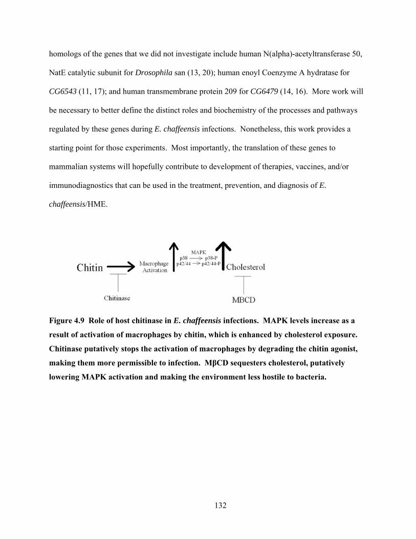
homologs of the genes that we did not investigate include human N(alpha)-acetyltransferase 50,
NatE catalytic subunit for Drosophila san (13, 20); human enoyl Coenzyme A hydratase for
CG6543 (11, 17); and human transmembrane protein 209 for CG6479 (14, 16). More work will
be necessary to better define the distinct roles and biochemistry of the processes and pathways
regulated by these genes during E. chaffeensis infections. Nonetheless, this work provides a
starting point for those experiments. Most importantly, the translation of these genes to
mammalian systems will hopefully contribute to development of therapies, vaccines, and/or
immunodiagnostics that can be used in the treatment, prevention, and diagnosis of E.
chaffeensis/HME.
Figure 4.9 Role of host chitinase in E. chaffeensis infections. MAPK levels increase as a
result of activation of macrophages by chitin, which is enhanced by cholesterol exposure.
Chitinase putatively stops the activation of macrophages by degrading the chitin agonist,
making them more permissible to infection. MβCD sequesters cholesterol, putatively
lowering MAPK activation and making the environment less hostile to bacteria.
132
REFERENCES 1. Agaisse, H., L. S. Burrack, J. A. Philips, E. J. Rubin, N. Perrimon, and D. E.
Higgins. 2005. Genome-wide RNAi screen for host factors required for intracellular bacterial infection. Science 309:1248-51.
2. Alvarez-Dominguez, C., A. M. Barbieri, W. Beron, A. Wandinger-Ness, and P. D. Stahl. 1996. Phagocytosed live Listeria monocytogenes influences Rab5-regulated in vitro phagosome-endosome fusion. J Biol Chem 271:13834-43.
3. Arakane, Y., and S. Muthukrishnan. 2010. Insect chitinase and chitinase-like proteins. Cellular and Molecular Life Sciences 67:201-216.
4. Azad, A., and C. Beard. 1998. Rickettsial pathogens and their arthropod vectors. A. F. Azad and C. B. Beard. Emerging Infectious Diseases 4:179–186.
5. Brandt, S. M., G. Jaramillo-Gutierrez, S. Kumar, C. Barillas-Mury, and D. S. Schneider. 2008. Use of a Drosophila model to identify genes regulating Plasmodium growth in the mosquito. Genetics 180:1671-8.
6. Bridges, P. 1941. A revision of the salivary gland 3R-chromosome map of Drosophila melanogaster. J Hered 32:299-300.
7. Brouqui, P., C. Lecam, J. Olson, and D. Raoult. 1994. Serologic diagnosis of human monocytic ehrlichiosis by immunoblot analysis. Clin Diagn Lab Immunol 1:645-9.
8. Cinco, M., F. Barbone, M. Grazia Ciufolini, M. Mascioli, M. Anguero Rosenfeld, P. Stefanel, and R. Luzzati. 2004. Seroprevalence of tick-borne infections in forestry rangers from northeastern Italy. Clin Microbiol Infect 10:1056-61.
9. Dionne, M. S., N. Ghori, and D. S. Schneider. 2003. Drosophila melanogaster is a genetically tractable model host for Mycobacterium marinum. Infect Immun 71:3540-50.
10. Elwell, C., and J. N. Engel. 2005. Drosophila melanogaster S2 cells: a model system to study Chlamydia interaction with host cells. Cell Microbiol 7:725-39.
11. Ensembl March 2010, posting date. enoyl Coenzyme A hydratase, short chain, 1, mitochondrial [Source:HGNC Symbol;Acc:3151]; Transcript: ECHS1-001 (ENST00000368547). [Online.]
12. Ensembl March 2010, posting date. Gene: UCK2 (ENSG00000143179). [Online.] 13. Ensembl March 2010, posting date. N(alpha)-acetyltransferase 50, NatE catalytic subunit
[Source:HGNC Symbol;Acc:29533]; Transcript: NAT13-001 (ENST00000240922). [Online.]
14. Ensembl March 2010, posting date. transmembrane protein 209; Transcript: TMEM209-001 (ENST00000397622). [Online.]
15. FlyBase March 19th, 2010, posting date. Gene Dmel\CG6364. FlyBase. [Online.] 16. FlyBase March 19, 2010, posting date. Gene Dmel\CG6479. [Online.] 17. FlyBase March 19, 2010, posting date. Gene Dmel\CG6543. [Online.] 18. FlyBase March 19, 2010, posting date. Gene Dmel\Cht11. [Online.] 19. FlyBase March 19, 2010, posting date. Gene Dmel\dp. [Online.] 20. FlyBase March 19, 2010, posting date. Gene Dmel\san. [Online.] 21. FlyBase March 19, 2010, posting date. Gene Dmel\tt. [Online.] 22. FlyBase March 19, 2010, posting date. Gene Dmel\whd. [Online.] 23. Ganta, R. R., C. Cheng, M. J. Wilkerson, and S. K. Chapes. 2004. Delayed clearance
of Ehrlichia chaffeensis infection in CD4+ T-cell knockout mice. Infect Immun 72:159-67.
133
24. Golitsina, N. L., F. T. Danehy, Jr., R. Fellows, E. Cretton-Scott, and D. N. Standring. Evaluation of the role of three candidate human kinases in the conversion of the hepatitis C virus inhibitor 2'-C-methyl-cytidine to its 5'-monophosphate metabolite. Antiviral Res 85:470-81.
25. Hall-Baker, P. A., J. Enrique Nieves, D. Ruth Ann Jajosky, D. A. Adams, P. Sharp, W. J. Anderson, J. J. Aponte, G. F. Jones, M. M. Aaron E. Aranas, M. Araceli Rey, M. Bernetta Lane, and M. S. W. . 2009. Summary of Notifiable Diseases --- United States, 2007. MMWR 56.
26. Kang, G. J., D. A. Cooney, J. D. Moyer, J. A. Kelley, H. Y. Kim, V. E. Marquez, and D. G. Johns. 1989. Cyclopentenylcytosine triphosphate. Formation and inhibition of CTP synthetase. J Biol Chem 264:713-8.
27. Kilsdonk, E. P., P. G. Yancey, G. W. Stoudt, F. W. Bangerter, W. J. Johnson, M. C. Phillips, and G. H. Rothblat. 1995. Cellular cholesterol efflux mediated by cyclodextrins. J Biol Chem 270:17250-17256.
28. Klein, U., G. Gimpl, and F. Fahrenholz. 1995. Alteration of the myometrial plasma membrane cholesterol content with -cyclodextrin modulates the binding affinity of the oxytocin receptor. Biochemisty 34:13784-13793.
29. Koide, S. 1998. Chitin-chitosan: Properties, benefits and risks. Nutrition Research 18:1091-1101.
30. Koo, I. C., Y. M. Ohol, P. Wu, J. H. Morisaki, J. S. Cox, and E. J. Brown. 2008. Role for lysosomal enzyme beta-hexosaminidase in the control of mycobacteria infection. Proc Natl Acad Sci U S A 105:710-5.
31. Korte, D., W. Haverkort, A. van Gennip, and D. Roos. 1985. Nucleotide profiles of normal human blood cells determined by high-performance liquid chromatography. Anal. Biochem. 147:197-209.
32. Lin, M., and Y. Rikihisa. 2003. Ehrlichia chaffeensis and Anaplasma phagocytophilum lack genes for lipid A biosynthesis and incorporate cholesterol for their survival. Infect Immun 71:5324-31.
33. Lindsley, D., and G. Zimm. 1992. The Genome of Drosophila melanogaster. Academic Press, Inc., San Diego, CA.
34. Luce-Fedrow, A., T. Von Ohlen, D. Boyle, R. R. Ganta, and S. K. Chapes. 2008. Use of Drosophila S2 cells as a model for studying Ehrlichia chaffeensis infections. Appl Environ Microbiol 74:1886-91.
35. Luce-Fedrow, A., T. Von Ohlen, and S. K. Chapes. 2009. Ehrlichia chaffeensis infections in Drosophila melanogaster. Infect Immun 77:4815-26.
36. Machado, R. Z., J. M. Duartes, A. S. Dagnone, and M. P. Szabo. 2006. Detection of Ehrlichia chaffeensis in Brazilain marsh deer (Blastocerus dichotomus. Vet Parasitol 139:262-266.
37. Morgan, T. 1929. Contributions to the genetics of Drosophila simulans and Drosophila melanogaster. Publs Carnegie Instn 399:169-199.
38. Moyer, J., N. Malinowski, S. Treanor, and V. Marquez. 1986. Antitumor activity and biochemical effects of cyclopentenyl cytosine in mice. Cancer REs 46:3325-9.
39. Nishiyama, A., T. Shinohara, T. Pantuso, S. Tsuji, M. Yamashita, S. Shinohara, Q. N. Myrvik, R. A. Henriksen, and Y. Shibata. 2008. Depletion of cellular cholesterol enhances macrophage MAPK activation by chitin microparticles but not by heat-killed Mycobacterium bovis BCG. Am J Physiol Cell Physiol 295:C341-9.
134

40. Ohtani, Y., T. Irie, K. Uekama, K. Fukunaga, and J. Pitha. 1989. Differential effects of -, - and gamma-cyclodextrins on human erythrocytes. Eur. J. Biochem 186:17-22.
41. Paddock, C. D., and J. E. Childs. 2003. Ehrlichia chaffeensis: a prototypical emerging pathogen. Clin Microbiol Rev 16:37-64.
42. Pfaffl, M. W. 2001. A new mathematical model for relative quantification in real-time RT-PCR. Nucleic Acids Res 29:e45.
43. Philips, J. A., E. J. Rubin, and N. Perrimon. 2005. Drosophila RNAi screen reveals CD36 family member required for mycobacterial infection. Science 309:1251-3.
44. Reese, T. A., H. E. Liang, A. M. Tager, A. D. Luster, N. Van Rooijen, D. Voehringer, and R. M. Locksley. 2007. Chitin induces accumulation in tissue of innate immune cells associated with allergy. Nature 447:92-6.
45. Santic, M., C. Akimana, R. Asare, J. C. Kouokam, S. Atay, and Y. A. Kwaik. 2009. Intracellular fate of Francisella tularensis within arthropod-derived cells. Environ Microbiol 11:1473-81.
46. Schimmel, K., H. Gelderblom, and H. Guchelaar. 2007. Cyclopentenyl Cytosine (CPEC): An Overview of its in vitro and in vivo Activity. Current Cancer Drug Targets 7:504-509.
47. Schneider, D., and M. Shahabuddin. 2000. Malaria parasite development in a Drosophila model. Science 288:2376-9.
48. Shimamoto, Y., K. Koizumi, H. Okabe, H. Kazuno, Y. Murakami, F. Nakagawa, A. Matsuda, T. Sasaki, and M. Fukushima. 2005. Sensitivity of Human Cancer Cells to the New Anticancer Ribo-nucleoside TAS–106 Is Correlated with Expression of Uridine-cytidine Kinase 2. Cancer Science 93:825-833.
49. Sirigireddy, K. R., and R. R. Ganta. 2005. Multiplex detection of Ehrlichia and Anaplasma species pathogens in peripheral blood by real-time reverse transcriptase-polymerase chain reaction. J Mol Diagn 7:308-16.
50. Stroman, P. 1974. Pyrimidine-sensitive drosophila wing mutants: withered (whd), tilt (tt) and dumpy (dp). Hereditas 78:157-68.
51. Van Rompay, A. R., A. Norda, K. Linden, M. Johansson, and A. Karlsson. 2001. Phosphorylation of uridine and cytidine nucleoside analogs by two human uridine-cytidine kinases. Mol Pharmacol 59:1181-6.
52. Verschuur, A. 2007. Cytidine triphosphate synthetase (CTP synthetase) as a druggable target in cancer. Drugs of the Future 32:1071-1080.
53. Verschuur, A. C., A. H. Van Gennip, R. Leen, R. Meinsma, P. A. Voute, and A. B. van Kuilenburg. 2000. In vitro inhibition of cytidine triphosphate synthetase activity by cyclopentenyl cytosine in paediatric acute lymphocytic leukaemia. Br J Haematol 110:161-9.
54. Vonkavaara, M., M. V. Telepnev, P. Ryden, A. Sjostedt, and S. Stoven. 2008. Drosophila melanogaster as a model for elucidating the pathogenicity of Francisella tularensis. Cell Microbiol 10:1327-38.
55. Waddington, C. 1940. The genetic control of wing development in Drosophila. J Genet 41:75-139.
56. Wen, B., W. Cao, and H. Pan. 2003. Ehrlichiae and ehrlichial diseases in china. Ann N Y Acad Sci 990:45-53.
135

57. Xiong, Q., X. Wang, and Y. Rikihisa. 2007. High-cholesterol diet facilitates Anaplasma phagocytophilum infection and up-regulates macrophage inflammatory protein-2 and CXCR2 expression in apolipoprotein E-deficient mice. J Infect Dis 195:1497-503.
58. Zacour, A., M. Silva, P. Cecon, E. Bambirra, and E. Vieira. 1992. Effect of dietary chitin on cholesterol absorption and metabolism in rats. J Nutr Sci Vitaminol (Tokyo). 32:609-613.
59. Zhang, J. Z., M. Sinha, B. A. Luxon, and X. J. Yu. 2004. Survival strategy of obligately intracellular Ehrlichia chaffeensis: novel modulation of immune response and host cell cycles. Infect Immun 72:498-507.
136
CHAPTER 5 - Conclusion
The research accomplished for this dissertation effectively demonstrates that Drosophila
melanogaster can be used for studying Ehrlichia chaffeensis infections. E. chaffeensis is an
emerging, tick-borne pathogen that causes human monocytic ehrlichiosis (HME). Reports of the
disease are on the rise and a 44% increase in reported HME cases was observed in just one year
(2006-2007) (13). Moreover, the reports of HME have increased 159% overall since the year
2003 (13). The increased cases of HME may be the result of growing vector populations,
increases in human contact with tick populations, and/or heightened surveillance and detection
techniques. In any case, a model system for studying E. chaffeensis infections will be most
helpful in better understanding the pathogenesis and resulting disease of these bacteria.
We have successfully used D. melanogaster as a model to determine host genes that are
necessary for E. chaffeensis pathogenesis. The importance of better understanding tick-borne
illness should not be underestimated. E. chaffeensis belongs to the Rickettsial group of diseases.
It was recently shown that 2% of imported fevers are the result of infection with Rickettsial
organisms and 20% of these fevers require hospitalization (14). The ticks and lice that vector
these diseases have wide ranging habitats and are adapted to living in ever-changing conditions.
The tick vector of E. chaffeensis is Amblyomma americanum, it has been expanding it range for
the past 30 years, and has been reported as far northeast as Maine (15). In addition, Rickettsial
diseases have a history of being considered for use as bioterrorism agents. In fact Rickettsia
prowazekii and Coxiella burnetii have been tested as weapons by the former Soviet Union, the
United States, and Japan (2). The conseqential increasing incidence of disease, spread of disease
vectors, and potential for the creation of weapons of mass destruction warrants careful study of
these organisms.
137

To our knowledge, our description of host genes that contribute to the replication of E.
chaffeensis is the first to be provided. Most importantly, these genes can be compared to their
mammalian homologs. For instance, the human homologs of the genes we have pinpointed in
playing a role in E. chaffeensis replication include human N(alpha)-acetyltransferase 50, NatE
catalytic subunit for Drosophila san (5, 11); human enoyl Coenzyme A hydratase for CG6543 (3,
9); human transmembrane protein 209 for CG6479 (6, 8); and human uridine cytidine kinase 2
(UCK2) for CG6364 (4, 7). No human homolog exists for CG3044. As a next step, it would be
interesting to determine the role of these genes during infections in a mammalian host or even in
the tick vector. Experiments that utilize RNAi based silencing would ultimately confirm the role
of the corresponding homologs during Ehrlichia infections in mammalian or tick systems. Our
contributions to the body of knowledge surrounding Ehrlichial and Rickettsial diseases should
ultimately lead to better characterization of the genes involved in infections in the mammalian
host(s). These are the types of studies that will be necessary to contribute to the development of
highly effective treatments, diagnostic procedures, and initial vaccines.
138
REFERENCES 1. Azad, A., and C. Beard. 1998. Rickettsial pathogens and their arthropod vectors.
A. F. Azad and C. B. Beard. Emerging Infectious Diseases 4:179–186. 2. Azad, A. F. 2007. Pathogenic rickettsiae as bioterrorism agents. Clin Infect Dis 45 Suppl
1:S52-5. 3. Ensembl March 2010, posting date. enoyl Coenzyme A hydratase, short chain, 1,
mitochondrial [Source:HGNC Symbol;Acc:3151]; Transcript: ECHS1-001 (ENST00000368547). [Online.]
4. Ensembl March 2010, posting date. Gene: UCK2 (ENSG00000143179). [Online.] 5. Ensembl March 2010, posting date. N(alpha)-acetyltransferase 50, NatE catalytic subunit
[Source:HGNC Symbol;Acc:29533]; Transcript: NAT13-001 (ENST00000240922). [Online.]
6. Ensembl March 2010, posting date. transmembrane protein 209; Transcript: TMEM209-001 (ENST00000397622). [Online.]
7. FlyBase March 19th, 2010, posting date. Gene Dmel\CG6364. FlyBase. [Online.] 8. FlyBase March 19, 2010, posting date. Gene Dmel\CG6479. [Online.] 9. FlyBase March 19, 2010, posting date. Gene Dmel\CG6543. [Online.] 10. FlyBase March 19, 2010, posting date. Gene Dmel\Cht11. [Online.] 11. FlyBase March 19, 2010, posting date. Gene Dmel\san. [Online.] 12. Golitsina, N. L., F. T. Danehy, Jr., R. Fellows, E. Cretton-Scott, and D. N.
Standring. Evaluation of the role of three candidate human kinases in the conversion of the hepatitis C virus inhibitor 2'-C-methyl-cytidine to its 5'-monophosphate metabolite. Antiviral Res 85:470-81.
13. Hall-Baker, P. A., J. Enrique Nieves, D. Ruth Ann Jajosky, D. A. Adams, P. Sharp, W. J. Anderson, J. J. Aponte, G. F. Jones, M. Aaron, M. E. Aranas, M. Araceli Rey, a. M. Bernetta Lane, and M. S. Wodajo. 2009. Summary of Notifiable Diseases --- United States, 2007. MMWR 56:1-94.
14. Jensenius, M., X. Davis, F. von Sonnenburg, E. Schwartz, J. S. Keystone, K. Leder, R. Lopez-Velez, E. Caumes, J. P. Cramer, L. Chen, and P. Parola. 2009. Multicenter GeoSentinel analysis of rickettsial diseases in international travelers, 1996-2008. Emerg Infect Dis 15:1791-8.
15. Magnarelli, L. 2001. Global importance of ticks and associated infectious disease agents. Clinical Microbiology Newsletter 31:33-37.
139

Appendix A - American Society for Microbiology License
for Chapter 2
This is a License Agreement between Alison Luce-Fedrow ("You") and American Society for Microbiology ("American Society for Microbiology") provided by Copyright Clearance Center ("CCC"). The license consists of your order details, the terms and conditions provided by American Society for Microbiology, and the payment terms and conditions.
All payments must be made in full to CCC. For payment instructions, please see information listed at the bottom of this form.
License Number: 2422570551583
License date: May 05, 2010
Licensed content publisher: American Society for Microbiology
Licensed content publication: Applied and Environmental Microbiology
Licensed content title: Use of Drosophila S2 Cells as a Model for Studying Ehrlichia chaffeensis Infections
Licensed content author: Alison Luce-Fedrow, Stephen K. Chapes
Licensed content date: Mar 15, 2008
Volume: 74
Issue: 6
Start page: 1886
End page: 1891
Type of Use: Dissertation/Thesis
Format: Electronic
Portion: Full article
Order reference number
140
Title of your thesis / dissertation: Drosophila melanogaster as a model for studying Ehrlichia chaffeensis infections
Expected completion date: May 2010
Estimated size(pages): 150
Total: 0.00 USD
Terms and Conditions
Publisher Terms and Conditions The publisher for this copyrighted material is the American Society for Microbiology (ASM). By clicking "accept" in connection with completing this licensing transaction, you agree that the following terms and conditions apply to this transaction (along with the Billing and Payment terms and conditions established by Copyright Clearance Center, Inc. ("CCC"), at the time that you opened your Rightslink account and that are available at any time at http://myaccount.copyright.com). 1. ASM hereby grants to you a non-exclusive license to use this material. Licenses are for one-time use only with a maximum distribution equal to the number that you identified in the licensing process; any form of republication must be completed within 1 year from the date hereof (although copies prepared before then may be distributed thereafter). The copyright of all material specified remains with ASM, and permission for reproduction is limited to the formats and products indicated in your license. The text may not be altered in any way without the express permission of the copyright owners. 2. The licenses may be exercised anywhere in the world. 3. You must include the copyright and permission notice in connection with any reproduction of the licensed material, i.e. Journal name, year, volume, page numbers, DOI and reproduced/amended with permission from American Society for Microbiology. 4. The following conditions apply to photocopies: a. The copies must be of high quality and match the standard of the original article. b. The copies must be a true reproduction word for word. c. No proprietary/trade names may be substituted. d. No additional text, tables or figures may be added to the original text. e. The integrity of the article should be preserved, i.e., no advertisements will be printed on the article. f. The above permission does NOT include rights for any online or other electronic reproduction. 5. The following conditions apply to translations: a. The translation must be of high quality and match the standard of the original article. b. The translation must be a true reproduction word for word. c. All drug names must be generic; no proprietary/trade names may be substituted.
141

d. No additional text, tables or figures may be added to the translated text. e. The integrity of the article should be preserved, i.e., no advertisements will be printed on the article. f. The translated version of ASM material must also carry a disclaimer in English and in the language of the translation. The two versions (English and other language) of the disclaimer MUST appear on the inside front cover or at the beginning of the translated material as follows: The American Society for Microbiology takes no responsibility for the accuracy of the translation from the published English original and is not liable for any errors which may occur. No responsibility is assumed, and responsibility is hereby disclaimed, by the American Society for Microbiology for any injury and/or damage to persons or property as a matter of product liability, negligence or otherwise, or from any use or operation of methods, products, instructions or ideas presented in the Journal. Independent verification of diagnosis and drug dosages should be made. Discussions, views, and recommendations as to medical procedures, choice of drugs and drug dosages are the responsibility of the authors. g. This license does NOT apply to translations made of manuscripts published ahead of print as "[ASM Journal] Accepts" papers. Translation permission is granted only for the final published version of the ASM article. Furthermore, articles translated in their entirety must honor the ASM embargo period, and thus may not appear in print or online until 6 months after the official publication date in the original ASM journal. 6. While you may exercise the rights licensed immediately upon issuance of the license at the end of the licensing process for the transaction, provided that you have disclosed complete and accurate details of your proposed use, no license is finally effective unless and until full payment is received from you (either by ASM or by CCC) as provided in CCC's Billing and Payment terms and conditions. If full payment is not received on a timely basis, then any license preliminarily granted shall be deemed automatically revoked and shall be void as if never granted. In addition, permission granted is contingent upon author permission, which you MUST obtain, and appropriate credit (see item number 3 for details). If you fail to comply with any material provision of this license, ASM shall be entitled to revoke this license immediately and retain fees paid for the grant of the license. Further, in the event that you breach any of these terms and conditions or any of CCC's Billing and Payment terms and conditions, the license is automatically revoked and shall be void as if never granted. Use of materials as described in a revoked license, as well as any use of the materials beyond the scope of an unrevoked license, may constitute copyright infringement and ASM reserves the right to take any and all action to protect its copyright in the materials. 7. ASM reserves all rights not specifically granted in the combination of (i) the license details provided by you and accepted in the course of this licensing transaction, (ii) these terms and conditions and (iii) CCC's Billing and Payment terms and conditions. 8. ASM makes no representations or warranties with respect to the licensed material and adopts on its own behalf the limitations and disclaimers established by CCC on its behalf in its Billing and Payment terms and conditions for this licensing transaction.
142

9. You hereby indemnify and agree to hold harmless ASM and CCC, and their respective officers, directors, employees and agents, from and against any and all claims arising out of your use of the licensed material other than as specifically authorized pursuant to this license. 10. This license is personal to you, but may be assigned or transferred by you to a business associate (or to your employer) if you give prompt written notice of the assignment or transfer to the publisher. No such assignment or transfer shall relieve you of the obligation to pay the designated license fee on a timely basis (although payment by the identified assignee can fulfill your obligation). 11. This license may not be amended except in a writing signed by both parties (or, in the case of ASM, by CCC on ASM 's behalf). 12. Objection to Contrary terms: ASM hereby objects to any terms contained in any purchase order, acknowledgment, check endorsement or other writing prepared by you, which terms are inconsistent with these terms and conditions or CCC's Billing and Payment terms and conditions. These terms and conditions, together with CCC's Billing and Payment terms and conditions (which are incorporated herein), comprise the entire agreement between you and ASM (and CCC) concerning this licensing transaction. In the event of any conflict between your obligations established by these terms and conditions and those established by CCC's Billing and Payment terms and conditions, these terms and conditions shall control. 13. The following terms and conditions apply to Commercial Photocopy and Commercial Reprint requests and should be considered by requestors to be additional terms. All other ASM terms and conditions indicating how the content may and may not be used also apply. Limitations of Use: The Materials you have requested permission to reuse in a commercial reprint or commercial photocopy are only for the use that you have indicated in your request, and they MAY NOT be used for either resale to others or republication to the public. Further, you may not decompile, reverse engineer, disassemble, rent, lease, loan, sell, sublicense, or create derivative works from the Materials without ASM's prior written permission. 14. Revocation: This license transaction shall be governed by and construed in accordance with the laws of Washington, DC. You hereby agree to submit to the jurisdiction of the federal and state courts located in Washington, DC for purposes of resolving any disputes that may arise in connection with this licensing transaction. ASM or Copyright Clearance Center may, within 30 days of issuance of this License, deny the permissions described in this License at their sole discretion, for any reason or no reason, with a full refund payable to you. Notice of such denial will be made using the contact information provided by you. Failure to receive such notice will not alter or invalidate the denial. In no event will ASM or Copyright Clearance Center be responsible or liable for any costs, expenses or damage incurred by you as a result of a denial of your permission request, other than a refund of the amount(s) paid by you to ASM and/or Copyright Clearance Center for denied permissions.
143
Appendix B - American Society for Microbiology License
for Chapter 3
This is a License Agreement between Alison Luce-Fedrow ("You") and American Society for Microbiology ("American Society for Microbiology") provided by Copyright Clearance Center ("CCC"). The license consists of your order details, the terms and conditions provided by American Society for Microbiology, and the payment terms and conditions.
All payments must be made in full to CCC. For payment instructions, please see information listed at the bottom of this form.
License Number: 2422570876460
License date: May 05, 2010
Licensed content publisher: American Society for Microbiology
Licensed content publication: Infection and Immunity
Licensed content title: Ehrlichia chaffeensis Infections in Drosophila melanogaster
Licensed content author: Alison Luce-Fedrow, Stephen K. Chapes
Licensed content date: Nov 1, 2009
Volume: 77
Issue: 11
Start page: 4815
End page: 4826
Type of Use: Dissertation/Thesis
Format: Electronic
Portion: Full article
Order reference number
Title of your thesis / dissertation : Drosophila melanogaster as a model for studying Ehrlichia chaffeensis infections
144
Expected completion date: May 2010
Estimated size(pages): 150
Total: 0.00 USD
Terms and Conditions
Publisher Terms and Conditions The publisher for this copyrighted material is the American Society for Microbiology (ASM). By clicking "accept" in connection with completing this licensing transaction, you agree that the following terms and conditions apply to this transaction (along with the Billing and Payment terms and conditions established by Copyright Clearance Center, Inc. ("CCC"), at the time that you opened your Rightslink account and that are available at any time at http://myaccount.copyright.com). 1. ASM hereby grants to you a non-exclusive license to use this material. Licenses are for one-time use only with a maximum distribution equal to the number that you identified in the licensing process; any form of republication must be completed within 1 year from the date hereof (although copies prepared before then may be distributed thereafter). The copyright of all material specified remains with ASM, and permission for reproduction is limited to the formats and products indicated in your license. The text may not be altered in any way without the express permission of the copyright owners. 2. The licenses may be exercised anywhere in the world. 3. You must include the copyright and permission notice in connection with any reproduction of the licensed material, i.e. Journal name, year, volume, page numbers, DOI and reproduced/amended with permission from American Society for Microbiology. 4. The following conditions apply to photocopies: a. The copies must be of high quality and match the standard of the original article. b. The copies must be a true reproduction word for word. c. No proprietary/trade names may be substituted. d. No additional text, tables or figures may be added to the original text. e. The integrity of the article should be preserved, i.e., no advertisements will be printed on the article. f. The above permission does NOT include rights for any online or other electronic reproduction. 5. The following conditions apply to translations: a. The translation must be of high quality and match the standard of the original article. b. The translation must be a true reproduction word for word. c. All drug names must be generic; no proprietary/trade names may be substituted. d. No additional text, tables or figures may be added to the translated text. e. The integrity of the article should be preserved, i.e., no advertisements will be printed on the article.
145
f. The translated version of ASM material must also carry a disclaimer in English and in the language of the translation. The two versions (English and other language) of the disclaimer MUST appear on the inside front cover or at the beginning of the translated material as follows: The American Society for Microbiology takes no responsibility for the accuracy of the translation from the published English original and is not liable for any errors which may occur. No responsibility is assumed, and responsibility is hereby disclaimed, by the American Society for Microbiology for any injury and/or damage to persons or property as a matter of product liability, negligence or otherwise, or from any use or operation of methods, products, instructions or ideas presented in the Journal. Independent verification of diagnosis and drug dosages should be made. Discussions, views, and recommendations as to medical procedures, choice of drugs and drug dosages are the responsibility of the authors. g. This license does NOT apply to translations made of manuscripts published ahead of print as "[ASM Journal] Accepts" papers. Translation permission is granted only for the final published version of the ASM article. Furthermore, articles translated in their entirety must honor the ASM embargo period, and thus may not appear in print or online until 6 months after the official publication date in the original ASM journal. 6. While you may exercise the rights licensed immediately upon issuance of the license at the end of the licensing process for the transaction, provided that you have disclosed complete and accurate details of your proposed use, no license is finally effective unless and until full payment is received from you (either by ASM or by CCC) as provided in CCC's Billing and Payment terms and conditions. If full payment is not received on a timely basis, then any license preliminarily granted shall be deemed automatically revoked and shall be void as if never granted. In addition, permission granted is contingent upon author permission, which you MUST obtain, and appropriate credit (see item number 3 for details). If you fail to comply with any material provision of this license, ASM shall be entitled to revoke this license immediately and retain fees paid for the grant of the license. Further, in the event that you breach any of these terms and conditions or any of CCC's Billing and Payment terms and conditions, the license is automatically revoked and shall be void as if never granted. Use of materials as described in a revoked license, as well as any use of the materials beyond the scope of an unrevoked license, may constitute copyright infringement and ASM reserves the right to take any and all action to protect its copyright in the materials. 7. ASM reserves all rights not specifically granted in the combination of (i) the license details provided by you and accepted in the course of this licensing transaction, (ii) these terms and conditions and (iii) CCC's Billing and Payment terms and conditions. 8. ASM makes no representations or warranties with respect to the licensed material and adopts on its own behalf the limitations and disclaimers established by CCC on its behalf in its Billing and Payment terms and conditions for this licensing transaction. 9. You hereby indemnify and agree to hold harmless ASM and CCC, and their respective officers, directors, employees and agents, from and against any and all claims arising out of your use of the licensed material other than as specifically authorized pursuant to this license.
146

10. This license is personal to you, but may be assigned or transferred by you to a business associate (or to your employer) if you give prompt written notice of the assignment or transfer to the publisher. No such assignment or transfer shall relieve you of the obligation to pay the designated license fee on a timely basis (although payment by the identified assignee can fulfill your obligation). 11. This license may not be amended except in a writing signed by both parties (or, in the case of ASM, by CCC on ASM 's behalf). 12. Objection to Contrary terms: ASM hereby objects to any terms contained in any purchase order, acknowledgment, check endorsement or other writing prepared by you, which terms are inconsistent with these terms and conditions or CCC's Billing and Payment terms and conditions. These terms and conditions, together with CCC's Billing and Payment terms and conditions (which are incorporated herein), comprise the entire agreement between you and ASM (and CCC) concerning this licensing transaction. In the event of any conflict between your obligations established by these terms and conditions and those established by CCC's Billing and Payment terms and conditions, these terms and conditions shall control. 13. The following terms and conditions apply to Commercial Photocopy and Commercial Reprint requests and should be considered by requestors to be additional terms. All other ASM terms and conditions indicating how the content may and may not be used also apply. Limitations of Use: The Materials you have requested permission to reuse in a commercial reprint or commercial photocopy are only for the use that you have indicated in your request, and they MAY NOT be used for either resale to others or republication to the public. Further, you may not decompile, reverse engineer, disassemble, rent, lease, loan, sell, sublicense, or create derivative works from the Materials without ASM's prior written permission. 14. Revocation: This license transaction shall be governed by and construed in accordance with the laws of Washington, DC. You hereby agree to submit to the jurisdiction of the federal and state courts located in Washington, DC for purposes of resolving any disputes that may arise in connection with this licensing transaction. ASM or Copyright Clearance Center may, within 30 days of issuance of this License, deny the permissions described in this License at their sole discretion, for any reason or no reason, with a full refund payable to you. Notice of such denial will be made using the contact information provided by you. Failure to receive such notice will not alter or invalidate the denial. In no event will ASM or Copyright Clearance Center be responsible or liable for any costs, expenses or damage incurred by you as a result of a denial of your permission request, other than a refund of the amount(s) paid by you to ASM and/or Copyright Clearance Center for denied permissions.
147