drosophila (diptera: drosophilidae) - sokolowski...
TRANSCRIPT

Journal of lnsect Behavior, Vol. 7, No. 4, 1994
Min irev iew
Sensorimotor Transformation from Light Reception to Phototactic Behavior in Drosophila Larvae (Diptera: Drosophilidae)
Elena P. Sawin, 1 Laurence R. Harris, 2 Ana R. Campos, 3 and Maria B. Sokoiowski 1'4
Accepted November 14, 1993; revised February 4, 1994
In this paper we examine the Drosophila metanogaster larval response to light. We survey the morphology o f the larval visual and motor systems in relation to larval locomotory behavior and phototaxis. In addition, this paper proposes a model o f sensorimotor transformation and examines the reversal in taxis oc- curring at the D. melanogaster larval wandering stage.
KEY WORDS: review; Drosophila; larva; phototaxis.
I N T R O D U C T I O N
The reaction of an animal to its external environment is dependent on two conditions: The animal must be sensitive to some features of the environment and the sensory receptors must be connected to the appropriate motor mecha- nisms. In all but the simplest reflex systems it is not adequate for the receptors to be connected directly to the motor effectors: the sensory input needs to be processed before it can provide appropriate motor control signals. The conver- sion of information from the format in which it is delivered by the senses to a format appropriate for the motor system is known as sensorimotor transforma- tion. An interesting and approachable (but for the most part unstudied) example
~Department of Biology, York University, 4700 Keele Street, North York, Ontario, Canada M3J 1P3.
2Department of Psychology, York University, North York, Ontario, Canada M3J 1P3. 3 Department of Biology, McMaster University, Hamilton, Ontario, Canada L8S 4KI. 4To whom correspondence should be addressed.
553
0892-7553/9410700-0553507.00/0 © 1994 Plenum Publishing Corporation

554 Sawin, Harris, Campos, and Sokolowski
of a sensorimotor transformation is found in the control systems that underlie the taxis behavior of the larva of the fruit fly Drosophila melanogaster.
A taxis is a movement that is directed with respect to some stimulus in the environment. Examples of stimuli and their respective taxes are light (photo- taxis), moisture (hydrotaxis), and gravity (geotaxis) (Wehner, 1981). During a taxic behavior the organism moves in some consistent way with respect to that stimulus. The movement might be directed toward (a positive taxis) or away from (a negative taxis) the stimulus or even at a fixed angle to it. This review concerns Drosophila larval phototaxis.
The D. melanogaster larval stage takes about 96 h at 25°C and is divided by two molts into three instars (Demerec and Kaufmann, 1940). During the first to the mid-third instar D. melanogaster larvae show a marked aversion to light, that is, upon exposure to light, larvae will attempt to move away from it (neg- ative phototaxis, Grossfield, 1978). In contrast, midway through the third instar, the larva's response to light becomes a positive phototaxis (Godoy-Herrera et al., 1992). This change occurs in conjunction with a series of other strain- and condition-dependent behaviors related to the selection of a suitable pupation site (see Rizki and Davis, 1953; Mishima, 1964; Kearsey and Kojima, 1967; Sameoto and Miller, 1968; Grossfield, 1978; Alvarez et al., 1979; Markow, 1979; Man- ning and Markow, 1981; Fogelman and Markow, 1982; Sokolowski and Han- sell, 1983; Wong et al., 1985; Schnebel and Grossfield, 1986; Sokolowski et al., 1986; Godoy-Herrera et al., 1989). The model developed to describe the sensorimotor transformations involved in the production of negative phototaxis in the first to mid-third instar larvae must be flexible enough to describe the reversal in phototaxis during the third instar.
D. melanogaster is a particularly suitable species for the study of larval phototactic behavior because of our extensive knowledge of its anatomy, behav- ior, and genetics (Morgan, 1907; for a comprehensive review see Ashburner and Wright, 1978). In particular, the knowledge of D. melanogaster genetics is especially relevant since the phototactic behavior in D. melanogaster is simple enough for its underlying neural circuitry to be amenable to genetic and phys- iological manipulation.
Genes involved in the D. melanogaster visual system have been extensively studied. They fall into three general categories.
1. Those which appear to affect the development of adult photoreceptor cells (reviewed by Hall et al., 1982).
2. Phototransduction mutations, where photoreceptor neurons necessary for receiving light signals from the physical environment and converting these signals into a form that the nervous system can interpret are dis- rupted (reviewed by Pak, 1979; Zuker, 1992).

Drosophila Larval Photobehavior 555
3. Structural brain mutations: those with morphological abnormalities of the optic ganglia (Heisenberg and Wolf, 1984; Campos et al., 1985, 1987; Steller et al., 1987).
In D. rnelanogaster, it is possible to isolate genetic variants that alter behav- ior pattems. By comparing the behavior of mutant animals with those of normal ones, we can determine what factors and processes are necessary for the pro- duction of normal behavior patterns (Ashbumer, 1989). The advantages of D. melanogaster for these inquiries are numerous. Once a mutant has been selected, the location of the mutant gene on the chromosome can be mapped. The gene can then be isolated by means of molecular cloning from the large proportion (90%) of the DNA in the genome that has already been assigned to certain chromosomal regions. Once the gene is cloned, the protein it encodes can be determined, as can the sites and times of its expression in the developing nervous system. The eventual participation of the protein in building the machinery of the functioning nervous system can be determined. To demonstrate that this particular gene (an actual segment of DNA) affects the behavior, a normal version of the gene can be put back (transformed) into the mutant animal to rescue or restore the normal behavior. Thus, in D. melanogaster, it is possible to prove that a given protein present in the nervous system, e.g., on a specific set or type of neuron in a restricted region of the central nervous system, is necessary for the performance of a normal behavior pattern.
Many mutations have been examined for their effects on visually related behavior in the adult fly. However, to date, few studies have concerned them- selves with the larval visual system. Once a number of genes that affect the larval visual system have been identified their role in larval photobehavior during foraging and wandering can be determined. An interesting subset of these genes may affect both larval and adult photobehavior.
Here we review the following:
(1) the Drosophila larval visual and locomotory systems, (2) mechanisms linking the Drosophila larval visual and locomotor systems
and possible implications for phototactic behavior, and (3) the significance of the transition from negative to positive phototactic
behavior.
We also propose a simple genetic manipulation that could provide a test between two possible methods by which a Drosophila larva might carry out its phototactic repertoire.
M O R P H O L O G Y OF T H E LARVAL VISUAL SYSTEM
The anterior end of the Drosophila larva has similarly organized sensory systems in each of the three instars (Grossfield, 1978). The anterior segment is equipped with at least two sensory organs, a centrally positioned terminal sense

556 Sawin, Harris, Campos, and Sokolowski
organ and a bilaterally symmetrical pair of light-receptive organs. The visual organs of Drosophila larvae have been localized near to the cephalopharyngeal skeleton (Steller et al., 1987). The visual organ is provided with a ganglion from which nerves extend to the central nervous system below the two spheres of the brain.
The components of the larval visual system in Drosophila have been studied by analogy to large flies (Musca domestica) by Bolwig (1946) and have been found to include axons from Bolwig's organ which originate from a cluster of 12 larval photoreceptor ceils near the mouth hooks above the pharyngeal sclerites of the cephalopharyngeal skeleton (Melamed and Trujillo-Cenoz, 1975; Zipur- sky et al., 1984; Steller et al., 1987) and a set of three larval neurons which establish contact with the larval optic nerve (Tix et al., 1989). Additionally, there are enveloping glial cells distributed along the length of the larval optic nerve (Campos et al., 1994). These neurons terminate at different sites in the developing optic lobes, most terminate superficially and a few terminate at a deeper level toward the central brain (Tix et al., 1989). The light-sensitive cells are enclosed in a slit-like pockets near the mouth hooks and just above the pharyngeal sclerites of the cephalopharyngeal skeleton (Steller et al., 1987). This is shown in Fig. 1. The embryonic development and morphology of the larval visual system have been described by Green et al. (1993) and Campos et al. (1994). The morphology of the visual system ensures that the larval central nervous system receives information about ambient illumination. Furthermore, it is likely that directional information about the source of the stimulus is also obtained.
MORPHOLOGY OF THE LARVAL MOTOR SYSTEM
The musculature system of the Drosophila larva consists of a segmented series of laterally paired hypodermal muscles, which form an almost-continuous multilayered sheet beneath the hypodermis. The larva has 28 segmentally repeated pairs of somatic hypodermal muscles in a range of orientations from longitudinal to almost circumferential.
The longitudinal and oblique muscles are attached at each intersegmentat junction, except for the anterior three segments, where the longitudinal muscles are continuous. The longitudinal muscles are effective as retractors of the head and pharynx. Lengthening of the body results from the constrictive action of the oblique muscles, with the resulting increase in pressure of the blood and viscera (Ashbumer and Wright, 1978).
Electromyographic readings from a copper wire inserted into abdominal segments of a freely moving Drosophila larva have not been done. However, recordings from the larva of the tobacco homworm Manduca sexta, which has a similar anatomical organization, indicate that the neural circuitry involved in

Drosophila Larval Photobehavior $57
- e t
- p e
=' SP-,
e~c , p s
:3.00/~
Fig. I. The visual cells of Dipteran larvae. (A) The mouth hooks and the cephalo- pharyngeal skeleton of a third-instar fly larva. The shading shows the position of the light-sensitive cells. The framed area is enlarged and shown in B. The outer wall of the pocket in the skeleton has been removed, and only the outline of the pocket is shown. (C) A horizontal section through the anterior end of the pharyngeal skeleton showing the pockets with light-sensitive cells, sc, light-sensitive cells; nv, nerve; et, epithelium; pc, prismatic epithelium; ps, pharyngeal skeleton; m, muscles (redrawn from Bolwig, 1946).
each muscle segment shows a repeated pattern of innervation among the thoracic and abdominal ganglia and is normally activated by the subesophageal ganglion and the supraesophageal ganglia (brain hemispheres) (Dominick and Truman, 1986a, b).
The paired subesophageal ganglia, in the Drosophila larva, along with three pairs of thoracic ganglia and eight pairs of abdominal ganglia, are fused to create the ventral ganglion (Kankel et al., 1978). Two large nerves are associated with each brain hemisphere, the antennal nerve and Bolwig's nerve (White and Kan- kel, 1978). Two maxillary nerves connect the sense organs of the head and the muscles with the most anterior portions of the subesophageal ganglion. A nerve is associated with each of the thoracic and abdominal ganglia and connects their respective body segments with these ganglia and the sense organs and associated musculature (Bodenstein, 1950).
Thus the musculature of the Drosophila larva is suitable for generating the basic motor patterns of extension and contraction. Much of the innervation is controlled locally, which means that fairly high-level commands, such as "lean

558 Sawin, Harris, Campos, and Sokolowski
left ," could be sent from the supraesophageal ganglia without needing to specify the activity of all 28 muscles in each segment. High-level instructions could, in principle, be executed by the local circuits. The innervation of each muscle seems to be sufficiently discrete to allow controlled turning or swaying in response to commands from the central nervous system. We now need to describe ori- entation behavior as well as the various relevant behaviors of Drosophila before speculating on the various processes that might take place between visual infor- mation arriving at the brain and such high-level motor commands leaving.
L O C O M O T O R Y BEHAVIOR IN T H E Drosophila LARVA
Since the maggot has no legs to direct its motions, it must accomplish all of its actions by movements of its body, but, so well developed to this end are the muscles of the body wall, that no act of shortening or lengthening, no contortionistic twist or turn is impossible to it (Snodgrass, 1924, p. 19)
Drosophila larvae, like most insect larvae, move by a cyclical, muscular peristalsis. This results in discrete steps of movement that, because of the oblique arrangement of the main anteroposterior muscles, shows a distinctive left/right alternation. In the absence of any particular stimulus conditions, the left and right steps are about equal, resulting in, on average, straight progress across the substrate.
Locomotion is achieved by a wave of extension running anteriorly from the posterior segments to the head, which throws the mouth hooks forward like a grappling iron where they can dig into the substrate. The larva then bends toward the substrate so that the terminal sense organ, which is on the most anterior segment, is pressed firmly against the surface. The anterior segments of the body behind the anchored front end then lifted, contracted, which moves them forward, and pressed down again against the substrate. The rest of the body then follows the advanced and anchored front segments by means of ante- riorly directed, peristaltic contraction of each segment in turn commencing with the posterior segments. Once the larva is fully contracted, the next movement begins with a renewed thrust of the anterior segments, and so it continues moving its anterior end alternately forward to the right and left. This is shown in the first part of Fig. 2, which shows the movement of a larva before a light is turned on. Now we are in a position to see how locomotion might be affected by changes in the environment, in particular, the direction of ambient light.
N E G A T I V E P H O T O T A X I S
Upon sudden exposure to light a Drosophila larva that has not yet reached the second half of the third instar usually stops moving and begins to swing its anterior end in all directions (Jennings, 1904; Mast, 1911, 1938; Grossfield,
Drosophila Larval Photobehavior 559
g
!
TI
ii1
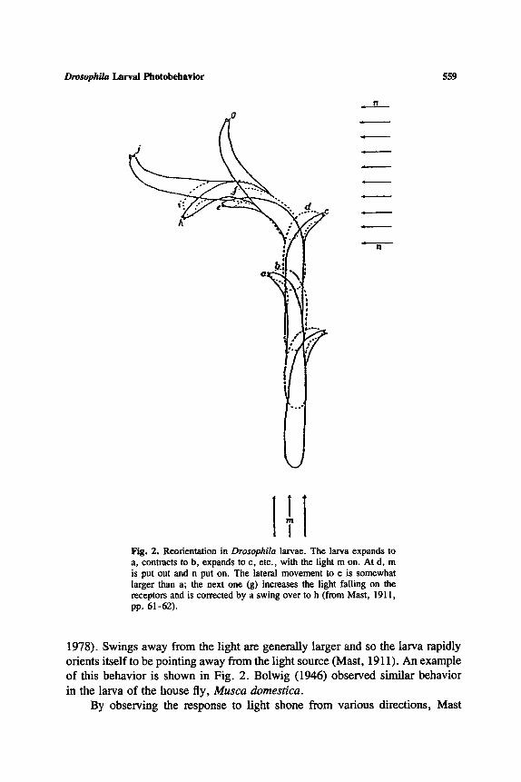
Fig. 2. Reorientation in Drosophila larvae. The larva expands to a, contracts to b, expands to e, etc., with the light m on. At d, m is put out and n put on. The lateral movement to e is somewhat larger than a; the next one (g) increases the light failing on the receptors and is corrected by a swing over to h (from Mast, 1911, pp. 61-62).
1978). Swings away from the light are generally larger and so the larva rapidly orients itself to be pointing away from the light source (Mast, 1911). An example of this behavior is shown in Fig. 2. Bolwig (1946) observed similar behavior in the larva of the house fly, Musca domestica.
By observing the response to light shone from various directions, Mast

560 Sawin, Harris, Campos, and Sokolowski
(1911) concluded that it was the amount of light falling on the larva's anterior end which influenced the behavior, that is, the larva sought positions in which the anterior end was exposed to the least amount of light. Ellsworth (1933) demonstrated that the cephalic lobes were involved in some way in the photo- tactic control of the behavior of the larva of the fleshfly, Lucilia sericata, whose behavior is similar to that of Drosophila. He severed the lobes and found that locomotion was no longer responsive to light.
Negative phototaxis is seen for most of the larval life of D. melanogaster. This is consistent with the lifestyle of the larva in this stage. It burrows into the nutritive substrate, e.g., fruit. Negative phototaxis likely helps direct the larva into a food source. This occurs in concert with other relevant sensory infor- mation such as the presence of certain chemicals and moisture concentrations (Lilly and Carlson, 1990). Near the end of the third and final instar the larva's behavior undergoes a dramatic change. In preparation for pupation it starts to seek dry surfaces, rather than the moist depths of fruit. Its previous negative phototaxis changes to positive phototaxis.
POSITIVE PHOTOTAXIS
As mentioned above, in the late third instar, a general change occurs in the behavior of D. metanogaster larvae: They cease foraging and commence what has been called "wandering behavior" (Sokolowski et al., 1984). This behavior precedes pupation and presumably has the effect of optimizing the choice of pupation site. One feature of wandering behavior is positive phototaxis (Godoy-Herrera et al., 1992). An example of positive phototaxis is shown in Fig. 3. The data illustrated here were obtained in our laboratory and are pre- sented in a format that allows comparison with the data presented in Fig. 2, which date from 1911. There are no obvious gross anatomical changes which occur at the time of the emergence of wandering behavior (A. R. Campos, unpublished results). The existing sensorimotor apparatus must be used but in a different, and in the case of phototaxis, opposite way.
The reversal of phototaxis provides an exciting opportunity to relate motor behavior to sensory input. Premorphogenic behavior is highly stereotyped in Drosophila and occurs at a specific developmental stage, thereby allowing a detailed analysis of the roles of internal factors, e.g., hormones such as ecdysone and juvenile hormone, as well as external factors, e.g., photoperiods, which may influence this behavior (Dominick and Truman, 1984, 1985).
SENSORIMOTOR TRANSFORMATIONS IN LARVAL PHOTOTAXIS
Phototactic behavior thus forms an interesting challenge to explain in terms of the sensorimotor transformations involved. Any model must be flexible enough to explain the reversal in phototaxis found in the third instar but must also be constrained to use the neural machinery available.

Drosophila Larval Photobehavior 561
LIGHT" AT
O ~
h "I'HROUC~H END [ ~
£.k- -8 --j (~I LIGI-IT A-l" a IHRoUEIH 8
Fig. 3. Positive phototaxis in a wandering D. melanogaster larva. The larva expands (a) and swings its anterior laterally, in response to onset of light right, then expands (c) and moves toward the light stimuli (d-f). Right light is put out and left light is put on. Larva swings laterally (h-m), contracts, and moves toward light (n-p).
Phototaxis involves both the neural assimilation of sensory information and the modulation of motor responses. What processes might take place between these parts? Broadly there are two ways of achieving phototaxis. The larva needs to ascertain which areas of the world are darker (or lighter) than others. It is necessary to compare samples of sensory information to make this decision, but the comparison can be made in space or time. Fraenkel and Gunn (1961) have used the terms klinotaxis and tropotaxis to describe these strategies. Klinotaxis is their term for comparison of stimulus intensity at successive movements in time; tropotaxis refers to a spatial comparison. The two possibilities are illus- trated in Fig. 4.
A temporally based decision (ldinotaxis) involves comparing the outputs of the sense organs at different times. This is illustrated in Fig. 4A. The method involves some kind of "memory ," since each sample of sensory input needs to be compared to a previous set. An example is provided by the negative photo- taxis of the blowfly larva Calliphora (Fraenkel and Gunn, 1961). The larva crawls away from light by turning its head alternately to the left and right and swinging its body away from the side to which the head is turned when a stronger light is received. It is when the light fails, rather than where the light falls, that matters. So if a light directly overhead is switched on only when the blowfly

562 Sawin, Harris, Campos, and Sokolowski
1 '"'"' 1 I I t l l l l
I I I I ! ! ! ! COMPARE IN TIME "make output smaller"
I t m lltll 1
444 ~ ~ 144 l COMPARE IN SPACE
"go to side with smal est output"
Patch right side
1 1
I I IIII
B LEFT TURNING
Patch right side
1 1
,111-4 4N~ I
D RIGHT TURNING
Fig, 4. Turning experiment in Drosophila, Diagrammatic rep- resentation of two possible mechanisms for negative photo- taxis in the Drosophila larva. The larva is shown as in Fig. 1 but with the translucent epithelium removed. Arrows indi- cate output from the photoreceptors located in shielded pock- ets beneath the condoyle spine (see Fig. 1). Underneath each arrow, the expected neural activity is indicated by the thick gray arrow. (A, B) The outputs are summed, making the an- imal like a photocell. By comparing the output at different times and applying a simple rule ("make output smaller"), the animal will move away from the light. (C, D) An alter- native mechanism in which the outputs of the two sides are compared. Again, by applying a simple rule ("go to side with smallest output"), negative phototaxis can be achieved. The two possibilities can be distinguished by patching one side. This will result in turning (see tex0 away from the patched side (to the left in the case of right patching; B) if the outputs are summed but toward the patched side (D) if the outputs are compared.
l a rva tu rns i ts h e a d t o w a r d the r ight , i t c r a w l s c o n t i n u o u s l y t o w a r d the lef t
( n e g a t i v e k l ino tax i s ) .
I t is no t n e c e s s a r y tha t the v i sua l o r g a n s o f la rva l D i p t e r a are d i rec t iona l
for k l ino tax is . I f t he re are d a r k e r o r l i gh t e r a reas ava i l ab le , the o r g a n i s m wil l

Drosophila Larval Photobehavior 563
eventually find the desired level by chance. Perhaps this is what Holmes (1905) had in mind when he talked about a "trial-and-error" mechanism.
A spatially based decision system (tropotaxis) compares the information coming from the different parts of space. This is illustrated in Fig. 4C. Tro- potaxis requires that the visual organs are directional and that they sample dif- ferent areas of space. Choosing to move in the direction sampled by the organ with the lowest activity leads to negative phototaxis. Daphnia, ArmadiUidum, and Planaria exhibit this sort of tropotaxis (Fraenkel and Gunn, 1961) and can move directly toward or away from a source of stimuli without turning alter- nately toward the left or right. A refinement is to compare different parts of the image of a single visual organ, as animals with optically focused eyes can do
Which system does the Drosophila larva use? The swinging of the head when a light is turned on is suggestive of klinotaxis. On the other hand, the anatomical structure of the visual organs at the bottom of little pockets suggests that they are directional, which is compatible with tropotaxis.
Figure 4 illustrates a simple test that could be performed on D. melano- gaster to distinguish between the two possible mechanisms. Removing the input from one eye makes opposite predictions for the two mechanisms. Both predict that turning should occur, but the two mechanisms predict turning in the opposite directions. The input from one visual organ could be removed by painting it over, patching it, removing it surgically, or, most elegantly, performing genetic manipulation (see below). Consider a negatively phototactic larva with a patch painted over the right visual organ and a constant source of light placed directly in front of the larva (Fig. 4). A temporally based direction (klinotaxis) results in turning away from the patched side (Fig. 4B). A spatially based decision mechanism (tropotaxis), however, predicts turning toward the side with the patch (Fig. 4D).
Whichever sensory strategy (klinotaxis or tropotaxis) is used, the ohtput of the sensory decision rule ("move to side with the smallest output" or "make output smallest") needs to be converted to a batch of signals in the appropriate form to the appropriate muscles. What pattern of signals should leave the central nervous system to cause appropriate body movements is not known. At first glace it would seem that, at the time of reversal, either the rule could be reversed or simply the output signal (from negative to positive phototaxis) could be redirected. This is not the case, however, since if the muscle arrangement was switched but the rule was kept the same, the system would be in open-loop, that is, the behavior would no longer tend toward canceling the sensory input and an unstable situation would result.
CONCLUSIONS
The change from negative to positive phototaxis is reminiscent of reversals in other parts of the animal kingdom that occur as the development phase of an animal puts opposing demands on the nervous system. The development of the

564 Sawin, Harris, Campos, and Sokolowski
flatfish is a well-documented example. In this species, the eyes migrate from lateral positions in the larvae to both being on the same side as an adult (Graf and Baker, 1983, 1990). Minimal anatomical rearrangement occurs in the central nervous system of an metamorphosing flatfish and an essentially identical ner- vous system has to control the oppositely directed eye movements of the adult for a given sensory input. This is achieved by altering synaptic weightings, a system that might also be employed by the Drosophila larva.
Of considerable interest is the possibility of external stimuli reconfiguring the neural circuitry at a critical time in the alteration of physiological develop- ment such as the onset of larval wandering in the late third instar. The Droso- phila larva can be at least as useful as the leech for investigating the ways that simple neural nets can carry out complex conversions of sensory to motor infor- mation and maintain flexibility (Churchland and Sejnowski, 1987; Keshishian and Chibe, 1993; Lockery and Sejnowski, 1993). What it loses by its incon- venient size compared to the leech, it gains by the immense power opened up by our superior genetic understanding of D. melanogaster and the possibility of directly exploring the genetic basis of behavior. The glass mutation, in which these mutants appear to be missing the larval visual organ, provides us with a genetic tool to test the model described above. This can be done by generating larvae that are mosaic (chimeras) for normal and mutant (in this case glass) tissues (Hall, 1978). In this way a larva can be constructed that is missing part of (e.g., the left or right side) its visual system. One can think of it as a way to "patch" different parts of the visual system genetically with mutant glass tissue. The data collected on the phototactic behavior of mosaic larvae will provide a test for the model presented in Fig. 4,
ACKNOWLEDGMENTS
The authors would like to thank Helen Rodd for help with statistical anal- yses. Gus Lagos and Brian McCormack provided technical assistance. This work was supported by a Human Frontiers Science Program Research Grant to M.B.S. which supported E.P.S. and a University Research Fellowship to M.B.S.L.R.H. is sponsored by the NSERC of Canada and the Institute of Space and Terrestrial Sciences. A.R.C. is supported by the NSERC of Canada.
REFERENCES
Alvarez, G., Farina, J., and Fontdevila, A. (1979). Density and frequency-dependent selection on the singed locus of Drosophila melanogaster. Genetica 50: 161-I66.
Ashbumer, M. (1989). Drosophila, a Laboratory Handbook, Cold Spring Harbor Laboratory Press, Cold Spring Harbor, NY.
Ashburner, M., and Wright, T. R. F. (1978). The Genetics and Biology of Drosophila, Vol. 2B, Academic Press, London.

Drosophila Larval Photobehavior 565
Bodenstein, D. (1950). The postembryonic development of Drosophila. In Demerec, M. (ed.), Biology of Drosophila, J. Wiley and Sons, New York, pp. 275-364.
Bolwig, N. (1946). Sense and sense organs of the anterior end of the house fly larvae. Vidensk. Medd. Dan. Natarhist. Foren. 109: 81-217.
Campos, A. R., Grossman, D., and White, K. (1985). Mutant alleles at the locus clay in Drosophila melanogaster lead to nervous system defects. A developmental-genetic analysis. J. Neurogenet. 2: 197-218.
Campos, A. R., Rosen, D. R., Robinow, S. N., and White, K. (1987) Molecular analysis of the locus elav in Drosophila melanogaster: A gene whose embryonic expression is neural specific. EMBO J. 6: 425--431.
Campos, A. R., Lee, K. J., and Stelter, H. (1994). Establishment of neuronal connectivity during development of the Drosophila larval visual system (submitted for publication).
Churchland, P. S., and Sejnowski, T. J. (1987). The Computational Brain, MIT, Boston. Demerec, M., and Kaufmann, B. P. (1940). Drosophila Guide, Carnegie Institution of Washington
Publication, Washington, DC. Dominick, O. S., and Truman, J. W. (1986a). The physiology of wandering behaviour in Manduca
sexta. III. Organization of wandering behaviour in the larval nervous system. J. Exp. Biol. 121: 115-132.
Dominick, O. S., and Truman, J. W. (1986b). The physiology of wandering behaviour in Manduca seta. IV. Hormonal induction of wandering behaviour from the isolated nervous system. J. Exp. Biol. 121: 131-151.
Dominick, O. S., and Truman, J. W. (1985). The physiology of wandering behaviour in Manduca sexta. 11. The endocrine control of wandering behaviour. J. Exp. Biol. 117: 45-68.
Dominick, O. S., and Truman, J. W. (1984). The physiology of wandering behaviour in Manduca sexta. I. Temporal organization and the influence of the internal and external environments. J. EXp. Biol. 110: 35-51.
Ellsworth, J. K. (1933). The photoreceptive organs of a fleshfly larvae, Lucilia sericata: An exper- imental and anatomical study. Ann. Entomol. Soc. Am. 26: 200-215.
Fischbach, K. F. (1983). Neural cell types surviving congenital sensory deprivation in the optic lobes of Drosophila melanogaster. Dev. Biol. 95: 1-18.
Fogelman, J. C., and Markow, T. A. (1982). Behavioral differentiation between two species of cactophilic Drosophila. II. Pupation site preference. Southwest Nat. 27: 315-320.
Fmenkel, G. S., and Gunn, D. L. (1961). The Orientation of Animals, Dover, New York. Godoy-Herrem, R., Cifuentes, L., de Arcaya, M. F. D., Fernandes, M., Fuentes, M., Reyes, I.,
and Valderrama, C~ (1989). The behaviour of Drosophila melanogaster larvae during pupation. Anim. Behav. 37: 820-829.
Godoy-Herrera, R., Alareon, M., Caceres, H., Loyola, I., Navarrete, I., and Vega, J. L. (1992). The development of photoresponse in Drosophila melanogaster larvae. Revista Chilena de Historia Natural 65: 91-10t.
Graf, W., and Baker, R. (1983). Adaptive changes of the vestibular-ocular reflex in flatfish are achieved by reorganization of central nervous pathways. Science 221: 777-779.
Graf, W., and Baker, R. (1990). Neuronal adaptation accompanying metamorphosis in the flatfish. J. Neurobiol. 21: 1136-1152.
Green, P., Hartenstein, A. Y., and Hartenstein, V. (1993). The embryonic development of the Drosophila visual system. Cell Tissue Res. 273: 583-598.
Grossfield, J. (1978). Non-sexual behavior of Drosophila. In Ashburner, M., and Wright, T. R. F. (eds.), The Genetics and Biology of Drosophila, Vol. 2B, Academic Press, New York, pp. 1-126.
Hall, J. C. (1978). Behavioral analysis in Drosophila mosaics. In Gehring, W. J. (ed.), Genetic Mosaics and Cell Differentiation, Springer-Verlag, Berlin, pp. 259-305.
Hall, J. C., Greenspan, R. J., and Harris, W. A. (1982). Genetic Neurobiology, MIT Press, Cambridge, MA.
Hardie, R., and Minke, B. (1992). The trp gene is essential for a light activated Ca 2+ channel in Drosophila photoreceptors. Neuron 8: 643-651.
Heisenberg, M., and Wolf, R. (1984). Vision in Drosophila Genetics of Microbehavior, Springer- Verlag, Berlin.

566 Sawin, Harris, Campos, and Sokolowski
Holmes, S. J. (1905). The selection of random movements as a factor in phototaxis. J. Comp. Neurol. Psychol. 15: 98-112.
Jennings, H. S. (1904). Contributions to the study of the behavior of lower organisms. Publ. Carnegie Inst. Wash. 16: 256.
Kankel, D. R., Ferrus, A., Garen, S. H., Harte, P. J., and Lewis, P. E. (1978). The structure and development of the nervous system. In Ashbumer, M., and Wright, T. R. F. (eds.), The Genetics and Biology of Drosophila, VoL 2D, Academic Press, New York, pp. 295-368.
Kearsey, M. J., and Kojima, K. (1967). The genetic architecture of body weight and egg hatchability in Drosophila melanogaster. Genetics 56: 23-37.
Keshishian, H., and Chiba, A. (1993). Neuromuscular development in Drosophila: Insights from single neurons and single genes. Trends Neurosci. 16: 278-283.
Lilly, M., and Carlson, J. (1990). smellblind: A gene required for Drosophila olfaction. Genetics 124: 293-302.
Lockery, S. R., and Sejnowski, T. J. (1993). The computational leech. Trends Neurosci. 19: 283- 290.
Manning, A., and Markow, T. A. (I981). Light-dependent pupation site preferences in Drosophila. II. D. melanogaster and D. simulans. Behav. Genet. 11: 557-563.
Markow, T. (1979). A survey of intra- and interspecific variation for pupation height in Drosophila. Behav. Genet. 9: 209-217.
Masai, I., Hosoya, T., Kojima, S., and Hotta, Y. (1992). Molecular cloning of a Drosophila diacylglycerel kinase gene that is expressed in the nervous system and muscle. Proc. Natl. Acad. Sci. USA 89: 6030-6034.
Mast, S. O. (1911). Light and the Behavior of Organisms, John Wiley & Sons, New York. Mast, S. O. (1938). Factors involved in the process of orientation of lower organisms in light. Biol.
Rev. 13: 186-224. Melamed, J., and Tmjillo-Cenoz, O. (1975). The fine structure of the eye imaginal discs in muscoid
flies. J. Uhrastruct. ICes. 51: 79-93. Meyerowitz, E. M., and Kankel, D. R. (1978). A genetic analysis of visual system development
in Drosophila melanogaster. Dev. Biol. 62:112-142. Mishima, J. (1964). The competition among Drosophila larvae in different growth stages. Res.
PopuL EcoL 6: 22-27. Morgan, T. H. (1907). Experimental Zoology, Macmillan, New York. Pak, W. L. (1979). Study of photoreceptor function using Drosophila mutants. In Breakfield,
X. O. (ed.), Neurogenetics, Genetic Approaches to the Nervous System, Elsevier, New York, pp. 67-99.
O'Tousa, J. E., Leonard, D. S., and Pak, W. L. (1989). Morphological defects in OraJ*84 pho- toreceptors caused by mutation in R1-6 opsin gene of Drosophila. J. Neurogenet. 6: 41-52.
Rizki, R. M., and Davis, C. (1953). Light as an ecological determinant of interspecific competition between 19. willistoni and D. melanogaster. Am. Nat. 87: 389-392.
Sameoto, D. D., and Miller, R. S. (1968). Selection of pupation site by Drosophila melanogaster and D. Simulans. Ecology 49: 177-180.
Schnebel, E. M., and Grossfield, J. (1986). The influence of light on pupation height in Drosophila. Behav. Genet. 16: 407-413.
Snodgrass, R. E. (1924). Anatomy and metamorphosis of the apple maggot. J. Agr. Res. 28: 1-36.
Sokolowski, M. B., and HanseU, R. I. (1983). Elucidating the behavioural phenotype of Drosophila melanogaster larvae: Correlations between larval foraging strategies and pupation height. Behav. Genet. 13: 267-280.
Sokolowski, M. B., Kent, C., and Wong, J. (1984). Drosophila larval foraging behaviour: Devel- opmental stages. Anim. Behav. 32: 645-651.
Sokolowski, M. B., Bauer, S. J., Wai-Ping, V., Rodriguez, L., Wong, J. L., and Kent, C. (1986). Ecological genetics and behaviour of Drosophila melanogaster larvae in nature. Anita. Behav. 34: 403-408.
Steele, F. R., Washburn, T., Rieger, R., and O'Tousa, J. E. (1992). Drosophila retinal degen- eration C rdgC encodes a novel serine/threonine protein phosphatase. Cell 69: 669-676.

Drosophila Larval Photobehavior 567
Steller, H., Fischbach, K. F., and Rubin, G. (1987). disconnected: A locus required for neuronal pathway function in the visual system of Drosophila. Cell 50: 1139-1153.
Tix, S., Minden, J. S., and Technau, G. M. (1989). Pre-existing neuronal pathways in the devel- oping optic lobes of Drosophila. Development 105: 739-746.
Vihtelic, T. S., Hyde, D. R, and O'Tousa, J. E. (1991). Isolation and characterization of the Drosophila retinal degeneration B rdgB gane. Genetic 127: 761-768.
Wehner, W. (1981). Spatial vision in anthropods. In Autrum, H. (ed.), Comparative Physiology and Evolution of Vision in Invertebrates C: Invertebrate Visual Centres and Behavior II, Springer-Vedag, Berlin/Heidelberg/New York, pp. 378-385.
White, K., and Kankel, D. R. (I978). Patterns of cell division and cell movement in the formation of the imaginal nervous system in Drosophila melanogaster. Dev. Biol. 65: 296-321.
Wong, J. L., Sokolowski, M. B., and Kent, C. (1985). Prepupation behaviour in Drosophila: Embedding. Behav. Genet. 15: 155-165.
Woodward, C., Alcorta, E., and Carlson, J. (1992). The rdgB gene of Drosophila: A link between vision and olfaction. J. Neurogenet. 8: 17-31.
Zipursky, S. L., Venkatesh, T. R., Teplow, D. B., and Benzer, S. (1984). Neuronal development in the Drosophila retina: Monoclonal antibodies as molecular probes. Cell 36: 15-26.
Zuker, C. S. (1992). Phototmnsduction in Drosophila: A paradigm for the genetic dissection of sensory transduction cascades. Curr. Opin. Neurobio. 2: 622-627.