dr. ye myint mbbs; da (london); frca sheffield, uk. education
TRANSCRIPT

Management of ShockDr. Ye Myint
MBBS; DA (London); FRCAConsultant in Anaesthesia and Critical
Care Medicine, Barnsley Hospital.Hon Senior Lecturer, University of
Sheffield, UK.Director of Postgraduate Medical
Education.
• Different types of shock• Early detection• General Management• Pathophysiology• Management• IV fluid
Different types
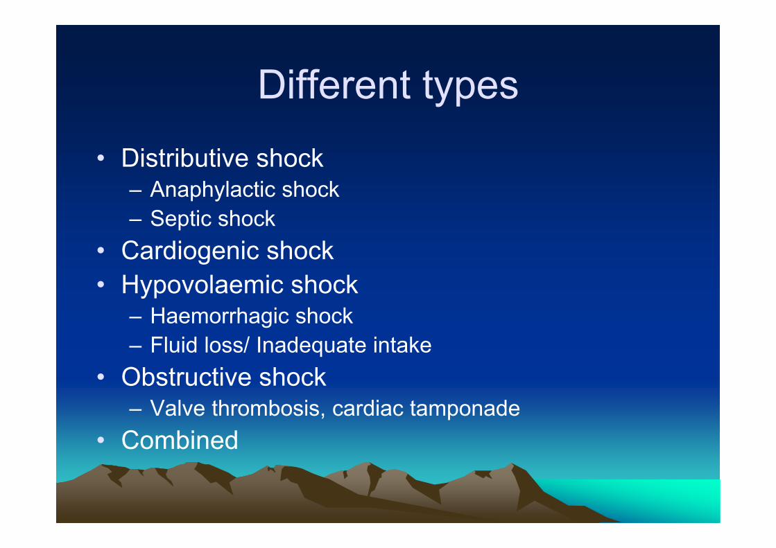
• Distributive shock– Anaphylactic shock– Septic shock
• Cardiogenic shock• Hypovolaemic shock
– Haemorrhagic shock– Fluid loss/ Inadequate intake
• Obstructive shock– Valve thrombosis, cardiac tamponade
• Combined
Resuscitation Guidelines 2010
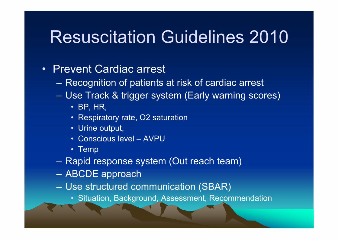
• Prevent Cardiac arrest– Recognition of patients at risk of cardiac arrest– Use Track & trigger system (Early warning scores)
• BP, HR, • Respiratory rate, O2 saturation • Urine output, • Conscious level – AVPU• Temp
– Rapid response system (Out reach team)– ABCDE approach– Use structured communication (SBAR)
• Situation, Background, Assessment, Recommendation

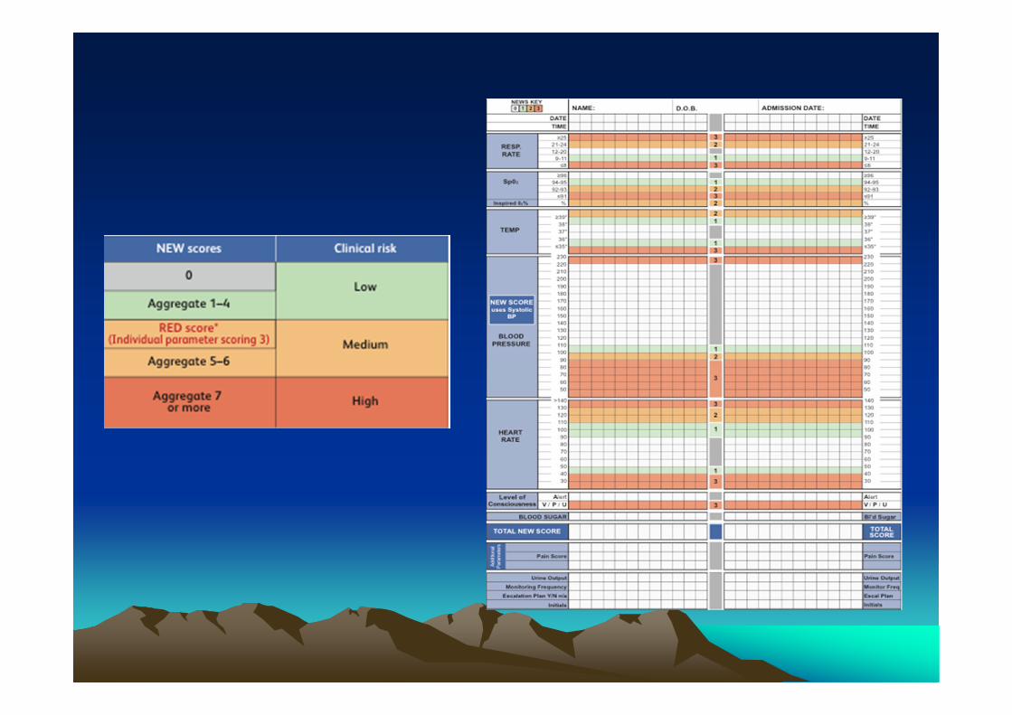
Early warning Scoring system
• National EWS• Local (Barnsley)• Inadequate O2 delivery despite
– Normotensive– Hypertensive

NEWS

Barnsley EWS
3 2 1 0 1 2 3HR <40 41-44 45-59 60-90 91-110 111-129 130
BP syst <85 85-95 96-160 161-190 >191Resps <10 10-20 21-25 26-34 35+
Oxygen Saturation Less than 94%
Temp <35.5 36-38 >38
Conscious level


Anaphylaxis
• Risk factors for severe anaphylaxis– Asthma– Medication
• β blocker (May need Glucagon 1 mg IV)• Angiotensin converting enzyme inhibitors, NSAID
– Acute respiratory infection– Mastocytosis– Alcohol, emotional stress, fever

Anaphylactic shock
• Call for help– Atypical presentation during anaesthesia
• Check A, B , C • High flow O2 (100% O2)• CPR if required• Stop giving the triggering drug
– Latex, Food, blood products
Anaphylactic shock
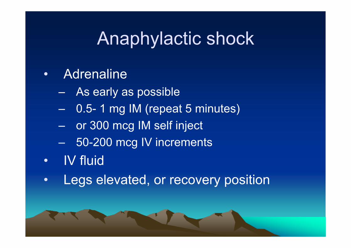
• Adrenaline– As early as possible– 0.5- 1 mg IM (repeat 5 minutes)– or 300 mcg IM self inject– 50-200 mcg IV increments
• IV fluid• Legs elevated, or recovery position

Anaphylactic shock
• Second line treatment– Antihistamine– Chlorphenamine 10 mg IV or IM– Hydrocortisone 200 mg IV– Bronchodilator
• Catecolamine infusion• Check Arterial blood gases• Check airway oedema

Anaphylactic shock• Bronchospasm
– Salbutamol (nebulizer or IV)– Ipratropium– IV Aminophylline or Magnesium sulphate
• Observe for delayed problems (Bi-phasic)• Oral antihistamine & corticosteroid 3 days• Arrhythmias, • Coronary spasm, ACS• Further investigation (allergy diagnosis)• Incident reporting

General Investigations
• Bedside– ECG– Haemoglobin– Arterial blood gases,– Lactate– Ultrasound, – Echocardiogram

General Investigations
• Bedside– ECG– Haemoglobin– Arterial blood gases,– Lactate– Ultrasound, – Echocardiogram

General Investigations
• Laboratory– Full blood count– Coagulation, D dimer– U & E– LFT– Cardiac enzymes– Cultures (urine, blood, sputum)– Toxicology
General Investigations
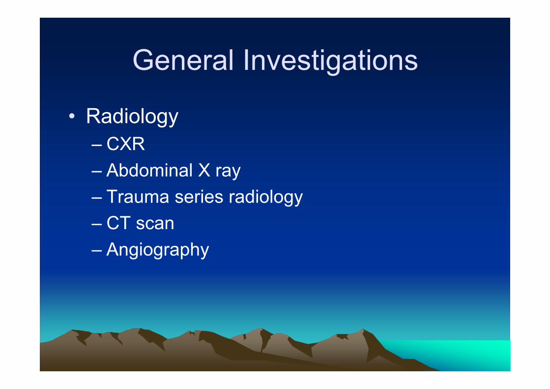
• Radiology– CXR– Abdominal X ray– Trauma series radiology– CT scan– Angiography

General Management of Shock
• Supply Oxygen• Vascular access• Volume resuscitation• Vasoactive drugs• Manage precipitating illness/ injury• Monitoring§

Fluid responsiveness
• Static measure– Intra cardiac pressure
• CVP (Limitation)• Pulmonary artery occlusion pressure
– Cardiovascular volume• Echo – LVEDV
– Oesophageal Doppler• Corrected Flow time• Peak velocity

Fluid responsiveness
• Dynaemic measure– Responsive to fluid challenge– Passive leg raising test
• Aortic flow• Pulse pressure
– Response to IPPV• Systolic pressure variation• Pulse pressure variation• Stroke volume variation

CVP
• Frank Starling law• CVP does NOT indicate volume status• Normal CVP does not exclude
hypovolaemia• High CVP
– May need fluid, may respond fluid challenge• Trend may be useful
Other tools to assess Volume status
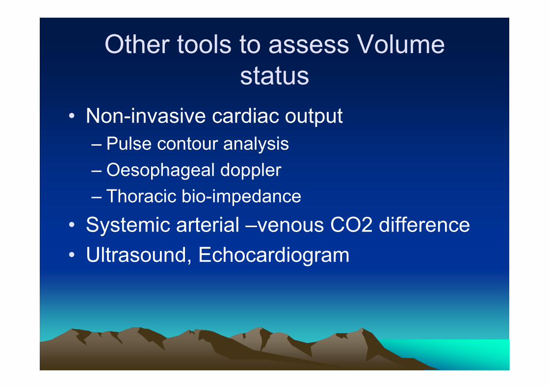
• Non-invasive cardiac output– Pulse contour analysis– Oesophageal doppler– Thoracic bio-impedance
• Systemic arterial –venous CO2 difference• Ultrasound, Echocardiogram
Cardiogenic shock
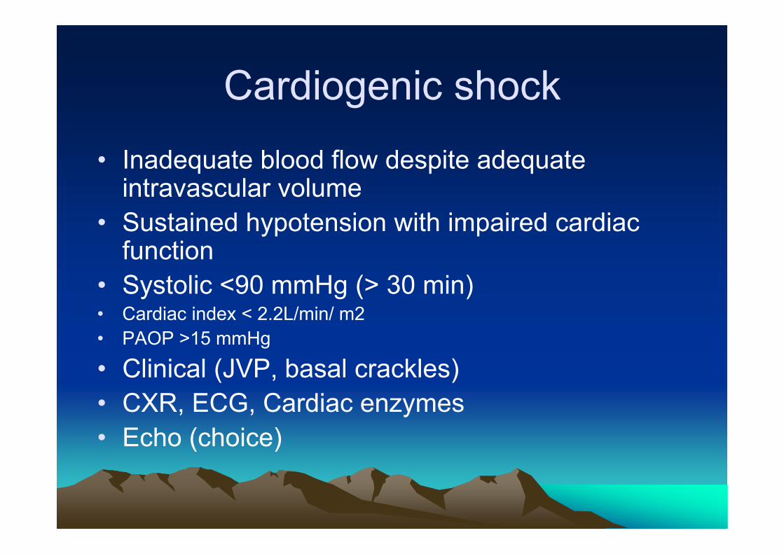
• Inadequate blood flow despite adequate intravascular volume
• Sustained hypotension with impaired cardiac function
• Systolic <90 mmHg (> 30 min)• Cardiac index < 2.2L/min/ m2• PAOP >15 mmHg
• Clinical (JVP, basal crackles)• CXR, ECG, Cardiac enzymes• Echo (choice)

Cardiogenic shockCauses
• Acute MI– Pump failure
• Mechanical complications– MR, VSD, Tamponade
• Others– Cardiomyopathy– Myocarditis– Cardiac contusion– Septic shock– Subarachnoid haemorrhage– Massive PE

• Systolic dysfunction– SV, Cardiac Output reduced– Hypotension– Reduced coronary perfusion pressure– Ischaemia
• Diastolic dysfunction– Pulmonary congestion– Hypoxaemia- Ischaemia
• RV infarct- Give Fluid (needs high filling pressure)

Cardiogenic shockManagement
• Urgent echocardiogram• Restore haemodynaemics, oxygenation
– Avoid arrythmias• Without significant pulmonary oedema
– O2– Fluid challenge– Vasopressor
• With pulmonary oedema– O2, CPAP (NIV)– Inotropes (Noradrenaline, Dobutamine)

Cardiogenic shockManagement
• Vasopressin• Phosphodiesterase inhibitors (Milrinone)
– RV infarct• Levosimendan ?? (calcium sensitizer)
– Coronary vasodilatation• Mechanical therapy
– Intra aortic balloon pump• Revascularization
– Thrombolyse, PCI, CABG

Cardiogenic shockPathophysiology- Microcirculation
• Microcirculatory function deteriorated during shock– Disturb flow to heart & brain– Vital organs
• Multi-organ failure• Monitor
– Cardiovascular MRI– Hand held video microscopy – sublingual
microcirculation

Cardiogenic shockPathophysiology-
• Persistent inflammatory response (SIRS) in severe heart failure
• Increased vascular permeability• Increased blood viscosity• Hypercoagulopathy (platelet activation)• Endothelial dysfunction (reduced NO)

Vasoactive agents
• Angiotensin II inhibitors– Improve microcirculation
• Intravenous Nitrogylcerin• Adrelanline- reduce microcirculatory flow
– Ischaemic vital organs
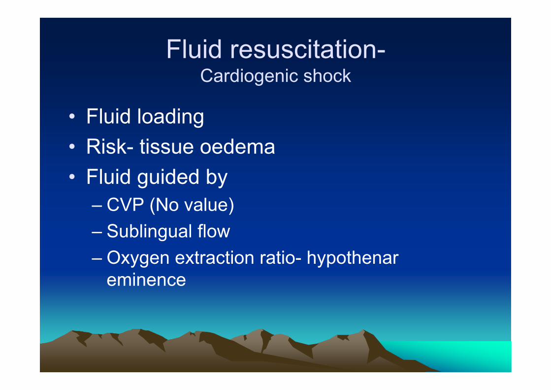
Fluid resuscitation-Cardiogenic shock
• Fluid loading• Risk- tissue oedema• Fluid guided by
– CVP (No value)– Sublingual flow– Oxygen extraction ratio- hypothenar
eminence

Heart rescue (ACS)
• Coronary revascularization– Fibrinolytic therapy– PCI (within 90 min)– CABG
• Cardiac surgery (VSD, Acute MR)• Mechanical circulatory support
– Intra –aortic balloon pump• LMWH• Antiplatelets

Post cardiac arrest
• Therapeutic hypothermia– 12-24 hours– 32-34 degree C
• Adverse Microcirculatory effect– 36 d C (NEJM, December 2013)

Hypovolaemic shock
• Trauma• Non trauma
– Medical– Obstetrics

Trauma (without head injury)• C (control bleeding) - ABC• Damage controlled resuscitation
– Hypotensive resuscitation– Damage controlled surgery
• Control haemorrhage & contamination• Definitive repair later
– Haemostatic resuscitation• Correct coagulopathy (early), hypothermia,
acidosis in ICU

Hypotensive resuscitation• Permissive hypotension
– End point of resuscitation 70-80mmHg • (Cannon & Fraser, JAMA 1918)
– Systolic BP 90 mmHg (80-100 mmHg),– except head injury-Systolic 120mmHg– Palpable radial pulse
• IV cannula (Intra-osseous route)• Minimal IV fluid
– Hypertonic saline

IV fluid
• Isotonic crystalloids– Ischaemia, reperfusion injury– Abdominal hypertension, Abdominal
compartment syndrome– ARDS, Multi-organ failure– Coagulopathy

Haemostatic resuscitation
• Acute Traumatic Coagulopathy (TIC)
• Early use of Tranexamic acid• (CRASH 2 trial, Lancet 2010)
– Within 3 hours– 1g over 10 min– 1 g over 8 hours– Cost effective

IV fluid -Hypertonic saline
– Rapid restoration of intravascular volume– Reduce intracranial pressure– Reverse endothelial swelling
(microcirculation)– Immunomodulation-
• Less cytokines– Lower ARDS, Renal failure, coagulopathy– Meta-analysis- Increased survival

Lethal triad(bloody vicious cycle)
• Hypothermia (keep >35 ° C)– More bleeding
• Affect clotting factors• Platelet dysfunction • sequestration in liver & spleen
• Acidosis– Reduce cardiac output (contractility)– Dysarrhythmia– bleeding
• Coagulopathy

Haemostatic resuscitation
• High FFP: RBC ratio (early)• Platelets• Cryoprecipitate• Calcium
– Keep >1.15 mmol/L• Activated Factor 7• Prothrombin complex ?

Trauma with head injury• ABC (O2 +Cervical spine)
– Prevent secondary injury• Maintain cerebral perfusion pressure
60-70 mmHg– Keep systolic >90 mmHg– Assume ICP of 20 mmHg in unconscious
• Role of hypertonic saline– More effective than Mannitol
• Early use of Vassopressors
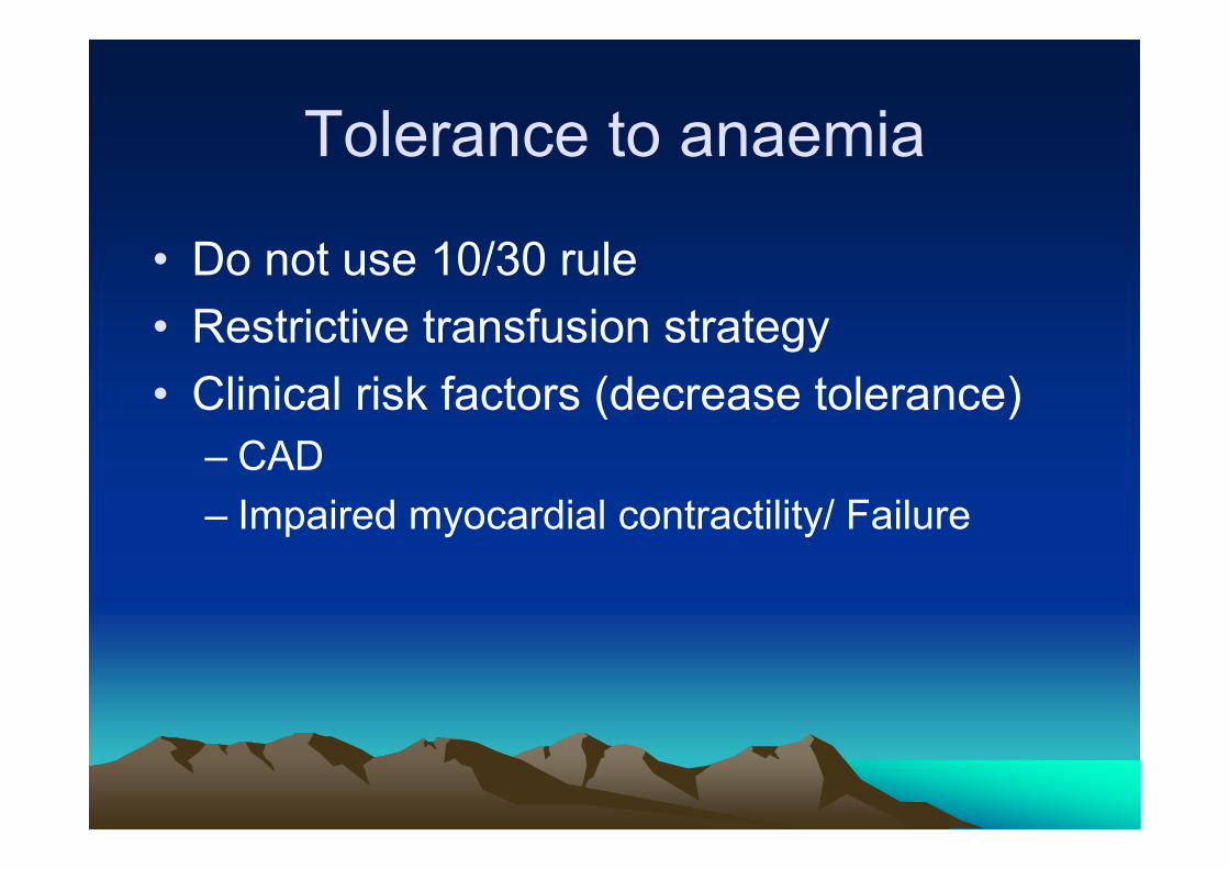
Tolerance to anaemia
• Do not use 10/30 rule • Restrictive transfusion strategy• Clinical risk factors (decrease tolerance)
– CAD– Impaired myocardial contractility/ Failure

Microvascular bleeding
• PT/ APTT >1.5 – give FFP• Platelets <50-100 – give platelets• Fibrinogen <1.5 g/L – give Cryoprecipitate
– <2 g/L in Obstetric – One adult dose raise Fibrinogen by 1 g/L– Consider Fibrinogen concentrate
• Dying from bleeding (Activated Factor 7)

Bleeding patient
• To reverse Warfarin– Vit K +/- Prothrombin Complex Concentrate– FFP when PCC is unavailable
• Give Platelets– Expect platelet <50 after 2 blood volume
replacement– Give Platelets when count is <50– Adult dose raise platelets by 20

Massive Blood Transfusion
• Replacement of > 1 blood volume (5L) in <24 hr.
• 50% blood volume lost in 3 hours• Loss 150 ml/min• Pathophysiology
– Dilution / consumption– DIC– Systemic fibrinolysis– Platelet dysfunction

Therapeutic goals
• Maintain tissue perfusion & oxygenation– Restore blood volume & Hb
• Stop bleeding (source)– Ultrasound, CT scan
• Correct coagulopathy• Shock pack box
– 2 Units of blood + 2 units FFP• Fresh blood (< 14 days old)

Management
• C – ABC– O2, 2 IV cannulae
• Inform– Blood bank– Haematology laboratory– Haematologist, – Surgeons– ICU consultants/ Anaesthetists

Accept hypotension
• Multiple trauma with active bleeding• Penetration injury• Major vessel or cardiac injury• Do not give large volume of fluid• Can feel a palpable pulse?

Management
• Colloid/ crystalloid• Blood transfusion
• Keep the patient warm– Avoid exacerbating coagulation problems
• Investigations

Investigations
• FBC – Haematocrit– Platelet count
• Coagulation screen• U & E• Request blood & blood products • Arterial Blood Gases

Blood Transfusion Guidelines
• Should not transfuse if Hb is > 10g/dl
• A strong indication - Hb < 7 g/dl
• Hb between 8 -10 g/dl is safe even for those with cardiorespiratory disease
• Symptomatic patients should be transfused

Recent British guidelines (2012)Critically Ill patients
o Transfusion threshold 70g/L • trigger not > 90 g/L• Target 70-90 g/L
o Traumatic brain injury – Target 70-90 g/L
o Single unit transfusions- recommended (especially in non-bleeding patients)

Recent British guidelines (2012)Critically Ill patients
o Subarachnoid Haemorrhage - 80-100 g/L
o Ischaemic stroke- maintained > 90g/L
o ICS – maintained >80-90 g/L

Summary
• Initial resuscitation & prevention of further bleeding
• Diagnosis & monitoring of bleeding• Rapid control of bleeding• Management of bleeding & coagulation• Tissue oxygenation, fluid & hypothermia

Summary
• Damage controlled resuscitation• Permissive hypotension• Haemostatic resuscitation
– Massive blood transfusion

Obstructive shock
• Support (ABC)• Treat the cause (Urgent)
– Cardiac tamponade– PE
• Thrombolysis• Anticoagulation• Thrombectomy

Septic shock
• Hypovolaemia- from fluid loss• Maldistribution- from vasodilatation
– Reduce peripheral vascular resistance• Increased permeability- tissue oedema• Reduced Contractility- Myocardial depressant
factors– Ischaemia
• Late- mitochondrial failure– Fluid fail to improve microcirculation

Septic Shock• Surviving sepsis campaign (2012)• Early goal directed therapy (Rivers 2001NEJM)
– First 6 hours– Fluid challenge– MAP >65 mmHg (vasopressors)
• Noradrenaline
– CVP goal 8-12 mmHg (Limited Value)– Central venous O2 saturation
• keep >70%• Fluid, (Blood transfusion), Dobutamine
• Sepsis bundle• Haemoglobin 7-9g/dl

Septic Shock
• Early recognition• 2 or > blood culture• Early & adequate antibiotic therapy
(within1 hour)– De-escalation therapy
• Source control• Early hemodynamic resuscitation support

Septic Shock
• Corticosteroids– Hydrocortisone 50 mg IV 6 hourly
• Metabolic support – Glycaemic control – 8-10 mmol/L
• NICE SUGAR study NEJM• SSC – Maintain below 10 mmol/L
– Early enteral nutrition – Immuno nutrition

Septic Shock• Respiratory support
– Tidal volume 6 ml/Kg– Limit pressure <30 cm H2O– Permissive hypercarbia– Adequate PEEP– Consider Prone -ARDS– 30-45 d head up
• Renal support• Stress Ulcer prophylaxis• DVT prophylaxis (daily assessment)

Management of septic shock
• AB (Correct low SaO2 - High flow O2) • C- Circulation
– correct hypovolaemia (colloid, crystalloid)– correct pump failure– early goal directed therapy– correct coagulopathy
• Specific (antibiotics)• Supportive measures
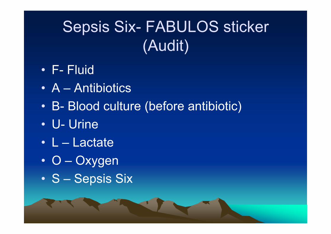
Sepsis Six- FABULOS sticker(Audit)
• F- Fluid• A – Antibiotics• B- Blood culture (before antibiotic)• U- Urine• L – Lactate• O – Oxygen• S – Sepsis Six

Central Venous O2 Saturation
• Global tissue hypoxia may persist after resuscitation
• Normal mixed venous O2 saturation (Sv O2 65-75% )
• Low SvO2 = Low O2 delivery or demand exceed the supply

Noradrenaline
• Improve MAP• Increase GFR• Improve renal function
• Adrenaline use should be limited

Vasopressin
• Relative deficiency• V1a receptors
• Vasopressin 0.01- 0.04 units/min• Terlipressin every 6 hr

Dobutamine
• Combined with N-Adrenaline• 5 -20 mcg/kg/min (septic shock)

Adrenaline
• Alone• combinations


IV Fluid
• Use fluid as a drug• NICE guidelines (December 2013)• Crystalloids vs colloid• Normal Saline• Balanced salt solution
– Hartmann• Colloid
– HES– Albumin

Normal Saline
• Abnormal• Hyper chloraemic acidosis
– Renal vasoconstriction• Hypernatraemia• More cytokines released • Risk of renal failure
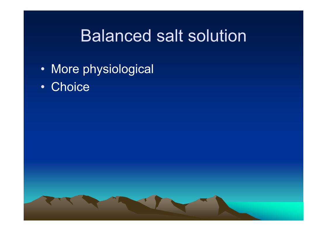
Balanced salt solution
• More physiological• Choice

Colloid
• Avoid HES (Renal failure)• Albumin
– Do not use in head injury

De-resuscitation
• Fluid overload- worse outcome• Ebb Phase• Persistent Ebb phase
– Impaired fluid mobilization• Flow Phase
– Conservation fluid– Diuretics– Renal replacement therapy (CVVH)- Negative
balance

An Early Warning System
Score 3 2 1 0 1 2 3
Pulse < 40 41-50 51-100 101-110 111-130 > 131
Resp rate 8 9-14 15-20 21-29 30
Temp 35.0 35.1-36 36.1-38 38.1-38.5 38.6
CNS level Unresp Pain Voice Alert New
confusion
Urine output <10ml/hr <0.5ml/kg/hr
BP <70 71-80 81-100 101-199 >200

Alert Plan of Assessment
No/UnsureYes
Yes
No