dr ricardo faundezklinikakoni.sggw.pl/attachments/article/82/wyk_5_postępy... · 2018. 4. 16. ·...
TRANSCRIPT

Dr Ricardo Faundez
Katedra Dużych Zwierząt z Kliniką
Wydział Medycyny Weterynaryjnej SGGW
*

*
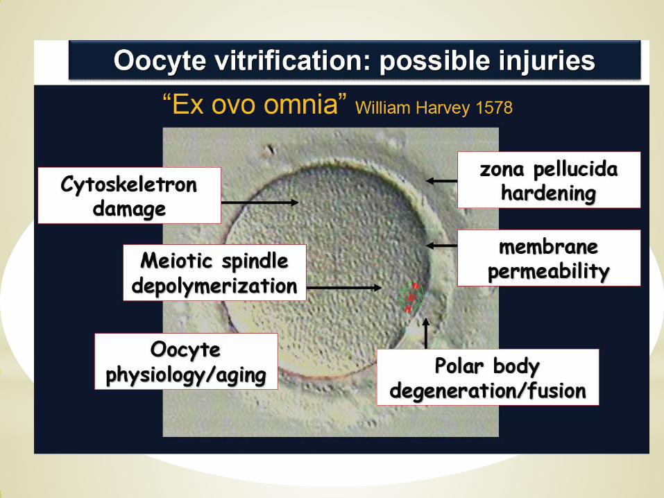
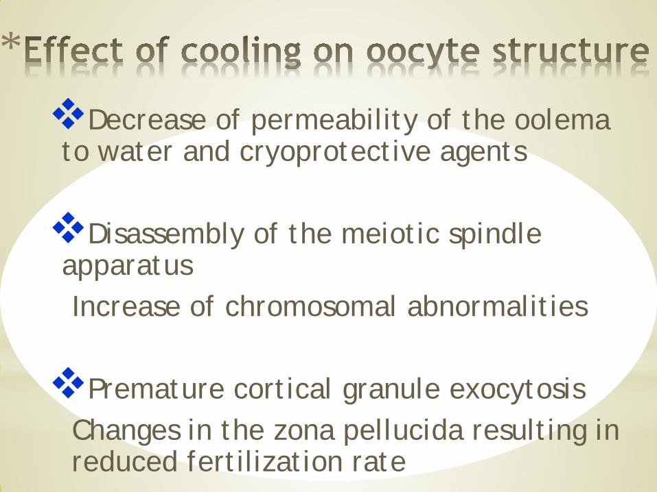
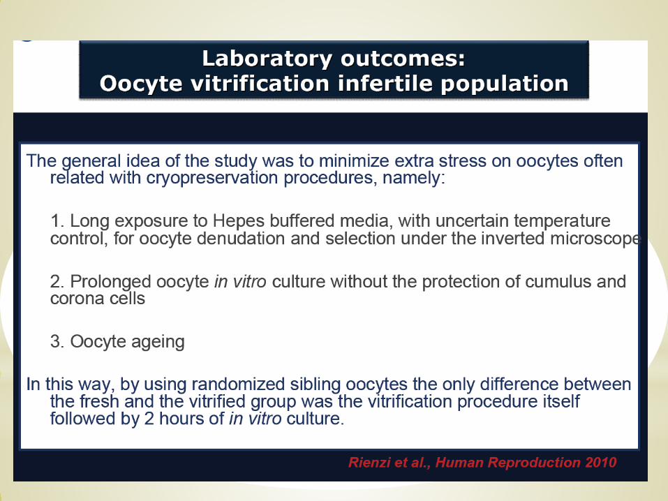
*Decrease of permeability of the oolemato water and cryoprotective agents
Disassembly of the meiotic spindle apparatusIncrease of chromosomal abnormalities
Premature cortical granule exocytosisChanges in the zona pellucida resulting in reduced fertilization rate
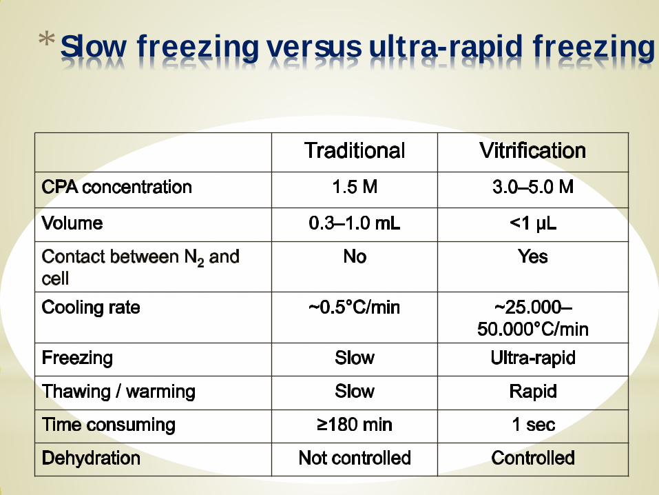
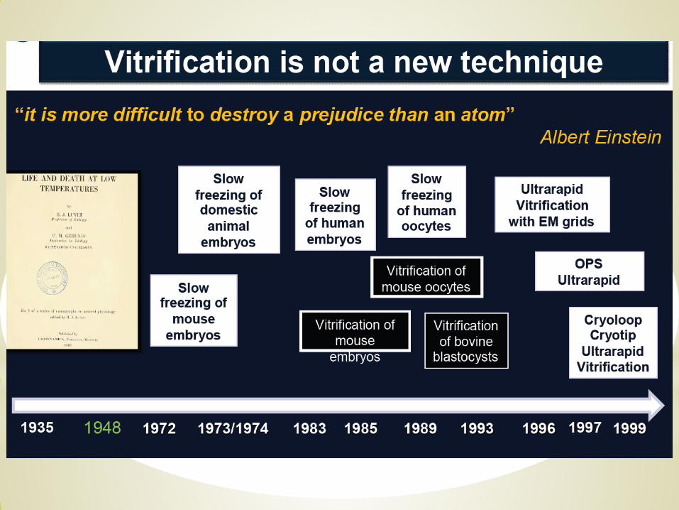
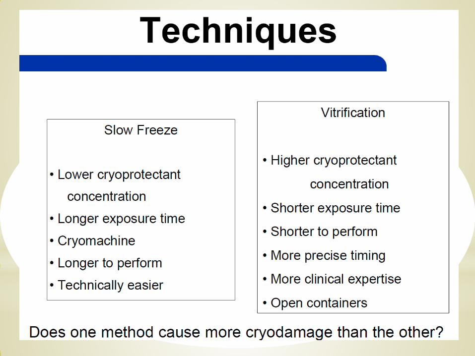
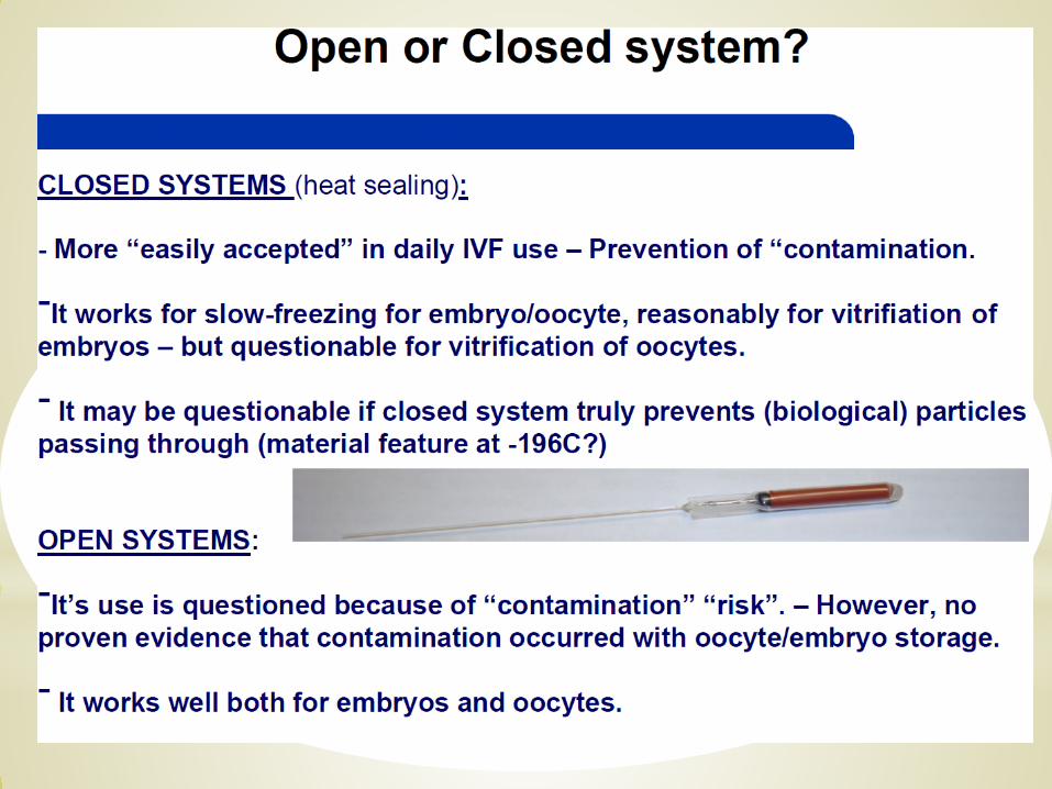
*Slow freezing versus ultra-rapid freezing
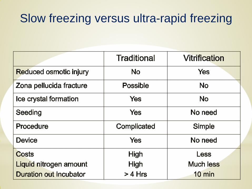
Slow freezing versus ultra-rapid freezing
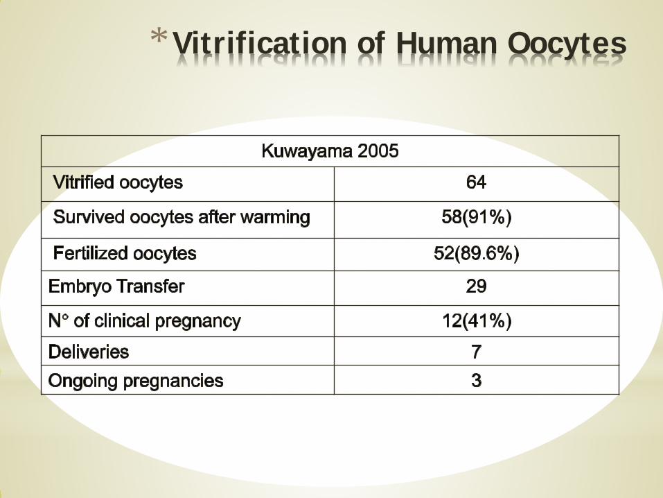
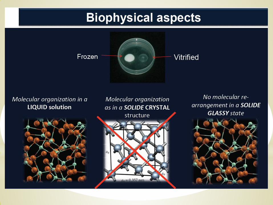
*Vitrification of Human Oocytes
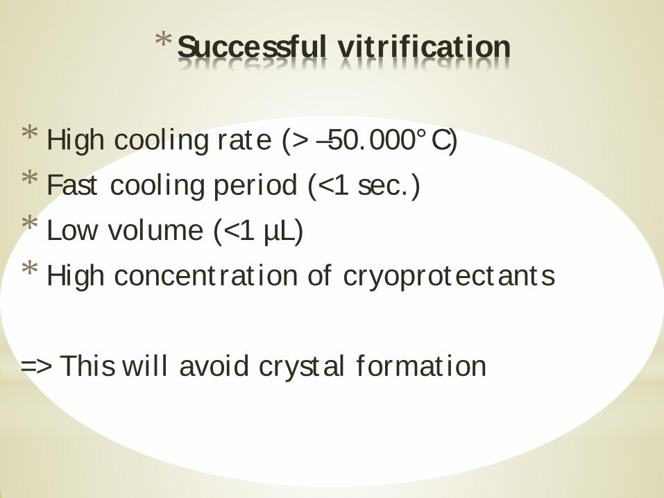
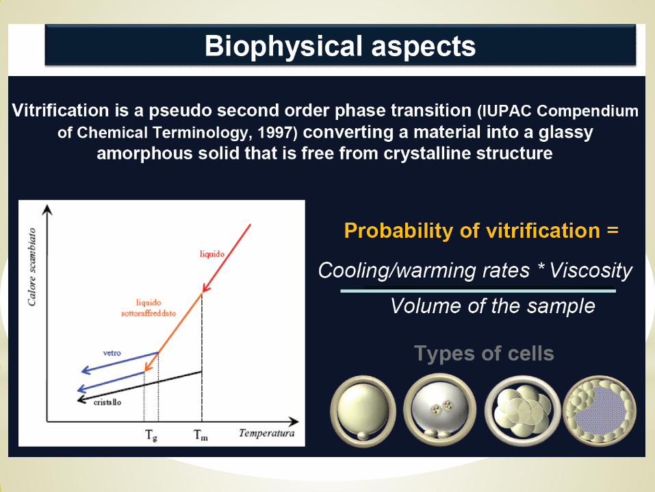
*Successful vitrification
* High cooling rate (> –50.000°C)
* Fast cooling period (<1 sec.)
* Low volume (<1 µL)
* High concentration of cryoprotectants
=> This will avoid crystal formation
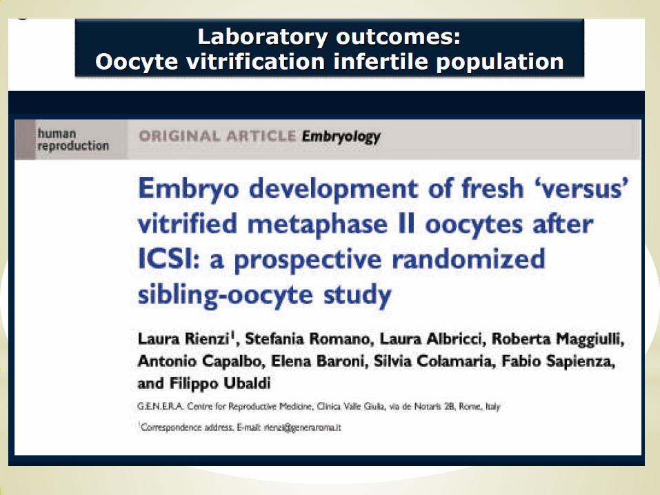
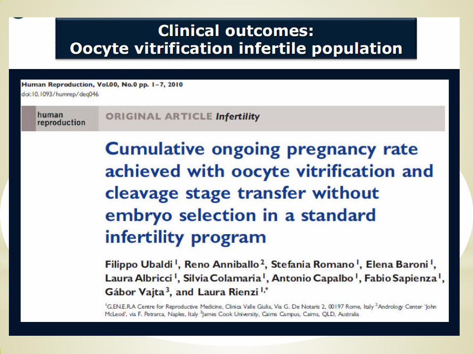
* Embryo Development of Fresh „Versus“ Vitrified Metaphase II Oocytes after ICSI: A
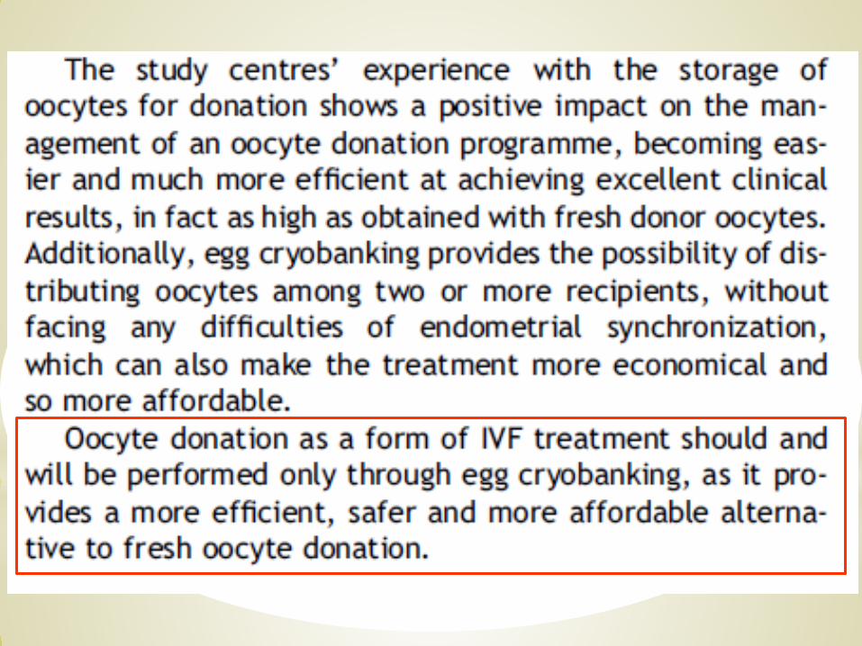
Prospective Randomised Sibling-Oocyte Study*Conclusion:
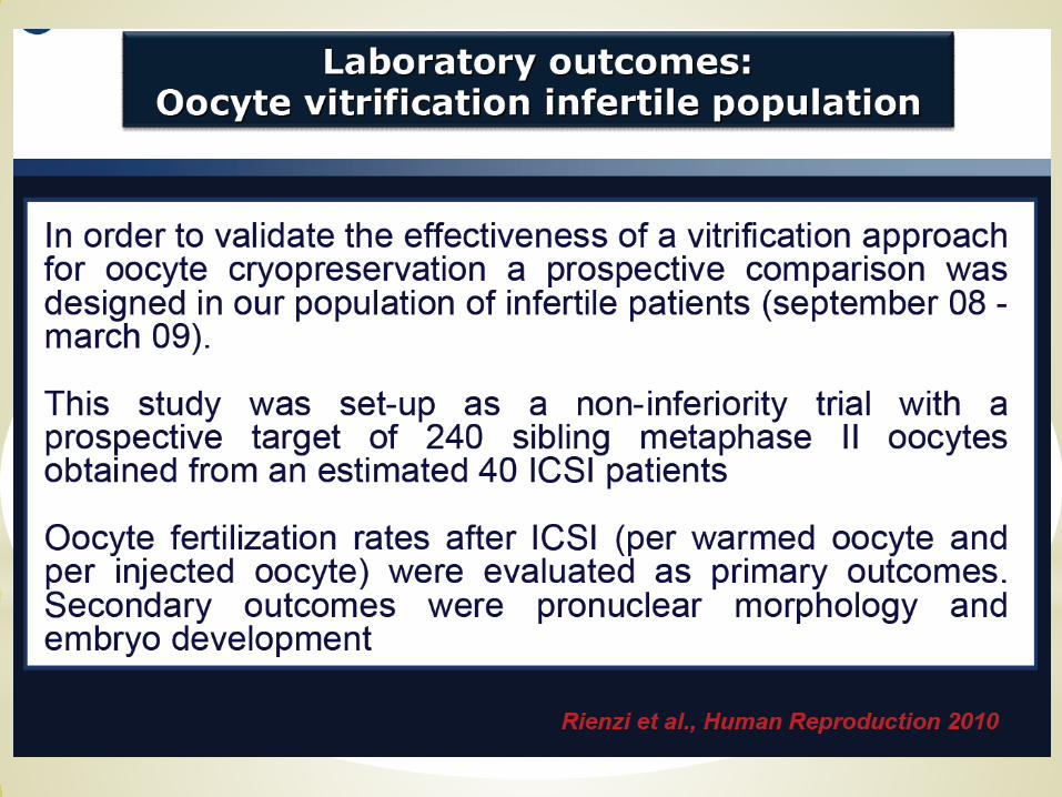
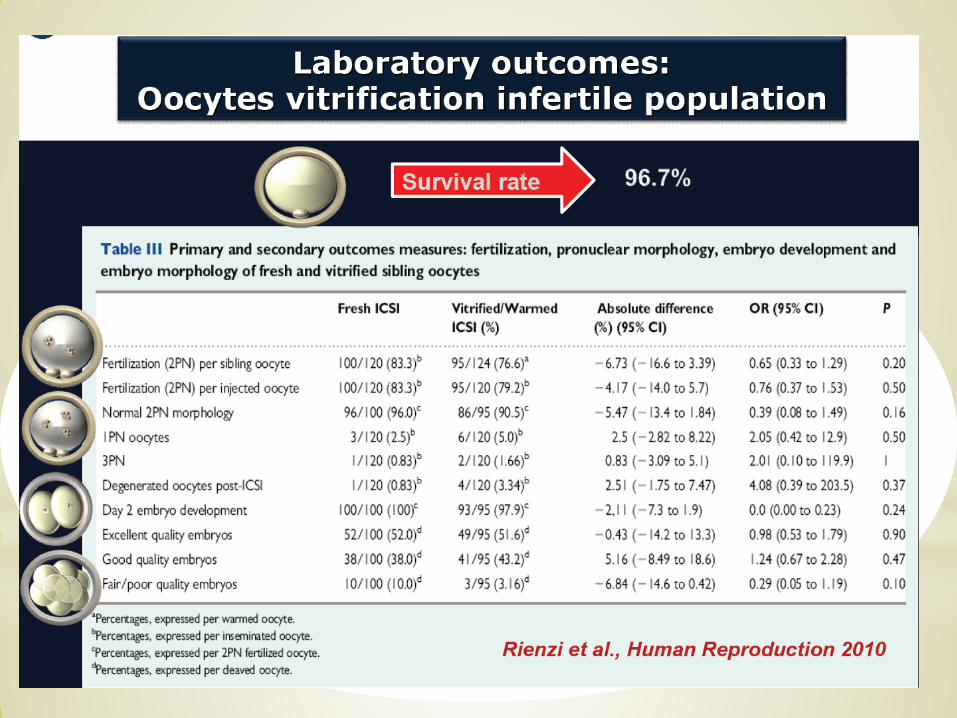
Our results indicate that oocyte vitrification procedure followed by ICSI is not inferior to fresh insemination procedure, with regard to fertilization and embryo developmental rates. Moreover, ongoing clinical pregnancy is comparable with this procedure, even with a restricted number of oocytes available for inseminat-ion. We believe that these results will help the spread of vitrification for human oocytes cryopreservation.The promising clinical results obtained, in a population of infertile patients, need to be confirmed on a larger scale.
Rienzi et al., 2010, Human Reprod., 25, 66-73
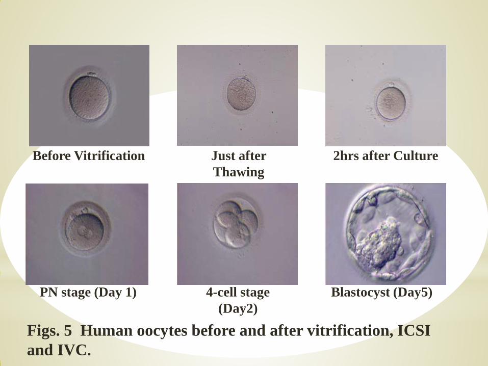
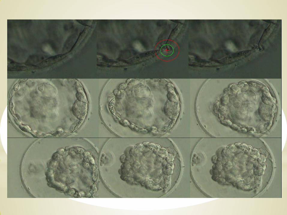
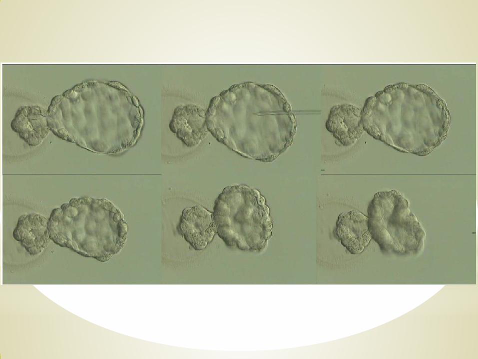
Figs. 5 Human oocytes before and after vitrification, ICSI and IVC.
Before Vitrification 2hrs after CultureJust after Thawing
4-cell stage (Day2)
Blastocyst (Day5)PN stage (Day 1)

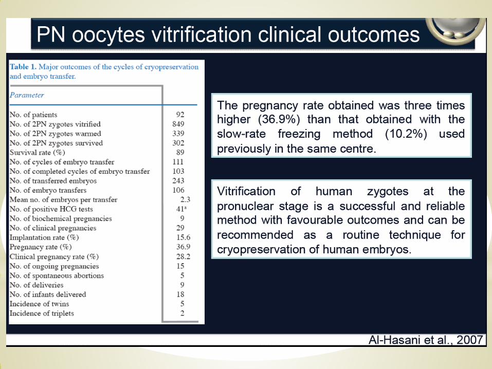

**znacznie wyższa skuteczność pod względem uzyskanych ciąż z zamrożonych komórek jajowych lub zarodków
*pod względem etycznym dla części pacjentek; witryfikacjaumożliwia świadomy wybór i rezygnację z mrożenia nadliczbowych zarodków na rzecz zamrożenia komórek jajowych
*skuteczne leczenie niepłodności dla kobiet źle reagujących na stymulację hormonalną (tzw. low responders). Witryfikacja umożliwia bankowanie oocytów i po zebraniu i zamrożeniu kilku podejście do pełnej procedury in vitro (wzrastają szanse na powodzenie zabiegu)
*szansa na macierzyństwo dla kobiet przed leczeniem onkologicznym, bankowanie własnych komórek jajowych i ich wykorzystanie po zakończeniu leczenia onkologicznego jest szansą na urodzenie dziecka po terapii onkologicznej

**W Polsce zazwyczaj mrozi się nadliczbową ilość zarodków powstałych w
skutek zabiegu in vitro. Są to zarodki, których nie podało się podczas pierwszego transferu pacjentce.
*Nadwyżka zarodków wynika z rekomendacji medycznych dotyczących ilości jednorazowo podawanych zarodków. Przyjęto, że można podawać maksymalnie 3 zarodki (najczęściej jednak podaje się tylko 1 lub 2 zarodki). Jest to podyktowane ochroną pacjentki przed ciążą mnogą (trojaczki, czworaczki).
*Jednym z rozwiązań problemu nadliczbowych zarodków jest mrożenie komórek jajowych bez ich zapłodnienia.
*Obecna metoda mrożenia tzw. slow freezing, ma jednak sporo wad i nie jest aż tak skuteczna, jak chcieliby tego lekarze i pacjentki. Po rozmrożeniu komórek jajowych znaczna ich część obumiera. Rozwiązaniem tego problemu jest witryfikacja.
*Witryfikowane komórki jajowe można wykorzystać, jeżeli pacjentka nie zajdzie w ciążę po podaniu niemrożonych zarodków

*

*
*
*
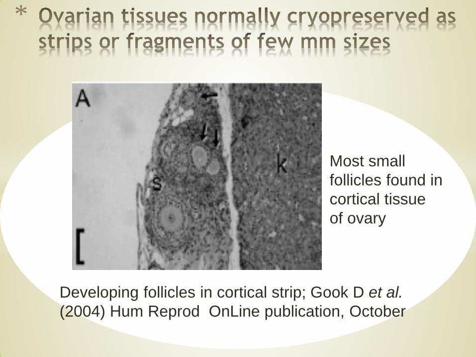
Most small follicles found in cortical tissue of ovary
Developing follicles in cortical strip; Gook D et al. (2004) Hum Reprod OnLine publication, October
*
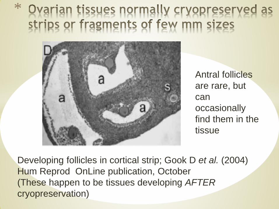
Antral follicles are rare, but can occasionally find them in the tissue
Developing follicles in cortical strip; Gook D et al. (2004) Hum Reprod OnLine publication, October(These happen to be tissues developing AFTERcryopreservation)
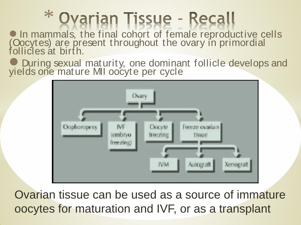
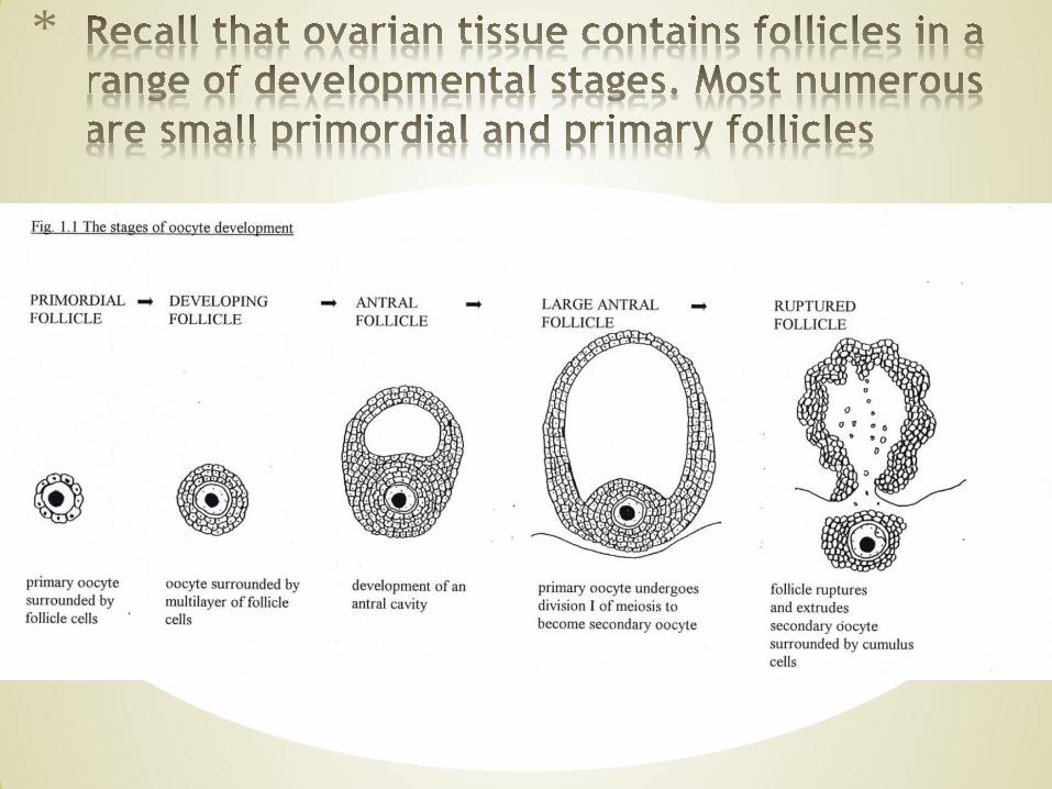
* In mammals, the final cohort of female reproductive cells (Oocytes) are present throughout the ovary in primordial follicles at birth. During sexual maturity, one dominant follicle develops and yields one mature MII oocyte per cycle
Ovarian tissue can be used as a source of immature oocytes for maturation and IVF, or as a transplant
*
*At birth, the primordial follicle population of ovarian tissue represents the lifetime supply of oocytes
*Primary ovarian failure may be induced in ‘young’ patients by anticancer treatments
*Oophorectomised tissues could provide donor oocytes for clinical infertility treatment or species conservation
*
*must preserve the ability of follicles (oocyte + supporting cells) to grow and acquire mature characteristics
*must maintain cell-cell communications and correct sequence of hormone responsiveness
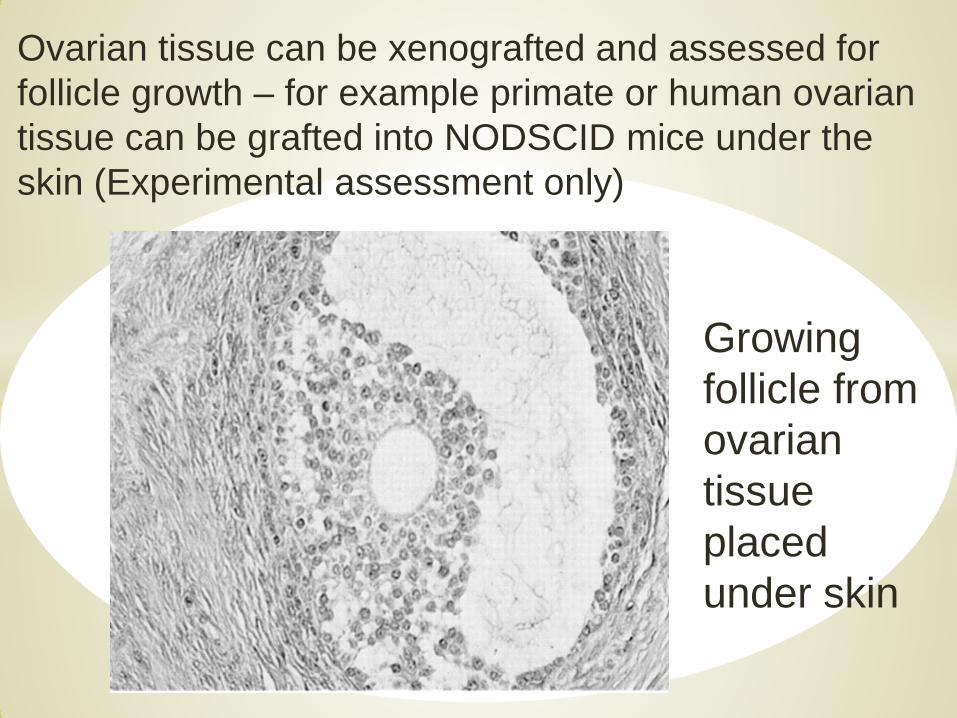
Ovarian tissue can be xenografted and assessed for follicle growth – for example primate or human ovarian tissue can be grafted into NODSCID mice under the skin (Experimental assessment only)
Growing follicle from ovarian tissue placed under skin
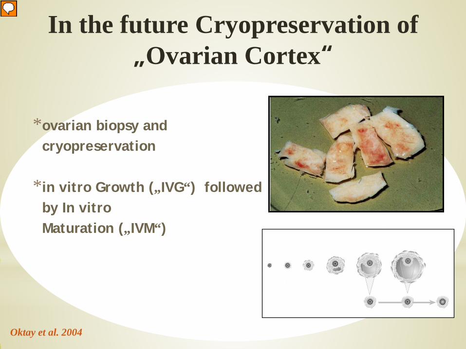
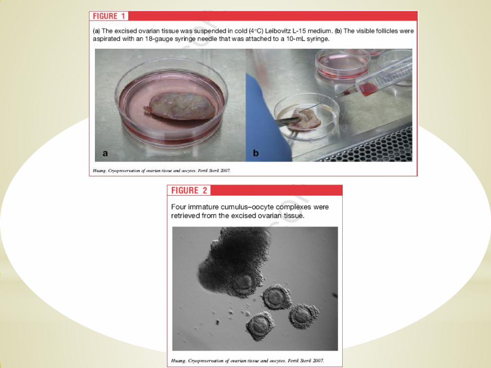
*ovarian biopsy and cryopreservation
*in vitro Growth („IVG“) followed by In vitro Maturation („IVM“)
In the future Cryopreservation of„Ovarian Cortex“
Oktay et al. 2004

*
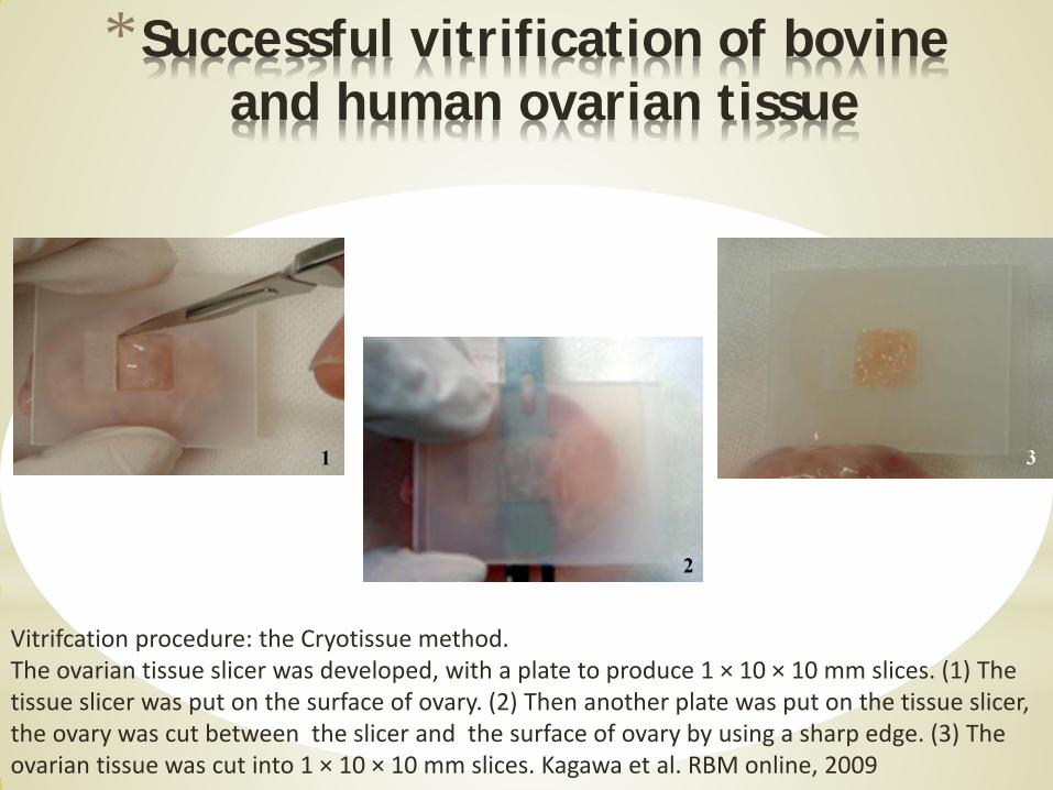
Vitrifcation procedure: the Cryotissue method. The ovarian tissue slicer was developed, with a plate to produce 1 × 10 × 10 mm slices. (1) The tissue slicer was put on the surface of ovary. (2) Then another plate was put on the tissue slicer, the ovary was cut between the slicer and the surface of ovary by using a sharp edge. (3) The ovarian tissue was cut into 1 × 10 × 10 mm slices. Kagawa et al. RBM online, 2009
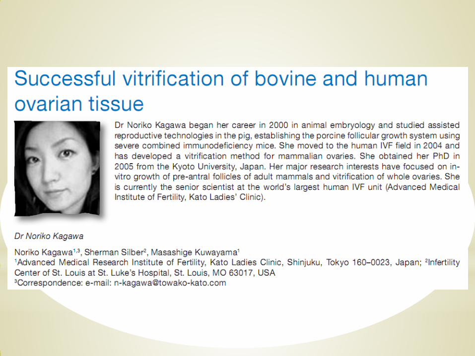
*Successful vitrification of bovine and human ovarian tissue
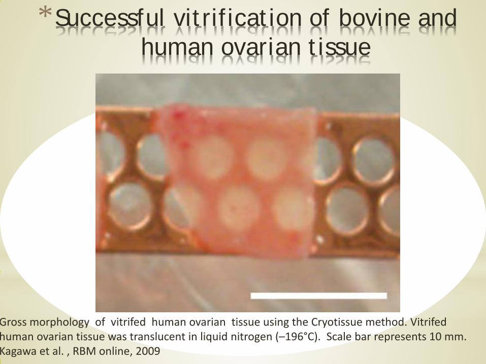
Gross morphology of vitrifed human ovarian tissue using the Cryotissue method. Vitrifedhuman ovarian tissue was translucent in liquid nitrogen (–196°C). Scale bar represents 10 mm.Kagawa et al. , RBM online, 2009
*Successful vitrification of bovine andhuman ovarian tissue
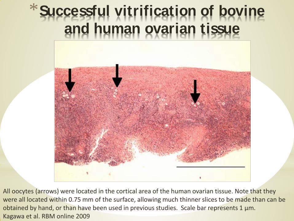
All oocytes (arrows) were located in the cortical area of the human ovarian tissue. Note that they were all located within 0.75 mm of the surface, allowing much thinner slices to be made than can be obtained by hand, or than have been used in previous studies. Scale bar represents 1 µm.Kagawa et al. RBM online 2009
*Successful vitrification of bovine and human ovarian tissue
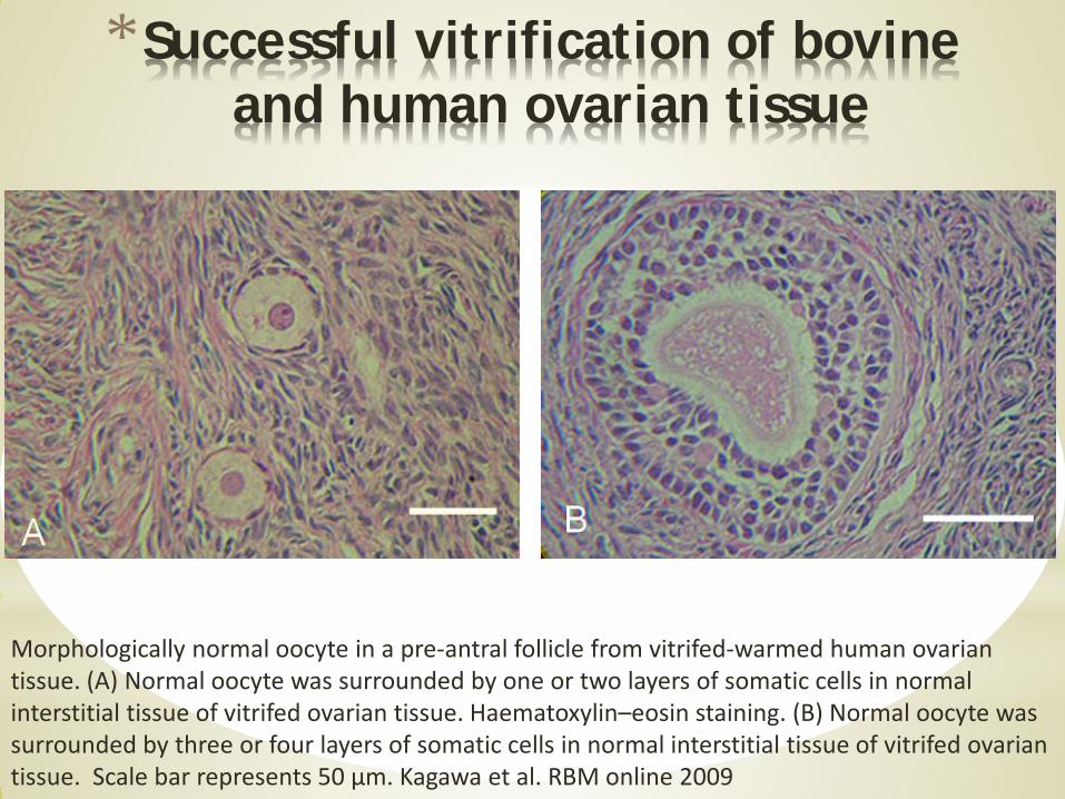
Morphologically normal oocyte in a pre-antral follicle from vitrifed-warmed human ovarian tissue. (A) Normal oocyte was surrounded by one or two layers of somatic cells in normal interstitial tissue of vitrifed ovarian tissue. Haematoxylin–eosin staining. (B) Normal oocyte was surrounded by three or four layers of somatic cells in normal interstitial tissue of vitrifed ovarian tissue. Scale bar represents 50 µm. Kagawa et al. RBM online 2009
*Successful vitrification of bovine and human ovarian tissue
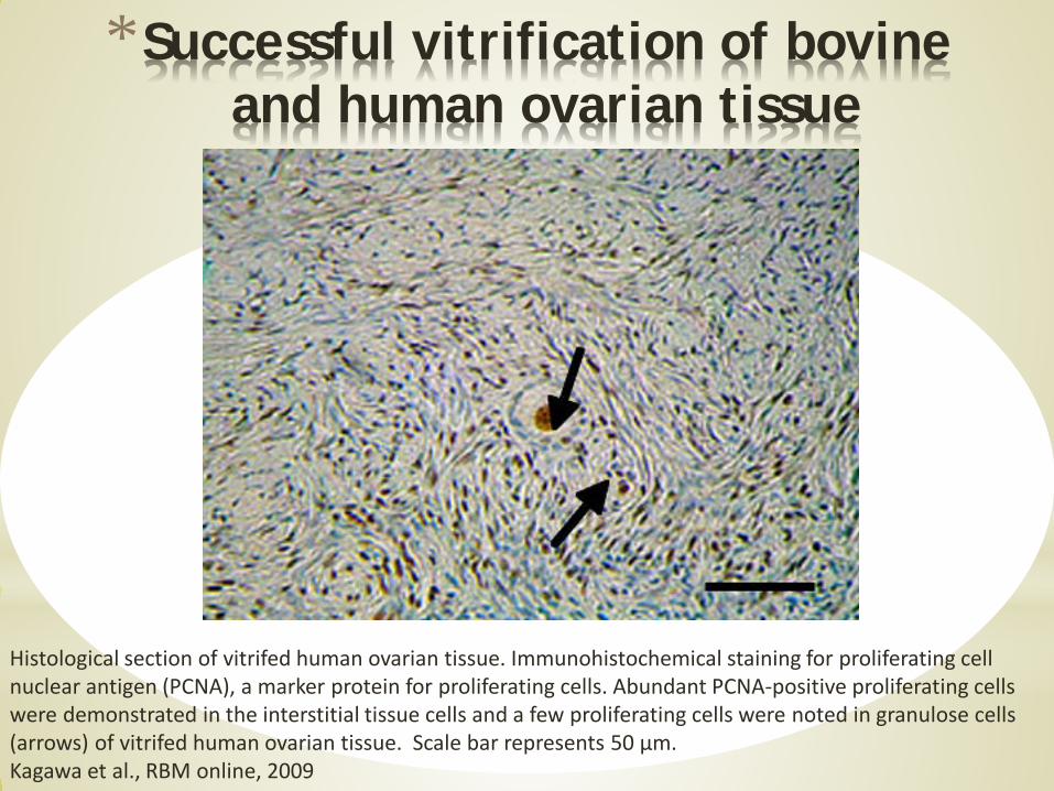
Histological section of vitrifed human ovarian tissue. Immunohistochemical staining for proliferating cellnuclear antigen (PCNA), a marker protein for proliferating cells. Abundant PCNA-positive proliferating cellswere demonstrated in the interstitial tissue cells and a few proliferating cells were noted in granulose cells(arrows) of vitrifed human ovarian tissue. Scale bar represents 50 µm.Kagawa et al., RBM online, 2009
*Successful vitrification of bovine and human ovarian tissue
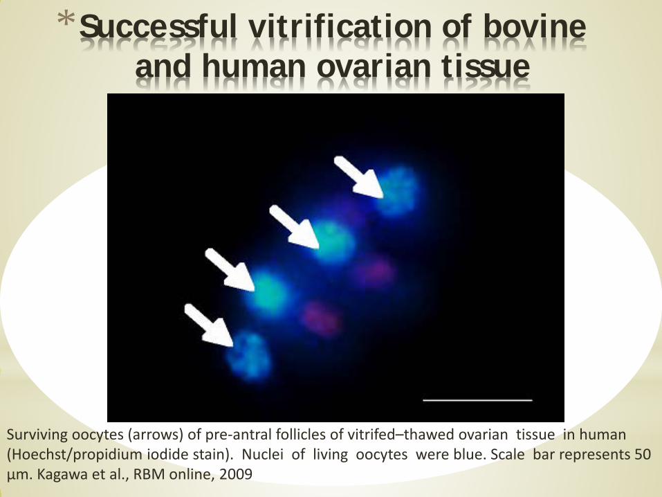
Surviving oocytes (arrows) of pre-antral follicles of vitrifed–thawed ovarian tissue in human (Hoechst/propidium iodide stain). Nuclei of living oocytes were blue. Scale bar represents 50 µm. Kagawa et al., RBM online, 2009
*Successful vitrification of bovine and human ovarian tissue
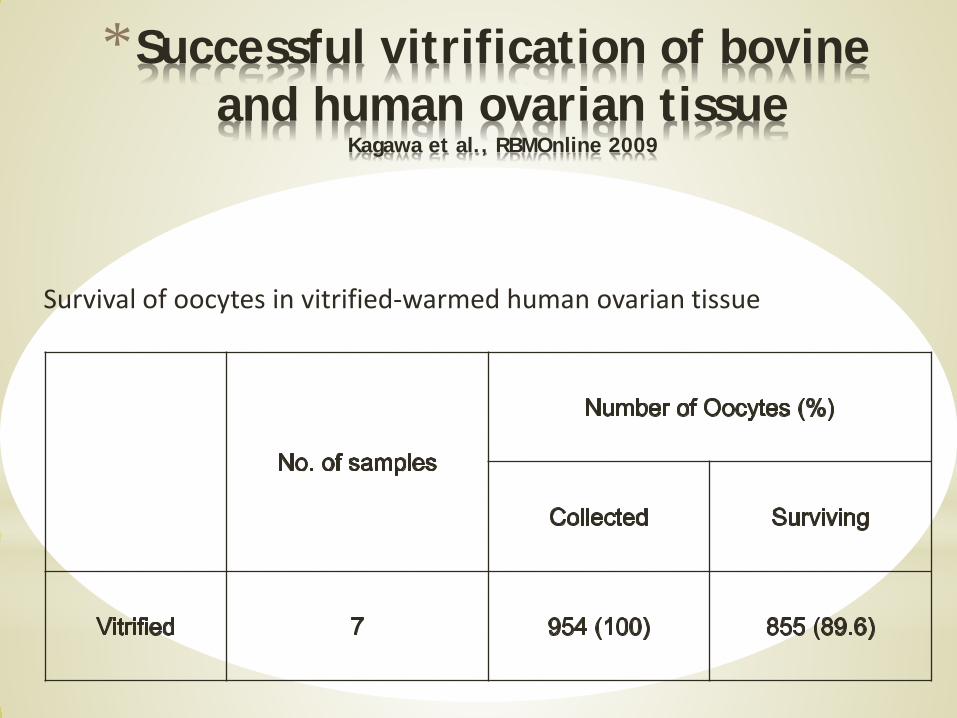
Survival of oocytes in vitrified-warmed human ovarian tissue
*Successful vitrification of bovine and human ovarian tissue
Kagawa et al., RBMOnline 2009
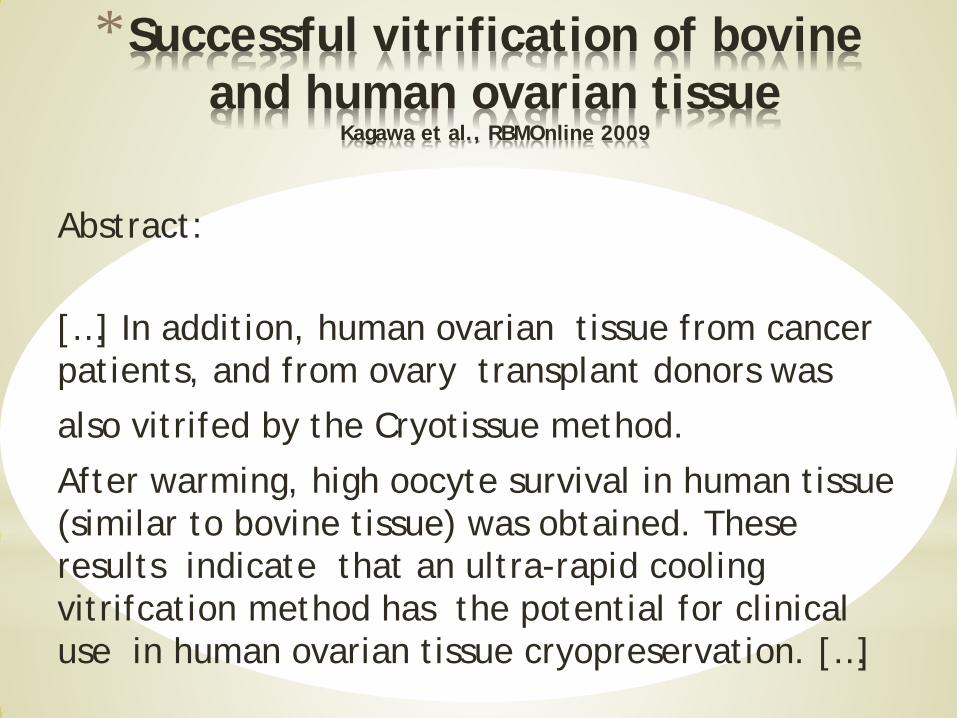
*Successful vitrification of bovine and human ovarian tissue
Kagawa et al., RBMOnline 2009
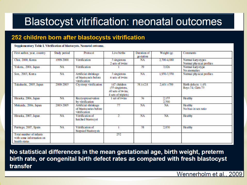
Abstract:
[…] In addition, human ovarian tissue from cancer patients, and from ovary transplant donors was
also vitrifed by the Cryotissue method.
After warming, high oocyte survival in human tissue (similar to bovine tissue) was obtained. These results indicate that an ultra-rapid cooling vitrifcation method has the potential for clinical use in human ovarian tissue cryopreservation. […]
*Why do we prefer the vitrificationprocedure now?
* No mechanical injury (extracellular crystal formation)
* Less osmotic stress to cells
* No intracellular crystal formation
* Less labour in laboratory daily work
* Simple protocol
* Useful for oocytes and blastocysts, which have less success with slow freezing
* No need for expensive devices
*Future Aspects
*Avoiding hyperstimulation syndrome in patients with PCOS by vitrification of all 2PN and replaced in a programmed cycle
*Cancelling of fresh ET in case of more than 10 Follicles
*Vitrification of all zygotes resulted from IVM programme
*An option for cancer patients to vitrify the oocytes instead of ovarian tissue
*In oocytes donation programme*Vitrification of the oocytes to postpone fertility*Mantains viability of specimens during long term storage

*Conclusions of vitrification* Easy to perform
* Low cost
* Future first choice procedure
* It was shown to be superior to slow freezing procedure
* Very high survival rates of oocytes and embryos at all stages of development
* It seems that the cryotop method is the most efficient procedure
* Revitrification is possible
* Ovarian Cortex is now also possible