dr. anand srinivasan. good regenerative capacity hence used for transplantation
TRANSCRIPT

Dr. ANAND SRINIVASAN
Good regenerative capacityHence used for transplantation

Able to : Describe, identify and draw the
histological features of : Liver Gall bladder Pancreas

2nd largest organ
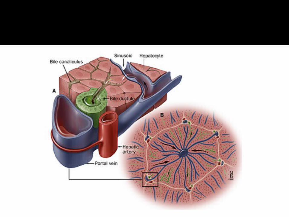
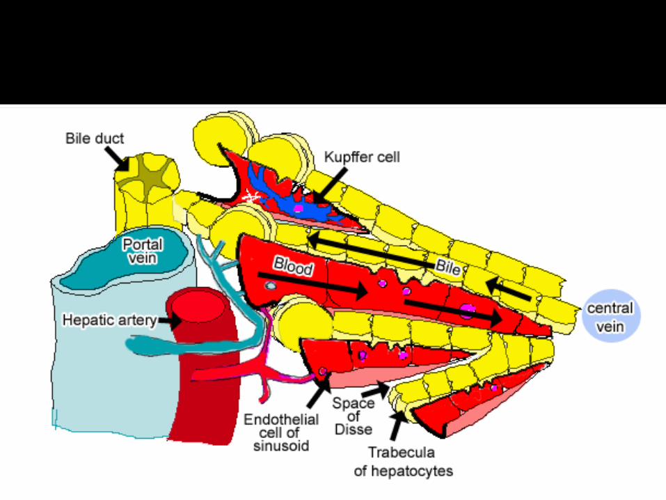
2 types of blood vessels Portal vein (70%) Hepatic A. (30%)
Surrounded by fibrous capsule (GLISSON’s CAPSULE) deep to peritoneum
Capsule through trabeculae divides the liver into “lobules”
Portal tract / Portal space / Portal canal Branches of Hepatic A, Portal V., Hepatic duct and
lymphatics present in trabeculae

Structural unit of liver
Hexagonal (polygonal) in shape
Central vein in the center
Liver lobules not completely demarcated by connective tissue septa (c.f. pig)
Lobules can be demarcated by connecting portal tracts

Structural and functional component of liver “Hepatocytes”
Hepatic plates usually single layer thick (c.f. children 2 plates)
Radiate from central vein to periphery
“Sinusoids” – spaces between hepatic plates lined by fenestrated endothelial cells
Some endothelial cells modified as “Kupffer’s cells”
Hepatic stellate / Ito cells (perisinusoidal lipocytes) also present within hepatocytes – activated in certain pathological conditions.
Sinusoids separated from hepatocytes by “Space of Disse”
Blood from portal vein and hepatic A. enter into sinusoid which then drains into central vein and finally to hepatic vein


Atleast 2 surfaces of hepatocytes are in contact with sinusoids through space of disse
Rest of the hepatocytes contact with adjacent and has a intercellular space called “Bile canaliculus” – first part of the bile duct system
Pass through “Cana of Hering” to hepatic ducule
Flow of bile is in a opposite direction to that of blood.


For studying certain pathological conditions it is useful to divide liver into portal lobule
Part of liver parenchyma that drains bile into hepatic ductule present in portal triad

Another functional unit of liver which is irrigated by terminal distributing branches of portal vein and hepatic artery
Diamond shaped


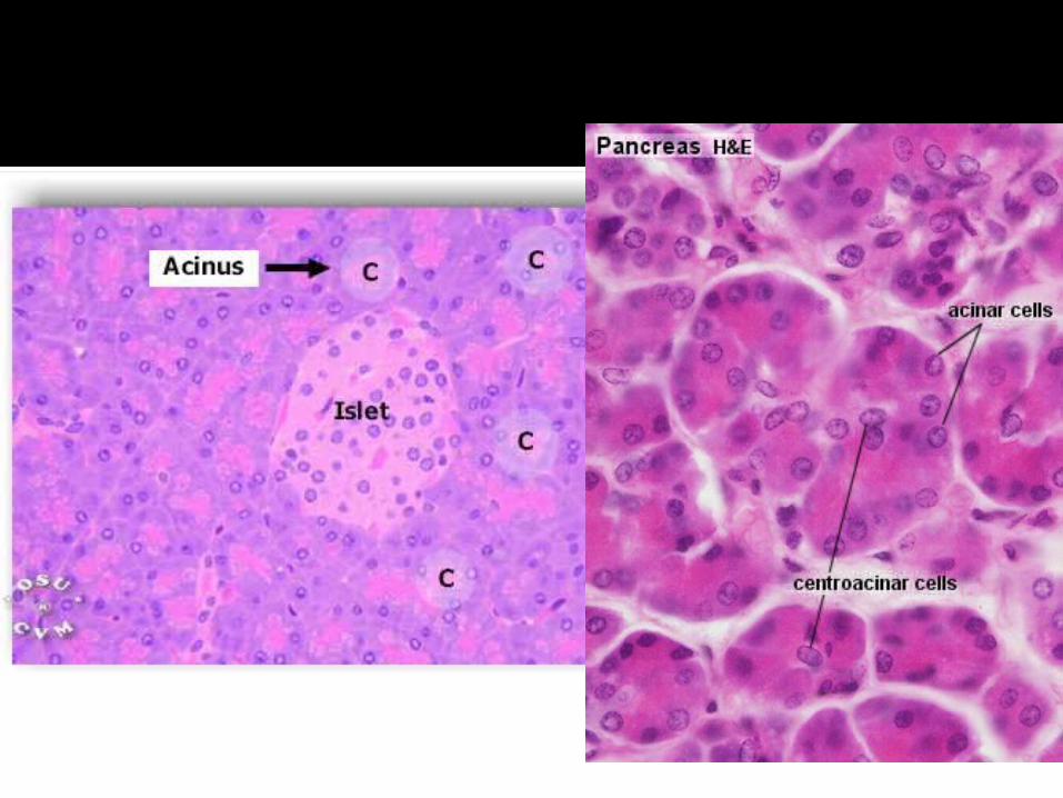
Exocrine and endocrine gland
EXOCRINE Darkly stained serous acini No myopeithelial cells but pancreatic
stellate cells present Some acini exhibit “Centroacinar cells” –
cuboidal cells representing intra-acinar part of intercalated duct
Intercalated duct intralobular ducts interlobular duct main pancreatic duct (epithelium varying from simple to stratified cuboidal)

ENDOCRINE Made of “Islets of Langerhans” – pale
staining spherical bodies amongst serous acini
More in the tail of pancreas 1 – 2 million islets in pancreas


ALPHA CELLS (20 %) Secrete glucagon
BETA CELLS (70%) Secrete insulin
DELTA CELLS (5%) Secrete somatostatin
F CELLS / PP CELLS Secrete pancreatic polypeptides

Sac situtated close to the liver
Concentrate bile
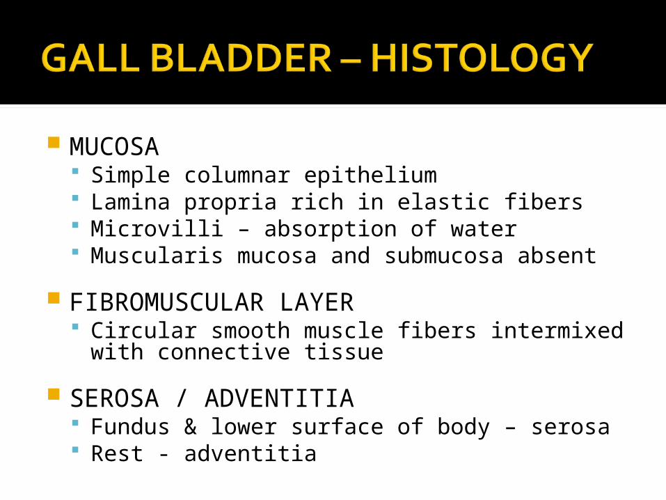
MUCOSA Simple columnar epithelium Lamina propria rich in elastic fibers Microvilli – absorption of water Muscularis mucosa and submucosa absent
FIBROMUSCULAR LAYER Circular smooth muscle fibers intermixed with
connective tissue
SEROSA / ADVENTITIA Fundus & lower surface of body – serosa Rest - adventitia


Wheater’s Functional Histology
Cell Biology & Histology – Board Review Series
Microanatomy workbook – RAKMHSU