dp71 ind a e - nahwoonahwoo.com/userdata/dp71_ind.pdf · high level of color accuracy ensures...
TRANSCRIPT
NEW
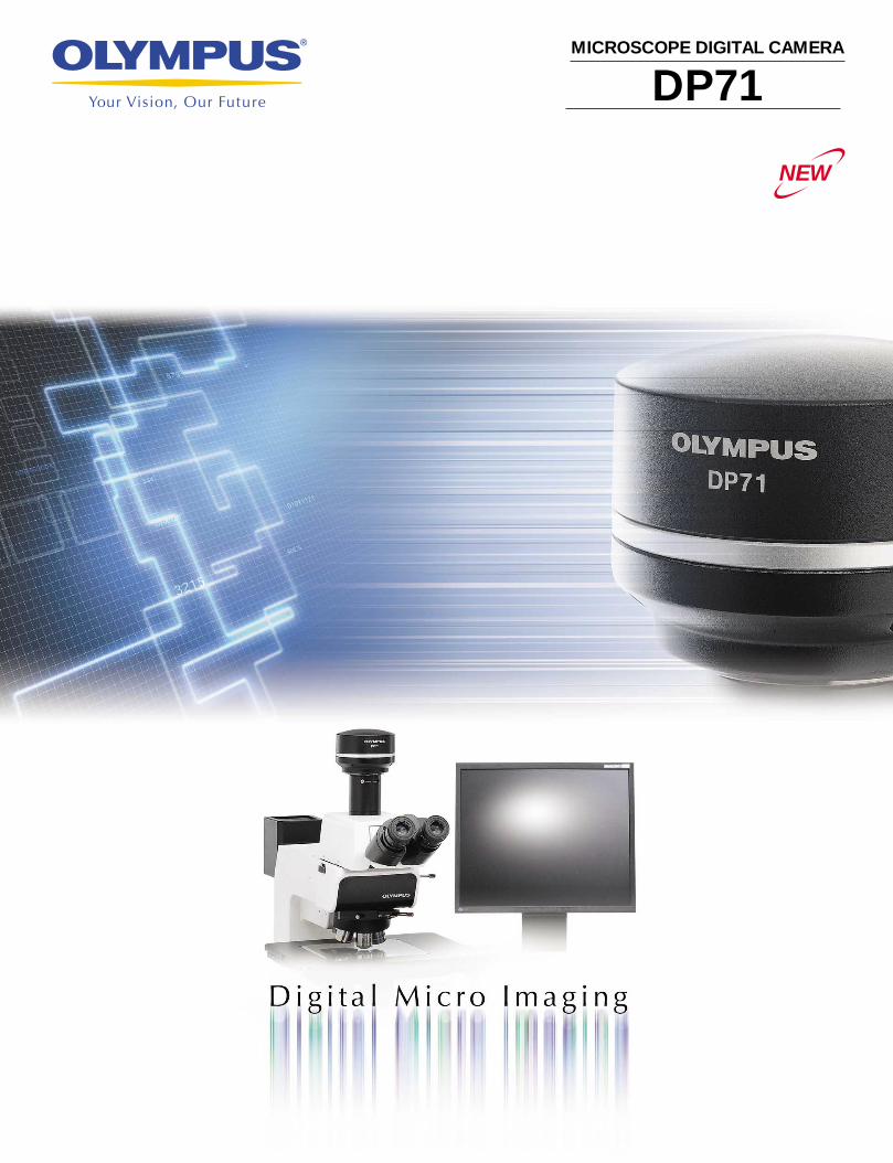
MICROSCOPE DIGITAL CAMERA
DP71
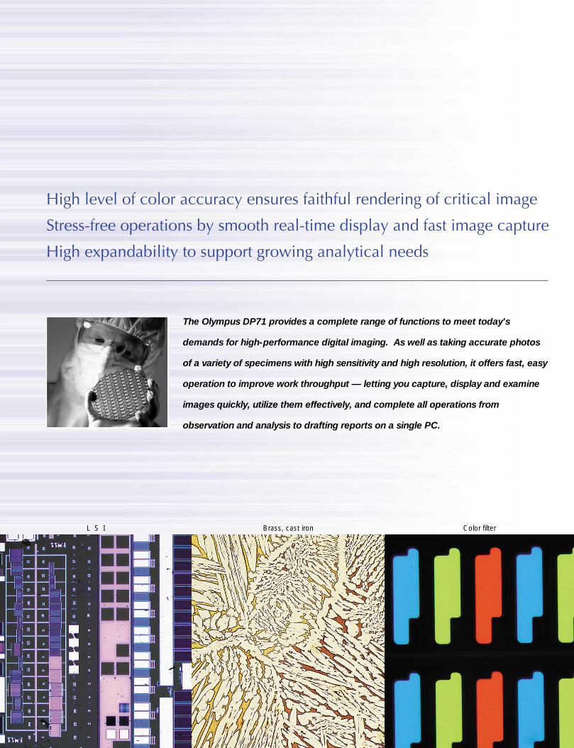
High level of color accuracy ensures faithful rendering of critical image
Stress-free operations by smooth real-time display and fast image capture
High expandability to support growing analytical needs
The Olympus DP71 provides a complete range of functions to meet today's
demands for high-performance digital imaging. As well as taking accurate photos
of a variety of specimens with high sensitivity and high resolution, it offers fast, easy
operation to improve work throughput — letting you capture, display and examine
images quickly, utilize them effectively, and complete all operations from
observation and analysis to drafting reports on a single PC.
1
L S I Brass, cast iron Color filter
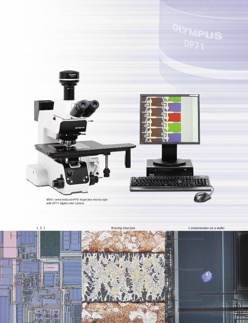
MX61 semiconductor/FPD inspection microscopewith DP71 digital color camera
2
L S I Brazing structure Contamination on a wafer
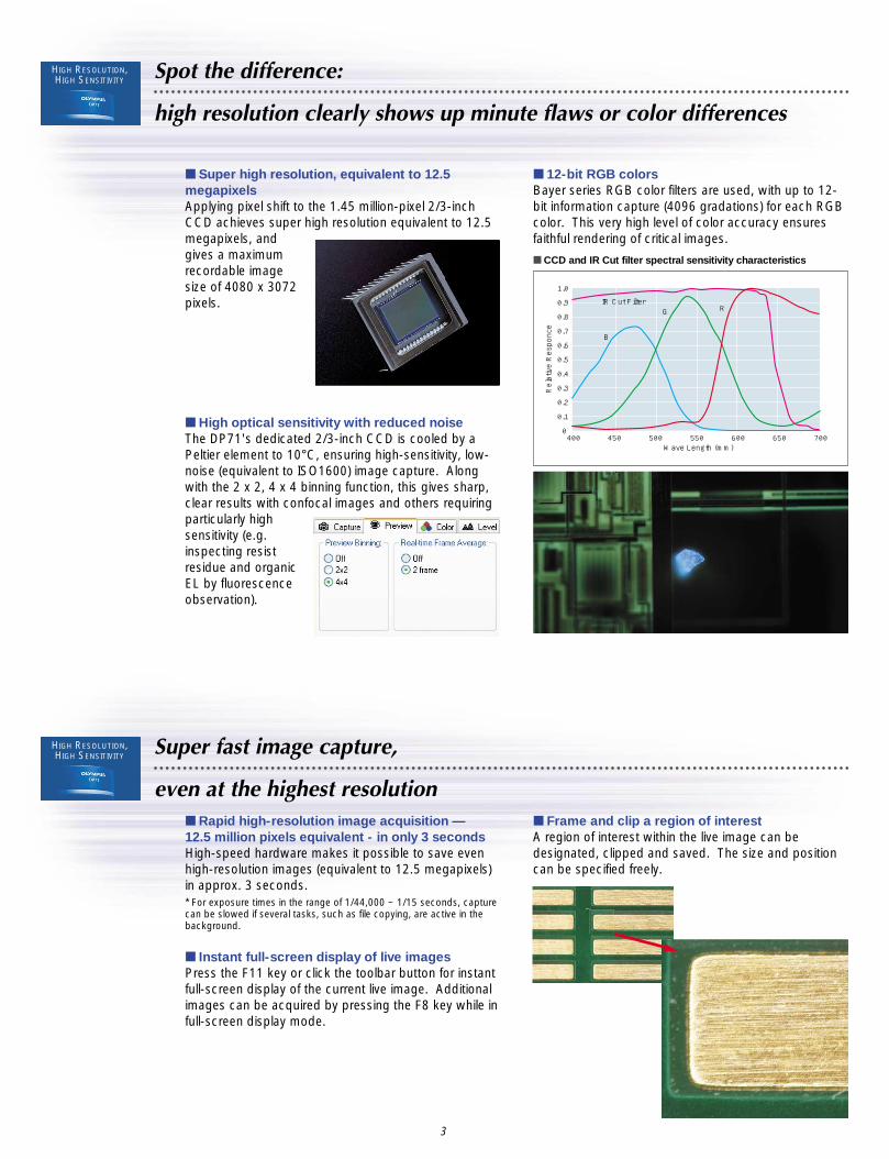
Spot the difference:
high resolution clearly shows up minute flaws or color differences
■ Super high resolution, equivalent to 12.5megapixelsApplying pixel shift to the 1.45 million-pixel 2/3-inchCCD achieves super high resolution equivalent to 12.5megapixels, andgives a maximumrecordable imagesize of 4080 x 3072pixels.
■ 12-bit RGB colorsBayer series RGB color filters are used, with up to 12-bit information capture (4096 gradations) for each RGBcolor. This very high level of color accuracy ensuresfaithful rendering of critical images.
IR Cut Filter
B
1.0
0.9
0.8
0.7
0.6
0.5
0.4
0.3
0.2
0.1
0400 450 500 550 600 650 700
G R
Wave Length (mm)
Relative Responce
■ CCD and IR Cut filter spectral sensitivity characteristics
■ High optical sensitivity with reduced noiseThe DP71's dedicated 2/3-inch CCD is cooled by aPeltier element to 10°C, ensuring high-sensitivity, low-noise (equivalent to ISO1600) image capture. Alongwith the 2 x 2, 4 x 4 binning function, this gives sharp,clear results with confocal images and others requiringparticularly highsensitivity (e.g.inspecting resistresidue and organicEL by fluorescenceobservation).
■ Rapid high-resolution image acquisition — 12.5 million pixels equivalent - in only 3 secondsHigh-speed hardware makes it possible to save evenhigh-resolution images (equivalent to 12.5 megapixels)in approx. 3 seconds.* For exposure times in the range of 1/44,000 ~ 1/15 seconds, capture can be slowed if several tasks, such as file copying, are active in thebackground.
■ Instant full-screen display of live imagesPress the F11 key or click the toolbar button for instantfull-screen display of the current live image. Additionalimages can be acquired by pressing the F8 key while infull-screen display mode.
■ Frame and clip a region of interestA region of interest within the live image can bedesignated, clipped and saved. The size and positioncan be specified freely.
Super fast image capture,
even at the highest resolution
3
HIGH RESOLUTION,HIGH SENSITIVITY
HIGH RESOLUTION,HIGH SENSITIVITY

Sharp, live images at1360 x 1204 pixels
15 fps real-time display
Smooth, sharp real-time display
for easy, stress-free observations
Easily choose and set photo conditions
according to the specimen or observation method
■ Pin-sharp image display in real time at 15 fpsSharp, detailed images (1360 x 1024 pixels) can bedisplayed live at 15 fps, making observations easy andcomfortable.
■ Easy, accurate focusingAn indicator function makes focusing easy, while theline profile function lets you focus accurately on specificregions. For liveimages, the center ofthe field of view canbe magnified to 2times.
■ Accurate automatic exposure for fluorescentspecimensThe SFL-Auto Exposure Mode makes it easy to acquirefluorescence images, since it sets the correct exposuretime automatically. Manualexposure mode can also beselected.
■ Photometric areas can be set freelyThree types of photometric area can be selected,depending on the specimen: 30%, 1%, 0.1%. Theselected area can be moved freely,allowing sharp exposures withoutchanging frames.
■ Include scale information or textA scale bar can be shown on the live image, andincluded when the image is saved. The same functionenables incorporation of captions and/or text.
Line profile function
4
FUNCTIONALITY
FUNCTIONALITY
Quickly find the image you want
and start using it
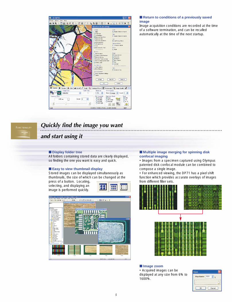
■ Return to conditions of a previously savedimageImage acquisition conditions are recorded at the time of a software termination, and can be recalledautomatically at the time of the next startup.
■ Image zoom• Acquired images can bedisplayed at any size from 6% to1600%.
5
■ Display folder treeAll folders containing stored data are clearly displayed,so finding the one you want is easy and quick.
■ Easy to view thumbnail display Stored images can be displayed simultaneously asthumbnails, the size of which can be changed at thepress of a button. Locating,selecting, and displaying animage is performed quickly.
■ Multiple image merging for spinning diskconfocal imaging • Images from a specimen captured using Olympuspatented disk confocal module can be combined tocompose a single image. • For enhanced viewing, the DP71 has a pixel shiftfunction which provides accurate overlays of imagesfrom different filter sets.
FUNCTIONALITY
Particle analysis
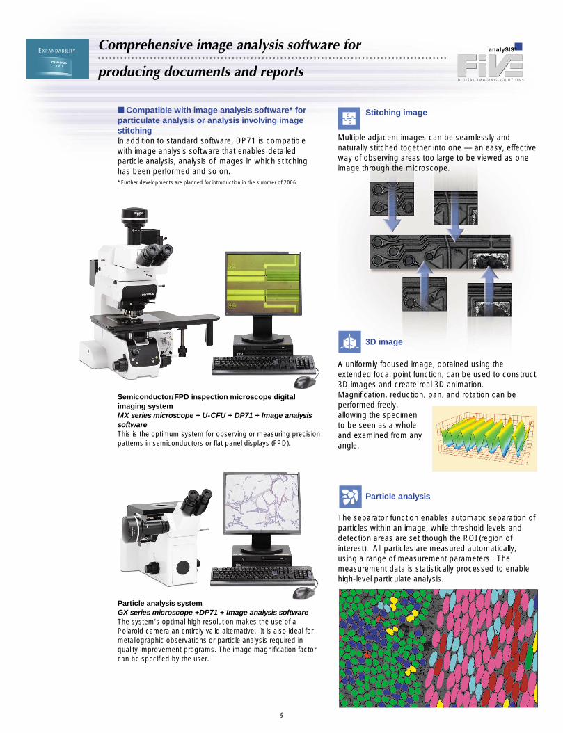
The separator function enables automatic separation ofparticles within an image, while threshold levels anddetection areas are set though the ROI (region ofinterest). All particles are measured automatically,using a range of measurement parameters. Themeasurement data is statistically processed to enablehigh-level particulate analysis.
Comprehensive image analysis software for
producing documents and reports
Stitching image■ Compatible with image analysis software* forparticulate analysis or analysis involving imagestitchingIn addition to standard software, DP71 is compatiblewith image analysis software that enables detailedparticle analysis, analysis of images in which stitchinghas been performed and so on.* Further developments are planned for introduction in the summer of 2006.
Semiconductor/FPD inspection microscope digitalimaging system MX series microscope + U-CFU + DP71 + Image analysissoftwareThis is the optimum system for observing or measuring precisionpatterns in semiconductors or flat panel displays (FPD).
Particle analysis system GX series microscope +DP71 + Image analysis softwareThe system's optimal high resolution makes the use of aPolaroid camera an entirely valid alternative. It is also ideal formetallographic observations or particle analysis required inquality improvement programs. The image magnification factorcan be specified by the user.
A uniformly focused image, obtained using theextended focal point function, can be used to construct3D images and create real 3D animation.Magnification, reduction, pan, and rotation can beperformed freely,allowing the specimento be seen as a wholeand examined from anyangle.
3D image
6
EXPANDABILITY
Multiple adjacent images can be seamlessly andnaturally stitched together into one — an easy, effectiveway of observing areas too large to be viewed as oneimage through the microscope.

Camera Type: Single CCD (Pixel shifting) Peltier cooling (max Ta-10°C)Image sensor Size: 2/3-inch
Effective pixels: 1.45million pixels Scanning method: Progressive scanning
Lens mount C mountRecorded image sizes 4080 x 3072/ 2040 x 1536/ 1360 x 1024/ 680 x 512/ 680 x 510/ 340 x 250ISO speed ISO 200/ 400/ 800/ 1600 equivalentA/D 12bitsMetering area 30%, 1%, 0.1% (measuring area can be moved in image freely)Exposure control Modes: Auto/ SFL auto/ Manual
AE lock: Available AE pause: AvailableCorrection range: ±2.0EV Step: 1/3EV
Exposure time 1/44,000 to 60sImage accumulation Modes: Integral,averaging
Accumulation count: 64frames (max)Image rotation Up/ down inversion, left/ right inversion, 180°White balance modes Area-specified auto/ Entire area-specified auto/ ManualBlack balance modes Area-specified auto/ Entire area-specified auto/ ManualColor modes Color/ standard gray scale/ custom gray scaleSharpness filter Low/ Standard/ HighMotion image display Max 15 frames/s (image size of 1360 x 1024)Focus indicator Contrast bar/ Numeric display/ HistogramImage transfer time Approx. 3 s* (Max resolution of 4080 x 3072)Image formats TIFF/ JPEG/ BMP /PICT /AVI/ MPEG-1Dimensions & weight Camera head: 112 (ø) x 87.8 (H) mm approx. 1,150g
PCI unit: 187.4 (W) x 125.7 (D) x 21.4 (H) mm approx. 250gInterface cable: approx. 2.8m
RECOMMENDED SPECIFICATIONS FOR PC CONTROLLERCPU Intel Pentium4 1.3 GHz or greater
(2.6GHz or greater, Hyper -Threading dual-core CPU recommended) Pentium D, Pentium EE
Chip set Intel i845, i850, i865, i875, i915, i925, i945, i955, i975 (i865 or later recommended)
RAM SDRAM, 512MB or more(PC2700 or after, dual-channel DDR/DDR2 recommended)
HDD Free space 500MB or moreGraphic Graphic RAM 16MB or more
Graphic card of the AGP specification with the capability of 32-bit color display of 1280 x 1024 or more, or PCI-Express x16 graphic card.*Onboard graphic also available when the chipset is i915 or after.
PCI bus PCI Rev.2.1 or 2.2OS Windows XP professional SP1a or later (Not compatible with x64 edition)
Windows 2000 professional SP4Drive CD-ROM or CD-R/RW etcMain body Half size PCI board compatiblePower supply 250W or more (with CE marking)
Camera head dimensions (unit: mm)
ø112
87.8
Weight: approximately 1,150g
• Replacement parts are available for 5 years afterpurchase.
* For exposure times in the range of 1/44,000 ~ 1/15 seconds, capture can be slowed if several tasks, such as file copying, are active inthe background.
This product contains precision electronic componentsthat can break or malfunction if subjected to strongvibrations or impacts. Please handle with care.
PC/AT compatible PCCamera head
Cables
DP-BSWBasic software
PCI board
DP-TRADTripod adapter
Commercial tripods
DP71
U-TV1X-2Direct image camera port
U-TV0.5XC-3*C-mount camera port with 0.5x lens
U-CMAD3C-mount adapter
Microscope
U-TV0.63XCC-mount camera port with 0.63x lens
SYSTEM DIAGRAM
Specifications are subject to change without any obligation on the part of the manufacturer.
•OLYMPUS CORPORATION obtains ISO9001/ISO14001.
DP71 SPECIFICATIONS
* Besides STM series measuring image microscopes
∗ All brands are trademarks or registered trademarks of their respective owners.
Printed in Japan M1592E-0606B