
1
Viscoelastic Characterisation of Pig Liver in Unconfined
Compression
G. Mattei a,b*, A. Tirella a,c, G. Gallone a,b and A. Ahluwalia a, c
a Research Centre "E. Piaggio", University of Pisa, Largo Lucio Lazzarino 1, 56122 Pisa, Italy
b Department of Civil and Industrial Engineering, University of Pisa, Largo Lucio Lazzarino 1, 56122
Pisa, Italy
c Institute of Clinical Physiology, National Research Council, Via Moruzzi 1, 56124 Pisa, Italy
*Corresponding Author
Address: Research Centre "E. Piaggio", University of Pisa, Largo Lucio Lazzarino 1, 56122 Pisa, Italy
Tel: +39 050 2217056
Fax: +39 050 2217051
E-mail: [email protected]
Keywords: Viscoelastic Models, Dynamic Mechanical Analysis, Strain Rate, Liver, Soft Tissues
brought to you by COREView metadata, citation and similar papers at core.ac.uk
provided by Archivio della Ricerca - Università di Pisa
2
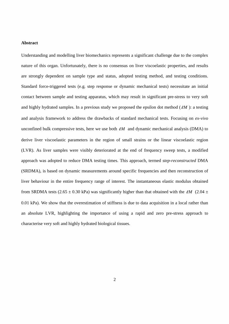
Abstract
Understanding and modelling liver biomechanics represents a significant challenge due to the complex
nature of this organ. Unfortunately, there is no consensus on liver viscoelastic properties, and results
are strongly dependent on sample type and status, adopted testing method, and testing conditions.
Standard force-triggered tests (e.g. step response or dynamic mechanical tests) necessitate an initial
contact between sample and testing apparatus, which may result in significant pre-stress to very soft
and highly hydrated samples. In a previous study we proposed the epsilon dot method (Mε& ): a testing
and analysis framework to address the drawbacks of standard mechanical tests. Focusing on ex-vivo
unconfined bulk compressive tests, here we use both Mε& and dynamic mechanical analysis (DMA) to
derive liver viscoelastic parameters in the region of small strains or the linear viscoelastic region
(LVR). As liver samples were visibly deteriorated at the end of frequency sweep tests, a modified
approach was adopted to reduce DMA testing times. This approach, termed step-reconstructed DMA
(SRDMA), is based on dynamic measurements around specific frequencies and then reconstruction of
liver behaviour in the entire frequency range of interest. The instantaneous elastic modulus obtained
from SRDMA tests (2.65 ± 0.30 kPa) was significantly higher than that obtained with the Mε& (2.04 ±
0.01 kPa). We show that the overestimation of stiffness is due to data acquisition in a local rather than
an absolute LVR, highlighting the importance of using a rapid and zero pre-stress approach to
characterise very soft and highly hydrated biological tissues.

3
1. Introduction
Although the mechanical properties of structural materials have been well described for decades using
various testing methods, there is still a scarcity of reliable and reproducible data for most biological
soft tissues. Characterising the heterogeneous, non-linear viscoelastic behaviour of soft non load-
bearing tissues (such as liver, kidney and brain) is very challenging, and a standard testing method is
needed to produce repeatable results that can be mathematically modelled to derive the mechanical
behaviour of a given tissue. Ideally, tissue characterisation via constitutive modelling requires the
control of both geometric and environmental boundary conditions. One of the main difficulties in
developing appropriate viscoelastic models for soft tissues is the establishment of suitable experimental
testing setups and protocols for the unique identification of material parameters. Generally, soft tissues
can be characterised under two different conditions, namely in-vivo and ex-vivo. In-vivo testing
maintains the tissue in its natural state, but has many limitations, such as accessibility, ill-defined
boundary conditions, ethical issues in using animals and potential risks to human subjects. Several in-
vivo mechanical measurements are reported in the literature, with datasets often limited to small
deformations. Furthermore, the interpretation of data is challenging due to difficulties in obtaining
appropriate alignment between the instrument and tested specimens, the presence of physiological
noise and the inability to account for and control the internal condition of the organ (Brown et al.,
2003; Gefen and Margulies, 2004; Tay et al., 2002). Conversely, ex-vivo experiments are preferable
when developing testing devices and protocols, as well for ease of testing, control of boundary
conditions and ethical considerations (Gao et al., 2010; Ocal et al., 2010; Raghunathan et al., 2010;
Sakuma et al., 2003; Valtorta and Mazza, 2005). They are also suitable for the development of
mechanically matched biomimetic scaffolds for in-vitro models and tissue engineering.

4
Like most internal organs, the liver essentially consists of a functional highly vascularised core
composed of cells embedded in a hydrated, porous and intrinsically viscoelastic extracellular matrix
(ECM). The organ is covered with a connective tissue capsule (i.e. the Glisson’s capsule) made of
densely interwoven collagen fibres, which ensures its structural integrity (Brunon et al., 2010).
Several methods and models are reported in the literature to characterise the mechanical behaviour of
liver either ex-vivo or in-vivo based on: direct measurements on tissue (e.g. rheological (Kalanovic et
al., 2003; Liu and Bilston, 2000; Marchesseau et al., 2010), compressive (Gao et al., 2010; Kemper et
al., 2013; Pervin et al., 2011; Raghunathan et al., 2010), indentation tests (Jordan et al., 2009; Kerdok
et al., 2006)) or imaging techniques (e.g. magnetic resonance (Asbach et al., 2008; Clarke et al., 2011;
Haghpanahi and Naeeni, 2010; Klatt et al., 2007; Venkatesh et al., 2008), ultrasound-based
elastography (Adebajo et al., 2012; Chenot et al., 2009; Ferraioli et al., 2013; Yoon et al., 2012)). The
first studies on the mechanical behaviour of animal and human livers were principally focused on the
investigation of static material properties (Carter et al., 2001; Nava et al., 2008; Roan and Vemaganti,
2007; Tay et al., 2002). Viscoelastic properties have been explored only recently, typically using stress-
relaxation and dynamic loading experiments to measure either time- or frequency-dependent material
properties, respectively (Liu and Bilston, 2000; Pervin et al., 2011). The development of imaging
elastography systems has also contributed to the study of the frequency dependence of liver mechanical
properties (Kruse et al., 2000; Valtorta and Mazza, 2005). Chatelin et al. compared in-vivo ultrasound-
based transient elastography (TE) and ex-vivo rheometry tests (DMA) on porcine livers, demonstrating
that the elastic properties measured by the two methods are equivalent (Chatelin et al., 2011). It is
worth noting that due to its highly heterogeneous structure, most mechanical models of liver published
to date are not material constitutive laws, but rather a means to quantify the gross tissue mechanical
properties and to determine the time scales of liver viscoelasticity, often attempting to correlate them

5
with different tissue conditions such as pathophysiological states or tissue ageing. Indeed, despite the
large number of investigations and reports, there is no consensus on the mechanical properties of liver,
or other soft tissues in general. Reported dynamic moduli span from few to tens of kPa (Marchesseau et
al., 2010) and are strongly dependent on the adopted testing method and experimental conditions as
well as sample type. In particular, the physical condition of the tissue (Kerdok et al., 2006), post-
mortem time or preservation period (Ocal et al., 2010), pathophysiological state (Mazza et al., 2007;
Wang et al., 1992; Yeh et al., 2002), tissue preload (Clarke et al., 2011; Yeh et al., 2002) and gravity
(Gao et al., 2010) can all affect sample status, and hence its mechanical properties. Moreover the
specific tissue model used, e.g. purely elastic versus viscoelastic, strongly conditions the estimated
tissue viscoelastic parameters.
Clearly, this is a vast and multifaceted area of research wherefrom emerges the need to clearly identify
the parameters of interest and then, based on available testing equipment, choose the appropriate
experimental set-up and analysis method. This need, coupled with the high variability and
inconsistency of published data on liver mechanical properties, motivated us to develop a reliable
quantitative testing and analysis framework for characterising the viscoelastic mechanical behaviour of
very soft and highly hydrated biological tissues ex-vivo. In this context, our main focus was the use of
common mechanical testing apparatus to measure hepatic tissue properties in the region of small and
physiologically relevant deformations, where soft tissues can be approximated as linear viscoelastic
materials. In general, soft tissue mechanical testing is beset by two main problems. First, the issues
related to establishing working conditions which ensure that each sample is in the same reproducible
status before testing. Secondly, standard force- or strain-triggered tests (e.g. step response or dynamic
mechanical tests) are affected by the long duration of tests and the need of an initial contact between
sample and testing apparatus, likely causing significant pre-stress and sample deterioration. Our aim

6
was thus to standardise sample preparation with defined, controlled and rapid testing conditions in
order to minimise tissue deterioration and guarantee a standard initial sample status.
Hence, two different testing methods were established to derive the viscoelastic parameters of hepatic
tissue through unconfined compressive tests within the linear viscoelastic region (LVR).
The first is based on the Mε& (epsilon dot method) which we recently proposed for testing and
parameter derivation of soft hydrated materials (Tirella et al., 2013), while the second is the dynamic
mechanical analysis (DMA) with a restricted number of discrete frequencies to reduce the duration of
the test (i.e. step-reconstructed DMA, or SRDMA).
With both methods, viscoelastic parameters of the hepatic tissue were estimated using a global fitting
approach with shared parameters. The SRDMA and Mε& are discussed and compared, highlighting the
similarities, advantages and limitations of the two methods for characterising the viscoelastic behaviour
of very soft, degradable and highly hydrated biological materials such as hepatic tissue.
2. Materials and methods
2.1. Sample preparation
Fresh porcine livers from 1 year old healthy pigs were collected as a slaughter by-product and frozen at
-20 °C within 3 hours of death. Prior to use, frozen livers were thawed at 4 °C overnight, then punched
to obtain regular 14 mm diameter cylinders which were subsequently cut in 3 mm thick samples with
parallel loading surfaces using a custom slicer and a microtome blade. Capsular connective tissue (i.e.
Glisson’s capsule) was not present in tested samples and particular attention was dedicated to avoid
macroscopic vasculature. We recently showed that the bulk compressive modulus (λ) of porcine fresh

7
liver does not change significantly with the sample harvesting site (i.e. different liver lobes) nor
between animals from the same slaughterhouse (Mattei et al., 2014). Furthermore, in agreement with
Tamura et al. (Tamura et al., 2002), our results showed that a freeze-thawing cycle (samples stored at -
20 °C, then thawed at 4 °C overnight prior to testing) does not significantly affect the liver compressive
modulus. Hence, thawed samples from multiple harvesting sites were used for all tests. To ensure
repeatable testing conditions, thawed liver samples were equilibrium swollen in PBS 1X at 4 °C and
then brought to room temperature prior to testing (Mattei et al., 2014; Yeh et al., 2002). Samples were
carefully measured in thickness (l0) and diameter (d) just before testing (hence accounting for any size
variations due the swelling process). Measurements were performed by gently placing the jaws of a
calliper (0.05 mm resolution) in contact with the sides of the sample, averaging readings from at least
three different points. Samples were tested partially immersed in PBS 1X to preserve their hydration
during the unconfined compression test (Mattei et al., 2014; Tirella et al., 2013). Mechanical tests were
performed within 2 weeks after sample collection.
2.2. Mε&
The Mε& is based on the application of a series of short compressions at different strain rates to
specimens while acquiring force and displacement versus time data in the LVR (Tirella et al., 2013).
Tests were performed with a uniaxial testing device (Zwick/Roell ProLine Z005) equipped with a 10 N
load cell (Zwick/Roell Xforce HP 10 N), applying strain rates of 0.001, 0.005, 0.01 and 0.05 s-1 to liver
samples. In particular, force and displacement versus time data were acquired starting with the upper
plate of the testing device (connected to the load cell) close to but not in contact with the sample, to
guarantee a zero pre-stress initial condition and a constant approach velocity. Experimental force- and

8
displacement-time series were respectively normalised to sample cross-sectional area (πd 2/4) and
thickness (l0) measured just prior to testing, obtaining stress- and strain-time series. Liver LVR was
identified as the region in which stress varies linearly with applied strain (R2 of at least 0.995). Then,
stress-time data within LVR obtained from measurements at different strain rates were used to derive
viscoelastic constants for lumped parameter models using the global fitting procedure with shared
parameters described in Supplementary Information (SI 3). Six liver samples were tested at each strain
rate, using a new sample for each repeat; total number of specimens = 24.
2.3. Dynamic mechanical testing method
Dynamic mechanical analysis (DMA), a standard force-triggered method, was used to determine
material viscoelastic properties by applying a small amplitude cyclic strain on a sample and measuring
the resultant cyclic stress response. The tests were performed compressing samples at room
temperature using a GABO Eplexor 150 N (Gabo GmbH, Ahlden, Germany). The trigger force was set
to a value of 10 mN, which we identified as the minimum reliable starting force of the instrument. In
this set of experiments, the LVR was considered as the range of strain amplitudes in which the storage
modulus changes by less than 5% of its initial value. To identify the LVR for frequency sweep tests, a
preliminary series of strain amplitude sweep tests at 1 Hz was conducted.
2.3.1. Conventional dynamic mechanical tests
To assess any possible change in liver mechanical properties due to tissue deterioration while standing,
equilibrium swollen samples were kept in PBS 1X at room temperature for different times prior to
testing (i.e. 0, 2, 4, 20, 24 h). Frequency sweep tests were then performed in triplicate in the frequency

9
range 0.05÷100 Hz, choosing 1% static and 0.5% dynamic strain amplitudes to guarantee a linear
response, as outlined in Supplementary Information SI 1. Note that each sweep test took about 1 hour
and 30 minutes to complete.
2.3.2. Step-reconstructed dynamic mechanical tests
The SRDMA approach is based on performing short frequency sweeps around selected frequencies (f =
0.5, 1, 10 and 50 Hz): specifically, measurements were performed at the selected frequency f, and f ±
0.1 Hz on the same sample. Equilibrium swollen samples were tested after being brought to room
temperature choosing 1% static and 0.5% dynamic strain amplitudes to lie within liver LVR. Liver
dynamic mechanical behaviour was then reconstructed over the whole investigated frequency-range
(SRDMA) as described in the Supplementary Information. Compared with the 0.05 ÷ 100 Hz
frequency sweep test, this approach enables a shorter testing phase (i.e. less than 5 minutes in the
longest test, 0.4 ÷ 0.6 Hz frequency range), hence preventing any significant sample deterioration. Note
that, since each sample was tested only once to prevent permanent alterations due to repeated testing
cycles, 12 liver samples were required to perform the SRDMA analysis in triplicate around the four
selected frequencies. Cyclic tests at each of the single frequencies investigated around 1 Hz were also
performed on independent samples. No significant differences in E’ and E’’ were measured between
independent samples tested at 0.9, 1 and 1.1 Hz with respect to those obtained by sequential tests at 0.9,
1 and 1.1 Hz on the same sample (data not reported), confirming the absence of sub-failure loading in
the SRDMA approach.

10
2.4. Lumped parameter estimation
Liver samples were treated as mechanically isotropic materials (Marchesseau et al., 2010; Mattei et al.,
2014; Pervin et al., 2011). In the region of small deformations, the viscoelastic behaviour of soft and
hydrated materials can be derived using classical lumped parameter models such as the Maxwell
Standard Linear Solid (SLS) (SI 1). As described in the Supplementary Information, two models, the
SLS and the GM2 (2-arm Generalised Maxwell model) were used to estimate the material coefficients
for liver tissue (SI 2). A global fitting approach was employed performing chi-square minimisation in a
combined parameter space (SI 3). In order to select suitable parameter initial guesses, an annealing
scheme, multiplying and dividing each initial parameter by 10 individually while keeping the
instantaneous modulus at a constant value (i.e. a constant sum of all springs in the model), was
adopted. In this way reliable and absolute hepatic viscoelastic parameters within the investigated
frequency range were obtained, while avoiding most of the local minima during the fitting procedure.
A lower boundary was set to prevent the fitting procedure returning negative values for the estimated
viscoelastic coefficients. Comparisons between parameter values were made using the Student’s t-test,
setting significance at p < 0.05.
3. Results
3.1. Mε&
As sketched in Fig. 1a, the measured sample stress is zero while the plate approaches the sample, prior
to the instant of contact to ensure no pre-stress acting on tested sample (zone A). Further advancement
results in a slight negative stress, mainly due to water mediated adhesive forces between the plate and
the sample (zone B). Zone C, represents the actual compression of the sample: in this region the stress
increases monotonically with time. Experimental stress-time data collected at various strain rates are

11
shown in Fig. 1b, where the time axis has been offset to be zero at the beginning of sample
compression (i.e. zone C). Only stress-time data belonging to the LVR are shown. In all experiments
the LVR extended up to a strain of 0.03, therefore the higher the strain-rate, the shorter the duration of
the stress-time series (Fig. 1b). Liver samples did not demonstrate any visible changes at the end of the
rapid Mε& compressive tests.
Figure 1. Epsilon dot method. a) Schematic of experimental testing setup and of a typical stress-time curve recorded during an Mε& test. The measured stress is zero while the plate approaches the sample (zone A). Then it becomes slightly negative
due to water mediated adhesive forces between the plate and the sample (zone B). Finally, the measured stress becomes positive, increasing monotonically with time, defining the zone of actual sample compression (zone C). b) Experimental stress-time data collected at various strain rates. The time axis has been offset to be zero at the beginning of the actual compressing phase (zone C) and only stress-time data belonging to the LVR (i.e. those used to estimate lumped parameters) are shown.
3.2. DMA: frequency sweep test
Using the conventional DMA approach, strain amplitude sweep tests underlined that sample LVR
extended up to 2% strain, in accordance with Marchesseau et al. (Marchesseau et al., 2010). However,
at the end of the frequency sweep test, samples were found to be highly compressed, dark brown and
dehydrated likely due to the very long testing time. This is clearly highlighted in Fig. 2 in which the

12
sample height decreases rapidly at the lower frequencies. As the sweep begins, cyclic stresses cause
fluid expulsion from the tested sample, so that additional compression is required to reach the trigger
contact force necessary for successive testing cycles.
Figure 2. Change in sample height during frequency sweep measurements reflecting sample degradation while testing, n = 3.
Consequently, the measured storage and loss moduli likely reflect sample deterioration and water
elimination rather than the dynamic mechanical properties. In fact, no meaningful trends in E’ and E’’
were found between measurements performed at different preservation times (Fig. 3) and the
experimental results were not used further to derive liver viscoelastic parameters.
3.3. SRDMA
We did not observe any deterioration in samples using the quicker SRDMA approach and sample
compression at the end of the test was measured to be less than 2% of its initial height.
SRDMA storage and loss moduli are presented in Fig. 4. As expected, the storage modulus increases
with frequency up to the relaxation frequency and then remains almost constant. On the other hand, the
loss modulus increases with frequency reaching a peak value at the relaxation frequency (~ 1 Hz, at
13
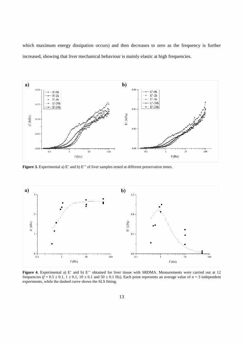
which maximum energy dissipation occurs) and then decreases to zero as the frequency is further
increased, showing that liver mechanical behaviour is mainly elastic at high frequencies.
Figure 3. Experimental a) E’ and b) E’’ of liver samples tested at different preservation times.
Figure 4. Experimental a) E’ and b) E’’ obtained for liver tissue with SRDMA. Measurements were carried out at 12 frequencies (f = 0.5 ± 0.1, 1 ± 0.1, 10 ± 0.1 and 50 ± 0.1 Hz). Each point represents an average value of n = 3 independent experiments, while the dashed curve shows the SLS fitting.
14
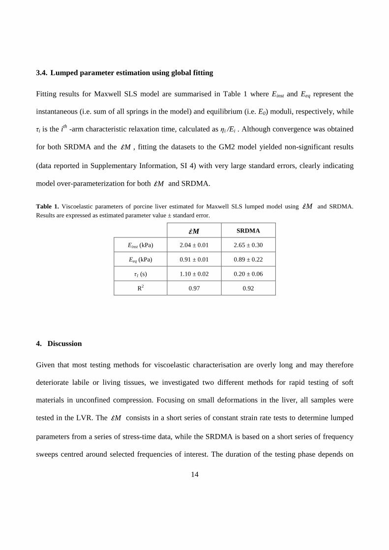
3.4. Lumped parameter estimation using global fitting
Fitting results for Maxwell SLS model are summarised in Table 1 where Einst and Eeq represent the
instantaneous (i.e. sum of all springs in the model) and equilibrium (i.e. E0) moduli, respectively, while
τi is the ith -arm characteristic relaxation time, calculated as ηi /Ei . Although convergence was obtained
for both SRDMA and the Mε& , fitting the datasets to the GM2 model yielded non-significant results
(data reported in Supplementary Information, SI 4) with very large standard errors, clearly indicating
model over-parameterization for both Mε& and SRDMA.
Table 1. Viscoelastic parameters of porcine liver estimated for Maxwell SLS lumped model using Mε& and SRDMA.
Results are expressed as estimated parameter value ± standard error.
Mε& SRDMA
Einst (kPa) 2.04 ± 0.01 2.65 ± 0.30
Eeq (kPa) 0.91 ± 0.01 0.89 ± 0.22
τ1 (s) 1.10 ± 0.02 0.20 ± 0.06
R2 0.97 0.92
4. Discussion
Given that most testing methods for viscoelastic characterisation are overly long and may therefore
deteriorate labile or living tissues, we investigated two different methods for rapid testing of soft
materials in unconfined compression. Focusing on small deformations in the liver, all samples were
tested in the LVR. The Mε& consists in a short series of constant strain rate tests to determine lumped
parameters from a series of stress-time data, while the SRDMA is based on a short series of frequency
sweeps centred around selected frequencies of interest. The duration of the testing phase depends on

15
the strain rate for Mε& or on the frequency employed for SRDMA: the lower the ε& or f the longer the
test. In case of Mε& we used strain rates between 0.001 and 0.05 s-1, while for SRDMA a frequency
range of 0.5 to 50 Hz was employed, thus tests were shorter than 30 s for the former and 300 s for the
latter. In terms of magnitude, SRDMA storage and loss moduli are comparable to those reported by Liu
and Bilston for bovine liver (G’ = 1 – 6 kPa, G’’ < 1 kPa, f = 0.006 – 20 Hz) (Liu and Bilston, 2000),
Kiss et al. for canine liver (E’ = 3 – 8 kPa, E’’ = 0.8 – 4 kPa, f = 0.1 – 100 Hz) (Kiss et al., 2004) and
Marchesseau et al. for porcine liver (G’ = 0.3 – 0.6 kPa, G’’ = 0.05 – 0.15 kPa, f = 0.1 – 4 Hz)
(Marchesseau et al., 2010). However, the estimated viscoelastic parameters of liver, or highly non-
linear soft materials in general, are strongly dependent on the testing conditions.
As mentioned in Section 2.3, a minimum contact force of 10 mN is necessary to trigger the GABO
Eplexor 150 N. Given that sample surface area of samples is about 1.5 cm2, the trigger force results in
an average sample pre-stress of 65 Pa with a consequent strain of about 4%. In this region, a local LVR
can nevertheless be identified (Figure 5), albeit with a smaller linear range and a higher slope with
respect to that obtained in the absence of pre-stress (i.e. the absolute LVR).
To perform measurements in the absolute LVR, it is necessary to use methods which do not require a
trigger force during the acquisition phase. In this perspective, the Mε& is very suited for the testing of
soft materials, as viscoelastic parameters can be derived with standard uniaxial testing devices, starting
in a contactless configuration (Tirella et al., 2013). In fact without pre-stress, Einst was found to be
significantly lower (2.04 ± 0.01 kPa) than the value estimated with SRDMA (2.65 ± 0.30 kPa, p =
0.0174), demonstrating that the unavoidable trigger contact force causes sample stiffening. The effect
of the trigger force is also reflected in the characteristic relaxation time: as shown in Table 1, the value

16
of τ estimated with the SRDMA was significantly lower than that obtained using the Mε& (p < 0.0001),
suggesting that pre-stressed samples exhibit a more elastic behaviour than in the absence of pre-stress.
Figure 5. Liver stress-strain plot up to 0.10 true compressive strain, data obtained from an Mε& test at 0.01 s-1. Clearly, the
absolute liver LVR is comprised in the 0 – 0.03 strain range, while local LVRs can be found depending on the measurement’s starting point (i.e. pre-strain). SRDMA analysis suffers from a 4% pre-strain due to GABO Eplexor’s triggering force, from which a local LVR can be found, characterised by a smaller linear range and a higher slope with respect to the absolute LVR obtained in the absence of pre-stress, as outlined in the table insert.
Clearly, the estimated time constants depend strongly on the range of strain rates (in the case ofMε& ) or
frequencies (in the case of SRDMA) employed during testing. In both cases, an a priori knowledge of

17
the relaxation behaviour of a material is desirable if one wishes to capture material viscoelastic features
within a time frame of interest.
5. Conclusion
The objective of this study was to formulate a reliable and reproducible method for the rapid testing
and measurement of liver viscoelastic properties in the true LVR. To this end, two different methods
(SRDMA and Mε& ) were developed and used to derive the viscoelastic parameters of porcine liver
samples. Attention was paid to tissue handling and treatment throughout the study, ensuring all samples
were in a repeatable initial state. The study highlights the advantages and disadvantages of SRDMA
and Mε& , in testing soft and degradable biomaterials. Although both methods permit considerable time
savings and good sample preservation, in our opinion the Mε& can give a better estimation of the
viscoelastic parameters than does SRDMA, since it avoids sample pre-stress and allows measurements
in the actual LVR. The Mε& is, as far as we know, the only testing and analysis framework which
enables the unique identification of viscoelastic parameters of soft materials - such as hepatic tissue -
through direct measurements and in the absence of pre-stress. The results can be used to design
mechanically matched hepatic tissue models and can also be extended to the characterisation and
constitutive modelling of other soft tissues and materials, enabling comparisons across different
studies.

18
Acknowledgements
The authors would like to thank Dr. Giovanni Aggravi and the public abattoir of the Azienda Speciale
Multiservizi in Colle di val d'Elsa (Siena, Italy), managed by Macellatori Senesi srl di Borghi e
Brunelli for supplying fresh hepatic tissue. The work leading to these results has received funding from
the European Union Seventh Framework Programme (FP7/2007-2013) under grant agreement n°
304961 (ReLiver).
Conflict of interest statement
The authors declare that there are no conflicts of interest.

19
References
Adebajo, C.O., Talwalkar, J.A., Poterucha, J.J., Kim, W.R., Charlton, M.R., 2012. Ultrasound-based
transient elastography for the detection of hepatic fibrosis in patients with recurrent hepatitis C
virus after liver transplantation: a systematic review and meta-analysis. Liver Transpl. 18, 323–31.
Asbach, P., Klatt, D., Hamhaber, U., Braun, J., Somasundaram, R., Hamm, B., Sack, I., 2008.
Assessment of liver viscoelasticity using multifrequency MR elastography. Magn. Reson. Med.
60, 373–9.
Brown, J.D., Rosen, J., Kim, Y.S., Chang, L., Sinanan, M.N., Hannaford, B., 2003. In-vivo and in-situ
compressive properties of porcine abdominal soft tissues. Stud. Health Technol. Inform. 94, 26–
32.
Brunon, A., Bruyère-Garnier, K., Coret, M., 2010. Mechanical characterization of liver capsule through
uniaxial quasi-static tensile tests until failure. J. Biomech. 43, 2221–7.
Carter, F.J., Frank, T.G., Davies, P.J., McLean, D., Cuschieri, A., 2001. Measurements and modelling
of the compliance of human and porcine organs. Med. Image Anal. 5, 231–6.
Chatelin, S., Oudry, J., Périchon, N., Sandrin, L., Allemann, P., Soler, L., Willinger, R., 2011. In vivo
liver tissue mechanical properties by Transient Elastography: comparison with Dynamic
Mechanical Analysis. Biorheology 48, 75–88.

20
Chenot, J., Melodelima, D., Parmentier, H., Souchon, R., Chapelon, J.-Y., 2009. Comparison of
different compression methods for ultrasound elastography in order to detect HIFU ablations in
the liver: preliminary in vivo results. In: 2009 IEEE International Ultrasonics Symposium. IEEE,
pp. 135–138.
Clarke, E.C., Cheng, S., Green, M., Sinkus, R., Bilston, L.E., 2011. Using static preload with magnetic
resonance elastography to estimate large strain viscoelastic properties of bovine liver. J. Biomech.
44, 2461–5.
Ferraioli, G., Tinelli, C., Dal Bello, B., Zicchetti, M., Lissandrin, R., Filice, G., Filice, C., Above, E.,
Barbarini, G., Brunetti, E., Calderon, W., Di Gregorio, M., Gulminetti, R., Lanzarini, P., Ludovisi,
S., Maiocchi, L., Malfitano, A., Michelone, G., Minoli, L., Mondelli, M., Novati, S., Patruno,
S.F.A., Perretti, A., Poma, G., Sacchi, P., Zanaboni, D., Zaramella, M., 2013. Performance of liver
stiffness measurements by transient elastography in chronic hepatitis. World J. Gastroenterol. 19,
49–56.
Gao, Z., Lister, K., Desai, J.P., 2010. Constitutive modeling of liver tissue: experiment and theory.
Ann. Biomed. Eng. 38, 505–16.
Gefen, A., Margulies, S.S., 2004. Are in vivo and in situ brain tissues mechanically similar? J.
Biomech. 37, 1339–52.
Haghpanahi, M., Naeeni, H.A., 2010. Investigation of viscoelastic properties of human liver tissue
using MR elastography and FE modeling. In: 2010 17th Iranian Conference of Biomedical
Engineering (ICBME). IEEE, pp. 1–4.

21
Jordan, P., Socrate, S., Zickler, T.E., Howe, R.D., 2009. Constitutive modeling of porcine liver in
indentation using 3D ultrasound imaging. J. Mech. Behav. Biomed. Mater. 2, 192–201.
Kalanovic, D., Ottensmeyer, M.P., Gross, J., Buess, G., Dawson, S.L., 2003. Independent testing of
soft tissue visco-elasticity using indentation and rotary shear deformations. Stud. Health Technol.
Inform. 94, 137–43.
Kemper, A.R., Santago, A.C., Stitzel, J.D., Sparks, J.L., Duma, S.M., 2013. Effect of strain rate on the
material properties of human liver parenchyma in unconfined compression. J. Biomech. Eng. 135,
104503–8.
Kerdok, A.E., Ottensmeyer, M.P., Howe, R.D., 2006. Effects of perfusion on the viscoelastic
characteristics of liver. J. Biomech. 39, 2221–31.
Kiss, M.Z., Varghese, T., Hall, T.J., 2004. Viscoelastic characterization of in vitro canine tissue. Phys.
Med. Biol. 49, 4207–18.
Klatt, D., Hamhaber, U., Asbach, P., Braun, J., Sack, I., 2007. Noninvasive assessment of the
rheological behavior of human organs using multifrequency MR elastography: a study of brain
and liver viscoelasticity. Phys. Med. Biol. 52, 7281–94.
Kruse, S.A., Smith, J.A., Lawrence, A.J., Dresner, M.A., Manduca, A., Greenleaf, J.F., Ehman, R.L.,
2000. Tissue characterization using magnetic resonance elastography: preliminary results. Phys.
Med. Biol. 45, 1579–90.

22
Liu, Z., Bilston, L., 2000. On the viscoelastic character of liver tissue: experiments and modelling of
the linear behaviour. Biorheology 37, 191–201.
Marchesseau, S., Heimann, T., Chatelin, S., Willinger, R., Delingette, H., 2010. Fast porous visco-
hyperelastic soft tissue model for surgery simulation: application to liver surgery. Prog. Biophys.
Mol. Biol. 103, 185–96.
Mattei, G., Di Patria, V., Tirella, A., Alaimo, A., Elia, G., Corti, A., Paolicchi, A., Ahluwalia, A., 2014.
Mechanostructure and composition of highly reproducible decellularized liver matrices. Acta
Biomater. 10, 875–82.
Mazza, E., Nava, A., Hahnloser, D., Jochum, W., Bajka, M., 2007. The mechanical response of human
liver and its relation to histology: an in vivo study. Med. Image Anal. 11, 663–72.
Nava, A., Mazza, E., Furrer, M., Villiger, P., Reinhart, W.H., 2008. In vivo mechanical
characterization of human liver. Med. Image Anal. 12, 203–16.
Ocal, S., Ozcan, M.U., Basdogan, I., Basdogan, C., 2010. Effect of preservation period on the
viscoelastic material properties of soft tissues with implications for liver transplantation. J.
Biomech. Eng. 132, 101007.
Pervin, F., Chen, W.W., Weerasooriya, T., 2011. Dynamic compressive response of bovine liver
tissues. J. Mech. Behav. Biomed. Mater. 4, 76–84.
Raghunathan, S., Evans, D., Sparks, J.L., 2010. Poroviscoelastic modeling of liver biomechanical
response in unconfined compression. Ann. Biomed. Eng. 38, 1789–800.

23
Roan, E., Vemaganti, K., 2007. The nonlinear material properties of liver tissue determined from no-
slip uniaxial compression experiments. J. Biomech. Eng. 129, 450–6.
Sakuma, I., Nishimura, Y., Chui, C.K., Kobayashi, E., 2003. In vitro Measurement of Mechanical
Properties of Liver Tissue under Compression and Elongation Using a New Test Piece Holding
Method with Surgical Glue, In Vitro, Lecture Notes in Computer Science. Springer Berlin
Heidelberg, Berlin, Heidelberg.
Tamura, A., Omori, K., Miki, K., Lee, J.B., Yang, K.H., King, A.I., 2002. Mechanical characterization
of porcine abdominal organs. Stapp Car Crash J. 46, 55–69.
Tay, B.K., Stylopoulos, N., De, S., Rattner, D.W., Srinivasan, M.A., 2002. Measurement of in-vivo
force response of intra-abdominal soft tissues for surgical simulation. Stud. Health Technol.
Inform. 85, 514–9.
Tirella, A., Mattei, G., Ahluwalia, A., 2013. Strain rate viscoelastic analysis of soft and highly hydrated
biomaterials. J. Biomed. Mater. Res. A.
Valtorta, D., Mazza, E., 2005. Dynamic measurement of soft tissue viscoelastic properties with a
torsional resonator device. Med. Image Anal. 9, 481–90.
Venkatesh, S.K., Yin, M., Glockner, J.F., Takahashi, N., Araoz, P.A., Talwalkar, J.A., Ehman, R.L.,
2008. MR elastography of liver tumors: preliminary results. AJR. Am. J. Roentgenol. 190, 1534–
40.

24
Wang, B.C., Wang, G.R., Yan, D.H., Liu, Y.P., 1992. An experimental study on biomechanical
properties of hepatic tissue using a new measuring method. Biomed. Mater. Eng. 2, 133–8.
Yeh, W.-C., Li, P.-C., Jeng, Y.-M., Hsu, H.-C., Kuo, P.-L., Li, M.-L., Yang, P.-M., Lee, P.H., 2002.
Elastic modulus measurements of human liver and correlation with pathology. Ultrasound Med.
Biol. 28, 467–474.
Yoon, K.T., Lim, S.M., Park, J.Y., Kim, D.Y., Ahn, S.H., Han, K.-H., Chon, C.Y., Cho, M., Lee, J.W.,
Kim, S.U., 2012. Liver stiffness measurement using acoustic radiation force impulse (ARFI)
elastography and effect of necroinflammation. Dig. Dis. Sci. 57, 1682–91.


