
ORIGINAL CONTRIBUTION
Trans-epithelial transport of the betalain pigments indicaxanthinand betanin across Caco-2 cell monolayers and influence of foodmatrix
L. Tesoriere • C. Gentile • F. Angileri •
A. Attanzio • M. Tutone • M. Allegra •
M. A. Livrea
Received: 23 January 2012 / Accepted: 27 June 2012 / Published online: 18 July 2012
� Springer-Verlag 2012
Abstract
Purpose This study investigated the absorption mecha-
nism of the phytochemicals indicaxanthin and betanin and
the influence of their food matrix (cactus pear and red beet)
on the intestinal transport.
Methods Trans-epithelial transport of dietary-consistent
amounts of indicaxanthin and betanin in Caco-2 cell
monolayers seeded on TranswellR inserts was measured in
apical to basolateral (AP-BL) and basolateral to apical
(BL-AP) direction, under an inwardly directed pH gradient
(pH 6.0/7.4, AP/BL) mimicking luminal and serosal sides
of human intestinal epithelium. The effect of inhibitors of
membrane transporters on the absorption was also evalu-
ated. Contribution of the paracellular route was investi-
gated after EDTA treatment of the cell monolayer. In vitro
digestion of betalainic food was performed to provide a
post-intestinal fraction containing bioaccessible pigments.
Results Apparent permeability coefficients (Papp) in the
absorptive direction were (4.4 ± 0.4) 9 10-6 and
(3.2 ± 0.3) 9 10-6 cm s-1 for indicaxanthin and betanin,
respectively. Transport of indicaxanthin was non-polarized,
linear as a function of time and concentration, and unaf-
fected by inhibitors of membrane transporters. Betanin
exhibited significantly different bidirectional Papp values
and non-linear efflux kinetics. The concentration-depen-
dent betanin efflux was described by a kinetic model
including one non-saturable (Kd = 0.042 lL cm-2 min-1)
and one saturable component identified as the apical
multidrug resistance–associated protein 2 (MRP2;
Km = 275 lM; Jmax = 42 pmol min-1 cm-2). Permeation
of both betalains increased remarkably after EDTA treat-
ment of the cell monolayer. Neither indicaxanthin nor
betanin underwent metabolic transformation. Food matrix
did not affect trans-epithelial transfer of indicaxanthin, but
reduced the absorption rate of betanin, red beet more than
cactus pear.
Conclusions Dietary indicaxanthin and betanin can sub-
stantially be absorbed through paracellular junctions of
intestinal epithelial cells. Additional trans-membrane per-
meation can be considered for betanin, whose absorption is
limited by a MRP2-mediated efflux and negatively affected
by its food matrix. Present findings are consistent with the
quite higher bioavailability of indicaxanthin over betanin
established in humans.
Keywords Betalains � Intestinal absorption �Caco-2 cells �Betalainic food � Indicaxanthin � Betanin
Introduction
Health benefits of dietary phytochemicals have been sug-
gested in recent years. Among thousands of different
compounds, phenolics, terpenoids, and sulfur-containing
compounds have been considered because of reducing
power and potential to affect redox-modulated cellular
processes [1–3]. Betalains, which occur in a number of
vegetables of the Cariophyllalae order, with cactus pear
(Opuntia genus) fruits and red beet (Beta vulgaris) as the
more representative dietary sources, are nitrogen-contain-
ing compounds, the structure of which comes from a
tyrosine derivative known as betalamic acid. Depending on
the components bonded to the main structure, violet-red
L. Tesoriere � C. Gentile � F. Angileri � A. Attanzio �M. Tutone � M. Allegra � M. A. Livrea (&)
Dipartimento di Scienze e Tecnologie Molecolari e
Biomolecolari (STEMBIO), Universita di Palermo,
Via Michele Cipolla 74, 90123 Palermo, Italy
e-mail: [email protected]
123
Eur J Nutr (2013) 52:1077–1087
DOI 10.1007/s00394-012-0414-5

betacyanins or orange-yellow betaxanthins arise, the for-
mer when the component is 3,4-dihydroxy-phenylalanine
(DOPA), which may be or may be not glycosylated, and
the latter if the conjugation partners are amino acids or
derived amines [4, 5]. The potential use of this class of
molecules as natural dyes has widely been explored in the
past, and experimental evidence has been provided about
safety and factors affecting their stability under a variety of
conditions [6, 7]. More recently, a number of bioactivities
have emerged from in vitro studies in solution and in cell
cultures. In particular, indicaxanthin and betanin (Fig. 1),
the adducts of betalamic acid with proline and cyclo-
DOPA 5-O-b glucoside, respectively, exhibited antioxidant
activity in biological lipid environments from human low-
density lipoproteins to membranes and whole cells [8–12],
were able to modulate redox-mediated signal transduction
pathways involved in inflammation in cultured endothe-
lium [13], and showed antiproliferative effects in human
tumor cell lines [14–16].
The health benefits of dietary phytochemicals cannot
easily be deduced from in vitro studies, one main reason
being that phytochemicals are processed as xenobiotics, that
is, they may be or may be not absorbed to a suitable amount,
transported in blood and distributed to tissues, metabolized
and excreted. Human studies after the ingestion of dietary
amounts of cactus pear fruit [17] or red beet juice [18, 19]
indicated that indicaxanthin and betanin reach plasma
concentrations of a micromolar order, which is quite higher
than other phytochemicals such as polyphenols [20, 21]. In
other studies [22], by a simulated gastro-intestinal diges-
tion, we were able to demonstrate that digestive stability
and additional factors relevant to the solubilization from
food matrix and style of food processing influence the
fraction of soluble betalains in the post-intestinal digesta
potentially available for trans-epithelial transfer. Other
factors that may concur to betalain bioavailability, includ-
ing intestinal metabolism and mechanism of trans-epithelial
transport, as well as interference of the food matrix on the
absorptive process, are still unknown. In this study, we
investigate intestinal permeation of indicaxanthin and
betanin using Caco-2 cell monolayers seeded on Trans-
well� insert, a well-established model of the intestinal
barrier. Originating from a human colorectal carcinoma,
these cells spontaneously differentiate into polarized mon-
olayers that exhibit morphological and functional charac-
teristics of the intestinal absorptive epithelium, including
intercellular tight junctions and apical microvilli, carriers
for nutrient as well as efflux proteins, and a number of
phase-2 enzymes required to xenobiotic disposition
[23, 24]. In addition, since food matrix can affect absorption,
the trans-epithelial permeation of betalains from various
betalainic food submitted to a simulated gastro-intestinal
digestion was compared with that of the pure compounds.
Materials and methods
Unless stated otherwise, all reagents and materials were
from Sigma Chemical Co. (St Louis, MO, USA) and sol-
vents were of the highest purity or HPLC grade.
Betalain pigments
Betanin and indicaxanthin were isolated from cactus pear
fruits as previously reported [25] and then purified
according to Stintzing et al. [26]. The betalains were
spectrophotometrically quantified using molar absorbance
of 65,000 at 536 nm and 42,800 at 482 nm for betanin and
indicaxanthin, respectively.
Physicochemical properties
Molecular descriptors of the betalains such as ClogP,
ClogD were computed by Qikprop 3.1 predict program
(Schrodinger, LLC, New York, NY, USA). The non-polar
surface area (NPSA) was obtained as the difference
between the molecular surface area (MSA) and polar sur-
face area (PSA), calculated by CODESSA PRO software
[27]. pKa values of indicaxanthin were obtained by two
different approaches, that is, the semi-empirical partial
charge related and the Hammet and Taft linear free-energy
relationships. Semi-empirical calculations were made by
means of Marvin Sketch 5.0.6.1 prediction program
(ChemAxon, Budapest, Hungary), based on the calculation
of Mulliken partial charge of atoms in the molecule. The
Hammet and Taft linear free-energy relationships were
calculated by Epik 1.6 software (Schrodinger, LLC, New
York, NY, USA), which adopts the combination of Ham-
met and Taf methods in conjunction with ionization and
tautomerization tools. The pKa values of betanin were
obtained from the literature [28].Fig. 1 Molecular structure of indicaxanthin and betanin
1078 Eur J Nutr (2013) 52:1077–1087
123

Cell culture
Caco-2 cells, obtained from the American Type Culture
Collection (Rockville, MD, USA), were cultured in Dul-
becco’s modified Eagle medium (DMEM; Gibco Life
Technologies, Grand Island, NY, USA) supplemented with
10 % fetal bovine serum (Gibco Life Technologies), 1 %
non-essential amino acids, 10 mM HEPES, 50 units/mL
penicillin, 50 lg/mL streptomycin, and 100 lg/mL genta-
micin and were maintained at 37 �C in 5 % CO2 and 95 %
humidity. Medium was changed 2–3 times per week. Caco-
2 cells were used between passages 27 and 31. The cells
were monitored by phase contrast microscopy, and fluo-
rescent staining of DNA by Hoechst 33,258 (Thermo
Fisher Scientific, Rockford, IL, USA) was used once a
month to rule out mycoplasma contamination.
In vitro simulated digestion and preparation
of the bioaccessible fraction from betalainic food
Fresh cactus pear fruits (O. ficus indica L. Mill.) from
yellow and red Sicilian cultivars and red beet roots
(B. Vulgaris L. ssp Vulgaris) from Tuscany (Italy) culti-
vations were obtained from a local market. Red beet juice
(Biotta AG, CH-8274, Tagewiler, Switzerland) was pur-
chased in a healthy food store. Cactus pear fruit juice was
prepared after brief homogenization of the pulp in a
kitchen-type blender and filtration through a colander
(0.2 mm mesh size). Aliquots (20 g of each food prepara-
tion) was chewed 10 times by a single investigator and
subsequently expelled in a tared beaker. The post-oral
material was briefly homogenized in Hanks’ balanced salt
solution, pH 7.4 (HBSS, 1:2, w:v) for 2 min in a laboratory
blender (Waring, New Hartford, CT), acidified at pH 2.0
with HCl, and 8 mg/mL porcine pepsin (3,200–4,500 units/
mg) was added. After incubation in a shaking (100 rpm)
water bath (type M 428-BD, Instruments s.r.l., Bernareggio,
Mi, Italy) at 37 �C, for 2 h to simulate the gastric phase, the
pH of the sample was immediately increased to 7.4 with
0.5 M NaHCO3. The small intestinal phase of digestion was
started after the addition of 2.4 mg/mL porcine bile extract
and 0.4 mg/mL pancreatin. After incubation in the shaking
water bath as above, for 2 h at 37 �C, the post-intestinal
digest was centrifuged at 167,000g, for 35 min at 4 �C in a
Beckman Optima TLX ultracentrifuge, equipped with an
MLA-55 rotor (Beckman Instruments, Inc., Palo Alto, CA,
USA), to separate the aqueous bioaccessible fraction (BF)
from particulate material. Digestive enzymes were removed
by ultracentrifugation through YM-10 membranes, and the
betalain content of BF was measured by HPLC. Food BF
was stored at -80 �C until use.
Cytotoxicity of the BFs on Caco-2 cells was checked in
pilot studies. Caco-2 cells were seeded at 5 9 105 cells
well in a six-well flat-bottom plate, and the medium was
changed three times a week. After 15 days from conflu-
ence, differentiated monolayers were washed three times
with HBSS, and then 2 mL of food BF was added. The BFs
were filtered through a Millex HV 0.2 lm filter (Millipore,
Billerica, MA, USA) immediately before the use. After a
90-min incubation, cells were washed and 50 lL HBSS
containing 5 mg/mL MTT was added. The medium was
discarded after a 4-h incubation at 37 �C, and formazan
blue formed in the cells was dissolved in DMSO. The
absorbance at 540 nm of MTT formazan of untreated cells
was taken as 100 % viability. In addition, viability of the
cells after the treatment was determined by trypan blue
exclusion and microscopy examination. Neither treatment
caused cell toxicity.
Trans-epithelial transport
Transport experiments were carried out using TranswellR
inserts (polycarbonate membrane, 0.4 lm pore size,
24 mm diameter, Corning Inc., Corning, NY, USA). Inserts
were placed in 6-well plates. Caco-2 cells were seeded at
5 9 104 cells per cm2 on the membrane insert with 1.5 mL
of medium in the apical/luminal side (AP) and 2.5 mL of
medium in the basolateral/serosal side (BL). Cells reached
confluence within 5 days post-seeding. Culture medium
was changed three times a week. On day 15 after reaching
confluence, the DMEM was removed and the cells were
rinsed three times with HBSS. The integrity of Caco-2 cell
monolayers was evaluated by measuring the trans-epithe-
lial electric resistance (TEER), according to Hidalgo et al.
[29]. TEER values across the cell monolayers were mea-
sured using a Millicell-ERS voltohmeter (Millipore Corp.,
Bedford, MA). Only monolayers with TEER [300 X/cm2
were utilized. After washing of the cells with HBSS as
reported above, 100 lM indicaxanthin, betanin or food BF
in HBSS was added to the donor compartment and HBSS
to the acceptor one. HBSS in the apical compartment was
buffered at pH 6.0 with 20 mM 2-(N-morpholine) ethane-
sulfonic acid (MES). When necessary, HBSS of various
either inhibitors (pravastatin, 5 mM; verapamil, 5 mM;
indomethacin, 10 mM) or substrates (ferulic acid, 10 mM;
acetic acid, 5 mM; valproic acid, 10 mM; glucose 10 mM)
of membrane transporters was added in the AP side.
Concentration-dependent trans-epithelial transport of either
indicaxanthin or betanin was measured by varying the
betalain concentrations between 100 lM and 2 mM under
all other conditions unaltered. Cultures were incubated
(37 �C, 5 % CO2) and the acceptor medium was taken at
15-min time-intervals between 0 and 90 min and replaced
with fresh HBSS. The acceptor medium was centrifuged at
1,000 g for 10 min at 4 �C and submitted to HPLC analysis
of betalains. In parallel experiments, the flux of marker
Eur J Nutr (2013) 52:1077–1087 1079
123

compounds, phenolsulfonaphtalein (phenol red, 5 mM) and
testosterone (100 lM), was evaluated by spectrophoto-
metric and HPLC analysis, respectively [30]. Thermal
stability of betalains (100 lM to 2 mM) under the condi-
tions of the experiments was checked in the absence of
cells. No significant loss of both pigments was observed
after 90-min incubation at 37 �C.
The effect of either purified betalains or food BF on the
barrier integrity of Caco-2 cell monolayers was assessed by
checking the TEER values at the end of each transport
experiment. In addition, the transfer of phenol red from the
AP-to-BL compartment was also measured. Under the
conditions applied, treatment with either betalains or BF
did not significantly modify the monolayer resistance or
barrier integrity.
The apparent permeability coefficients (Papp) were cal-
culated according to the equation
Papp ¼V
ACo
dC
dtcm s�1 ð1Þ
where V = the volume of solution in the receiving com-
partment, A = the membrane surface area (4.71 cm2),
Co = the initial concentration in the donor compartment,
and dC/dt = the steady-state flux across the monolayer
calculated as the slope of the curve betalain concentration
in the receiving compartment versus time [31].
The mass balance, calculated as the percentage of the
original pigment mass accounted for at the end of the trans-
epithelial transport experiments, was assessed by evaluat-
ing the compound recovered in the donor and receiving
chambers, and associated with cells. To this end, the cell
monolayer was washed three times with 2 mL of HBSS
containing 5 mmol/L sodium taurocholate, to remove the
pigment adhered to cell surface. Rinsed cells were scraped
in methanol, and each well was washed three times with
the same solvent. Cells with washing solvent were imme-
diately extracted, sonicated in ice bath for 5 min, and
centrifuged at 2,000 g for 5 min. Methanol supernatant was
collected, and the cells were re-suspended in methanol and
extracted again as above. The methanol extracts were
gathered and reconstituted in suitable solvent for HPLC
analysis of betalains.
Kinetic parameters for the efflux of betanin were
obtained by fitting the data to a model incorporating satu-
rable and non-saturable components, according to Eq. 2
(Gepasi software package, 3.30):
J ¼ JmaxC
Km þ Cþ KdC pmol min�1 cm�2 ð2Þ
where J is the flux normalized to unit surface area, Jmax is
the maximum efflux rate, Km is the kinetic constant for
saturable transport, Kd is the kinetic constant for non-
saturable transport, and C is the betanin concentration.
Transport after alteration in the tight junction barrier
A 10 lM EDTA solution was prepared using PBS solution
without Ca2?/Mg2? and applied to the apical and baso-
lateral sides of Caco-2 cell monolayers for 5 min at 37 �C
[32]. After the solution was removed, cells were washed
three times with the PBS without Ca2?/Mg2?, before
adding betalains (100 lM), phenol red (5 mM) or testos-
terone (100 lM), at the apical compartment, under pH
gradient conditions (pH 6.0/7.4; AP/BL).
HPLC analysis of betalains
HPLC measurements of betalain pigments were performed
as reported [25] using a RP-18e Performance column
(100 9 4.6 mm; Merck, Darmstadt, Germany), equipped
with RP-18e Chromolith guard cartridge (5 9 4.6 mm,
Merck), eluted with a 20-min linear gradient elution from
solvent A (1 % acetic acid in water) to 20 % solvent B
(1 % acetic acid in acetonitrile), at a flow rate of
1 mL/min. Spectrophotometric detection of indicaxanthin
(Rt 9.3 min) or betanin (Rt 12.5 min) was at 482 nm or
536 nm, respectively. Quantitation was by reference to
curves constructed with 1–100 ng of the purified com-
pounds, and by relating the amount of compound under
analysis to the peak area.
Statistical analysis of data
All data are expressed as mean ± SD. Three independent
observations were made for each experiment. All experi-
ments have been replicated two to three times, to have
6 \ n \ 9. Statistical difference was calculated using
unpaired t test. Significance was accepted if the null
hypothesis was rejected at the p \ 0.05 level. Calculations
and graphs were obtained by Instat-3 statistical software
(GraphPad Software Inc., San Diego, CA).
Results
Physicochemical parameters
Physicochemical parameters and ionization constants cal-
culated for indicaxanthin and betanin are reported in
Table 1. Indicaxanthin and betanin are cationized mole-
cules, with a positive charge localized in proximity of the
N1 nitrogen (Fig. 1). Both molecules possess a number of
ionizable carboxyl groups, with pKa between 2.0 and 5.4
from our calculations and literature data. In addition,
betanin bears a phenol hydroxyl at the cyclo-Dopa, the pKa
of which has been reported more acidic than expected
[28]. In accordance with the measurements, indicaxanthin
1080 Eur J Nutr (2013) 52:1077–1087
123

mainly exists as a bis-anion at both pH 6.0 and 7.4, whereas
betanin must shift toward a tris-anion around pH 7.0
[18, 28].
Trans-epithelial transport of pure betalains
The bidirectional trans-epithelial transport of indicaxanthin
and betanin across Caco-2 cell monolayers was investi-
gated under a pH gradient (6.0/7.4; AP/BL). Functional
characteristics of the monolayer were preliminarily
checked using phenol red and testosterone as markers for
paracellular and trans-cellular permeation, respectively.
The values of Papp (AP-to-BL) calculated for these com-
pounds were 0.28 ± 0.01 9 10-6 cm s-1 and 30.1 ±
0.6 9 10-6cm s-1, respectively. Unless specified, transport
experiments were done with 100 lM of either betalains and
the amount of pigment in the receiving compartment was
monitored at 15-min time-intervals for 90 min, under initial
rate conditions.
The Papp measured for indicaxanthin across the Caco-2
cell monolayer in the absorptive AP-to-BL direction
(4.2 ± 0.4 9 10-6 cm s-1) was comparable with that
measured in the secretive BL-to-AP direction (4.4 ± 0.4 9
10-6 cm s-1, Fig. 2), indicating a non-polarized transport.
In contrast, the calculated Papp of betanin was significantly
higher in the absorptive AP-to-BL direction (3.2 ± 0.33 9
10-6 cm s-1) than in the efflux direction (2.5 ±
0.23 9 10-6 cm s-1, Fig. 2). Due to the –OH phenol
group, the pKa value of which is near to the pH in the donor
BL compartment, variation in the ratio between bis-anion
and tris-anion species in favor of the latter could limit the
efflux of betanin, resulting in an asymmetric flux of the
phytochemical.
HPLC measurements of the medium in the apical and in
basolateral chambers were done to evaluate the mass bal-
ance of either indicaxanthin or betanin at the end of each
experiment. Peaks relevant to the parent compounds pro-
vided evidence of a complete mass balance of both beta-
lains, on the basis of the sum of the cumulative amounts
recovered in the receiving chamber and the residual com-
pound in the donor one, expressed as percent of the original
pigment added. Only traces of the molecules were found in
cells. These data indicate that no significant metabolism of
betalains had occurred in intestinal cells.
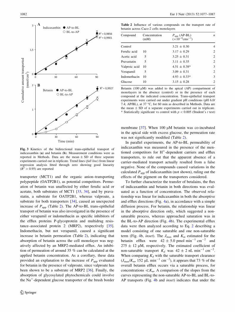
The cumulative amounts of either indicaxanthin or
betanin transported into the receiving chamber as a func-
tion of time are shown in Fig. 3. The amount of indica-
xanthin crossing the monolayer increased linearly with
time within 90 min, in both the absorptive and efflux
direction, with comparable transport rates (Fig. 3a). For
betanin, the relationship was linear in the absorptive
direction, whereas a quite different relationship was
observed in the efflux transport (Fig. 3b).
In the presence of the proton gradient applied, H?-
dependent influx carriers were considered. Then, the AP-
to-BL trans-epithelial transport of betanin was measured in
the presence of substrates for the monocarboxylate
Table 1 Physicochemical parameters of indicaxanthin and betanin
MW logPa logDb PSA (A´ 2) NPSA (A
´ 2) pKa
(COOH)c (phenol-OH)c
Indicaxanthin 309 0.362 -7.25 126.92 161.11 5.0(2);3.7(11);2.6(13)d
Betanin 551 -1.767 -15.63 246.53 229.26 \3.4(2);3.4(15);3.4(17)e \7.4(6)
e
The molecular surface is described by the polar surface area (PSA) and the non-polar surface area (NPSA)a Octanol/water partition coefficientb Octanol/buffer pH 6.0 partition coefficientc C atom in bracketd Theoretically calculated pKa values (see ‘‘Materials and methods’’)e pKa values from Ref [28]
Fig. 2 Bidirectional apparent permeability coefficient (Papp) of
betalains across Caco-2 cell monolayers under inwardly directed pH
gradient (AP pH 6.0/BL pH 7.4). Trans-epithelial transport AP-to-BL
(black bars) and BL-to-AP (white bars) and Papp measurements were
as reported in Methods. Values are the mean ±SD of three separate
experiments carried out in triplicate. *Statistically significant vs the
relevant Papp (AP-to-BL) direction with p \ 0.001 (Student’s t test)
Eur J Nutr (2013) 52:1077–1087 1081
123
transporter (MCT1) and the organic anion–transporting
polypeptide (OATP2B1), as potential competitors. Perme-
ation of betanin was unaffected by either ferulic acid or
acetate, both substrates of MCT1 [33, 34], and by prava-
statin, a substrate for OATP2B1, whereas valproate, a
substrate for both transporters [34], caused an unexpected
increase of Papp (Table 2). The AP-to-BL trans-epithelial
transport of betanin was also investigated in the presence of
either verapamil or indomethacin as specific inhibitors of
the efflux proteins P-glycoprotein and multidrug resis-
tance–associated protein 2 (MRP2), respectively [35].
Indomethacin, but not verapamil, caused a significant
increase in betanin permeation (Table 2), indicating that
absorption of betanin across the cell monolayer was neg-
atively affected by an MRP2-mediated efflux. An inhibi-
tion of permeation of around 35 % can be calculated at the
applied betanin concentration. As a corollary, these data
provided an explanation to the increase of Papp evaluated
for betanin in the presence of valproate, since valproate has
been shown to be a substrate of MRP2 [36]. Finally, the
absorption of glycosylated phytochemicals could involve
the Na?-dependent glucose transporter of the brush border
membrane [37]. When 100 lM betanin was co-incubated
in the apical side with excess glucose, the permeation rate
was not significantly modified (Table 2).
In parallel experiments, the AP-to-BL permeability of
indicaxanthin was measured in the presence of the men-
tioned competitors for H?-dependent carriers and efflux
transporters, to rule out that the apparent absence of a
carrier-mediated transport actually resulted from a false
negative. None of the compounds caused variations in the
calculated Papp of indicaxanthin (not shown), ruling out the
effects of the pigment on the transporters considered.
To further characterize the transfer of betalains, the flux
of indicaxanthin and betanin in both directions was eval-
uated as a function of concentration. The observed rela-
tionship was linear for indicaxanthin in both the absorptive
and efflux directions (Fig. 4a), in accordance with a simple
diffusion process. For betanin, the relationship was linear
in the absorptive direction only, which suggested a non-
saturable process, whereas approached saturation was in
the BL-to-AP direction (Fig. 4b). The experimental efflux
data were then analyzed according to Eq. 2 describing a
model consisting of one saturable and one non-saturable
term (Fig. 4b, inset). The Jmax and Km estimated for the
betanin efflux were 42 ± 5.0 pmol min-1 cm-2 and
275 ± 12 lM, respectively. The estimated coefficient of
non-saturable transport Kd was 42 ± 2 nL min-1 cm-2.
When comparing Kd with the saturable transport clearance
(Jmax/Km, 152 lL min-1 cm-2), it appears that 73 % of the
overall betanin efflux occurs via a saturable process, for
concentrations �Km. A comparison of the slopes from the
curves representing the non-saturable AP-to-BL and BL-to-
AP transports (Fig. 4b and inset) indicates that under the
Fig. 3 Kinetics of the bidirectional trans-epithelial transport of
indicaxanthin (a) and betanin (b). Measurement conditions were as
reported in Methods. Data are the mean ± SD of three separate
experiments carried out in triplicate. Trend lines (full line) from linear
regression analysis fitted through zero showing good linearity
(R2 [ 0.95) are reported
Table 2 Influence of various compounds on the transport rate of
betanin across Caco-2 cells monolayers
Compound Concentration
(mM)
Papp (AP-BL)
(910-6cms-1)
n
Control 3.21 ± 0.30 4
Ferulic acid 10 3.17 ± 0.29 2
Acetic acid 5 3.25 ± 0.31 2
Pravastatin 5 3.11 ± 0.35 2
Valproic acid 10 4.51 ± 0.38* 3
Verapamil 5 3.09 ± 0.31 2
Indomethacin 10 4.93 ± 0.33* 3
Glucose 10 3.15 ± 0.28 2
Betanin (100 lM) was added to the apical (AP) compartment of
monolayers in the absence (control) or in the presence of each
compound at the indicated concentration. Trans-epithelial transport
experiments were carried out under gradient pH conditions (pH 6.0/
7.4; AP/BL), at 37 �C, for 60 min as described in Methods. Data are
the mean ± SD of n separate experiments carried out in triplicate.
* Statistically significant vs control with p \ 0.005 (Student’s t test)
1082 Eur J Nutr (2013) 52:1077–1087
123

experimental conditions applied, betanin diffusion is
fourfold higher in the influx than in the efflux direction.
The Papp of the AP-to-BL transfer of indicaxanthin and
betanin across Caco-2 cell monolayers was evaluated after
treatment of the monolayer with EDTA, which affects
paracellular permeability via loosening of the tight junctions
[31], in comparison with markers for paracellular and trans-
cellular pathways. The Papp value of indicaxanthin increased
around 11-fold, and that of betanin sevenfold (Fig. 5). As
expected, an effective transfer of phenol red across the epi-
thelial cell layer was observed only after loosening of the
TJs, whereas the permeability of testosterone, the transfer of
which occurs via a passive trans-cellular pathway, was
almost unaffected by the EDTA treatment of the cells
(Fig. 5). It was concluded that the transport of both betalains
substantially occurred through paracellular permeation.
Transport of betalains from food digesta
The absorption of betalains from digested betalainic food
was evaluated. Cactus pear fresh fruits and juice as a source
of indicaxanthin and betanin, as well as raw red beet and red
beet juice, as a source of betanin, were processed through a
simulated in vitro digestion, and the BFs so obtained were
placed at the apical side of Caco-2 cells layered on a
Transwell insert, after measuring their betalain content.
Permeation of indicaxanthin and betanin is reported in
Table 3. Whereas permeation of indicaxanthin from cactus
pear either fruit or juice was quite comparable with that of the
pure pigment, the food matrix reduced the transport rate of
betanin, red beet more than cactus pear (Table 3).
Discussion
Bi-directional transfer of indicaxanthin and betanin
through Caco-2 cell monolayers
The intestinal absorption of two bioavailable dietary bet-
alains, betanin and indicaxanthin, has been investigated
Fig. 4 Concentration-dependent transport of indicaxanthin (a) and
betanin (b) across Caco-2 cell monolayers. Measurement conditions
are reported in Methods. Data are the mean ± SD of three separate
experiments carried out in triplicate. Trend lines (full line) from linear
regression analysis fitted through zero showing good linearity
(R2 [ 0.95) are reported. Inset B: the solid, dashed, and dotted linesrepresent the best fit to the efflux data, saturable and non-saturable
components of the transport, respectively, according to Eq. 2
Fig. 5 Apparent permeability coefficient (Papp) for betalains across
Caco-2 cell monolayer with intact tight junctions (open bars) and
after opening of tight junctions with 5 mM EDTA (black bars), in
comparison with phenol red and testosterone. Data are the
mean ± SD of two separate experiments carried out in triplicate.
*Statistically significant with p \ 0.001 (Student’s t test)
Table 3 Apparent permeation coefficient (Papp, AP-BL) across Caco-2
cell monolayers of betalains from the bioaccessible fraction (BF) of
betalainic foods
Betalain BF source Concentration
(lM)
Papp
(10-6 cms-1)
Indicaxanthin Cactus yellow fruit 30.5 ± 2.3 4.32 ± 0.35
Cactus yellow fruit
juice
29.8 ± 3.2 4.15 ± 0.41
Betanin Raw red beet 62.5 ± 5.3 1.98 ± 0.21*
Red beet juice 11.7 ± 1.1 1.82 ± 0.20*
Cactus red fruit 12.2 ± 1.5 2.78 ± 0.34**
Cactus red fruit
juice
13.1 ± 1.2 2.61 ± 0.31**
Preparation of BF from foods and incubation conditions of Caco-2
monolayers were as reported in methods
Values are the mean ± SD of three determinations carried out on two
different food samples. Significantly different from Papp of the rele-
vant pure compound, * p \ 0.001, ** p \ 0.05 (Student’s t test)
Eur J Nutr (2013) 52:1077–1087 1083
123

using Caco-2 cell monolayers grown on permeable poly-
ester membranes as a model for the small intestinal
mucosal epithelium, and the influence of the betalainic
food matrix on the absorption process has been evaluated.
Different tracts of the gastro-intestinal lumen are charac-
terized by different pH values, and further pH changes are
measured during digestion [38]. The pH in the upper gas-
tro-intestinal tract under fasting conditions ranges from 5.0
to 6.5, and it has been reported to be 5.8 to 6.3 just above
the absorbing epithelial cell layer [39]. We then measured
the flux of betalains across the Caco-2 cell monolayer
under conditions of an inwardly directed pH gradient (pH
6.0/7.4) approaching the pH microclimate at the luminal
and serosal sides of human intestinal cells, with main-
taining first-order conditions to mimic physiological
absorption circumstances at the small intestine. In addition,
the bulk of the experiments were carried out using a
100-lM betalain concentration, which approached the
amount in the intestinal digesta from dietary amounts of
either cactus pear fruit pulp (200 g, 24 mg indicaxanthin)
or raw red beet (100 g, 71 mg betanin) [22], when con-
sidering an intestinal volume of 600 mL [40].
Like for xenobiotics, the intestinal absorption of phy-
tochemicals may occur passively, through trans-cellular
permeation or paracellular route in accordance with
molecular mass and physicochemical characteristics, and
could involve either influx or efflux membrane transport-
ers. Our computational analysis provided solubility
parameters and the polar and NPSA of indicaxanthin and
betanin, as well as dissociation constants of the indica-
xanthin carboxyl groups. According to our calculations and
literature data [18, 28], both compounds mainly occur as
bis-anions at pH 6.0. The octanol/water partition coeffi-
cients ClogP and ClogD (pH 6.0) indicated that indica-
xanthin is moderately less polar than betanin. Finally, the
calculated NPSA provided evidence that both betalains
have a quite large non-polar surface, accounting for around
50 % of surface area, which substantiated previous obser-
vations on the ability of these molecules to interact with
lipid environments from membranes [9, 19, 41, 42] to low-
density lipoproteins [8, 11, 17].
Generally, unless utilizing transporters in the epithelial
membrane, charged solutes of a suitable molecular mass
should diffuse through the paracellular route and be
transported passively by solvent drag [43–46]. Quite con-
sistent with the physicochemical features of indicaxanthin
and betanin, our study shows that both compounds may
cross the epithelial cell layer by passive diffusion via the
paracellular pathway, without any metabolic transforma-
tion. Some findings, however, suggest a mixture of para-
cellular and trans-cellular transport for betanin.
The effects of perturbation on the permeation of solutes
are used as criteria to determine the preferred route of
intestinal transport [32, 47]. If the permeation is signifi-
cantly affected by perturbation of cellular tight junctions
(TJs), then the permeation is considered to occur pre-
dominantly by the paracellular route. We observed that an
opening of TJs with EDTA induced a remarkable increase
in the indicaxanthin and betanin influx, consistent with a
paracellular transport of both compounds across the Caco-2
cell monolayer. Indeed, the trans-epithelial transfer of in-
dicaxanthin did not appear to be polarized, did not show
saturation kinetics, nor the absorption was varied by
inhibiting membrane transporters, in accordance with the
simple diffusion-solubility criteria governing paracellular
transports. On the other hand, the absorption of the gly-
cosylated betanin appeared polarized, and the cumulative
transfer in the efflux direction was not linear with time nor
with concentration. Kinetic modeling of the efflux data
revealed parallel contribution of one non-saturable and one
saturable component, identified as the apical efflux trans-
porter MRP2. Quantitative analysis of the data showed that
the betanin efflux mainly occurs through the saturable
pathway (73 %) at betanin concentrations far below Km
(i.e. �275 lM), whereas the betanin diffusion rate is
fourfold higher in the influx than in the efflux direction.
Then, in spite of the apical efflux, under the present
experimental set-up mimicking physiological pH and
betalain concentration conditions, the permeation rate of
betanin was higher in the absorptive than in the secretive
direction, which should assure that absorption at the
intestine will prevail. It may be worthwhile to mention that
our experiments have been done with amounts of betalains
consistent with the intestinal content after a reasonable
dietary intake, which may be critical when dealing with
carrier-mediated processes.
The involvement of MRP2 in the transport discloses
interesting features of the betanin ability to migrate through
the Caco-2 cell monolayer. While indicating that the bio-
availability of this phytochemical is limited by an absorption
barrier, our findings suggest that trans-cellular transfer of
betanin could occur in parallel with the paracellular one.
Partition of various ionized species into chemical bilayers
has been determined [48], and trans-cellular transport of
ionized species across Caco-2 cell monolayers has been
suggested in other studies [49, 50]. Moreover, previously
performed chemico-physical partition studies in our labo-
ratory provided evidence that betanin locates at the
phospholipid core of the bilayer in an aqueous phosphati-
dylcholine liposomal system [41]. Then, it seems reasonable
to suppose betanin permeation of the cell membrane at the
apical side, with access to the apical membrane efflux
transporter, and possibly lateral diffusion to the basal side of
the cell and release to the basolateral compartment.
With solutes of like charge, paracellular permeability is
a function of the molecular mass and decreases with the
1084 Eur J Nutr (2013) 52:1077–1087
123

increase in molecular weight [43, 44, 46]. Since indica-
xanthin and betanin have a comparable ionic charge at pH
6.0, the molecular mass should favor the paracellular per-
meation of indicaxanthin. Noteworthy that in the presence
of indomethacin, that is, blocking the MRP2 efflux trans-
porter, the Papp for the absorption of betanin has appeared
as high as that of indicaxanthin may be an indirect evi-
dence that additional trans-membrane transport of betanin
does occur.
The intestinal absorption of xenobiotics is considered
negligible if the Papp \ 0.1 9 10-6 cm s-1 and essentially
complete if the trans-epithelial Papp [ 5.0 9 10-6 cm s-1
[51, 52]. With the observed permeability coefficients, the
trans-epithelial gradient of indicaxanthin and betanin at the
intestinal lumen after ingestion, and their continuous
removal by the bloodstream at the serosal side, could
account for a significant intestinal transport. Present data
indeed are consistent with and appear to validate the high
fraction of dietary indicaxanthin absorbed in vivo [17].
Bioavailability of betanin in humans has been shown much
lower than that of indicaxanthin [17–19]. According to
previous studies [22], around 50 % of betanin is lost during
the digestive process; however, its recovery in human urine
was found no more than 3 % of the compound ingested
with various foods [17–19]. Present findings on the intes-
tinal processing and the calculated Papp do not fully consist
with the bioavailability measurements. The complexity of
the in vivo system could involve other significant losses,
possibly hydrolytic processes by glycosidases of the
intestinal microflora, and/or some oxidation of the pigment
in the body [12]. Other factors, including the food matrix,
could play additional roles.
Influence of betalainic food matrix on the intestinal
permeation of the pigments
The absorption efficacy of phytochemicals can be affected
by the mixture of their food matrix [53]. With respect to the
pure compounds, the trans-epithelial transport rate of
betanin from the soluble fraction of betalainic food digesta
was strongly reduced, whereas that of indicaxanthin was
not. Phenolic groups of phytochemicals may be involved in
hydrogen bonding with protein moiety [54], and then
complexes between betanin and soluble protein fragments
in the post-intestinal digesta could prevent a fraction of the
pigment to be absorbed. Eventual competition by other
betalainic food components for the paracellular pathway
could also be hypothesized. Present observations that
betanin from cactus pear appeared more readily absorbed
than from red beet are quite in accordance with previous
studies in humans, showing higher bioavailability of
betanin from dietary cactus pear fruit than from red beet
juice [17–19]. This may deserve consideration for nutri-
tional purposes.
Conclusive remarks
Dietary bioactive phytochemicals are now considered
potential nutraceuticals/pharmacological molecules [55],
and then analyzing mechanisms and factors affecting
intestinal transfer could help to predict their eventual
effects in the body. Definite evidence has been provided
that the very high bioavailability of dietary indicaxanthin
in humans [17] results not only from relatively high
stability of the molecule to the digestive process [22], but
also from favorable intestinal absorption through para-
cellular route by solvent drag, and easy release from
food. Betanin bioavailability, instead, appears to be lim-
ited by digestive instability [22] and by an intestinal
efflux mechanism reducing the absorption by around
35 % for betanin concentrations consistent with a nutri-
tional intake. Furthermore, its intestinal permeation may
negatively be influenced by its food matrix. Importantly,
indicaxanthin and betanin do not need metabolic trans-
formations to be released in plasma and circulate as
unconjugated molecules. Taking all these facts into con-
sideration, beneficial effects of dietary betalain pigments,
as well as the impact of betalainic foods on human health
[56], may be considered and results from appropriately
planned in vitro studies may be interpreted to suggest real
effects in vivo. Present data may provide a basis for
research on the potential health effects of these sub-
stances, eventually to be orally administered as purified
compounds.
Conflict of interest The authors declare that there are no conflicts
of interest.
References
1. Scalbert A, Johnson IT, Saltmarsch M (2005) Polyphenols:
antioxidants and beyond. Am J Clin Nutr 81:215S–217S
2. Raederstorff D (2009) Antioxidant activity of olive polyphenols
in humans: a review. Int J Vitam Nutr Res 79:152–165
3. Herr I, Buchler MW (2010) Dietary constituents of broccoli and
other cruciferous vegetables: implications for prevention and
therapy of cancer. Cancer Treat Rev 36:377–383
4. Piattelli M (1981) The betalains: structure, biosynthesis and
chemical taxonomy. In: Conn EE (ed) The biochemistry of
plants: a comprehensive treatise, vol 7. Academic Press, New
York, pp 557–575
5. Steglich W, Strack D (1990) Betalains. In: Brossi A (ed) The
alkaloids, chemistry and pharmacology. Academic Press, Lon-
don, pp 1–62
6. Herbach KM, Stintzing FC, Carle R (2006) Betalain stability and
degradation. Structural and chromatic aspects. J Food Sci
71:R41–R50
Eur J Nutr (2013) 52:1077–1087 1085
123

7. Reynoso R, Garcia FA, Morales D, Gonzalez de Mejia E (1997)
Stability of betalain pigments from a cactacea fruit. J Agric Food
Chem 45:2884–2889
8. Tesoriere L, Butera D, D’Arpa D, Di Gaudio F, Allegra M,
Gentile C, Livrea MA (2003) Increased resistance to oxidation of
betalain-enriched human low density lipoproteins. Free Radic
Res 37:689–696
9. Tesoriere L, Butera D, Allegra M, Fazzari M, Livrea MA (2005)
Distribution of betalain pigments in red blood cells after con-
sumption of cactus pear fruits and increased resistance of the cells
to ex vivo-induced oxidative hemolysis in humans. J Agric Food
Chem 53:1266–1270
10. Tesoriere L, Allegra M, Butera D, Gentile C, Livrea MA (2006)
Cytoprotective effects of the antioxidant phytochemical indica-
xanthin in beta-thalassemia red blood cells. Free Radic Res
40:753–761
11. Tesoriere L, Allegra M, Butera D, Gentile C, Livrea MA (2007)
Kinetics of the lipoperoxyl radical-scavenging activity of indi-
caxanthin in solution and unilamellar liposomes. Free Radic Res
41:226–233
12. Tesoriere L, Allegra M, Gentile C, Livrea MA (2009) Betacya-
nins as phenol antioxidants. Chemistry and mechanistic aspects
of the lipoproxyl radical scavenging activity in solution and lip-
osomes. Free Radic Res 43:706–717
13. Gentile C, Tesoriere L, Allegra M, Livrea MA, D’Alessio P
(2004) Antioxidant betalains from cactus pear (Opuntia ficus
indica) inhibit endothelial ICAM-1 expression. Ann NY Acad Sci
1028:481–486
14. Reddy MK, Alexander-Lindo RL, Nair MG (2005) Relative
inhibition of lipid peroxidation, cyclooxygenase enzymes, and
human tumor cell proliferation by natural food colors. J Agric
Food Chem 53:9268–9273
15. Sreekantah D, Arunasree MK, Roy KR, Reddy TC, Reddy GV,
Reddanna P (2007) Betanin a betacyanin pigment purified from
fruits of Opuntia ficus indica induces apoptosis in human chronic
myeloid leukemia cell line-K562. Phytomedicine 14:739–746
16. Kapadia GJ, Azuine MA, Rao GS, Arai T, Iida A, Tokuda H
(2011) Cytotoxic effect of the red beetroot (Beta vulgaris, L9
extract compared to doxorubicin (Adriamycin) in the human
prostate (PC-3) and breast (MCF-7) cancer cell lines. Anticancer
Agents Med Chem 11:280–284
17. Tesoriere L, Allegra M, Butera D, Livrea MA (2004) Absorption,
excretion, and distribution in low density lipoproteins of dietary
antioxidant betalains. Potential health effects of betalains in
humans. Am J Clin Nutr 80:941–945
18. Frank T, Stintzing FC, Carle R, Bitsch I, Quaas D, Strass G,
Bitsch R, Netzel M (2005) Urinary pharmacokinetics of betalains
following consumption of red beet juice in healthy humans.
Pharmacol Res 52:290–297
19. Kanner J, Harel S, Granit R (2001) Betalains-a new class of dietary
cationized antioxidants. J Agric Food Chem 49:5178–5185
20. Holt RR, Lazarus SA, Sullards MC, Zhu QY, Schramm DD,
Hammrstone JF, Fraga GC, Schmitz HH, Keen C (2002) Procy-
anidin dimer B2 [epicathechin-(4beta-8)-epicathechin] in human
plasma after the consumption of a flavonol-rich cocoa. Am J Clin
Nutr 76:798–804
21. Schroeter H, Heiss C, Balzer J, Kleinbongrad P, Keen CL, Hol-
lenberg NK, Sies H, Kwik-Uribe CL, Schmitz HH, Kelm M
(2006) (-)-Epicathechin mediates beneficial effects of flavonol-
rich cocoa on vascular function in humans Proc Nat Acad Sci
USA 103:1024–1029
22. Tesoriere L, Fazzari M, Angileri F, Gentile C, Livrea MA (2008)
In vitro digestion of betalainic foods. Stability and bioaccessi-
bility of betaxanthins and betacyanins and antioxidative potential
of food digesta. J Agric Food Chem 56:10487–10492
23. Galijatovic A, Otake Y, Walle UK, Walle T (2001) Induction of
UDP-glucuronyltransferase UGT1A1 by the flavonoid chrysin in
Caco2 cells- potential role in carcinogen bioactivation. Pharm
Res 18:374–379
24. Sun DX, Lennernas H, Welage LS, Barnett JL, Landowski CP,
Foster D, Fleischer D, Lee KD, Amidon JL (2002) Comparison of
human duodenum and Caco2 gene expression profiles for 12,000
gene sequence tags and correlation with permeability of 26 drugs.
Pharm Res 19:1400–1416
25. Butera D, Tesoriere L, Di Gaudio F, Bongiorno A, Allegra M,
Pintaudi AM, Kohen R, Livrea MA (2002) Antioxidant activities
of sicilian prickly pear (Opuntia ficus indica) fruit extracts and
reducing properties of its betalains: betanin and indicaxanthin.
J Agric Food Chem 50:6895–6901
26. Stintzing FC, Schieber A, Carle R (2002) Identification of beta-
lains from yellow beet (Beta vulgaris, L) and cactus pear
(Opuntia ficus indica, L Mill) by high performance liquid chro-
matography-electrospray ionization mass spectrometry. J Agric
Food Chem 50:2302–2307
27. Katritzky AR, Lobanov VS, Karelson M (1996) Quantum-
chemical descriptors in QSAR/QSPR studies. Chem Rev
96:1027–1043
28. Gliszczynska-Swiglo A, Szymusiak H, Malinowska P (2006)
Betanin, the main pigment of red beet: molecular origin of its
exceptionally high free radical-scavenging activity. Food Addit
Contam 23:1079–1087
29. Hidalgo IJ, Raub TJ, Borchardt RT (1989) Characterization of the
human colon carcinoma cell line (Caco-2) as a model system for
intestinal epithelial permeability. Gastroenterology 96:736–749
30. Behrens I, Stenberg P, Artusson P, Kissel T (2001) Transport of
lipophilic drug molecules in a new mucus-secreting cell culture
model based on HT29_MTX cells. Pharmacol Res 18:1138–1145
31. Artursson P (1990) Epithelial transport of drugs in cell culture. I:
a model for studying the passive diffusion of drugs over intestinal
(Caco-2) absorptive cells. J Pharm Sci 79:476–482
32. Artursson P, Magnusson C (1990) Epithelial transport of drugs in
cell culture. II: effect of extracellular calcium concentration on
the paracellular transport of drugs of different lipophilicities
across monolayers of intestinal epithelial (Caco-2) cells. J Pharm
Sci 79:595–600
33. Konishi Y, Shimizu M (2003) Transepithelial transport of ferulic
acid by monocarboxylic acid transporter in Caco-2 cell mono-
layers. Biosci Biotechnol Biochem 67:856–862
34. Thwaites DT, Anderson CMH (2007) H ? -coupled nutrient,
micronutrient and drug transporters in the mammalian small
intestine. Exp Physiol 92:603–619
35. Shugarts S, Benet LZ (2009) The role of transporters in the
pharmacokinetics of orally administered drugs. Pharm Res
26:2039–2045
36. Ogawa K, Yumoto R, Hamada N, Nagai J, Takano M (2006)
Interaction of valproic acid and carbapenem antibiotics with
multidrug resistance-associated proteins in rat erythrocyte mem-
branes. Epilepsy Res 71:76–87
37. Walgren RA, Lin J-T, Kinne RK-H, Walle T (2000) Cellular
uptake of dietary flavonoid quercetin 40-b-glucoside by sodium-
dependent glucose transporter SLGT1. J Pharmacol Experim
Therap 294:837–843
38. Dressman JB, Berardi RR, Dermentzoglou LC, Russel TL,
Schmaltz SP, Barnett JL, Jarvenpaa KM (1990) Upper gastroin-
testinal (GI) pH in young, healthy men and women. Pharm Res
7:756–761
39. Lucas M (1983) Determination of acid surface pH in vivo in rat
proximal jejunum. Gut 24:734–739
40. Mahe S, Huneau JF, Marteau P, Thuillier F, Tome D (1992)
Gastroileal nitrogen and humans. Am J Clin Nutr 56:410–416
1086 Eur J Nutr (2013) 52:1077–1087
123

41. Turco Liveri ML, Sciascia L, Lombardo R, Tesoriere L, Passante
E, Livrea MA (2007) Spectrophotometric evidence for the solu-
bilization site of betalain pigments in membrane biomimetic
systems. J Agric Food Chem 55:2836–2840
42. Turco Liveri ML, Sciascia L, Allegra M, Tesoriere L, Livrea MA
(2009) Partition of indicaxanthin in membrane biomimetic sys-
tems. A kinetic and modeling approach. J Agric Food Chem
57:10959–10963
43. Adson A, Raub TJ, Burton PS, Barshun CL, Hilgers AR, Audus
KL, Ho NF (1994) Quantitative approach to delineate paracel-
lular diffusion in cultured epithelial cell monolayer. J Pharm Sci
83:1529–1536
44. Karlsson J, Ungell AL, Artursson P (1994) Effect of an oral
rehydration solution on paracellular drug transport in intestinal
epithelial cells and tissues: assessment of charge and tissue
selectivity. Pharm Res 11:S248
45. Pade V, Stavchansky S (1997) Estimation of the relative contri-
bution of the transcellular and paracellular pathway to the
transport of passively absorbed drugs in the Caco-2 cell culture
model. Pharm Res 14:1210–1215
46. Knipp GT, Ho NF, Barsuhn CL, Borchardt R (1997) Paracellular
diffusion in Caco-2 cell monolayers: effect of perturbation on the
transport of hydrophilic compounds that vary in charge and size.
J Pharm Sci 86:1105–1110
47. Gan LS, Hsyu PH, Pritchard JF, Thakker D (1993) Mechanism of
intestinal absorption of ranitidine and ondansetron: transport
across Caco-2 cell monolayer. Pharm Res 10:1722–1725
48. Avdeef A, Box KJ, Comer JEA, Hibbert C, Tam KY (1998) pH-
metric logP 10. Determination of liposomal membrane-water
partition coefficients of ionizable drugs. Pharm Res 15:209–215
49. Palm K, Luthman K, Ros J, Grasjo J, Artursson P (1999) Effect of
molecular charge on intestinal epithelial drug transport: pH-
dependent transport of cationic drugs. J Pharmacol Exp Ther
291:435–443
50. Menez C, Buyse M, Dugave C, Farinotti R, Barratt G (2007)
Intestinal absorption of miltefosine: contribution of passive
paracellular transport. Pharm Res 24:546–554
51. Hillgren KM, Kato A, Borchardt RT (1995) In vitro systems for
studying intestinal drug absorption. Med Res Rev 15:83–109
52. Artursson P, Karlsson J (1991) Correlation between oral drug
absorption in humans and apparent drug permeability coefficients
in human intestinal epithelial (Caco-2) cells. Biochem Biophys
Res Commun 175:880–885
53. Boyer J, Brown D, Liu RH (2004) Uptake of quercetin 3-glu-
coside from whole onion and apple peel extracts by Caco-2 cell
monolayers. J Agric Food Chem 52:7172–7179
54. Hagerman AE, Butler LG (1981) The specificity of proanthocy-
anidin-protein interactions. J Biol Chem 256:4494–4497
55. Konishi T (2011) From herb to kitchen and bedside: food factors
are pharmacological molecules with antioxidant activity. Free
Radic Res 45:863–864
56. Tesoriere L, Butera D, Pintaudi AM, Allegra M, Livrea MA
(2004) Supplementation with cactus pear (Opuntia ficus-indica)
fruit decreases oxidative stress in healthy humans: a comparative
study with vitamin C. Am J Clin Nutr 80:391–395
Eur J Nutr (2013) 52:1077–1087 1087
123


