
warwick.ac.uk/lib-publications
Original citation: Eissa, Ahmed M., Abdulkarim, Ali, Sharples, Gary J. and Cameron, Neil R.. (2016) Glycosylated nanoparticles as efficient antimicrobial delivery agents. Biomacromolecules, 17 (8). pp. 2672-2679.
Permanent WRAP URL: http://wrap.warwick.ac.uk/84530 Copyright and reuse: The Warwick Research Archive Portal (WRAP) makes this work by researchers of the University of Warwick available open access under the following conditions. Copyright © and all moral rights to the version of the paper presented here belong to the individual author(s) and/or other copyright owners. To the extent reasonable and practicable the material made available in WRAP has been checked for eligibility before being made available. Copies of full items can be used for personal research or study, educational, or not-for profit purposes without prior permission or charge. Provided that the authors, title and full bibliographic details are credited, a hyperlink and/or URL is given for the original metadata page and the content is not changed in any way. Publisher’s statement: “This document is the Accepted Manuscript version of a Published Work that appeared in final form in Biomacromolecules,. copyright © American Chemical Society after peer review and technical editing by the publisher. To access the final edited and published work http://pubs.acs.org/page/policy/articlesonrequest/index.html .” A note on versions: The version presented here may differ from the published version or, version of record, if you wish to cite this item you are advised to consult the publisher’s version. Please see the ‘permanent WRAP URL above for details on accessing the published version and note that access may require a subscription. For more information, please contact the WRAP Team at: [email protected]

1
Glycosylated Nanoparticles as Efficient Antimicrobial Delivery Agents
Ahmed M. Eissa,a,b,c,d * Ali Abdulkarim,a Gary J. Sharples,e Neil R. Camerona,b,c *
a Department of Chemistry, University of Durham, South Road, Durham, DH1 3LE, U.K.
b School of Engineering, University of Warwick, Coventry, CV4 7AL, U.K.
c Department of Materials Science and Engineering, Monash University, Clayton 3800, Victoria,
Australia.
d Department of Polymers, Chemical Industries Research Division, National Research Centre
(NRC), El-Bohoos Street, Dokki, Cairo, Egypt.
e School of Biological and Biomedical Sciences, Biophysical Sciences Institute, Department of
Chemistry, University of Durham, Durham DH1 3LE, U.K.
Synthetic polymer nanoparticles that can be tailored through multivalent ligand display on the
surface, while at the same time allowing encapsulation of desired bioactive molecules, are
especially useful in providing a versatile and robust platform in the design of specific delivery
vehicles for various purposes. Glycosylated nanoparticles (glyco-NPs) of a poly(n-butyl acrylate)
(pBA) core and poly(N-2-(β-D-Glccosyloxy)ethyl acrylamide) (p(NβGlcEAM)) or poly(N-2-(β-
D-galactosyloxy)ethyl acrylamide) (p(NβGalEAM)) corona were prepared via nanoprecipitation
in aqueous solutions of preformed amphiphilic glycopolymers. Well-defined block copolymers

2
of (poly(pentafluorophenyl acrylate) (pPFPA) and pBA were first prepared by RAFT
polymerization followed by post-polymerization functionalization with aminoethyl glycosides to
yield p(NβGlcEAM-b-BA) and p(NβGalEAM-b-BA) which were then used to form glyco-NPs
(glucosylated and galactosylated NPs, Glc-NPs and Gal-NPs, respectively). The glyco-NPs were
characterized by dynamic light scattering (DLS) and TEM. Encapsulation and release of
ampicillin, leading to nanoparticles that we have termed ‘glyconanobiotics’, were studied. The
ampicillin-loaded glyco-NPs were found to induce aggregation of Staphylococcus aureus and
Escherichia coli and resulted in antibacterial activity approaching that of ampicillin itself. This
glyconanobiotics strategy represents a potential new approach for the delivery of antibiotics
close to the surface of bacteria by promoting bacterial aggregation. Defined release in the
proximity of the bacterial envelope may thus enhance antibacterial efficiency and potentially
reduce the quantities of agent required for potency.
3
Over the last century, morbidity and mortality as a consequence of infectious diseases have
decreased drastically, due to the wide-spread development and deployment of vaccines and
antimicrobial agents.1 However, bacterial resistance to our available antibiotic repertoire is now
reaching a critical level, potentially bringing about a post-antibiotic era.2 Considerable effort has
been expended to overcome this problem through the discovery of new antibiotics and chemical
modification of existing antibacterial drugs. Nevertheless, the development of new antimicrobial
drugs is unlikely to outpace the emergence of microbial pathogen resistance. The continued
evolution of antimicrobial resistance mechanisms has prompted the scientific community to seek
longer-term solutions to this ever-growing problem.3 Metallic nanomaterials (e.g. Ag, Au, and
Cu) have been found to display strong antimicrobial properties.4 Nevertheless, their applications
as antimicrobial agents are limited by their potential toxicity to human cells as a result of their
unusual physicochemical properties and/or physical or chemical production techniques.5
Encapsulation or conjugation of antibiotics into different classes of nanocarriers has been
recently represented as a potential approach to combat infectious diseases.6-8 This approach can
not only offer efficient intracellular delivery to pathogens but also help in controlling the amount
and frequency of drug dosage and hence reduces the toxicity associated to therapy and may
overcome bacterial resistance.
Strategies that do not kill the pathogens yet interfere with their pathogenic mechanisms may
provide a promising alternative. One such strategy is anti-adhesion therapy, which interferes with
the early stages of infection in which pathogens attach to the mammalian cell surface.9-11 This
adhesion is often mediated by protein-carbohydrate interactions: proteins on the pathogen
binding to displayed carbohydrate structures or receptors on the eukaryotic cell. These
interactions are often highly specific and determine the preference of the pathogen for certain
4
tissue types, known as tropism. Free carbohydrates can be harnessed to interfere with bacterial
attachment, thus preventing initial colonization and subsequent infection. This principle is
operative daily as part of the innate immune system, for example human breast milk contains
many oligosaccharides that act as anti-adhesives.12 However, the limited affinity of monovalent
carbohydrates for target proteins that are often multivalent hinders the application of this
approach. Therefore, designing multivalent glycosylated constructs that can inhibit protein-
carbohydrate interactions may be productive in finding a way forward.
During the last decade, nanoparticles presenting carbohydrate/glycan functionalities at their
surfaces have been reported to exhibit antibacterial actions towards different classes of bacteria
including Gram-positive, Gram-negative and mycobacteria [ref]. Most of these reports focused
on using nanoparticles with an inorganic core, such as gold, silver, iron oxide, silica, copper,
bismuth, palladium, and platinum.13 A major reason for the special interest in these inorganic
materials is their attractive physical properties, such as plasmonic effects, luminescence and/or
magnetic susceptibility which make them especially useful for both imaging and therapeutic
(theranostic) applications. One of the most studied examples of carbohydrate-mediated targeting
that leads to bacterial growth inhibition exerted by aggregation has been shown with
mannosylated silver and gold nanoparticles, interacting with fimbriated E. coli strains.14, 15
Bacterial growth inhibition has also been demonstrated with non-targeting glycosylated
nanoparticles, where the carbohydrate moieties serve as stabilising and/or solubilizing agents.
For example, silver nanoparticles functionalized with kocuran (an exopolysaccharide produced
by Kocuria rosea strain BS-1) were probed against a range of bacterial strains, of which S.
aureus was affected the most.16 The binding affinity and specificity of glycosylated nanoparticles
to many bacterial strains is not well-studied in the literature and needs further attention.

5
Here, we report the construction of polymeric glycosylated nanoparticles (glyco-NPs) that
encapsulate an antibiotic (ampicillin) as an amalgamated system, which we refer to as
glyconanobiotics. Our hypothesis is that antibiotic delivery will be enhanced if the nanoparticle
delivery vehicle is capable of binding to, and aggregating, the bacteria, and therefore releasing
the antibiotic in the proximity of the bacterial surface. As an example, the pili of E. coli contain
at their tips the FimH protein which has binding specificity for glucose and mannose.17
Consequently, multivalent glucosylated nanoparticles loaded with antibacterial agent could form
such a glyconanobiotic delivery system. Ampicillin was used in this study as a well-
characterized and potent antibiotic. It is a semi-synthetic derivative of penicillin, with a relatively
short-term stability in aqueous solutions.18 It is used clinically for the treatment of a broad range
of bacterial infections.19, 20 Improvement of its activity and reduction of its allergic and toxic
reactions have been achieved by means of topical formulations21 and the use of liposomal
nanoparticles as passive delivery systems.22, 23 Moreover, from the pharmaceutical application
perspective, these liposomal nano-carriers provide endocytozable formulations for intracellular
chemotherapy, since β-lactam antibiotics do not diffuse readily through the lysosomal membrane
because of their ionic character at neutral extracellular or cytoplasmatic pH.24 Polymeric
nanoparticles offer, over liposomes, certain advantageous features with respect to chemical and
biological stability and prolonged circulation times in the bloodstream.25 Furthermore, they can
be prepared in such a way that they present on their external surface biologically active
functionalities,26, 27 for example carbohydrates as employed in this study.
EXPERIMENTAL METHODS
Typical synthesis of amphiphilic glycopolymers
6
PFPA (476 mg, 2 mmol), benzyl 2-hydroxyethyl carbonotrithioate (32 mg, 0.13 mmol), AIBN
(4.5 mg, 0.025 mmol) and benzene (5 mL) were placed into a Schlenk flask equipped with a
stirrer bar. After degassing by purging with nitrogen under an ice bath for 30 min, the solution
was heated to 70 °C and stirred for 6 h. Conversion of monomer to polymer was determined by
1H NMR spectroscopy. The polymerization reaction was quenched by cooling and exposure to
air. n-Butyl acrylate (2.76 g, 21.6 mmol) and AIBN (4.5 mg, 0.025 mmol) in benzene (8 mL)
were added to the crude PFPA homopolymer solution. After degassing by purging with nitrogen
under an ice bath for 30 min, the mixture was heated to 70 °C and stirred for 18 h. After
quenching the reaction, the solvent was removed under reduced pressure and the product was
reprecipitated twice from THF into cold methanol (at 0 °C). The block copolymer was dried
under reduced pressure to yield a yellow powder which was fully characterized by SEC, 1H and
19F NMR spectroscopy, and ATR-FTIR spectroscopy.
The obtained PFPA/BA block copolymer was first dissolved in toluene (30 mL) in the
presence of a large excess of AIBN (30 eq.). After degassing by purging with nitrogen under an
ice bath for 30 min, the mixture was heated to 70 °C and stirred for 6 h. After quenching the
reaction, the solvent was removed under reduced pressure and the product was reprecipitated
from THF into cold methanol (at 0 °C) to yield p(PFPA15-b-BA120) as an off-white powder. 2’-
aminoethyl-β-D-glucopyranoside (100 mg, 0.45 mmol) was mixed with triethylamine (100 µL) in
water (1 mL). While stirring, the sugar solution was added slowly to p(PFPA15-b-BA120) (200
mg) solution in DMF (3 mL). The mixture was stirred at 30 °C for 18 h. The product was then
dialyzed against deionized water for 24 h and freeze-dried to yield p(NβGlcEAM15-b-BA120)
which was fully characterized by SEC, 1H and 19F NMR spectroscopy, and ATR-FTIR
spectroscopy.

7
In vitro drug release from ampicillin-loaded glyco-NPs
Ampicillin-encapsulated glyco-NPs were prepared using the nanoprecipitation method with the
addition of 0.5 mg of ampicillin to the organic phase. Samples with a specific amount of
ampicillin-encapsulated glyco-NP suspensions were subjected to high-speed centrifugation of
10,000 rpm for 30 min. The supernatant containing the unencapsulated ampicillin was isolated.
The ampicillin content in the supernatant was assayed by HPLC and the encapsulation efficiency
(EE) was calculated. Full details are given in the Supporting Information file.
Turbidimetric Assay
Concanavalin A (Con A) was dissolved in HBS buffer (10 mM HEPES, 90 mM NaCl, 1 mM
MgCl2, 1 mM CaCl2, 1 mM MnCl2, pH = 7.4) (1 mg/mL), and the resulting solution was gently
mixed. Turbidity measurements were performed by adding the Con A solution (400 µL) to a dry
quartz cuvette (500 µL volume, 1 cm path length). A solution of the glyco-NP of interest in HBS
buffer (prepared by nanoprecipitation method as described above) was then added (100 µL at 2
mg/mL). Upon addition, the solution was mixed vigorously using a micropipette before
placement in a Varian Cary-100 UV–Vis spectrophotometer. Absorbance data were recorded at
420 nm for 10 min.
Bacteriological experiments
The bacteriological studies of the glyco-NPs (both Glc-NPs and Gal-NPs) and the ampicillin
loaded glyco-NPs were investigated using an Escherichia coli K-12 wild-type strain (W3110 /
ATCC27325, F-, λ-, rpoS(Am), rph-1, Inv(rrnD-rrnE)), Staphylococcus aureus (3R7089 strain

8
Oxford / ATCC9144) and Staphylococcus epidermidis (laboratory strain from clinical isolate) as
representative Gram-negative (E. coli) and Gram-positive (S. aureus and S. epidermidis) species.
MIC was defined as the lowest concentration which completely inhibited bacterial growth after
incubation at 37°C for 16 h with agitation. Absorbance measurements at A650nm were obtained
using a Biotek Synergy H4 Plate Reader. Full details are given in the Supporting Information
file.
RESULTS AND DISCUSSION
We designed amphiphilic glycopolymers with different compositions (hydrophilic to
hydrophobic block ratio) that could assemble in aqueous solution, using the nanoprecipitation
method,28, 29 into NPs with sugar moieties presented on the surface. Our synthetic approach was
based on the reversible addition fragmentation chain transfer (RAFT) polymerization of an
activated ester, pentafluorophenyl acrylate (PFPA), followed by chain extension using n-butyl
acrylate (BA) for subsequent modification with aminoethyl glycosides (Fig. 1A). We first
polymerized PFPA using benzyl 2-hydroxyethyl carbonotrithioate (BHECTT) as a chain transfer
agent. The resulting pPFPA macroRAFT agents were then used to polymerize BA to generate
block copolymers with different compositions as revealed by 1H-NMR spectroscopy (Fig. S1,
supporting information). After purification by reprecipitation, the block copolymers were
analyzed by SEC which showed monomodal distributions with dispersities of about ca. 1.2. The
RAFT end group on the block copolymers was removed by treatment with azobisisobutyronitrile
(AIBN). The block copolymers were subsequently modified by reaction with 2’-aminoethyl-β-D-
glucopyranoside or 2’-aminoethyl-β-D-galactoside. Under optimized experimental conditions, a
high yield with total consumption of pentafluorophenyl ester as reveled by 1H- and 19F-NMR and
9
FTIR spectroscopy were achieved (Fig. S2–4, supporting information). The final glycopolymers
were characterized by SEC as shown in Table S1 (supporting information).
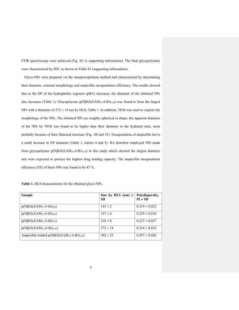
Glyco-NPs were prepared via the nanoprecipitation method and characterized by determining
their diameter, external morphology and ampicillin encapsulation efficiency. The results showed
that as the DP of the hydrophobic segment (pBA) increases, the diameter of the obtained NPs
also increases (Table 1). Glucopolymer p(NβGlcEAM15-b-BA120) was found to form the largest
NPs with a diameter of 272 ± 14 nm by DLS, Table 1. In addition, TEM was used to explore the
morphology of the NPs. The obtained NPs are roughly spherical in shape; the apparent diameter
of the NPs by TEM was found to be higher than their diameter in the hydrated state, most
probably because of their flattened structure (Fig. 1B and 1F). Encapsulation of ampicillin led to
a small increase in NP diameter (Table 1, entries 4 and 5). We therefore employed NPs made
from glycopolymer p(NβGlcEAM15-b-BA120) in this study which showed the largest diameter
and were expected to possess the highest drug loading capacity. The ampicillin encapsulation
efficiency (EE) of these NPs was found to be 47 %.
Table 1. DLS measurements for the obtained glyco-NPs.
Sample Size by DLS (nm) ±
SD
Polydispersity,
PI ± SD
p(NβGlcEAM15-b-BA30) 145 ± 2 0.219 ± 0.022
p(NβGlcEAM15-b-BA45) 197 ± 6 0.229 ± 0.018
p(NβGlcEAM15-b-BA75) 218 ± 8 0.227 ± 0.027
p(NβGlcEAM15-b-BA120) 272 ± 14 0.234 ± 0.032
Ampicillin loaded p(NβGlcEAM15-b-BA120) 302 ± 23 0.297 ± 0.026

10
Figure 1. A) Schematic representation of amphiphilic glycopolymers synthesis (Conditions: a)
BHECTT and AIBN in benzene at 70 °C for 6 h, b) BA and AIBN in benzene at 70 °C for 6 h
followed excess AIBN at 70 °C for 6 h, c) 2’-aminoethyl-β-D-glucopyranoside and TEA in
DMF-water at 30 °C for 18 h. B)-F) TEM images of glyco-NPs made from B) p(NβGlcEAM15-

11
b-BA30), C) p(NβGlcEAM15-b-BA45), D) p(NβGlcEAM15-b-BA75), E) p(NβGlcEAM15-b-BA120),
and F) ampicillin loaded glyco-NPs made from P(NβGlcEAM15-b-BA120). Scale bar = 200 nm.
The typical in vitro release profile of a drug from NPs often displays two distinct regions; an
early, rapid release, while the remainder is liberated slowly during an extended period of time.
The early release represents the loss of surface-associated and poorly entrapped drug. The
magnitude of this burst effect is dependent on the quantity of drug bound to the outer surface of
the NPs. However, the drug release from within the NP core is controlled by diffusion.30 In our
case, the percentage cumulative ampicillin release versus time plot did not show an initial burst
release phase. Instead, slow and constant zero-order ampicillin release kinetics was observed.
The cumulative ampicillin release from NPs after 21 days was found to be 56 % (Fig. 2).
Figure 2. In vitro release profile of ampicillin-loaded P(NβGlcEAM15-b-BA120) glyco-NPs in
PBS (pH 7.4) at 37 °C.

12
The ability of the glycosylated NPs to bind to the glucose/mannose specific lectin
Concanavalin-A (Con-A) was evaluated using a well-established turbidimetric assay.31 In this
assay, the Con-A tetramer is mixed with an excess of the multivalent ligand under investigation,
inducing rapid precipitation. The change in turbidity is related directly to the formation of Con-A
clusters in solution mediated by the appropriate multivalent ligand. The glucose-containing NPs
were found to bind readily to Con-A; however, galactose-containing NPs did not show any
binding affinity (Fig. 3). These results indicate that D-glucose moieties are presented on the
surface of the NPs in a densely multivalent manner and are available for lectin binding. These
results also show the potential of the glyco-NPs to bring about targeted delivery to cells
displaying glucosyl-binding lectins.
Figure 3. Assessment of the binding of NPs to Con-A by turbidimetry, from left to right: Con-A
alone (control); Con-A plus glucosylated nanoparticles; Con-A plus galactosylated nanoparticles.
Commented [NC1]: Replace Glu with Glc
13
We performed bacterial minimal inhibitory concentration (MIC) assays32 using both
glucosylated and galactosylated NPs (Glc-NPs and Gal-NPs, respectively), ampicillin loaded
Glc-NPs and free ampicillin against representative Gram-positive (Staphylococcus aureus and
Staphylococcus epidermidis) and Gram-negative (Escherichia coli) bacteria. Unloaded Glc-NPs
and Gal-NPs showed no significant antibacterial activity against these bacterial species (Fig. 4A-
C), although there was some indication of a reduction in growth in the 5-25 µg/ml concentration
range for Glc-NPs mixed with E. coli (Fig. 4A). Visual inspection of the plates revealed that this
is due to aggregation of E. coli correlating with an increased Glc-NP concentration (Fig. 4D and
also evident in the increased absorbance at the highest Gal-NP concentrations in Fig. 4A) (no
aggregation was evident in any other wells). Carbohydrate recognition sites have been identified
on the pili of E. coli. The FimH protein, present in the W3110 strain used here33 which is
exposed at the tip of the pili, has binding specificity for glucose and mannose on human cell
surfaces17 and so this is a likely explanation for the E. coli aggregation observed with Glc-NPs
but not with the non-binding Gal-NPs. The affinity of FimH for galactose is 10-fold lower than
that for glucose in a surface plasmon resonance assay34.

14
Figure 4. Evaluation of the growth and aggregation of bacteria in the presence of glucosylated
nanoparticles (blue circles) and galactosylated nanoparticles (red squares): A) E. coli, B) S.
aureus, C) S. epidermidis. D) Section of a 96-well plate showing bacterial aggregation of E. coli
as a consequence of incubation with glucosylated nanoparticles for 16 h.
Representative examples of bacterial cultures grown in liquid media in the presence of Glc-
NPs or ampicillin loaded Glc-NPs are shown in Fig. 5A (the results for E. coli only are shown,
although similar results were obtained with S. aureus and S. epidermidis). Bacterial growth is
evident in liquid media and in the presence of Glc-NPs (Fig. 5A, left and centre), while inclusion
of ampicillin in the NPs completely inhibited the bacterial growth (Fig. 5A, right).
Commented [NC2]: Replace Glu with Glc

15
Figure 5. A) E. coli grown in liquid media (left), in the presence of Glc-NPs (centre) and
ampicillin loaded Glc-NPs (right). Evaluation of bacterial growth in the presence of ampicillin
loaded Glc-NPs (red squares) and ampicillin (yellow triangles). B) E. coli, C) S. aureus, D) S.
epidermidis.
Both ampicillin-loaded Glc-NPs and free ampicillin show antibacterial activity against E. coli,
S. aureus and S. epidermidis (Fig. 5B-D). The MIC values of ampicillin loaded Glc-NPs and free
ampicillin against different bacterial strains are shown in Table 2. Ampicillin-loaded Glc-NPs
exhibited MICs 4-fold and 2-fold higher than free ampicillin against the Gram-negative strain E.
coli and Gram-positive strains S. epidermidis and S. aureus, respectively. This presumably
reflects the slower release of ampicillin from the Glc-NPs, compared to free ampicillin. The
extent of bacterial killing is nonetheless quite remarkable given the small amount of ampicillin
Commented [NC3]: Replace Glu with Glc

16
released over the 16h timeframe of the bacterial culture experiments (Fig. 2). The Glc-NPs alone
display some antibacterial effect even without encapsulation of ampicillin, the reason for which
requires further investigation.
Table 2. MIC assays on E. coli, S. epidermidis and S. aureus.
Strain Glc-NP
(µg mL-1)
Glc-NPamp
(µg mL-1)
Free Ampicillin
(µg mL-1)
E. coli 25 6.25 1.56
S. epidermidis — 25 12.5
S. aureus — 0.196 0.098
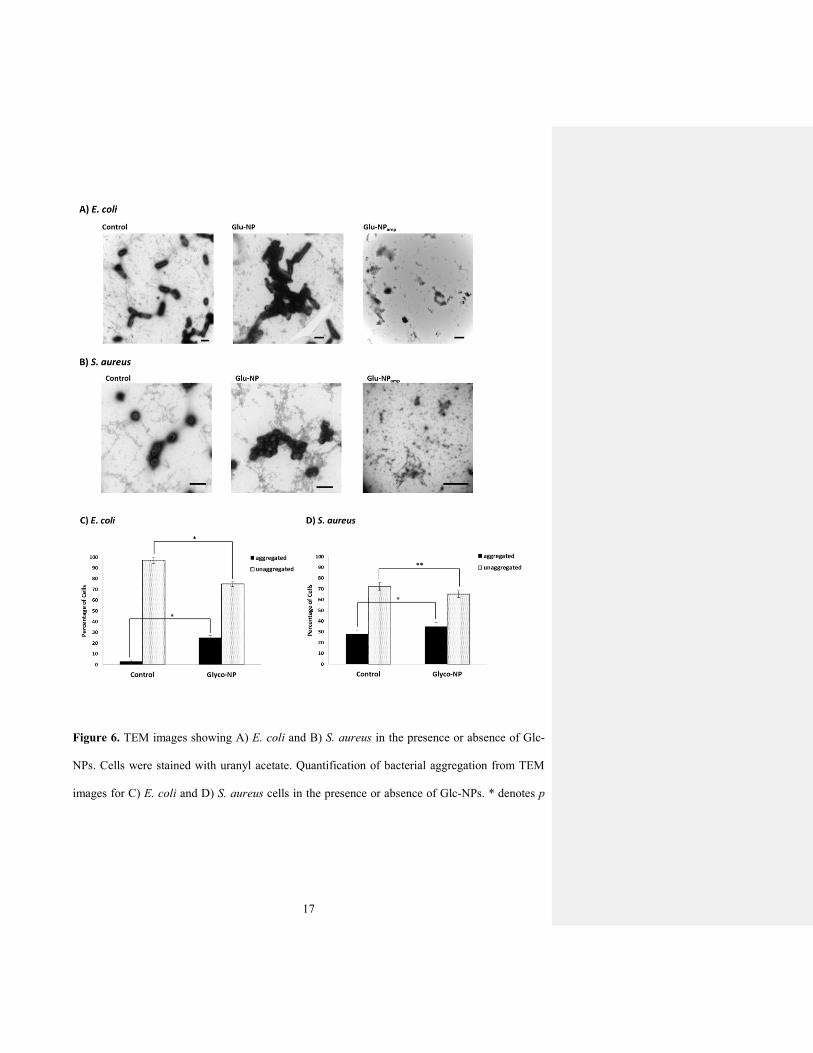
We also investigated bacterial morphology and aggregation in the presence or absence of Glc-
NPs using TEM (Fig. 6A and 6B). The bacterial cultures were dried onto TEM grids, stained
with uranyl acetate, and imaged using TEM. The resulting micrographs confirm that bacteria in
the presence of Glc-NPs form significantly more aggregates compared to the control experiment
without addition of Glc-NPs. Indeed, these TEM micrographs are representative of each sample
as indicated by the collected statistical data (Fig. 6C and 6D).
17
Figure 6. TEM images showing A) E. coli and B) S. aureus in the presence or absence of Glc-
NPs. Cells were stained with uranyl acetate. Quantification of bacterial aggregation from TEM
images for C) E. coli and D) S. aureus cells in the presence or absence of Glc-NPs. * denotes p
18
value < 0.15; ** p value < 0.0001 as determined by Student’s t test (15 fields of view per
experiment)
We refer to any cluster of ten or more cells attached to each other as “aggregated”; fewer than
ten associated cells was defined as “unaggregated”. Based on this collected data, we noted a
marked increase in the aggregation percent of E. coli cells in the presence of Glc-NPs compared
to the control experiment without Glc-NPs (Fig. 6C). This was also the case for S. aureus cells
(Fig. 6D), although the difference between the aggregation percentage of cells in the presence of
Glc-NPs and in their absence (control) was much less obvious. The suggested explanation for E.
coli aggregation is the presence of the Glc-binding FimH protein present on the tips of the
bacterial pili. No such Glc-binding protein has been identified for S. aureus, so the significant
ability of the Glc-NPs to aggregate these bacteria is as yet unexplained. These results are
consistent with the MIC and culture assays presented earlier. We suggest that this bacterial
aggregation induced by the glyconanobiotics accounts for their antibacterial effect, perhaps by
facilitating release or delivery of the encapsulated antibiotic.
CONCLUSIONS
In this study, glycosylated polymeric nanoparticles (glyco-NPs) composed of a poly(n-butyl
acrylate) (pBA) core and a poly(N-2-(β-D-Glccosyloxy)ethyl acrylamide) (pNβGlcEAM) corona
were prepared by nanoprecipitation in PBS of preformed RAFT block copolymers. The
antibiotic ampicillin was encapsulated in the core of these glycosylated NPs and released
following a zero-order kinetic profile. The glycopolymer chains were presented to the outside
environment of the NP and could bind specifically to the lectin Con A. The glycosylated NPs

19
were bioactive, exhibiting significant adhesive interactions with several bacterial strains (E. coli,
S. epidermidis and S. aureus) as judged by the ability to induce bacterial aggregation. In the case
of E. coli, glucosylated NPs were able to induce substantial bacterial aggregation compared to
the galactosylated analogues. The ability of our glycosylated NPs to display the dual function of
bacterial aggregation and antibiotic delivery is potentially a powerful route to produce
antimicrobials with improved efficacy. Indeed, it was found that bacterial killing abilities only
slightly lower than that of free ampicillin were obtained, despite the much lower instantaneous
concentration of antibiotic in the vicinity of the glyco-NPs. By tuning the carbohydrate epitope
on the NP tethers through polymer design and hence affinities in cell aggregation, a new class of
glyconanobiotics may emerge.
ASSOCIATED CONTENT
Supporting Information. Materials and instrumentation used; detailed experimental
procedures; characterization data for block glycopolymers; calculation of ampicillin
encapsulation efficiency. This information is available free of charge on the ACS Publications
website at DOI: xxxx.
AUTHOR INFORMATION
Corresponding Authors
*E-mail: [email protected]
*E-mail: [email protected].
Author Contributions
20
The manuscript was written through contributions of all authors. All authors have given approval
to the final version of the manuscript.
Notes
The authors declare no competing financial interests.
ACKNOWLEDGEMENTS
The Leverhulme Trust is thanked for funding (F/00128/BO).
REFERENCES
1. Cohen, M. L., Nature 2000, 406, 762-767.
2. Walsh, C., Nature 2000, 406, 775-781.
3. Taylor, P. W.; Stapleton, P. D.; Luzio, J. P., Drug Discovery Today 2002, 7, 1086-1091.
4. Miller, K. P.; Wang, L.; Benicewicz, B. C.; Decho, A. W., Chem. Soc. Rev. 2015, 44,
7787-7807.
5. Schrand, A. M.; Rahman, M. F.; Hussain, S. M.; Schlager, J. J.; Smith, D. A.; Ali, S. F.,
Wiley Interdiscip. Rev.: Nanomed. Nanobiotechnol. 2010, 2, 544-568.
6. Rizzello, L.; Cingolani, R.; Pompa, P. P., Nanomedicine 2013, 8, 807-821.
7. Abed, N.; Couvreur, P., Int. J. Antimicrob. Agents 2014, 43, 485-496.
8. Seil, J. T.; Webster, T. J., Int. J. Nanomed. 2012, 7, 2767-2781.
9. Zopf, D.; Roth, S., Lancet 1996, 347, 1017-1021.
10. Ofek, I.; Hasy, D. L.; Sharon, N., FEMS Immunol. Med. Microbiol. 2003, 38, 181-191.
11. Bavington, C.; Page, C., Respiration 2005, 72, 335-344.

21
12. Kunz, C.; Rudloff, S.; Baier, W.; Klein, N.; Strobel, S., Ann. Rev. Nutr. 2000, 20, 699-
722.
13. Ramstrom, O.; Yan, M. D., Chem. Eur. J. 2015, 21, 16310-16317.
14. Ramtenki, V.; Raju, D.; Mehta, U. J.; Ramana, C. V.; Prasad, B. L. V., New J. Chem.
2013, 37, 3716-3720.
15. Tseng, Y. T.; Chang, H. T.; Chen, C. T.; Chen, C. H.; Huang, C. C., Biosens. Bioelectron.
2011, 27, 95-100.
16. Kumar, C. G.; Sujitha, P., Nanotechnology 2014, 25.
17. Pieters, R. J., Med. Res. Rev. 2007, 27, 796-816.
18. Oliyai, R.; Lindenbaum, S., Int. J. Pharm. 1991, 73, 33-36.
19. Mandell, G. L.; Douglas, R. G. J.; Bennett, J. E., Principles and Practice of Infectious
Diseases, 3rd Ed. Elsevier: Philadelphia, 1990.
20. Ahren, I. L.; Karlsson, E.; Forsgren, A.; Riesbeck, K., J. Antimicrob. Chemother. 2002,
50, 903-906.
21. Rothbard, J. B.; Wender, P. A.; McGrane, P. L.; Sista, L. V. S.; Kirschberg, T. A., US
Pat. 08623833, 2014.
22. Schumacher, I.; Margalit, R., J. Pharm. Sci. 1997, 86, 635-641.
23. Huh, A. J.; Kwon, Y. J., J. Contr. Rel. 2011, 156, 128-145.
24. Bakkerwoudenberg, I., Adv. Drug Delivery Rev. 1995, 17, 5-20.
25. Mora-Huertas, C. E.; Fessi, H.; Elaissari, A., Int. J. Pharm. 2010, 385, 113-142.
26. Eissa, A. M.; Cameron, N. R., Glycopolymer Conjugates. In Bio-Synthetic Polymer
Conjugates, Schlaad, H., Ed. 2013; Vol. 253, pp 71-114.

22
27. Eissa, A. M.; Smith, M. J. P.; Kubilis, A.; Mosely, J. A.; Cameron, N. R., J. Polym. Sci.
Pt. A-Polym. Chem. 2013, 51, 5184-5193.
28. Barichello, J. M.; Morishita, M.; Takayama, K.; Nagai, T., Drug Dev. Ind. Pharm. 1999,
25, 471-476.
29. Zili, Z.; Sfar, S.; Fessi, H., Int. J. Pharm. 2005, 294, 261-267.
30. Xiong, X. Y.; Li, Y. P.; Li, Z. L.; Zhou, C. L.; Tam, K. C.; Liu, Z. Y.; Xie, G. X., J.
Controlled Release 2007, 120, 11-17.
31. Cairo, C. W.; Gestwicki, J. E.; Kanai, M.; Kiessling, L. L., J. Am. Chem. Soc. 2002, 124,
1615-1619.
32. Andrews, J. M., J. Antimicrob. Chemother. 2001, 48, 5-16.
33. Hayashi, K.; Morooka, N.; Yamamoto, Y.; Fujita, K.; Isono, K.; Choi, S.; Ohtsubo, E.;
Baba, T.; Wanner, B. L.; Mori, H.; Horiuchi, T., Mol. Syst. Biol. 2006, 2, 2006.0007.
34. Bouckaert, J.; Berglund, J.; Schembri, M.; De Genst, E.; Cools, L.; Wuhrer, M.; Hung, C.
S.; Pinkner, J.; Slattegard, R.; Zavialov, A.; Choudhury, D.; Langermann, S.; Hultgren, S. J.;
Wyns, L.; Klemm, P.; Oscarson, S.; Knight, S. D.; De Greve, H., Mol. Microbiol. 2005, 55, 441-
55.


