template for electronic submission to acs...
TRANSCRIPT
warwick.ac.uk/lib-publications
Original citation: Joubert, Fanny, Yeo, R. Paul, Sharples, Gary J., Musa, Osama M., Hodgson, David R. W. and Cameron, Neil R.. (2015) Preparation of an antibacterial poly(ionic liquid) graft copolymer of hydroxyethyl cellulose. Biomacromolecules, 16 (12). pp. 3970-3979. Permanent WRAP URL: http://wrap.warwick.ac.uk/85216 Copyright and reuse: The Warwick Research Archive Portal (WRAP) makes this work by researchers of the University of Warwick available open access under the following conditions. Copyright © and all moral rights to the version of the paper presented here belong to the individual author(s) and/or other copyright owners. To the extent reasonable and practicable the material made available in WRAP has been checked for eligibility before being made available. Copies of full items can be used for personal research or study, educational, or not-for profit purposes without prior permission or charge. Provided that the authors, title and full bibliographic details are credited, a hyperlink and/or URL is given for the original metadata page and the content is not changed in any way. Publisher’s statement: “This document is the Accepted Manuscript version of a Published Work that appeared in final form Biomacromolecules copyright © American Chemical Society after peer review and technical editing by the publisher. To access the final edited and published work http://pubs.acs.org/page/policy/articlesonrequest/index.html .” A note on versions: The version presented here may differ from the published version or, version of record, if you wish to cite this item you are advised to consult the publisher’s version. Please see the ‘permanent WRAP URL above for details on accessing the published version and note that access may require a subscription. For more information, please contact the WRAP Team at: [email protected]

1
Preparation of an antibacterial poly(ionic liquid)
graft copolymer of hydroxyethyl cellulose
Fanny Joubert a,c, R. Paul Yeo b,c, Gary J. Sharples b,c, Osama M. Musa d, David R. W. Hodgson
a,c,e, Neil R. Cameron* a,c,f,g
a Department of Chemistry, Durham University, Science Laboratories, Durham DH1 3LE, UK
b School of Biological and Biomedical Sciences, Durham University, Science Laboratories,
Durham DH1 3LE, UK
c Biophysical Sciences Institute, Durham University, Science Laboratories, Durham DH1 3LE,
UK
d Ashland Speciality Ingredients, 1005 Route 202/206, Bridgewater, NJ 08807, USA
e Centre for Sustainable Chemical Processes, Durham University, Science Laboratories, Durham
DH1 3LE, UK
f Department of Materials Science and Engineering, Monash University, Clayton, Victoria, 3800,
Australia
g School of Engineering, University of Warwick, Coventry, CV4 7AL, UK
* To whom correspondence should be addressed: [email protected]; +61399020774.

2
Keywords: Hydroxyethyl cellulose, poly(ionic liquid)s, “grafting to”, RAFT polymerization,
‘click’ chemistry, antibacterial properties.
Abstract
Poly(ionic liquid)s (P(IL)s) of different degrees of polymerisation (10, 50 and 100) were
prepared via RAFT polymerisation using an alkyne-terminated xanthate as transfer agent, with a
monomer conversion of up to ~80% and a ƉM of 1.5 for P(IL)100. Subsequently, P(IL) chains
were coupled to 15N-labelled azido-functionalized HEC, forming graft copolymers of HEC with
different chain length and graft densities which were characterised using (13C and 15N) CP-MAS
NMR and FT-IR spectroscopies. The antibacterial activities of HEC-g-P(IL)s were tested against
E. coli and S. aureus and were comparable to ampicillin, a well-known antibiotic, demonstrating
efficient activity of the graft copolymers against bacteria. Moreover, HEC-g-P(IL)s were slightly
more effective against E. coli than S. aureus. A decrease in graft density of P(IL)10 on the HEC
backbone decreased the activity of the graft copolymers against both bacteria. These findings
suggest that HEC-g-P(IL) could find applications as an antiseptic compound, for example in
paint formulation.
Introduction
Synthetic, man-made polymers are produced from petroleum, a non-renewable resource. This
will eventually result in decreased production of synthetic polymers with consequent societal
problems because of their ubiquity in everyday life (packaging, furniture, containers, electronics,
etc.). Polymers are also found in abundance in nature, in the form of biopolymers such as
3
cellulose, starch, proteins, amylose and chitin. In contrast to petroleum-based synthetic polymers,
these polymers come from renewable sources. Cellulose is the most abundant biopolymer on
Earth and is principally found in the cell walls of plants1-3. Cellulose, which is defined by the
assembly of -D-anhydroglucopyranose (AGU) units4, presents interesting properties such as
biocompatibility and high strength, however, its principal drawback is its insolubility in both
organic and aqueous solvents, limiting considerably its use in industry3, 5-7. Chemical
modification of the hydroxyl groups of cellulose overcomes this problem to some extent. In fact,
hydroxyethyl cellulose (HEC) (Figure 1), where the hydroxyl groups have been modified with
ethylene oxide, shows solubility in dimethyl sulfoxide and water due to the removal of the H-
bonding networks of cellulose. Although the chemical modification of cellulose can improve
considerably its physical properties, cellulose derivatives are still less competitive than synthetic
polymers due to the latter’s better solubility and easier production.
Figure 1. Chemical structure of HEC
Tashiro8 and, more recently, Muñoz-Bonilla9 reviewed advances in the preparation of
antibacterial polymers which contain, for instance, quaternary ammonium salts, biguanide group
and quaternary pyridinium, phosphonium or sulfonium salts. A new class of monomer, ionic

4
liquid monomers (IL), which include vinylimidazolium, methacryloyl and styrene-based ILs
(Figure 2), have been developed recently and present promising properties such as high thermal
stability, high ionic conductivity, low vapour pressure and bactericidal effects10-12. The
antibacterial properties arise from the presence of the positive charge which disrupts the bacterial
membrane leading to cell death.
Figure 2. Structure of ionic liquid monomers (a) methacryloyl-, (b) vinyl-imidazolium- and (c)
styrene- based IL
ILs have been polymerised successfully using conventional free radical polymerisation13 and
CRP techniques such as ATRP14-16 and RAFT polymerisation17, 18. Regarding RAFT
polymerisation, Mori et al.17 used a xanthate-type chain transfer agent (‘CTA’) to polymerise
vinylimidazolium salt-based ionic liquid (IL) monomers leading to a monomer conversion of up
to 78% and dispersity (ƉM) ranging from 1.2 to 1.4. Vijayakrishna et al.18, however, used a
dithiobenzoate CTA to polymerise methacryloyl-based IL monomers to a lower monomer
conversion after a longer reaction time. The ƉM was not reported, however, the degree of

5
polymerisation was estimated using 1H NMR spectroscopy and was in agreement with the
monomer conversion.
Although HEC is used extensively as an emulsifier, stabiliser, thickener and cosmetic film-
former in the formulation of personal care products and paints19-21, it lacks antibacterial
properties. Antibacterial compounds such as antibiotics are commonly mixed with cellulose to
prevent the growth of microorganisms such as bacteria. In this work we sought to develop a
hybrid HEC material possessing antibacterial properties via the introduction of cationic
polymers, such as P(IL)s, as grafts on the HEC backbone. To prepare HEC-g-P(IL), our reported
method22 based on a “grafting to” approach, combining RAFT polymerisation and CuAAC, was
used. An acryloyl-based IL monomer was chosen for the RAFT polymerisation because of the
predicted higher reactivity of such a monomer compared to vinyl-imidazolium-based ILs. Here,
we report the use of this method to form well-defined HEC graft copolymers containing different
graft densities and graft lengths of P(IL), as well as their antibacterial behaviours and
cytotoxicities against an immortalised lung alveolar cell line (A549).
2. EXPERIMENTAL
2.1. Materials
2-Hydroxyethyl cellulose (HEC) (Mw =90 kDa, MS =2.5), sodium azide (NaN3), 15N-labelled
sodium azide (Na15N), carbon tetrabromide, triphenylphosphine, propargyl bromide solution
(80% wt. in toluene), potassium ethyl xanthogenate, 2,2′-azobis(2-methylpropionitrile) (AIBN),
11-bromo-1-undecanol, p-toluene sufonic acid, monomethyl ether hydroquinone (MEHQ),

6
acrylic acid, 1-methylimidazole, sodium L-ascorbate, copper (II) sulphate pentahydrate,
N,N,N′,N′-tetramethylethylenediamine (TMEDA), dimethylformamide (DMF), pentane,
cyclohexane, sodium hydroxide, magnesium sulfate, ampicillin sodium salt and Luria broth were
purchased from Sigma Aldrich. Iso-sensitest broth was obtained from OxoidTM. Toluene, diethyl
ether, acetone, tetrahydrofuran (THF) and dialysis tubing of 500-1,000 Da and 3,500 Da MWCO
were supplied by Fisher Scientific. DMSO-d6 was purchased from Apollo Scientific, and dialysis
tubing of 50 kDa MWCO was supplied by Spectrumlabs. Both bacteria, Escherichia coli K-12
wild-type strain (W3110 / ATCC27325, F-, λ-, rpoS(Am), rph-1, Inv(rrnD-rrnE)) and
Staphylococcus aureus (3R7089 strain Oxford / ATCC9144) were prepared in the laboratory.
The A549 immortalized lung alveolar cell line was provided from the American tissue culture
collection (ATCC). Dulbecco’s Modified Eagle Medium (DMEM), Fetal Calf Serum (FCS) and
Almar blue were purchased from Life Technologies Limited (UK).
2.2. Characterisations
Solution state NMR spectra were recorded using a Bruker Avance 400 spectrometer at 400.13
MHz (1H) and 100.60 MHz (13C). For solid state NMR spectroscopy, a Varian VNMRS
spectrometer with a 9.4 T magnet was used and 13C (100.562 MHz) and 15N (40.527 MHz)
experiments were run using the cross polarisation method. IR spectra were recorded on a Perkin-
Elmer 1600 Series FT-IR spectrometer. Molecular weight and ƉM data were obtained using triple
detection SEC on a Viscotek TDA 301 with refractive index, viscosity and light scattering
detectors and 2 × 300 mL PL HFIPgel 9 µm columns. 1,1,1,3,3,3-Hexafluoropropan-2-ol with 25
mM of trifluoroacetic acid salt (NaTFAc) was used as the eluent at a flow rate of 0.8 mL/min

7
and at a constant temperature of 40 ºC. Experiments were performed by Smithers Rapra,
Shawbury, UK.
2.3. Synthesis and characterisation
2.3.1. Preparation of N3-HEC
The procedure followed that reported in the literature23 (see Scheme 1). In a round-bottomed
flask fitted with a condenser, 2-hydroxyethyl cellulose 1 (Mw =90,000 g/mol, MS =2.5, 2.5 g,
9.19×10⁻³ mol, 1 eq), sodium azide (3.8 g, 5.85×10⁻² mol, 6 eq.) and 15N-labelled sodium azide
Na15N3 (0.1 g, 1.54×10⁻³ mol, 0.1 eq) were dissolved in DMF (100 mL). The mixture was heated
at 80 °C for 1 h in order to dissolve HEC. The mixture was cooled to room temperature and
carbon tetrabromide (14.3 g, 4.31×10⁻² mol, 5 eq) was added. Triphenylphosphine (11.8 g,
4.50×10⁻² mol, 5 eq) dissolved in DMF (12.5 mL) was added carefully to the HEC mixture. The
reaction was then left for 24 h at room temperature under magnetic stirring. The product was
precipitated by addition of toluene (500 mL) and collected by filtration. The solid was dissolved
in DMF (50 mL) and re-precipitated using diethyl ether (500 mL). After being filtered, the solid
was washed with acetone (100 mL) and dried under vacuum at 50 °C overnight. The product 2
was obtained as a light yellow solid in quantitative yield (3.1 g). The product was characterised
using solid state (13C and 15N) CP-MAS NMR and FT-IR spectroscopies.
2.3.2. Preparation of the alkyne-terminated xanthate

8
The procedure followed that reported in the literature24 (see Scheme S1). In a one-neck round-
bottomed flask, propargyl bromide solution 3 (80% wt. in toluene, 1.02 g, 8.57×10⁻³ mol) and
potassium ethyl xanthogenate 4 (1 g, 6.24×10⁻³ mol) were dissolved in THF (10 mL). The flask
was covered with aluminium foil and the reaction was run overnight at room temperature. THF
(100 mL) was added and the mixture was filtered to remove KOH. The excess solvent was
evaporated and distilled water (10 mL) was added to the residues. The product was extracted
with diethyl ether (3×30 mL). The diethyl ether was removed under vacuum and the final
product 5 was purified via column chromatography using pentane as eluent and dried overnight
under vacuum. The product was obtained as a pale yellow oil in a yield of 48% (0.48 g). 1H
NMR (400 MHz, DMSO-d6): δH (ppm) 1.37 (t, J =7.0 Hz, 3H, CH3-CH2-O), 3.22 (t, J =2.6 Hz,
1H, CH≡C-), 3.97 (d, J =2.6 Hz, 2H, CH≡C-CH2-), 4.65 (q, J =7.0 Hz, 2H, -CH2-O); 13C NMR
(400 MHz, DMSO-d6): δC (ppm) 13.5 (CH3-CH2-O-), 23.5 (-CH2-S-), 70.6 (-CH2-O-), 74.1
(CH≡C-), 78.6 (CH≡C-), 211.8 (-C=S).
2.3.3. Preparation of 1-(11-acryloyloxyundecyl)-3-methylimidazolium bromide
1-(11-acryloyloxyundecyl)-3-methylimidazolium bromide was prepared in a two-step
procedure (Scheme 2) according to Harmand et al.25
First step: In a two-neck round-bottomed flask fitted with both a condenser and a dropping
funnel, 11-bromo-1-undecanol 6 (2.5 g, 9.95×10⁻³ mol, 1 eq.), p-toluene sulfonic acid (0.2 g,
1.16×10⁻³ mol, 1 eq.) and MEHQ (0.02 g, 1.6×10⁻⁴ mol, 0.02 eq.) were dissolved in cyclohexane
(27 mL) and the mixture was refluxed for 1 h. Acrylic acid 7 (0.8 mL, 1.16×10⁻² mol, 1 eq.) was
dissolved in cyclohexane (4 mL) and the solution was dropped into the reaction mixture using a
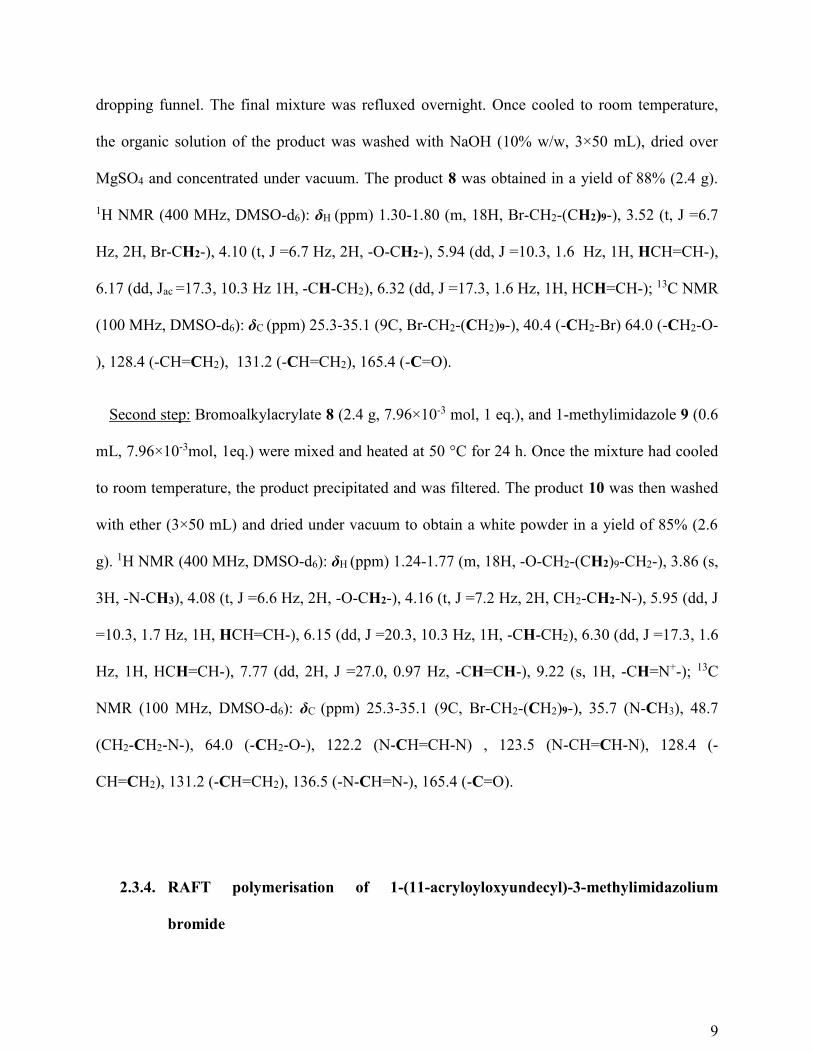
9
dropping funnel. The final mixture was refluxed overnight. Once cooled to room temperature,
the organic solution of the product was washed with NaOH (10% w/w, 3×50 mL), dried over
MgSO4 and concentrated under vacuum. The product 8 was obtained in a yield of 88% (2.4 g).
1H NMR (400 MHz, DMSO-d6): δH (ppm) 1.30-1.80 (m, 18H, Br-CH2-(CH2)9-), 3.52 (t, J =6.7
Hz, 2H, Br-CH2-), 4.10 (t, J =6.7 Hz, 2H, -O-CH2-), 5.94 (dd, J =10.3, 1.6 Hz, 1H, HCH=CH-),
6.17 (dd, Jac =17.3, 10.3 Hz 1H, -CH-CH2), 6.32 (dd, J =17.3, 1.6 Hz, 1H, HCH=CH-); 13C NMR
(100 MHz, DMSO-d6): δC (ppm) 25.3-35.1 (9C, Br-CH2-(CH2)9-), 40.4 (-CH2-Br) 64.0 (-CH2-O-
), 128.4 (-CH=CH2), 131.2 (-CH=CH2), 165.4 (-C=O).
Second step: Bromoalkylacrylate 8 (2.4 g, 7.96×10⁻³ mol, 1 eq.), and 1-methylimidazole 9 (0.6
mL, 7.96×10⁻³mol, 1eq.) were mixed and heated at 50 °C for 24 h. Once the mixture had cooled
to room temperature, the product precipitated and was filtered. The product 10 was then washed
with ether (3×50 mL) and dried under vacuum to obtain a white powder in a yield of 85% (2.6
g). 1H NMR (400 MHz, DMSO-d6): δH (ppm) 1.24-1.77 (m, 18H, -O-CH2-(CH2)9-CH2-), 3.86 (s,
3H, -N-CH3), 4.08 (t, J =6.6 Hz, 2H, -O-CH2-), 4.16 (t, J =7.2 Hz, 2H, CH2-CH2-N-), 5.95 (dd, J
=10.3, 1.7 Hz, 1H, HCH=CH-), 6.15 (dd, J =20.3, 10.3 Hz, 1H, -CH-CH2), 6.30 (dd, J =17.3, 1.6
Hz, 1H, HCH=CH-), 7.77 (dd, 2H, J =27.0, 0.97 Hz, -CH=CH-), 9.22 (s, 1H, -CH=N+-); 13C
NMR (100 MHz, DMSO-d6): δC (ppm) 25.3-35.1 (9C, Br-CH2-(CH2)9-), 35.7 (N-CH3), 48.7
(CH2-CH2-N-), 64.0 (-CH2-O-), 122.2 (N-CH=CH-N) , 123.5 (N-CH=CH-N), 128.4 (-
CH=CH2), 131.2 (-CH=CH2), 136.5 (-N-CH=N-), 165.4 (-C=O).
2.3.4. RAFT polymerisation of 1-(11-acryloyloxyundecyl)-3-methylimidazolium
bromide

10
The following procedure was inspired by the work of Mori et al.17 and Vijayakrishna et al.18
(see Scheme 3). 1-(11-acryloyloxyundecyl)-3-methylimidazolium bromide (IL) 10 (10, 50 or 100
eq.), xanthate 5 (1 eq.) and AIBN (0.3 eq.) were dissolved in DMF (10 mL) and the mixture was
purged with N2 for 10 min. The flask was sealed and the reaction was heated at 70 °C for 17 h.
The mixture was dialysed against water for 2 days using a dialysis tubing of MWCO of 500-
1,000 g/mol for P(IL)10 and 3,500 g/mol for P(IL)50 and P(IL)100. Subsequently the dialysed
material was freeze-dried overnight and the polymer 11 was obtained as a sticky white solid in a
yield of 70-80% (Table S1). The polymer was characterised using solution state NMR
spectroscopy and SEC. NMR spectra were recorded in DMSO-d6 and the Mn and ƉM values were
determined using SEC with conventional calibration (PMMA standards).
2.3.5. CuAAC between N3-HEC and alkyne-terminated poly(1-(11-
acryloyloxyundecyl)-3-methylimidazolium bromide)
General procedure: in a round-bottomed flask fitted with a drying tube, N3-HEC 2, alkyne-
terminated P(IL) 11, sodium L-ascorbate (2 eq.), copper (II) sulfate pentahydrate (1 eq.) and
N,N,N′,N′-tetramethylethylenediamine (1 eq.) were dissolved in DMF (20 mL). The flask was
heated at 30 °C for 24 h. The mixture was cooled to room temperature and dialyzed for 3 days
using a 50 kDa MWCO membrane against Milli-Q water (18.2 Mega Ohm). HEC-g-P(IL) 12
was further lyophilized and obtained in a yield ranging from 64-100% (Table S2). The graft
copolymers were characterized using solid state (13C and 15N) CP-MAS NMR and FT-IR
spectroscopies.

11
2.4. Biological study
2.4.1. Anti-bacterial testing
The bacteriological effects of the HECm-g-P(IL)ns 18 containing P(IL) with different chain
lengths (n= 10, 50 and 100; m =1) and different graft densities (m =1, 0.3 and 0.17; n =10) were
investigated using an Escherichia coli K-12 wild-type strain (W3110)26, and Staphylococcus
aureus (3R7089 strain Oxford)27 selected as representative Gram-negative and Gram-positive
species, respectively.
Bacterial growth inhibition assay on agar plates
HECm-g-P(IL)ns were dissolved in OxoidTM iso-sensitest broth at a concentration of 10 mg/mL
and 6 sequential 2-fold dilutions were prepared in the same medium. 10 µL of each dilution was
spotted onto a Luria-Bertani (LB) agar plate containing bacteria in a 0.6% soft agar overlay (5
mL) and the plates incubated overnight at 30 °C. Samples were analysed in triplicate and a zone
of growth inhibition was observed due to the presence of the HECm-g-P(IL)ns.
Determination of minimum inhibitory concentration (MIC)
MIC determination followed the protocol described by Andrews28. 10 mg/mL dilution of
HECm-g-P(IL)ns in iso-sensitest broth were prepared and 11 sequential 2-fold dilutions were
made using the medium as diluent. The inoculum was prepared from a single bacterial colony
grown on LB agar plates and inoculation of 5 mL of iso-sensitest broth followed by incubation
overnight at 37 °C. 20-50 µL of overnight culture was inoculated into 1 mL of iso-sensitest broth

12
and incubated at 37 °C with aeration until an inoculum density of approximatively 104 cfu/spot
was reached as determined by an optical density A650nm of 0.07, equivalent to a 0.5 MacFarland
standard (240 µM BaCl2 in 0.18 M H2SO4 aq.). This inoculum was diluted 10-fold in iso-sensitest
broth for use in MIC determination.
In a 96-well microtiter plate, 50 µL of each dilution of HECm-g-P(IL)n was mixed with 50 µL
of inoculum for each dilution. Ampicillin was diluted in a similar manner as HECm-g-P(IL)ns and
was used as a positive antibacterial control. The plate was incubated at 37 °C overnight with
shaking and the optical density A650nm of each well was recorded using a Biotek Synergy HT
Multi-Mode Microplate Reader. Samples were analysed in triplicate.
2.4.2. Measurement of cytotoxicity
A549, an immortalized lung alveolar cell line29, was cultured in DMEM supplemented with
10% FCS. Approximately 5×10³ cells in a volume of 100 μL per well were used to seed a 96-
well plate. Cells were allowed to grow in a humidified incubator with an atmosphere of 5% CO2
at 37 °C until 70% confluence was achieved. HECm-g-P(IL)ns to be tested were dissolved in
culture medium to a concentration of 1 mg/mL and 100 μL was added to each well of the first
column of wells to give a final concentration of 500 μg/mL. Subsequently 2-fold serial dilutions
were performed from columns 1-11 with the final column being left as a negative control. After
further incubation the effect of the treatment was determined by a cell viability assay using
Almar Blue30 (GIBCO) that was analysed on a fluorescent plate reader (BioTek, FL500) using
the GEN5 software package. Samples were analysed four times to ensure the reliability of the
results. The fluorescence was read using a fluorescence excitation and emission wavelengths of

13
540–570 nm (peak excitation is 570 nm) and 580–610 nm (peak emission is 585 nm)
respectively. The evolution of the fluorescence intensity as a function of the concentration of
HEC-g-P(IL) permitted the graphical determination of LD50.
3. RESULTS AND DISCUSSION
3.1. Graft copolymers of HEC
3.1.1. Azide functionalized hydroxyethyl cellulose: N3-HEC
N3-HEC 2 was prepared from HEC 1 using a one-step procedure23 (Scheme 1). In order to aid
the detection of nitrogen by 15N NMR spectroscopy, sodium azide was doped with Na15N3. The
characterisation of the partially labelled N3-HEC has already been described in our previous
work22, where the efficiency of a method combining click reaction and RAFT polymerisation to
prepare graft copolymers of HEC was demonstrated, and resulted in the full functionalization of
the primary alcohols of HEC with NaN3. For simplicity, Scheme 1 only shows substitution at the
C6 position, however, substitution at positions C2 and C3 cannot be ruled out.
Scheme 1. Synthesis of partially 15N-labelled N3-HEC 2.

14
3.1.2. Poly(1-(11-acryloyloxyundecyl)-3-methylimidazolium bromide)
1-(11-acryloyloxyundecyl)-3-methylimidazolium bromide 10 was synthesised in 75% overall
yield following the two-step procedure of Harmand et al.25, as shown in Scheme 2. It was then
polymerised by RAFT polymerisation using xanthate 5 as chain transfer agent (Scheme 3). A
xanthate CTA was chosen because the imidazolium ring could potentially reduce the reactivity
of the acrylate functionality.
Scheme 2. Preparation of 1-(11-acryloyloxyundecyl)-3-methylimidazolium bromide 10.
The polymerisation of IL 10 (Scheme 3) was inspired by the work of Mori et al.17 and
Vijayakrishna et al.18. RAFT polymerisation was performed at 70 °C in DMF using a ratio of
xanthate to initiator (AIBN) of 1:0.3 which led to a good comprise between monomer conversion
and dispersity. Different degrees of polymerisation (10, 50 and 100) were targeted in order to
produce graft copolymers of HEC with different chain lengths. After 17 h of polymerisation, the
monomer conversion determined using 1H NMR spectroscopy was found to be approximatively
70-80% (Table S1). Subsequently, the P(IL)s 11 were characterised by NMR spectroscopy and
SEC.

15
Scheme 3. RAFT polymerisation of 1-(11-acryloyloxyundecyl)-3-methylimidazolium bromide
(IL) 10.
In the 1H NMR spectrum of P(IL)100 (Figure 3), signals δH ~1.2-1.8 ppm are assigned to the -
CH2 protons along the side chain and the backbone of P(IL). The signal δH ~2.2 ppm is assigned
to the methine group of the backbone and the signal δH ~3.9 ppm is assigned to the -CH3 group
which is the substituent group of the ring. The signal δH ~4 ppm is assigned to -CH2- next to the
imidazolium ring and the signal δH ~4.2 ppm is assigned to the protons –CH2 next to the acrylate
group. The signal δH~ 7.7-7.8 and 9.4 ppm are assigned to the protons of the imidazolium ring.
The signals of the end groups were not found, perhaps because they are hidden by the signals of
P(IL) because they are too weak. The absence of peaks in the range δH ~6.0-6.5 ppm in the 1H
NMR spectrum confirms that dialysis was successful in removing any residual monomer, and
thus the purity of the polymer can be estimated at > 99%. The spectra of the P(IL)50 and P(IL)10
did not show any new signals, thus, the estimation of the chain length from end groups analysed
by NMR spectroscopy was not feasible.

16
Figure 3. Solution state 1H NMR spectrum of poly(1-(11-acryloyloxyundecyl)-3-
methylimidazolium bromide) (DPtargeted =100)
The molecular weight and dispersity of P(IL) were determined using SEC. The SEC trace for
P(IL)100 is displayed in Figure 4. Conventional calibration with PMMA standards was used to
determine Mn and was found to be 26,600 g/mol. This is in good agreement with the predicted
Mn at 71% monomer conversion (27,600 g/mol). A ƉM of 1.5 was measured, indicating some
control of the polymerisation. A value of ƉM below 1.2 is widely accepted to indicate full control
of the polymerisation, while a value between 1.2 to 1.5 indicates some control of the
polymerisation. A value greater than 1.5 means the loss of control resulting in a broader
h, d
j + k
m
l
i
g e
h, d

17
molecular weight distribution. Typically, polymers produced via conventional free radical
polymerisation have a ƉM value higher than 1.5. The dispersity value of 1.5 for P(IL)100 was
borderline for a polymer which was prepared from a reversible deactivation radical
polymerization (RDRP) process. Regarding P(IL)s with shorter chain lengths (DP = 10 and 50),
ƉM values are expected to be similar or slightly higher because the lower DP accentuates
differences in chain lengths.
Figure 4. SEC trace of P(IL)100.
3.1.3. Graft copolymers HEC-g-poly(1-(11-acryloyloxyundecyl)-3-methylimidazolium
bromide): HEC-g-P(IL)s
70
90
110
130
150
170
190
210
230
250
12 14 16 18 20 22 24
Ref
ract
ive
Ind
ex
Retention volume (mL)

18
Ionic liquid polymers P(IL)n 11 of different chain lengths (n =10, 50 or 100) were coupled to
each AGU unit of N3-HEC 2 and this resulted in the preparation of graft copolymers HEC-g-
P(IL)ns 12 (Scheme 4) where the grafts were composed of 10, 50 or 100 repeat units of ionic
liquid monomer. These fully substituted HEC graft copolymers thus have a graft density of 1.
Additionally, HECm-g-P(IL)10s were prepared with different graft densities (m =0.3 and 0.17) via
the coupling of one P(IL)10 chain per 3 and 6 AGU units of N3-HEC respectively. In order to
remove the homopolymer P(IL)n, the reaction mixture was dialysed against water using a dialysis
tubing of a MWCO of 50,000 g/mol ensuring the recovery of only HECm-g-P(IL)ns. The graft
copolymers were characterised using solid state (13C and 15N) CP-MAS NMR and FT-IR
spectroscopies. Their solubility permits characterisation by solution state NMR spectroscopy,
however the resulting spectra do not show the presence of HEC and thus evidence of the
coupling could not be demonstrated. The chain length (defined by “n”) of P(IL) did not influence
the detection of the HEC backbone by solid state NMR spectroscopy, however, decreasing the
graft density (defined by “m”) allowed the presence of HEC to be confirmed. The
characterisation of HECm-g-P(IL)10s (m =1, 0.3 and 0.17) only are reported here.

19
Scheme 4. CuAAC between alkyne-ended P(IL)n and partially 15N-labelled N3-HEC
FT-IR spectra of N3-HEC, P(IL)10 and HECm-g-P(IL)10s are displayed in Figure 5. The total
loss of the azide signal at 2136 cm-1 in the spectrum of HEC1-g-P(IL)10 (Figure 5d) indicates the
complete consumption of the azide group of N3-HEC (Figure 5a) due to the formation of the
triazole with the alkyne-terminated P(IL)10. This is crucial evidence of the grafting because,
otherwise, the IR spectrum of HEC1-g-P(IL)10 is the same as that of P(IL)10 (Figure 5e), and no
absorption bands of HEC were detected because they are probably hidden by those of P(IL)10.
On decreasing the graft density (Figure 5b & c), the azide signal at 2136 cm-1 was still detected,

20
however a decrease of its relative intensity was observed compared to the IR spectrum of N3-
HEC (Figure 5a) indicating partial consumption of the azide. In fact, increasing the graft density
produces a decrease of the relative intensity of the azide signal (Figure 5c vs. 5b). Except for this
signal, the others are characteristic of the presence of P(IL)10. The bands at 3400 cm-1 are
assigned to -OH groups, at ~3142 cm-1 to the -CH groups of the ring, at 2856-2926 cm-1 to the
alkyl -CH groups, at 1734 cm-1 to the carbonyl group, at 1676 cm-1 to the -C=C- and -C=N-
groups, at 1576 cm-1 to the -C-C- and -C-N- groups, at 1468 cm-1 to the -CH alkyl groups and at
~1170 cm-1 to the alkyl -CH groups.
Figure 5. FT-IR spectrum of: (a) N3-HEC; (b) HEC0.17-g-P(IL)10; (c) HEC0.3-g-P(IL)10; (d)
HEC1-g-P(IL)10; and (e) P(IL)10.
400900140019002400290034003900
Tra
nsm
itta
nce
(%
)
Wavenumber (cm-1)
(a)
(b)
(c)
(d)
(e) N3

21
The solid state 13C CP-MAS NMR spectrum of HEC1-g-P(IL)10 (Figure 6c) shows only signals
of the P(IL)10 grafts. The signal δC ~174.4 ppm is assigned to the carbonyl group, δC ~139.5 and
~123.6 ppm are assigned to the alkyl -CH groups of the ring, δC ~64.9 ppm to the -CH2-O group,
δC ~49.1 ppm to the -CH2-N group, δC ~41.4 ppm to the methine group and the signals δC ~36.0-
30.6 ppm to the methylene and methyl groups. The absence of signals for the HEC backbone was
expected because of the relatively high molecular weight of the P(IL)10 grafts. In our previous
work22, coupling of polymers with N3-HEC was demonstrated more easily because of the low
molecular weight of the grafted chains (PVP10 or PNIPAAM10) which did not exceeded 1,000
g/mol. The signals assigned to the HEC backbone, which were relatively weak compared to
those of the grafts, were detected in the solid state 13C NMR spectra22, however, we suspected
that an increase in the molecular weight of the grafts would result in the disappearance of the
signals of HEC. This is what we observed with P(IL)10 grafts, which have a molecular weight of
approximatively 4,000 g/mol. However, a decrease of the graft density permitted the detection of
the HEC backbone. In fact, signals assigned to HEC were clearly detected in the spectrum of
HEC0.17-g-P(IL)10 (Figure 6a) which possesses the lowest graft density of P(IL)10. The signal δC
~103.6 ppm is assigned to the carbon at the C1 position, ~82.6 ppm to carbon at the C4 position,
~74.9 ppm to the set of carbons at C2, C3 and C5 positions, ~71.2 ppm to the set of carbons at
C6, C7, C8 and C9 positions and ~51.1 ppm to the carbon at C10 position. These signals
assigned to the HEC backbone were still detected in the spectrum of HEC0.3-g-P(IL)10 (Figure
6b) but their relative intensities are lower due to the increase of the graft density of P(IL)10
compared to those in the spectrum of HEC0.17-g-P(IL)10.

22
Figure 6. Solid state 13C CP-MAS NMR spectra of HECm-g-P(IL)10: (a) HEC0.17-g-P(IL)10; (b)
HEC0.3-g-P(IL)10; (c) HEC1-g-P(IL)10.
The solid state 15N spectra of HEC1-g-P(IL)n with n =10, 50 and 100 were all identical, thus
only the spectrum of HEC1-g-P(IL)10 is reported here (Figure 7b) with the spectrum of N3-HEC
(Figure 7a) used as a reference. The spectrum of N3-HEC showed two signals δN ~ –310 ppm
and ~ –170 ppm which are assigned to Nα and Nγ respectively (Scheme 4)31, 32. The differences in
intensity can be explained by the differences in their proton environment. Nα is next to a
methylene group resulting in a higher intensity of its signal compared to that of Nγ because
nitrogen is detected through the excitation of the protons. From our previous investigation22, only
the Nα signal remained in the 15N CP-MAS NMR spectrum after cycloaddition of N3-HEC with
l, h
f m
j, k g
i
e
C1 C4
C10 C2, C3, C5
C6 to C9 (a)
(b)
(c)

23
alkyne-terminated synthetic polymers, however, the signal was shifted from ~ –310 ppm to ~ –
134 ppm, providing evidence of the success of the “click” reaction. Here, the spectrum of HEC1-
g-P(IL)10 showed two signals δN ~ –208.0 and ~ –194.3 ppm which are assigned to the
imidazolium ring, based on the literature33. More precisely, the former is assigned to the -N-CH3
group and the latter to the N-CH2- group. The absence of the signal δN ~ –134 ppm, assigned to
Nα after the cycloaddition, is most likely caused by the high molecular weight of the P(IL)10
grafts which decreases the relative intensity of the Nα signal. Decreasing the graft density of
P(IL)10 resulted in the loss of signals assigned to the nitrogen atoms nitrogen present in the
grafts, however, the signals assigned to the triazole and azide were also absent. In fact, a
decrease of the graft density did not change the ratio of Nα (after cycloaddition) to P(IL)10, and
this is why Nα remains undetectable. Nα and Nγ (before cycloaddition) were also not detected
upon decreasing the graft density because their concentration is too low compared to the rest of
the sample.

24
Figure 7. Solid state 15N CP-MAS spectra: (a) N3-HEC; (b) HEC1-g-P(IL)10
To summarise, the characterisation of the graft copolymers demonstrated the presence of P(IL)
grafts by NMR and FT-IR spectroscopies, however, detection of the HEC backbone was more
challenging due to the high molecular weight of the IL monomer. When the graft density of
P(IL)10 was decreased, the HEC backbone was detected by 13C CP-MAS NMR spectroscopy
indicating the presence of HEC in the product. The use of 15N CP-MAS NMR spectroscopy did
not demonstrate the formation of the triazole, most probably because of the relatively low level
of enrichment over the 0.4% background. However, evidence of coupling between P(IL)s and
N3-HEC came from the disappearance of the azide band at 2100 cm-1 in the FT-IR spectrum of
the graft copolymers. Furthermore, a quantitative yield for each coupling reaction (Table S2)
strongly implies coupling of the azide in N3-HEC, resulting in the preparation of HEC based
(a)
(b)

25
P(IL) graft copolymers. The final graft copolymers were purified by extensive dialysis using a
high molecular weight cut off membrane (50,000 g/mol) so it can reasonably be expected that
any uncoupled P(IL) chains were removed.
3.2. Biological activities of HEC-g-P(IL)s
The successful preparation of HEC-g-P(IL)s 12 permitted the study of their effects on bacteria
and on an immortalized lung alveolar cell line (A549) to investigate potential applications.
Furthermore, the influence of the chain length and the graft density of P(IL) on biological
activity were investigated.
3.2.1. Antibacterial effects
Two tests were performed to evaluate the bacteriological effects of the HEC-g-P(IL)s. The first
was a qualitative test to visualise any effects on growth inhibition, whereas the second was a
quantitative test permitting a determination of the minimum inhibition concentration (MIC)
which is the lowest concentration of a compound which inhibits bacterial growth. E. coli (Gram-
negative) and S. aureus (Gram-positive) were chosen as representative bacteria.
Solutions of each HECm-g-P(IL)n were prepared using iso-sensitest broth and series of 11 x 2-
fold dilutions were prepared resulting in concentrations ranging from 10 mg/mL to 2 μg/mL. In
the first test, which shall be referred to as the zone inhibition assay, 10 μL of each solution of the
first 6 dilutions were spotted onto an agar plate containing an overlay of E. coli or S. aureus

26
bacteria. The plates were incubated overnight to allow the lawn of bacteria to grow (Figure 8).
To determine the MIC (2nd test), 50 μL of each graft copolymer solution was pipetted into a 96-
well plate containing 50 μL of bacteria solution (both in iso-sensitest broth). The 96-well plate
was incubated overnight and the optical density (OD) at A650nm was determined in each well to
monitor bacterial growth (Figure 9). A 10 mg/mL dilution series of ampicillin was used as a
positive control.
For E. coli, growth inhibition (Figure 8a) was obtained from solutions of P(IL)n-g-HEC1s with
concentrations ranging from 10 to 1.75 mg/mL, independent of the graft chain length (n = 10, 50
and 100). At 0.875 mg/mL HEC1-g-P(IL)n (with n = 100, 50 and 10) concentration, inhibition
was not observed indicating the loss of antibacterial effect at this concentration. At 1.75 mg/mL,
the size of the zones were similar independent of the length of P(IL) grafts, indicating there was
no major influence of the chain length on antibacterial properties. However, the graft density of
P(IL)10 on HEC affected to a slight extent the inhibitory effect. The inhibitory effect of the graft
copolymers with lower graft densities (m =0.3 and 0.17) was shown at higher concentration than
HEC1-g-P(IL)10. At a 1.75 mg/mL HECm-g-P(IL)10 (with m =0.3 and 0.17) concentration,
inhibition zones were not observed indicating the lack of an inhibitory effect. Furthermore, the
size of the inhibition zones for HEC0.3-g-P(IL)10 and HEC0.17-g-P(IL)10 were similar at a
concentration of 2.5 mg/mL, demonstrating a limited influence of the graft density on inhibition
at these densities. However, HEC1-g-P(IL)n (n =10, 50 and 100) also have a higher inhibitory
effect compared to those of lower graft density.

27
Figure 8. Results of the zone inhibition assays for the graft copolymers HEC-g-P(IL)s against
(a) E. coli (b) S. aureus.
(a)
(b)

28
For S. aureus, zones of growth inhibition were observed at 10 and 5 mg/mL concentrations for
each graft copolymers (Figure 8b), however, at 2.5 mg/mL concentration, HEC1-g-P(IL)10 and
HEC0.3-g-P(IL)10 still showed a slight growth inhibition, whereas HEC1-g-P(IL)50, HEC1-g-
P(IL)100 and HEC0.17-g-P(IL)10 did not. Samples with longer grafts of IL (50 and 100) or a
decreased graft density (HEC0.17-g-P(IL)10) had a similar effect on the growth of S. aureus
bacteria.
In order to corroborate these observations, MICs for each bacterial strain with the different
graft copolymers were determined. Regarding E. coli, the graft copolymers HEC1-g-P(IL)ns (n
=10, 50 and 100) and HECm-g-P(IL)10s (m =0.3 and 0.17) gave MIC values of 10 and 19 μg/mL
respectively (Figure 9a). Furthermore, a MIC value of 39 μg/mL was measured for ampicillin,
higher than that determined for HEC-g-P(IL)s demonstrating a stronger inhibitory effect of the
graft copolymers against Gram-negative bacteria. For S. aureus, MIC values for HEC1-g-P(IL)10
and HEC0.3-g-P(IL)10 were estimated to be 19 μg/mL whereas a MIC value of 39 μg/mL was
determined for HEC1-g-P(IL)n (n =50 and 100) and HEC0.17-g-P(IL)10 (Figure 9b). The MICs of
HEC1-g-P(IL)n (n =100 and 50) and HEC0.17-g-P(IL)10 were similar to that of ampicillin,
however, the MICs of HECm-g-P(IL)10s (m =1 and 0.3) were lower, suggesting a potentially
stronger efficacy of these graft copolymers against Gram-positive bacteria compared to a well-
known antibiotic.
To summarise, growth inhibition effects of the graft copolymers on both Gram-positive and
Gram-negative bacteria were observed. The magnitude of the effects were comparable to those
produced by ampicillin, a commonly used antibiotic. The cationic charge of the imidazolium ring
is likely to be responsible for this effect8. Surprisingly, the chain length of the grafts, which
determines the number of charges, did not significantly influence the antibacterial properties of
29
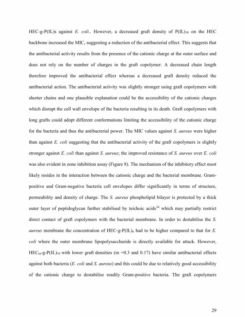
HEC-g-P(IL)s against E. coli.. However, a decreased graft density of P(IL)10 on the HEC
backbone increased the MIC, suggesting a reduction of the antibacterial effect. This suggests that
the antibacterial activity results from the presence of the cationic charge at the outer surface and
does not rely on the number of charges in the graft copolymer. A decreased chain length
therefore improved the antibacterial effect whereas a decreased graft density reduced the
antibacterial action. The antibacterial activity was slightly stronger using graft copolymers with
shorter chains and one plausible explanation could be the accessibility of the cationic charges
which disrupt the cell wall envelope of the bacteria resulting in its death. Graft copolymers with
long grafts could adopt different conformations limiting the accessibility of the cationic charge
for the bacteria and thus the antibacterial power. The MIC values against S. aureus were higher
than against E. coli suggesting that the antibacterial activity of the graft copolymers is slightly
stronger against E. coli than against S. aureus; the improved resistance of S. aureus over E. coli
was also evident in zone inhibition assay (Figure 8). The mechanism of the inhibitory effect most
likely resides in the interaction between the cationic charge and the bacterial membrane. Gram-
positive and Gram-negative bacteria cell envelopes differ significantly in terms of structure,
permeability and density of charge. The S. aureus phospholipid bilayer is protected by a thick
outer layer of peptidoglycan further stabilised by teichoic acids34 which may partially restrict
direct contact of graft copolymers with the bacterial membrane. In order to destabilise the S.
aureus membrane the concentration of HEC-g-P(IL)n had to be higher compared to that for E.
coli where the outer membrane lipopolysaccharide is directly available for attack. However,
HECm-g-P(IL)10 with lower graft densities (m =0.3 and 0.17) have similar antibacterial effects
against both bacteria (E. coli and S. aureus) and this could be due to relatively good accessibility
of the cationic charge to destabilise readily Gram-positive bacteria. The graft copolymers

30
therefore display promising antibacterial properties which could extend the use of cellulosic
materials in a variety of applications.
Figure 9. Determination of the MIC of HEC-g-P(IL)s for: (a) E. coli and (b) S. aureus. The MIC
for each graft polymer is indicated.
3.2.2. Cytotoxicity
0,0
0,2
0,4
0,6
0,8
1,0
1,2
1,4
1,6
1,8
2,0
5000 2500 1250 625 312 156 78 39 19 10 5 2,5
OD
(A
65
0n
m)
Concentration (µg/mL)
HEC1-g-P(IL)100
HEC1-g-P(IL)50
HEC1-g-P(IL)10
HEC0.3-g-P(IL)10
HEC0.17-g-P(IL)10
Amp.
0,0
0,2
0,4
0,6
0,8
1,0
1,2
1,4
1,6
1,8
2,0
5000 2500 1250 625 312 156 78 39 19 10 5 2,5
OD
(A
65
0n
m)
Concentration (µg/mL)
(b) S. aureus
(a) E. coli

31
To evaluate the possible use of HECm-g-P(IL)ns in medical applications, an estimation of
cytotoxicity was essential. Cytotoxicity is measured using LD50, the lethal dose that kills 50% of
cells. An immortalized lung alveolar cell line (A549) was used. A 500 µg/mL concentration
solution was prepared for each graft copolymer followed by 11 x 2-fold serial dilutions. Each
solution was spotted into a 96-well plate containing the cells. After incubating overnight, Almar
blue solution was added and the fluorescence of each well containing both cells and HEC-g-
P(IL) was determined. The evolution of fluorescence intensity as a function of the concentration
of each graft copolymer is displayed in Figure 10.
Figure 10. Determination of LD50 for HEC-g-P(IL)s.
The fluorescence decreases with the death of the cells and approaches zero when all cells have
died, however, LD50 corresponds to the concentration of HEC-g-P(IL) at which half of the
0
20
40
60
80
100
120
141664256
Rel
ativ
e fl
uo
resc
ence
in
ten
sity
(%)
Log2 (Concentration (µg/mL))
HEC1-g-P(IL)100
HEC1-g-P(IL)50
HEC1-g-P(IL)10
HEC0.3-g-P(IL)10
HEC0.17-g-P(IL)10

32
fluorescence was lost. For HEC1-g-P(IL)ns (n =10, 50 and 100), LD50 was ~25 µg/mL whereas
~45 µg/mL LD50 was measured for HECm-g-P(IL)10s (m =0.3 and 0.17). The chain length did not
significantly influence the toxicity of the graft copolymers, however, a decrease of the graft
density of P(IL)10 onto HEC reduced the toxicity of the graft copolymers towards the cells. The
cationic charge is responsible for the attachment between the cell membrane and the graft
copolymers resulting in the necrosis of the cell, i.e. their death35, 36. One plausible reason of the
reduction of the toxicity observed when decreasing the graft density is the decrease of the
number of cationic charges on the outer surface of the graft copolymers. The LD50 values were
similar to the MIC values, indicating that the doses required to inhibit the growth of the bacteria
are close to those that kill 50% of the cells.
4. CONCLUSION
Graft copolymers of HEC and a poly(ionic liquid) (P(IL)) were successfully prepared using a
versatile method combining RAFT polymerisation and CuAAC. P(IL)s 11 with different DPs
(10, 50 and 100) were synthesised with monomer conversions up to ~80% using a xanthate as a
chain transfer agent. The dispersity of P(IL)100 was measured using SEC and was found to be
~1.5. P(IL)s were further coupled to azido-functionalized HEC 2, which was prepared via the full
functionalization of the primary alcohols of HEC with sodium azide. This produced graft
copolymers having both different chain lengths of P(IL)n (n =10, 50 and 100) per AGU unit and
different graft densities of P(IL)10 onto HECm (m =0.3 and 0.17; i.e. 1 chain of P(IL)10 per 3 or 6
AGU units). Subsequently, the graft copolymers were characterised using (13C and 15N) CP-
MAS NMR and FT-IR spectroscopies. The observed decrease of the graft density of P(IL)10 onto

33
HEC provided evidence of the success of the “click” reaction. Antibacterial properties of the
graft copolymers were evaluated against E. coli (Gram-negative) and S. aureus (Gram-positive)
and were comparable to those of a commonly employed antibiotic, ampicillin. For HEC1-g-
P(IL)n (n =10, 50 and 100), the minimum inhibition concentration (MIC) against E. coli was the
same (10 µg/mL) indicating no influence of the chain length, however, a decreased graft density
reduced the inhibitory effect (MIC =19 µg/mL) of HEC0.3-g-P(IL)10 and HEC0.17-g-P(IL)10. This
suggests that the antibacterial properties arise from the number of cationic charges on the outer
surface of the graft copolymers. For the Gram-positive bacterium (S. aureus), higher MICs were
obtained indicating weaker inhibitory effects of the graft copolymers as compared to E. coli,
potentially because of their different cell envelopes. However, the shortest chain length (n =10)
had a slightly stronger inhibitory effect (MIC =19 µg/mL) compared to graft copolymers with a
longer graft chain length (MIC =39 µg/mL) possibly because of higher accessibility of the
cationic charge due to the limitation of the conformational flexibility. Furthermore, a decrease of
the graft density of HECm-g-P(IL)10 to 0.3 did not affect the antibacterial properties, however, a
further decrease to 0.17 reduced the inhibitory effect to a value comparable to those of HEC1-g-
P(IL)n (n =100 and 50). This is most likely due to the considerable decrease of the number of
cationic charges on the outer surface. To evaluate the potential of this antibacterial HEC graft
copolymer, the cytotoxicity was evaluated using LD50 measurements. Values close to the MIC
values were found consistent with toxicity due to membrane damage in both the prokaryotic and
eukaryotic cells tested here. This suggests that these HEC-g-P(IL) graft copolymers are most
likely to be appropriate for use as antibacterial coatings.
Supporting Information

34
The Supporting Information includes a scheme for the synthesis of the chain-transfer agent
xanthate, the reaction conditions of the RAFT polymerisations of the ionic liquid monomer and
the reaction conditions of the “click” reaction between P(IL) and N3-HEC. This material is
available free of charge via the Internet at http://pubs.acs.org.
Acknowledgements
We thank Dr David Apperley and other staff members of the EPSRC solid state NMR National
Facility at Durham University for their assistance.
Author Contributions
The manuscript was written through contributions of all authors. All authors have given
approval to the final version of the manuscript.
Funding sources
We thank EPSRC and Ashland Inc. for financial support (studentship to FJ).
Conflict of Interest Disclosure
Dr Osama Musa is an employee of Ashland Inc., which is a producer of hydroxyethyl
cellulose.

35
References
1. Klemm, D.; Schmauder, H.-P.; Heinze, T. Cellulose. In Biopolymers Online; Steinbüchel,
A., Eds.; Wiley-VCH Verlag GmbH & Co. KGaA, 2005.
2. D. R. Nobles, Jr.; R. M. Brown, Jr., Cellulose 2008, 15, 691-701.
3. Polysaccharides: Structural Diversity and Functional Versatility; Dumitriu, S. E., Eds.;
Marcel Dekker: New York, 2005.
4. O'Sullivan, A. Cellulose 1997, 4, 173-207.
5. Biopolymers: New Materials for Sustainable Films and Coatings; Plackett D. E., Eds.;
John Wiley & Sons: Chichester, 2011.
6. Kamel, S.; Ali, N.; Jahangir, K.; Shah S. M.; El-Gendy, A. A. Express Polym. Lett. 2008,
2, 758-778.
7. Cork: Biology, Production and Uses; Pereira, H., Eds.; Elsevier: Amsterdam, 2007.
8. Tashiro, T. Macromol. Mater. Eng. 2001, 286, 63-87.
9. Muñoz-Bonilla, A.; Fernández-García, M. Prog. Polym. Sci. 2012, 37, 281-339.
10. Gilmore, B. F.; Andrews, G. P.; Borberly, G.; Earle, M. J.; Gilea, M. A.; Gorman, S. P.;
Lowry, A. F.; McLaughlin, M.; Seddon, K. R. New J. Chem. 2013, 37, 873-876.
11. Demberelnyamba, D.; Kim, K.-S.; Choi, S.; Park, S.-Y.; Lee, H.; Kim C.-J.; Yoo, I.-D.
Bioorg. Med. Chem. 2004, 12, 853-857.

36
12. Cole, M. R.; Li, M.; El-Zahab, B.; Janes, M. E.; Hayes D.; Warner, I. M. Chem. Biol.
Drug Des. 2011, 78, 33-41.
13. Yuan, J.; Antonietti, M. Polymer 2011, 52, 1469-1482.
14. Ding, S.; Tang, H.; Radosz, M.; Shen, Y. J. Polym. Sci., Part A: Polym. Chem. 2004, 42,
5794-5801.
15. He, H.; Zhong, M.; Luebke, D.; Nulwala H.; Matyjaszewski, K. J. Polym. Sci., Part A:
Polym. Chem. 2014, 52, 2175-2184.
16. Ma, X.; Ashaduzzaman, M.; Kunitake, M.; Crombez, R.; Texter, J.; Slater L.; Mourey, T.
Langmuir 2011, 27, 7148-7157.
17. Mori, H.; Yahagi M.; Endo, T. Macromolecules 2009, 42, 8082-8092.
18. Vijayakrishna, K.; Jewrajka, S. K.; Ruiz, A.; Marcilla, R.; Pomposo, J. A.; Mecerreyes,
D.; Taton D.; Gnanou, Y. Macromolecules 2008, 41, 6299-6308.
19. Advances in Food Research, Stewart, G. F. E., Mrak, E. M., Chischester C. O., Eds.;
Academic Press: London, 1963.
20. Coatings Materials and Surface Coatings; Tracton, A. A., Eds.; CRC Press: , 2006.
21. Controlled Release in Oral Drug Delivery; Wilson, C. G., Crowley, P. J., Eds.; Springer:
London, 2011.
22. Joubert, F.; Musa, O.; Hodgson, D. R. W.; Cameron, N. R. Polym. Chem. 2015, 6, 1567-
1575.

37
23. Eissa, A. M.; Khosravi, E.; Cimecioglu, A. L. Carbohydr. Polym. 2012, 90, 859-869.
24. Akeroyd, N.; Pfukwa, R.; Klumperman, B. Macromolecules 2009, 42, 3014-3018.
25. Harmand, J.; Rogalski, M.; Sindt, M.; Mieloszynski, J.-L. Environ. Chem. Lett. 2009, 7,
255-260.
26. Hayashi, K.; Morooka, N.; Yamamoto, Y.; Fujita, K.; Isono, K.; Choi, S.; Ohtsubo, E.;
Baba, T.; Wanner, B. L.; Mori H.; Horiuchi, T. Mol. Syst. Biol. [Online] 2006, DOI:
10.1038/msb4100049. http://msb.embopress.org/content/msb/2/1/2006.0007.full.pdf (accessed
Oct 29, 2015)
27. Rosenbach, F. I. Mikroorganismen Bei Den Wund-Infections- Krankheiten Des
Menschen, Bergmann, J. F., Eds.; Wiesbaden, 1884.
28. Andrews, J. M. J. Antimicrob. Chemother. 2001, 48, 5-16.
29. Lieber, M.; Smith, B.; Szakal, A.; Nelsonrees, W.; Todaro, G. Int. J. Cancer 1976, 17,
62-70.
30. Andreas, B. Patent CA 2089870, 1993.
31. Corredor, M.; Bujons, J.; Messeguer, A.; Alfonso, I. Org. Biomol. Chem. 2013, 11, 7318-
7325.
32. Wrackmeyer, B. Z. Naturforsch. B. 2011, 66, 1079-1082.
33. Lyčka, A.; Doleček, R.; Šimûnek, P.; Macháček, V. Magn. Reson. Chem. 2006, 44, 521-
523.

38
34. Silhavy, T. J.; Kahne, D.; Walker, S. Cold Spring Harbor Perspect. Biol. [Online] 2010,
DOI: 10.1101/cshperspect.a000414.
http://cshperspectives.cshlp.org/content/2/5/a000414.full.pdf+html (accessed Oct 29, 2015)
35. Lu, G.; Wu, D.; Fu, R. React. Funct. Polym. 2007, 67, 355-366.
36. Fischer, D.; Li, Y.; Ahlemeyer, B.; Krieglstein, J.; Kissel, T. Biomaterials 2003, 24,
1121-1131.

39
Table of Contents Graphic