1
Reconstitution of membrane proteins - a GPCR as an example.
Alan D. Goddard1, Patricia M. Dijkman2, Roslin J. Adamson2, Rosana Inácio dos
Reis2 and Anthony Watts2*.
1School of Life Sciences, University of Lincoln, Lincoln, LN6 7TS, UK. 2Biomembrane Structure Unit, Department of Biochemistry, University of Oxford,
Oxford, OX1 3QU, UK.
*Corresponding author: email address: [email protected]
Contents
1. Introduction
2. Production of liposomes
2.1. Lipid preparation
2.2. Sizing of liposomes
3. Reconstitution into liposomes
3.1. Rapid dilution
3.2. Hydrophobic adsorption
4. Reconstitution into nanodiscs
4.1. Production of MSP for nanodiscs
4.2. NTS1 reconstitution into nanodiscs
5. Reconstitution into lipodisqs
5.1. bR reconstitution into lipodisqs
6. Determination of lipid-to-protein ratio
6.1. Liposome/lipid concentration determination
6.2. Liposome scatter in UV-Vis: Labelled lipid marker
6.3. Sucrose density gradient
7. Concentration methods
Summary
References
Abstract
Membrane proteins are the gatekeepers to the cell, and are essential to the function of
all cells, controlling the flow of molecules and information across the cell membrane.
Much effort has been put into the development of systems for studying membrane
proteins in simplified environments that nevertheless mimic their native lipid
environment. After isolation and production of purified membrane proteins in
detergent, it is often necessary to reconstitute them into a lipid structure such as
liposome, nanodisc or lipodisq. Each of these has the advantage of returning the
protein to a defined lipid environment, and the choice of system depends on the
application. Regardless of the system to be used, the fundamental process involves the
removal of detergent and incorporation of the protein into a stable lipid system. This
chapter details methodologies we have developed, mainly focussed on the model G
protein-coupled receptor (GPCR) neurotensin receptor 1, and the GPCR-homologue
and model, bacteriorhopdopsin.
1. Introduction
All cells are surrounded by a lipid membrane which modulates the flow of molecules,
and information, into and out of the cell. However, these membranes do not consist
solely of lipids, but incorporate a wide variety of proteins such as receptors,
© 2015, Elsevier. Licensed under the Creative Commons Attribution-NonCommercial-NoDerivatives 4.0 Internationalhttp://creativecommons.org/licenses/by-nc-nd/4.0/
2
transporters and structural proteins which modulate cellular behaviour. The protein
content of a membrane can be variable (from 500:1 to 10:1 molar ratio, depending on
functional criteria), but usually amounts to 50-90% by mass. Membrane protein copy
numbers are also highly variable, ranging from 1-2 per cell to >106 per cell.
Membrane proteins are notoriously difficult to study due to the requirement for
detergents to solubilise them out of the membrane in which they are either
endogenously present, or have been expressed. Detergents, whilst stabilising the
protein for purification, do not represent a native environment and many proteins will
require specific lipids to establish functional activity or stability. In the case of
transporters and signalling receptors, it is also often necessary to have a system in
which the protein can be orientated in order to check for full activity. As such, the
production of lipid systems in which proteins can be reconstituted has become a major
avenue of research.
In this chapter, we will focus on some of the more common systems for reconstitution
of membrane proteins, including liposomes, nanodiscs and lipodisqs using examples
and principles from our own work with the G protein-coupled receptor neurotensin
receptor 1 (NTS1) and bacteriorhodopsin (bR). The other GPCR for which significant
reconstitution methods have been developed for both functional and biophysical
studies, is (frog and bovine) rhodopsin, and details have been reported previously
(Albert, Yeagle, Whiteway, Watts, & Epand, 1994; Jackson & Litman, 1982; Spooner
et al., 2004).
2. Production of liposomes
Lipids spontaneously form liposomes when rehydrated from a dried film into an
aqueous buffer. Liposomes formed by such rehydration will range in size (generally
between ~30 nm and 50 m in diameter) and may well form multilamellar vesicles
possessing multiple concentric bilayers. Such a population of heterogeneous and
multilamellar liposomes may be less desirable for the study of membrane proteins
such as receptors and transporters, only those present in the outer layer will be
available for study. Therefore, it is usual to create better-characterised unilamellar
liposomes for the creation of proteoliposomes.
2.1. Lipid preparation
Lipids are commercially available as liquid stocks which are generally supplied in
chloroform or chloroform:methanol at 25 mg/ml (e.g. from Avanti Polar Lipids, Inc.).
Alternatively, stocks can be prepared from lyophilized powders at similar
concentrations. Lipids can be difficult to weigh accurately (due to the hygroscopic
nature of unsaturated lipids) and are prone to oxidation. Therefore, it is most
convenient to work with lipids supplied in solvent. To prevent evaporation of the
solvent during storage, opened stocks are best transferred to Teflon-capped glass vials
and closed under an N2 atmosphere.
Materials for liposome creation:
Lipid stocks in chloroform at 25 mg/ml;
Liposome buffer e.g. 50 mM Tris pH7.4, 100 mM NaCl, 1mM EDTA;
Rotary evaporator (optional);
Temperature-controlled waterbath sonicator (optional).
3
Note, do not use plastic tubes or pipette tips for transferring solvents.
1. To create the desired lipid composition for the proteoliposomes, appropriate
volumes of lipid stocks are mixed, along with sterols such as cholesterol
which are poorly soluble in aqueous solvents.
2. To create a lipid film, the lipid mixture (or single lipid) is placed into a round
bottomed flask and the solvent is evaporated. This can be achieved by a
variety of methods, e.g. by blowing a stream of argon or nitrogen over the
chloroform. Air should not be used as lipids are prone to oxidation. This
solvent removal method is suitable for small (milliliter) volumes, but can be
time consuming and difficult to perform for larger volumes. Thus, for large
volume samples, it is often easiest to use a rotary evaporator with appropriate
solvent trap.
3. The lipid should be fully dried until a film is generated on the bottom of the
flask.
4. Films are then thoroughly dried under vacuum (< 10-5 Torr) overnight. Dried
lipid films are stable for months if stored under nitrogen at < -20 °C.
5. Lipid films are then rehydrated in aqueous buffer, typically to a final lipid
concentration of 5 mg/ml. The choice of aqueous buffer for rehydration is very
much dependent on the protein to be reconstituted. Generally, the same buffer
can be used as the final purified protein preparation, but lacking detergents.
Rehydration may also be performed in a variety of manners, but it is important
to note that this should be done above any phase transition temperature of the
lipid (or mix of lipids) (Marsh, 2013). The buffer should be pre-warmed above
the highest phase transition temperature within the lipids being used, and
rehydration should be done at the same temperature.
6. In the case of lipids with a phase transition temperature above ambient, one of
the simplest methods is to place the sample on a rotary evaporator (with the
vacuum off) and heat the waterbath to an appropriate temperature. Such
rehydration should be performed for at least an hour, which should yield an
homogenous suspension of liposomes. In the case of lipids with a lower phase
transition temperature it is possible to vortex and sonicate the sample in a
waterbath for 1-5 min, again above any phase transition temperature.
7. Resuspended lipid dispersion will contain multilamellar liposomes, which can
be broken down by repeated freeze thawing in liquid nitrogen 6-8 times.
2.2. Sizing of liposomes
Multilamellar liposomes are often an undesirable form of proteoliposomes due to the
limited accessibility of proteins within the internal bilayers. Thus, it is often useful to
create unilamellar liposomes of defined size in advance of protein reconstitution.
There are a variety of methods to do this with the simplest being sonication.
Materials for sizing of liposomes:
(Temperature-controlled) waterbath sonicator or probe sonicator;
Extruder, e.g. mini-extruder (Avanti Polar Lipids, Inc.);
Polycarbonate filters of appropriate pore size (e.g. Whatman).
Generating small unilamellar vesicles (SUVs):
4
1. A temperature-controlled sonicating waterbath set above the phase transition
temperature of the lipid can be used and lipid dispersions sonicated for
approximately 30-60 minutes, depending on the lipid composition.
2. Alternatively, a probe sonicator can be used although care must be taken to
use a plastic container. This is a more rapid method (typically on the order of
minutes) although care should be taken to avoid foaming and overheating, by
using short (typically 30 s) pulses. Probe sonication is more difficult if the
lipid mixture has a phase transition temperature above ambient due to the need
for a (controlled) heat source.
3. Either method generates small unilamellar vesicles (SUVs) of 30-50 nm
diameter yielding a clarified solution as the size of the liposomes drops below
the wavelength of visible light.
4. It should be noted that the high level of curvature and surface tension makes
SUVs unstable and they should be used promptly after creation. Stability can
be checked through turbidity monitoring (~400-700 nm), since larger
structures are formed with time (within hours).
Generating large unilamellar vesicles (LUVs):
1. Extrusion can also be used to create large unilamellar vesicles (LUVs) of
defined size. Extrusion involves repeatedly passing the lipid mixture through a
polycarbonate filter with defined pore size. An excellent guide to extrusion is
provided by Avanti Polar Lipids Inc. (http://www.avantilipids.com).
2. It is important to select an appropriate pore size when creating liposomes. We
generally extrude to 100 nm although extrusion to 400 nm is also common and
can be performed prior to the 100 nm filter to make extrusion easier.
3. As liposomes of 100 nm diameter are smaller than the wavelength of visible
light the solution clarifies.
4. It is vital that extrusion is performed an odd number of times in order to retain
any large aggregates in the original syringe, as these are undesirable during
reconstitution.
5. Extruded liposomes can be stored at -80 oC for weeks with no obvious signs of
fusion. This can be checked via techniques such a dynamic light scattering or
turbidity measurements.
6. One drawback of extrusion can be lipid loss during the process. It is therefore
advisable to assay the final lipid concentration before use e.g. via a phosphate
assay (Chen Jr, Toribara, & Warner, 1956) as described in section 6.1.1. Thin
layer chromatography (Skipski, Peterson, & Barclay, 1964) can be used to
ensure that the correct ratio of lipids is present.
3. Reconstitution into liposomes
Detergents are typically required to solubilise integral membrane proteins for isolation
and purification whether they are expressed into membranes (Link, Skretas, Strauch,
Chari, & Georgiou, 2008; Rigaud & Levy, 2003; Weiss & Grisshammer, 2002), or
inclusion bodies in bacteria (Baneres & Parello, 2003; Kiefer, Maier, & Vogel, 1999)
or are expressed in cell-free systems (Ishihara et al., 2005; Klammt et al., 2007).
There are different methods for the reconstitution of detergent-solubilised membrane
proteins and the most common ones, dilution and dialysis methods or hydrophobic
adsorption (Figure 1), are described below.
5
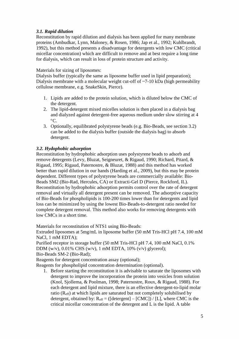
3.1. Rapid dilution
Reconstitution by rapid dilution and dialysis has been applied for many membrane
proteins (Ambudkar, Lynn, Maloney, & Rosen, 1986; Jap et al., 1992; Kuhlbrandt,
1992), but this method presents a disadvantage for detergents with low CMC (critical
micellar concentration) which are difficult to remove and at best require a long time
for dialysis, which can result in loss of protein structure and activity.
Materials for sizing of liposomes:
Dialysis buffer (typically the same as liposome buffer used in lipid preparation);
Dialysis membrane with a molecular weight cut-off of ~7-10 kDa (high permeability
cellulose membrane, e.g. SnakeSkin, Pierce).
1. Lipids are added to the protein solution, which is diluted below the CMC of
the detergent.
2. The lipid-detergent mixed micelles solution is then placed in a dialysis bag
and dialyzed against detergent-free aqueous medium under slow stirring at 4
C.
3. Optionally, equilibrated polystyrene beads (e.g. Bio-Beads, see section 3.2)
can be added to the dialysis buffer (outside the dialysis bag) to absorb
detergent.
3.2. Hydophobic adsorption
Reconstitution by hydrophobic adsorption uses polystyrene beads to adsorb and
remove detergents (Levy, Bluzat, Seigneuret, & Rigaud, 1990; Richard, Pitard, &
Rigaud, 1995; Rigaud, Paternostre, & Bluzat, 1988) and this method has worked
better than rapid dilution in our hands (Harding et al., 2009), but this may be protein
dependent. Different types of polystyrene beads are commercially available: Bio-
Beads SM2 (Bio-Rad, Hercules, CA) or Extracti-Gel D (Pierce, Rockford, IL).
Reconstitution by hydrophobic adsorption permits control over the rate of detergent
removal and virtually all detergent present can be removed. The adsorptive capacity
of Bio-Beads for phospholipids is 100-200 times lower than for detergents and lipid
loss can be minimized by using the lowest Bio-Beads-to-detergent ratio needed for
complete detergent removal. This method also works for removing detergents with
low CMCs in a short time.
Materials for reconstitution of NTS1 using Bio-Beads:
Extruded liposomes at 5mg/mL in liposome buffer (50 mM Tris-HCl pH 7.4, 100 mM
NaCl, 1 mM EDTA); Purified receptor in storage buffer (50 mM Tris-HCl pH 7.4, 100 mM NaCl, 0.1%
DDM (w/v), 0.01% CHS (w/v), 1 mM EDTA, 10% (v/v) glycerol);
Bio-Beads SM-2 (Bio-Rad);
Reagents for detergent concentration assay (optional);
Reagents for phospholipid concentration determination (optional).
1. Before starting the reconstitution it is advisable to saturate the liposomes with
detergent to improve the incorporation the protein into vesicles from solution
(Knol, Sjollema, & Poolman, 1998; Paternostre, Roux, & Rigaud, 1988). For
each detergent and lipid mixture, there is an effective detergent-to-lipid molar
ratio (Reff) at which lipids are saturated but not completely solubilised by
detergent, obtained by: Reff = ([detergent] – [CMC]) / [L], where CMC is the
critical micellar concentration of the detergent and L is the lipid. A table
6
listing these parameters for the most common detergents can be found in
(Rigaud & Levy, 2003). For DDM, Rsat is 1:1 (mol/mol) and Rsol is 1:1.6
(mol/mol).
2. Based on the detergent-to-lipid molar ratio, an appropriate amount of
detergent is added to lipid solution.
3. If using DDM, incubate the mixture at room temperature for 3 h with stirring
to ensure complete equilibration of the detergent with lipids.
4. Following saturation and before the addition of the protein solution, the lipid-
detergent mixture is bath sonicated for 10 minutes at room temperature. Bio-
Beads are washed several times with methanol following several washes with
water and can be stored at 4 oC in water.
5. Prior to use, Bio-Beads are washed in liposome buffer. The receptor is added
to the lipid-detergent solution at an appropriate concentration to achieved the
desired lipid-to-protein ratio.
6. The reaction mixture is incubated for ~1 h with gentle mixing before the
addition of Bio-Beads. If the detergents used to solubilise the lipids are
deleterious to the activity of the protein, this incubation period may be
shortened.
7. Washed Bio-Beads are added to the protein-lipid-detergent solution at a ratio
of 10:1 (w/w) of wet Bio-Beads to detergent.
8. The sample is incubated above the phase transition temperature of the lipids
for 1-2 h and then aspirated into fresh Bio-Beads and incubated overnight with
rotation.
9. Turbidity measurements can be used to monitor reconstitution and a detergent
assay can be performed to confirm complete detergent removal (within the
sensitivity limit of the assay) (Mallya & Pattabiraman, 1997).
10. Proteoliposomes can be recovered by centrifugation (100,000g, 1-3 h, 4 C)
and resuspended in an appropriate volume of liposome buffer depending on
end use.
11. If required, proteoliposomes can be isolated from empty liposomes using
sucrose density gradient centrifugation (0-35% in liposome buffer) in 5 %
steps for a typical receptor (MW ~40-100 KDa) at medium high lipid-to-
protein molar ratios (> 500 to 1). For denser liposomes higher percentages of
sucrose may be required.
12. The sample is loaded on top of the gradient and centrifuged (100,000g, 16 h,
4 C).
13. The gradient is then fractionated, and fraction can be analysed by SDS-PAGE.
14. Proteoliposomes fractions may be dialyzed or diluted and centrifuged
(100,000g, 1-3 h, 4 C) to remove sucrose.
4. Reconstitution into nanodiscs
Nanodiscs consist of a lipid bilayer disc stabilised by an amphiphilic so-called
membrane scaffold protein (MSP, an engineered construct of the human
apolipoprotein A-1). A dimer of MSP wraps around a lipid bilayer forming a disc
with a diameter of 10 nm or larger depending on the length of the MSP construct
used; several constructs are available containing multiples of the same repeat domain,
allowing formation of larger discs, to suit the incorporation of membrane proteins of
varying sizes (Denisov, Grinkova, Lazarides, & Sligar, 2004). Nanodiscs can in
7
principle be made with any bilayer forming lipids to suit individual protein
requirements, although in practice some lipids yield better results than others, and
reconstitution conditions need to be determined empirically for each membrane
protein and lipid mix (Ritchie et al., 2009).The use of nanodiscs for reconstitution of
membrane proteins has become an increasingly popular and useful alternative to
liposomes, not least because they are homogenously-sized, soluble while still
maintaining the protein in a membrane environment, and relatively rapid to prepare
(Bayburt, Grinkova, & Sligar, 2002; Denisov et al., 2004; Shih, Arkhipov,
Freddolino, Sligar, & Schulten, 2007). Furthermore, the protein is accessible from
both sides of the membrane, making nanodiscs highly suitable for simultaneously
studying intracellular and extracellular membrane protein interactions in vitro using
techniques such as microscale thermophoresis or surface plasmon resonance. In our
hands, preparation of NTS1-loaded and empty nanodiscs follows the protocol
developed by Sligar and co-workers (Ritchie et al., 2009) with some modifications
based on the lipid types used and empirically determined optimal MSP:lipid:NTS1
ratios, with reference to (Bayburt, Grinkova, & Sligar, 2006; Inagaki et al., 2012;
Leitz, Bayburt, Barnakov, Springer, & Sligar, 2006). A schematic of the method is
illustrated in Figure 2.
4.1. Production of MSP for nanodiscs
The MSP belt protein is commercially available from Sigma-Aldrich, or can be
expressed in house as described by Ritchie et al. (Ritchie et al., 2009). The use of a
fermenter can be replaced with standard bacterial expression in 2L flasks. If the
scaffold protein is to be used without its His-tag, it can be incubated with TEV
protease overnight at 4 C at a 1:10 TEV:MSP molar ratio, and then reverse purified
by collecting the flow-through of a Ni2+ column.
4.2. NTS1 reconstitution into nanodiscs
Materials:
Purified MSP
Purified receptor in storage buffer (50mM Tris-HCl pH 7.4, 100mM NaCl, 0.1%
DDM (w/v), 0.01% CHS(w/v), 1mM EDTA, 10% (v/v) glycerol)
Lipids (purchased from Avanti Polar Lipids, Inc.)
Lipid sodium cholate buffer (50 mM Tris-HCl pH 7.4, 100 mM sodium cholate, 100
mM NaCl, 1 mM EDTA)
Bio-Beads SM-2 (Bio-Rad), prepared as described (section 3.2)
1. A lipid films is prepared made as in section 2.1, using the desired lipid
mixture, e.g. 3:1:1 POPC:POPG:POPE with 25 mol % cholesterol; 1:1
POPC:POPG; 3:1 POPC:POPE or 1.07:1.5:1 BPL:POPC:POPG.
2. At least 8-10 mol lipid per batch of discs is made up to 50 mM in lipid
sodium cholate buffer..
3. The lipid suspension is freeze-thawed with liquid nitrogen three times.
4. Detergent-solubilised lipids can be stored overnight at 4 C, or snap-frozen in
liquid nitrogen and stored at -80 C.
5. The following guidelines are used for successful nanodisc formation: the final
[lipid] must be 4 mM, final [sodium cholate] must be 12-40 mM, final
[glycerol], must be less than 4 %.
8
6. MSP-to-lipid ratios of 1:50 to 1:60 are most effective for NTS1. Empty
nanodiscs are reconstituted with 5 additional lipid molecules per MSP, and
are prepared simultaneously with loaded nanodiscs. Ratios of between? 80:1
and 50:1 MSP:NTS1 are effective.
7. NTS1 (2-5 nmol at ~0.2-0.5 mg/ml), or NTS1 buffer alone (for empty
nanodiscs) is added to sodium cholate-solubilised lipid mixtures.
8. MSP1D1 (4-10 mg, or 0.16-0.25 mol at ~4.5 mg/ml) is added in a final
volume of 2.5-3.5 ml, with final concentrations of receptor at approximately 1
M, MSP at 1.6-3.5 mg/ml (70-170 M) and lipid at 7 mM.
9. The solution is incubated for 1 h with rotation at 4-6 rpm at 4 °C.
10. Washed Bio-Beads (0.8-1.0 g/ml) are added to the solution and rotation
continues overnight at 4 °C.
11. Bio-Beads are then replaced (0.5 g/ml) and the sample is further incubated for
1 h.
12. Bio-Beads are removed and the sample is concentrated (using e.g. Vivaspin 6,
10,000 MWCO, Satorius) followed by centrifugation (10,000g, 4 C, 10
min).
13. The supernatant is aspirated off any pellet that forms and filtered through a
Nanosep MF low-volume 0.2 m centrifugal filter (PALL Corporation).
14. Nanodiscs are separated from other material on a (calibrated) Superdex 200
10/300 GL column (GE Healthcare) equilibrated in 50 mM Tris HCl pH 7.4,
100 mM NaCl, 5 mM MgCl2.
15. Peak fractions containing NTS1 as well as MSP are pooled and concentrated
if required.
16. NTS1 nanodiscs may be enriched by purification using an affinity tag on the
receptor, e.g. via FLAG-tag using anti-FLAG M2 resin (Sigma-Aldrich)
according to the manufacturer’s instructions, or for via a His-tag, in which
case the His-tag on MSP1D1 must be cleaved with TEV protease prior to
nanodisc formation and loaded nanodiscs may be enriched from the mixed
nanodisc population using a nickel column.
17. Nanodiscs can then extensively be dialysed at 4 C in three changes of
dialysis buffer appropriate for end use.
5. Reconstitution into lipodisqs
Lipodisqs are another discoidal nanoscale lipid bilayer system, similar to nanodiscs,
forming particles in which both sides of the inserted membrane protein remain
accessible (Orwick et al., 2012). However instead of using a protein belt, lipodisqs are
formed using a styrene-maleic acid polymer (SMA). This aspect makes them
attractive for techniques such as protein NMR where additional signals arising from
the MSP belt would be undesirable.
SMA polymers are sensitive to pH; in its charged form the polymer is extended, but
as pH is lowered and charge is lost it collapses into a compact coil. At the critical
collapsing pH lipodisqs will form most efficiently. SMA is available in a variety of
styrene-to-maleic acid ratios, influencing the charge distribution in the molecule and
thus optimal pH range at which it can be used. As the polymer length is not exactly
fixed, unlike the length of a particular MSP construct used in nanodisc formation, the
range of particle sizes obtained in lipodisq formation can be more variable (depending
on the protein and lipids used) and experimental conditions may need to be adjusted
to obtain a more homogenous sample size, in combination with careful selection of
9
sample fractions after gel filtration. The final sample size and dispersion thereof can
be verified by dynamic light-scattering experiments and/or electron microscopy.
Lipodisqs have been used in our laboratory to reconstitute bacteriorhodopsin (bR, a
7TM receptor) from its native membrane (Orwick-Rydmark et al., 2012), and single
TM proteins (unpublished). Others have used SMA to solubilise for example intact
mitochondrial complexes directly from mitochondrial membranes (Long et al., 2013),
bacterial cell division machinery directly from staphylococcal cells (Paulin et al.,
2014), or to reconstituted labelled, purified membrane proteins for spectroscopy
studies (Sahu et al., 2013). Here, we provide a protocol for lipodisq formation of bR.
5.1. Preparation of bR proteoliposomes and bR-proteolipodisqs
Materials:
Pre-hydrolysed SMA at a 3:1 ratio of styrene-to-maleic acid was used, which is
optimal for working around neutral pH (Malvern Cosmeceutics Ltd, can be purchased
from Sigma Aldrich);
400 nm extruded DMPC unilamellar vesicles in 50 mM Tris-HCl, pH 8 at 20 mg/ml
(prepared as described in 2.2.);
bR expressed and purified in its native membrane as previously described (Oesterhelt
& Stoeckenius, 1974);
Reconstitution buffer (50 mM Tris-HCl, pH 8).
1. Native bR membranes (containing ~4 mg of protein) are resuspended in 1 ml
of the DMPC-liposomes (20 mg) at a lipid-to-protein molar ratio of 172:1 and
sonicated for 30 minutes in a bath sonicator.
2. A 2.5% (w/v) SMA-polymer solution is prepared in reconstitution buffer, and
added drop-wise to the bR-DMPC suspension.
3. Lipodisq formation and kinetics thereof can be followed by monitoring light
scattering at 540 nm as a function of time (Jasco V-630 UV
spectrophotometer).
4. After incubation for 1 h at room temperature, the resulting bR lipodisq
solution is centrifuged for 30 minutes at 40,000g to remove any
unsolubilised bR.
5. Lipodisqs are further purified from non-incorporated bR by gel filtration
(Superdex 200, X16/100 column in 50 mM Tris, 300 mM NaCl, pH 8.0), and
the resulting purple fractions with an absorbance at 555 nm are pooled.
6. Analysis of the main gel filtration peaks by DLS (Viscotek 801 particle sizer,
Malvern Instruments) shows resulting bR lipodisqs to be between 10-20 nm in
diameter.
6. Determination of lipid-to-protein ratio
The lipid-to-protein (L:P) ratio of a sample can be used for estimating the receptor
density in proteoliposomes. This knowledge can be useful for assessing how
experimental conditions compare to the conditions found in vivo, or how they affect
biophysical experiments such as energy transfer in FRET, where very high densities
might lead to bystander FRET, and is essential in for example protein-to-lipid
stoichiometry experiments with spin-labelled lipids.
6.1. Liposome/lipid concentration determination A number of ways can be used to estimate the L:P ratio of proteoliposmes. If the
protein concentration can be determined reliably by absorbance measurements at 280
10
nm, this can be compared with the lipid concentration determined from a colorimetric
phosphate assay (Chen Jr et al., 1956). This assay determines the total phosphorus
content of the sample. Although it cannot directly account for cholesterol, which does
not have a phosphate headgroup, the cholesterol concentration can be calculated from
the molar ratio once the phosphorus concentration is known.
Materials for phosphate assay:
2.5 % (w/v) ammonium molybdate (VI) tetrahydrate solution;
8.9 N H2SO4;
10 % (w/v) ascorbic acid;
phosphorus standard solution;
H2O2.
1. A standard curve is constructed from triplicate samples of 0-0.228 μmol
phosphorus made up from 0.65 mM stock phosphorus solution (Sigma-
Aldrich 661-9).
2. The standard samples and triplicate samples of prepared liposomes, nanodiscs
or lipid stocks are acid-hydrolysed by addition of 0.45 ml 8.9 N H2SO4 to each
tube with heating in an heat block at 200-215 °C for 25 min.
3. The tubes are removed from the heating block and allowed to cool for five
minutes before 150 μl H2O2 is added to each tube.
4. Heating is continued above 200 °C for a further 30 min. De-ionised water (3.9
ml) is added to each tube, followed by 0.5 ml 2.5 % (w/v) ammonium
molybdate (VI) tetrahydrate solution.
5. All tubes are vortexed five times, 0.5 ml 10 % (w/v) ascorbic acid added, and
vortexed five times each again.
6. Tubes are covered with glass marbles to prevent evaporation and heated at 100
°C for 7 min.
7. Once cooled to room temperature the absorbance at 820 nm of all standards
and samples is determined. The 0 μmol phosphorus standard is used for blank-
correction and triplicate averages plotted as a function of phosphorus amount.
8. The phosphorus concentration, and thus the phospholipid concentration of the
samples are determined from the equation of a regression line fitted to the
data.
6.2. Liposome scatter in UV-Vis: Labelled lipid marker
Unfortunately, the scatter from lipid vesicles often hampers accurate determination of
the protein absorbance signal at 280 nm, especially at high sample concentrations
and/or high L:P ratios. In such a case one can include a small amount of fluorescently
labelled lipid, e.g. rhodamine-PE (Rho-PE) in the reconstituted samples (~0.8:1 Rho-
PE:protein) depending on the molar extinction coefficient of the protein, as well as
making a protein-free lipid-only background sample containing Rho-PE. The
rhodamine signal at 573 nm in both samples can then be used to correct for the lipid
scatter: the rhodamine peak from the background sample is normalised to that in the
proteoliposome sample, and the scaled background spectrum is then subtracted. The
protein concentration can be calculated from the scatter-corrected signal at 280 nm
and then used together with a phosphate assay as described above to determine the
lipid-to-protein ratio. The protein concentration can also be determined by protein
assays such as the BCA assay, or other comparisons with bovine serum albumin, but
these methods are less accurate.
11
Instead of using a phosphate assay to determine the lipid concentration, when
employing the Rho-PE method one can also simply use the Rho-PE absorption to
calculate the total lipid content by using the Rho-PE-to-total-lipid ratio employed in
the reconstitution. This of course relies on the assumption that reconstitution
efficiency of Rho-PE does not differ from the other lipids used.
6.3. Sucrose density gradient
Alternatively, a sucrose density gradient can be used to determine the L:P ratio. This
method relies on the different densities that proteoliposomes with varying protein
content will have; low L:P proteoliposomes will be heavier and thus run further down
a sucrose gradient than samples lower L:P.
1. A sucrose density gradient with appropriate concentrations of sucrose added to
liposome buffer is prepared (for NTS1 reconstituted in BPL at initial L:P
ratios of >500:1 a step gradient of 35-5% with 5% steps is typically
employed).
2. The reconstituted sample is layered on top of the gradient, and the gradients
are centrifuged overnight in a swing-out rotor (e.g. Beckmann SW28 for large
(>1.5 mL) and SW41 for smaller samples) at ~100,000g.
3. The sucrose gradient is then fractionated and the presence of reconstituted
receptor can be verified by SDS-PAGE analysis.
4. The position of the proteoliposome band on the gradient can be used to
estimate the density and thus the L:P ratio of the samples using the equation
derived by Barber (Barber, 1966):
2 2 2 2
1 2 3 4 5 6 7 8 9 T B B T B T B B T B T Y B B T B T Y
where is the density (kg/dm3), T the temperature (°C), Y the fraction of sucrose in
solution and 1 9B are constants( 1 1.00037B , 5
2 3.96805 10 B ,
6
3 5.85133 10 B , 4 0.389824B , 3
5 1.05789 10 B , 5
6 1.23928 10 B ,
7 0.1700976B , 4
8 4.75301 10 B , and 6
9 8.92397 10 B ).
5. The partial specific volume ( v in mL/g) follows from that (as the reciprocal
of the density), and can be used to estimate the L:P ratio (w/w) by solving
(1 )proteoliposomes lipid proteinx xv v v
for x (fraction of lipid), where proteoliposomesv is the partial specific volume of
the sample determined from the sucrose gradient, lipidv is the contribution of
the lipid and can be determined by running lipid-only sample on a sucrose
gradient (~0.986 mL/g for BPL was determined in our laboratory), and
proteinv is the contribution of the protein and can be calculated from its amino
acid composition using the relation derived by Cohn and Edsall (Cohn &
Edsall, 1943) modified for the effect of temperature (Durchschlag, 1986)
12
4 4
25 4.25 10 25 4.25 10 25i i i
T
i i
n MT T
n M
vv v
where Tv is the partial specific volume at temperature T , and 25v that at 25
°C, calculated from the number of moles ( in ) for each particular amino acid (
i ) with molecular weight iM and partial specific volume iv .
6. The fraction of lipid x in w/w can then be used to calculate a molar L:P ratio
using the molecular weight of the lipid and protein.
7. Concentration methods
Different experimental methods require more or less concentrated protein samples.
Proteoliposome samples can be concentrated very easily to high protein
concentrations by pelleting the liposomes via high-speed centrifugation (~100,000g)
followed by resuspension in small volumes of liposome buffer to yield the final
concentration desired.
If high concentration/small sample volume are critical (e.g. for NMR or EPR
application) or high sample homogeneity is required, it is advisable to run the samples
on a preparative sucrose density gradient (as described in section 6.3), to separate
loaded proteoliposomes from empty liposomes which may be present to varying
extent, depending on the method of reconstitution and initial L:P ratio used, which
can also be used to estimate the final L:P ratio as discussed in section 6.
Discoidal reconstitutions (nanodiscs or lipodisqs) can be concentrated by the same
methods available for membrane proteins solubilised in detergents, such as centrifugal
concentrators (e.g. Vivaspin concentrators, Satorius) or by affinity chromatography
with small resin volumes.
Summary
For many studies of membrane proteins it is essential that they are present in a lipid
bilayer. In this chapter we have detailed methods for incorporating membrane proteins
into liposomes and also discoidal lipid structures such as nanodsics and liposdisqs.
Whilst the choice of lipid environment is largely dependent upon the downstream
application, the importance of investigating membrane proteins in their native
environments is clear.
References
Albert, A. D., Yeagle, P. L., Whiteway, C., Watts, A., & Epand, R. (1994). Effect of
cholesterol on retinal rod outer segment disk membranes. Paper presented at
the Investigative Ophthalmology & Visual Science.
Ambudkar, S. V., Lynn, A. R., Maloney, P. C., & Rosen, B. P. (1986). Reconstitution
of ATP-dependent calcium transport from streptococci. Journal of Biological
Chemistry, 261(33), 15596-15600.
Baneres, J. L., & Parello, J. (2003). Structure-based analysis of GPCR function:
evidence for a novel pentameric assembly between the dimeric leukotriene B4
receptor BLT1 and the G-protein. J Mol Biol, 329(4), 815-829.
13
Barber, E J. (1966). Calculation of density and viscosity of sucrose solutions as a
function of concentration and temperature. National Cancer Institute
monograph, 21, 219.
Bayburt, T. H., Grinkova, Y. V., & Sligar, S. G. (2002). Self-assembly of discoidal
phospholipid bilayer nanoparticles with membrane scaffold proteins. Nano
Lett, 2(8), 853-856.
Bayburt, T. H., Grinkova, Y. V., & Sligar, S. G. (2006). Assembly of single
bacteriorhodopsin trimers in bilayer nanodiscs. Arch Biochem Biophys,
450(2), 215-222.
Chen Jr, P. S., Toribara, T. Y., & Warner, Huber. (1956). Microdetermination of
phosphorus. Analytical chemistry, 28(11), 1756-1758.
Cohn, E J, & Edsall, J T. (1943). Proteins, Amino Acids and Peptides as Ions and
Dipolar Ions. Reinhold, New York.
Denisov, I. G., Grinkova, Y. V., Lazarides, A. A., & Sligar, S. G. (2004). Directed
self-assembly of monodisperse phospholipid bilayer Nanodiscs with
controlled size. J Am Chem Soc, 126(11), 3477-3487.
Specific volumes of biological macromolecules and some other molecules of
biological interest 45-128 (Springer 1986).
Harding, P. J., Attrill, H., Boehringer, J., Ross, S., Wadhams, G. H., Smith, E. (2009).
Constitutive dimerization of the G-protein coupled receptor, neurotensin
receptor 1, reconstituted into phospholipid bilayers. Biophys J, 96(3), 964-973.
Inagaki, S., Ghirlando, R., White, J. F., Gvozdenovic-Jeremic, J., Northup, J. K., &
Grisshammer, R. (2012). Modulation of the interaction between neurotensin
receptor NTS1 and Gq protein by lipid. J Mol Biol, 417(1-2), 95-111.
Ishihara, G., Goto, M., Saeki, M., Ito, K., Hori, T., Kigawa, T. (2005). Expression of
G protein coupled receptors in a cell-free translational system using detergents
and thioredoxin-fusion vectors. Protein Expr Purif, 41(1), 27-37.
Jackson, Marilyn L., & Litman, Burton J. (1982). Rhodopsin-phospholipid
reconstitution by dialysis removal of octyl glucoside. Biochemistry, 21(22),
5601-5608.
Jap, B. K., Zulauf, M., Scheybani, T., Hefti, A., Baumeister, W., Aebi, U. (1992). 2D
crystallization: from art to science. Ultramicroscopy, 46(1-4), 45-84.
Kiefer, H., Maier, K., & Vogel, R. (1999). Refolding of G-protein-coupled receptors
from inclusion bodies produced in Escherichia coli. Biochem Soc Trans, 27(6),
908-912.
Klammt, C., Schwarz, D., Eifler, N., Engel, A., Piehler, J., Haase, W. (2007). Cell-
free production of G protein-coupled receptors for functional and structural
studies. J Struct Biol, 158(3), 482-493.
Knol, J., Sjollema, K., & Poolman, B. (1998). Detergent-mediated reconstitution of
membrane proteins. Biochemistry, 37(46), 16410-16415.
Kuhlbrandt, W. (1992). Two-dimensional crystallization of membrane proteins. Q
Rev Biophys, 25(1), 1-49.
Leitz, A. J., Bayburt, T. H., Barnakov, A. N., Springer, B. A., & Sligar, S. G. (2006).
Functional reconstitution of Beta2-adrenergic receptors utilizing self-
assembling Nanodisc technology. Biotechniques, 40(5), 601-602, 604, 606,
passim.
Levy, D., Bluzat, A., Seigneuret, M., & Rigaud, J. L. (1990). A systematic study of
liposome and proteoliposome reconstitution involving Bio-Bead-mediated
Triton X-100 removal. Biochim Biophys Acta, 1025(2), 179-190.
14
Link, A. J., Skretas, G., Strauch, E. M., Chari, N. S., & Georgiou, G. (2008). Efficient
production of membrane-integrated and detergent-soluble G protein-coupled
receptors in Escherichia coli. Protein Sci, 17(10), 1857-1863.
Long, Ashley R, O'Brien, Catherine C, Malhotra, Ketan, Schwall, Christine T, Albert,
Arlene D, Watts, Anthony. (2013). A detergent-free strategy for the
reconstitution of active enzyme complexes from native biological membranes
into nanoscale discs. BMC biotechnology, 13, 41.
Mallya, H. Madhukar, & Pattabiraman, T. N. (1997). Effect of Acid Concentration on
Chromogen Formation from Hexoses in Sulfuric Acid-Based Reactions.
Analytical Biochemistry, 251(2), 299-301.
Marsh, Derek. (2013). Handbook of lipid bilayers (Second Edition ed.): CRC Press.
Oesterhelt, D., & Stoeckenius, W. (1974). Isolation of the cell membrane of
Halobacterium halobium and its fractionation into red and purple membrane.
Methods Enzymol, 31, 667-678.
Orwick-Rydmark, M., Lovett, J. E., Graziadei, A., Lindholm, L., Hicks, M. R., &
Watts, A. (2012). Detergent-free incorporation of a seven-transmembrane
receptor protein into nanosized bilayer Lipodisq particles for functional and
biophysical studies. Nano Lett, 12(9), 4687-4692.
Orwick, M. C., Judge, P. J., Procek, J., Lindholm, L., Graziadei, A., Engel, A. (2012).
Detergent-free formation and physicochemical characterization of nanosized
lipid-polymer complexes: Lipodisq. Angew Chem Int Ed Engl, 51(19), 4653-
4657.
Paternostre, M. T., Roux, M., & Rigaud, J. L. (1988). Mechanisms of membrane
protein insertion into liposomes during reconstitution procedures involving the
use of detergents. 1. Solubilization of large unilamellar liposomes (prepared
by reverse-phase evaporation) by triton X-100, octyl glucoside, and sodium
cholate. Biochemistry, 27(8), 2668-2677.
Paulin, Sarah, Jamshad, Mohammed, Dafforn, Timothy R, Garcia-Lara, Jorge, Foster,
Simon J, Galley, Nicola F. (2014). Surfactant-free purification of membrane
protein complexes from bacteria: application to the staphylococcal penicillin-
binding protein complex PBP2/PBP2a. Nanotechnology, 25, 285101.
Richard, P., Pitard, B., & Rigaud, J. L. (1995). ATP synthesis by the F0F1-ATPase
from the thermophilic Bacillus PS3 co-reconstituted with bacteriorhodopsin
into liposomes. Evidence for stimulation of ATP synthesis by ATP bound to a
noncatalytic binding site. J Biol Chem, 270(37), 21571-21578.
Rigaud, J. L., & Levy, D. (2003). Reconstitution of membrane proteins into
liposomes. Methods Enzymol, 372, 65-86.
Rigaud, J. L., Paternostre, M. T., & Bluzat, A. (1988). Mechanisms of membrane
protein insertion into liposomes during reconstitution procedures involving the
use of detergents. 2. Incorporation of the light-driven proton pump
bacteriorhodopsin. Biochemistry, 27(8), 2677-2688.
Ritchie, T. K., Grinkova, Y. V., Bayburt, T. H., Denisov, I. G., Zolnerciks, J. K.,
Atkins, W. M. (2009). Reconstitution of membrane proteins in phospholipid
bilayer nanodiscs. Methods Enzymol, 464, 211-231.
Sahu, Indra D, McCarrick, Robert M, Troxel, Kaylee R, Zhang, Rongfu, Smith,
Hubbell J, Dunagan, Megan M. (2013). DEER EPR measurements for
membrane protein structures via bifunctional spin labels and lipodisq
nanoparticles. Biochemistry, 52, 6627-6632.

15
Shih, A. Y., Arkhipov, A., Freddolino, P. L., Sligar, S. G., & Schulten, K. (2007).
Assembly of lipids and proteins into lipoprotein particles. J Phys Chem B,
111(38), 11095-11104.
Skipski, V. P., Peterson, R. F., & Barclay, M. (1964). Quantitative analysis of
phospholipids by thin-layer chromatography. Biochem J, 90(2), 374-378.
Spooner, Paul J. R., Sharples, Jonathan M., Goodall, Scott C., Bovee-Geurts, Petra H.
M., Verhoeven, Michiel A., Lugtenburg, Johan. (2004). The ring of the
rhodopsin chromophore in a hydrophobic activation switch within the binding
pocket. J Mol Biol, 343(3), 719-730.
Weiss, H. M., & Grisshammer, R. (2002). Purification and characterization of the
human adenosine A(2a) receptor functionally expressed in Escherichia coli.
Eur J Biochem, 269(1), 82-92.
16
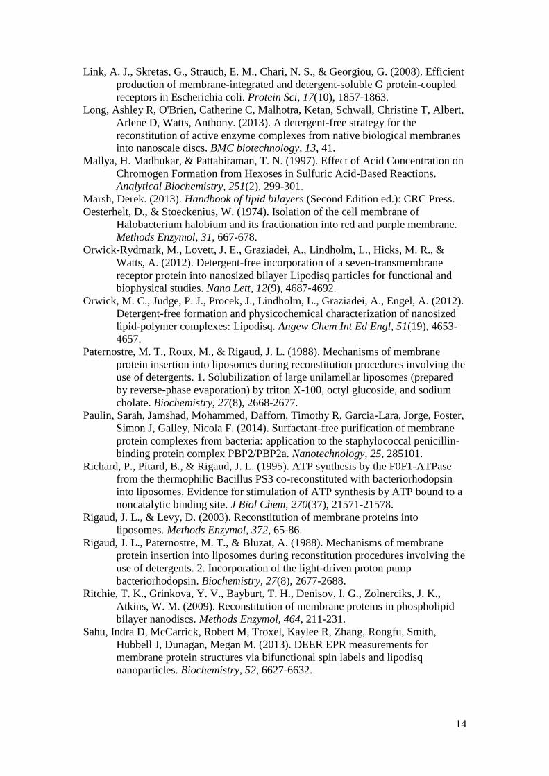
Figure 1: Reconstitution by dilution and dialysis (A) and by using Bio-Beads for
detergent removal (B). (A): Lipids, detergent, and protein are mixed at the desired
ratios and incubated for 1 h, after which the reconstitution mixture is rapidly diluted
below the CMC of the detergent and the detergent monomers are removed by
extensive dialysis. (B): Detergent-solubilised receptor is mixed with liposomes
desestabilised by detergent at the required concentration (incubation of mixture for 1
h), after which detergent is removed with Bio-Beads at a 10:1 Bio-Beads-to-detergent
ratio (w/w).
17
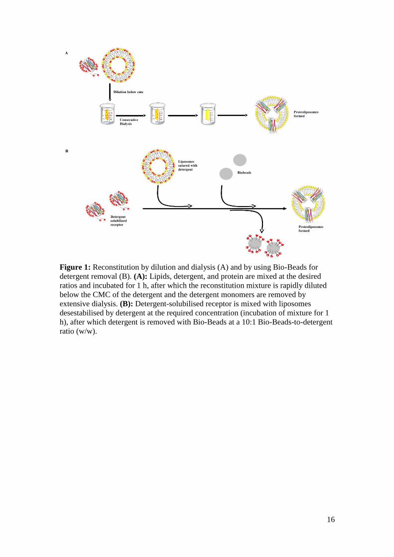
Figure 2: Schematic diagram illustrating nanodisc formation. A lipid film is
solubilised in 100 mM sodium cholate to a final concentration of 50 mM lipid.
Detergent-solubilised, purified NTS1 (cyan ribbon structure surrounded by blue
detergent molecules) is added to the lipid-detergent mixture at the target concentration
(lipid molecules are orange with gold rectangles representing cholesterol). Membrane
scaffold protein is added to the reaction mixture at the target MSP:lipid and
MSP:NTS1 ratio, and the sample is incubated with rotation at 4 C for 1 h. Bio-Beads
(1g/ml) are added to remove the detergent overnight, and nanodiscs spontaneously
form.
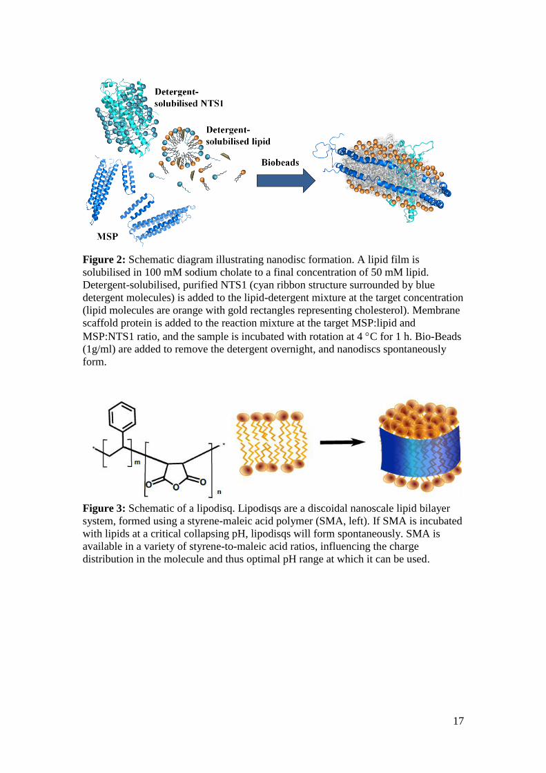
Figure 3: Schematic of a lipodisq. Lipodisqs are a discoidal nanoscale lipid bilayer
system, formed using a styrene-maleic acid polymer (SMA, left). If SMA is incubated
with lipids at a critical collapsing pH, lipodisqs will form spontaneously. SMA is
available in a variety of styrene-to-maleic acid ratios, influencing the charge
distribution in the molecule and thus optimal pH range at which it can be used.


