Hasegawa & Urrea-Mendoza, Cogent Medicine (2017), 4: 1331601https://doi.org/10.1080/2331205X.2017.1331601
NEUROLOGY | CASE REPORT
Prognosis of post-cardiac-arrest anoxic encephalopathy using felbamate: A case reportHisanori Hasegawa1,2,3* and Enrique Urrea-Mendoza1
Abstract: Reliable prognostic methods for cerebral functional outcome of post cardiac-arrest anoxic encephalopathy (AAE) are necessary. Evolution of the Electroencephalogram (EEG) background pattern is a robust predictor of poor or good outcome of patients in this condition. During the first 24 h a rapid recovery toward continuous patterns is associated with a good neurological outcome, but a lack of improvement in this time window represents the opposite, although epilep-tiform patterns are of unknown significance and effects of treatment with anti-epileptic drugs are indistinct. Generalized periodic epileptiform discharges (GPEDs) and bilateral independent periodic lateralized epileptiform discharges (BIPLEDs) after a severe hypoxemia carried a poor prognosis for survival. Treatment of AAE is symptomatic. Barbiturate coma was historically applied to decrease cerebral metabolic rate of O2 but the clinical benefit was insignificant and minimally af-fected the grave outcomes. Whether or not treatment of electrographic status epilepticus improves outcome is still under review in the randomized multicenter Treatment of Electroencephalographic STatus epilepticus After cardiopulmonary Resuscitation (TELSTAR) trial (NCT02056236), and no clear data of a standard man-agement has been published. This is a report of cases whose EEG showed severe epileptic abnormality due to AAE and demonstrated remarkable EEG improvement by compassionate use of felbamate.
*Corresponding author: Hisanori Hasegawa, Greenville Health System, Greenville, SC, USA; Saginaw VA Medical Center, 1500 Weiss St. Saginaw, MI 48602, USA; Bronson Methodist Hospital, Kalamazoo, MI, USA Email: [email protected]
Reviewing editor:Udo Schumacher, University Medical Center Hamburg-Eppendorf, Germany
Additional information is available at the end of the article
ABOUT THE AUTHORSHisanori Hasegawa, MD Neurologist, Clinical Epileptologist. Diplomate of ABPN with Epilepsy subspecialty. Learned under Frank Morrell in Rush Epilepsy Center. My primary interest is treatment of chronic seizure and EEG. Clinical research experiences include high-resolution EEG, the secondary epileptogenesis.
Enrique Urrea-Mendoza, MD Neurologist, Fellow in Movement Disorders. Certificate in Clinical and Translational Research. My primary interest is brain oscillations and neural rhythms in movement disorders and epilepsy. Clinical research experiences include high frequency Deep Brain Stimulation and neural rhythms in Parkinson’s Disease and other movement disorders.
PUBLIC INTEREST STATEMENTWhen the patients are comatose after resuscitation is referred as acute-anoxic encephalopathy (AAE) and this condition is a devastating complication, having high mortality rate. It is mostly fatal if electroencephalogram showed no evidence of improvement after 24 h. Treatment of AAE is symptomatic. Barbiturate coma was historically applied to decrease metabolic rate of O2 but the clinical benefit was insignificant and minimally affected the grave outcomes. This article describes three cases that anoxic encephalopathy reversed by compassionate application of felbamate, and describe the response based on EEG findings. The findings suggest the clinical benefit of felbamate to the devastating situation which could cause grave prognosis otherwise. The potential utilization of felbamate is suggested although their efficacy and success rate was not discussed because this is compassionate use.
Received: 03 March 2017Accepted: 15 May 2017First Published: 25 May 2017
© 2017 The Author(s). This open access article is distributed under a Creative Commons Attribution (CC-BY) 4.0 license.
Page 1 of 10
Page 2 of 10
Hasegawa & Urrea-Mendoza, Cogent Medicine (2017), 4: 1331601https://doi.org/10.1080/2331205X.2017.1331601
Subjects: Epilepsy; Neurological Rehabilitation; Cardiology; Emergency Medicine
Keywords: felbamate; acute-anoxic encephalopathy; EEG monitoring; cardiac arrest; prognosis
1. IntroductionAbout 450,000 Americans have cardiac arrest annually, and about 80% of cardiac arrests occur at home with a high rate of mortality (Callans, 2004). The presence of coma after resuscitation, termed acute-anoxic encephalopathy (AAE), is a devastating complication. The incidence of AAE is 300,000 per year in USA and has increased as the success rate of cardiac resuscitation has improved in the past two decades (Geocadin, Koenig, Jia, Stevens, & Peberdy, 2008). Treatment of AAE is sympto-matic. Barbiturate coma was historically applied to decrease cerebral metabolic rate of O2 but the clinical benefit was insignificant and minimally affected the grave outcomes (Kassell, Hitchon, & Gerk, 1980). Hypothermia was introduced to treat acute phase of AAE for mild improvement of clini-cal outcome (Lafferty, Keykhah, Shapiro, Horn, & Behar, 1978). However, if a patient remains unre-sponsive and unreactive to painful stimulation for 48 h after the cardiac arrest, the prognosis is still poor regardless of hypothermia (Wijdicks, Hijdra, Young, Bassetti, & Wiebe, 2006). Electroencephalogram (EEG) may be utilized in intensive care unit (ICU) to predict the outcome and has been described some findings with clinical outcomes following after cardiac arrest. Scollo-Lavizzari and Bassetti (1987), classified the EEG in terms of increasing severity in five categories. Grade I EEG (normal alpha with theta-delta activity) being a normal pattern with an excellent prog-nosis. Grade II (dominant theta-delta activity with detectable normal alpha) and grade III (domi-nant theta-delta activity without detectable normal alpha), patterns with variable prognosis. Grade IV (low-voltage delta, possibly with short isoelectric intervals); dominant, monomorphic, non-reac-tive alpha-activity (alpha coma); periodic generalized phenomena (spikes, sharp waves, slow waves with very low background activity), and grade V (very flat to isoelectric EEG) patterns related with an ominous prognosis.
Others authors agreed that patterns such as generalized periodic epileptiform discharges (GPEDS), suppression-burst pattern, and subclinical electrographic seizure pattern suggest a very serious prognosis (Husain, Mebust, & Raatke, 1999; San-juan, Chiappa, & Costello, 2009; Wijdicks et al., 2006). Anticonvulsants are used for the treatment of seizures when they occur. However, Wijdicks et al. (2006), pointed out that the seizures in AAE are resistant to antiepileptic drugs (AEDs), most of which are either gamma aminobutyric acid (GABA) enhancers or sodium channel blockers (Wasterlain, Baldwin, Suchomelova, et al., 2011). Although AAE process is enhanced by cascading over-excitation of N-methyl-D-aspartate (NMDA) glutamatergic receptors, NMDA glutamate receptor blockers are rarely used.
The main mechanism of action of felbamate (FBM) includes inhibition of NMDA receptor responses and action on GABA receptors. It inhibits NMDA subtype of glutamate receptor (Kwan, Sills, & Brodie, 2001). This medication was approved by the Food and Drug Administration (FDA) in 1993, but after a good initial therapeutic response in patients with partial seizures and Lennox Gastaut syndrome, in September 1994 the FDA issued a warning of a high incidence of aplastic anemia and hepatic failure among patients with this treatment and this medication was forgotten (French, Smith, Faught, & Brown, 1999).
Page 3 of 10
Hasegawa & Urrea-Mendoza, Cogent Medicine (2017), 4: 1331601https://doi.org/10.1080/2331205X.2017.1331601
2. PurposeAlthough this is a difficult position and after have balanced the risk and benefit ratio in different scenarios, we report three cases of post-cardiac arrest AAE that were treated with adjunctive FBM and describe the response based on EEG findings on a compassionate basis.
3. CasesThe following three cases of AAE after cardiac arrest were encountered by in-hospital neurological consultations between 24 and 48 h after cardiac arrest. In all cases, the patients were already in the rewarming phase of hypothermia. These cases were encountered in Bronson Methodist Medical Center in Kalamazoo, MI during 2011. FBM was administered only after the family received a full explanation of the severe nature of the disease and poor prognosis of AAE and understood the risks and benefits of individual medications. The specific risks and adverse effects of FBM were explained; an informed consent was signed and documented in the individual chart prior to the administration of the medication. Because of the risk of aplastic anemia and hepatic failure, complete blood count (CBC) and liver function test (LFTs) were monitored. None of the patients were in the high-risk group for development of aplastic anemia (a history of cytopenia, autoimmune disorder, or a positive anti-nuclear antibody titer) (Wasterlain et al., 2011). The family recognized the risks of FBM treatment but agreed that the anticipated neurological consequence from anoxic brain injury carried greater mor-bidity and mortality than the infrequent severe side effects of FBM.
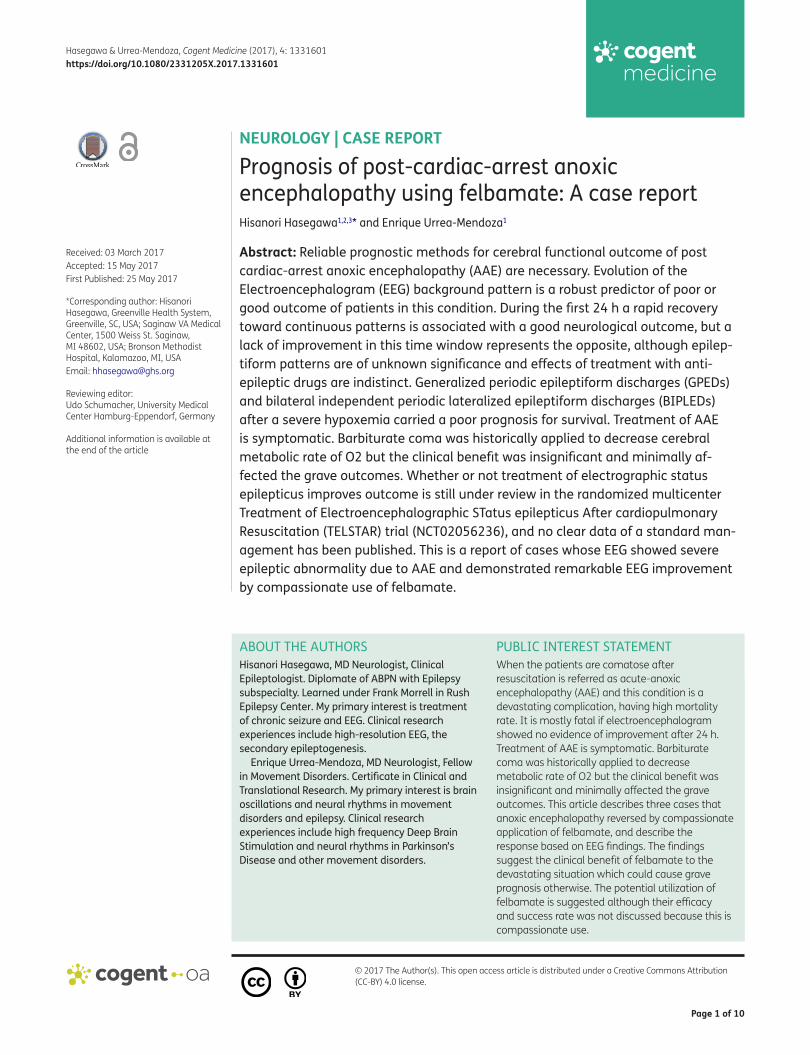
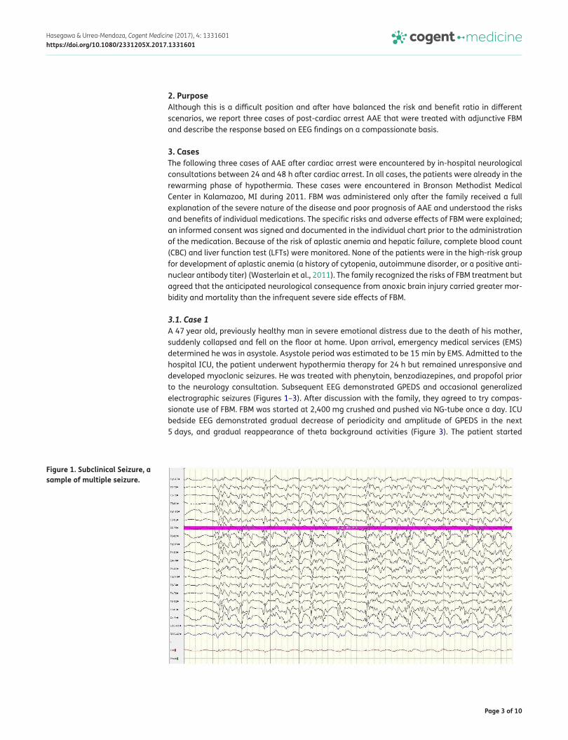
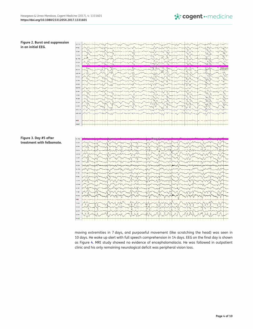
3.1. Case 1A 47 year old, previously healthy man in severe emotional distress due to the death of his mother, suddenly collapsed and fell on the floor at home. Upon arrival, emergency medical services (EMS) determined he was in asystole. Asystole period was estimated to be 15 min by EMS. Admitted to the hospital ICU, the patient underwent hypothermia therapy for 24 h but remained unresponsive and developed myoclonic seizures. He was treated with phenytoin, benzodiazepines, and propofol prior to the neurology consultation. Subsequent EEG demonstrated GPEDS and occasional generalized electrographic seizures (Figures 1–3). After discussion with the family, they agreed to try compas-sionate use of FBM. FBM was started at 2,400 mg crushed and pushed via NG-tube once a day. ICU bedside EEG demonstrated gradual decrease of periodicity and amplitude of GPEDS in the next 5 days, and gradual reappearance of theta background activities (Figure 3). The patient started
Figure 1. Subclinical Seizure, a sample of multiple seizure.
Page 4 of 10
Hasegawa & Urrea-Mendoza, Cogent Medicine (2017), 4: 1331601https://doi.org/10.1080/2331205X.2017.1331601
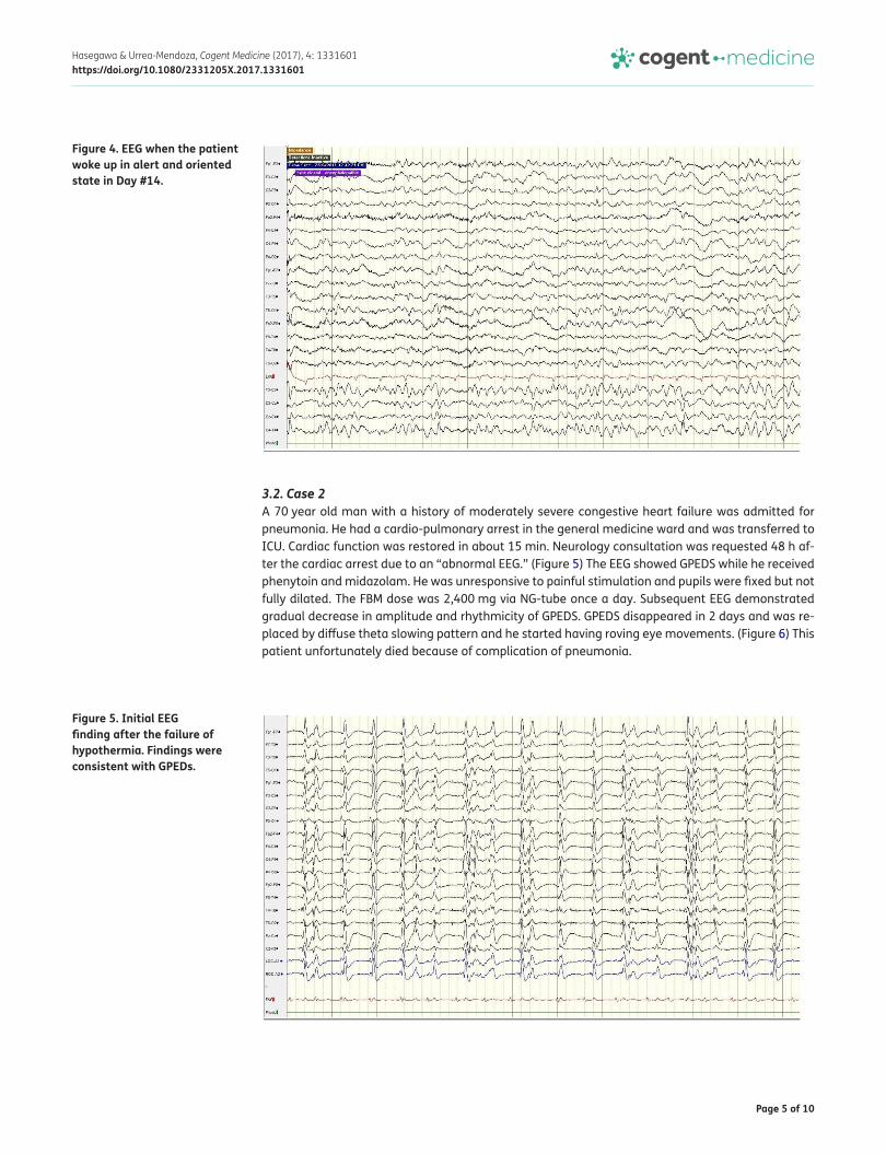
moving extremities in 7 days, and purposeful movement (like scratching the head) was seen in 10 days. He woke up alert with full speech comprehension in 14 days. EEG on the final day is shown as Figure 4. MRI study showed no evidence of encephalomalacia. He was followed in outpatient clinic and his only remaining neurological deficit was peripheral vision loss.
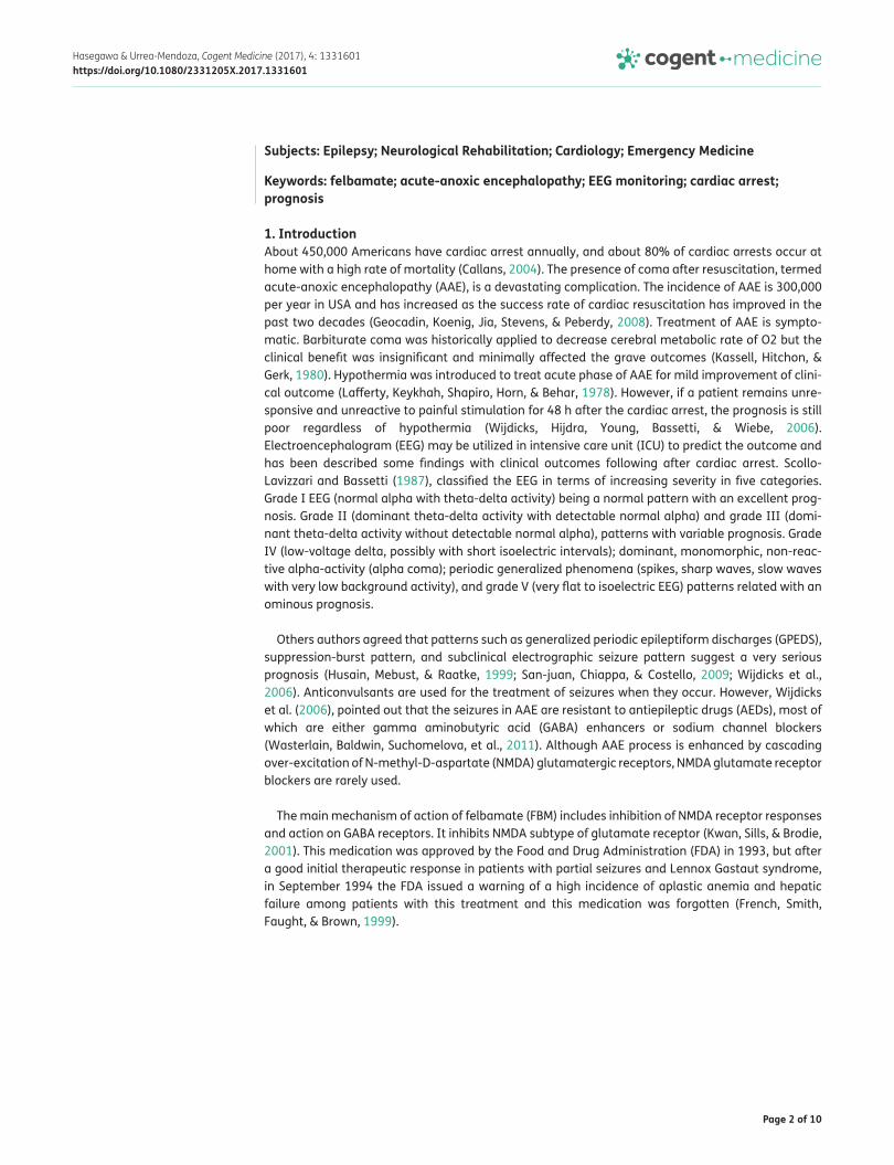
Figure 2. Burst and suppression in an initial EEG.
Figure 3. Day #5 after treatment with felbamate.
Page 5 of 10
Hasegawa & Urrea-Mendoza, Cogent Medicine (2017), 4: 1331601https://doi.org/10.1080/2331205X.2017.1331601
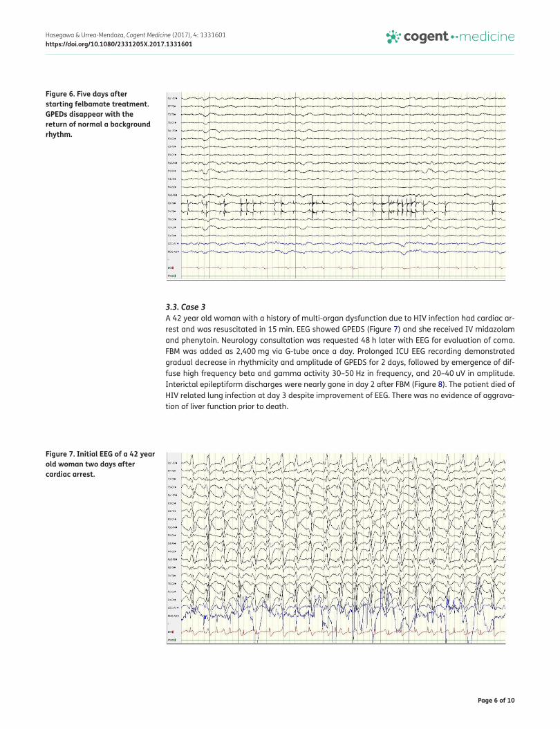
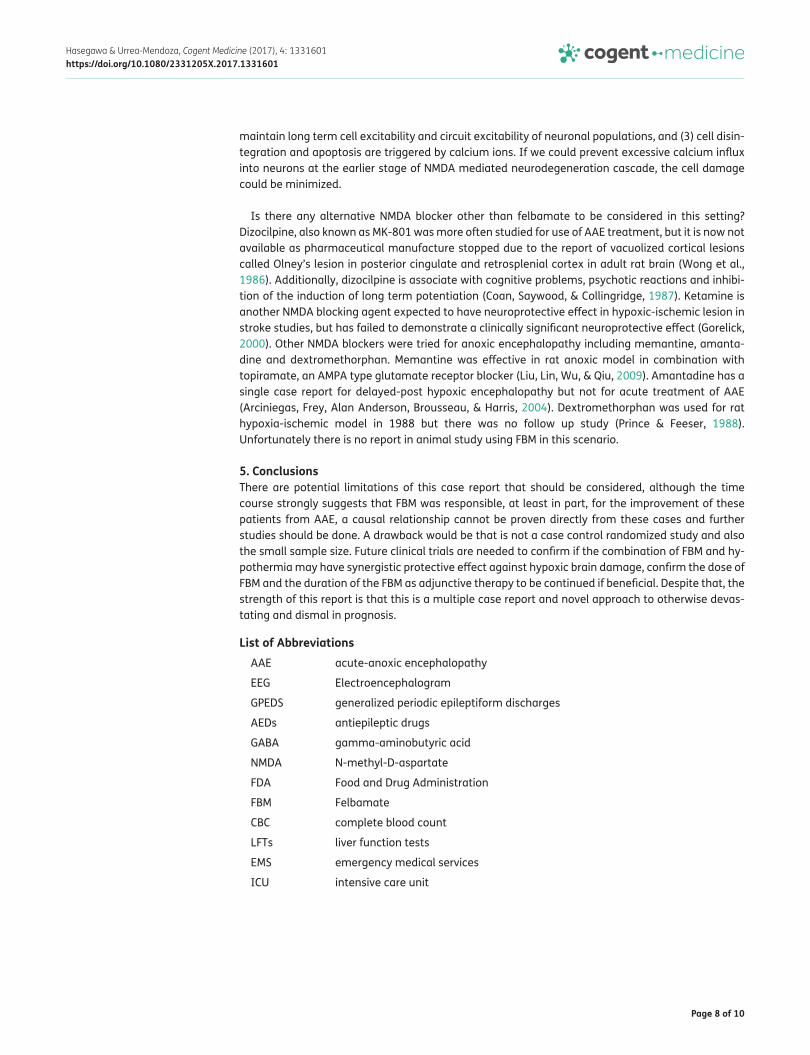
3.2. Case 2A 70 year old man with a history of moderately severe congestive heart failure was admitted for pneumonia. He had a cardio-pulmonary arrest in the general medicine ward and was transferred to ICU. Cardiac function was restored in about 15 min. Neurology consultation was requested 48 h af-ter the cardiac arrest due to an “abnormal EEG.” (Figure 5) The EEG showed GPEDS while he received phenytoin and midazolam. He was unresponsive to painful stimulation and pupils were fixed but not fully dilated. The FBM dose was 2,400 mg via NG-tube once a day. Subsequent EEG demonstrated gradual decrease in amplitude and rhythmicity of GPEDS. GPEDS disappeared in 2 days and was re-placed by diffuse theta slowing pattern and he started having roving eye movements. (Figure 6) This patient unfortunately died because of complication of pneumonia.
Figure 4. EEG when the patient woke up in alert and oriented state in Day #14.
Figure 5. Initial EEG finding after the failure of hypothermia. Findings were consistent with GPEDs.
Page 6 of 10
Hasegawa & Urrea-Mendoza, Cogent Medicine (2017), 4: 1331601https://doi.org/10.1080/2331205X.2017.1331601
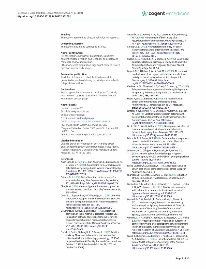
3.3. Case 3A 42 year old woman with a history of multi-organ dysfunction due to HIV infection had cardiac ar-rest and was resuscitated in 15 min. EEG showed GPEDS (Figure 7) and she received IV midazolam and phenytoin. Neurology consultation was requested 48 h later with EEG for evaluation of coma. FBM was added as 2,400 mg via G-tube once a day. Prolonged ICU EEG recording demonstrated gradual decrease in rhythmicity and amplitude of GPEDS for 2 days, followed by emergence of dif-fuse high frequency beta and gamma activity 30–50 Hz in frequency, and 20–40 uV in amplitude. Interictal epileptiform discharges were nearly gone in day 2 after FBM (Figure 8). The patient died of HIV related lung infection at day 3 despite improvement of EEG. There was no evidence of aggrava-tion of liver function prior to death.
Figure 6. Five days after starting felbamate treatment. GPEDs disappear with the return of normal a background rhythm.
Figure 7. Initial EEG of a 42 year old woman two days after cardiac arrest.

Page 7 of 10
Hasegawa & Urrea-Mendoza, Cogent Medicine (2017), 4: 1331601https://doi.org/10.1080/2331205X.2017.1331601
4. DiscussionTo our knowledge this is the first case report of severe AAE after cardiac arrest treated by compas-sionate use of FBM. FBM is the most readily available NMDA blocking anticonvulsant in this country and it is inexpensive. It is a potent noncompetitive glutamate receptor blocker of NMDA type, selec-tively bound to NR1-2B subunits (Kwan et al., 2001). FBM is a lipophilic dicarbamate having no ioniz-able chemical group moiety and is not charged at physiological pH (Kleckner, Glazewski, Chen, & Moscrip, 1999), therefore it is biochemically stable. After oral/NG-tube ingestion, FBM is rapidly ab-sorbed with >90% bioavailability (Shumaker, Fantel, Kelton, et al., 1990). Neuroprotective efficacy of FBM against anoxic brain damage was reported by Wasterlain et al. (1993) in a rat model of post-hypoxic encephalopathy.
Could FBM be of valuable for treatment of AAE after cardiac arrest? After FBM administration, a single patient (Case 1) demonstrated significant clinical improvement. Case 2 died of complication of pneumonia and Case 3 died of HIV related pneumonia while EEG showed significant improvement in both cases. All of three cases showed evidence of electrophysiological improvement in EEG after receiving FBM during the rewarming phase of hypothermia in addition to standard therapy. These patients remained unreactive to painful stimulation with EEG findings of GPEDS, burst-suppression pattern and subclinical generalized electrographic seizures, which are poor prognostic indicators per sec. Improvement of EEG findings from GPEDS to diffuse theta slowing was seen in these cases. All cases had been treated with standard and therapeutics dose of anticonvulsants and propofol, but it was only after the addition of FBM that the GPEDS disappeared. It may be that neuroprotective mechanisms of FBM played a role in the unexpected improvement of these patients. Alternatively, it may be that FBM acted as an anticonvulsant and finally succeeded in treating concomitant noncon-vulsive status epilepticus. Retrospectively, blocking NMDA receptors for treatment of AAE is not new idea. NMDA receptors play a key role in the mediation of hypoxia-induced excitotoxicity (DeLorenzo, Pal, & Sombati, 1998). Choi (1990) summarized three cascading stages of neurodegenerative pro-cess triggered by NMDA over-excitation by hypoxic insults: (1) NMDA was over-activated and it al-lows uncontrolled influx of Ca++ and Na+ ions and depolarizing neurons. (2) Build-up of intracellular calcium ion increases the free Ca++ concentration that activate sets of enzymes to adversely
Figure 8. EEG on day #2 after using felbamate 2,400 mg.
Page 8 of 10
Hasegawa & Urrea-Mendoza, Cogent Medicine (2017), 4: 1331601https://doi.org/10.1080/2331205X.2017.1331601
maintain long term cell excitability and circuit excitability of neuronal populations, and (3) cell disin-tegration and apoptosis are triggered by calcium ions. If we could prevent excessive calcium influx into neurons at the earlier stage of NMDA mediated neurodegeneration cascade, the cell damage could be minimized.
Is there any alternative NMDA blocker other than felbamate to be considered in this setting? Dizocilpine, also known as MK-801 was more often studied for use of AAE treatment, but it is now not available as pharmaceutical manufacture stopped due to the report of vacuolized cortical lesions called Olney’s lesion in posterior cingulate and retrosplenial cortex in adult rat brain (Wong et al., 1986). Additionally, dizocilpine is associate with cognitive problems, psychotic reactions and inhibi-tion of the induction of long term potentiation (Coan, Saywood, & Collingridge, 1987). Ketamine is another NMDA blocking agent expected to have neuroprotective effect in hypoxic-ischemic lesion in stroke studies, but has failed to demonstrate a clinically significant neuroprotective effect (Gorelick, 2000). Other NMDA blockers were tried for anoxic encephalopathy including memantine, amanta-dine and dextromethorphan. Memantine was effective in rat anoxic model in combination with topiramate, an AMPA type glutamate receptor blocker (Liu, Lin, Wu, & Qiu, 2009). Amantadine has a single case report for delayed-post hypoxic encephalopathy but not for acute treatment of AAE (Arciniegas, Frey, Alan Anderson, Brousseau, & Harris, 2004). Dextromethorphan was used for rat hypoxia-ischemic model in 1988 but there was no follow up study (Prince & Feeser, 1988). Unfortunately there is no report in animal study using FBM in this scenario.
5. ConclusionsThere are potential limitations of this case report that should be considered, although the time course strongly suggests that FBM was responsible, at least in part, for the improvement of these patients from AAE, a causal relationship cannot be proven directly from these cases and further studies should be done. A drawback would be that is not a case control randomized study and also the small sample size. Future clinical trials are needed to confirm if the combination of FBM and hy-pothermia may have synergistic protective effect against hypoxic brain damage, confirm the dose of FBM and the duration of the FBM as adjunctive therapy to be continued if beneficial. Despite that, the strength of this report is that this is a multiple case report and novel approach to otherwise devas-tating and dismal in prognosis.
List of AbbreviationsAAE acute-anoxic encephalopathy
EEG Electroencephalogram
GPEDS generalized periodic epileptiform discharges
AEDs antiepileptic drugs
GABA gamma-aminobutyric acid
NMDA N-methyl-D-aspartate
FDA Food and Drug Administration
FBM Felbamate
CBC complete blood count
LFTs liver function tests
EMS emergency medical services
ICU intensive care unit
Page 9 of 10
Hasegawa & Urrea-Mendoza, Cogent Medicine (2017), 4: 1331601https://doi.org/10.1080/2331205X.2017.1331601
FundingThe authors received no direct funding for this research.
Competing InterestsThe authors declare no competing interest.
Author contributionHH conception, manuscript preparation, significant content-related direction and feedback on all relevant materials, review and critique.EUM manuscript preparation, significant content-related direction, review and critique.
Consent for publicationAvailable of data and materials: All relevant data generated or analyzed during this study are included in this published article.
DeclarationsEthics approval and consent to participate: This study was reviewed by Bronson Methodist Medical Center in Kalamazoo, ethical group.
Author detailsHisanori Hasegawa1,2,3
E-mail: [email protected] Urrea-Mendoza1
E-mail: [email protected] ID: http://orcid.org/0000-0002-1588-89331 Greenville Health System, Greenville, SC, USA.2 Saginaw VA Medical Center, 1500 Weiss St., Saginaw, MI
48602, USA.3 Bronson Methodist Hospital, Kalamazoo, MI, USA.
Citation informationCite this article as: Prognosis of post-cardiac-arrest anoxic encephalopathy using felbamate: A case report, Hisanori Hasegawa & Enrique Urrea-Mendoza, Cogent Medicine (2017), 4: 1331601.
ReferencesArciniegas, D. B., Frey, K. L., Alan Anderson, C., Brousseau, K. M.,
& Harris, S. N. (2004). Amantadine for neurobehavioural deficits following delayed post-hypoxic encephalopathy. Brain Injury, 18, 1309–1318. https://doi.org/10.1080/02699050410001720130
Callans, D. J. (2004). Out-of-hospital cardiac arrest—The solution is shocking. New England Journal of Medicine, 351, 632–634. https://doi.org/10.1056/NEJMp048174
Choi, D. W. (1990). Cerebral hypoxia: Some new approaches and unanswered questions. Journal of Neuroscience, 10, 2493–2501.
Coan, E. J., Saywood, W., & Collingridge, G. L. (1987). MK-801 blocks NMDA receptor-mediated synaptic transmission and long term potentiation in rat hippocampal slices. Neuroscience Letters, 80, 111–114. https://doi.org/10.1016/0304-3940(87)90505-2
DeLorenzo, R. J., Pal, S., & Sombati, S. (1998). Prolonged activation of the N-methyl-D aspartate reseptor-Ca2+ transuction pathway causes spontaneous recurrent epileptiform discharges in hippocampal neurons in culture. Proceedings of the National Academy of Sciences, 95, 14482–14487. https://doi.org/10.1073/pnas.95.24.14482
French, J., Smith, M., Faught, E., & Brown, L. (1999). Practice advisory: The use of felbamate in the treatment of patients with intractable epilepsy. Neurology, 52, 1540. (Approved by the AAN Quality Standards Subcommittee October 17, 1998, Reaffirmed October 18, 2003 ad October 28, 2006.)
Geocadin, R. G., Koenig, M. A., Jia, X., Stevens, R. D., & Peberdy, M. A. (2008). Management of brain injury after resuscitation from cardiac arrest. Neurologic Clinics, 26, 487–506. https://doi.org/10.1016/j.ncl.2008.03.015
Gorelick, P. B. (2000). Neuroprotective therapy for acute ischemic stroke: A tale of for whom the bell tolls? The Lancet, 355, 1925–1926. https://doi.org/10.1016/S0140-6736(00)02318-7
Husain, A. M., Mebust, K. A., & Raatke, R. A. (1999). Generalized periodic epileptiform discharges: Etiologies. Relationship to Status Epilepticus, and Prognosis Journal of Clinical Neurophysiology, 16, 51–58.
Kassell, N. F., Hitchon, P. W., & Gerk, M. K. (1980). Alterations in cerebral blood flow, oxygen metabolism, and electrical activity produced by high dose sodium thiopental. Neurosurgery, 7, 598–603. https://doi.org/10.1227/00006123-198012000-00011
Kleckner, N. W., Glazewski, J. C., Chen, C. C., Moscrip, T.D. (1999). Subtype- selective antagonism of N-Methyl-D-Aspartate receptors by felbamate: Insight into the mechanism of action. JPET, 286, 886–894.
Kwan, P., Sills, G., & Brodie, M. (2001). The mechanisms of action of commonly used antiepileptic drugs. Pharmacology & Therapeutics, 90, 21–34. https://doi.org/10.1016/S0163-7258(01)00122-X
Lafferty, J. J., Keykhah, M. M., Shapiro, H. M., Horn, K., & Behar, M. G. (1978). Cerebral hypometabolism obtained with deep pentobarbital anesthesia and hypothermia (30C). Anesthesiology, 49, 159–164. https://doi.org/10.1097/00000542-197809000-00002
Liu, C., Lin, N., Wu, B., & Qiu, Y. (2009). Neuroprotective effect of memantine combined with topiramate in hypoxic–ischemic brain injury. Brain Research, 1282, 173–182. https://doi.org/10.1016/j.brainres.2009.05.071
Prince, D. A., & Feeser, H. R. (1988). Dextromethorphan protects against cerebral infarction in a rat model of hypoxia-ischemia. Neuroscience Letters, 86, 291–296. https://doi.org/10.1016/0304-3940(88)90581-2
San-juan, O. D., Chiappa, K. H., Costello, D. J., & Cole, A. J. (2009). Periodic epileptiform discharges in hypoxic encephalopathy BiPLEDS and GPEDS as poor prognosis for survival. Seizure, 18, 365–368. https://doi.org/10.1016/j.seizure.2009.01.003
Scollo-Lavizzari, G., & Bassetti, C. (1987). Prognostic value of EEG in post-anoxic coma after cardiac arrest. European neurology, 26, 161–170.
Shumaker, R. C., Fantel, C., Kelton, E., et al. (1990). Evaluation of the elimination of (14C) felbamate in healthy men. Epilepsia, 31, 642.
Wasterlain, C. G., Adams, L. M., Schwartz, P. H., Hattori, H., Sofia, R. D., & Wichmann, J. K. (1993). Posthypoxic treatment with felbamate in neuroprotective in a rat model of hypoxia-ischemia. Neurology, 43, 2303–2310. https://doi.org/10.1212/WNL.43.11.2303
Wasterlain, C. G., Baldwin, R., Suchomelova, L., Niquet, J. (2011). Mono-versus polytherapy in the treatment of status epilepticus. Epilepsy Research Lab, VA GLAHS & Department of Neurology, David Geffen School of Medicine at UCLA, (Poster presentation), American Epilepsy Society Annual Conference, Baltimore, ME.
Wijdicks, E. F. M., Hijdra, A., Young, G. B., Bassetti, C. L., & Wiebe, S. (2006). Practice parameter: Prediction of outcome in comatose survivors after cardiopulmonary resuscitation: Report of the quality standards subcommittee of the American Academy of Neurology. Neurology, 67, 203–210. https://doi.org/10.1212/01.wnl.0000227183.21314.cd
Wong, E. H., Kemp, J. A., Priestley, T., Knight, A. R., Woodruff, G. N., & Iversen, L. L. (1986). The anticonvulsant MK-801 is a potent NMDA antagonist. Proceedings of the National Academy of Sciences, 83, 7104–7108. https://doi.org/10.1073/pnas.83.18.7104
Page 10 of 10
Hasegawa & Urrea-Mendoza, Cogent Medicine (2017), 4: 1331601https://doi.org/10.1080/2331205X.2017.1331601
© 2017 The Author(s). This open access article is distributed under a Creative Commons Attribution (CC-BY) 4.0 license.You are free to: Share — copy and redistribute the material in any medium or format Adapt — remix, transform, and build upon the material for any purpose, even commercially.The licensor cannot revoke these freedoms as long as you follow the license terms.
Under the following terms:Attribution — You must give appropriate credit, provide a link to the license, and indicate if changes were made. You may do so in any reasonable manner, but not in any way that suggests the licensor endorses you or your use. No additional restrictions You may not apply legal terms or technological measures that legally restrict others from doing anything the license permits.
Cogent Medicine (ISSN: 2331-205X) is published by Cogent OA, part of Taylor & Francis Group. Publishing with Cogent OA ensures:• Immediate, universal access to your article on publication• High visibility and discoverability via the Cogent OA website as well as Taylor & Francis Online• Download and citation statistics for your article• Rapid online publication• Input from, and dialog with, expert editors and editorial boards• Retention of full copyright of your article• Guaranteed legacy preservation of your article• Discounts and waivers for authors in developing regionsSubmit your manuscript to a Cogent OA journal at www.CogentOA.com


