-RESEARCH ARTICLE-
Maturation and Gonad Development of Yellowspotted Puffer Torquigener
flavimaculosus (Osteichthyes: Tetraodontidae) from Iskenderun Bay, North-eastern
Mediterranean
Şehriban Çek- Yalnız1*, Funda Turan1, Servet A. Doğdu2
1 Iskenderun Technical University, Faculty of Marine Science and Technology, Department
of Aquaculture, Turkey. 2 Molecular Ecology and Fisheries Genetics Laboratory, Faculty of Marine Sciences and
Technology, Iskenderun Technical University, Iskenderun, Turkey
Abstract
Marine pufferfish of the family Tetraodontidae accumulate the highest levels of tetrodotoxin (TTX)
in the ovary. The level of TTX accumulation is reported to differ between males and females and
fluctuate through gonadal development and maturation stages. Therefore, in the present work,
maturation and gonad development of Yellowspotted Puffer (Torquigener flavimaculosus) from
Iskenderun Bay, North-eastern Mediterranean were investigated histologically and
morphologically. Mean length and weight of specimens were 12.1±0.58 cm and 20.25±0.17 g
respectively. Histological examination of the gonads showed that maturation occurs in every single
male T. flavimaculosus collected in summer 2017. Moreover, vitellogenic and matured oocytes
were also consistently found in every female collected during the summer months. These data
suggest that both sexes are reproductively active at the same time of the year. Where the spawning
season for both males and females was detected in summer. T. flavimaculosus was found to be
dioecious. Five and six developmental stages were indicated for testis and ovaries, respectively.
The developmental pattern of ovaries was categorized as the asynchronous or group synchronous
type.
Keywords:
Yellowspotted Puffer, Gametogenesis, Histology, Iskenderun Bay
Article history:
Received 08 November 2017, Accepted 25 November 2017, Available online 19 December 2017
* Corresponding Author: Şehriban Çek, e-mail: [email protected]
Supplement, 2017, 2(3): 1-11
Natural and Engineering Sciences 2
Introduction
Pufferfish are among the most poisonous animals on earth. Tetrodotoxin (TTX) is known to be the
substance of pufferfish poison. TTX is 100 times more toxic than cyanide and it has been suggested
that TTX in pufferfish is a result of accumulation through food chain, which starts from marine
bacteria Vibrio alginolyticus, Shewanella sp., S. putrefaciens, Alteromonas tetraodonis (Nouguchi
& Arakawa, 2008). Itoi et al., (2015), suggested that Takifugu niphobles ingested the toxic eggs of
another pufferfish Takifugu pardalis to toxify themselves more efficiently via a TTX loop
consisting of TTX bearing organisms at a higher trophic level in the food chain. The origin of TTX
was suggested to be exogenous by Jal & Khora (2015). Fouad (2005) and El-Dayem (2013) have
proved anti-cancer activity of TTX. In their study, the anti-tumor activity of the TTX increased by
46%, in addition to decreasing the number of tumor cells. Biomedical and pharmacological
potential of TTX-producing bacteria was isolated from a marine pufferfish by Bragadeeswaran et
al. (2010). China and Canada have performed clinical trials for using TTX as an analgesic for
reducing the pain in the cancer patients (Alonso et al., 2003). Recently, antimicrobial, hemolytic
activity and cytotoxicity of pufferfish have also been detected (Priya & Khora, 2013).
Yellow-spotted puffer fish (Torquigener flavimaculosus) belongs to the Tetraodontidae
family. The species belonging to the tetraodontidae contain high amount of TTX (Hardy, 1983; Ha
& Sato, 2013; Azman et al., 2014). T. flavimaculosus is distributed in tropical and temperate
regions of the east Africa, India, and Persian Gulf. Recently, it has been recorded in Turkey (Hardy
& Randall, 1983; Bilecenoğlu, 2003;2005; Ergüden et al., 2015; Engin & Seyhan, 2017), in the
northern red sea (Golani & Lerner, 2007), in the eastern Mediterranean Egyptian coast (Farrag et
al., 2016), in the Aegean sea (Corsini-Foka et al., 2006). It is a reef-associated fish dwelling at
depths of 3 to 57 m. It is known as Yellowspotted Puffer in Turkey and cannot be used
commercially for human consumption (Turan et al., 2007; Turan, 2010). Studies on the T.
flavimaculosus are very limited and most of the studies are related to the distribution of it (Golani,
1987; Bilecenoglu, 2003; 2005; Ergüden & Gürlek, 2010; Turan, 2010; Froese and Pauly, 2013;
Sabour et al., 2014; Farrag et al., 2016). The other solely published study was related the burrowing
behavior of the T. flavimaculosus which was investigated by Bilecenoglu (2005). The author
suggested that burrowing behavior was an anti-predator adaptation by this species.
TTX is the most spectacular substance of pharmacological importance extracted from
pufferfish. Thattiyaphong et al., (2014), performed a comprehensive study on pufferfish TTX
extraction. They found out that the highest concentrations of TTX accumulated in the gonads
followed by the liver of the pufferfish. Acar et al. (2017) were also detected the highest
concentration of TTX in the ovary of a pufferfish. TTX concentration was fluctuated regarding to
the spawning season. It has also been suggested that maximum TTX content of the pufferfish might
be affected by the gonadal development (Ikeda et al., 2010; Itoi et al., 2012; 2015). Recently, Itoi
et al. (2016) found out that TTX content was significantly higher during the oocytes maturation
and spawning period. Study was performed on pufferfish Takifugu niphobles. Male and female T.
niphobles deposited TTX differently during the spawning period (Itoi et al. 2016). Yin et al. (2017)
revealed a novel function of the vitellogenin subdomain as binding with TTX, and its involvement
in the toxification of the pufferfish ovary. In their study, TTX was suggested to transfer from the
liver to the ovary in female pufferfish during maturation. TTX was also suggested to act as a sexual
pheromone to attract male pufferfish (Yin et al., 2017).
Natural and Engineering Sciences 3
In order to extract TTX from the gonads and liver of the T. flavimaculosus, spawning
season, maturation stages, and reproductive pattern of fish should be known. Currently there is no
studies on the developmental pattern, developmental stages of ovaries and spawning season. In the
present study, maturation stages, spawning season, formation of oocytes and sperm were examined.
Material and Methods
Specimen Collection
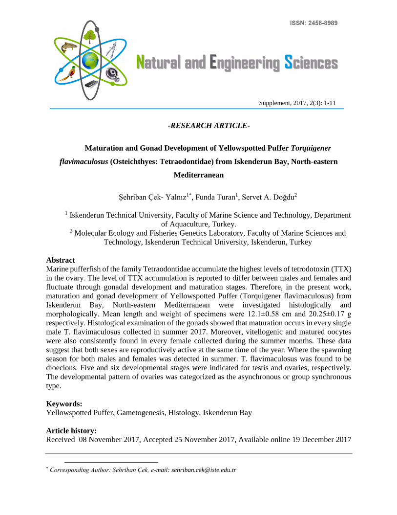
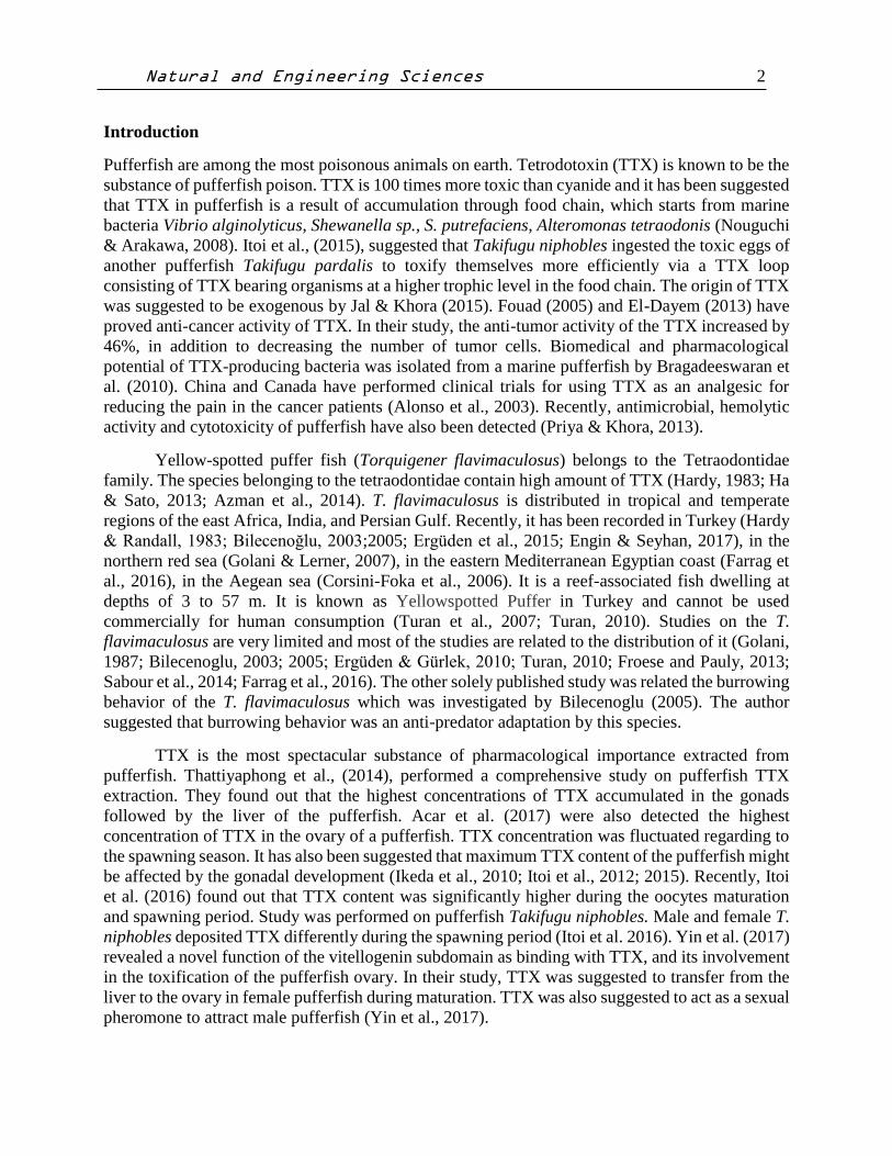
Eight specimens of T. flavimaculosus was caught by gill nets from the Iskenderun Bay southeast
coast of Turkey, and immediately brought fresh to the laboratory (Fig. 1A and 1B). Fish were
preliminary identified by morphological characteristics as previously reported (Golani, 1987;
Ergüden & Gürlek, 2010; Turan et al., 2010). They were sexed and their body weight and length
were recorded.
Figure 1. A). Picture of the yellowspotted puffer, Torquigener flavimaculosus, Hardy and
Randall, 1983. Photograph taken by Servet A. Doğdu. B). Capture locality of T. flavimaculosus
in the Iskenderun Bay (Indicated by an asterisk).
Natural and Engineering Sciences 4
Histological Procedures
The gonads were directly fixed in 10% neutral buffered formalin (prepared in neutral buffered
saline modified for use with teleost tissue, 4 g NaH2PO4, 6.5 g NaHPO4, 100 ml formaldehyde and
900 ml distilled water). A cross section from the center of each ovary and testis was fixed in
formalin, dehydrated in graded ethanol, embedded in paraffin, sectioned at 5μm thickness by
microtome and sticked on slides (sizes) before stained with haematoxylin and eosin (MERCK,
Germany) for histological examination (Çek, 2006; Turan et al. 2006; Çek & Turan, 2007; Çek &
Yılmaz, 2007). After histological work, all slides were examined under a light microscope (CH-2
Olympus, Japan). Photomicrographs were taken to illustrate developmental stages of ovary and
testis. Spermatozoa classification was based on the histological criteria adapted from Grier (1981).
Oocytes were classified by developmental stages modified from Çek et al. (2001). Maturation
stages were detected.
Results
Sex Ratio
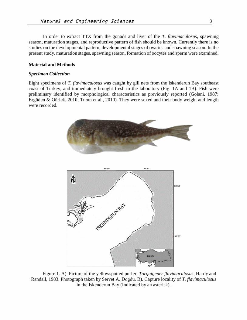
During the study, mean length (ort.±S.E) and weight (ort.±S.E.) of specimens were 12.1±0.58cm
and 20.25±0.17g respectively. Based on morphological and histological examination of the gonads,
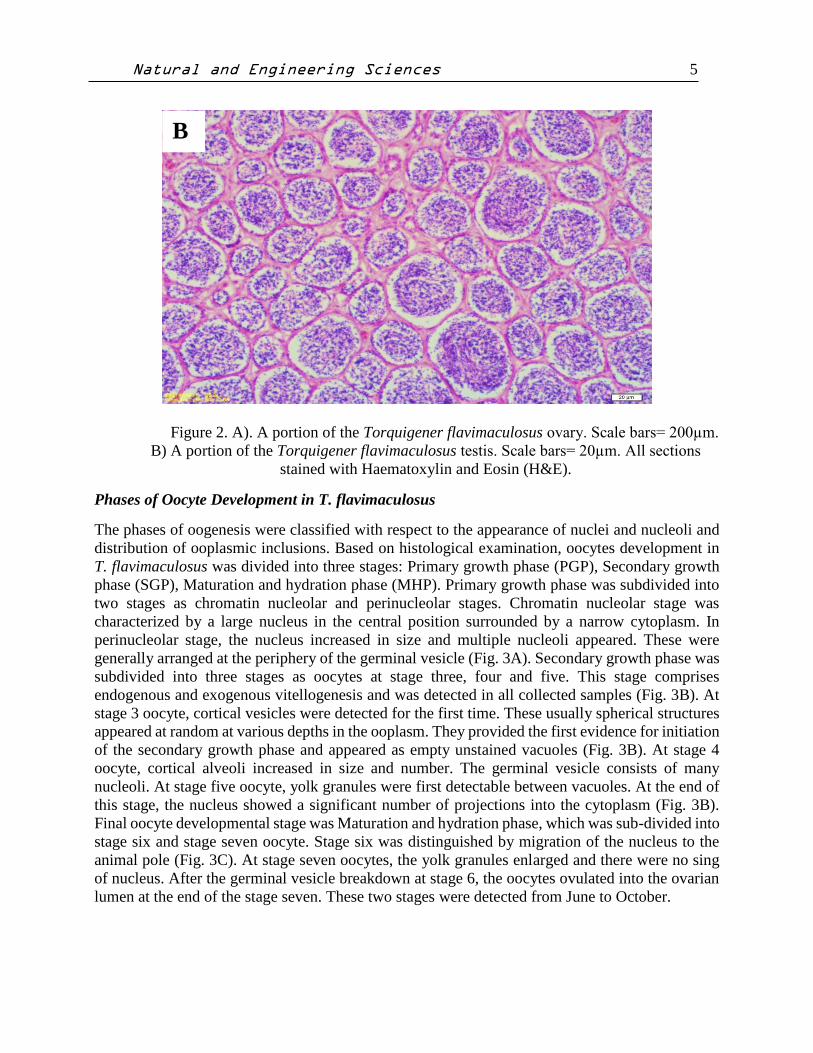
the population seems to consist of gonochoristic individuals (Fig. 2A and 2B). Of the 2017,
Iskenderun Bay 8 T. flavimaculosus examined five (62.5%) were females and three (37.5%) were
males. Because of the small number of specimens caught, the annual sex ratio could not be
calculated using a Chi-square test. Nevertheless, a female biased sex ratio was recorded (5♀, 3♂).
A
Natural and Engineering Sciences 5
Figure 2. A). A portion of the Torquigener flavimaculosus ovary. Scale bars= 200µm.
B) A portion of the Torquigener flavimaculosus testis. Scale bars= 20µm. All sections
stained with Haematoxylin and Eosin (H&E).
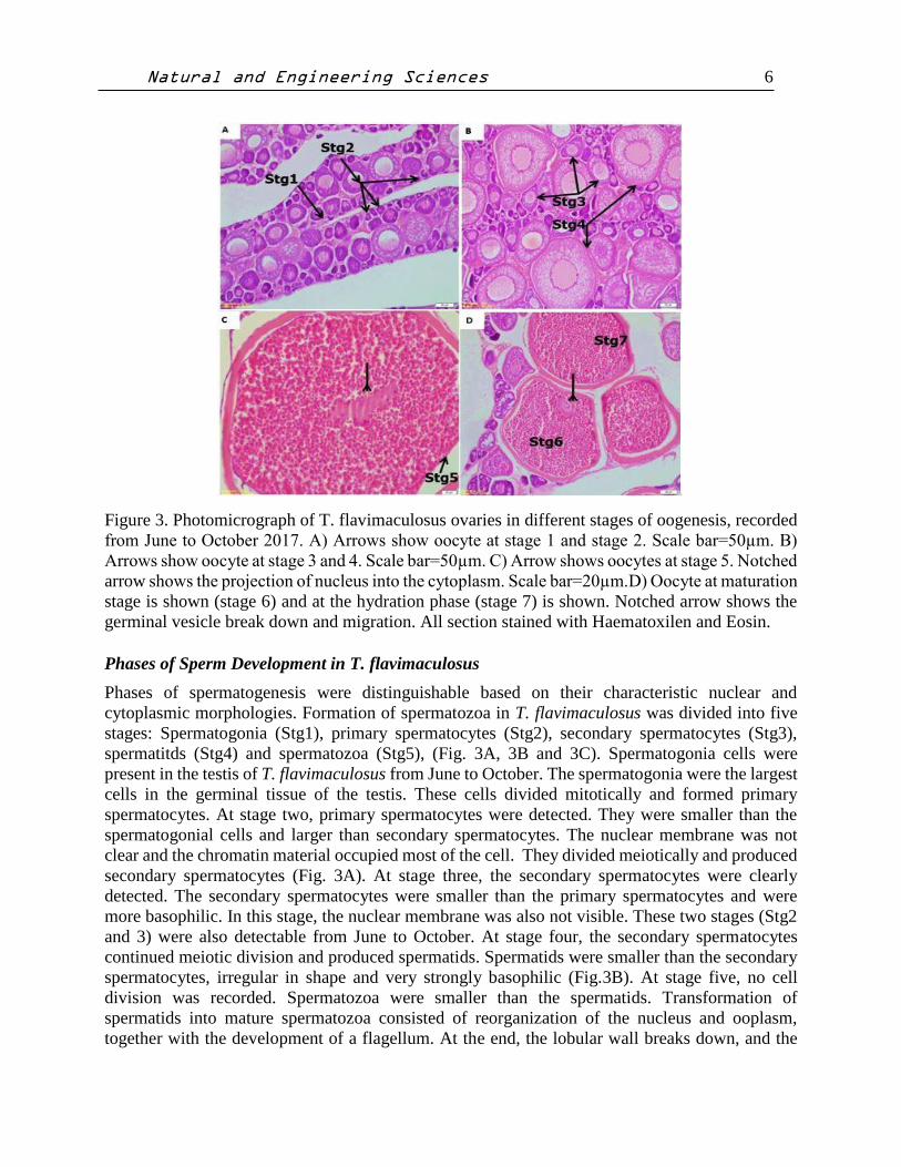
Phases of Oocyte Development in T. flavimaculosus
The phases of oogenesis were classified with respect to the appearance of nuclei and nucleoli and
distribution of ooplasmic inclusions. Based on histological examination, oocytes development in
T. flavimaculosus was divided into three stages: Primary growth phase (PGP), Secondary growth
phase (SGP), Maturation and hydration phase (MHP). Primary growth phase was subdivided into
two stages as chromatin nucleolar and perinucleolar stages. Chromatin nucleolar stage was
characterized by a large nucleus in the central position surrounded by a narrow cytoplasm. In
perinucleolar stage, the nucleus increased in size and multiple nucleoli appeared. These were
generally arranged at the periphery of the germinal vesicle (Fig. 3A). Secondary growth phase was
subdivided into three stages as oocytes at stage three, four and five. This stage comprises
endogenous and exogenous vitellogenesis and was detected in all collected samples (Fig. 3B). At
stage 3 oocyte, cortical vesicles were detected for the first time. These usually spherical structures
appeared at random at various depths in the ooplasm. They provided the first evidence for initiation
of the secondary growth phase and appeared as empty unstained vacuoles (Fig. 3B). At stage 4
oocyte, cortical alveoli increased in size and number. The germinal vesicle consists of many
nucleoli. At stage five oocyte, yolk granules were first detectable between vacuoles. At the end of
this stage, the nucleus showed a significant number of projections into the cytoplasm (Fig. 3B).
Final oocyte developmental stage was Maturation and hydration phase, which was sub-divided into
stage six and stage seven oocyte. Stage six was distinguished by migration of the nucleus to the
animal pole (Fig. 3C). At stage seven oocytes, the yolk granules enlarged and there were no sing
of nucleus. After the germinal vesicle breakdown at stage 6, the oocytes ovulated into the ovarian
lumen at the end of the stage seven. These two stages were detected from June to October.
B
Natural and Engineering Sciences 6
Figure 3. Photomicrograph of T. flavimaculosus ovaries in different stages of oogenesis, recorded
from June to October 2017. A) Arrows show oocyte at stage 1 and stage 2. Scale bar=50µm. B)
Arrows show oocyte at stage 3 and 4. Scale bar=50µm. C) Arrow shows oocytes at stage 5. Notched
arrow shows the projection of nucleus into the cytoplasm. Scale bar=20µm.D) Oocyte at maturation
stage is shown (stage 6) and at the hydration phase (stage 7) is shown. Notched arrow shows the
germinal vesicle break down and migration. All section stained with Haematoxilen and Eosin.
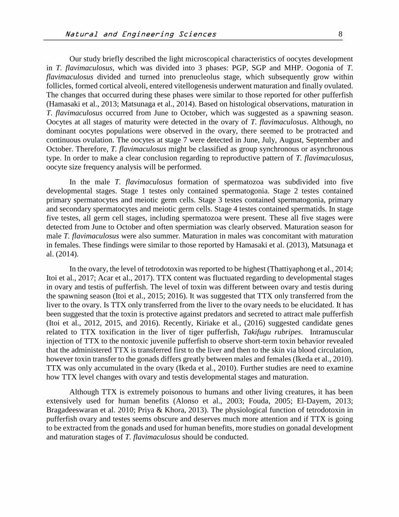
Phases of Sperm Development in T. flavimaculosus
Phases of spermatogenesis were distinguishable based on their characteristic nuclear and
cytoplasmic morphologies. Formation of spermatozoa in T. flavimaculosus was divided into five
stages: Spermatogonia (Stg1), primary spermatocytes (Stg2), secondary spermatocytes (Stg3),
spermatitds (Stg4) and spermatozoa (Stg5), (Fig. 3A, 3B and 3C). Spermatogonia cells were
present in the testis of T. flavimaculosus from June to October. The spermatogonia were the largest
cells in the germinal tissue of the testis. These cells divided mitotically and formed primary
spermatocytes. At stage two, primary spermatocytes were detected. They were smaller than the
spermatogonial cells and larger than secondary spermatocytes. The nuclear membrane was not
clear and the chromatin material occupied most of the cell. They divided meiotically and produced
secondary spermatocytes (Fig. 3A). At stage three, the secondary spermatocytes were clearly
detected. The secondary spermatocytes were smaller than the primary spermatocytes and were
more basophilic. In this stage, the nuclear membrane was also not visible. These two stages (Stg2
and 3) were also detectable from June to October. At stage four, the secondary spermatocytes
continued meiotic division and produced spermatids. Spermatids were smaller than the secondary
spermatocytes, irregular in shape and very strongly basophilic (Fig.3B). At stage five, no cell
division was recorded. Spermatozoa were smaller than the spermatids. Transformation of
spermatids into mature spermatozoa consisted of reorganization of the nucleus and ooplasm,
together with the development of a flagellum. At the end, the lobular wall breaks down, and the
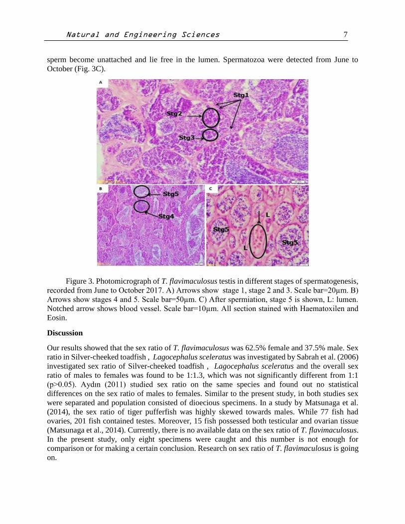
Natural and Engineering Sciences 7
sperm become unattached and lie free in the lumen. Spermatozoa were detected from June to
October (Fig. 3C).
Figure 3. Photomicrograph of T. flavimaculosus testis in different stages of spermatogenesis,
recorded from June to October 2017. A) Arrows show stage 1, stage 2 and 3. Scale bar=20µm. B)
Arrows show stages 4 and 5. Scale bar=50µm. C) After spermiation, stage 5 is shown, L: lumen.
Notched arrow shows blood vessel. Scale bar=10µm. All section stained with Haematoxilen and
Eosin.
Discussion
Our results showed that the sex ratio of T. flavimaculosus was 62.5% female and 37.5% male. Sex
ratio in Silver-cheeked toadfish , Lagocephalus sceleratus was investigated by Sabrah et al. (2006)
investigated sex ratio of Silver-cheeked toadfish , Lagocephalus sceleratus and the overall sex
ratio of males to females was found to be 1:1.3, which was not significantly different from 1:1
(p>0.05). Aydın (2011) studied sex ratio on the same species and found out no statistical
differences on the sex ratio of males to females. Similar to the present study, in both studies sex
were separated and population consisted of dioecious specimens. In a study by Matsunaga et al.
(2014), the sex ratio of tiger pufferfish was highly skewed towards males. While 77 fish had
ovaries, 201 fish contained testes. Moreover, 15 fish possessed both testicular and ovarian tissue
(Matsunaga et al., 2014). Currently, there is no available data on the sex ratio of T. flavimaculosus.
In the present study, only eight specimens were caught and this number is not enough for
comparison or for making a certain conclusion. Research on sex ratio of T. flavimaculosus is going
on.
Natural and Engineering Sciences 8
Our study briefly described the light microscopical characteristics of oocytes development
in T. flavimaculosus, which was divided into 3 phases: PGP, SGP and MHP. Oogonia of T.
flavimaculosus divided and turned into prenucleolus stage, which subsequently grow within
follicles, formed cortical alveoli, entered vitellogenesis underwent maturation and finally ovulated.
The changes that occurred during these phases were similar to those reported for other pufferfish
(Hamasaki et al., 2013; Matsunaga et al., 2014). Based on histological observations, maturation in
T. flavimaculosus occurred from June to October, which was suggested as a spawning season.
Oocytes at all stages of maturity were detected in the ovary of T. flavimaculosus. Although, no
dominant oocytes populations were observed in the ovary, there seemed to be protracted and
continuous ovulation. The oocytes at stage 7 were detected in June, July, August, September and
October. Therefore, T. flavimaculosus might be classified as group synchronous or asynchronous
type. In order to make a clear conclusion regarding to reproductive pattern of T. flavimaculosus,
oocyte size frequency analysis will be performed.
In the male T. flavimaculosus formation of spermatozoa was subdivided into five
developmental stages. Stage 1 testes only contained spermatogonia. Stage 2 testes contained
primary spermatocytes and meiotic germ cells. Stage 3 testes contained spermatogonia, primary
and secondary spermatocytes and meiotic germ cells. Stage 4 testes contained spermatids. In stage
five testes, all germ cell stages, including spermatozoa were present. These all five stages were
detected from June to October and often spermiation was clearly observed. Maturation season for
male T. flavimaculosus were also summer. Maturation in males was concomitant with maturation
in females. These findings were similar to those reported by Hamasaki et al. (2013), Matsunaga et
al. (2014).
In the ovary, the level of tetrodotoxin was reported to be highest (Thattiyaphong et al., 2014;
Itoi et al., 2017; Acar et al., 2017). TTX content was fluctuated regarding to developmental stages
in ovary and testis of pufferfish. The level of toxin was different between ovary and testis during
the spawning season (Itoi et al., 2015; 2016). It was suggested that TTX only transferred from the
liver to the ovary. Is TTX only transferred from the liver to the ovary needs to be elucidated. It has
been suggested that the toxin is protective against predators and secreted to attract male pufferfish
(Itoi et al., 2012, 2015, and 2016). Recently, Kiriake et al., (2016) suggested candidate genes
related to TTX toxification in the liver of tiger pufferfish, Takifugu rubripes. Intramuscular
injection of TTX to the nontoxic juvenile pufferfish to observe short-term toxin behavior revealed
that the administered TTX is transferred first to the liver and then to the skin via blood circulation,
however toxin transfer to the gonads differs greatly between males and females (Ikeda et al., 2010).
TTX was only accumulated in the ovary (Ikeda et al., 2010). Further studies are need to examine
how TTX level changes with ovary and testis developmental stages and maturation.
Although TTX is extremely poisonous to humans and other living creatures, it has been
extensively used for human benefits (Alonso et al., 2003; Fouda, 2005; El-Dayem, 2013;
Bragadeeswaran et al. 2010; Priya & Khora, 2013). The physiological function of tetrodotoxin in
pufferfish ovary and testes seems obscure and deserves much more attention and if TTX is going
to be extracted from the gonads and used for human benefits, more studies on gonadal development
and maturation stages of T. flavimaculosus should be conducted.
Natural and Engineering Sciences 9
References
Acar, C., Ishizaki, S., Nagashima Y. (2017). Toxicity of the lessepsian pufferfish Lagocephalus
scleratus from eastren Mediterranean coast of Turkey and species identification by rapid
PCR amplification. EuropeanFood Research Technology, 243: 49-57.
Alonso, D., Khalilb, Z., Satkunanthanb, N. and Livettc, BG. (2003). Drug from the sea: Conotoxins
as drug leads for neuropathic pain and other neurological conditions. Mini Reviews in
Medicinal Chemistry, 3: 785-787.
Aydın, M. (2011). Growth, reproduction and diet of Pufferfish (Lagocephalus sceleratus, Gmelin,
1789) from Turkey’s Mediterranean sea coast. Turkish Journal of Fisheries and Aquatic
Sciences, 11: 569-576.
Azman, A. M. N., Samsur M., Othman M. (2014). Distribution of tetrodotoxin among tissues of
puffer fish from Sabah and Sarawak waters. Sains Malaysiana, 43(7): 1003-1011.
Bilecenoğlu, M. (2003). Kızıldeniz göçmeni balon balığı (Torquigener flavimaculosus Hardy &
Randall, 1983), Türkiye kıyılarından ilk gözlemler. Sualtı Dünyası Dergisi, 74: 38-39.
Bilecenoğlu, M. (2005). Observation on the burrowing behaviour of the Dwarf Blaasop
Torquigener flavimaculosus (Osteichthyes: Tetraodontidae) along the coast of Fethiye,
Turkey. Zoology in the Middle East, 35: 29-34.
Bragadeeswaran S., Therasa D., Prabhu K & Kathiresan K. (2010). Biomedical and
pharmacological potential of tetrodotoxin-producing bacteria isolated from marine
pufferfish Arothron hispidus (Muller, 1841). The Journal of Venomous Animals and
Toxins Including Tropical Diseases, 16(3): 421-431.
Corsini-Foka, M., Margies, P., Kondilatos, G., & Economidis, P. S. (2006). Torquigener
flavimaculosus Hardy and Randall, 1983 (Pisces: Tetraodontidae) off Rhodes island
marine area: a new alien fish in the Hellenic waters. Mediterranean Marine Science, 7(2),
73-76.
Çek, S. and Turan, F. (2007). Masculinization of Convict Cichlid (Cichlosoma nigrofasciatum)
by Immersion in Tribulus terrestris extract. Aquaculture International, 15: 109-119.
Çek, Ş. and E, Yılmaz. (2007). Gonad development and sex ratio of Sharptooth catfish (Clarias
gariepinus, Burchell 1822) cultured under laboratory conditions,’’ Turkish Journal of
Zoology 37, 1-12.
Çek, Ş, (2006). Early gonadal development and sex differentiation in rosy barb (Puntius
conchonius).’’ Animal Biology, 56(3), 335-350.
Çek, Ş., N. Bromage, C. Randal, & K. Rana. (2001). Oogenesis, Hepatosomatic and
Gonadosomatic Indexes, and Sex Ratio in Rosy barb (Puntius conchonius).’’ Turkish
Journal of Fisheries and Aquatic Sciences, 1(1), 33-41.
El-Dayem S. M. A., Fouda F. M., Ali E. H. A. (2013). The antitumor effects of tetrodotoxin and/or
doxorubicin on ehrlich ascites carcinoma-bearing female mice. Toxicology and Industrial
Health, 29(5);404-417.
Engin, S. and Seyhan, D. (2017). A new species of Pomatoschistus (Teleostei, Gobiidae): the
Mediterranean’s smallest marine fish. Journal of. Fish Biology, doi:10.1111/jfb.13455
Erguden, D., & Gurlek, M. (2010). The Presence of Indo-Pacific Puffer Fish Torquigener
filavimaculosus Harddy & Randall, 1983 In The Iskenderun Bay, The Eastern
Mediterranean Coast of Turkey. Rapp. Comm. int. Mer Médit., 39.
Ergüden, D., Ergüden S. A., Gürlek M. (2015). Length-weight relationships fors ix fish species in
Iskenderun bay (Eastern Mediterranean Sea coast of Turkey. Journal of Applied
Ichthyology, 31, 1148-1149.
Natural and Engineering Sciences 10
Farrag, M. M. S., El-Haweet, A. A. K., Akel, E. A., Moustafa, M. A. (2016). Occurrence of puffer
fishes (Tetraodontidae) in the eastern Mediterranean Egyptian coast-filling in the gap.
BioInvasions Records, 5(1): 47-54.
Fouda, F. M. (2005). Anti-tumor activity of tetrodotoxin extracted from the masked puffer fish
Arothron diadematus. Egyptian Journal of Biology, 7; 1-13.
Froese, R & Pauly, D. (2013). Fishbase. World wide web electronic publication.
www.Fishbase.org,version (06/2013).
Golani, D. (1987). The Red sea pufferfish, Turquigener flavimaculosus Hardy and Randall 1983.
A new suez canal migrant to the eastern mediterrenean (Pisces; Tetraodontidae).
Senckenbergiana Maritima, 19; 339-343.
Golani, D & Lerner, A. (2007). A long-term study of the sandy shore ichthyofauna in the northern
red sea (gulf of aqaba) with reference to adjacent mariculture activity. The Reffles Bullettin
of zoology, 14: 255-264.
Grier, H. J. (1981). Cellular organisationof the testis and spermatogenesis in fishes. American
Zoology: 21:345-357.
Ha, D. V. & Sato S. (2013). The toxicity of the fishsource made from a tetrodotoxin-bearing puffer
Torquigener gloerfelti. Tap Chi Khoa Hoc vᾁ Cong nghề Biền, 13(3): 261-265.
Hamasaki, M., Takeuchi, Y., Miyaki, K., Yoshizaki, G. (2013). Gonadal development and fertility
of triploid grass puffer Takifugu niphobles induced by cold shock treatment. Marine
Biotechnology, 15: 133-144.
Hardy, G.S., & Randall, J. E. (1983). Description of a new species of puffer fish
(Tetraodontiformes: Tetraodontidae) from the Red Sea and adjacent. Israel Journal of
Zoology, 32(1): 13-20.
Hardy, G. S. (1983). Revision of Australian species of Torquigener whitley (Tetraodontiformes:
Tetraodontidae), and two new generic names for Australian puffer fishes. Journal of the
Royal Society of New Zealand, 13(112): 1-48.
Ikeda, K., Emoto, Y., Tatsuno, R., Wang, JJ., Ngy, L., Taniyama, S., Takatani, T., Arakawa, O.
(2010). Maturation-associated changes in toxicity of the pufferfish Takifugu poecilonotus.
Toxicon, 55: 289-297.
Itoi, S., Ishizuka, K., Mitsuoka, R., Takimoto, N., Yokoyama, N., Detake, A., Takayanagi, C.,
Yoshikawa, S., Sugita, H. (2016). Seasonal changes in the tetrodotoxin content of the
pufferfish Takifugu niphobles. Toxicon, 14: 53-58.
Itoi, S., Kozaki, A., Komori, K., Tsunashima, T., Noguchi, S., Kawane, M. Sugita, H. (2015). Toxic
Takifugu pardalis eggs found in Takifugu niphobles gut implications for TTX
accumulations in the pufferfish. Toxicon, 108: 141-146.
Itoi, S., Yoshikawa, S., Tatsuno, R., Suziki, M., Asahina, K., Yamamoto, S., Takanashi, S.,
Takatani, T., Arakawa, O., Sakakura, Y., Sugita. H. (2012). Difference in the localization
of tetrodotoxin between the female and male fish pufferfish Takifugu niphobles during
spawning. Toxicon, 60: 1000-1004.
Jal, S. & Khora, S. S. (2015). An overview on the origin and production of tetrodotoxin a potent
neurotoxin. Journal of Applied Microbiology, 119: 907-916
Kiriake, A., Ohta A., Suga E.,Matsumoto T., Shoichiro I., Nagashima Y. (2016). Comparison of
tetrodotoxin uptake and gene exoression in the liver between juvenile and adult tiger
pufferfish, Takifugu rubripes, Toxicon. 111: 6-12.
Matsunaga, T., Ieda, R., Hosoya, S., Kuroyanagi, M., Suziki, S., Suetake, H., Tasumi, S., Suzuki
Y., Miyadai, T., Kikuchi, K. (2014). An efficient molecular technique for sexing tiger
Natural and Engineering Sciences 11
pufferfish (fugu) and the occurrence of sex reversal in a hatchery population. Fish Science,
80; 933-942.
Nouguchi, T., Arakawa, O. (2008). Tetrodotoxin; distribution and accumulation in aquatic
organisms and cases of human intoxication. Marine Drugs, 6(2): 220-242.
Priya, K. M., & Khora, S. S. (2013). Antimicrobial, hemolytic and cytotoxic activities of the puffer
fish Arothron hispidus from the Southeast coast of India. International Journal of Drug
Development & Research, 5(2): 317-322.
Sabour, W., Saad, A., Jawad, L. (2014). First record of the yellowspotted puffer Torquigener
flavimaculosus Hardy &Randall, 1983 (Osteichthys: Tetraodontidae from the
Mediterranean sea coasts of Syria. Thalassia Salentina, 36: 29-34.
Sabrah, M. M., El-Ganainy, A. A., Zaky M. A. (2006). Biology and toxicity of the pufferfish
Logocephalus sceleratus (Gmelin, 1789) from the gulf of suez. Egyptian Journal of
Aquatic Research, 32(1): 283-297.
Thattiyaphong, A., Unahalekhaka, J., Mekha, N., Nispa, W., Kluengklangdon, P., Rojanapantip,
L. (2014). Efficiency of a rapid test for detection of tetrodotoxin in pufferfish. Journal of
Immunoassay and Immunochemistry, 5: 111-119.
Turan, C. (2010). Status and trend of lessepsian species in marine waters of Turkey. FAO EastMed.
04: 109-118.
Turan, F.,Çek, Ş., Atık, E (2006). Productıon of Monosex Male Guppy, Poecılıa retıculata, by
17α-Methyltestosterone. Aquaculture Research, 37(2): 200-203.
Turan, C., Ergüden, D., Gürlek, M., Yaglıoğlu, D., Keskin, C (2007).Lessepsian Fishes of Turkey.
In: Turan C (ed), Atlas and Systematics of Marine Bony Fishes of Turkey. Nobel
Publishing House, Adana, Turkey.pp 531.
Yin, X., Kriake, A., Ohta, A., Kitani, Y., Shizaki, S., Nagashima, Y. (2017). A novel function of
vitellogenin subdomain, vWF type D, as a toxin-binding protein in the pufferfish Takifugu
pardalis ovary. Toxicon, 136: 56-66.


