10.1261/rna.2815911Access the most recent version at doi: 2011 17: 1821-1830 originally published online August 2, 2011RNA
Bhaskara Reddy Madina, Gokulan Kuppan, Ajay A. Vashisht, et al.
Trypanosoma bruceiendoribonuclease in Guide RNA biogenesis involves a novel RNase III family
MaterialSupplemental http://rnajournal.cshlp.org/content/suppl/2011/07/25/rna.2815911.DC1.html
References http://rnajournal.cshlp.org/content/17/10/1821.full.html#ref-list-1
This article cites 61 articles, 30 of which can be accessed free at:
serviceEmail alerting
click heretop right corner of the article orReceive free email alerts when new articles cite this article - sign up in the box at the
http://rnajournal.cshlp.org/subscriptions go to: RNATo subscribe to
Copyright © 2011 RNA Society
Cold Spring Harbor Laboratory Press on January 31, 2012 - Published by rnajournal.cshlp.orgDownloaded from
Guide RNA biogenesis involves a novel RNase III family
endoribonuclease in Trypanosoma brucei
BHASKARA REDDY MADINA,1 GOKULAN KUPPAN,1 AJAY A. VASHISHT,2 YU-HE LIANG,3 KURTISM. DOWNEY,4 JAMES A. WOHLSCHLEGEL,2 XINHUA JI,3 SING-HOI SZE,1,5 JAMES C. SACCHETTINI,1
LAURIE K. READ,4 and JORGE CRUZ-REYES1,6
1Department of Biochemistry and Biophysics, Texas A&M University, College Station, Texas 77843, USA2Department of Biological Chemistry, David Geffen School of Medicine, University of California, Los Angeles, Los Angeles,California 90095-1737, USA3Center for Cancer Research, National Cancer Institute, Frederick, Maryland 21702, USA4Department of Microbiology and Immunology, University at Buffalo, State University of New York, Buffalo, New York 14214, USA5Department of Computer Science and Engineering, Texas A&M University, College Station, Texas 77843, USA
ABSTRACT
The mitochondrial genome of kinetoplastids, including species of Trypanosoma and Leishmania, is an unprecedented DNAstructure of catenated maxicircles and minicircles. Maxicircles represent the typical mitochondrial genome encodingcomponents of the respiratory complexes and ribosomes. However, most mRNA sequences are cryptic, and their maturationrequires a unique U insertion/deletion RNA editing. Minicircles encode hundreds of small guide RNAs (gRNAs) that partiallyanneal with unedited mRNAs and direct the extensive editing. Trypanosoma brucei gRNAs and mRNAs are transcribed aspolycistronic precursors, which undergo processing preceding editing; however, the relevant nucleases are unknown. We reportthe identification and functional characterization of a close homolog of editing endonucleases, mRPN1 (mitochondrial RNAprecursor-processing endonuclease 1), which is involved in gRNA biogenesis. Recombinant mRPN1 is a dimeric dsRNA-dependent endonuclease that requires Mg2+, a critical catalytic carboxylate, and generates 2-nucleotide 39 overhangs. Thecleavage specificity of mRPN1 is reminiscent of bacterial RNase III and thus is fundamentally distinct from editingendonucleases, which target a single scissile bond just 59 of short duplexes. An inducible knockdown of mRPN1 in T. bruceiresults in loss of gRNA and accumulation of precursor transcripts (pre-gRNAs), consistent with a role of mRPN1 in processing.mRPN1 stably associates with three proteins previously identified in relatively large complexes that do not contain mRPN1, andhave been linked with multiple aspects of mitochondrial RNA metabolism. One protein, TbRGG2, directly binds mRPN1 and isthought to modulate gRNA utilization by editing complexes. The proposed participation of mRPN1 in processing ofpolycistronic RNA and its specific protein interactions in gRNA expression are discussed.
Keywords: guide RNA processing; RNase III endonuclease; mitochondria; RNA editing; Trypanosoma brucei
INTRODUCTION
Kinetoplastid protozoa include early-branched parasites thatcause Leishmaniasis and Trypanosomiasis, such as Chagas’disease and sleeping sickness. Each cell has one mitochon-drion and a single mitochondrial genome (kDNA) in anunusual catenated structure containing a dozen copies of amaxicircle and hundreds of different minicircles. Trypano-soma brucei maxicircle genes are tightly packed in bothstrands, including two rRNAs, 18 (edited and never-edited)
mRNAs, and two guide RNAs (gRNAs). rRNAs and mRNAsare similar to those in other mitochondria, but the formerare the smallest known examples in eukaryotes, and 12mRNAs require editing. U insertion alone may account formore than half of the final sequence in some cases (Supple-mental Fig. S1A). Since several maxicircle genes overlap, theformation of mature ends must be controlled (Clement et al.2004). Each T. brucei minicircle encodes three to five gRNAson only one strand, but low-level transcription of an un-known function in the complementary (antisense) strand hasbeen detected (Supplemental Fig. S1B; Aphasizheva andAphasizhev 2010). There are about 80–100 different mini-circle sequence classes in this species (Simpson et al. 2000;Hong and Simpson 2003; Ochsenreiter et al. 2007), with nocorrelation between editing and expression (Koslowsky et al.
6Corresponding author.E-mail [email protected] published online ahead of print. Article and publication date are
at http://www.rnajournal.org/cgi/doi/10.1261/rna.2815911.
RNA (2011), 17:1821–1830. Published by Cold Spring Harbor Laboratory Press. Copyright � 2011 RNA Society. 1821
Cold Spring Harbor Laboratory Press on January 31, 2012 - Published by rnajournal.cshlp.orgDownloaded from
1992). Each gRNA gene may be able to initiate transcription,producing a polycistronic precursor (pre-gRNA) extendingpast downstream gRNAs. Maturation of the 59-most gRNA isconsistent with its detection via capping of the 59 triphos-phate in the primary transcript, implying post-transcrip-tional 39 cleavage of precursor sequence. This 39 processingwould be followed by the addition and trimming of a shortU tail. (Blum and Simpson 1990; Grams et al. 2000; Clementet al. 2004; Aphasizheva and Aphasizhev 2010; Zimmer et al.2011). However, the precise structure of pre-gRNAs, in-cluding their transcription initiation and termination sites,and processing mechanisms are unknown.
U-insertion/deletion editing is catalyzed by the exten-sively studied multiprotein RNA editing core complex(RECC; the 20 S editosome or L-complex). Several mRNAsare edited at over a hundred sites, and at each site, editingstarts by a nuclease cleavage directed by a complementarygRNA (Rusche et al. 1997; Stuart et al. 2005; Li et al. 2009;Simpson et al. 2010). There are three accepted specializedediting endonucleases, REN1, REN2, and REN3 (Carneset al. 2005; Trotter et al. 2005; Kang et al. 2006; Simpsonet al. 2010), although only REN1 has been produced in anactive recombinant form (Kang et al. 2006). RENs havea conserved RNase III domain and two domains of un-known function (Worthey et al. 2003). After cleavage,U-addition or U-deletion occurs, followed by re-ligation.Since one gRNA directs the editing of only a few sites,multiple gRNAs are needed to edit an entire mRNA.
Several ancillary mitochondrial factors, often in multipro-tein complexes, copurify with editing complexes via uniden-tified RNA linkers. Some factors affect editing indirectly,for example, by impacting the level of pre-mRNA precursorsor the turnover of gRNA or mRNA in vivo, or possiblythrough associated activities identifiedin vitro: RNA annealing, unwinding, or39 remodeling (Aphasizhev et al. 2002;Pelletier and Read 2003; Mingler et al.2006; Etheridge et al. 2008; Fisk et al.2008; Hashimi et al. 2008; Panigrahiet al. 2008; Weng et al. 2008; Koslowsky2009; Aphasizheva and Aphasizhev 2010;Hernandez et al. 2010). Finally, a mito-chondrial activity was found to catalyze39 processing of polycistronic pre-gRNAs,but the responsible nucleases are notknown (Grams et al. 2000).
Here, we report an endonucleasemRPN1 (mitochondrial RNA precursor-processing endonuclease 1) that is involvedin gRNA metabolism. mRPN1 is a closehomolog of the REN endonucleases butis not present in RECC or in the knownmultiprotein ancillary factors. Cleavageby recombinant mRPN1 requires Mg2+,a critical catalytic carboxylate, and its
specificity is reminiscent of bacterial RNase III towardduplexed RNA. In contrast, RECC specifically cleaves atsingle/double-stranded mRNA junctions with gRNA. Induc-ible knockdown of mRPN1 results in the loss of gRNAs andaccumulation of precursor sequences (pre-gRNAs), consistentwith a role in processing. Finally, cell purified mRPN1 hasnuclease-resistant interactions with three other proteins thatwere previously identified in relatively large MRB (mitochon-drial RNA binding)-related complexes, which do not havemRPN1, and appear to have multiple roles in mitochondrialRNA metabolism (Hashimi et al. 2008; Panigrahi et al. 2008;Weng et al. 2008; Hernandez et al. 2010; Ammerman et al.2011). One of these factors (TbRGG2) directly binds mRPN1and was previously linked with gRNA utilization duringediting, which may include entry into the editing pathway.The proposed role of mRPN1 and its protein associations ingRNA metabolism is discussed.
RESULTS
mRPN1 is a homolog of the REN endonucleases, butmRPN1 and editing complexes exhibit fundamentallydifferent specificity
A search of the T. brucei genome revealed mRPN1(Tb11.01.0150) as a homolog of REN editing endonucleases(Fig. 1). The overall average amino acid sequence homologyof mRPN1 with these protein homologs is z16%–18% (Sup-plemental Fig. S2A). mRPN1 and REN proteins are (class I)RNase III family members, which also include Drosha (classII) and Dicer (class III) involved in the maturation ofmiRNAs (both) and the creation of siRNAs (the latter) in anumber of organisms. The RNase III domain of mRPN1 is
FIGURE 1. Similar domain organization of mRPN1 and REN proteins. (A) Aligned RNase IIIendonuclease domain and two other conserved sequences representing a U1-like C2H2 zincfinger and a putative dsRBD, which is more divergent in mRPN1 and REN1. Other regionshave no significant homology. (B) Multiple sequence alignment of the RNase III domain,including the prototype domain of A. aeolicus, shows the most conserved motifs, their e-values,four invariant catalytic carboxylates, and a lysine residue (*) that corresponds to a catalyticresidue in the mouse Dicer (Zhang et al. 2004). Cylinders denote the a-helices in the crystalstructure (1JFZ) of the A. aeolicus domain (Blaszczyk et al. 2001).
Madina et al.
1822 RNA, Vol. 17, No. 10
Cold Spring Harbor Laboratory Press on January 31, 2012 - Published by rnajournal.cshlp.orgDownloaded from
between 27% and 35% identical to that of RENs (Supple-mental Fig. S2A). This domain includes four invariant catalyticresidues and has been associated in most family members withdouble-stranded RNA (dsRNA) cleavage activity (Blaszczyket al. 2001; Pertzev and Nicholson 2006). RENs and mRPN1share two other domains of unknown function and a predictedN-terminal mitochondrial leader (Fig. 1; Supplemental Fig.S2B; data not shown). These domains represent a significantlydivergent U1-like zinc finger, with the signature C2H2 res-idues, and a possible dsRBD (Worthey et al. 2003). We foundan mRPN1 ortholog in Trypanosoma cruzi, but not in therelated Leishmania genus.
Importantly, mRPN1 was not detected in reported puri-fications of either native or tandem-affinity purified (TAP)editing complexes, whether RENs or other core subunits weretagged, indicating a distinction of function between mRPN1and REN proteins (Rusche et al. 1997; Panigrahi et al. 2006,2007; Hernandez et al. 2008).
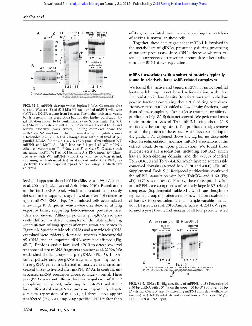
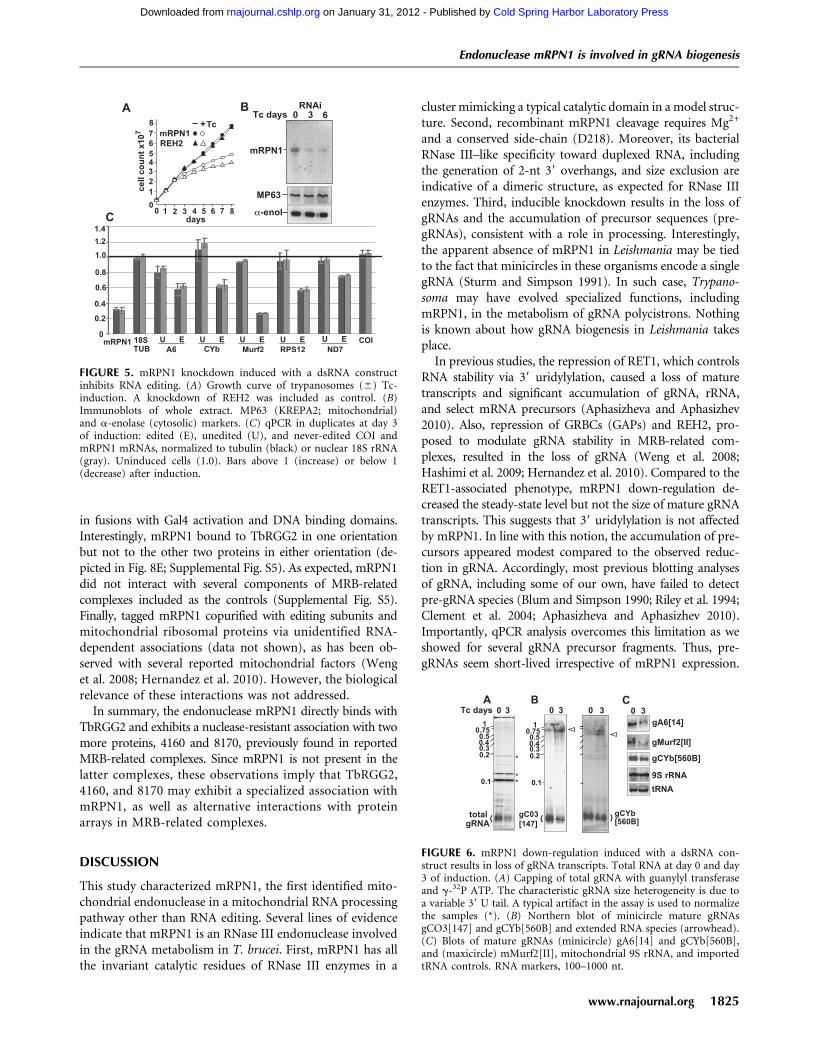
We investigated the possibility that mRPN1 may beprocessive. We first performed structure homology-mod-eling of the putative nuclease domain, using the Aquifexaeolicus (Aa) RNase III and RNase III–dsRNA structures astemplates (Blaszczyk et al. 2001; Gan et al. 2008). Thealignment showed that the RNase III fold indeed brings thecatalytically important mRPN1 side-chains in a cluster,mimicking the catalytic Aa–RNase III structure, includingthe potential to dimerize (Fig. 2). Based on this informa-tion, we expressed in bacteria tagged mRPN1 wild-type(WT) or with a substitution of the invariant Aspartate 218for Alanine (D218A) and tested these proteins for activityin vitro. A bacterially expressed recombinant protein of theexpected size reacted with anti-mRPN1 antibodies, and sizeexclusion indeed indicates that mRPN1 forms a dimer (Fig.3A,B; Supplemental Fig. S3). WT mRPN1 catalyzed a majorcleavage and a few nearby cuts within the duplex in a modeldsRNA with a 59 overhang (Fig. 3C). This activity requiredMg2+, both substrate strands, and D218 (Fig. 3D–F). Incontrast to the mRPN1 activity, purified editing complexescleave specifically at the dsRNA–ssRNA junction of thisminimized substrate (Hernandez et al. 2008). Also, mRPN1cleaved both strands of a short 30-bp dsRNA, which has beentested with RNase III and Dicer in vitro (Zhang et al. 2004).Interestingly, the mRPN1 specificity with the latter substratewas closer to RNase III than Dicer, and this activity alsorequired Mg2+ (Fig. 4). Like these enzymes, mRPN1 induceda major cleavage that leaves 2-nucelotide (nt) 39 overhangs,as well as minor nearby cuts in the duplex. Thus, mRPN1 isa dsRNA-dependent endonuclease that exhibits an RNaseIII–like specificity.
mRPN1 down-regulation results in a loss of gRNAand accumulation of pre-gRNA transcripts
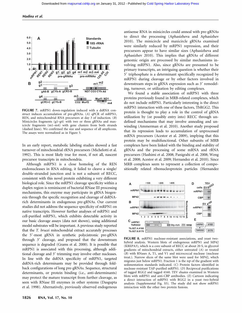
We examined the role of mRPN1 in trypanosomes via RNAi-mediated repression through the tetracycline induction of
a dsRNA construct targeting mRPN1. This RNAi constructinduced a growth defect that became apparent by day 3 andcontinued over 10 d without recovery. Similar results wereobtained with independent clonal lines, a hairpin RNAi con-struct for mRPN1, and an RNAi construct for another factor,REH2, which is also important in gRNA metabolism (Fig. 5A;Hashimi et al. 2009; Hernandez et al. 2010; data not shown).mRPN1 was diminished, but control mitochondrial and cyto-solic proteins were unaffected after several days (Fig. 5B).Since cells appeared normal at day 3, implying limited or nosecondary effects, we performed all experiments at this post-induction time. Real-time RT-PCR (qPCR) of several editingsubstrates showed a decreased abundance of edited mRNAsupon mRPN1 suppression (Fig. 5C). Changes in uneditedmRNAs were not statistically significant, as in other studiesshowing sublethal editing effects, including with our REH2construct above, where unedited RNAs are not always evi-dently affected (Supplemental Fig. S4; Hashimi et al. 2008;Acestor et al. 2009). The never-edited mRNA COI was notaffected, while the A6 mRNA may only be slightly decreased.We asked whether gRNA biogenesis was affected. Pre-gRNAtranscripts are rarely detected in Northern blots or radioactivecapping of a 59 triphosphate, due to their low steady-state
FIGURE 2. Homology model of mRPN1. The A. aeolicus (Aa)-RNaseIII (PDB entry 1JFZ) and the Aa-RNase III-dsRNA (2NUF) structuresas starting models (Blaszczyk et al. 2001; Gan et al. 2008). (A) Ribbondiagram of the mRPN1 RNase III domain with helices (a1–a6). Thefour conserved catalytic side-chains (E214, D218, D288, E291,clustered on a3 and a6) are in red. (B) Model of the mRPN1–dsRNAcomplex. One subunit is in yellow, the other in cyan, and the dsRNAin orange. The acidic catalytic side-chains and magnesium ions(labeled in one cleavage site only) are in red and magenta, re-spectively. Size exclusion chromatography of recombinant mRPN1indicates a dimer, as expected for RNase III family members (seeahead Supplemental Fig. S3).
Endonuclease mRPN1 is involved in gRNA biogenesis
www.rnajournal.org 1823
Cold Spring Harbor Laboratory Press on January 31, 2012 - Published by rnajournal.cshlp.orgDownloaded from
level and apparent short half-life (Riley et al. 1994; Clementet al. 2004; Aphasizheva and Aphasizhev 2010). Examinationof the total gRNA pool, which is abundant and readilydetected in the capping assay, showed an over 40% decreaseupon mRPN1 RNAi (Fig. 6A). Induced cells accumulateda few large RNA species, which were only detected at longexposure times, suggesting heterogeneous precursor sizes(data not shown). Although potential pre-gRNAs are gen-erally difficult to detect, examples of the blots exhibitingaccumulation of long species after induction are shown inFigure 6B. Specific minicircle gRNAs and a maxicircle gRNAexamined were evidently decreased, whereas mitochondrial9S rRNA and an imported tRNA were not affected (Fig.6B,C). Previous studies have used qPCR to detect low-levelunprocessed pre-mRNA fragments (Acestor et al. 2009). Weestablished similar assays for pre-gRNAs (Fig. 7). Impor-tantly, polycistronic pre-gRNA fragments spanning two orthree gRNA genes in different minicircles examined in-creased three- to fivefold after mRPN1 RNAi. In contrast, un-processed mRNA precursors appeared largely normal. Thesepre-gRNAs were not affected by down-regulation of REH2(Supplemental Fig. S4), indicating that mRPN1 and REH2have different roles in gRNA expression. Importantly, despitea z70% repression of mRPN1, all three RENs appearunaffected (Fig. 7A), implying specific RNAi rather than
off-targets on related proteins and suggesting that catalysisof editing is normal in these cells.
Together, these data suggest that mRPN1 is involved inthe metabolism of gRNAs, presumably during processingof nascent precursors, since gRNAs decrease whereas ex-tended unprocessed transcripts accumulate after induc-tion of mRPN1 down-regulation.
mRPN1 associates with a subset of proteins typicallyfound in relatively large MRB-related complexes
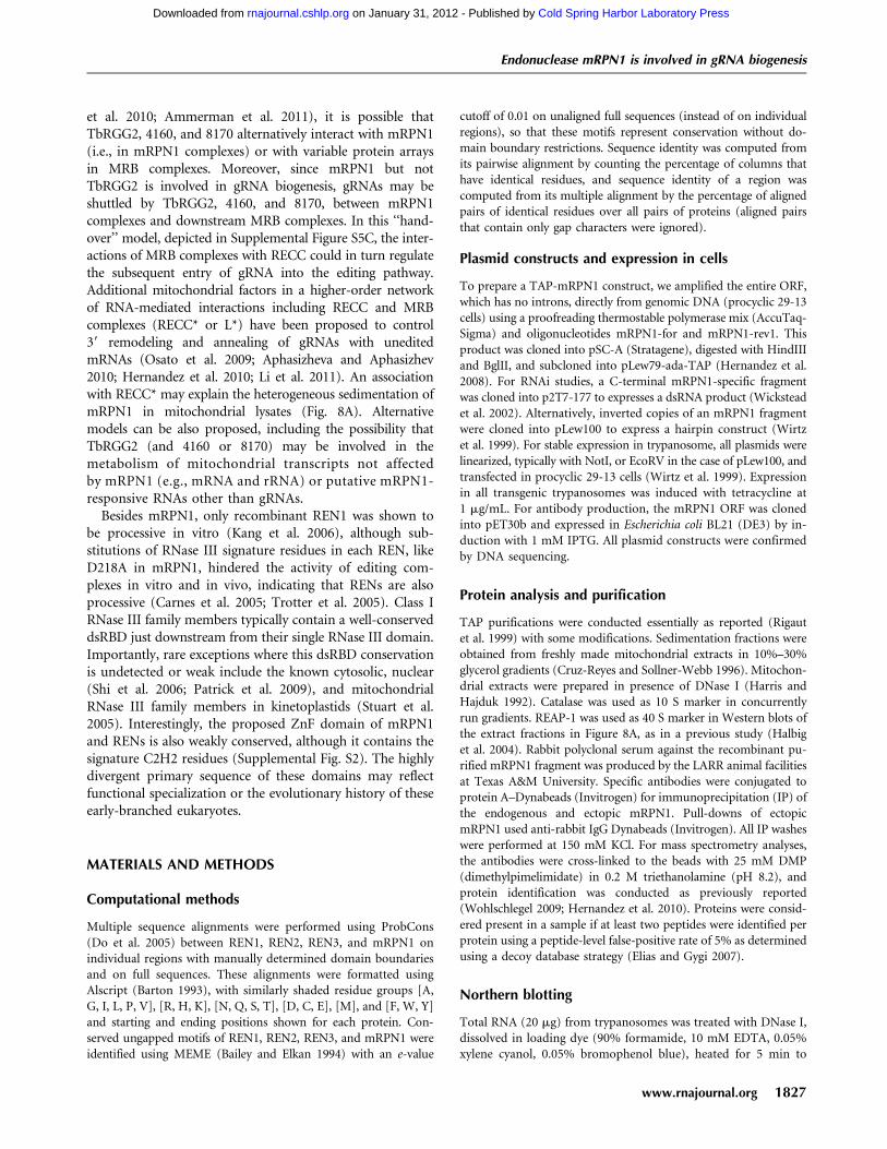
We found that native and tagged mRPN1 in mitochondriallysates exhibit equivalent broad sedimentation, with clearaccumulation in low density (top fractions) and a shallowpeak in fractions containing about 20 S editing complexes.However, most mRPN1 shifted to low-density fractions, awayfrom editing complexes, after nuclease treatment or affinity-purification (Fig. 8A,B; data not shown). We performed massspectrometric analyses of TAP mRPN1 using about 20 Sfractions as the starting extract. This purification helps removemost of the protein in the extract, which lies near the top ofthe gradient. As explained above, the tag has no discernibleeffect on sedimentation, and most mRPN1 associations in theextract break down upon purification. We found threenuclease-resistant associations, including TbRGG2, whichhas an RNA-binding domain, and the z80% identicalTb927.8.8170 and Tb927.4.4160, which have no recognizableconserved domains (termed here 8170 and 4160) (Fig. 8C;Supplemental Table S1). Reciprocal purifications confirmedthe mRPN1 association with both TbRGG2 and 4160 (Fig.8D). 8170 was not tested. Notably, these three proteins, butnot mRPN1, are components of relatively large MRB-relatedcomplexes (Supplemental Table S1), which are thought torepresent a group of protein assemblies with a core scaffold ofat least six to seven subunits and multiple variable interac-tions (Hernandez et al. 2010; Ammerman et al. 2011). We per-formed a yeast two-hybrid analysis of all four proteins tested
FIGURE 3. mRPN1 cleavage within duplexed RNA. Coomassie blue(A) and Western (B) of 57.3 kDa His-tag purified mRPN1 wild-type(WT) and D218A mutant from bacteria. Two higher molecular weightbands present in this preparation but not after further purification bygel filtration appear to be contaminants (see Supplemental Fig. S3).(C) Model 33-bp duplex with a 18-nt 59 overhang. Cleaved bonds andrelative efficiency (black arrows). Editing complexes cleave thessRNA–dsRNA junction in this minimized substrate (white arrow)(Hernandez et al. 2010). (D) Cleavage assay with z10 fmol of gel-purified dsRNA (32P = *), z1.2, 2.4, or 3.6 pmol of recombinant WTmRPN1 and Mg2+. A �Mg2+ lane has 3.6 pmol of WT mRPN1.Alkaline hydrolysis or T1 RNase cuts 39 at Gs. (E) Cleavage withincreasing mRPN1 WT or D218A. Lane 3 is RNA input. (F) Cleav-age assay with WT mRPN1 without or with the bottom strand,i.e., using single-stranded (ss) or double-stranded (ds) RNA, re-spectively. The same major cut reproduced in all assays is indicated byan arrow.
FIGURE 4. RNase III–like specificity of mRPN1. (A,B) Processing ofa 30-bp dsRNA with a 59 32P on the upper (30 bp U*) or lower (30 bpL*) strand. Cleavage sites by increasing mRPN1 and relative efficiency(arrows). (C) dsRNA substrate and cleaved bonds. Reactions 6Mg2+.Lane 1 in B is RNA input.
Madina et al.
1824 RNA, Vol. 17, No. 10
Cold Spring Harbor Laboratory Press on January 31, 2012 - Published by rnajournal.cshlp.orgDownloaded from
in fusions with Gal4 activation and DNA binding domains.Interestingly, mRPN1 bound to TbRGG2 in one orientationbut not to the other two proteins in either orientation (de-picted in Fig. 8E; Supplemental Fig. S5). As expected, mRPN1did not interact with several components of MRB-relatedcomplexes included as the controls (Supplemental Fig. S5).Finally, tagged mRPN1 copurified with editing subunits andmitochondrial ribosomal proteins via unidentified RNA-dependent associations (data not shown), as has been ob-served with several reported mitochondrial factors (Wenget al. 2008; Hernandez et al. 2010). However, the biologicalrelevance of these interactions was not addressed.
In summary, the endonuclease mRPN1 directly binds withTbRGG2 and exhibits a nuclease-resistant association with twomore proteins, 4160 and 8170, previously found in reportedMRB-related complexes. Since mRPN1 is not present in thelatter complexes, these observations imply that TbRGG2,4160, and 8170 may exhibit a specialized association withmRPN1, as well as alternative interactions with proteinarrays in MRB-related complexes.
DISCUSSION
This study characterized mRPN1, the first identified mito-chondrial endonuclease in a mitochondrial RNA processingpathway other than RNA editing. Several lines of evidenceindicate that mRPN1 is an RNase III endonuclease involvedin the gRNA metabolism in T. brucei. First, mRPN1 has allthe invariant catalytic residues of RNase III enzymes in a
cluster mimicking a typical catalytic domain in a model struc-ture. Second, recombinant mRPN1 cleavage requires Mg2+
and a conserved side-chain (D218). Moreover, its bacterialRNase III–like specificity toward duplexed RNA, includingthe generation of 2-nt 39 overhangs, and size exclusion areindicative of a dimeric structure, as expected for RNase IIIenzymes. Third, inducible knockdown results in the loss ofgRNAs and the accumulation of precursor sequences (pre-gRNAs), consistent with a role in processing. Interestingly,the apparent absence of mRPN1 in Leishmania may be tiedto the fact that minicircles in these organisms encode a singlegRNA (Sturm and Simpson 1991). In such case, Trypano-soma may have evolved specialized functions, includingmRPN1, in the metabolism of gRNA polycistrons. Nothingis known about how gRNA biogenesis in Leishmania takesplace.
In previous studies, the repression of RET1, which controlsRNA stability via 39 uridylylation, caused a loss of maturetranscripts and significant accumulation of gRNA, rRNA,and select mRNA precursors (Aphasizheva and Aphasizhev2010). Also, repression of GRBCs (GAPs) and REH2, pro-posed to modulate gRNA stability in MRB-related com-plexes, resulted in the loss of gRNA (Weng et al. 2008;Hashimi et al. 2009; Hernandez et al. 2010). Compared to theRET1-associated phenotype, mRPN1 down-regulation de-creased the steady-state level but not the size of mature gRNAtranscripts. This suggests that 39 uridylylation is not affectedby mRPN1. In line with this notion, the accumulation of pre-cursors appeared modest compared to the observed reduc-tion in gRNA. Accordingly, most previous blotting analysesof gRNA, including some of our own, have failed to detectpre-gRNA species (Blum and Simpson 1990; Riley et al. 1994;Clement et al. 2004; Aphasizheva and Aphasizhev 2010).Importantly, qPCR analysis overcomes this limitation as weshowed for several gRNA precursor fragments. Thus, pre-gRNAs seem short-lived irrespective of mRPN1 expression.
FIGURE 5. mRPN1 knockdown induced with a dsRNA constructinhibits RNA editing. (A) Growth curve of trypanosomes (6) Tc-induction. A knockdown of REH2 was included as control. (B)Immunoblots of whole extract. MP63 (KREPA2; mitochondrial)and a-enolase (cytosolic) markers. (C) qPCR in duplicates at day 3of induction: edited (E), unedited (U), and never-edited COI andmRPN1 mRNAs, normalized to tubulin (black) or nuclear 18S rRNA(gray). Uninduced cells (1.0). Bars above 1 (increase) or below 1(decrease) after induction.
FIGURE 6. mRPN1 down-regulation induced with a dsRNA con-struct results in loss of gRNA transcripts. Total RNA at day 0 and day3 of induction. (A) Capping of total gRNA with guanylyl transferaseand g-32P ATP. The characteristic gRNA size heterogeneity is due toa variable 39 U tail. A typical artifact in the assay is used to normalizethe samples (*). (B) Northern blot of minicircle mature gRNAsgCO3[147] and gCYb[560B] and extended RNA species (arrowhead).(C) Blots of mature gRNAs (minicircle) gA6[14] and gCYb[560B],and (maxicircle) mMurf2[II], mitochondrial 9S rRNA, and importedtRNA controls. RNA markers, 100–1000 nt.
Endonuclease mRPN1 is involved in gRNA biogenesis
www.rnajournal.org 1825
Cold Spring Harbor Laboratory Press on January 31, 2012 - Published by rnajournal.cshlp.orgDownloaded from
In an early report, metabolic labeling studies showed a fastturnover of mitochondrial rRNA precursors (Michelotti et al.1992). This is most likely true for most, if not all, nascentprecursor transcripts in mitochondria.
Although mRPN1 is a close homolog of the RENendonucleases in RNA editing, it failed to cleave a single/double-stranded junction and is not a subunit of RECC,consistent with this novel protein exhibiting a very differentbiological role. Since the mRPN1 cleavage specificity within aduplex region is reminiscent of bacterial RNase III processingmechanisms, this enzyme may participate in gRNA biogen-esis through the specific recognition and cleavage of dsRNA-rich determinants in endogenous pre-gRNAs. Our currentstudies did not address the sequence specificity of mRPN1 onnative transcripts. However further analyses of mRPN1 andcell-purified mRPN1, which exhibits detectable activity inour basic cleavage assays (data not shown), using additionalmodel substrates will be important. A previous study reportedthat the T. brucei mitochondrial extract accurately processesthe 59-most gRNA in synthetic polycistronic pre-gRNAthrough 39 cleavage, and proposed that the downstreamsequence is degraded (Grams et al. 2000). It is possible thatmRPN1 is associated with this processing, although addi-tional cleavage and 39 trimming may involve other nucleases.In line with the dsRNA specificity of mRPN1, targeteddsRNA-rich determinants may be provided through fold-back configurations of long pre-gRNAs. Sequence, structuraldeterminants, or protein binding (i.e., anti-determinants)may protect the mature transcript from further cleavage, asseen with RNase III enzymes in other systems (Dasguptaet al. 1998). Alternatively, previously observed endogenous
antisense RNA in minicircles could anneal with pre-gRNAsto direct the processing (Aphasizheva and Aphasizhev2010). The minicircle and maxicircle gRNAs examinedwere similarly reduced by mRPN1 repression, and theirprecursors appear to have similar sizes (Aphasizheva andAphasizhev 2010). This implies that gRNAs of differentgenomic origin are processed by similar mechanisms in-volving mRPN1. Also, since gRNAs are presumed to beprimary transcripts, an intriguing question is whether their59 triphosphate is a determinant specifically recognized bymRPN1 during cleavage or by other factors involved indownstream steps in gRNA expression such as 39 remodel-ing, turnover, or utilization by editing complexes.
We found a stable association of mRPN1 with threeproteins previously found in MRB-related complexes, whichdo not include mRPN1. Particularly interesting is the directmRPN1 interaction with one of these factors, TbRGG2. Thisprotein is thought to play a role in the control of gRNAutilization by (or possibly entry into) RECC through un-defined mechanisms that may involve annealing and un-winding (Ammerman et al. 2010). Another study proposedthat its repression leads to accumulation of unprocessedmRNA precursors (Acestor et al. 2009), implying that thisprotein may be multifunctional. Other subunits of MRBcomplexes have been linked with the binding and stability ofgRNAs and the processing of some mRNA and rRNAprecursors (Hashimi et al. 2008; Panigrahi et al. 2008; Wenget al. 2008; Acestor et al. 2009; Hernandez et al. 2010). SinceMRB complexes seem to represent a collection of compo-sitionally related ribonucleoprotein particles (Hernandez
FIGURE 7. mRPN1 down-regulation induced with a dsRNA con-struct induces accumulation of pre-gRNAs. (A) qPCR of mRPN1,REN, and mitochondrial RNA precursors at day 3 of induction. (B)Minicircles fragments (g1–g4) with two or three gRNAs and max-icircle fragments (m1–m4) with gene clusters from both strands(dashed lines). We confirmed the size and sequence of all amplicons.The assays were normalized as in Figure 5.
FIGURE 8. mRPN1 nuclease-resistant associations, and yeast two-hybrid analysis. Western blots of endogenous mRPN1 and MP42(KREPA3), which is a core subunit of RECC at about 20 S, in glycerolgradients of mitochondrial extracts, either untreated (A) or treated(B) with RNases A, T1, and V1 and micrococcal nuclease (nucleasetreat.). Narrow slices of the same blot were used for MP42, whichmigrates just below mRPN1. Fraction 1 is the top of the gradient withsedimentation standards indicated. (C) Protein factors identified innuclease-resistant TAP-purified mRPN1. (D) Reciprocal purificationsof tagged RGG2 and tagged 4160. TEV eluates examined in Westernblots with mRPN1 and anti-CBP antibodies. (E) Cartoon indicatinga direct interaction of mRPN1 with RGG2 in a yeast two-hybridanalysis (Supplemental Fig. S5). The study did not show mRPN1interaction with the other two protein fusions.
Madina et al.
1826 RNA, Vol. 17, No. 10
Cold Spring Harbor Laboratory Press on January 31, 2012 - Published by rnajournal.cshlp.orgDownloaded from
et al. 2010; Ammerman et al. 2011), it is possible thatTbRGG2, 4160, and 8170 alternatively interact with mRPN1(i.e., in mRPN1 complexes) or with variable protein arraysin MRB complexes. Moreover, since mRPN1 but notTbRGG2 is involved in gRNA biogenesis, gRNAs may beshuttled by TbRGG2, 4160, and 8170, between mRPN1complexes and downstream MRB complexes. In this ‘‘hand-over’’ model, depicted in Supplemental Figure S5C, the inter-actions of MRB complexes with RECC could in turn regulatethe subsequent entry of gRNA into the editing pathway.Additional mitochondrial factors in a higher-order networkof RNA-mediated interactions including RECC and MRBcomplexes (RECC* or L*) have been proposed to control39 remodeling and annealing of gRNAs with uneditedmRNAs (Osato et al. 2009; Aphasizheva and Aphasizhev2010; Hernandez et al. 2010; Li et al. 2011). An associationwith RECC* may explain the heterogeneous sedimentation ofmRPN1 in mitochondrial lysates (Fig. 8A). Alternativemodels can be also proposed, including the possibility thatTbRGG2 (and 4160 or 8170) may be involved in themetabolism of mitochondrial transcripts not affectedby mRPN1 (e.g., mRNA and rRNA) or putative mRPN1-responsive RNAs other than gRNAs.
Besides mRPN1, only recombinant REN1 was shown tobe processive in vitro (Kang et al. 2006), although sub-stitutions of RNase III signature residues in each REN, likeD218A in mRPN1, hindered the activity of editing com-plexes in vitro and in vivo, indicating that RENs are alsoprocessive (Carnes et al. 2005; Trotter et al. 2005). Class IRNase III family members typically contain a well-conserveddsRBD just downstream from their single RNase III domain.Importantly, rare exceptions where this dsRBD conservationis undetected or weak include the known cytosolic, nuclear(Shi et al. 2006; Patrick et al. 2009), and mitochondrialRNase III family members in kinetoplastids (Stuart et al.2005). Interestingly, the proposed ZnF domain of mRPN1and RENs is also weakly conserved, although it contains thesignature C2H2 residues (Supplemental Fig. S2). The highlydivergent primary sequence of these domains may reflectfunctional specialization or the evolutionary history of theseearly-branched eukaryotes.
MATERIALS AND METHODS
Computational methods
Multiple sequence alignments were performed using ProbCons(Do et al. 2005) between REN1, REN2, REN3, and mRPN1 onindividual regions with manually determined domain boundariesand on full sequences. These alignments were formatted usingAlscript (Barton 1993), with similarly shaded residue groups [A,G, I, L, P, V], [R, H, K], [N, Q, S, T], [D, C, E], [M], and [F, W, Y]and starting and ending positions shown for each protein. Con-served ungapped motifs of REN1, REN2, REN3, and mRPN1 wereidentified using MEME (Bailey and Elkan 1994) with an e-value
cutoff of 0.01 on unaligned full sequences (instead of on individualregions), so that these motifs represent conservation without do-main boundary restrictions. Sequence identity was computed fromits pairwise alignment by counting the percentage of columns thathave identical residues, and sequence identity of a region wascomputed from its multiple alignment by the percentage of alignedpairs of identical residues over all pairs of proteins (aligned pairsthat contain only gap characters were ignored).
Plasmid constructs and expression in cells
To prepare a TAP-mRPN1 construct, we amplified the entire ORF,which has no introns, directly from genomic DNA (procyclic 29-13cells) using a proofreading thermostable polymerase mix (AccuTaq-Sigma) and oligonucleotides mRPN1-for and mRPN1-rev1. Thisproduct was cloned into pSC-A (Stratagene), digested with HindIIIand BglII, and subcloned into pLew79-ada-TAP (Hernandez et al.2008). For RNAi studies, a C-terminal mRPN1-specific fragmentwas cloned into p2T7-177 to expresses a dsRNA product (Wicksteadet al. 2002). Alternatively, inverted copies of an mRPN1 fragmentwere cloned into pLew100 to express a hairpin construct (Wirtzet al. 1999). For stable expression in trypanosome, all plasmids werelinearized, typically with NotI, or EcoRV in the case of pLew100, andtransfected in procyclic 29-13 cells (Wirtz et al. 1999). Expressionin all transgenic trypanosomes was induced with tetracycline at1 mg/mL. For antibody production, the mRPN1 ORF was clonedinto pET30b and expressed in Escherichia coli BL21 (DE3) by in-duction with 1 mM IPTG. All plasmid constructs were confirmedby DNA sequencing.
Protein analysis and purification
TAP purifications were conducted essentially as reported (Rigautet al. 1999) with some modifications. Sedimentation fractions wereobtained from freshly made mitochondrial extracts in 10%–30%glycerol gradients (Cruz-Reyes and Sollner-Webb 1996). Mitochon-drial extracts were prepared in presence of DNase I (Harris andHajduk 1992). Catalase was used as 10 S marker in concurrentlyrun gradients. REAP-1 was used as 40 S marker in Western blots ofthe extract fractions in Figure 8A, as in a previous study (Halbiget al. 2004). Rabbit polyclonal serum against the recombinant pu-rified mRPN1 fragment was produced by the LARR animal facilitiesat Texas A&M University. Specific antibodies were conjugated toprotein A–Dynabeads (Invitrogen) for immunoprecipitation (IP) ofthe endogenous and ectopic mRPN1. Pull-downs of ectopicmRPN1 used anti-rabbit IgG Dynabeads (Invitrogen). All IP washeswere performed at 150 mM KCl. For mass spectrometry analyses,the antibodies were cross-linked to the beads with 25 mM DMP(dimethylpimelimidate) in 0.2 M triethanolamine (pH 8.2), andprotein identification was conducted as previously reported(Wohlschlegel 2009; Hernandez et al. 2010). Proteins were consid-ered present in a sample if at least two peptides were identified perprotein using a peptide-level false-positive rate of 5% as determinedusing a decoy database strategy (Elias and Gygi 2007).
Northern blotting
Total RNA (20 mg) from trypanosomes was treated with DNase I,dissolved in loading dye (90% formamide, 10 mM EDTA, 0.05%xylene cyanol, 0.05% bromophenol blue), heated for 5 min to
Endonuclease mRPN1 is involved in gRNA biogenesis
www.rnajournal.org 1827
Cold Spring Harbor Laboratory Press on January 31, 2012 - Published by rnajournal.cshlp.orgDownloaded from
65°C, and resolved by 8 M urea/9% PAGE. RNA was transferred tobright star plus membrane (Ambion) by electrotransfer in 0.53 TBEfor 1.5 h at 100 V and UV cross-linked twice at 0.12 J/Cm2. DNAoligonucleotide probes (10 pmol) were labeled with T4 kinase and50 mCi of [g-32P] ATP (6000 Ci/mmol). Overnight hybridizations(30°C) were performed in ULTRAhyb-oligo solution (Ambion) with6 3 106 cpm/mL of radiolabeled probe. The membrane was washedthree times with 50 mL of 43 SSPE, 0.5%SDS at 25°C and was ex-posed to phosphor screen. gRNA-labeling assays of total RNA wereperformed as reported (Hernandez et al. 2008). Protein samplescoupled to Dynabeads were treated with the following nucleasemix at the given final concentration: RNase A (0.1 U/mL), T1 (0.125U/mL), V1 (0.001 U/mL), and micrococcal nuclease (0.03 U/mL)for 60 min in ice. Quantitation of the density of bands in Northernblots was performed with the software Quantity one (Bio-Rad).
Structure-modeling studies
The homology modeling of the mRPN1 endonuclease domain wasdone using the SWISS-MODEL protein structure homology-modeling server (http://swissmodel.expasy.org/) in the alignmentmode (sequence alignment was provided) with the Aa–RNase IIIstructure (Protein Data Bank [PDB] entry 1JFZ) as the startingmodel (Blaszczyk et al. 2001). The resulting model from the serverwas used to construct the mRPN1–dsRNA complex based on theAa–RNase III–dsRNA structure (PDB entries 2EZ6, 2NUF) (Ganet al. 2008). Manual adjustments, using program COOT, were carriedout to remove steric clashes and the adjusted model was subject toenergy minimization using the CNS program suite (Brunger and Rice1997; Emsley and Cowtan 2004). The stereochemical quality of thefinal model was checked using PROCHECK (Laskowski et al. 1993)and WHATIF (Vriend 1990).
Endonuclease cleavage assay in vitro
The synthesized top and bottom strands of the dsRNA substratewere annealed and the duplex isolated from a native gel asdescribed (Hernandez et al. 2008, 2010). The dephosphorylatedtop strand was 59-end labeled with [g-32P]ATP (6000 Ci/mmol).Individual reactions of 20 mL were set up with z50 cps (z10fmol) of dsRNA; reaction buffer (final concentration: 10 mM KCl,1 mM EDTA at pH 8, 25 mM Tris-HCl at pH 8, and 5% glycerol);1 U of SUPERase, with or without 2 mM MgCl2; and the indicatedamount of recombinant mRPN1. One hundred nanograms of pro-tein was estimated to contain z1.2 pmol of mRPN1 based on amolecular weight of 54 kDa and at least 70% purity. After incu-bation for 60 min at 26°C, the mixture was deproteinized and theRNA resolved in 9% acrylamide, 7 M urea gels. Each panel in thefigures corresponds to one of two replica series performed simul-taneously (i.e., one experiment). At least two independent experi-ments were performed for each figure, and the data shown arerepresentative.
SUPPLEMENTAL MATERIAL
Supplemental material is available for this article. Detailed pro-cedures for real-time RT-PCR, site-directed mutagenesis, thepreparation of recombinant mRPN1 and anti-mRPN1 antibodies,yeast two-hybrid and oligonucleotide sequences used in this studyare described in the Supplemental Methods section.
ACKNOWLEDGMENTS
We thank Damian Terry and Vikas Kumar in our laboratory forcareful comments and discussion on the manuscript. RicardoZavala provided technical assistance with the preparation ofplasmids. Craig Kaplan in our department provided detailedadvice and access to equipment for real-time PCR. Ken Stuartand Paul Michels kindly provided monoclonal antibodies againstcore subunits of editing complexes and anti-serum againstenolase, respectively. This work was supported by grants fromthe National Institutes of Health to J.C.-R. (GM067130,AI081529) and J.A.W. (GM089778) and by funding from theJonsson Cancer Center at UCLA to J.A.W. and from the Intra-mural Research Program of the NIH, National Cancer Institute,Center for Cancer Research to X.J.
Received May 13, 2011; accepted June 29, 2011.
REFERENCES
Acestor N, Panigrahi AK, Carnes J, Zikova A, Stuart KD. 2009. TheMRB1 complex functions in kinetoplastid RNA processing. RNA15: 277–286.
Ammerman ML, Presnyak V, Fisk JC, Foda BM, Read LK. 2010.TbRGG2 facilitates kinetoplastid RNA editing initiation and pro-gression past intrinsic pause sites. RNA 16: 2239–2251.
Ammerman ML, Hashimi H, Novotna L, Cicova Z, McEvoy SM,Lukes J, Read LK. 2011. MRB3010 is a core component of theMRB1 complex that facilitates an early step of the kinetoplastidRNA editing process. RNA 17: 865–877.
Aphasizhev R, Sbicego S, Peris M, Jang SH, Aphasizheva I, SimpsonAM, Rivlin A, Simpson L. 2002. Trypanosome mitochondrial39 terminal uridylyl transferase (TUTase): the key enzyme inU-insertion/deletion RNA editing. Cell 108: 637–648.
Aphasizheva I, Aphasizhev R. 2010. RET1-catalyzed uridylylationshapes the mitochondrial transcriptome in Trypanosoma brucei.Mol Cell Biol 30: 1555–1567.
Bailey TL, Elkan CP. 1994. Fitting a mixture model by expectationmaximization to discover motifs in biopolymers. Proc Int ConfIntell Sys Mol Biol 2: 28–36.
Barton GJ. 1993. ALSCRIPT: a tool to format multiple sequencealignments. Protein Eng 6: 37–40.
Blaszczyk J, Tropea JE, Bubunenko M, Routzahn KM, Waugh DS,Court DL, Ji X. 2001. Crystallographic and modeling studies ofRNase III suggest a mechanism for double-stranded RNA cleavage.Structure 9: 1225–1236.
Blum B, Simpson L. 1990. Guide RNAs in kinetoplastid mitochondriahave a nonencoded 39 oligo(U) tail involved in recognition of thepreedited region. Cell 62: 391–397.
Brunger AT, Rice LM. 1997. Crystallographic refinement by simulatedannealing: methods and applications. Methods Enzymol 277: 243–269.
Carnes J, Trotter JR, Ernst NL, Steinberg A, Stuart K. 2005. Anessential RNase III insertion editing endonuclease in Trypanosomabrucei. Proc Natl Acad Sci 102: 16614–16619.
Clement SL, Mingler MK, Koslowsky DJ. 2004. An intragenic guideRNA location suggests a complex mechanism for mitochondrialgene expression in Trypanosoma brucei. Eukaryot Cell 3:862–869.
Cruz-Reyes J, Sollner-Webb B. 1996. Trypanosome U-deletional RNAediting involves guide RNA-directed endonuclease cleavage, ter-minal U exonuclease, and RNA ligase activities. Proc Natl Acad Sci93: 8901–8906.
Dasgupta S, Fernandez L, Kameyama L, Inada T, Nakamura Y, PappasA, Court DL. 1998. Genetic uncoupling of the dsRNA-binding andRNA cleavage activities of the Escherichia coli endoribonuclease
Madina et al.
1828 RNA, Vol. 17, No. 10
Cold Spring Harbor Laboratory Press on January 31, 2012 - Published by rnajournal.cshlp.orgDownloaded from
RNase III—the effect of dsRNA binding on gene expression. MolMicrobiol 28: 629–640.
Do CB, Mahabhashyam MS, Brudno M, Batzoglou S. 2005. ProbCons:probabilistic consistency-based multiple sequence alignment. Ge-nome Res 15: 330–340.
Elias JE, Gygi SP. 2007. Target-decoy search strategy for increasedconfidence in large-scale protein identifications by mass spec-trometry. Nat Methods 4: 207–214.
Emsley P, Cowtan K. 2004. Coot: model-building tools for moleculargraphics. Acta Crystallogr D Biol Crystallogr 60: 2126–2132.
Etheridge RD, Aphasizheva I, Gershon PD, Aphasizhev R. 2008.39 adenylation determines mRNA abundance and monitorscompletion of RNA editing in T. brucei mitochondria. EMBO J27: 1596–1608.
Fisk JC, Ammerman ML, Presnyak V, Read LK. 2008. TbRGG2, anessential RNA editing accessory factor in two Trypanosoma bruceilife cycle stages. J Biol Chem 28: 23016–23025.
Gan J, Shaw G, Tropea JE, Waugh DS, Court DL, Ji X. 2008. Astepwise model for double-stranded RNA processing by ribonu-clease III. Mol Microbiol 67: 143–154.
Grams J, McManus MT, Hajduk SL. 2000. Processing of polycistronicguide RNAs is associated with RNA editing complexes in Trypa-nosoma brucei. EMBO J 19: 5525–5532.
Halbig K, De Nova-Ocampo M, Cruz-Reyes J. 2004. Completecycles of bloodstream trypanosome RNA editing in vitro. RNA10: 914–920.
Harris ME, Hajduk SL. 1992. Kinetoplastid RNA editing: in vitroformation of cytochrome b gRNA-mRNA chimeras from syntheticsubstrate RNAs. Cell 68: 1091–1099.
Hashimi H, Zikova A, Panigrahi AK, Stuart KD, Lukes J. 2008.TbRGG1, an essential protein involved in kinetoplastid RNAmetabolism that is associated with a novel multiprotein complex.RNA 14: 970–980.
Hashimi H, Cicova Z, Novotna L, Wen YZ, Lukes J. 2009. Kineto-plastid guide RNA biogenesis is dependent on subunits of themitochondrial RNA binding complex 1 and mitochondrial RNApolymerase. RNA 15: 588–599.
Hernandez A, Panigrahi A, Cifuentes-Rojas C, Sacharidou A, Stuart K,Cruz-Reyes J. 2008. Determinants for association and guide RNA-directed endonuclease cleavage by purified RNA editing complexesfrom Trypanosoma brucei. J Mol Biol 381: 35–48.
Hernandez A, Madina BR, Ro K, Wohlschlegel JA, Willard B, KinterMT, Cruz-Reyes J. 2010. REH2 RNA helicase in kinetoplastidmitochondria: ribonucleoprotein complexes and essential motifsfor unwinding and guide RNA (gRNA) binding. J Biol Chem 285:1220–1228.
Hong M, Simpson L. 2003. Genomic organization of Trypanosomabrucei kinetoplast DNA minicircles. Protist 154: 265–279.
Kang X, Gao G, Rogers K, Falick AM, Zhou S, Simpson L. 2006.Reconstitution of full-round uridine-deletion RNA editing withthree recombinant proteins. Proc Natl Acad Sci 103: 13944–13949.
Koslowsky DJ. 2009. Complex interactions in the regulation oftrypanosome mitochondrial gene expression. Trends Parasitol 25:252–255.
Koslowsky DJ, Riley GR, Feagin JE, Stuart K. 1992. Guide RNAs fortranscripts with developmentally regulated RNA editing arepresent in both life cycle stages of Trypanosoma brucei. Mol CellBiol 12: 2043–2049.
Laskowski RA, MacArthur MW, Moss DS, Thornton JM. 1993.PROCHECK: a program to check stereochemical quality of pro-tein structures. J Appl Crystallogr 26: 283–291.
Li F, Ge P, Hui WH, Atanasov I, Rogers K, Guo Q, Osato D, FalickAM, Zhou ZH, Simpson L. 2009. Structure of the core editingcomplex (L-complex) involved in uridine insertion/deletion RNAediting in trypanosomatid mitochondria. Proc Natl Acad Sci 106:12306–12310.
Li F, Herrera J, Zhou S, Maslov DA, Simpson L. 2011. TrypanosomeREH1 is an RNA helicase involved with the 39-59 polarity of
multiple gRNA-guided uridine insertion/deletion RNA editing.Proc Natl Acad Sci 108: 3542–3547.
Michelotti EF, Harris ME, Adler B, Torri AF, Hajduk SL. 1992.Trypanosoma brucei mitochondrial ribosomal RNA synthesis, pro-cessing and developmentally regulated expression. Mol BiochemParasitol 54: 31–41.
Mingler MK, Hingst AM, Clement SL, Yu LE, Reifur L, Koslowsky DJ.2006. Identification of pentatricopeptide repeat proteins in Try-panosoma brucei. Mol Biochem Parasitol 150: 37–45.
Ochsenreiter T, Cipriano M, Hajduk SL. 2007. KISS: the kinetoplastidRNA editing sequence search tool. RNA 13: 1–4.
Osato D, Rogers K, Guo Q, Li F, Richmond G, Klug F, Simpson L.2009. Uridine insertion/deletion RNA editing in trypanosomatidmitochondria: in search of the editosome. RNA 15: 1338–1344.
Panigrahi AK, Ernst NL, Domingo GJ, Fleck M, Salavati R, Stuart KD.2006. Compositionally and functionally distinct editosomes inTrypanosoma brucei. RNA 12: 1038–1049.
Panigrahi AK, Schnaufer A, Stuart KD. 2007. Isolation and compo-sitional analysis of trypanosomatid editosomes. Methods Enzymol424: 3–24.
Panigrahi AK, Zikova A, Dalley RA, Acestor N, Ogata Y, Anupama A,Myler PJ, Stuart KD. 2008. Mitochondrial complexes in Trypano-soma brucei: a novel complex and a unique oxidoreductasecomplex. Mol Cell Proteomics 7: 534–545.
Patrick KL, Shi H, Kolev NG, Ersfeld K, Tschudi C, Ullu E. 2009.Distinct and overlapping roles for two Dicer-like proteins in theRNA interference pathways of the ancient eukaryote Trypanosomabrucei. Proc Natl Acad Sci 106: 17933–17938.
Pelletier M, Read LK. 2003. RBP16 is a multifunctional generegulatory protein involved in editing and stabilization of specificmitochondrial mRNAs in Trypanosoma brucei. RNA 9: 457–468.
Pertzev AV, Nicholson AW. 2006. Characterization of RNA sequencedeterminants and antideterminants of processing reactivity fora minimal substrate of Escherichia coli ribonuclease III. NucleicAcids Res 34: 3708–3721.
Rigaut G, Shevchenko A, Rutz B, Wilm M, Mann M, Seraphin B.1999. A generic protein purification method for protein complexcharacterization and proteome exploration. Nat Biotechnol 17:1030–1032.
Riley GR, Corell RA, Stuart K. 1994. Multiple guide RNAs foridentical editing of Trypanosoma brucei apocytochrome b mRNAhave an unusual minicircle location and are developmentallyregulated. J Biol Chem 269: 6101–6108.
Rusche LN, Cruz-Reyes J, Piller KJ, Sollner-Webb B. 1997. Purifica-tion of a functional enzymatic editing complex from Trypanosomabrucei mitochondria. EMBO J 16: 4069–4081.
Shi H, Tschudi C, Ullu E. 2006. An unusual Dicer-like1 protein fuelsthe RNA interference pathway in Trypanosoma brucei. RNA 12:2063–2072.
Simpson L, Thieman OH, Savill NJ, Alfonzo JD, Maslov DA. 2000.Evolution of RNA editing in trypanosome mitochondria. Proc NatlAcad Sci 97: 6986–6993.
Simpson L, Aphasizhev R, Lukes J, Cruz-Reyes J. 2010. Guide to thenomenclature of kinetoplastid RNA editing: a proposal. Protist161: 2–6.
Stuart KD, Schnaufer A, Ernst NL, Panigrahi AK. 2005. Complexmanagement: RNA editing in trypanosomes. Trends Biochem Sci30: 97–105.
Sturm NR, Simpson L. 1991. Leishmania tarentolae minicircles ofdifferent sequence classes encode single guide RNAs located in thevariable region approximately 150 bp from the conserved region.Nucleic Acids Res 19: 6277–6281.
Trotter JR, Ernst NL, Carnes J, Panicucci B, Stuart K. 2005. A deletion siteediting endonuclease in Trypanosoma brucei. Mol Cell 20: 403–412.
Vriend G. 1990. WHAT IF: a molecular modeling and drug designprogram. J Mol Graph 8: 52–56.
Weng J, Aphasizheva I, Etheridge RD, Huang L, Wang X, Falick AM,Aphasizhev R. 2008. Guide RNA-binding complex from mito-chondria of trypanosomatids. Mol Cell 32: 198–209.
Endonuclease mRPN1 is involved in gRNA biogenesis
www.rnajournal.org 1829
Cold Spring Harbor Laboratory Press on January 31, 2012 - Published by rnajournal.cshlp.orgDownloaded from
Wickstead B, Ersfeld K, Gull K. 2002. Targeting of a tetracycline-inducibleexpression system to the transcriptionally silent minichromosomes ofTrypanosoma brucei. Mol Biochem Parasitol 125: 211–216.
Wirtz E, Leal S, Ochatt C, Cross GA. 1999. A tightly regulatedinducible expression system for conditional gene knock-outs anddominant-negative genetics in Trypanosoma brucei. Mol BiochemParasitol 99: 89–101.
Wohlschlegel JA. 2009. Identification of SUMO-conjugated proteinsand their SUMO attachment sites using proteomic mass spec-trometry. Methods Mol Biol 497: 33–49.
Worthey EA, Schnaufer A, Mian IS, Stuart K, Salavati R. 2003.Comparative analysis of editosome proteins in trypanosomatids.Nucleic Acids Res 31: 6392–6408.
Zhang H, Kolb FA, Jaskiewicz L, Westhof E, Filipowicz W. 2004.Single processing center models for human Dicer and bacterialRNase III. Cell 118: 57–68.
Zimmer SL, McEvoy SM, Li J, Qu J, Read LK. 2011. A novel memberof the RNase D exoribonuclease family functions in mitochondrialguide RNA metabolism in Trypanosoma brucei. J Biol Chem 286:10329–10340.
Madina et al.
1830 RNA, Vol. 17, No. 10
Cold Spring Harbor Laboratory Press on January 31, 2012 - Published by rnajournal.cshlp.orgDownloaded from


