Flagellar glycosylation in Clostridium botulinumSusan M. Twine1, Catherine J. Paul1,2, Evgeny Vinogradov1, David J. McNally1, Jean-RobertBrisson1, James A. Mullen1, David R. McMullin1, Harold C. Jarrell1, John W. Austin2, John F. Kelly1
and Susan M. Logan1
1 NRC-Institute for Biological Sciences, Ottawa, Canada
2 Bureau of Microbial Hazards, HFPB, Health Canada, Sir Frederick G. Banting Research Centre, Ottawa, Canada
Clostridium botulinum is a Gram-positive spore-form-
ing anaerobic bacterium that produces the potent
botulinum neurotoxin (BoNT). Botulism is a descend-
ing symmetrical paralysis caused by BoNT. Foodborne
botulism occurs after ingestion of food contaminated
with the neurotoxin, and is associated with a case
fatality rate in the USA of 4% [1]. Infant botulism, an
intestinal toxaemia, typically occurs in infants under
the age of 12 months, and is caused by spores that
colonize and produce toxin in the intestine. Infant bot-
ulism is now the most common form of botulism in
the USA, and adult intestinal colonization botulism is
extremely rare, with less than 15 cases reported [1a].
Wound botulism occurs when C. botulinum infects
wounds and produces toxin. It is largely associated
with injection drug use and is now the most common
form of botulism in the UK [2]. The process by which
the organism is able to colonize the gastrointestinal
tract or wound tissue is currently unknown, but
bacterial cell surface-associated factors are envisaged
as playing an important role in this process.
Glycosylation of proteins is known to impart novel
physical properties and biological roles to proteins
from both eukaryotes and prokaryotes. Glycoproteins
Keywords
Clostridium botulinum; flagellin; legionaminic
acid; protein glycosylation
Correspondence
S. M. Logan, Institute for Biological
Sciences, National Research Council, Room
3037, 100 Sussex Drive, Ottawa, ON,
K1A 0R6, Canada
Fax: +1 613 952 9092
Tel: +1 613 990 0839
E-mail: [email protected]
(Received 6 June 2008, revised 3 July 2008,
accepted 7 July 2008)
doi:10.1111/j.1742-4658.2008.06589.x
Flagellins from Clostridium botulinum were shown to be post-translationally
modified with novel glycan moieties by top-down MS analysis of purified
flagellin protein from strains of various toxin serotypes. Detailed analyses
of flagellin from two strains of C. botulinum demonstrated that the protein
is modified by a novel glycan moiety of mass 417 Da in O-linkage. Bio-
informatic analysis of available C. botulinum genomes identified a flagellar
glycosylation island containing homologs of genes recently identified in
Campylobacter coli that have been shown to be responsible for the
biosynthesis of legionaminic acid derivatives. Structural characterization of
the carbohydrate moiety was completed utilizing both MS and NMR
spectroscopy, and it was shown to be a novel legionaminic acid derivative,
7-acetamido-5-(N-methyl-glutam-4-yl)-amino-3,5,7,9-tetradeoxy-d-glycero-
a-d-galacto-nonulosonic acid, (aLeg5GluNMe7Ac). Electron transfer disso-
ciation MS with and without collision-activated dissociation was utilized to
map seven sites of O-linked glycosylation, eliminating the need for chemical
derivatization of tryptic peptides prior to analysis. Marker ions for novel
glycans, as well as a unique C-terminal flagellin peptide marker ion, were
identified in a top-down analysis of the intact protein. These ions have the
potential for use in for rapid detection and discrimination of C. botulinum
cells, indicating botulinum neurotoxin contamination. This is the first
report of glycosylation of Gram-positive flagellar proteins by the ‘sialic
acid-like’ nonulosonate sugar, legionaminic acid.
Abbreviations
BoNT, botulinum neurotoxin; CAD, collision activated dissociation; ETD, electron transfer dissociation; FGI, flagellar glycosylation island; Leg,
5,7-diacetamido-3,5,7,9-tetradeoxy-D-glycero-D-galacto-nonulosonic acid; aLeg5GluNme7Ac, 7-acetamido-5(N-methyl-glutam-4-yl)-amino-
3,5,7,9-tetradeoxy-D-glycero-a-D-galacto-nonulosonic acid; VR, variable region.
4428 FEBS Journal 275 (2008) 4428–4444 ª 2008 National Research Council – Institute for Biological Sciences. Journal compilation ª 2008 FEBS
of bacteria have received considerable attention
recently, most notably those identified in pathogenic
species and localized on the bacterial cell surface where
they may be involved in interactions with the host. In
Gram-negative bacteria, examples of surface-associated
glycoproteins are the pilins of Pseudomonas aeruginosa
[3–5] and Neisseria spp.[6], the adhesins TibA and
AIDA-1 of Escherichia coli [7,8] and HMW1 of
Haemophilus influenzae [9], and the flagellins of
P. aeruginosa[10], Helicobacter pylori [11,12] and
Campylobacter jejuni ⁄ coli [13]. Although the full signif-
icance of glycosylation of these proteins has yet to be
defined, there are a number of reports describing the
contribution of these modifications to virulence and
colonization [9,14,15].
For Gram-positive bacteria, extensive characteriza-
tion of S-layer proteins from a number of organisms
has revealed glycosylation as a major structural modi-
fication [16,17]. The platelet aggregation-associated
protein of Streptococcus sanguis was one of the first
prokaryotic virulence factors shown to be modified by
an N-linked rhamnose polymer [18]. More recently, a
unique family of high-molecular-mass serine-rich pro-
teins found in staphyococcal and streptococcal species
that play a major role in bacterial interactions with
host components has been characterized. The Fap1
fimbriae of Streptococcus parasanguis and the Fap1-like
protein adhesins GspB of Streptococcus gordonii [19]
and SrpA of S. sanguis [20] are all glycosylated in a
novel fashion through O-linkage. The glycan moieties
of the Fap1 fimbriae were recently shown to play a
role in biofilm development [21]. The flagellins of
Listeria monocytogenes are glycosylated with b-GlcNAc
O-linked at up to six sites per flagellin monomer,
although the biological significance of this modification
is not yet understood [22].
Glycosylation of proteins within the genus
Clostridium has been reported. S-layer proteins from
Clostridium difficile have been identified as glycosylated
[23] and the S-layer protein S102-70 of Clostrid-
ium thermohydrosulfuricum is modified with a
tyrosine-linked hexasaccharide [24]. The Clostrid-
ium thermocellum S-layer protein p130 is glycosylated,
as are several of the component proteins of its
cellulosome [25,26]. The Clostridium cellulolyticum
cellulosome contains up to four glycoproteins, and
these are thought to be N-linked [27]. Flagellins of
Clostridium tyrobutyricum, Clostridium acetobutylicum
and C. difficile have been examined, and indirect
staining and aberrant molecular mass indicated that
these proteins are also glycosylated [28–31].
Many of the standard methods for identification
of strains of C. botulinum involve determining the
BoNT serotype or gene sequence. These methods
have limited ability to unequivocally identify a strain
due to a high degree of structural and DNA
sequence conservation across the various BoNT types
[32]. The designation of BoNT as a bio-warfare and
terrorism agent has led to renewed interest in the
development of techniques capable of (a) detecting
C. botulinum ⁄BoNT contamination, (b) identifying
and characterizing the BoNT type and ⁄or subtype,
and (c) forensic identification of individual strains
[33,34]. Previously, Paul et al. [35,36] demonstrated
that variations in the flaA1 and flaA2 gene sequences
could be used to distinguish strains independently of
the BoNT type; however, this method has only lim-
ited ability to distinguish between strains, as several
unrelated C. botulinum strains possessed the same fla
variable region (VR) type and BoNT serotype. In
addition, it has been shown that the flagellins of
C. botulinum are post-translationally modified: flagel-
lin proteins isolated from strains with the same
flaVR type and BoNT serotype migrated differently
on SDS–PAGE gels.
The present study examines in detail the structural
basis of this mass difference, and demonstrates that
flagellins of C. botulinum are glycosylated with novel
O-linked glycan moieties. MS analysis of the flagellin
protein provides an opportunity to explore strain
diversity at the flagellin post-translational level, and to
utilize flagellin-specific marker ions for identification
and characterization of C. botulinum. The identifica-
tion of flagellin in culture supernatants containing
BoNT demonstrates that flagellin detection may be
utilized as a surrogate biomarker for BoNT detection.
Results
Intact mass analysis of C. botulinum flagellins
In a previous study of C. botulinum flagellin diversity,
it was shown that sheared flagella preparations from a
number of group I strains of C. botulinum contained
one to three species of flagellin monomers with molec-
ular masses ranging from 29 to 32 kDa as observed by
SDS–PAGE analysis [36]. Peptide MS ⁄MS analysis
confirmed that these flagellin monomers were the prod-
ucts of either the flaA1 or flaA2 genes of each strain
[35]. In the present study, we have determined the
mass of flagellin proteins from a number of strains
belonging to distinct toxin types and of both the prote-
olytic group (I) and non-proteolytic group (II). The
mass of each flagellin protein was obtained following
infusion into a QTOF2 mass spectrometer (Table 1).
In each case, the MS spectrum showed an envelope of
S. M. Twine et al. Flagellar glycosylation in Clostridium botulinum
FEBS Journal 275 (2008) 4428–4444 ª 2008 National Research Council – Institute for Biological Sciences. Journal compilation ª 2008 FEBS 4429
multiply charged protein ions, from which the
reconstructed molecular mass profile was calculated.
Flagellin from strains belonging to groups I and II
and toxin types A, B, A ⁄B and F all produced flagel-
lins with a molecular mass approximately 7–10% lar-
ger than that predicted from the translated sequence of
respective flagellin structural genes (Table 1). In con-
trast, the group II type E strains examined (Bennett
and Gordon) produced a larger flagellin monomer
protein, the product of the flaB structural gene
(52 155 Da). The intact mass of these flagellin mono-
mers matched precisely the mass predicted from the
translated protein sequence, indicating that these fla-
gellins were not post-translationally modified. A repre-
sentative spectrum of flagellin from C. botulinum strain
FE9909ACS Alberta is shown in Fig. 1A, and the
reconstructed molecular mass profile shown in Fig. 1B.
Three major peaks at 31 462, 31 879 and 32 297 Da
were observed in the reconstructed molecular mass
profile (Fig. 1A). Peaks of much lower intensity were
observed at 31 045, 30 627 and 21 531 Da (data not
shown). Each of the major intact mass peaks were
separated by a mass of 417 Da.
Top-down analysis of C. botulinum flagellins
Examination of intact proteins by top-down MS has
been shown to readily identify the labile glycan oxoni-
um ions found on flagellin glycoproteins [37]. Analysis
of the group I C. botulinum flagellins using this method
revealed at least two distinct marker ions for each
flagellin examined (Table 1 and Fig. 1A,C–H). The
first of these was common to all C. botulinum flagellins
(m ⁄ z 512.2), bute the second ion identified was unique
to particular strains of C. botulinum (Table 1) (m ⁄ z418, Fig. 1A,C; m ⁄ z 259, Fig. 1D,E; m ⁄ z 301, Fig. 1D;
m ⁄ z 299, 317, Fig. 1H). The inset in Fig. 1A is an
expanded view of the lower mass region of the
FE9909ACS Alberta FlaA spectrum, showing the
presence of two marker ions of high intensity at m ⁄ z
Table 1. Diversity of flagellin proteins determined by mass spectrometry. Flagellin from a diverse array of strains was purified from cultured
cells by mechanical shearing, and the accurate mass of the protein was determined by infusion into QTOF2 mass spectrometer (Waters).
Marker ions are labile protein ions observed during tandem mass spectrometry experiments using intact protein. The 512.32+ ion was
observed as a labile marker ion in all Clostridium botulinum flagellins studied. ND, not determined.
Strain FlaVR Region Source MWa Exact protein massb (kDa) Marker ionsc
Group I
Type A
FE9909ACS Alberta 3 Alberta, Canada Feces 29.4 32.30, 31.89, 31.40 512.32+, 418.1+
FE303A1YO 3 Ontario, Canada Feces 29.4 32.30, 31.88, 31.46 512.32+, 418.1+
MUL0109ASA 1 Gulf of Kuwait, Kuwait Mullet fish 29.80 32.63, 32.60, 32.27, 32.24, 31.88 512.32+, 418.1+
FE0205A1AK 1 Alberta, Canada Feces 29.59 32.32, 32.35, 32.28 512.32+, 301.1+, 259.1+
17A 5 – – 30.0 32.88, 32.45, 32.24 512.32+
Type B
PA9508B 2 Quebec, Canada Pate 29.6 32.92, 32.88, 32.49 512.32+, 259.1+
FE9904BMT 4 Ontario, Canada Feces 29.4 32.96, 32.80, 32.47 512.32+, 299.1+ (317.1+)
Type AB
FE9504ACG 1 Quebec, Canada Feces 29.7 32.36 512.32+, 259.1+
Type F
Langeland 3 Denmark Liver paste 29.4 32.32, 31.91, 31.49 512.32+, 418.1+
H461297F 1 Wisconsin, USA Honey 29.8 32.27, 32.31, 32.35 512.32+, 301.1+, 259.1+
Group II
Type B
17B 9 Pacific, USA Sediment 29.1 32.18d 512.32+, 697.2+
Kap-B3 California, USA Kapchunka (fish) 29.1 32.89d 512.32+
Type E
Bennett 8 Newfoundland, Canada Gastric fluid 52.1 52.15 512.32+
Gordon 10 Quebec, Canada Clinical ND 52.15 512.32+
Type F
610F 9 Oregon, USA Salmon 29.3 32.18, 32.20, 32.22* 512.32+, 697.2+
a Predicted molecular mass from DNA sequence. b Protein molecular mass of intact Clostridium flagellins, determined by mass spectrometry.
Intact proteins were infused into the QTOF2 mass spectrometer and the ion profile was recorded. The exact mass was determined using a
maximum-entropy algorithm. c Ions observed during tandem mass spectrometry of multiple charged intact protein ions. Examples are shown
in Fig. 1. d Peaks of much lower intensity, that were not well resolved above the baseline noise, were observed at approximately 38 and
43 kDa. These correspond to protein bands previously visualized by SDS–PAGE of flagellin preparations [36].
Flagellar glycosylation in Clostridium botulinum S. M. Twine et al.
4430 FEBS Journal 275 (2008) 4428–4444 ª 2008 National Research Council – Institute for Biological Sciences. Journal compilation ª 2008 FEBS
418.2 and 512.1. Increasing the RF lens 1 voltage pro-
moted the formation of fragment ions in the ori-
fice ⁄ skimmer region of the mass spectrometer, allowing
MS ⁄MS spectra of the marker ions to be recorded.
Second-generation ion spectra of m ⁄ z 512.1 gave a
typical peptide MS ⁄MS spectrum, with a clear series of
type y and b ions (Fig. 2A), the sequence of which
(PQGVLQLLR) corresponded to the C-terminal
amino acid sequence of FlaA1 and FlaA2 proteins.
This peptide is found in flagellins from all C. botulinum
isolates for which flagellin gene sequence data have
been obtained, including the strain Hall A for which
the genome has been sequenced. Second-generation
ion spectra of other flagellin marker ions showed no
peaks characteristic of peptide type y or b ions. The
MS ⁄MS spectrum of m ⁄ z 259.1 showed fragments ions
at m ⁄ z 200.1, 182.1, 158.1, 154.1, 126.1 and 112.1,
characteristic of a di-N-acetylhexuronic acid, previ-
ously observed as part of a trisaccharide modification
on Methanococcus voltae flagellin [38] (Fig. S1A ). The
MS ⁄MS spectra of m ⁄ z 301.1 revealed fragment ions
identical to those observed for fragmentation of the
m ⁄ z 259.1 di-N-acetylhexuronic acid moiety, with the
additional mass of 42 Da. It appears that this m ⁄ z301.1 marker ion corresponds to a tri-N-acetylhexu-
ronic acid moiety (Fig. S1B). In contrast, the MS ⁄MS
spectrum of the ion observed at m ⁄ z 299.1 resulted
from neutral loss of water from m ⁄ z 317.1. The
MS ⁄MS of the latter oxonium ion showed distinct
fragment ions at m ⁄ z 299.1, 261.1, 239.1, 222.1, 221.1,
180.1, 162.1 and 135.1 (Fig. 2C and Fig. S1C), charac-
teristic of the fragmentation pattern of nonulosonic
acids such as pseudaminic or legionaminic acid. These
derivatives have been found previously on Campylo-
bacter and Helicobacter flagellins [12,39].
Second-generation ion spectra of the m ⁄ z 418 ion
gave an MS ⁄MS spectrum with predominant peaks at
274, 240 and 181 Da (Fig. 2C). The fragmentation
Fig. 1. Electrospray mass spectrometry and
tandem mass spectrometry analyses of
intact flagellin protein from strains of
C. botulinum. (A) Electrospray mass spec-
trometry of intact flagellin from C. botulinum
strain FE9909ACS Alberta. The inset shows
the predominant ions observed in the lower
mass region of the mass spectrum at m ⁄ z512.32+ and 418.2+. (B) The reconstructed
molecular mass profile of C. botulinum
strain FE9909ACS Alberta, showing three
major peaks at 33 297, 31 879 and
31 462 Da. (C–H) Tandem mass spectro-
metry analyses of multiply charged protein
ions: (C) m ⁄ z 1089.530+ from C. botulinum
Alberta, (D) m ⁄ z 1155.428+ from
C. botulinum FE0205A1AK, (E) m ⁄ z940.535+ from C. botulinum PA9508B, (F)
m ⁄ z 1094.448+ from C. botulinum Bennett,
(G) m ⁄ z 1039.031+ from C. botulinum 17B,
(H) m ⁄ z 1144.229+ from C. botulinum
FE9904BMT. Spectra were acquired at a
collision energy of 15–25 V using argon as
the collision gas. Ions at m ⁄ z 512.3 were
observed in the tandem mass spectrometry
analysis of all purified flagellin proteins stud-
ied. The corresponding singly charged ion at
m ⁄ z 1023.6+ was observed in some but not
all spectra. Other intense ions were
observed in all spectra recorded, except
those of strain Bennett and Gordon, at (C)
m ⁄ z 418.2+, (D) m ⁄ z 301.1+, 259.1+, (E)
m ⁄ z 259.1+, (G) m ⁄ z 647.2+ and (H) m ⁄ z299.1+, 317.1+.
S. M. Twine et al. Flagellar glycosylation in Clostridium botulinum
FEBS Journal 275 (2008) 4428–4444 ª 2008 National Research Council – Institute for Biological Sciences. Journal compilation ª 2008 FEBS 4431
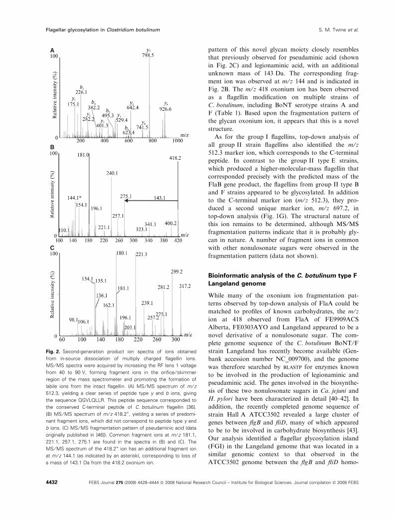
pattern of this novel glycan moiety closely resembles
that previously observed for pseudaminic acid (shown
in Fig. 2C) and legionaminic acid, with an additional
unknown mass of 143 Da. The corresponding frag-
ment ion was observed at m ⁄ z 144 and is indicated in
Fig. 2B. The m ⁄ z 418 oxonium ion has been observed
as a flagellin modification on multiple strains of
C. botulinum, including BoNT serotype strains A and
F (Table 1). Based upon the fragmentation pattern of
the glycan oxonium ion, it appears that this is a novel
structure.
As for the group I flagellins, top-down analysis of
all group II strain flagellins also identified the m ⁄ z512.3 marker ion, which corresponds to the C-terminal
peptide. In contrast to the group II type E strains,
which produced a higher-molecular-mass flagellin that
corresponded precisely with the predicted mass of the
FlaB gene product, the flagellins from group II type B
and F strains appeared to be glycosylated. In addition
to the C-terminal marker ion (m ⁄ z 512.3), they pro-
duced a second unique marker ion, m ⁄ z 697.2, in
top-down analysis (Fig. 1G). The structural nature of
this ion remains to be determined, although MS ⁄MS
fragmentation patterns indicate that it is probably gly-
can in nature. A number of fragment ions in common
with other nonulosonate sugars were observed in the
fragmentation pattern (data not shown).
Bioinformatic analysis of the C. botulinum type F
Langeland genome
While many of the oxonium ion fragmentation pat-
terns observed by top-down analysis of FlaA could be
matched to profiles of known carbohydrates, the m ⁄ zion at 418 observed from FlaA of FE9909ACS
Alberta, FE0303AYO and Langeland appeared to be a
novel derivative of a nonulosonate sugar. The com-
plete genome sequence of the C. botulinum BoNT ⁄Fstrain Langeland has recently become available (Gen-
bank accession number NC_009700), and the genome
was therefore searched by blastp for enzymes known
to be involved in the production of legionaminic and
pseudaminic acid. The genes involved in the biosynthe-
sis of these two nonulosonate sugars in Ca. jejuni and
H. pylori have been characterized in detail [40–42]. In
addition, the recently completed genome sequence of
strain Hall A ATCC3502 revealed a large cluster of
genes between flgB and fliD, many of which appeared
to be to be involved in carbohydrate biosynthesis [43].
Our analysis identified a flagellar glycosylation island
(FGI) in the Langeland genome that was located in a
similar genomic context to that observed in the
ATCC3502 genome between the flgB and fliD homo-
A
B
C
Fig. 2. Second-generation product ion spectra of ions obtained
from in-source dissociation of multiply charged flagellin ions.
MS ⁄ MS spectra were acquired by increasing the RF lens 1 voltage
from 40 to 90 V, forming fragment ions in the orifice ⁄ skimmer
region of the mass spectrometer and promoting the formation of
labile ions from the intact flagellin. (A) MS ⁄ MS spectrum of m ⁄ z512.3, yielding a clear series of peptide type y and b ions, giving
the sequence QGVLQLLR. This peptide sequence corresponded to
the conserved C-terminal peptide of C. botulinum flagellin [36].
(B) MS ⁄ MS spectrum of m ⁄ z 418.2+, yielding a series of predomi-
nant fragment ions, which did not correspond to peptide type y and
b ions. (C) MS ⁄ MS fragmentation pattern of pseudaminic acid (data
originally published in [46]). Common fragment ions at m ⁄ z 181.1,
221.1, 257.1, 275.1 are found in the spectra in (B) and (C). The
MS ⁄ MS spectrum of the 418.2+ ion has an additional fragment ion
at m ⁄ z 144.1 (as indicated by an asterisk), corresponding to loss of
a mass of 143.1 Da from the 418.2 oxonium ion.
Flagellar glycosylation in Clostridium botulinum S. M. Twine et al.
4432 FEBS Journal 275 (2008) 4428–4444 ª 2008 National Research Council – Institute for Biological Sciences. Journal compilation ª 2008 FEBS
logs (Fig. 3). The order of the first 29 ORFs of the
FGI (FGI-I) was completely conserved between Lange-
land and ATCC3502, with the homology of each pre-
dicted protein product never below 80% amino acid
identity. A number of genes in this conserved region
appear to be involved in carbohydrate biosynthesis
and show homology to capsular biosynthetic proteins
of group B Streptococcus agalactia, in particular the
predicted gene products of CLI_2752–2755, which
show homology to CpsE, CpsD, CpsB and CpsC of
S. agalactia. Homologs of the sialic acid biosynthetic
genes neuB (CLI_2745) and neuA (CLI_2746) were
also present [44]. In contrast, the region immediately
downstream of the conserved region and extending to
the flaA structural genes (CBO2730 and CBO2731
ATCC3502; CLI_2781 in Langeland) differed signifi-
cantly between Langeland and ATCC3502 in terms of
their genetic content. This second section of the FGI
(FGI-II) contains a 22 kb deletion in Langeland, and
the organization of the remaining ORFs differs sub-
stantially. The ATCC3502 genome contained tandem
copies of the flagellin structural genes (flaA1 and
flaA2), but only a single flaA gene was found at the
analogous position in the Langeland FGI. Both
Langeland and ATCC3502 genomes contained a num-
ber of carbohydrate biosynthetic genes in this region,
including homologs of a second set of the sialic acid
biosynthetic genes, neuA and neuB. However, only the
Langeland locus contained predicted gene products
with sequence similarity to proteins recently shown to
be involved in legionaminic acid biosynthesis in
Ca. coli [45]. These genes and the Campylobacter
homologs are listed in Table 2.
Nano-LC-MS ⁄ MS analysis of a C. botulinum
FE9909ACS Alberta flagellin peptide digest
To precisely assign the location of the post-translational
modifications, flagellar tryptic peptides of C. botulinum
strain FE9909ACS Alberta were analysed by nano-LC-
Table 2. Identification of legionaminic acid biosynthetic gene homologs in C. botulinum type F Langeland. The E values were obtained by
BLASTP of the Campylobacter jejuni 11168 protein sequence against the C. botulinum type F Langeland genome, and results are for the
Langeland ORF that gave the highest E value.
C. jejuni gene
number Gene name
Langeland
homologous ORF E value Function in Campylobacter jejuni Reference
Cj1319 Unknown CLI_2770 3e-98 Unknown [52]
Cj1320 Unknown CLI_2769 4e-58 Unknown [52]
Cj1325 ptmH CLI_2776 3e-06 Methyl transferase [52]
Cj1327 ptmC CLI_2775 1e-58 Legionaminic acid synthase [62]
Cj1328 ptmD CLI_2777 1e-68 Legionaminic acid biosynthetic gene [62]
Cj1329 ptmE CLI_2778 1e-67 Legionaminic acid biosynthetic gene [62]
Cj1331 ptmB CLI_2773 4e-23 CMP-legionaminic acid synthase [13,52]
Fig. 3. Flagellar glycosylation island locus of C. botulinum strain Langeland.
S. M. Twine et al. Flagellar glycosylation in Clostridium botulinum
FEBS Journal 275 (2008) 4428–4444 ª 2008 National Research Council – Institute for Biological Sciences. Journal compilation ª 2008 FEBS 4433
MS ⁄MS. Many of the MS ⁄MS spectra could be readily
assigned, but a number of peptide spectra were observed
that did not match any expected tryptic peptides. In
each case, an intense ion at m ⁄ z 418 was observed for
each of these unassignable peptides. This ion did not
correspond to any peptide fragmentation and also
accounted for the mass difference between predicted
and observed peptide ion masses, confirming that the
putative oxonium ion observed in top-down studies of
the intact protein corresponds to the flagellin glycan
modification. The MS ⁄MS spectrum of tryptic peptide
169-178 (VGSINIGSGK) is shown in Fig. 4A, with the
type y and b ions series indicated. Nano LC-MS ⁄MS
analyses were performed upon tryptic and endo protei-
nase GluC (Staphylococcus areus protease V8; New Eng-
land Biolabs, Ipswich, MA, USA) digests of the protein,
and resulted in 98% sequence coverage. This process
identified both modified and unmodified peptides
(Fig. 4C).
Determination of glycan attachment site by
electron transfer dissociation
Examination of the modified peptide sequences showed
no evidence of the classic eukaryotic N-linked or the
more recently defined prokaryotic N-linked consensus
sequons [46]. The glycan linkage was therefore sus-
pected to be O-linked. Although the modified glyco-
peptides were readily detected, based upon mass
differences from the predicted sequence, the precise
sites of glycosylation were not. The labile nature of the
glycosidic bond between the hydroxyl amino acid of
serine or threonine and the carbohydrate residue made
it impossible to detect fragment ions corresponding to
the intact modification on the native glycopeptide. To
date, mapping of O-linked glycans has required chemi-
cal derivatization via alkaline hydrolysis of each glyco-
peptide prior to collision-activated dissociation (CAD)
to map the precise site of glycosylation. Electron trans-
fer dissociation (ETD) differs from traditional CAD in
that it preserves the post-translational modifications of
peptides, thereby allowing direct determination of the
sites of post-translational modification without prior
chemical treatment [47–50]. HPLC fractions containing
glycopeptides from FE9909ACS Alberta FlaA were
fragmented using ETD, and seven sites of modification
were identified at Ser126, Ser139, Ser142, Ser165,
Ser171, Ser176 and Ser182 (Fig. 4C). The ETD spec-
trum of peptide T is shown in Fig. 4B, and the corre-
sponding c and z ions are highlighted. The mass
increase imparted by these modifications of the intact
protein was consistent with the measured masses indi-
cating occupancy of 3–7 sites, with a glycan of mass
417 Da on each FlaA monomer. Some microhetero-
geneity was observed at certain serine residues, for
example peptide T was found in two forms, with either
one or both serine residues modified. Accurate mass
measurements were performed on the glycan oxonium
ions in the MS ⁄MS spectra to determine a plausible
A
B
C
Fig. 4. MS ⁄ MS and electron transfer dissociation mass spectrum
of the VGS*INIGS*GK tryptic peptide (asterisks indicate modified
amino acids). (A) MS ⁄ MS spectrum of peptide T of FlaA. Peptide
type y and b fragmentation ions are indicated, and an intense ion at
m ⁄ z 418 was observed, corresponding to the putative glycan oxoni-
um ion. (B) ETD mass spectrum obtained on the [M + 3H]3+ ion at
m ⁄ z 589.4, corresponding to the same peptide T as FlaA. Observed
fragment ions of type c and z are indicated. This peptide was found
to harbor two glycan modifications of 417 Da, on serines 171 and
186 respectively. (C) Peptide sequence coverage obtained from
MS ⁄ MS sequencing of FlaA digested with trypsin or GluC.
Sequenced peptides are indicated in bold, and glycosylation sites
are underlined.
Flagellar glycosylation in Clostridium botulinum S. M. Twine et al.
4434 FEBS Journal 275 (2008) 4428–4444 ª 2008 National Research Council – Institute for Biological Sciences. Journal compilation ª 2008 FEBS
empirical formula for the glycan moiety. The known
masses of peptide fragment ions were used as internal
standards as previously described [39]. The accurate
mass of the glycan oxonium ion was determined to be
418.1822 Da (± 0.0015 Da). Thus the top-ranked
plausible elemental composition for the glycan residue
was C17H28N3O9.
NMR structural analysis of the flagellin
glycopeptide
Structural elucidation of the glycan was accomplished
using a purified tryptic glycopeptide (glycopeptide 1),
and glycopeptide 2, which was produced from exten-
sive proteolytic digestion of intact flagellin using pro-
teinase K (Figs 5 and 6). MS analysis showed that
glycopeptide 2 had the sequence GSAK. Homonuclear
and heteronuclear NMR experiments on these samples
allowed complete assignment of the 1H and 13C reso-
nance shifts (Table 3) and determination of the glycan
structure, which was identical for both peptides.
For the tryptic glycopeptide 1, due to the small
amount material that was available, the signal-to-noise
ratio was maximized by concentrating the sample from
150 to 50 lL by the use of two immiscible liquid plugs.
Diffusion-weighted NMR spectroscopy was used to
differentiate resonances of the higher-molecular-mass
peptidyl glycan from those of contaminants. Unusual
resonances were observed that did not correspond to
known structures, especially the presence of a singlet
at 2.74 p.p.m. (Fig. 5). This experiment effectively
attenuated signals from the lower-molecular-mass
contaminants (higher diffusion rates), leaving reso-
nances from the larger peptide as well as the glycan as
the dominant peaks in the NMR spectrum (Fig. S2).
Selective TOCSY experiments on the putative glycan
resonances were then used to assign the proton reso-
nances and measure coupling constants (Fig. 5). The
Fig. 5. Structure and proton NMR analyses
of the O-linked glycan found on the glyco-
peptides isolated from the flagellin of
C. botulinum. (A) Stucture and atom num-
bering for aLeg5GluNMe7Ac. (B) 1H-NMR
spectrum (16 scans) of protein-
ase K-digested glycopeptide 2 (GS*AK).
(C) 1H-NMR spectrum (256 scans) of the
tryptic glycopeptide (VGS*INIGS*GK). (E–G)
Selective TOCSY experiments for the tryptic
glycopeptide using a mixing time of 80 ms
for the Leg-H3ax (D), Leg-H9 (E) and
glutamate H4 (F) resonances.
S. M. Twine et al. Flagellar glycosylation in Clostridium botulinum
FEBS Journal 275 (2008) 4428–4444 ª 2008 National Research Council – Institute for Biological Sciences. Journal compilation ª 2008 FEBS 4435
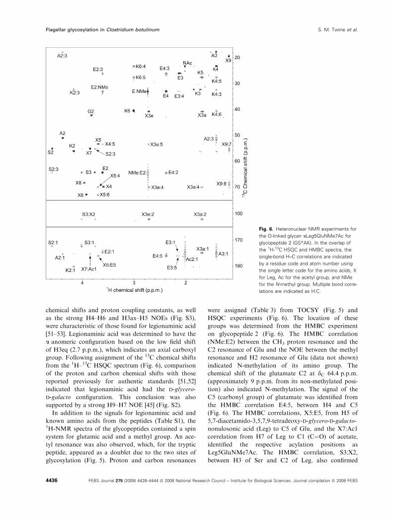
chemical shifts and proton coupling constants, as well
as the strong H4–H6 and H3ax–H5 NOEs (Fig. S3),
were characteristic of those found for legionaminic acid
[51–53]. Legionaminic acid was determined to have the
a anomeric configuration based on the low field shift
of H3eq (2.7 p.p.m.), which indicates an axial carboxyl
group. Following assignment of the 13C chemical shifts
from the 1H–13C HSQC spectrum (Fig. 6), comparison
of the proton and carbon chemical shifts with those
reported previously for authentic standards [51,52]
indicated that legionaminic acid had the d-glycero-
d-galacto configuration. This conclusion was also
supported by a strong H9–H7 NOE [45] (Fig. S2).
In addition to the signals for legionaminic acid and
known amino acids from the peptides (Table S1), the1H-NMR spectra of the glycopeptides contained a spin
system for glutamic acid and a methyl group. An ace-
tyl resonance was also observed, which, for the tryptic
peptide, appeared as a doublet due to the two sites of
glycosylation (Fig. 5). Proton and carbon resonances
were assigned (Table 3) from TOCSY (Fig. 5) and
HSQC experiments (Fig. 6). The location of these
groups was determined from the HMBC experiment
on glycopeptide 2 (Fig. 6). The HMBC correlation
(NMe:E2) between the CH3 proton resonance and the
C2 resonance of Glu and the NOE between the methyl
resonance and H2 resonance of Glu (data not shown)
indicated N-methylation of its amino group. The
chemical shift of the glutamate C2 at dC 64.4 p.p.m.
(approximately 9 p.p.m. from its non-methylated posi-
tion) also indicated N-methylation. The signal of the
C5 (carbonyl group) of glutamate was identified from
the HMBC correlation E4:5, between H4 and C5
(Fig. 6). The HMBC correlations, X5:E5, from H5 of
5,7-diacetamido-3,5,7,9-tetradeoxy-d-glycero-d-galacto-
nonulosonic acid (Leg) to C5 of Glu, and the X7:Ac1
correlation from H7 of Leg to C1 (C¼O) of acetate,
identified the respective acylation positions as
Leg5GluNMe7Ac. The HMBC correlation, S3:X2,
between H3 of Ser and C2 of Leg, also confirmed
Fig. 6. Heteronuclear NMR experiments for
the O-linked glycan aLeg5GluNMe7Ac for
glycopeptide 2 (GS*AK). In the overlap of
the 1H-13C HSQC and HMBC spectra, the
single-bond H–C correlations are indicated
by a residue code and atom number using
the single letter code for the amino acids, X
for Leg, Ac for the acetyl group, and NMe
for the N-methyl group. Multiple bond corre-
lations are indicated as H:C.
Flagellar glycosylation in Clostridium botulinum S. M. Twine et al.
4436 FEBS Journal 275 (2008) 4428–4444 ª 2008 National Research Council – Institute for Biological Sciences. Journal compilation ª 2008 FEBS
O-linked glycosylation to Ser. Based on these NMR
results, the glycan O-linked to Ser on the flagellin gly-
copeptides was determined to be 5,7,9-tetradeoxy-d-
glycero-a-d-galacto-nonulosonic acid, N-acylated with
N-methylated glutamate at C5 and acetate at C7
(aLeg5GluNMe7Ac).
Detection of flagellin signature ions in
C. botulinum culture supernatants
We next examined a C. botulinum culture supernatant,
containing BoNT, to determine whether unique flagel-
lin fragment ions can be used as surrogate markers for
detection of BoNT contamination. A culture superna-
tant from C. botulinum strain FE9909ACS Alberta was
prepared, which contained 20 000 mouse lethal doses ⁄mL by the mouse bioassay. This preparation was then
heat-inactivated. We examined this fraction by MS-
based methods, and were indeed able to detect flagellin
protein as well as the C. botulinum flagellin-specific
marker ions at m ⁄ z 418 and m ⁄ z 512. (Fig. 7A,B). The
deconvoluted mass profile is presented in the inset to
Fig. 7A, and the protein mass is in close agreement
with that obtained for purified flagellin from this strain
(Fig. 1B). Multiply charged protein ions at m ⁄ z 1197
and 1346 were selected for tandem mass spectrometry.
Spectra were acquired at 15–25 V collision energy using
argon as the collision gas, with marker ions characteris-
tic C. botulinum flagellin observed at m ⁄ z 418.2, 512.3
and 1023.6. Increasing the RF lens 1 voltage from 40
to 90 V resulted in non-specific fragmentation in the
orifice ⁄ skimmer region of the mass spectrometer,
increasing the intensity of flagellin marker ions at m ⁄ z418.2 and 512, and allowing MS ⁄MS spectra to be
obtained for each ion. The MS ⁄MS spectrum of m ⁄ z512.3 yielded a clear series of peptide type y and b ions,
giving the sequence PQGVLQLLR. MS ⁄MS spectra
were obtained for m ⁄ z 418.2, yielding a series of frag-
ment ions characteristic of the glycan modification of
C. botulinum strain FE9909ACS Alberta flagellin,
thereby confirming that the labile flagellin marker ions
can be detected in a complex protein mixture that con-
tains BoNT.
Discussion
In this study, we have demonstrated that the flagellins
of C. botulinum are glycosylated in O-linkage with
novel glycans at up to seven sites per monomer. Top-
down MS analysis of flagellin proteins prepared from
Table 3. NMR data for the O-linked nonulosonic acid derivative,
aLeg5GluNMe7Ac, found on glycopeptides from flagella of
C. botulinum. Carbon and proton chemical shifts were referenced
to an internal acetone standard (dH 2.225 p.p.m. and dC
31.5 p.p.m.). The error for dH is ± 0.02 p.p.m., that for dC is
± 0.4 p.p.m., and that for JH,H is ± 0.2 Hz. J, coupling constant;
d, chemical shift. AC, acetyl; GluNMe, N-methyl-glutamyl.
Residue
Proton
(1H)
dH
(p.p.m.)
Carbon
(13C)
dC
(p.p.m.) JH,H J (Hz)
Leg C1 174.6
C2 101.8 J3ax,3eq 13.2
H3ax 1.68 C3 40.7 J3ax,4 11.0
H3eq 2.71 J3eq,4 3.9
H4 3.59 C4 69.9 J4,5 10.0
H5 3.69 C5 53.3 J5,6 10.4
H6 3.92 C6 73.0 J6,7 3.0
H7 3.85 C7 55.2 J7,8 3.0
H8 3.95 C8 68.4 J8,9 6.3
H9 1.16 C9 19.4
GluNMe C1 174.6
H2 3.61 C2 64.4
H3 2.12 C3 26.1
H4 2.38 C4 32.9
C5 176.0
Me 2.73 Me 33.1
Ac C=O 175.2
CH3 2.02 CH3 23.3
A
B
Fig. 7. Detection of flagellin in culture supernatants from C. botu-
linum strain FE9909ACS Alberta. (A) Electrospray mass spectrum
of crude BoNT preparation. The inset shows the deconvoluted
mass profile, indicating an intact protein mass of 32 300 Da. The
arrows indicate protein ions that were selected for tandem mass
spectrometry. (B) Tandem mass spectrometry analyses of a multi-
ply charged protein ion at m ⁄ z 1197.027+. Ions at m ⁄ z 418.2+,
512.32+ and 1023.6+ were observed in the tandem mass spectrum.
S. M. Twine et al. Flagellar glycosylation in Clostridium botulinum
FEBS Journal 275 (2008) 4428–4444 ª 2008 National Research Council – Institute for Biological Sciences. Journal compilation ª 2008 FEBS 4437
strains belonging to a number of unique toxin types
revealed diversity in glycan structure amongst isolates.
Glycan mass and MS ⁄MS fragmentation patterns of
each of the glycan oxonium ions showed that group I
C. botulinum strains are able to glycosylate flagellin
monomers with derivatives of either a nonulosonate
sugar, a di-acetamido-substituted hexuronic acid sugar
or a tri-acetamido-substituted hexuronic acid sugar. In
contrast, preliminary studies of group II C. botulinum
strains revealed that, while type B and type F strains
produce flagellins of increased mass that were glycosy-
lated with a novel glycan of mass 696 Da, the FlaB
flagellin from group II type E strains is distinctive in
that it is a higher-molecular-mass flagellin protein
that is not glycosylated. It has been demonstrated
indirectly that numerous clostridial species appear to
glycosylate their flagellin proteins, but this is the first
study to provide specific structural details of clostridial
flagellar glycan modifications. Previous work demon-
strating the sensitivity of the glycan from flagella of
C. acetobutylicum to neuraminidase treatment provided
the first evidence that glycosylation may involve
a sialic acid-related sugar [28]. More recent comparative
genomic analysis revealed the presence of homologs of
sialic acid biosynthetic genes in the completed genomes
of C. acetobutylicum and C. tetani [43,54].
In the present study, NMR and MS were used for
structural assignment of a novel 417 Da glycan found
on flagellins of the group I type A strain FE9909ACS
Alberta and the type F strain Langeland. Using a com-
bination of MS and NMR techniques, we have demon-
strated that these strains of C. botulinum glycosylate
flagellin not with sialic acid but with a novel derivative
of the sialic acid-like nonulosonate sugar legionaminic
acid, Leg5GluNMe7Ac. Legionaminic acid belongs to
the class of nonulosonic acids that consists of 5,7-diami-
no-3,5,7,9-tetradeoxy nonulosonate derivatives uniquely
found in microorganisms. The sugars of this class have
been reported as components of a variety of capsular
polysaccharides and lipopolysaccharides from a number
of Gram-negative bacteria [51]. More recently, these
nonulosonate derivatives have been shown to be com-
ponents of the pilin glycoprotein from P. aeruginosa [3]
as well as of the flagellar glycoproteins of Ca. jejuni ⁄ coli,H. pylori and Aeromonas caviae [12,12,39].
This is the first report of the biosynthesis of a nonu-
losonate sugar in a Gram-positive, anaerobic bacteria.
It remains to be established whether the presence of
these nonulosonate sugars facilitiates a host ⁄pathogeninteraction or imparts a novel biological function to
C. botulinum cells. However, the increasing number of
examples of pathogenic species that incorporate these
sugars on prominent cell-surface glycoconjugate struc-
tures suggests a potential role in host ⁄pathogeninteraction. It is noteworthy that three of the four
strains associated with infant botulism (FE9909ACS
Alberta, FE0303AYO and FE9904BMT) produced
glycan fragmentation patterns characteristic of legio-
naminic acid derivatives, while the flagellins from three
of four strains not associated with C. botulinum
infections (PA9508, 17A and H461297F) were modified
with a di-N-acetylhexuronic acid derivative. It has been
suggested that the similarity of the bacterial nonulo-
sonic acids to sialic acids may provide an immune
‘cloak’, whereby the host response to the invading
bacterium is less intense [51].
Future work will be directed towards examination
of flagellin glycans from distinct epidemiological clus-
ters of C. botulinum to determine whether there is
indeed a correlation between glycan structure and
pathogenic potential. Until recently, the genetic manip-
ulation of Clostridium spp. has been difficult. However,
with the recent report of a mutagenesis system based
on the mobile GpII intron for Clostridium spp., it will
now be possible to perform functional studies on puta-
tive flagellar glycosylation genes [55]. This system will
provide the means to determine the role of these dis-
tinct glycan moieties in both flagellar assembly and
other biological interactions that may be important for
C. botulinum pathogenesis.
Many of the tools for the identification and genetic
discrimination of group I C. botulinum strains are
based on a very limited number of structurally and
genetically similar toxin types. The recent work of
Paul et al. [35,36] using flagellin gene sequencing has
provided an additional means to more accurately dis-
tinguish C. botulinum isolates. We demonstrate here
that top-down MS analysis of the flagellin protein
can be utilized to rapidly detect C. botulinum through
identification of a common C-terminal peptide marker
ion (m ⁄ z 512). In addition, this top-down analysis has
revealed the presence of unique flagellar glycan mar-
ker ions that are found exclusively on individual
strains and could also be utilized for strain typing.
Genomic comparisons of the FGI from the two
C. botulinum strains for which the genome sequence is
available (ATCC3502 and Langeland) indicated that
diversity in the gene content at this locus may also
provide additional discriminatory power for typing of
isolates.
In a similar manner, extensive genomic analysis of
multiple Campylobacter strains revealed that the FGI
was a hypervariable region of the chromosome. Diver-
gence in genetic content at the Campylobacter flagellar
glycosylation locus led to identification of a cluster of
genes within this locus that were shown to be a genetic
Flagellar glycosylation in Clostridium botulinum S. M. Twine et al.
4438 FEBS Journal 275 (2008) 4428–4444 ª 2008 National Research Council – Institute for Biological Sciences. Journal compilation ª 2008 FEBS
marker for a livestock-associated clade [56]. Two of
the genes identified from this cluster – ptmG (Cj1324)
and ptmH (Cj1325 ⁄ 6) – have been shown to be
involved in the biosynthesis of the flagellar glycans
Leg5Am7Ac (ptmG) and Leg5AmNMe7Ac (ptmH)
[45]. In the present study, bioinformatic analysis identi-
fied a homolog of ptmH in the C. botulinum Langeland
genome (CLI_2776), together with homologs of other
legionaminic acid biosynthetic genes (see Table 2). It
remains to be established whether diversity in flagellar
glycan biosynthetic capacity in C. botulinum is related
to host specificity and the colonization ability of
isolates.
Detailed characterization of prokaryotic glycopro-
teins still presents technical challenges. MS and
MS ⁄MS techniques provide substantial information on
the nature of a particular glycan, the type of linkage
and the site of attachment, although the lability of par-
ticular glycan modifications may still be problematic.
In this study we utilized the electron transfer dissocia-
tion soft fragmentation technique to map all glycan
attachment sites without the need for chemical deriva-
tization. In this manner, the amino acid sequence and
glycan attachment sites for all glycopeptides, regardless
of size or charge state, were successfully identified. A
larger glycopeptide ion failed to yield informative frag-
ment ions by ETD alone, instead mainly generating
charge-reduced precursor ions. In this instance, frag-
ment ions were generated by performing gentle colli-
sion-activated dissociation with a low Q value on the
charge-reduced precursor ions [57]. This process
favored the production of c and z fragment ions over
production of b and y ions, allowing mapping of the
glycan attachment site.
Detailed structural elucidation of novel glycan moie-
ties or glycopeptides by NMR requires substantially
higher amounts of material (lg) of considerably
greater purity than is required for MS analysis (ng).
These requirements can provide a considerable techni-
cal challenge. This is especially true for glycoproteins
from fastidious microorganisms or organisms that
require bio-containment. In this study, we have dem-
onstrated that, even for the very ‘sample limited’ and
partially purified peptidyl glycan samples that are
obtained from C. botulinum flagellin, diffusion-
weighted experiments and sample concentration with
immiscible plugs enable these challenges to be over-
come, facilitating rigorous definition of structures by
NMR techniques.
Preparation of glycopeptide by trypsin digest pro-
vided sufficient quantity and quality of sample to eluci-
date the basic glycan structure by NMR, as done
previously for determination of pseudaminic acid on
Ca. coli flagellin [39]. Due to the unusual nature of the
N-methyl glutamate modification, larger amounts were
required in order to detect the weak HMBC correla-
tions required to determine unambiguously the loca-
tion of acetyl and N-methyl-glutamyl groups on
legionaminic acid. In this case, extensive proteinase K
hydrolysis of the flagellin protein produced glycan
attached to only three or four amino acids, which
could be more readily fractionated from non-glycosy-
lated peptides by gel chromatographic separation in a
manner similar to that described by Voisin et al. [38].
Different glycopeptide preparations also helped to dis-
tinguish the signals from the amino acid linked to Leg
from those in the peptide. Both purification methods
are complementary, and their use depends on the nat-
ure of the glycan being isolated and characterized.
The ability to use mass spectrometry to rapidly iden-
tify the flagellar modifications of C. botulinum opens
the possibility for screening large numbers of strains
for unique sugars to potentially establish biological
correlations for strains of C. botulinum. This could
include geographic distributions of strains, coloniza-
tion potential and type of disease caused (i.e. coloniza-
tion versus wound botulism). Flagellar modification
with nonulosonate sugars may be correlated with the
ability of organisms such as C. botulinum to establish
infections in the gastrointestinal tract, and will be the
subject of future studies.
Experimental procedures
Bacterial strains
C. botulinum strains were stored and archived at the
Botulism Reference Service for Canada at )86 �C on
Microbank� beads (Pro-Lab Diagnostics, Richmond Hill,
Canada), and their sources have been detailed elsewhere [35].
All C. botulinum cultures were routinely grown in SPGY
broth containing 5% w ⁄ v special peptone (Oxoid Inc.,
Basingstoke, UK), 0.5% w ⁄ v peptone (Difco, Tucker, GA,
USA), 2% w ⁄ v yeast extract (Difco), 0.4% w ⁄ v glucose
(Difco) and 0.1% w ⁄ v sodium thioglycolate (Sigma-Aldrich,
St Louis, MO, USA), adjusted to a pH of 7.2 using HCl.
Cells were grown for 24–48 h in an atmosphere of 10% H2,
10% CO2 and 80% N2 at either 35 �C (group I strains) or
room temperature (group II strains).
Purification of flagella
Flagella proteins were isolated as described previously [36].
In brief, cultures were grown overnight in 100 mL SPGY
broth. Cells were harvested and flagella were sheared from
the cell surface using a 50 mL tissue homogenizer. Follow-
S. M. Twine et al. Flagellar glycosylation in Clostridium botulinum
FEBS Journal 275 (2008) 4428–4444 ª 2008 National Research Council – Institute for Biological Sciences. Journal compilation ª 2008 FEBS 4439
ing removal of whole cells by low-speed centrifugation,
flagellar filaments were collected by centrifugation at
130 000 g for 1 h, pellets were washed in ultrapure water,
re-centrifuged, and resuspended in ultrapure water. Before
any downstream analysis, all flagellin protein preparations
were heat-treated for 25 min at 75 �C to denature any
BoNT present in the sample. The purity of flagellar prepa-
rations was determined by SDS–PAGE.
Mass spectrometry of isolated flagellins
Purified flagellin was dialysed in aqueous 0.2% v ⁄ v formic
acid using a Centricon YM-30 membrane filter (Millipore,
Billerica, MA, USA). The resulting solution was infused in
a QTOF2 hybrid mass spectrometer (Waters, Milford, MA,
USA) at a flow rate of 0.5 lLÆmin)1. Top-down experi-
ments were performed as described by Schirm et al. [37]
using argon collision gas with collision energies ranging
from 20–30 V. The RF lens 1 voltage was increased from
30 to 125 V in order to obtain second-generation fragment
ion spectra.
Solution enzymatic digests
To identify the type and location of glycosylation sites,
flagellin (50–200 lg) was digested with trypsin (Promega,
Madison, WI, USA) at a protein:enzyme ratio of 30 : 1
(v ⁄ v) in 50 mm ammonium bicarbonate at 37 �C overnight
or with endoproteinase GluC (Sigma) at a protein:enzyme
ratio of 25 : 1 (v ⁄ v) in 100 mm phosphate buffer, pH 7.8,
at 37 �C overnight. Protein digests were analysed by nano-
LC-MS ⁄MS using a QTOF2 or QTOF Ultima hybrid
quadrupole time-of-flight mass spectrometer coupled to a
CapLC capillary HPLC system (Waters). MS ⁄MS spectra
were acquired automatically on doubly, triply and quadru-
ply charged ions. Peak lists were automatically generated
by proteinlynx software (Waters) with the following
parameters: smoothing – four channels, two smooths,
Savitzky–Golay mode; centroid – minimum peak width at
half height of four channels, centroid top 80%. Tryptic
peptides were analysed by nano-LC-MS ⁄MS, and spectra
were searched against the National Centre for Biotechno-
logy nonredundant database and an in-house database of
sequenced C. botulinum flagellin proteins using mascot
2.0.1 (Matrix Science, London, UK), as described previ-
ously [35]. MS ⁄MS spectra that did not correspond to
predicted tryptic peptides were examined manually.
HPLC purification of tryptic peptides
Tryptic digests of flagellin were fractionated using an
Agilent 1100 series HPLC with a diode array detector
(Agilent Technologies, Palo Alto, CA, USA). Each tryptic
digest (100 lL) was separated using a 4.6 · 250 mm Jupiter
C18 reverse-phase column with a Phenomenex pre-column
(SecurityGuard, Torrance, CA, USA). Peptides were sepa-
rated using a linear gradient of 5–60% acetonitrile, 0.5%
formic acid over 40 min at a flow rate of 1 mLÆmin)1. A
post-column splitter was used to divert approximately
60 lLÆmin)1 of column eluate to the electrospray interface
of the QTOF2 to allow real-time monitoring of ion elution
profiles, allowing specific peptides to be isolated as they
eluted from the column. A 7 lL aliquot of each fraction
was retained, and the remainder immediately evaporated to
dryness and stored at )20 �C. Aliquots of each fraction
were screened by nano-LC-MS ⁄MS using the QTOF2 to
confirm the peptide contents of each HPLC fraction. Pure
tryptic glycopeptide (glycopeptide 1) was thus obtained.
Accurate mass measurement of C. botulinum
glycan
A glycopeptide-containing HPLC fraction was infused in
the QTOF Ultima at 1 lLÆmin)1 and the MS ⁄MS spectrum
was recorded over a period of 30 min. Accurate mass deter-
mination of the glycan oxonium ion was achieved using a
number of neighboring peptide fragment ions as internal
mass standards.
Determination of glycan linkage sites by electron
transfer dissociation
Electron transfer dissociation preserves delicate modifica-
tions during the fragmentation process and is ideal for
identifying the linkage sites of O-glycans [47,49,58]. Glyco-
peptide-containing HPLC fractions were infused at 1 lLÆmin)1 into the electrospray ionization source of an LTQ XL
linear ion trap (Thermo Fisher Scientific, Nepean, Canada)
capable of performing ETD. Initially, CAD MS ⁄MS analysis
was performed on the glycopeptide ions to confirm their
identity. ETD was performed using fluoranthene as the anio-
nic reagent and with supplementary activation enabled. The
ETD reaction time was adjusted for optimal fragmentation
of each glycopeptide (typically 35 ms). One glycopeptide ion
failed to yield informative fragment ions by ETD alone,
instead mainly generating charged-reduced precursor ions. In
this instance, c and z fragment ions were generated by per-
forming gentle CAD (10 V) on the individual charge-reduced
precursor ions generated by ETD. Production of c and z frag-
ment ions was favored over b and y ions by reducing the
Q value from its default of 0.25 to 0.15 [57].
Preparation of glycopeptide using proteinase K
digestion
A preparation of flagellin (8 mg) in water was adjusted to
pH 8.5 by addition of Na2HPO4. Proteinase K (10 mg) was
Flagellar glycosylation in Clostridium botulinum S. M. Twine et al.
4440 FEBS Journal 275 (2008) 4428–4444 ª 2008 National Research Council – Institute for Biological Sciences. Journal compilation ª 2008 FEBS

added, and the solution was kept at 50 �C for 1 week. The
resulting digest was filtered through a SepPak C18 column
(Waters) (pre-washed with methanol and then water) and
desalted on a Sephadex G15 column (1.6 · 80 cm; Sigma-
Aldrich) in pyridine (0.4%) ⁄ acetic acid (1%) in H2O, moni-
tored by a refractive index detector. All fractions (5 mL
each) eluted before the salt peak were dried and analyzed by1H-NMR. A relatively clean fraction containing a glycopep-
tide (glycopeptide 2) as well as mixtures of this and other
glycopeptides were collected and analyzed by NMR and MS.
NMR analysis of purified glycopeptide
The HPLC-purified glycopeptide 1 sample [VGSINIGSGK
(169–178)] was lyophilized and resuspended in 99% D2O
(Cambridge Isotope Laboratories Inc., Andover, MD,
USA) and analyzed using NMR spectroscopy. To increase
the level of signal to noise by approximately threefold over
a conventional aqueous NMR sample, the sample was
resuspended in a minimum amount of D2O (50 lL) and
centered between two insoluble liquid plugs of FC43
(Sigma-Aldrich) in a 3 mm NMR tube. The aqueous sam-
ple was centered in the RF detection coil of the NMR
probe. The fraction containing glycopeptide 2 was lyophi-
lized and resuspended in 150 uL D2O in a 3 mm NMR
tube. For both samples, standard homo- and heteronuclear-
correlated two-dimensional 1H-NMR, 13C-HSQC, HMBC,
DQCOSY, NOESY and ROESY pulse sequences from
Varian (Palo Alto, CA, USA) were used for general assign-
ments. Selective one-dimensional TOCSY experiments with
a Z-filter were used for complete residue assignments and
measurement of proton coupling constants [59]. NMR spec-
tra were acquired on a Varian INOVA 500 MHz (1H) spec-
trometer with a Varian Z-gradient 3 mm triple resonance
probe (1H, 13C and 31P) or a Varian 600 MHz (1H) spec-
trometer equipped with a Varian 5 mm Z-gradient triple
resonance (1H, 13C and 15N) cryogenically cooled probe
(cold probe) for optimized sensitivity. NMR experiments
were acquired at 25 �C with suppression of the HOD reso-
nance at 4.78 p.p.m. by solvent presaturation. For proton
and carbon experiments, the methyl resonance of acetone
was used as an internal reference (dH 2.225 p.p.m. and dC31.5 p.p.m.). Diffusion-weighted NMR spectroscopy at
500 MHz was performed using a bipolar pulse pair-longitu-
dinal encode ⁄ decode (BPP-LED) to attenuate signals aris-
ing from low-molecular-mass contaminants and enhance
signals originating from larger molecules, thereby simplify-
ing the NMR spectrum [60].
Bioinformatic analysis of flagellar glycosylation
loci
The FGI in the genome sequence of C. botulinum strain
Langeland (Genbank accession number NC_009699) was
identified by first locating the ORFs corresponding to
homologs of the FGI flanking proteins FlgB and FliD by
blastp (http://www.ncbi.nlm.nih.gov/BLAST/). Open read-
ing frames between FlgB and FliD were then compared to
either the Hall A genome sequence (Genbank accession
number NC_009495), the Ca. jejuni NCTC11168 genome
sequence (Genbank accession number NC_002163) or the
complete NCBI protein sequence database to assign puta-
tive identities. The flagellar glycosylation locus map was
compiled using enhance map draw version 2.0 (Sci-Ed
Software, Carey, NC, USA).
Preparation of C. botulinum culture supernatants
Culture supernatants of C. botulinum were prepared using
the standard method employed by the Botulism Reference
Service of Canada with minor modifications. Briefly,
C. botulinum strain FE9909ACS was grown in 100 mL
SPGY broth for 5 days. Bacteria were removed by centrifu-
gation at 10 000 g for 10 min at 4 �C and the supernatant
divided. One half was filter-sterilized using a 0.22 lm filter
for toxin quantification by mouse bioassay (see below). The
remaining supernatant was heated for 25 min at 75 �C to
inactivate BoNT, but was not filter-sterilized. Heated super-
natant was dialyzed into Milli-Q water (Millipore) and con-
centrated tenfold using an Amicon spin filter (molecular
mass cut-off 10 000 Da) and diluted to the desired concen-
trations for MS detection of FlaA.
MS detection of flagellar marker ions in
C. botulinum culture supernatants
Concentrated culture supernatant (50 lL) was injected onto
a protein microtrap (Chromatographic Specialties, Brock-
ville, Canada) connected to the Agilent HPLC and washed
offline with 500 lL of Milli-Q water. An HPLC gradient of
5–60% acetonitrile, 0.2% formic acid (1 mLÆmin)1) over
60 min was used to resolve the protein mixture. A pre-col-
umn splitter was used to direct approximately 60 lLÆmin)1
of the HPLC mobile phase through the trap and into the
electrospray interface of the QTOF2 to allow real-time
monitoring of ion elution profiles. The MS ⁄MS spectra of
multiply charged ion peaks were recorded as described
above. When low-intensity ions at m ⁄ z 418 and 512 were
observed, the RF lens voltage was increased until the inten-
sity of these ions was sufficient to collect MS ⁄MS data.
MS ⁄MS data were then collected for each ion to confirm
that these were the labile flagellin-associated ions.
Mouse bioassay for BoNT
The mouse bioassay was conducted as previously described
[61]. Briefly, samples of flagellin or filter-sterilized culture
supernatant were diluted in gelatin phosphate buffer up to
1 ⁄ 1 000 000. Samples were tested by intraperitoneal injection
S. M. Twine et al. Flagellar glycosylation in Clostridium botulinum
FEBS Journal 275 (2008) 4428–4444 ª 2008 National Research Council – Institute for Biological Sciences. Journal compilation ª 2008 FEBS 4441
into a minimum of two female CFI white mice. Mice were
observed for 72 h for symptoms of BoNT intoxication. The
observational endpoint used was pinched waist with labored
breathing, after which animals were killed. These experiments
were carried out in accordance with the guidelines laid down
by the National Institutes of Health in the USA regarding
the care and use of animals for experimental procedures.
Acknowledgements
This work was supported by project CRTI-02-0091TA
of the Defense Research and Development Canada
Chemical, Biological, Radiological and Nuclear Res-
earch and Technology Initiative (J.W.A. and S.M.L.).
We thank Luc Tessier for technical assistance with mass
spectrometers and Greg Sanders and Jeff Bussey for
conducting mouse bioassays for BoNT activity.
References
1 Sobel J, Tucker N, Sulka A, McLaughlin J & Maslanka
S (2004) Foodborne botulism in the United States,
1990–2000. Emerg Infect Dis 10, 1606–1611.
1a Fenicia L, Annibali F & Aureli P (2007) Intestinal
toxemia botulism in Italy 1984–2005. Eur J Microbiol
Infect Dis 26, 385–394.
2 Akbulut D, Dennis J, Gent M, Grant KA, Hope V,
Ohai C, McLauchlin J, Mithani V, Mpamugo O, Ncube
F et al. (2005) Wound botulism in injectors of drugs:
upsurge in cases in England during 2004. Euro Surveill
10, 172–174.
3 Castric P, Cassels FJ & Carlson RW (2001) Structural
characterization of the Pseudomonas aeruginosa 1244
pilin glycan. J Biol Chem 276, 26479–26485.
4 Smedley JG III, Jewell E, Roguskie J, Horzempa J,
Syboldt A, Stolz DB & Castric P (2005) Influence of
pilin glycosylation on Pseudomonas aeruginosa 1244
pilus function. Infect Immun 73, 7922–7931.
5 Voisin S, Kus JV, Houliston S, St Michael F, Watson
D, Cvitkovitch DG, Kelly J, Brisson JR & Burrows LL
(2007) Glycosylation of Pseudomonas aeruginosa strain
Pa5196 type IV pilins with mycobacterium-like a-1,5-linked d-Araf oligosaccharides. J Bacteriol 189, 151–
159.
6 Stimson E, Virji M, Makepeace K, Dell A, Morris HR,
Payne G, Saunders JR, Jennings MP, Barker S &
Panico M (1995) Meningococcal pilin: a glycoprotein
substituted with digalactosyl 2,4-diacetamido-2,4,6-tride-
oxyhexose. Mol Microbiol 17, 1201–1214.
7 Lindenthal C & Elsinghorst EA (1999) Identification of
a glycoprotein produced by enterotoxigenic Escherichia
coli. Infect Immun 67, 4084–4091.
8 Benz I & Schmidt MA (2001) Glycosylation with
heptose residues mediated by the aah gene product is
essential for adherence of the AIDA-I adhesin. Mol
Microbiol 40, 1403–1413.
9 Grass S, Buscher AZ, Swords WE, Apicella MA,
Barenkamp SJ, Ozchlewski N & St Geme J III (2003)
The Haemophilus influenzae HMW1 adhesin is glycosy-
lated in a process that requires HMW1C and phospho-
glucomutase, an enzyme involved in lipooligosaccharide
biosynthesis. Mol Microbiol 48, 737–751.
10 Brimer CD & Montie TC (1998) Cloning and compari-
son of fliC genes and identification of glycosylation in
the flagellin of Pseudomonas aeruginosa a-type strains.
J Bacteriol 180, 3209–3217.
11 Josenhans C, Vossebein L, Friedrich S & Suerbaum S
(2002) The neuA ⁄ flmD gene cluster of Helicobacter
pylori is involved in flagellar biosynthesis and flagellin
glycosylation. FEMS Microbiol Lett 210, 165–172.
12 Schirm M, Soo EC, Aubry AJ, Austin J, Thibault P &
Logan SM (2003) Structural, genetic and functional
characterization of the flagellin glycosylation process in
Helicobacter pylori. Mol Microbiol 48, 1579–1592.
13 Doig P, Kinsella N, Guerry P & Trust TJ (1996) Char-
acterization of a post-translational modification of
Campylobacter flagellin: identification of a sero-specific
glycosyl moiety. Mol Microbiol 19, 379–387.
14 Guerry P, Ewing CP, Schirm M, Lorenzo M, Kelly J,
Pattarini D, Majam G, Thibault P & Logan SM (2006)
Changes in flagellin glycosylation affect Campylobacter
autoagglutination and virulence. Mol Microbiol 60,
299–311.
15 Arora SK, Neely AN, Blair B, Lory S & Ramphal R
(2005) Role of motility and flagellin glycosylation in the
pathogenesis of Pseudomonas aeruginosa burn wound
infections. Infect Immun 73, 4395–4398.
16 Schaffer C & Messner P (2004) Surface-layer glycopro-
teins: an example for the diversity of bacterial glycosyla-
tion with promising impacts on nanobiotechnology.
Glycobiology 14, 31R–42R.
17 Sara M & Sleytr UB (2000) S-Layer proteins. J Bacte-
riol 182, 859–868.
18 Erickson PR & Herzberg MC (1993) Evidence for the
covalent linkage of carbohydrate polymers to a glyco-
protein from Streptococcus sanguis. J Biol Chem 268,
23780–23783.
19 Takahashi Y, Sandberg AL, Ruhl S, Muller J & Cisar
JO (1997) A specific cell surface antigen of Streptococ-
cus gordonii is associated with bacterial hemagglutina-
tion and adhesion to alpha2-3-linked sialic acid-
containing receptors. Infect Immun 65, 5042–5051.
20 Plummer C, Wu H, Kerrigan SW, Meade G, Cox D &
Ian Douglas CW (2005) A serine-rich glycoprotein of
Streptococcus sanguis mediates adhesion to platelets via
GPIb. Br J Haematol 129, 101–109.
21 Wu H, Zeng M & Fives-Taylor P (2007) The glycan
moieties and the N-terminal polypeptide backbone of a
Flagellar glycosylation in Clostridium botulinum S. M. Twine et al.
4442 FEBS Journal 275 (2008) 4428–4444 ª 2008 National Research Council – Institute for Biological Sciences. Journal compilation ª 2008 FEBS
fimbria-associated adhesin, Fap1, play distinct roles in
the biofilm development of Streptococcus parasanguinis.
Infect Immun 75, 2181–2188.
22 Schirm M, Kalmokoff M, Aubry A, Thibault P, Sandoz
M & Logan SM (2004) Flagellin from Listeria mono-
cytogenes is glycosylated with beta-O-linked N-acetyl-
glucosamine. J Bacteriol 186, 6721–6727.
23 Calabi E, Ward S, Wren B, Paxton T, Panico M, Mor-
ris H, Dell A, Dougan G & Fairweather N (2001)
Molecular characterization of the surface layer proteins
from Clostridium difficile. Mol Microbiol 40, 1187–1199.
24 Christian R, Schulz G, Schuster-Kolbe J, Allmaier G,
Schmid ER, Sleytr UB & Messner P (1993) Complete
structure of the tyrosine-linked saccharide moiety from
the surface layer glycoprotein of Clostridium thermohy-
drosulfuricum S102-70. J Bacteriol 175, 1250–1256.
25 Lemaire M & Beguin P (1993) Nucleotide sequence of
the celG gene of Clostridium thermocellum and charac-
terization of its product, endoglucanase CelG. J Bacte-
riol 175, 3353–3360.
26 Gerwig GJ, Kamerling JP, Vliegenthart JF, Morag E,
Lamed R & Bayer EA (1993) The nature of the carbo-
hydrate–peptide linkage region in glycoproteins from
the cellulosomes of Clostridium thermocellum and Bacte-
roides cellulosolvens. J Biol Chem 268, 26956–26960.
27 Gehin A & Petitdemange H (1995) The effects of
tunicamycin on secretion, adhesion and activities of the
cellulase complex of Clostridium cellulolyticum, ATCC
35319. Res Microbiol 146, 251–262.
28 Lyristis M, Boynton ZL, Petersen D, Kan Z, Bennett
GN & Rudolph FB (2000) Cloning, sequencing and
characterisation of the gene encoding flaC and the post-
translational modification of flagellin from Clostridium
acetobutylicum ATCC824. Anaerobe 6, 69–79.
29 Bedouet L, Arnold F, Robreau G, Batina P, Talbot F
& Binet A (1998) Evidence for an heterogeneous glyco-
sylation of the Clostridium tyrobutyricum ATCC 25755
flagellin. Microbios 94, 183–192.
30 Arnold F, Bedouet L, Batina P, Robreau G, Talbot F,
Lecher P & Malcoste R (1998) Biochemical and immu-
nological analyses of the flagellin of Clostridium tyrobu-
tyricum ATCC 25755. Microbiol Immunol 42, 23–31.
31 Tasteyre A, Barc MC, Karjalainen T, Dodson P, Hyde
S, Bourlioux P & Borriello P (2000) A Clostridium diffi-
cile gene encoding flagellin. Microbiology 146, 957–966.
32 Hill KK, Smith TJ, Helma CH, Ticknor LO, Foley BT,
Svensson RT, Brown JL, Johnson EA, Smith LA,
Okinaka RT et al. (2007) Genetic diversity among
Botulinum neurotoxin-producing clostridial strains.
J Bacteriol 189, 818–832.
33 Lindstrom M, Kiviniemi K & Korkeala H (2006) Haz-
ard and control of group II (non-proteolytic) Clostrid-
ium botulinum in modern food processing. Int J Food
Microbiol 108, 92–104.
34 Arnon SS, Schechter R, Inglesby TV, Henderson DA,
Bartlett JG, Ascher MS, Eitzen E, Fine AD, Hauer J,
Layton M et al. (2001) Botulinum toxin as a biological
weapon: medical and public health management. J Am
Med Assoc 285, 1059–1070.
35 Paul CJ, Tran S, Tam KJ & Austin JW (2007) A
unique restriction site in the flaA gene allows rapid dif-
ferentiation of group I and group II Clostridium botu-
linum strains by PCR-restriction fragment length
polymorphism analysis. J Food Prot 70, 2133–2139.
36 Paul CJ, Twine SM, Tam KJ, Mullen JA, Kelly JF,
Austin JW & Logan SM (2007) Flagellin diversity in
Clostridium botulinum groups I and II: a new strategy
for strain identification. Appl Environ Microbiol 73,
2963–2975.
37 Schirm M, Schoenhofen IC, Logan SM, Waldron KC
& Thibault P (2005) Identification of unusual bacterial
glycosylation by tandem mass spectrometry analyses of
intact proteins. Anal Chem 77, 7774–7782.
38 Voisin S, Houliston RS, Kelly J, Brisson JR, Watson
D, Bardy SL, Jarrell KF & Logan SM (2005) Identifica-
tion and characterization of the unique N-linked glycan
common to the flagellins and S-layer glycoprotein of
Methanococcus voltae. J Biol Chem 280, 16586–16593.
39 Thibault P, Logan SM, Kelly JF, Brisson JR, Ewing
CP, Trust TJ & Guerry P (2001) Identification of the
carbohydrate moieties and glycosylation motifs in Cam-
pylobacter jejuni flagellin. J Biol Chem 276, 34862–
34870.
40 Schoenhofen IC, McNally DJ, Vinogradov E, Whitfield
D, Young M, Dick S, Wakarchuk WW, Brisson J-R &
Logan SM (2006) Functional characterisation of dehy-
dratase ⁄ aminotransferase pairs from Helicobacter and
Campylobacter: enzymes distinguishing the pseudaminic
acid and bacillosamine biosynthetic pathways. J Biol
Chem 281, 723–732.
41 Schoenhofen IC, McNally DJ, Brisson JR & Logan SM
(2006) Elucidation of the CMP-pseudaminic acid path-
way in Helicobacter pylori: synthesis from UDP-N-acet-
ylglucosamine by a single enzymatic reaction.
Glycobiology 16, 8C–14C.
42 Schoenhofen IC, Lunin VV, Julien JP, Li Y, Ajamian
E, Matte A, Cygler M, Brisson JR, Aubry A, Logan
SM et al. (2006) Structural and functional characteriza-
tion of PseC, an aminotransferase involved in the
biosynthesis of pseudaminic acid, an essential flagellar
modification in Helicobacter pylori. J Biol Chem 281,
8907–8916.
43 Sebaihia M, Peck MW, Minton NP, Thomson NR,
Holden MT, Mitchell WJ, Carter AT, Bentley SD,
Mason DR, Crossman L et al. (2007) Genome sequence
of a proteolytic (group I) Clostridium botulinum strain
Hall A and comparative analysis of the clostridial
genomes. Genome Res 17, 1082–1092.
S. M. Twine et al. Flagellar glycosylation in Clostridium botulinum
FEBS Journal 275 (2008) 4428–4444 ª 2008 National Research Council – Institute for Biological Sciences. Journal compilation ª 2008 FEBS 4443
44 Cieslewicz MJ, Chaffin D, Glusman G, Kasper D,
Madan A, Rodrigues S, Fahey J, Wessels MR &
Rubens CE (2005) Structural and genetic diversity of
group B streptococcus capsular polysaccharides. Infect
Immun 73, 3096–3103.
45 McNally DJ, Aubry AJ, Hui JP, Khieu NH, Whitfield D,
Ewing CP, Guerry P, Brisson JR, Logan SM & Soo EC
(2007) Targeted metabolomics analysis of Campylo-
bacter coli VC167 reveals legionaminic acid derivatives
as novel flagellar glycans. J Biol Chem 282, 14463–
14475.
46 Kowarik M, Young NM, Numao S, Schulz BL, Hug I,
Callewaert N, Mills DC, Watson DC, Hernandez M,
Kelly JF et al. (2006) Definition of the bacterial
N-glycosylation site consensus sequence. EMBO J 25,
1957–1966.
47 Coon JJ, Shabanowitz J, Hunt DF & Syka JE (2005)
Electron transfer dissociation of peptide anions. J Am
Soc Mass Spectrom 16, 880–882.
48 Mikesh LM, Ueberheide B, Chi A, Coon JJ, Syka JE,
Shabanowitz J & Hunt DF (2006) The utility of ETD
mass spectrometry in proteomic analysis. Biochim
Biophys Acta 1764, 1811–1822.
49 Syka JE, Coon JJ, Schroeder MJ, Shabanowitz J &
Hunt DF (2004) Peptide and protein sequence analysis
by electron transfer dissociation mass spectrometry.
Proc Natl Acad Sci USA 101, 9528–9533.
50 Udeshi ND, Shabanowitz J, Hunt DF & Rose KL
(2007) Analysis of proteins and peptides on a chromato-
graphic timescale by electron-transfer dissociation MS.
FEBS J 274, 6269–6276.
51 Knirel YA, Shashkov AS, Tsvetkov YE, Jansson PE &
Zahringer U (2003) 5,7-diamino-3,5,7,9-tetradeoxynon-
2-ulosonic acids in bacterial glycopolymers: chemistry
and biochemistry. Adv Carbohydr Chem Biochem 58,
371–417.
52 Tsvetkov YE, Shashkov AS, Knirel YA & Zahringer U
(2001) Synthesis and NMR spectroscopy of nine stereo-
isomeric 5,7-diacetamido-3,5,7,9-tetradeoxynon-2-ulo-
sonic acids. Carbohydr Res 335, 221–243.
53 Tsvetkov YE, Shashkov AS, Knirel YA & Zahringer U
(2001) Synthesis and identification in bacterial lipopoly-
saccharides of 5,7-diacetamido-3,5,7,9-tetradeoxy-d-gly-
cero-d-galacto- and –d-glycero-d-talo-non-2-ulosonic
acids. Carbohydr Res 331, 233–237.
54 Bruggemann H (2005) Genomics of clostridial patho-
gens: implication of extrachromosomal elements in
pathogenicity. Curr Opin Microbiol 8, 601–605.
55 Heap JT, Pennington OJ, Cartman ST, Carter GP &
Minton NP (2007) The ClosTron: a universal gene
knock-out system for the genus Clostridium. J Microbiol
Methods 70, 452–464.
56 Champion OL, Gaunt MW, Gundogdu O, Elmi A,
Witney AA, Hinds J, Dorrell N & Wren BW (2005)
Comparative phylogenomics of the food-borne patho-
gen Campylobacter jejuni reveals genetic markers predic-
tive of infection source. Proc Natl Acad Sci USA 102,
16043–16048.
57 Swaney DL, McAlister GC, Wirtala M, Schwartz JC,
Syka JE & Coon JJ (2007) Supplemental activation
method for high-efficiency electron-transfer dissociation
of doubly protonated peptide precursors. Anal Chem
79, 477–485.
58 Hogan JM, Pitteri SJ, Chrisman PA & McLuckey SA
(2005) Complementary structural information from a
tryptic N-linked glycopeptide via electron transfer
ion ⁄ ion reactions and collision-induced dissociation.
J Proteome Res 4, 628–632.
59 Brisson JR, Sue SC, Wu WG, McManus G, Nghia PT
& Uhrin D (2002) NMR of carbohydrates: 1D homo-
nuclear selective methods. In NMR Spectroscopy of
Glycoconjugates (Jimenez-Barbero J & Peters T, eds),
pp 59–93. Wiley-VCH, Weinheim.
60 Gmeiner WH, Hudalla CJ, Soto AM & Marky L
(2000) Binding of ethidium to DNA measured using a
2D diffusion-modulated gradient COSY NMR experi-
ment. FEBS Lett 465, 148–152.
61 Austin JW, Dodds KL, Blanchfield B & Farber JM
(1998) Growth and toxin production by Clostridium
botulinum on inoculated fresh-cut packaged vegetables.
J Food Prot 61, 324–328.
62 Logan SM, Kelly JF, Thibault P, Ewing CP & Guerry
P (2002) Structural heterogeneity of carbohydrate modi-
fications affects serospecificity of Campylobacter flagel-
lins. Mol Microbiol 46, 587–597.
Supporting information
The following supporting information is available:
Fig. S1. Second-generation product ion spectra of
multiply charged flagellin ions.
Fig. S2. BPP-LED NMR analysis of the tryptic glyco-
peptide.
Fig. S3. NOESY NMR 1D traces of tryptic glycopep-
tide.
Table S1. Tryptic glycopeptide chemical shifts.
This supporting information can be found in the
online version of this article.
Please note: Blackwell Publishing are not responsible
for the content or functionality of any supporting
information supplied by the authors. Any queries
(other than missing material) should be directed to the
corresponding author for the article.
Flagellar glycosylation in Clostridium botulinum S. M. Twine et al.
4444 FEBS Journal 275 (2008) 4428–4444 ª 2008 National Research Council – Institute for Biological Sciences. Journal compilation ª 2008 FEBS


