
1/8
Protocol
Project Title: Toxicity screening of nanoparticles in zebrafish embryos
CEIN Principal Investigator: Dr. Andre Nel Theme# 2-‐2 Molecular, Cellular and Organism
High Throughput Screening for Hazard Assessment
Version Number: 1.0 Production Start Date: 07/13/2011 Version 1.0 Date: 07/13/2011 Authors: Sijie Lin Institution: University of California, Los Angeles Department: UC CEIN Contact Phone #’s: 310-‐983-‐3359 Email: [email protected] Reviewed/Revised by: Ellie Fairbairn, Carol Vines
This protocol has been published in whole or in part in the following journal articles: 1) T. Xia, Y. Zhao, T. Sager, S. George, S. Pokhrel, N. Li, D. Schoenfeld, H. Meng, S. Lin, X. Wang,
M. Wang, Z. Ji, J. I. Zink, L. Madler, V. Castranova, S. Lin, A. Nel. Decreased Dissolution of ZnO by Iron Doping Yields Nanoparticles with Reduced Toxicity in the Rodent Lung and Zebrafish Embryos. ACS Nano, 2011, 5, 1223-‐1235.
2) S. George, T. Xia, R. Rallo, Y. Zhao, Z. Ji, S. Lin, X. Wang, H. Zhang, B. France, D. Schoenfeld, R. Damoiseaux, R. Liu, S. Lin, K. A. Bradley, Y. Cohen, A. Nel. Use of a High-‐Throughput Screening Approach Coupled with In Vivo Zebrafish Embryo Screening To Develop Hazard Ranking for Engineered Nanomaterials. ACS Nano, 2011, 5, 1805-‐1817.
3) S. Lin, Y. Zhao, T. Xia, H. Meng, Z. Ji, R. Liu, S. George, S. Xiong, X. Wang, H. Zhang, S. Pokhrel, L. Madler, R. Damoiseaux, S. Lin, A. Nel. High Content Screening in Zebrafish Speeds up Hazard Ranking of Transition Metal Oxide Nanoparticles. ACS Nano, 2011, 5, 7284-‐7295.
Summary
This protocol describes the method for toxicity screening of nanoparticles using wild-‐type zebrafish
embryos. This screening method focuses on detecting morphological abnormalities, interference of
hatching and survival based on bright-‐field imaging of embryos.

2/8
Background and Project Goals
The zebrafish serves as an excellent model organism for toxicity screening of chemicals and engineered
nanomaterials. This study aims to use wild-‐type zebrafish embryos for developmental toxicity screening
and hazard ranking of nanoparticles. Example metal and metal oxide nanoparticles (listed in Table 1) will
be used for illustrative purposes and to demonstrate how bright-‐field imaging can be used for data
collection over a 5 day observation period. The protocol describes the implementation of high content
imaging to speed up the hazard ranking of engineered nanomaterials (ENMs).
Table 1: Example ENMs screened so far:
Nanoparticles Primary particle size (nm)
Au
Ag
Pt
Al2O3
Fe3O4
SiO2
CdSe/ZnS QD
ZnO
CuO
NiO
Co3O4
12
13
13
12
8
19
10
23
18
40
12
Materials & Reagents
Materials/Reagents/Equipment Disposables 96-‐well transparent plate (round bottom w/ lid) 15 mL Falcon tubes 50 mL Falcon tubes 1.7 mL microcentrifuge tubes Glass transfer pipets Petri-‐dishes
Vendor Corning Various Various Various Various Various Sigma
Stock Number CLS3603 Z369659

3/8
Sealing films Reagents Holtfreter’s medium Alginic acid sodium salt Ethyl 3-‐aminobenzoate methanesulfonate salt (Tricaine) Equipment Stereomicroscope ImageXpress
In-‐house Sigma Sigma Zeiss Molecular Devices
180947 A5040 Stemi 2000
Laboratory Safety Precautions
Nanoparticles (dry powders) handling has to be done in chemical fume hood and with N95 filter mask. Scientists performing this procedure must wear a lab coat and gloves. In situations where there might be a chance of an accidental splash to the eyes, safety glasses must be worn. Please refer to the Nanotoolkit produced by the California Nanosafety Consortium of Higher Education for recommendations regarding safe handling and disposal of nanomaterials. Prior to suspension of the nanoparticles, use engineering controls, work practices, and PPE as specified for Category 2 (Moderate Exposure Potential); after suspension, use use engineering controls, work practices, and PPE as specified for Category 1 (Low Exposure Potential) as specified in the Nanotoolkit. As described in the Nanotoolkit, NIOSH has determined that workers may be at risk of developing adverse respiratory health effects if exposed to certain nanomaterials for a working lifetime at the upper limit of quantitation (LOQ) using NIOSH Method 5040, which is currently the recommended analytical method for measuring airborne CNTs. The LOQ for CNTs using NIOSH Method 5040 is 7 μg/m3. Animal data-‐based risk estimates from NIOSH indicate that workers may have >10% excess risk of developing early stage pulmonary fibrosis if exposed over a full working lifetime at the upper LOQ for NIOSH Method 5040. Until improved sampling and analytical methods can be developed, and until data become available to determine if an alternative exposure metric to mass may be more biologically relevant, NIOSH is recommending a REL of 7 μg/m3 elemental carbon (EC) as an 8-‐hr TWA respirable mass airborne concentration.a Likewise, NIOSH recommends airborne exposure limits of 2.4 mg/m3 for fine TiO2 and 0.3 mg/m3 for ultrafine (including engineered nanoscale) TiO2, as time-‐weighted average (TWA) concentrations for up to 10 hr/day during a 40-‐hour work week. These recommendations represent levels that over a working lifetime are estimated to reduce risks of lung cancer to below 1 in 1,000. The recommendations are based on using chronic inhalation studies in rats to predict lung tumor risks in humans.b Citations: aNIOSH. (2010). Occupational Exposure to Carbon Nanotubes and Nanofiber. Current Intelligence Bulletin. bNIOSH. (2011). Occupational Exposure to Titanium Dioxide. Current Intelligence Bulletin.

4/8
Calibration Check
n/a
Workflow
1) Establish spawning conditions for adult zebrafish;
2) Collect embryos and pick-‐and-‐place in multiwall plates;
3) Prepare nanoparticle suspensions;
4) Add nanoparticles in incremental concentrations to the multi-‐well plates;
5) Examine embryo development using bright-‐field microscopy image capture and scoring the
rates of morphological abnormalities, hatching interference and survival;
6) Modify procedure for high content screening and automation.
Reagent/Stock Preparation
1) Holtfreter’s medium with 100 mg/L of alginic acid
(a) Weigh 7.0 g NaCl, 0.4 g NaHCO3, 0.1 g KCl, and 0.235 g CaCl2 and dissolve in 1900 mL of
DI water;
(b) Adjust pH to 7.0 by adding 1M HCl or NaOH
(c) Add DI water until 2000 mL;
(d) Filter the solution through 0.45 um filters;
(e) Weigh 200.0 mg alginic acid sodium salt and dissolve in the filtered solution.
2) Tricaine solution (1.7 wt%, for larvae anesthetization)
(a) Weigh 1.6 g of tricaine powder (Sigma Cat# A-‐5040) and dissolve in 97.9 mL of DI water;
(b) Add 2.1 mL of 1M Tris buffer (pH 9.0);
(c) Adjust pH to 7.0 using 1 M HCl or NaOH, and store in freezer.
Procedure
1. Setup adult fish for spawning1
5/8
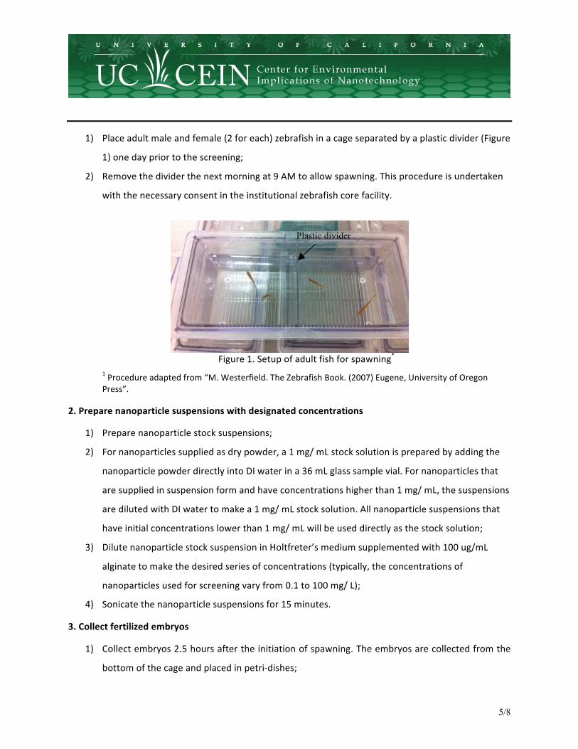
1) Place adult male and female (2 for each) zebrafish in a cage separated by a plastic divider (Figure
1) one day prior to the screening;
2) Remove the divider the next morning at 9 AM to allow spawning. This procedure is undertaken
with the necessary consent in the institutional zebrafish core facility.
Figure 1. Setup of adult fish for spawning*
1 Procedure adapted from “M. Westerfield. The Zebrafish Book. (2007) Eugene, University of Oregon Press”.
2. Prepare nanoparticle suspensions with designated concentrations
1) Prepare nanoparticle stock suspensions;
2) For nanoparticles supplied as dry powder, a 1 mg/ mL stock solution is prepared by adding the
nanoparticle powder directly into DI water in a 36 mL glass sample vial. For nanoparticles that
are supplied in suspension form and have concentrations higher than 1 mg/ mL, the suspensions
are diluted with DI water to make a 1 mg/ mL stock solution. All nanoparticle suspensions that
have initial concentrations lower than 1 mg/ mL will be used directly as the stock solution;
3) Dilute nanoparticle stock suspension in Holtfreter’s medium supplemented with 100 ug/mL
alginate to make the desired series of concentrations (typically, the concentrations of
nanoparticles used for screening vary from 0.1 to 100 mg/ L);
4) Sonicate the nanoparticle suspensions for 15 minutes.
3. Collect fertilized embryos
1) Collect embryos 2.5 hours after the initiation of spawning. The embryos are collected from the
bottom of the cage and placed in petri-‐dishes;
Plastic divider
6/8
2) Wash the embryos in petri-‐dishes using Holtfreter’s medium 3 times.
4. Pick and place embryos in 96-‐well plate, with one embryo in each well
1) Identify embryos at the same developmental stages (at 4 hpf)1;
2) Hand pick and place one embryo in each well of 96-‐well plates using a glass transfer pipette.
5. Add 100 µL of Holtfreter’s medium or designated concentration of the nanoparticle suspension. Use 12 replicate wells for each concentration
6. Put the 96-‐well plates in an incubator (Thermal Scientific, Inc.) and keep the temperature at 28 °C
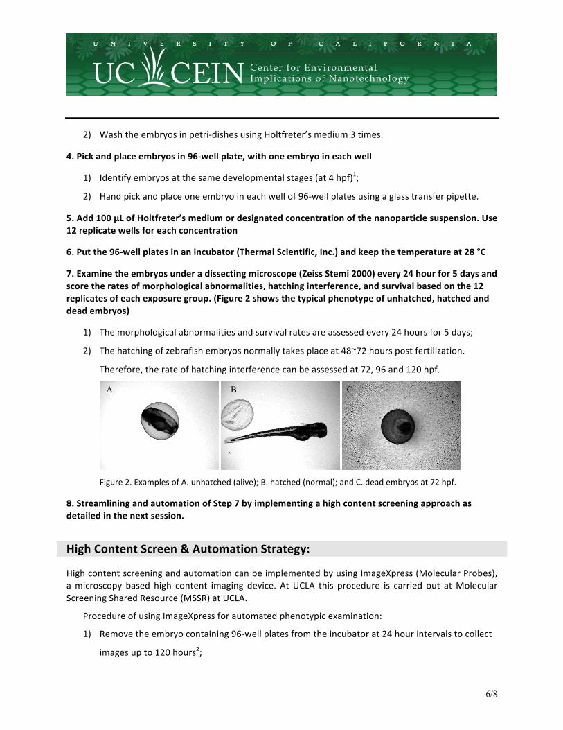
7. Examine the embryos under a dissecting microscope (Zeiss Stemi 2000) every 24 hour for 5 days and score the rates of morphological abnormalities, hatching interference, and survival based on the 12 replicates of each exposure group. (Figure 2 shows the typical phenotype of unhatched, hatched and dead embryos)
1) The morphological abnormalities and survival rates are assessed every 24 hours for 5 days;
2) The hatching of zebrafish embryos normally takes place at 48~72 hours post fertilization.
Therefore, the rate of hatching interference can be assessed at 72, 96 and 120 hpf.
Figure 2. Examples of A. unhatched (alive); B. hatched (normal); and C. dead embryos at 72 hpf.
8. Streamlining and automation of Step 7 by implementing a high content screening approach as detailed in the next session.
High Content Screen & Automation Strategy:
High content screening and automation can be implemented by using ImageXpress (Molecular Probes), a microscopy based high content imaging device. At UCLA this procedure is carried out at Molecular Screening Shared Resource (MSSR) at UCLA.
Procedure of using ImageXpress for automated phenotypic examination:
1) Remove the embryo containing 96-‐well plates from the incubator at 24 hour intervals to collect
images up to 120 hours2;
A B C
7/8
2For embryos after hatching (after 72 hpf), add 1 uL of 1.7% tricaine solution to each well of 96-‐well plates
before imaging
2) Seal the plate with a transparent sealing film (Sigma Cat# Z369659) to prevent liquid spilling
during imaging;
3) Open up ImageXpress software and choose the imaging protocol by clicking “2X-‐TM-‐96” for 96-‐
well plates;
4) Load one 96-‐well plate into the imaging chamber;
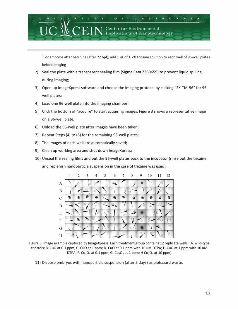
5) Click the bottom of “acquire” to start acquiring images. Figure 3 shows a representative image
on a 96-‐well plate;
6) Unload the 96-‐well plate after images have been taken;
7) Repeat Steps (4) to (6) for the remaining 96-‐well plates;
8) The images of each well are automatically saved;
9) Clean up working area and shut down ImageXpress;
10) Unseal the sealing films and put the 96-‐well plates back to the incubator (rinse out the tricaine
and replenish nanoparticle suspension in the case of tricaine was used).
Figure 3. Image example captured by ImageXpress. Each treatment group contains 12 replicate wells. (A. wild-‐type controls; B. CuO at 0.1 ppm; C. CuO at 1 ppm; D. CuO at 0.1 ppm with 10 uM DTPA; E. CuO at 1 ppm with 10 uM
DTPA; F. Co3O4 at 0.1 ppm; G. Co3O4 at 1 ppm; H Co3O4 at 10 ppm)
11) Dispose embryos with nanoparticle suspension (after 5 days) as biohazard waste.
1 2 3 4 5 6 7 8 9 10 11 12
A
B
C
D
E
F
G
H
8/8
SOP Approval
DEPARTMENT APPROVED BY DATE Principle Investigator Andre Nel 2-‐15-‐12
MSSR


