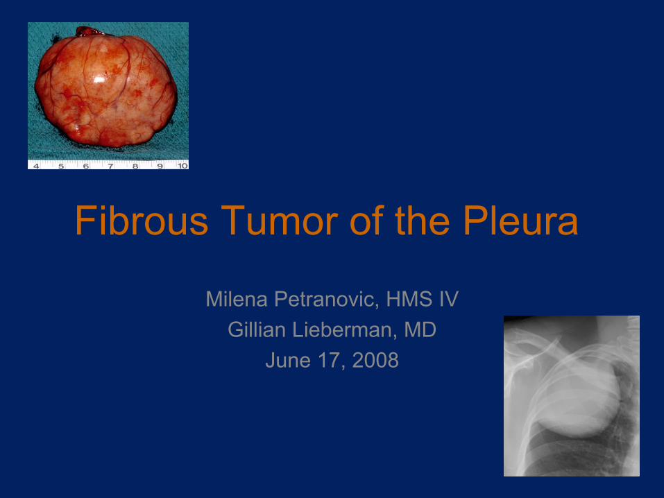
Fibrous Tumor of the Pleura
Milena Petranovic, HMS IVGillian Lieberman, MD
June 17, 2008
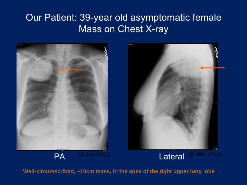
Our Patient: 39-year old asymptomatic female Mass on Chest X-ray
PA Lateral
Well-circumscribed, ~10cm mass, in the apex of the right upper lung lobe
BIDMC, PACS BIDMC, PACS
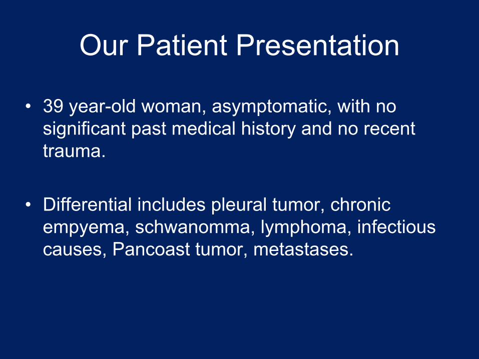
Our Patient Presentation
•
39 year-old woman, asymptomatic, with no significant past medical history and no recent trauma.
•
Differential includes pleural tumor, chronic empyema, schwanomma, lymphoma, infectious causes, Pancoast
tumor, metastases.
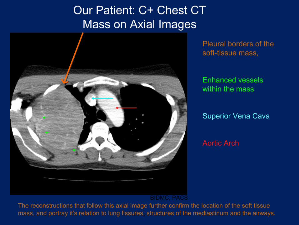
Our Patient: C+ Chest CT Mass on Axial Images
Pleural borders of the soft-tissue mass,
Enhanced vessels within the mass
Superior Vena Cava
Aortic Arch
The reconstructions that follow this axial image further confirm
the location of the soft tissue mass, and portray it’s relation to lung fissures, structures of the mediastinum
and the airways.
BIDMC, PACS
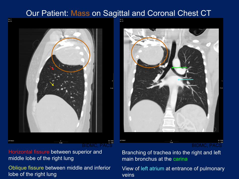
Our Patient: Mass
on Sagittal
and Coronal Chest CT
Horizontal fissure
between superior and middle lobe of the right lung
Oblique fissure
between middle and inferior lobe of the right lung
Branching of trachea into the right and left main bronchus at the
carina
View of
left atrium
at entrance of pulmonary veins
BIDMC, PACS BIDMC, PACS
http://fau.pearlashes.com/anatomy/Chapter%2036/Chapter%2036.htm
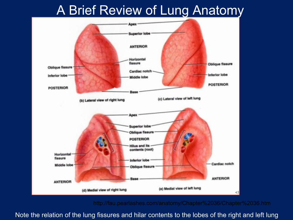
A Brief Review of Lung Anatomy
Note the relation of the lung fissures and hilar
contents to the lobes of the right and left lung

A Brief Review of Pleural Anatomy
http://www.breader.com/diagram-teaching-files/index.html
Note the divisons
of the visceral and parietal pleura as they would appear on a frontal chest x-ray
and on axial images from CT chest scan.
Benign pleural disorders•
Pleural disorders broadly divided into:–
Effusions–
Thickening–
Masses–
Pneumothorax•
Normal amount of pleural fluid in healthy people is 2-10ml•
Parietal pleura supplied by systemic capillary vessels -
drains into right atrium via the azygous, hemiazygous, and internal mammary veins
•
Visceral pleura supplied by pulmonary arterial capillaries and runs mainly into the pulmonary veins
•
Lymphatic vessels play major role in clearance of pleural fluid -
drainage mainly through parietal pleural lymphatics.
Fibrous Tumors of the Pleura•
Localized pleural tumors fall into one of two categories: fibrous tumors of the pleura or lipomas.
•
Pathologic characteristics of solitary fibrous tumor of the pleura first described by Klemperer and Rabin in 1931
•
~ 800 cases reported in the literature between 1931 and 2002 and
has been referred to as localized mesothelioma, localized fibrous tumor, fibrous mesothelioma, or a pleural fibroma.
•
Rare neoplasm: incidence is 2.8 per 100,000 registered hospital patients and accounts for 8% of benign pathologic diseases of chest
Clinical Presentation of Fibrous Pleural Tumors
•
Usually found incidentally
by chest radiography•
Greatest occurrence in fourth to sixth decade, no hx
of asbestos exposure, relatively equal gender distribution (some report slight female preponderance)
•
Presenting symptoms (~50-60% are symptomatic): intrathoracic
symptoms (dyspnea, chest pain, hemoptysis), systemic symptoms (hypoglycemia, hypertrophic osteoarthropathy), nonspecific symptoms (fever, weight loss, fatigue)
•
Most behave as slowly growing, painless masses
although large tumors may also give rise to compression symptoms.
•
Associated hypertrophic pulmonary osteoarthropathy
and episodic hypoglycemia
(due to production of insulin-like growth factor) may be present in 4-5% of cases.
•
80% arise from visceral pleura
and 20% from parietal pleura•
Calcification present in ≤5%, central necrosis is common in the larger tumors.•
Behavior is unpredictable with ~10-15% behaving aggressively so follow-up is mandatory
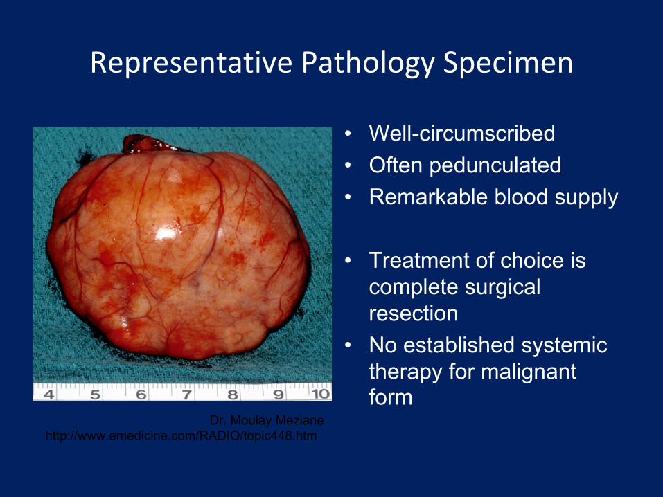
Representative Pathology Specimen
•
Well-circumscribed•
Often pedunculated
•
Remarkable blood supply
•
Treatment of choice is complete surgical resection
•
No established systemic therapy for malignant form
Dr. Moulay
Meziane
http://www.emedicine.com/RADIO/topic448.htm
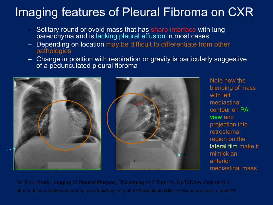
Imaging features of Pleural Fibroma
on CXR–
Solitary round or ovoid mass that has sharp interface
with lung parenchyma and is lacking pleural effusion
in most cases–
Depending on location may be difficult to differentiate from other pathologies
–
Change in position with respiration or gravity is particularly suggestive of a pedunculated
pleural fibroma
Dr. Paul Stark. Imaging of Pleural Plaques, Thickening and Tumors. UpToDate. Online16.1http://utdol.com/online/content/topic.do?topicKey=int_pulm/7040&selectedTitle=2~2&source=search_result#1
Note how the blending of mass with left mediastinal
contour on PA view
and projection into retrosternal
region on the lateral film
make it mimick
an anterior mediastinal
mass
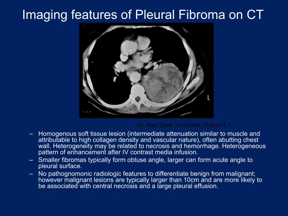
Imaging features of Pleural Fibroma
on CT
–
Homogenous soft tissue lesion (intermediate attenuation similar to muscle and attributable to high collagen density and vascular nature), often abutting chest wall. Heterogeneity may be related to necrosis and hemorrhage. Heterogeneous pattern of enhancement after IV contrast media infusion.
–
Smaller fibromas
typically form obtuse angle, larger can form acute angle to pleural surface.
–
No pathognomonic
radiologic features to differentiate benign from malignant; however malignant lesions are typically larger than 10cm and are
more likely to be associated with central necrosis and a large pleural effusion.
Dr. Paul Stark. UpToDate. Online16.1
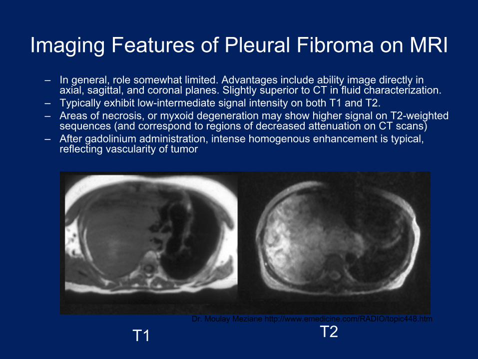
Imaging Features of Pleural Fibroma
on MRI–
In general, role somewhat limited. Advantages include ability image directly in axial, sagittal, and coronal planes. Slightly superior to CT in fluid characterization.
–
Typically exhibit low-intermediate signal intensity on both T1 and T2.–
Areas of necrosis, or myxoid
degeneration may show higher signal on T2-weighted sequences (and correspond to regions of decreased attenuation on
CT scans) –
After gadolinium administration, intense homogenous enhancement is typical, reflecting vascularity
of tumor
T1 T2Dr. Moulay
Meziane
http://www.emedicine.com/RADIO/topic448.htm
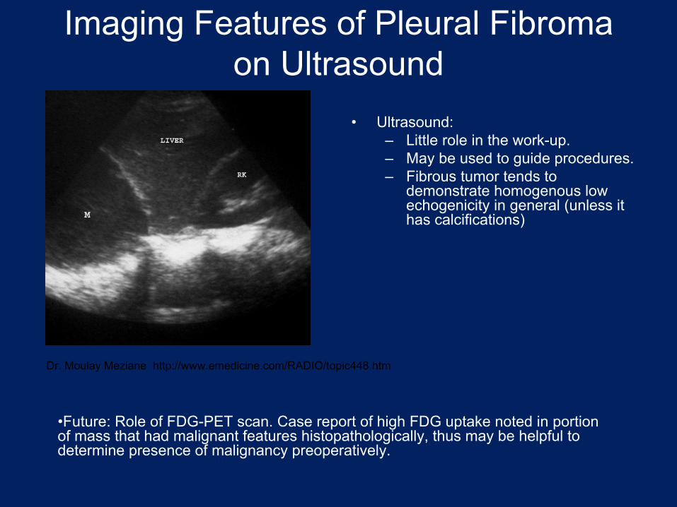
Imaging Features of Pleural Fibroma on Ultrasound
•
Ultrasound:–
Little role in the work-up. –
May be used to guide procedures.–
Fibrous tumor tends to demonstrate homogenous low echogenicity
in general (unless it has calcifications)
•Future: Role of FDG-PET scan. Case report of high FDG uptake noted in portion of mass that had malignant features histopathologically, thus may be helpful to determine presence of malignancy preoperatively.
Dr. Moulay
Meziane
http://www.emedicine.com/RADIO/topic448.htm
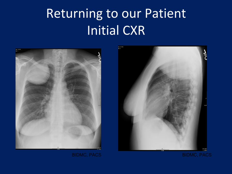
Returning to our Patient Initial CXR
BIDMC, PACS BIDMC, PACS

Our Patient: Post-operative CXR Full surgical excision of mass was performed
BIDMC, PACS BIDMC, PACS
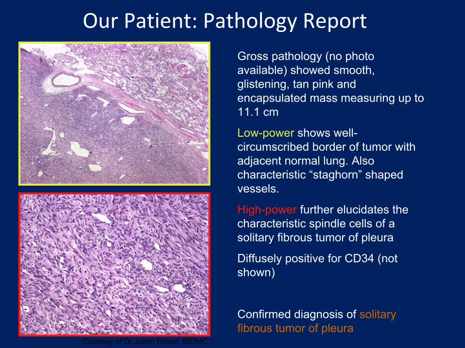
Our Patient: Pathology ReportGross pathology (no photo available) showed smooth, glistening, tan pink and encapsulated mass measuring up to 11.1 cm
Low-power
shows well-
circumscribed border of tumor with adjacent normal lung. Also characteristic “staghorn”
shaped vessels.
High-power
further elucidates the characteristic spindle cells of a solitary fibrous tumor of pleura
Diffusely positive for CD34 (not shown)
Confirmed diagnosis of solitary fibrous tumor of pleura
Courtesy of Dr.Justin Hilson, BIDMC

References:
•
Gallardo et al. Benign pleural diseases. Eur
Journal of Rad
34 (2000) 87-97.•
Janssen et al. Quiz case of the month. Eur
Radiol. (2001) 11:527--528.•
Karbulut
N, Goodman LR -
Pedunculated
Solitary Fibrous Tumor of the Interlobar
Fissure: A Wandering Chest Mass. AJR:173, August 1999) •
Kouki
et al. Solitary fibrous tumor of the lung. Gen Thorac
Cardiovasc
Surg
(2008) 56:249-251.
•
McLoud, Theresa. CT and MR in Pleural Disease. Clinics in Chest Medicine 1998; 19(2): 261-276.
•
Meziane
et al. Localized Fibrous Tumor of the Pleura. eMedicine. Accessed 6/10/2008.
•
Qureshi
NR, Gleeson FV. Imaging of Pleural Disease. Clin
Chest Med 27 (2006): 193-213.
•
Stark, Paul. Imaging of pleural plaques, thickening, and tumors.
UpToDate. Accessed 6/9/2008.
•
Sung et al. Solitary Fibrous Tumors of the Pleura: Surgical Outcome and Clinical Course. Ann Thorac
Surg
2005;79:303-7
Acknowledgments:•
Daniel Siegal, MD –
Department of Radiology
•
Justin Hilson, MD –
Department of Pathology•
Maria Levantakis
-
Department of Radiology


