done by: dina shaban th date: 30 of october...
TRANSCRIPT

Done By: Dina Shaban Date: 30th of October 2013
1
Done By: Dina Shaban Date: 30th of October 2013
2
Virology Lecture by Dr. Ashraf Khasawneh
Lecture 6
From the previous lecture:
Cytopathic effects – (Virus induced damage to cells):
1. Change in size and shape.
2. Cytoplasmic inclusion bodies.
3. Nuclear inclusion bodies.
4. Cells fuse to form multinucleated cells.
5. Cell lysis (death).
6. Alter DNA (changes in the DNA/genome).
7. Transform cells into cancerous cells.
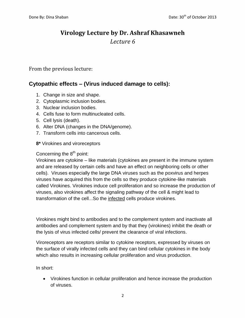
8* Virokines and viroreceptors
Concerning the 8th point:
Virokines are cytokine – like materials (cytokines are present in the immune system
and are released by certain cells and have an effect on neighboring cells or other
cells). Viruses especially the large DNA viruses such as the poxvirus and herpes
viruses have acquired this from the cells so they produce cytokine-like materials
called Virokines. Virokines induce cell proliferation and so increase the production of
viruses, also virokines affect the signaling pathway of the cell & might lead to
transformation of the cell...So the infected cells produce virokines.
Virokines might bind to antibodies and to the complement system and inactivate all
antibodies and complement system and by that they (virokines) inhibit the death or
the lysis of virus infected cells/ prevent the clearance of viral infections.
Viroreceptors are receptors similar to cytokine receptors, expressed by viruses on
the surface of virally infected cells and they can bind cellular cytokines in the body
which also results in increasing cellular proliferation and virus production.
In short:
Virokines function in cellular proliferation and hence increase the production
of viruses.

Done By: Dina Shaban Date: 30th of October 2013
3
They interfere in the immune response were they bind to antibodies and
complement proteins and deactivate them.
Again: virokines are acquired by recombination from normal cells and after that mutation were introduced by the viruses to become viral produced particles.
Cytopathic changes in cells :( also mentioned in previous lecture)
(a) First picture shows multinucleated giant cells and the mechanism by which this happens is:
1. Fusion of the viral envelope with the cell membrane upon entry of virus.
2. Viral glycoproteins present on the viral envelope remain on cell membrane and
attach to receptors on surrounding cells. 3. This leads to fusion between the cells’ membranes and hence a large
multinucleated cell is formed.
* It has one cell membrane with multiple nuclei inside.
(b) Second picture shows inclusion bodies. Inclusion bodies are aggregates mostly derived from viral capsid proteins and therefore they indicate the site of virus replication. There are cytoplasmic inclusion bodies (virus replicated in cytoplasm) and nuclear inclusion bodies (virus replicated in nucleus). Generally for DNA viruses you would observe intranuclear inclusion bodies and for RNA viruses intracytoplasmic inclusion bodies. There are also single or multiple inclusion bodies.

Done By: Dina Shaban Date: 30th of October 2013
4
*Some notes on table 6.4: 1) Respiratory syncytial virus: response in animal cell: multinucleated giant cell formation. 2) Rabies Virus: Negri bodies are diagnostic on pathological examination of the tissues.
Patterns of viral infection: 1. Inapparent infection (subclinical infection): the virus might infect a person and that person might not develop any symptoms/ illness.
2. Apparent Infection: if a person develops a disease from the infection, this can be divided further into:
a) Acute Infection b) Persistent Infection which can be divided into
i. Chronic Infections. ii. Latent Infections. iii. Slow virus infections.
Done By: Dina Shaban Date: 30th of October 2013
5
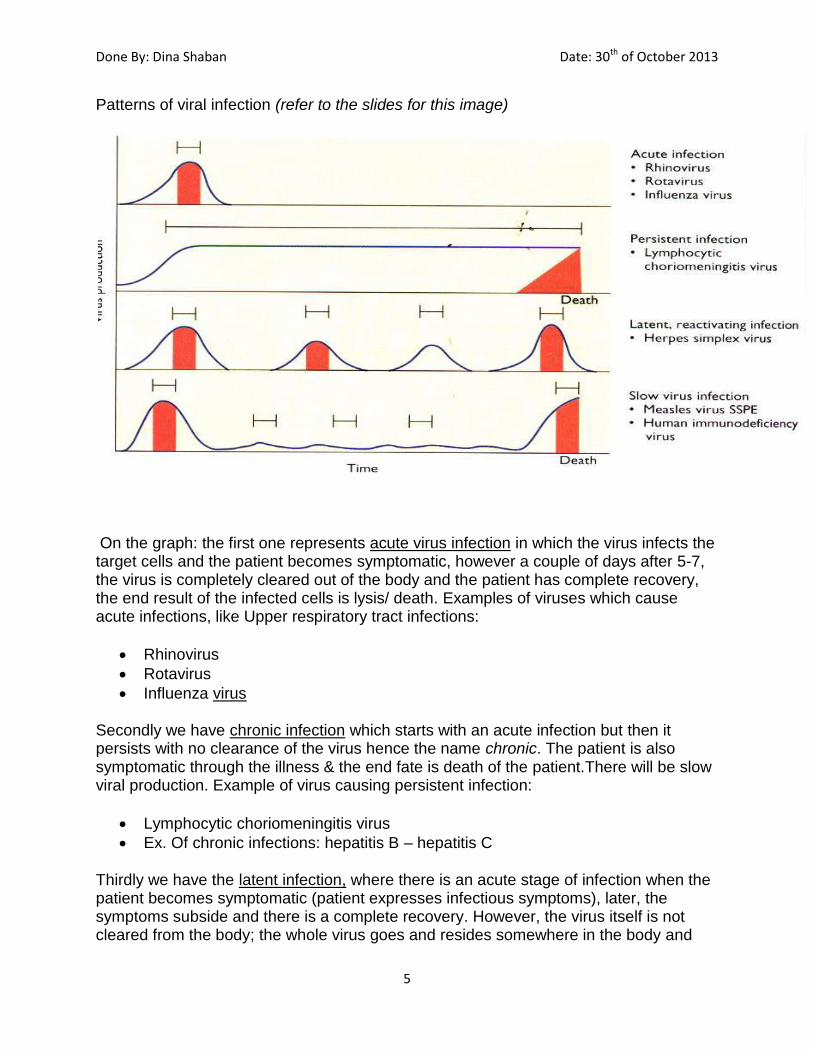
Patterns of viral infection (refer to the slides for this image)
On the graph: the first one represents acute virus infection in which the virus infects the target cells and the patient becomes symptomatic, however a couple of days after 5-7, the virus is completely cleared out of the body and the patient has complete recovery, the end result of the infected cells is lysis/ death. Examples of viruses which cause acute infections, like Upper respiratory tract infections:
Rhinovirus
Rotavirus
Influenza virus
Secondly we have chronic infection which starts with an acute infection but then it persists with no clearance of the virus hence the name chronic. The patient is also symptomatic through the illness & the end fate is death of the patient.There will be slow viral production. Example of virus causing persistent infection:
Lymphocytic choriomeningitis virus
Ex. Of chronic infections: hepatitis B – hepatitis C
Thirdly we have the latent infection, where there is an acute stage of infection when the patient becomes symptomatic (patient expresses infectious symptoms), later, the symptoms subside and there is a complete recovery. However, the virus itself is not cleared from the body; the whole virus goes and resides somewhere in the body and

Done By: Dina Shaban Date: 30th of October 2013
6
could reactivate and cause infection. An example: the Herpes simplex virus this virus goes to the dorsal root ganglia of nerves and resides there, and is reactivated once again with suppression in immune system and old age.
Ex. Herpes viridae family is known for latent infections, there are 8 subdivisions, ex. Chicken box and shingles.
The fourth pattern shows slow virus infection, they are characterized by:
* Long incubation period
* opposite to chronic infection, in slow virus infection, the patient is asymptomatic- the patient doesn’t show any symptoms, while in chronic infection, the patient is symptomatic all the time!
Ex. of this pattern of infection: JC papovavirus & an example on viruses causing slow V infections is Human Immunodeficiency Virus (HIV) which starts as an acute infection and in the case of HIV we get flu-like symptoms after that the immune system takes over and symptoms subside almost but not completely. Every time the patient with HIV experiences a drop in the immune system the patient becomes symptomatic for a while, however these viruses have very long incubation periods of up to 10 years and after that when the immune system is completely suppressed/dramatic injuries to the immune system occur, the illness appears in its full grown picture and the fate of the patient is death within a short period of time.
Lastly, the slow chronic infection long incubation period, the infection initially starts when the virus reproduces in small amount, patient is asymptomatic, and then builds up and increases over time, then the patient is symptomatic
i. Chronic Infection: virus can be continuously detected; mild or no clinical symptoms may be evident, such as HTLV-1 leukemia and hepatitis B and hepatitis C.

Done By: Dina Shaban Date: 30th of October 2013
7
ii. Latent infection: The Virus persists in an occult, or cryptic, from most of the time. There will be intermittent flare-ups of clinical disease; Infectious virus can be recovered during flare-ups. Latent virus infections typically persist for the entire life of the host. Ex. Shingles & varicella-zoster.
iii. Slow virus infection: A prolonged incubation period, lasting months or years, during which virus continues to multiply. Clinical symptoms are usually not evident during the long incubation period (after this long incubation period the disease episode starts). An example is the JC papovavirus.
Overall fate of the cell:
The cell dies in cytocidal infections (meaning: the cell dies or lysis), this may be acute (when infection is brief and self-limiting) example: most upper respiratory tract infections like influenza, respiratory syncytial virus (RSV) or parainfleunza// Rota virus infections that lead to gastroenteritis most of them

Done By: Dina Shaban Date: 30th of October 2013
8
are acute and end by cell death or cell lysis. It could also be chronic (drawn out, only a few cells infected while the rest proliferate)-Cytocidal effect examples include hepatitis B or hepatitis C (not all the hepatocytes are infected with the virus, the cells which die/lyse are only the ones infected with the virus, but there are other hepatocytes that remain healthy, the liver cells have the ability to regenerate- cells that die regenerate and might get infected again, so it is still in balance).
The cell lives in persistent infections, this may be productive or nonproductive (refers to whether or not virions are produced) or it may alternate between the two by way of latency and reactivation-Steady state infection. *Productive - Chronic infections (↑ as in hepatitis B and C ) *Non-productive – Latent infections, in case of Herpes viridae
Another fate of the cell is transformation, which is the change of the cell from normal cell into (cancerous - malignant or tumor) cell. This can be seen in RNA/DNA viruses:
RNA tumor viruses usually transform cells to a malignant phenotype by integrating their own genetic material into the cellular genome and may also produce infectious progeny. Mostly we refer to retroviruses or HIV and certain viruses such as hepatitis B and C virus. The mechanism by which Hepatitis C virus transforms the cell, is due to the chronic infection which leads to chronic inflammation of the hepatocytes, the second one is that one of the viral proteins which is NS3 might be oncogenic – itself; the expression of this protein induces the change of a normal -مسبب للسرطان cell into a cancerous cell. In retroviruses there are three mechanisms by which transformation to cancerous cells (oncogenesis) might occur: 1. Acute transforming viruses: We’ll talk about Cellular SRC (CSRC), CSRC is a
Cellular gene & a proto-oncogene, its present in normal cells but doesn’t transform them into cancerous ones ,it plays a role in the translation process and signaling pathway in the cell, viruses at long period of infection acquire the cellular genes (CSRC) & incorporate them into their genes(( so the origin of proto-oncogenes is from the cells and the viruses got them by copy choice recombination1)) … after that, mutations were inserted into the acquired CSRC during the viral cell replication and they became Viral SRC (VSRC), so the expressions of VSRC and those mutations, transform the gene from being proto-oncogene into an oncogene(that causes the transformation)
1we will talk about this in the coming lectures concerning viral genetics.

Done By: Dina Shaban Date: 30th of October 2013
9
What do oncogenes do? They make the cell proliferate more and affect the signaling pathways of the cells; with uncontrolled cell proliferation they transform the cell into a cancerous one.
2. Insertional mutagenesis: inappropriate expression of proto-oncogenes. How? Here we also have oncogenes and proto-oncogenes (the origin of the gene is from the cells and the virus got it with mutations, just like the 1st point). We know that in order for genes to be expressed we need promoters or enhancers that precede them – مكانهم قبل الجين-, in insertional mutagenesis the viral promoter of the viral gene is inserted just before the cellular gene (so the enhancer is from the virus but the gene is from the cell) this makes the promoter to overproduce the cellular gene- proto-oncogene affecting the signaling pathway and transforming the cell from normal to cancerous… So the viral genes activate the cellular genes. *the 1st & 2nd mechanisms are not associated with cancerous transformations or illnesses in HUMANS.
3. Transactivating factors: certain viruses such as HTLV-1 have certain genes; in HTLV there is an oncogenic gene to the cells called TAX gene, the expression of this gene leads to formation of TAX protein which is toxic & oncogenic to the cell and this induces the change from normal to cancerous cell.
DNA tumor virus infections are often cytocidal; thus transformation is associated with abortive or restrictive infections in which few viral genes are expressed.
So in DNA viruses we have 2 things that must occur to transform the cell: 1. Persistence of at least part of the viral genome or the whole viral genome
within the cell is required for cell transformation. 2. Continual expression from a number of viral genes.
Examples: human papilloma virus, herpes viruses, hepatitis B virus & adeno virus. - There are also other mechanisms which include p53 and pRb
(retinoblastoma) they are called: cell cycle regulators, *For P53, it protects the cellular genome and prevents any damage to the cellular DNA; it plays a role in correction of any defects that occur in DNA, once the defects are detected, P53 repairs them, and if the damage to the DNA is irreparable, p53 might induce the death of the cell / apoptosis (PCD –Programmed Cell Death). *For pRb, it doesn’t have the same mechanism of P53; it doesn’t repair the genome & doesn’t induce cell death.

Done By: Dina Shaban Date: 30th of October 2013
10
* So certain viruses can produce enzymes which block the effect of such regulators the cell will enter an unregulated cell cycle uncontrolled cell division cancerous cell.
Types of Viral infections at the cellular level: Type Virus Production Fate of cell
Abortive - No effect
Cytolytic + Death
Persistent -Productive -Latent
+ -
Senescence No effect
Transforming -DNA viruses -RNA viruses
- +
Immortalization Immortalization
Mechanisms of viral cytopathogenesis:
Inhibition of cellular protein synthesis Polioviruses, HSV, poxviruses, togaviruses
Inhibition and degradation of cellular DNA Herpesviruses
Alteration of cell membrane structure Glycoprotein insertion Syncytia formation (Multinucleate giant cell formation) Disruption of cytoskeleton Permeability
All enveloped viruses HS HSV, VZ virus, HIV
HSV, HIV, RSV Togaviruses, herpesviruses
Inclusion bodies (Negri bodies) Rabies Toxicity of Virion components Adenovirus fibers
مش مطلوب حفظ الفيروسات في الجدول أعاله، المهمين برجع بركز عليهم الدكتور بالشرح *
Possible consequences to a cell that is infected by a virus:
•Lytic infections: Result in the destruction of the host cell; are caused by virulent viruses, which inherently bring about the death of the cells that they infect.

Done By: Dina Shaban Date: 30th of October 2013
11
•Persistent infections: Infections that occur over relatively long periods of time, where the release of the viral particles may be slow and the host cell may not be lysed. •Latent infections: Delay between the infection by the virus and the appearance of symptoms.
•Transformation: Some animal viruses have the potential to change a cell from a normal cell into a tumor cell which grows without restraint.
New topic: Immunology of viral infections (we are going to address the clearing of viral infections)
Introduction to immunology
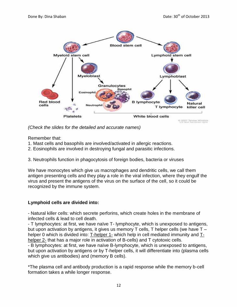
We’ll talk about the origin of immune cells at first, starting with the stem cells in the bone marrow, which produces myeloid progenitor & the lymphoids,
*the myeloid cells give platelets, RBCs, mast cells & myeloblasts(which give monocytes, baso/ eosino/neutro phils).
Done By: Dina Shaban Date: 30th of October 2013
12
(Check the slides for the detailed and accurate names) Remember that: 1. Mast cells and basophils are involved/activated in allergic reactions. 2. Eosinophils are involved in destroying fungal and parasitic infections.
3. Neutrophils function in phagocytosis of foreign bodies, bacteria or viruses
We have monocytes which give us macrophages and dendritic cells, we call them antigen presenting cells and they play a role in the viral infection, where they engulf the virus and present the antigens of the virus on the surface of the cell, so it could be recognized by the immune system.
Lymphoid cells are divided into: - Natural killer cells: which secrete perforins, which create holes in the membrane of infected cells & lead to cell death. - T lymphocytes: at first, we have naïve T- lymphocyte, which is unexposed to antigens, but upon activation by antigens, it gives us memory T cells, T helper cells (we have T – helper 0 which is divided into: T-helper 1- which help in cell mediated immunity and T-helper 2- that has a major role in activation of B-cells) and T cytotoxic cells. - B lymphocytes: at first, we have naïve B-lymphocyte, which is unexposed to antigens, but upon activation by antigens or by T-helper cells, it will differentiate into (plasma cells which give us antibodies) and (memory B cells).
*The plasma cell and antibody production is a rapid response while the memory b-cell formation takes a while longer response.

Done By: Dina Shaban Date: 30th of October 2013
13
Immunity to Microbes: The body’s defense mechanisms/response to viral infections are of two types:
Defense against infections is mediated by the early reactions of innate immunity and the
later response of adaptive immunity. The innate immune response controls infection
long enough for an adaptive response to eradicate the infection. Adaptive immunity
involves T and B cells, while innate involves first line of defense, cytokines, natural killer
cells and macrophages. First response is the innate immunity, which can’t clear/kill
viruses or viral infected cells, it would only minimize viral infection effects, but a later
adaptive immunity kicks in and eradicates it.
An immune system is specialized to generate different effective mechanisms for
different types of microbes; extracellular microbes can be tackled by antibodies (they
neutralize glycoproteins of the spikes which are the most immunogenic), phagocytes
and t-helper cells, intracellular microbes by phagocytes, cytotoxic t-cells and natural
killer cells.
The non-specific (innate immunity): the body has defenses which are not specifically
directed at particular infectious agents but serve as non-specific barriers to
infections…many pathogenic microbes resist innate immunity; so its not that efficient:
1. Skin (1st line of defense): an effective barrier unless breached by an injurious
disease, viruses might enter through any cuts or wounds.
2. Respiratory Tract: upward flow of mucous by ciliated epithelium removes virus
particles to prevent invasion of the lower respiratory tract.
3. GI Tract: acidity of gastric juices and sterilizing effect of the bile salts.
4. Urinary Tract: is protected only by the continuous flow of urine (flushing effect) so any
obstruction to urine flow might lead to infection and it would be a good media for
bacterial infections.
5. Congectiva: tears flush viruses from the eye and it is considered the least protective.
Non-specific/ innate Specific/ adaptive

Done By: Dina Shaban Date: 30th of October 2013
14
Phagocytosis: an important defense mechanism in bacterial infection and in viral
infection. Ingestion is carried out by two types of cells; neutrophils
(polymorphonuclear leukocytes) and macrophages (free macrophages present
in the blood, the lung alveoli and peritoneum, and fixed macrophages present
in the lymph nodes, spleen, liver, connective tissues and CNS).
Phagocytosis is enhanced by antibodies, and complement system, this effect is
known as opsonization antigen presenting cells (get activated by cytokines),
engulf these antigens and represent them on their surface and after that
antibodies and the adaptive immune system take place and destroy the viruses.
When T-lymphocytes are activated they release cytokines which promotes the
function of macrophages and attracts them to the site of infection this is the
principle of chemotaxis.
Cytokines are released from one cell and have an effect on other cells. They are
basically small protein molecules released by many cells including lymphocytes and
macrophages, they function as signals or mediators to activate and modulate the
immune response.
Types of cytokines: interleukins, interferons and tumor necrosis factor (TNF).
Cytokines are numerous and of different types. In addition to their role in immune
response, they possess other physiologic functions such as tissue repair,
differentiation and signaling activity of the CNS.
1. Interferons:
- Small proteins produced by certain cells. We have the α, ß and γ interferons.
- Alpha released by lymphocytes and macrophages, beta by fibroblasts &
epithelial cells and gamma prduced in response to T- cells.
- In viral infections we see alpha and beta interferons most commonly activated
-produced in response to viruses, specifically in response to antigens or RNA of
the viruses.
- They’re produced to affect neighboring cells
- They bind to cell surfaces and induce expression of antiviral proteins.
- They inhibit the expression of cancer genes.
Mechanism of action of interferon: Is the induction of the following enzymes:
1. Protein kinase which inhibits protein synthesis.
2. Oligoadenylate Synthase which leads to the degradation of viral mRNA.
3. Phosphodiesterase which inhibits tRNA.

Done By: Dina Shaban Date: 30th of October 2013
15
All three of those steps play a role in inhibition of the translation of the viral gene
or the production of viral proteins as well as degradation of the viral genome.
According to a figure in the slides, which shows the infected cell and the nearby
cells surrounding it, as a result of the presence of viral nucleic acids and the
antigens, the interferon gene is activated, hence interferons are synthesized and
released to the nearby cells attacking the interferon receptors expressed on them,
which leads to suppression in the translation of viral genes & leads to degradation
of viral nuclei acids.
The specific or adaptive immunity defense mechanisms:
1. Humoral: affect the neutralization of viruses (antigen antibody reaction) it is
responsible for protective immunity involves antibodies derived from plasma
cells from B lymphocytes.
-Antibodies are immunoglobulins which react specifically with antigens(proteins),
in the case of viral infections those antigens are the glycoproteins or spikes (most
antigenic) on the surface of the virus structures against which antibodies are
produced.
-B-lymphocytes have immunoglobulins on their surface which act as receptors for
virus antigen upon an encounter with a viral antigen they differentiate into
plasma cells. This differentiation requires most of the time the presence of the T-
helper cells.
-T-helper zero cells differentiate into T-helper 1 and T-helper 2. T-helper 2 aids in
the cell mediated immunity and is required for the activation of the B-cells where it
helps with the virus antigen exposure hence recognition by B-cell becomes a
plasma cell also formation of memory B-cells.
2. Cellular: main affect is: localization of lesions & killing virus infected cells,
involves T-cytotoxic cells.

Done By: Dina Shaban Date: 30th of October 2013
16
What are immunoglobulins?
A Y-shaped protein consists of 4 polypeptide chains containing (2 heavy chains and 2
light chains connected with each other by disulfide bonds). There are five types of
immunoglubulins: IgM, IgG, IgD, IgA and IgE.
Hypervariable region is the antigen binding site, whereas the Fc portion binds to
macrophages or complement proteins to destroy the antigen.
IgG: is a monomer, single Y-shaped unit.
IgM: is a Pentamer, 5 subunits tied together by disulfide bonds & J-chain ties the
subunits as well. Its presence indicates an acute infection since it is the first antibody to
be present at the site of infection. It is followed by IgG which indicates the presence of a
second exposure or a chronic infection.
*Just before the decline of IgM, IgG arises.
*The purpose of vaccines is to generate those IgG immunoglobulins, there should be
multiple vaccinations not only one; to prevent the IgG from dropping in the immune
system.
IgA: exists as a dimer in the secretions, and in the blood as a monomer. It is
responsible for a long term immunity.
This sheet was written without the slides, my apologies for any mistakes, please do
refer to the slides this sheet doesn’t include everything.