“do or do not. there is no try.” (master yoda ... · vadivel, dr. rajesh alphonse, melanie...
TRANSCRIPT

“Do or do not. There is no try.” (Master Yoda – “Star Wars”, 1977)

University of Alberta
Evaluation of Mesenchymal Stem Cell-Based Therapies
for Inflammatory Lung Diseases
by
Lavinia Iuliana Ionescu
A thesis submitted to the Faculty of Graduate Studies and Research
in partial fulfillment of the requirements for the degree of
Doctor of Philosophy
Department of Physiology
© Lavinia Iuliana Ionescu
Fall 2012
Edmonton, Alberta
Permission is hereby granted to the University of Alberta Libraries to reproduce single
copies of this thesis and to lend or sell such copies for private, scholarly or scientific research
purposes only. Where the thesis is converted to, or otherwise made available in digital form,
the University of Alberta will advise potential users of the thesis of these terms.
The author reserves all other publication and other rights in association with the copyright
in the thesis and, except as herein before provided, neither the thesis nor any substantial
portion thereof may be printed or otherwise reproduced in any material form whatsoever
without the author's prior written permission.
Dedication
To those who have given me all the chances to be who I am. To my
grandfather, Ion Leulescu, who has defined me before anything else could have.
I miss him to this day and my strongest wish is to be who he believed I will be.
To my mother, Carmen Leulescu, who is quietly encouraging everything about
me. To my grandmother, Niculina Leulescu, who is not quiet about the value of
life. To my younger brother, Leontin Ionescu, for teaching me the joy and trust
of sharing by being first to share. To those who smiled with me, to those who
smiled for me, to those who were too stubborn to abandon the uncertain path
by my side throughout my struggles.
To Georgiana, for trustfully being there for so long. To Raluca, for being
the role-model sister-by-choice. To Bev, for being a genuine embodiment of joy.
To Farah, for her unabashed patience. To Anda, for bringing an improbable
friendship alive. To Ahmed, for having the courage to challenge my fears. To
Mamoona, for knowing me before I did. To Mira, for being within my reach. To
Ioana, for her timely presence in my life. To Mădălina, for her soft-spoken
strength. To Vijay, for his persistence in believing I would understand who I am.
To both my homelands.
Thank you.
ABSTRACT
Recent discoveries in stem cell biology have generated enthusiasm
about the possibility of harnessing stem cells for organ repair and
regeneration. The ability of pluri- and multipotent stem cells to differentiate
along various cellular lineages has placed them at the core of research that
seeks to protect endogenous stem cell populations or to deliver exogenous
stem cells to sites of organ injury. Lung diseases are a major health care
concern and the prevalence of chronic lung diseases such as asthma,
pulmonary fibrosis and chronic obstructive pulmonary disease is expected to
continue to rise over the next decades. Meanwhile, improved perinatal care
has allowed the survival of extremely premature infants that constitute a
particularly vulnerable subpopulation because of their risk of developing
bronchopulmonary dysplasia (BPD) with potential life-long complications.
Research that aims to evaluate the therapeutic potential of exogenous
stem cells in lung diseases placed an initial emphasis on the engraftment and
differentiation of these cells in the lung. More recent studies demonstrate
that multipotent stem cell populations, such as mesenchymal stromal cells
(MSCs), exert paracrine activity that can modulate local inflammatory and
immune responses in experimental lung disease models including asthma,
acute lung injury (ALI) and pulmonary fibrosis. The studies presented hereby
demonstrate that factors secreted by adult bone marrow-derived MSCs can
prevent the development of inflammatory lung diseases in mouse models of

asthma and ALI and provide mechanistic insight into the anti-inflammatory
properties of MSCs.
The perspective of using pluripotent stem cells as therapeutic agents
has been revived by the landmark discovery of induced pluripotency, where
“embryonic stem cell (ESC)-like” cells can be generated by reprogramming
terminally differentiated somatic cells. While the field of induced pluripotent
stem cell (iPSC) biology is still in its effervescent infancy, this finding may
relieve many ESCs-related ethical concerns and may open the way to large-
scale production and evaluation of pluripotent stem cells for organ
regeneration and repair. The additional study presented provides proof-of-
concept for the utility of iPSC.
Together, the studies presented hereby advocate the potential of stem
cells as a novel clinical option for the treatment of severe lung diseases.

ACKNOWLEDGMENTS
My gratitude goes toward all those who made the work presented here
possible: Dr. Bernard Thébaud, my supervisor; Dr. Marek Duszyk, who has initially
co-supervised me when I was a starting Master of Science program student; Dr.
Christopher I. Cheeseman and Dr. Harissios Vliagoftis, my Supervisory Committee
members, who have thoughtfully contributed to designing my research path; our
collaborators, particularly Dr. Zamaneh Kassiri; Dr. Lisa Cameron (University of
Alberta); Dr. Kenneth Walsh and Dr. Tamar Aprahamian (Boston University), Dr.
James Ellis (University of Toronto); Dr. Valentin Duţă, whose advice accelerated the
development of the mouse model of asthma; many current and former members of
Dr. Thébaud’s and Dr. Vliagoftis’ laboratories: Farah Eaton, Beverly Morgan, Dr. Arul
Vadivel, Dr. Rajesh Alphonse, Melanie Abel, Thuraya Marshall, Dr. Paul Waszak, Dr.
Narcy Arizmendi, Dr. Gaia Weissmann; staff members, especially Bronwyn
Appleyard (Department of Pediatrics), Lynette Elder, Alana Eshpeter (Alberta
Diabetes Institute Histology Core), Dorothy Kratochwil-Otto (University of Alberta
flow-cytometry facility), Dr. Nathan Bosvik and Dr. Greg Parks (Health Sciences
Laboratory Animal Services), Drew Nahirney (Dr. Duszyk’s laboratory), Dr. Wilma
Suarez-Pinzon for their work and advice.
I would also like to thank the Department of Physiology (Dr. Keir Pearson,
Sharon Orescan, Barb Armstrong) and gratefully acknowledge the support I have
received from the CIHR-MFN Strategic Training Program (Paul Jacquier).

TABLE OF CONTENTS
CHAPTER 1 – INTRODUCTION ……………………………………………………….… 1
1.1. Overview……………………………………………………………………………..2
1.2. Lung Development and Regeneration……………………..…….……....3
1.3. Stem Cells…………………………………….…………………………………….. 8
1.4. Resident Lung Stem/Progenitor Cells……………………………..…...13
1.5. Therapeutic Potential of Exogenous Stem/Progenitor Cells….15
1.5.1. Cell Replacement by MSCs………………………..………......15
1.5.2. Cell Replacement Versus Paracrine Activity of MSCs.17
1.5.3. ESCs and Induced Pluripotent Stem Cells (iPSCs) ......19
1.5.4. Stem Cells and Carcinogenesis ………………………….......20
1.6. Biotechnology – Engineering Lung Tissue……………………………22
1.6.1. Human Ex Vivo Lung Project (HELP) …………………..…22
1.6.2. Artificial Lung - NovaLung®………………….……….…..…..23
1.6.3. Bioengineered Lung Tissue…………………...………….……24
1.7. Clinical Studies: Experience, Outcome, Limitations………………25
1.8. References……………………………………………..………………………....30
CHAPTER 2 – AIRWAY DELIVERY OF SOLUBLE FACTORS FROM
PLASTIC-ADHERENT BONE MARROW CELLS PREVENTS MURINE
ASTHMA………………………………………………………………………………..…..……62

2.1. Abstract…………………………………………………….………………………...………..63
2.2. Introduction…………………………………………………………………….…………...65
2.3. Materials and Methods……………………………….…………………………………67
2.3.1. BMCs Isolation and Culture………………………………...……………67
2.3.2. Lung Fibroblasts Isolation and Culture………………..……………69
2.3.3. Fluorescence-Activated Cell Sorting………………………….………69
2.3.4. BMCs and Lung Fibroblasts CdM Preparation………..……….…70
2.3.5. Animal Model………………………………………………………………..…72
2.3.6. Bronchoalveolar Lavage Fluid (BALF) analysis………….………73
2.3.7. Lung Function Testing (LFT) ……………………………………..…….74
2.3.8. Cytokine Quantification………………………………………..……….….75
2.3.9. Lung Histological Analysis…..………………………………………...…76
2.3.10. Statistical Analysis..…………………………………………………….….77
2.4. Results……………………………………………………….…………………………………78
2.4.1. BMCs Differentiation Along Mesenchymal Lineages and
Surface Marker Profile………………………………………………………………78
2.4.2. BMCs CdM Prevents Airway Inflammation in Experimental
Asthma………………………………..…………………………………………..……….78
2.4.3. BMCs CdM Prevents AHR in Experimental
Asthma……………………………………………………………………………….……79

2.4.4. BMCs CdM Improves Bronchial Responsiveness to
Salbutamol and Prevents Chronic ASM Thickening and
Peribronchial Inflammation……………...……..……………………..80
2.4.5. BMCs CdM Attenuates T helper-2 (Th2) Lymphocytes
Cytokine Response…………………………….…….…………………….81
2.4.6. APN Mediates Protective Effects of BMCs CdM on
AHR…………………………………………………….………………………..81
2.4.7. APN Mediates Protective Effects of BMCs CdM on
Airway Remodeling…………….……………………….…………………82
2.4.8. BMCs CdM Induces Subsets of IL-10-Producing T
Regulatory Cells (Tregs) and Macrophages……………………..83
2.5. Discussion………………………………………………………………………….84
2.6. References…………………………………………………………………………94
2.7. Figures…………………………………………………………………...………..108
CHAPTER 3 – STEM CELL CONDITIONED MEDIUM PREVENTS ACUTE
LUNG INJURY IN MICE: IN VIVO EVIDENCE FOR STEM CELL PARACRINE
ACTION…………………………………………………………………………………………125
3.1. Abstract…………………………………………………...………………………126
3.2. Introduction…………………………………….……….………………………128
3.3. Materials and Methods…………………………………...…..…………….130
3.3.1. MSCs and Lung Fibroblasts Isolation, Culture and
Characterization………………………………………………….….…….………..130
3.3.2. Conditioned Medium (CdM) Preparation….……………………..131
3.3.3. Murine LPS-Induced ALI………………………………………..……….132
3.3.4. BALF Analysis and Alveolar Macrophages (AM) Isolation..133
3.3.5. Assessment of Lung Permeability……………………………...……134
3.3.6. Lung Histological Analysis……………………………………………...134
3.3.7. AM LPS Exposure and AM Phenotype Characterization……135
3.3.8. Antibody Array of MSC CdM……………………………………....…...137
3.3.9. Statistical Analysis…………………………………………………..……..137
3.4. Results………………………………………………………………………………..….…..138
3.4.1. MSCs Display Functional Characteristics and Surface Marker
Phenotype of Murine MSCs…………………………………………….…….…138
3.4.2. MSC CdM Decreased Lung Inflammation and Lung Vascular
Permeability in LPS-Induced Lung Injury……………………………..….138
3.4.3. MSC CdM Failed to Prevent LPS-Induced Body Weight
Loss…………………………………………………………………………………….…139
3.4.5. MSC CdM Improved LPS-Induced Lung Injury…………………139
3.4.6. MSC CdM Determined Alternative Activation of AMs
Following LPS Exposure In Vitro and In Vivo…………………………….139
3.4.7. MSC CdM Contains Soluble Factors That May Convey
Therapeutic Benefit……………………………………………...…………………140

3.5. Discussion………………………………………….…………………….....……….……..142
3.6. References………………………………………………..………………...………………149
3.6. Figures…………………………………………………..………………………...…………161
CHAPTER 4 – GENERAL DISCUSSION………………………….………………….171
4.1. Overview…………………………………..………………………….………….172
4.2. Study Limitations……………………………………………..………………175
4.3. Conclusions and Future Perspectives on Lung Regenerative
Therapies……………………………………………………………………………….177
4.4. References…………………………………………………………………….....180
APPENDIX 1- INDUCED PLURIPOTENT STEM CELLS (IPSC)
DIFFERENTIATE INTO ALVEOLAR EPITHELIAL CELLS IN VITRO AND
PREVENT HYPEROXIA-INDUCED LUNG INJURY IN VIVO….…….……….187

LIST OF TABLES
Table 1-1. Summary of putative endogenous lung stem cell
populations…………………………………………………………………………………………29
Table 2-1. Cytokine profile in the acute asthma model……………………….107
Table 2-2. Cytokine profile in the chronic asthma model……………...…….108

LIST OF FIGURES
Figure 2-2. Mesenchymal lineage differentiation of BALB/c BMCs…..….108
Figure 2-3. Flow-cytometric characterization of BALB/c BMCs surface
marker profile…………………………………………………………..………………………109
Figure 2-4. BALF analysis…………………………………………………………...…….111
Figure 2-5. Invasive LFT……………………………………………………...……………113
Figure 2-6. Bronchodilator response to salbutamol…………………………..115
Figure 2-7. Airway remodeling in chronic asthma………..…………………….116
Figure 2-8. Effects of APN on AHR…………………………………………………….118
Figure 2-9. Effects of APN on chronic airway remodeling parameters…120
Figure 2-10. FACS of lung and draining lymph nodes lymphocytes..……122
Figure 2-11. FACS of lung lymphocytes. …………………………...……………….123
Figure 2-12. Flow-cytometric evaluation of CD45, Sca-1, CD29 expression
by WT BMCs, CD45+ BMCs and BM-MSCs……………………………….…………..124
Figure 3-1. Mesenchymal lineage differentiation of C57BL/6 BM-
MSCs......................................................................................................................... ..............162
Figure 3-2. Flow-cytometric characterization of C57BL/6 BM-MSCs
surface marker profile. ……………………………………………………………….…….163
Figure 3-3. BALF and lung permeability analyses. ……………….…………….164

Figure 3-4. LPS-induced body weight loss and lung histological
assessment. …………………………………………………………………………...…………166
Figure 3-5. AM polarization following in vitro LPS exposure………...…….168
Figure 3-6. AM polarization following in vivo LPS administration…...…..169
Figure 3-7. MSC secretome analysis. ………………………………………………...170
Figure 3-8. Factors up- or downregulated in MSC compared to LF
secretome. ………………………………………………………………………………….…….171

LIST OF ABBREVIATIONS
ALI acute lung injury
AHR airway hyperresponsiveness
AM alveolar macrophage
ANOVA analysis of variance
APC allophycocyanin
APN adiponectin
ARDS acute respiratory distress syndrome
Arg-1 arginase-1
AT2 alveolar epithelial type 2 cells
ASM airway smooth muscle
AT1 alveolar epithelial type I cell
AT2 alveolar epithelial type II cell
BALF bronchoalveolar lavage fluid
BASC bronchoalveolar stem cell
Bmi1 B lymphoma Mo-MLV insertion region 1 homolog
BMC plastic-adherent bone marrow-derived stromal cell
BMSC bone marrow-derived stromal cell
BOOP bronchiolitis obliterans organizing pneumonia
BPD bronchopulmonary dysplasia
c-kit proto-oncogene c- ( tyrosine-protein kinase) Kit

CCL chemokine (C-C motif) ligand
CCSP Clara cell secretory protein (also: CC10, Scg1b1)
CdM conditioned medium
CD cluster of differentiation
CFTR cystic fibrosis transmembrane regulator
CGRP calcitonin gene-related peptide
CHI3L3 chitinase-3-like-3 (also: ECF-L, Ym1)
COPD chronic obstructive pulmonary disease
CXC chemokine (C-X-C motif)
CXCL chemokine (C-X-C motif) ligand
DALY disability-adjusted life years
DMEM Dulbecco’s modified Eagle medium
DNA deoxyribonucleic acid
ECF-L T-lymphocyte-derived eosinophil chemotactic factor (also:
CHI3L3, Ym1)
ECM extracellular matrix
ECMO extracorporeal membrane oxygenation
EDTA ethylenediaminetetraacetic acid
ELISA enzyme-linked immunosorbent assay
eNOS endothelial nitric oxide synthase
EPC endothelial progenitor cell
EpCAM epithelial cell adhesion molecule
ESC embryonic stem cell
FACS fluorescence-activated cell sorting
FBS fetal bovine serum
FcγRIIB Fc receptor for immunoglobulin G
Fib fibroblast
FiO2 oxygen fraction
FITC fluorescein
FIZZ1 found in inflammatory zone-1 (also: RELM-α)
Foxp3 forkhead-box p3
H & E hematoxylin and eosin
HELP Human Ex Vivo Lung Project
HSC hematopoietic stem cell
IBMX isobutyl methyl xanthine (1-methyl-3-(2-methylpropyl)-7H-
purine-2,6-dione)
IFN-γ interferon gamma
IGF-1 insulin-like growth factor 1
IGF-BP-6 insulin-like growth factor binding protein 6
IL interleukin
IL-1ra IL-1 receptor antagonist
iNOS inducible nitric oxide synthase
iPSC induced pluripotent stem cell
i.n. intranasal

i.p. intraperitoneal
i.t. intratracheal
i.v. intravenous
IVIG intravenous immunoglobulin
JAK Janus-activated kinase
KGF keratinocyte growth factor
KO knock-out
LF lung fibroblast
LFT lung function testing
LIX lipopolysaccharide-inducible CXC chemokine
LPS lipopolysaccharide
M1 classically (canonically) activated macrophages
M2 alternatively activated macrophages
MAPK mitogen-activated protein kinase
MCP-1 monocyte chemotactic protein-1 (also: CCL2, small
inducible cytokine A2)
M-CSF macrophage / monocyte colony stimulating factor
MDC macrophage-derived chemoattractant / chemokine
MDSC myeloid-derived suppressor cells
MIP-2 macrophage inflammatory protein-2
MIP-1α macrophage inflammatory protein-1 alpha
MSC mesenchymal stem cells

NEB neuroendocrine body
OVA ovalbumin
PaO2 partial arterial pressure of oxygen
PBS phosphate-buffered saline
PE phycoerythrin
PEEP positive end-expiratory pressure
PF4 platelet factor 4 (also: CXCL4)
PI3K phosphoinositide 3-kinase
PLSD probable least significant difference
PMN polymorphonuclear cell
PMSF phenyl-methyl-sulphonyl fluoride
PNEC pulmonary neuroendocrine cell
PTEN phosphatase and tensin homolog
PSF penicillin - streptomycin - fungizone (amphotericin B)
RANTES regulated on activation, normal T cell expressed and
secreted
RIPA radioimmunoprecipitation assay
RPMI-1640 Roswell Park Memorial Institute medium-1640
qRT-PCR quantitative reverse transcriptase – polymerase chain
reaction
RELM- resistin-like molecule alpha (also: FIZZ1)
Sca-1 stem cell antigen-1

SCF stem cell factor
SDF-1α stromal-derived factor-1 alpha
SEM standard error of the mean
SP side population cells
SP-C surfactant protein C
STAT6 signal transducer and activator of transcription 6
sTNFRII soluble TNF-α receptor II
TARC thymus and activation regulated chemokine
TGF-β transforming growth factor beta
TLC total lung capacity
TGF-β transforming growth factor beta
Th1 T-helper 1
Th2 T-helper 2
Tr1 interleukin-10-induced and –secreting regulatory T cell
Treg regulatory T cell
TNF-α tumor necrosis factor alpha
VCAM-1 vascular cell adhesion molecule-1
VEGFR2 vascular endothelial growth factor receptor 2
WT wild-type
Ym-1 see: CHI3L3, ECF-L

1
CHAPTER 1 – GENERAL INTRODUCTION
This chapter was written by LII and edited by BT. Fragments of this
chapter have been published as part of:
Coltan L, Thébaud B. Chapter 30: Lung. in Regenerative Medicine, Steinhoff,
Gustav (Ed.), 1st Edition., 2011. ISBN 978-90-481-9074-4

2
1.1. Overview
The respiratory system supports the vital function of breathing. It
can be viewed as the interface between the oxygen-rich environment and
the carbon dioxide-producing living organism. The failure of the lungs to
complete their function is immediately life-threatening.
From a functional and anatomical viewpoint, the respiratory
system comprises two compartments: the conducting airways (nasal
cavity, pharynx, larynx, trachea, bronchi and bronchioles) and the gas-
exchanging airways (respiratory bronchioles and the saccular-alveolar
compartment, where alveolar walls come in close contact with capillary
walls in order to facilitate the exchange of oxygen and carbon dioxide).
Lung injury can occur at any of these levels leading to impairment of
breathing function, which can be reversible or irreversible. Obstructive
respiratory diseases, such as asthma and chronic bronchitis, are caused by
damage at the airway level, which limits airflow, whereas restrictive
pulmonary diseases, such as lung fibrosis, acute respiratory distress
syndrome (ARDS) and sarcoidosis are determined by inflammatory
processes in the lung interstitium, which lead to reduced lung compliance
with limitation of lung expansion. Although the advancements of
biomedical research over the past decades have brought novel therapeutic
approaches for respiratory disorders, many lung diseases, such as chronic

3
lung disease of prematurity (or bronchopulmonary dysplasia, BPD),
chronic obstructive pulmonary disease (COPD) and cystic fibrosis are still
lacking efficient treatments. According to the WHO World Health Report
2000, lung diseases contribute to a total of 17.4% of deaths and 13.3% of
disability-adjusted life years (DALY) worldwide [180]. These facts
highlight the absolute necessity to study the potential applicability of
recent developments in the field of regenerative medicine as therapeutic
options for lung diseases.
1.2. Lung Development and Regeneration
According to one of its most recent definitions, regenerative medicine
is “an interdisciplinary field of research (…) focused on the repair,
replacement or regeneration of cells, tissues, or organs to restore
impaired function resulting from any cause (…). It uses a combination of
converging technological approaches (…) [which] may include (…) the use
of soluble molecules, gene therapy, stem and progenitor cell therapy,
tissue engineering, and the reprogramming of cell and tissue types.” [32]
This field has evolved dramatically over the past couple of decades and
even more so in the recent years. A search for scientific publications using
the keyword “regenerative medicine” on the United States Library of
Medicine / National Institutes of Health database is returning 12,000

4
results [158]. As for specialized journals, the 17% increase in the number
of articles published in “Cell Transplantation – The Regenerative Medicine
Journal” over only one year (2008 compared to 2007) can serve as an
eloquent example [109].
The fundamental paradigms in regenerative medicine are:
(i) in situ organ regeneration following injury may occur as long as
the organ framework has been sufficiently preserved;
(ii) the regeneration principles would normally follow
evolutionary principles and would likely recapitulate ontogeny
[26, 168]. This brings forth the need to understand organ
development, as new developmental concepts may have
immediate applicability in regenerative medicine.
The intrauterine development of the lung has traditionally been
subdivided in five overlapping stages, on the basis of gross histological
features [24]. The respiratory system starts forming as early as the third
week of gestation as an outpouching of the primitive forgut bifurcating
into the two main stem bronchi (embryonic stage). During the following
pseudo-glandular stage, rudimentary bronchi divide by dichotomous
branching; the resulting tubular structures are lined by columnar
epithelium surrounded by mesenchymal tissue. The canalicular stage is
characterized by the bifurcation of the last generations of distal bronchi.
In this stage there is also capillary invasion and differentiation of the air

5
space epithelium into alveolar type 2 cells (AT2, responsible for surfactant
production) and type 1 cells (AT1, which form the thin air-blood barriers).
Next, during the saccular stage, the peripheral air spaces expand in
length and width, and, around 36 weeks of gestation these spaces form
saccules at the expense of the intervening mesenchyme. Alveolarization,
the final stage of lung development, begins in the near-term lung prior to
birth but primarily occurs postnatally, during the first 2–3 years of life,
and may continue beyond childhood, albeit at a slower rate.
The alveolus is the definitive gas-exchanging unit- of the lung. Alveoli
are, in part, formed by subdivision (septation) of the saccules. Septation
involves budding of septal crests, which is followed by elongation of the
septal walls to form individual alveoli. Septation increases the gas-
exchange surface area, without a proportionate increase of lung volume
(i.e. alveoli have a larger surface/volume ratio than saccules).
Microvascular maturation, the final important step in lung development,
follows and partly overlaps the alveolar stage. Initially, capillaries form
double layers in the immature gas-exchange region; during the maturation
step, these microvessels remodel to form a single capillary layer. The
thickness of the alveolar wall decreases by about 20% and the distance
between alveolar gas and capillary blood diminishes by about 25%.
Morphometric studies show that from birth to adulthood the alveolar and

6
capillary surface areas expand about 20-fold and the capillary volume 35-
fold.
While the histological changes are well described [76, 87, 190], much
more needs to be learned about the mechanisms that regulate normal lung
development in order to harness these processes for therapeutic purposes
[168]. This is particularly relevant for the perinatal care of extremely
premature infants who are born at the late canalicular stage, before the
completion of the alveolar stage. The immaturity of the lungs, together
with the ventilator support required places these infants at risk of
developing BPD, which may lead to an irreversible arrest in alveolar
development and impaired lung function beyond childhood [179].
The lungs are particularly vulnerable organs due to their role as one of
the ports-of-entry for environmental toxins and allergens. The complexity
of the lungs and their developmental programme, together with the
current lack of efficient therapies that would prevent or repair lung
damage, render the lung an especially challenging candidate for the
current arsenal of regenerative medicine. Traditionally, respiratory
diseases have been assigned to several pathophysiological categories;
however, new insights into disease mechanisms may offer new
approaches to “old” problems. One example is cystic fibrosis, which is
caused by mutations in the gene encoding for the cystic fibrosis

7
transmembrane regulator (CFTR), an ion channel normally present in
epithelial cells. This monogenic disease has been classically regarded as a
purely “electrophysiological” disease due to the CFTR dysfunction;
however, recent findings suggest that CFTR is also expressed in immune
effector cells, such as macrophages [22], which opens new therapeutic
perspectives that place more emphasis on the inflammatory aspects of
cystic fibrosis.
Aside from and also alike cystic fibrosis, numerous lung diseases are
currently lacking efficient therapies: while ALI / ARDS or asthma have an
overwhelming inflammatory component, other types of injury may have a
more obscure etiology (emphysema, COPD, pulmonary fibrosis). BPD
impairs lung development, yet, in recent years, advances in perinatal care
have permitted the survival of extremely premature babies whose lungs
are in earlier developmental stages; whereas the “old” BPD had a stronger
fibrotic component, the “new” BPD is an arrest in alveolarization with
minimal inflammation [11]. The prolonged “stalemate” in finding solutions
for patients suffering from these incurable diseases has brought stem cell
research at the core of RM today. The hallmark abilities of stem cells to
self-renew and differentiate along multiple cell lineages (“stem cell
plasticity”) have rendered both tissue-resident and circulating stem and
progenitor cells extremely appealing for tissue regeneration purposes.
8
Together with advances in creating animal models of lung disease, the
promise of stem cells has become a crucial research avenue in lung
regenerative medicine [80].
1.3. Stem Cells
The concept of a “plastic” cell type that can respond to the demands of
its microenvironment by acquiring traits specific to other cell types was
proposed by the German pathologist Julius Cohnheim in 1867 [27]. He
theorized that the fibroblasts that participate in wound healing had a bone
marrow origin, a hypothesis that to this day has not yet been starkly
resolved. It was not until the dawn of the twentieth century that a similar
vision of the bone marrow as an origin and reservoir of blood cells was
elaborated by the Russian scientist Alexander Maximow. He developed a
novel theory of hematopoiesis, in which these elusive cells that he named
“stem cells” [92], had an overarching role. It is acknowledged that
Maximow’s communication during a scientific congress in Berlin in 1908
was the first instance of the term “stem cells”. His work described complex
cellular interactions where the marrow stroma orchestrated the
conditions of hematopoietic stem cell differentiation [46, 93]. The
modern milestone of stem cell research was set in 1961 with J.E. Till and
E.A. McCulloch’s description of colonies consisting of multiple cell types in

9
the spleens of irradiated mice that had received unirradiated marrow cells
[153]. Before the end of that decade, the first allogeneic bone marrow
transplantations [54] were to provide the clinical proof for the existence
of hematopoietic stem cells (HSCs). These cells are the basis of the
complete reconstitution of blood cells following transplantation of bone
marrow into irradiated patients. To date, HSCs have been extensively
characterized and are often employed as a model of stem cell hierarchy
[145, 173], while research methods initially developed to study HSCs, such
as lineage tracing methods [45], have been enthusiastically adopted by
other fields of research.
The unprecedented emergence of results that suggested a continuous,
organism-wide renewal of postnatal tissues also comprised the pioneering
reports of Joseph Altman and Gopal Das, indicating postnatal neurogenesis
in several animal species: rats (1965) [7], Guinea pigs (1967) [8] and cats
(1971) [34]; however, the existence of a neural stem cell had to await
more than a few years for the confirmation by Samuel Weiss’ group in
1992 [125].
Meanwhile, evidence had started to gather with regards to the
presence of a different type of stem cell harbored by the bone marrow,
this time in the stromal compartment, which was, at the time, mainly
regarded as a “support” for HSCs. These cells were the focus of A.
Friedenstein’s 1976 report that named them “colony forming units –

10
fibroblast (CFU-F)” [47]; later, these cells were going to be assigned the
term “mesenchymal stem cells (MSCs)” [4, 115].
Stem cells are defined as cells that have clonogenic and self-renewal
potential and are able to differentiate along multiple cell lineages [173].
The size, shape and cellular compartments of the adult organs are
determined by embryonic and fetal stem/progenitor cell behavior
[121].Traditionally, stem cells are categorized based on their origin and
differentiation potential into embryonic and adult (postnatal) stem cells.
Embryonic stem cells (ESCs), definitively established in culture in
1981 [41, 90], are isolated from the inner mass of the trophoblast and are
pluripotent, i.e. able to differentiate along multiple cell lineages
originating in any of the three germ layers: ectoderm, mesoderm or
endoderm, whereas the differentiation potential of adult stem cells
(multipotent or, for progenitor cells, oligo- or unipotent) has been
considered to be restricted to their original germ layer. However, recent
studies are challenging this paradigm, as stem cells derived from bone
marrow, classically considered to be partially committed to either the
hematopoietic or mesenchymal lineages, have been shown to cross lineage
boundaries and transdifferentiate along lineages derived from a different
germ layer.
11
The discovery of ESCs determined another golden era of
exponential discovery and reconceptualized biomedical research in its
entirety by allowing the generation of genetically engineered (specifically
“knock-out”) mice [25, 40, 138] that are now widely used to model the
effects of an abundance of factors. To date, ESCs are better characterized
than adult stem cells. Yet, the lineage relationship between embryonic and
adult stem/progenitor cells has not been clearly described.
One of the defining features of stem cells is their ability to divide
either symmetrically, generating two identical “daughter” cells or
asymmetrically, giving rise to an identical “daughter” stem cell and a more
specialized, lineage-committed progenitor cell [121, 149] that lacks the
self-renewal ability but possesses a higher proliferation rate compared to
its parent stem cell.
It becomes obvious that a tight regulatory control of the balance
between symmetrical and asymmetrical division, as well as the
proliferation rate of these cells, is critical for organ development and
homeostasis. For instance, it has been proposed that at each stage of lung
development the stem cells divide mostly in an asymmetrical fashion,
leaving the specialized progenitor behind as the identical “daughter” cell
moves distally with the budding lung tips [121].
Regenerative approaches may therefore follow several therapeutic
directions:

12
1) Targeting of endogenous local (resident) stem cell
populations – protecting / stimulating these cells (potential
regenerative mechanisms effectors) as a means to promote
organ regeneration or, conversely, targeting cancer stem
cells (e.g. lung cancer stem cells [6]) in a stem cell-oriented
therapeutic approach to treat cancer;
2) Cell replacement by exogenous stem cells, as stem cells
may be able to regenerate damaged organs by
differentiating and engrafting in vivo. This holds promise for
degenerative diseases (e.g. multiple sclerosis, Parkinson’s
disease) or genetic diseases (cystic fibrosis, alpha-1
antitrypsin deficiency);
3) Standardized stem-cell based preparations (e.g.
conditioned medium) may eliminate the risks typically
associated with heterologous stem cell transplantation
(infectious agents carried over to the recipient [130],
immune rejection, tumorigenesis [5, 155]) and even allow
for autologous therapy.
4) Regeneration/ reconstruction of damaged organs using
stem cells as a source of terminally differentiated cells for
tissue engineering.

13
1.4. Resident Lung Stem/Progenitor Cells
At birth, the normally developed lung is comprised of more than 40
cell types that originate in both endoderm and mesoderm layers. In
healthy adults, lung cellular homeostasis is viewed as a slow process
compared to highly proliferating tissues such as the bone marrow,
intestine or skin, which makes it more difficult to study lung resident
stem/progenitor cells. However, it is widely accepted that
stem/progenitor cells contribute to maintenance of lung cell populations
and there is evidence that
(i) stem cell proliferation rate in the lung increases
dramatically following injury;
(ii) the type and amplitude of injury also determines the
intensity, duration and type of cellular response [56].
Current approaches in lung regeneration include therapeutic
approaches aimed at the protecting and/or exogenously administering
both lung-resident and circulating stem/progenitor cells. Local
stem/progenitor cells divide to replace injured or postmitotic cells and
require strict control over their proliferation rate. Traditionally, local
endoderm-derived adult stem/progenitor cell populations have been
considered to reside in well-delineated niches and categorized by lung
region. Several cell populations have displayed progenitor-like behavior

14
following chemical-induced lung injury in rodents, and the common
feature generally employed to functionally define these cells has been
their ability to incorporate [H3]-thymidine into their DNA [30, 91, 139].
Lung cell populations that have been ascribed stem/progenitor cell
functions [reviewed in 18, 84, 105, 119, 120, 122] are summarized in
Table 1-1.
One particular category of cells, endothelial progenitor cells
(EPCs), have been traditionally considered to be circulating cells (found in
the bloodstream) that contribute to the homeostasis of the endothelium
[64, 142, 186, 187], consistent with previous findings demonstrating the
beneficial effect of angiogenic growth factors in experimental lung disease
models [79, 151]. There is evidence that circulating EPCs may contribute
to the maintenance of the lung parenchyma in LPS- [183], hyperoxia- [15]
and elastase-induced lung injury [66, 67]. In patients, the number of
circulating EPCs correlates with survival and disease severity in acute lung
injury [23], severe COPD [106] or restrictive lung diseases [42], idiopathic
pulmonary arterial hypertension [36, 71, 146] and pneumonia [184].
Recent findings have identified the presence of resident EPCs
within the pulmonary microvascular endothelium with angiogenic
capacity [9], highlighting the potential of new tools in stem cell biology to
identify resident lung progenitor cells. The significance of these cells in

15
health and disease as well as their therapeutic potential is currently being
explored.
1.5. Therapeutic Potential of Exogenous Stem/Progenitor cells
1.5.1. Cell Replacement by MSCs
Beside local stem/progenitor cell populations, there is evidence
that non-resident stem/progenitor cells contribute to lung repair
following injury [1, 2, 56, 97, 100, 137, 144, 169-172, 178]. Kotton et al.
[77] and Krause et al. [78] showed that bone marrow-derived stem cells
can give rise to “daughter” cells in the airways. This ability of the cells to
engraft and differentiate has led to the hypothesis that they may
reconstitute injured tissues by replacing the damaged cells. There is now a
large body of evidence in support of the hypothesis that bone marrow-
derived multipotent MSCs can differentiate into airway [166, 177] or
alveolar [159] epithelial cells in vitro, engraft and differentiate in vivo and
prevent lung injury in various disease models including bleomycin-
induced lung fibrosis [102, 103, 129, 131, 191], lipopolysaccharide (LPS)-
induced ALI/ARDS [57, 181, 183, 184], oxygen-induced BPD [13, 159],
radiation [1]- and naphthalene [135]-induced lung injury, haemorrhhagic
shock [110]. The ability of adult MSCs to differentiate into lung cells has

16
rendered them particularly important candidates for lung regeneration
approaches; today, MSCs are the most widely used stem cells in
regenerative medicine. The establishment of a minimal set of criteria for
defining human MSCs [37] created a frame of reference for comparison of
reports from different groups; however, an equivalent for rodent MSCs is
still lacking. MSCs have proven therapeutic abilities in numerous organ
injury models, including myocardial infarction [16], acute renal failure
[61, 154], type 1 diabetes [43, 53, 162] and neurodegenerative diseases
[70]. As of May, 2012, there are 238 clinical trials listed on
clinicaltrials.gov, a website hosted by the United States National Institutes
of Health. A so far unique clinical success employed the ability of MSCs to
differentiate into chondrocytes that were used to repopulate an acellular
tracheal acellular scaffold [86]. The engineered structure was successfully
transplanted as main bronchus into a patient whose own airway had been
irreversibly damaged. Beyond their classically described mesenchymal
lineage differentiation ability, the fact that MSCs can cross lineage
boundaries and differentiate into lung epithelial cells could be harnessed
for diseases such as cystic fibrosis, in which the symptoms are mainly
caused by mutations in the gene encoding for the CFTR, a chloride channel
typically expressed in the apical membrane of epithelial cells. The stem
cells would be engineered to overexpress functional CFTR and act as a
delivery vehicle to the damaged tissues, including the lung [22]. The same
17
approach would be applicable for other monogenic diseases that severely
affect the lung such as alpha-1 antitrypsin deficiency (which leads to
irreversible emphysema-like lesions) or surfactant protein B deficiency
(which results in fatal respiratory failure in newborns). Moreover, it has
been suggested that stem cells could act as drug delivery vehicles [107]
based on observed therapeutic effects in myocardial infarction [83] and
cancer [73, 85]. The possibility of isolating MSCs from a variety of sources,
including the cord blood and the adipose tissue, makes autologous therapy
a very promising clinical approach in the close future.
1.5.2. Cell Replacement Versus Paracrine Activity of MSCs
Despite the hope originally placed in cell replacement-driven
studies, numerous reports that evaluated the therapeutic potential of stem
cell transplantation in animal models of lung disease shared one common
feature: the degree of stem cell engraftment in the target organs was
generally low and therefore alternate mechanisms needed to be
considered in order to account for the observed therapeutic benefit.
Moreover, MSCs have been shown effective in inflammatory diseases
[136], such as graft-versus-host disease in humans [81] and rodent
models of LPS-induced ALI [57, 95], fulminant hepatic failure [108], sepsis
[98] and asthma [20, 55, 99], where the local cell engraftment may not be

18
the primary beneficial component. This has led to the current view that
stem cells act through a paracrine mechanism by secreted factors [116].
Indeed, MSCs secrete anti-apoptotic, angiogenic, and immuno-modulatory
factors. Since the initial report indicating paracrine-mediated protective
effects of MSCs overexpressing the pro-survival gene Akt in the ischemic
heart [52], this paracrine activity has now extensively been explored in
vitro [57, 62, 159], showing cell-protective, pro-angiogenic and anti-
inflammatory properties. Ex vivo [82] and in vivo in oxygen- [13],
ventilator-[31] and LPS- [57] induced lung injury, MSC-derived
conditioned medium conferred therapeutic benefit, even when compared
directly with whole-cell therapy [13, 82]. The immunomodulatory,
paracrine activity of MSCs may also have therapeutic potential in other
inflammatory diseases [68, 116, 136]. Several factors found in the MSCs
secretome, among which are interleukin-10 [98], transforming growth
factor-beta (TGF-β) [92], stanniocalcin-1 [19] and keratinocyte growth
factor (KGF) [82], have been proposed to mediate MSCs cross-talk with
various effector cell types, such as macrophages [98]. Recent observations
also indicate cell-protective effects of MSCs on endogenous stem cells,
such as bronchoalveolar stem cells (BASC) [157]. Identification of these
MSC-secreted factors, along with clarification of their mechanisms of
action, may allow the development of new treatments.

19
1.5.3. ESCs and Induced Pluripotent Stem Cells (iPSCs)
ESCs represent the most pluripotent stem cells but the clinical
applicability of ESC-based clinical solution is hampered by the ethical
controversy surrounding the need to isolate these cells from the early
embryo. The recent landmark generation of “ESC-like”, induced
pluripotent stem cells (iPSC) using viral delivery of pluripotency genes to
somatic cells [148] may relieve many ethical concerns related to the use of
ESCs for research and has opened the way to large-scale production and
evaluation of pluripotent stem cells for lung regeneration and repair.
When maintained in conditions that support the undifferentiated
state, pluripotent stem cells show unlimited proliferation potential, which
renders them ideal candidates for studies in developmental biology
regeneration. ESCs can be directed to differentiate into definitive
endoderm from which they may be further differentiated into lung cells
using specific factors [128, 161]. Another method employed was the
exposure of ESCs to microenvironments mimicking lung conditions
(coculture with lung mesenchyme or lung cell extracts) [160]. Although
there are isolated reports indicating the attainment of fully differentiated
proximal airway-like tissue [28], airway epithelium [133], distal lung
progenitors [127] or even pure populations of AT2 cells [165] from ESCs,
most of the available literature indicates cellular heterogeneity of the
20
cultures with a relatively low yield of lung cells. In vivo administration of
ESCs or progenitor cells derived from ESCs or iPSCs has also generated
inconclusive results so far, with limited and transient ESC expression in
the lung [128, 174]. Further steps, such as stable differentiation and
purification of desired cell populations need to be taken in order to assess
the potential of ESCs and iPSCs for lung diseases.
1.5.4. Stem Cells and Carcinogenesis
The term “lung cancer” encompasses several different pathological
entities: squamous cell carcinomas, small cell carcinomas and
adenocarcinomas which appear with different frequency in different areas
of the lung, suggesting that local lung environment may act upon cell fate.
The hypothesis of tumor-initiating cells (cancer stem cells) could explain
the relapse of certain tumors owing to the fact that these cells might be
resistant to many conventional cancer therapies [111, 113, 182]. The
identification of putative resident stem cells in lung tumors [75] leads to
the question whether the resident cells that survive pollutant-induced
injury may in fact be such a cancer stem cell. The existence of cancer stem
cells in the lung is supported by work indicating that CD133+ is a marker
of self-renewing cells that sustain tumor propagation in mice [39]. These
cells are resistant to cisplatin treatment [17]; however, the proportion of

21
cells expressing this marker lacks prognostic value [132]. Other work
suggests that activation of the k-ras gene, whose activation was shown to
be directly linked to early-onset lung cancer [69], upregulates the SP-
C+/CCSP+ (BASC) cells and leads to development of lung
adenocarcinomas [75]. Similarly, deletion of phosphatase and tensin
homolog (PTEN), phosphoinositide 3-kinase (PI3K) or p38a mitogen-
activated protein kinase (MAPK) led to proliferation of SP-C+/CCSP + cells
simultaneous with the increase in susceptibility to develop lung
neoplasms [185], whereas Bmi1 (B lymphoma Mo-MLV insertion region 1
homolog) deletion had opposite effects [38]. Bmi1 has been shown to be
crucial for stem cell self-renewal [189]. However, it has not yet been
clearly determined whether there is a link between the CD133-expressing
and the dual SP-C/CCSP-expressing cell population or whether either of
these populations acts as an initiator or propagator of lung malignant
tumors. Also, the cells in small cell carcinomas have been shown to
express basal cell markers, whereas small cell carcinomas have been
found to express markers reminiscent of PNECs [51], but the direct
relationship between the putative stem/progenitor cells and the
neoplastic cells, as well as the proposed contributions of bone marrow-
derived cells to cancer progression [48] has yet to be investigated.
Although much work is still needed to identify and characterize cancer

22
stem cells-initiating cells, the discovery opens therapeutic avenues for
designing specific cellular targets for the treatment of cancer [192].
1.6. Biotechnology – Engineering Lung Tissue
Currently, lung transplantation is the only viable solution for
incurable lung disease in patients under 65 years of age. These lung
diseases include lung fibrosis, COPD, cystic fibrosis, primary pulmonary
hypertension, sarcoidosis, lymphangioleiomyomatosis. However, the
mortality rate from the moment the potential recipients are placed on the
waiting list until they receive the transplant is currently around 30%
[118]. Moreover, lung transplantation is not an option for patients with
other major accompanying health problems. This highlights the necessity
to seek for alternative approaches, such as the development of the
artificial lung or bioengineered lung components.
1.6.1. Human Ex-Vivo Lung Project (HELP)
Currently, the supply of donor lungs does not match the demand
and one of the facts that contribute to this shortage is that only about 20%
of donor organs are considered acceptable for transplantation [118].
Improper oxygenation capacity (reflected by a PaO2 below 300 mm Hg

23
after oxygenation with a FiO2 of 100% for 5 min and PEEP greater than 5
cm H2O) leads to rejection of donor lungs. HELP involves the concept of
reconditioning and transplantation of these otherwise rejected donor
lungs. Lungs are reconditioned ex vivo by continuous perfusion with a lung
evaluation–preservation solution (Steen solution) [175] mixed with
erythrocytes for several hours, until the functional parameters reach
acceptable values. After reconditioning, these lungs can be transplanted
immediately or stored at 8°C in ex vivo extracorporeal membrane
oxygenation (ECMO) until transplantation can be performed [63]. The first
transplant of lungs harvested from a donor and reconditioned ex vivo was
performed successfully in 2007 [141]. The impact of this promising
strategy remains to be evaluated.
1.6.2. Artificial Lung - NovaLung®
The artificial lung is a relatively new method, similar in concept to
dialysis and designed to support respiratory function while the potential
lung transplant recipient is waiting for the donor lungs [44]. The patient’s
blood flows into a device that removes carbon dioxide and enriches the
blood in oxygen. As compared to conventional ECMO, the artificial lung
eliminates the need for an extracorporeal blood pump and can be used for
extended periods of time (up to 100 days) [163] in centres where ECMO is

24
not available. Other advantages of this system over ECMO are reduced
anticoagulation and avoidance of long-term mechanical ventilation [150,
164].
1.6.3. Bioengineered Lung Tissue
The structural and functional complexity of the lung has so far
restricted the development of bioengineered lung tissue, when compared
to the progress made in engineering less complex organs, such as the skin
or the urinary bladder [12, 14]. A recent in silico model of the alveolar-
capillary interface has been developed employing biomaterials and human
alveolar epithelial cells at air-liquid interface, along with human
pulmonary microvascular endothelial cells [60]. This type of biomimetic
microsystems could facilitate drug screening and toxicology studies by
allowing high-throughput processing. On a larger scale, so far both ESCs
and adult multipotent stem cells, as well as mixed cell populations
containing progenitor cells or terminally differentiated cells such as
fibroblasts or chondrocytes have been used with promising results to
generate lung cell lineages or bioengineered lung components, including
recellularization of a human tracheal scaffold with MSCs-derived
chondrocytes, followed by surgical implantation as a main bronchus [86].
However, the lack of conclusive information with respect to the

25
tumorigenic potential of stem cells, especially ESCs, known for their
karyotypic instability, together with the unanswered question regarding
the local progenitor cells as potential cancer stem cells, demand careful
safety evaluation of stem cell-based approaches. Also, the biomaterials
used as scaffolds on which the lung tissue would be grown need to be
evaluated with regards to their biocompatibility in terms of elasticity,
adsorption kinetics, porosity and degradation kinetics [101]. So far,
scaffolds composed of natural polymers like collagen, Matrigel (a mixture
of basement membrane proteins and / or synthetic polymers, such as
polyacrylamide have been used in attempts to engineer lung tissue [12].
Aside from constructed scaffolds, a recent breakthrough in lung
bioengineering has been achieved by demonstrating that decellularized
lung matrices have the ability to support repopulation with newly-seeded
epithelial and endothelial cells and, moreover, to sustain lung function
following transplantation into animals [104, 112, 140].
1.7. Clinical Studies: Experience, Outcome, Limitations
Several limitations have hampered clinical trials of stem cell-based
therapies for lung diseases. There are certain risks to heterologous cell
transplantation. The cells may carry infectious agents, which poses an
even enhanced peril in the case of recipients who have developed graft-
26
versus-host disease [130]. Furthermore, there have been reports of
bronchiolitis obliterans organizing pneumonia (BOOP) in patients who
had undergone HSC transplantation [58]. Both heterologous and
autologous transplantation bear the risk of tumor formation. ESCs and
iPSCs develop teratomas in vivo and there are also reports indicating that
transplantation of neural stem cells led to the development of tumors in
the recipient brain [10]. MSCs, generally considered less prone to
acquiring karyotypic abnormalities compared to ESCs, may also pose
tumorigenic risks [155]. However, these dangers may be overcome: recent
findings indicating that stem cell-secreted factors exert therapeutic
benefits may abrogate the need to deliver the cells themselves to the
damaged tissues.
Another limitation is the insufficient characterization of stem cells
in terms of both phenotype and function. For MSCs, minimal criteria for
defining human MSCs, established by the International Society for Cellular
Therapy [37], have reduced some of the variations with regards to cellular
composition of MSC populations isolated according to different protocols.
Lung injury prevention obtained with MSCs in various animal models of
lung disease, together with their ease of isolation and culture, as well as
their immuno-modulatory properties make these cells very promising
candidates for clinical trials.

27
Thus far, MSCs have been transplanted in humans as part of whole
bone marrow transplantation for various disorders (including leukemia
and genetic diseases of the immune system). Gender-mismatched
transplantation (male donor bone marrow to female recipient) has proven
to be a useful tool in assessing the impact of stem cell transplantation on
other organs than bone marrow. Donor male cells were identified in the
lungs of recipients as epithelial and endothelial cells [147] and also in the
liver [152], heart [35], brain [29, 96] and kidney [114]. Also, in the reverse
case where males were recipients of sex-mismatched organ transplants,
the Y chromosome indicating recipient origin was identified in a variable
proportion of organ-specific cells. With regards to lungs, the chimerism
was present in bronchial epithelial cells, AT2 and seromucous glands
[115].
Currently, one phase I clinical trial aimed at evaluating the
tolerability and safety of progenitor cells for the treatment of pulmonary
arterial hypertension (Pulmonary Hypertension: Assessment of Cell
Therapy, PHACeT) is underway. Autologous endothelial progenitor cells
are engineered ex vivo to express endothelial nitric oxide synthase (eNOS),
followed by injection of the cells via a pulmonary artery line. Previous
pilot studies have supported the feasibility of this approach in idiopathic
pulmonary hypertension [167, 193].

28
On the basis of initial reports of safety and efficacy following
allogeneic administration of MSCs to patients with Crohn’s disease or with
graft-versus-host disease, several trials studying the effect of MSCs in
patients with lung diseases (COPD, BPD, idiopathic pulmonary fibrosis,
emphysema) are ongoing and programs such as the Production Assistance
for Cellular Therapies (PACT) [124] in the United States have been
initiated to facilitate the translation of cell-based therapies to the clinical
environment. Information on current clinical trials involving the use of
stem cells or stem cell-derived products is regularly updated on the
United States National Institute of Health’s website
www.ClinicalTrials.gov.

29
Cell type Location Phenotype Lung injury model
Remarks
Basal and parabasal cells
proximal airway epithelium - submucosal glandular ducts and intercarti-laginous zone
cytokeratin 5/14+
polydocanol or SO2-induced [21]
clonogenic capacity, multilineage differentiation [134] ; repopulate airway epithelium post-injury [59]
Type A (new, variant) Clara cells
bronchioalveolar junction; proximity of neuroepithelial bodies (NEBs) [49]
Clara cell secretory protein (CCSP)+; no secretory granules, no smooth ER
naphthalene-or ozone-induced
retain labeled DNA precursors [121]; repopulate injured airway epithelium with both mature, quiescent Clara cells and ciliated epithelial cells
Bronchio-alveolar stem cells (BASC)
Sca1+/CD34+/CD45−/CD31− [74, 75]
may co-express SP-C
Type II alveolar epithelial cells (AT2)
SP-C proliferate and generate AT1 cells following injury [4, 123]. AT1 differentiate into AT2 in vitro [33]: alternate progenitor cells depending on type of lung injury
Side population (SP) [146]
heterogeneous population efflux the DNA dye Hoechst [50]
derived from bone marrow, identified in lung [88]; differentiate along endoderm- and mesoderm-derived lineages [89, 143]; endothelial potential (newborn mice); decreased in oxygen-induced arrested alveolar growth [65]
Pulmonary neuro-endocrine cells (PNECs)
found with type A Clara cells in NEBs-associated regenerative foci [126].
calcitonin gene-related peptide (CGRP)
naphthalene-induced
oxygen-sensing [188]; if both naphthalene-sensitive and -resistant Clara cells are ablated airway epithelium does not regenerate - PNECs are not airway epithelial progenitors [59]
Lung epithelial progenitors
EpCAM(hi)/ CD104+/ CD24(low)
give rise to bronchial and alveolar epithelium [94]
Multipotent lung progenitors
c-kit+
self-renewing, clonogenic [72]
Table 1-1. Summary of putative endogenous lung stem cell populations.

30
1. 8. References
1. Abe S, Lauby G, Boyer C, Rennard SI, Sharp JG (2003) Transplanted
BM and BM side population cells contribute progeny to the lung
and liver in irradiated mice. Cytotherapy 5, 523-533
2. Abe S Boyer C, Liu X, Wen FQ, Kobayashi T, Fang Q, Wang X,
Hashimoto M, Sharp JG, Rennard SI (2004) Cells derived from the
circulation contribute to the repair of lung injury. Am. J. Respir. Crit.
Care Med 170, 1158-1163
3. Adamson IY, Bowden DH (1974) The type 2 cell as progenitor of
alveolar epithelial regeneration. A cytodynamic study in mice after
exposure to oxygen. Lab. Invest 30, 35-42
4. Afanasyev BV, Elstner EE, Zander RA (2009) A. J. Friedenstein,
founder of the mesenchymal stem cell concept. Cellular Therapy
and Transplantation (CTT), Vol. 1, No. 3, doi: 10.3205/ctt-2009-en-
000029.01
5. Aguilar S, Nye E, Chan J, Loebinger M, Spencer-Dene B, Fisk N,
Stamp G, Bonnet D, Janes SM (2007) Murine but not human
mesenchymal stem cells generate osteosarcoma-like lesions in the
lung. Stem Cells 25(6):1586-94
6. Alison MR, Lebrenne AC, Islam S (2009) Stem cells and lung cancer:
future therapeutic targets? Expert Opin Biol Ther 9, 1127-1141

31
7. Altman J, Das GD (1965) Post-natal origin of microneurones in the
rat brain. Nature 207(5000):953-6
8. Altman J, Das GD (1967) Postnatal neurogenesis in the guinea-pig.
Nature 214(5093):1098-101
9. Alvarez DF, Huang L, King JA, ElZarrad MK, Yoder MC, Stevens T.
(2008) Lung microvascular endothelium is enriched with
progenitor cells that exhibit vasculogenic capacity. Am J Physiol
Lung Cell Mol Physiol 294(3):L419-30
10. Amariglio N, Hirshberg A, Scheithauer BW, Cohen Y, Loewenthal R,
Trakhtenbrot L, Paz N, Koren-Michowitz M, Waldman D, Leider-
Trejo L, Toren A, Constantini S, Rechavi G (2009) Donor-derived
brain tumor following neural stem cell transplantation in an ataxia
telangiectasia patient. PLoS Med 6(2):e1000029
11. Ambalavanan N, Carlo WA (2004) Bronchopulmonary dysplasia:
new insights. Clin Perinatol 31(3):613-28
12. Ashton RS, Keung AJ, Peltier J, Schaffer DV (2011) Progress and
prospects for stem cell engineering. Annu Rev Chem Biomol Eng
2:479-502
13. Aslam M, Baveja R, Liang OD, Fernandez-Gonzalez A, Lee C,
Mitsialis SA, Kourembanas S (2009) Bone marrow stromal cells
attenuate lung injury in a murine model of neonatal chronic lung
disease. Am J Respir Crit Care Med 180(11):1122-30

32
14. Atala A. (2007) Engineering tissues, organs and cells. J Tissue Eng
Regen Med 1(2):83-96
15. Balasubramaniam V, Mervis CF, Maxey AM, Markham NE, Abman
SH. (2007) Hyperoxia reduces bone marrow, circulating, and lung
endothelial progenitor cells in the developing lung: implications for
the pathogenesis of bronchopulmonary dysplasia. Am J Physiol
Lung Cell Mol Physiol 292(5):L1073-84
16. Beitnes JO, Lunde K, Brinchmann JE, Aakhus S (2011) Stem cells for
cardiac repair in acute myocardial infarction. Expert Rev Cardiovasc
Ther 9(8):1015-25
17. Bertolini G, Roz L, Perego P, Tortoreto M, Fontanella E, Gatti L,
Pratesi G, Fabbri A, Andriani F, Tinelli S, Roz E, Caserini R, Lo Vullo
S, Camerini T, Mariani L, Delia D, Calabrò E, Pastorino U, Sozzi G.
(2009) Highly tumorigenic lung cancer CD133+ cells display stem-
like features and are spared by cisplatin treatment. Proc Natl Acad
Sci U S A 106, 16281-16286
18. Bertoncello I, McQualter J (2010) Endogenous lung stem cells: what
is their potential for use in regenerative medicine? Expert Rev
Respir Med 4(3):349-62
19. Block GJ, Ohkouchi S, Fung F, Frenkel J, Gregory C, Pochampally R,
DiMattia G, Sullivan DE, Prockop DJ (2009) Multipotent stromal
cells are activated to reduce apoptosis in part by upregulation and

33
secretion of stanniocalcin-1. Stem Cells 27(3):670-81
20. Bonfield TL, Koloze M, Lennon DP, Zuchowski B, Yang SE, Caplan AI
(2010) Human mesenchymal stem cells suppress chronic airway
inflammation in the murine ovalbumin asthma model. Am J Physiol
Lung Cell Mol Physiol 299(6):L760-70
21. Borthwick DW, Shahbazian M, Krantz QT, Dorin JR, Randell SH
(2001) Evidence for stem-cell niches in the tracheal epithelium. Am
J Respir Cell Mol Biol 24(6):662-670
22. Bruscia EM, Zhang PX, Ferreira E, Caputo C, Emerson JW, Tuck D,
Krause DS, Egan ME (2009) Macrophages directly contribute to the
exaggerated inflammatory response in cystic fibrosis
transmembrane conductance regulator-/- mice. Am J Respir Cell
Mol Biol 40(3):295-304
23. Burnham EL, Taylor WR, Quyyumi AA, Rojas M, Brigham KL, Moss
M. (2005) Increased circulating endothelial progenitor cells are
associated with survival in acute lung injury. Am J Respir Crit Care
Med 172(7):854-60
24. Burri PH. (2006) Structural aspects of postnatal lung development
- alveolar formation and growth. Biol Neonate 89(4):313-22
25. Capecchi MR (2008) The making of a scientist II (Nobel Lecture).
Chembiochem 9, 1530–1543
26. Cardoso WV, Whitsett JA (2008) Resident Cellular Components of

34
the Lung: Developmental Aspects. Proc Am Thorac Soc 5, 767-771
27. Cohnheim J (1867) Über entzündung und Eiterung. Arch Path Anat
Physiol Klin Med 40: 1−79
28. Coraux C, Nawrocki-Raby B, Hinnrasky J, Kileztky C, Gaillard D,
Dani C, Puchelle E (2005) Embryonic stem cells generate airway
epithelial tissue. Am J Respir Cell Mol Biol 32(2):87-92
29. Crain BJ, Tran SD, Mezey E (2005) Transplanted human bone
marrow cells generate new brain cells. J Neurol Sci 233(1-2):121-3
30. Crystal RG, Randell SH, Engelhardt JF, Voynow J, Sunday ME (2008)
Airway Epithelial Cells: Current Concepts and Challenges. Proc Am
Thorac Soc 5, 772-777
31. Curley GF, Hayes M, Ansari B, Shaw G, Ryan A, Barry F, O'Brien T,
O'Toole D, Laffey JG (2011) Mesenchymal stem cells enhance
recovery and repair following ventilator-induced lung injury in the
rat. Thorax 67(6):496-501
32. Daar AS, Greenwood HL (2007) A proposed definition of
regenerative medicine. J Tissue Eng Regen Med 1(3):179-84
33. Danto SI, Shannon JM, Borok Z, Zabski SM, Crandall ED (1995)
Reversible transdifferentiation of alveolar epithelial cells. Am J
Respir Cell Mol Biol 12(5):497-502
34. Das GD, Altman J (1971) Postnatal neurogenesis in the cerebellum
of the cat and tritiated thymidine autoradiography. Brain Res
35
30(2):323-30
35. Deb A, Wang S, Skelding KA, Miller D, Simper D, Caplice NM (2003)
Bone marrow-derived cardiomyocytes are present in adult human
heart: A study of gender-mismatched bone marrow transplantation
patients. Circulation 107(9):1247-9
36. Diller GP, van Eijl S, Okonko DO, Howard LS, Ali O, Thum T, Wort SJ,
Bédard E, Gibbs JS, Bauersachs J, Hobbs AJ, Wilkins MR, Gatzoulis
MA, Wharton J (2008) Circulating endothelial progenitor cells in
patients with Eisenmenger syndrome and idiopathic pulmonary
arterial hypertension. Circulation 117(23):3020-30
37. Dominici M, Le Blanc K, Mueller I, Slaper-Cortenbach I, Marini F,
Krause D, Deans R, Keating A, Prockop Dj, Horwitz E (2006)
Minimal criteria for defining multipotent mesenchymal stromal
cells. The International Society for Cellular Therapy position
statement. Cytotherapy 8(4):315-7
38. Dovey JS, Zacharek SJ, Kim CF, Lees JA (2008) Bmi1 is critical for
lung tumorigenesis and bronchioalveolar stem cell expansion. Proc
Natl Acad Sci U S A 105, 11857-11862
39. Eramo A, Lotti F, Sette G, Pilozzi E, Biffoni M, Di Virgilio A,
Conticello C, Ruco L, Peschle C, De Maria R (2007) Identification
and expansion of the tumorigenic lung cancer stem cell population.
Cell Death Differ 15, 504-514

36
40. Evans M (2011) Discovering pluripotency: 30 years of mouse
embryonic stem cells. Nat Rev Mol Cell Biol 12(10):680-6. doi:
10.1038/nrm3190
41. Evans MJ, Kaufman MH (1981) Establishment in culture of
pluripotential cells from mouse embryos Nature 292, 154–156
42. Fadini GP, Schiavon M, Cantini M, Baesso I, Facco M, Miorin M,
Tassinato M, de Kreutzenberg SV, Avogaro A, Agostini C (2006)
Circulating progenitor cells are reduced in patients with severe
lung disease. Stem Cells 24(7):1806-13
43. Fiorina P, Jurewicz M, Augello A, Vergani A, Dada S, La Rosa S, Selig
M, Godwin J, Law K, Placidi C, Smith RN, Capella C, Rodig S, Adra
CN, Atkinson M, Sayegh MH, Abdi R (2009) Immunomodulatory
function of bone marrow-derived mesenchymal stem cells in
experimental autoimmune type 1 diabetes. J Immunol 183(2):993-
1004
44. Fischer S, Simon AR, Welte T, Hoeper MM, Meyer A, Tessmann R,
Gohrbandt B, Gottlieb J, Haverich A, Strueber M (2006) Bridge to
lung transplantation with the novel pumpless interventional lung
assist device NovaLung. J Thorac Cardiovasc Surg 131(3):719-23
45. Fox, DT, Morris LX., Nystul T, Spradling, AC (2009) Lineage analysis
of stem cells. StemBook, ed. The Stem Cell Research Community,
StemBook, doi/10.3824/stembook.1.33.1

37
46. Friedenstein A (2009) Stromal-hematopoietic interrelationships:
Maximov's ideas and modern models. Cell Ther Transplant
1:e.000033.01. doi:10.3205/ctt-2009-en-000033.01; Republished
from Modern Trends in Human Leukemia VIII (1989), Ed. R. Neth,
with kind permission by Springer Science and Business Media
47. Friedenstein AJ, Gorskaja JF, Kulagina NN (1976) Fibroblast
precursors in normal and irradiated mouse hematopoietic organs.
Exp Hematol 4(5):267-74
48. Gao D, Mittal V (2009) The role of bone-marrow-derived cells in
tumor growth, metastasis initiation and progression. Trends Mol
Med 15, 333-343
49. Giangreco A, Reynolds SD, Stripp BR (2002) Terminal bronchioles
harbor a unique airway stem cell population that localizes to the
bronchoalveolar duct junction. Am J Pathol 161, 173-182
50. Giangreco A, Shen H, Reynolds SD, Stripp BR (2004) Molecular
phenotype of airway side population cells. Am J Physiol Lung Cell
Mol Physiol 286(4):L624-30
51. Giangreco A, Groot KR, Janes SM (2007) Lung cancer and lung stem
cells: strange bedfellows? Am J Respir Crit Care Med 175, 547-553
52. Gnecchi M, He H, Liang OD, Melo LG, Morello F, Mu H, Noiseux N,
Zhang L, Pratt RE, Ingwall JS, Dzau VJ (2005) Paracrine action
accounts for marked protection of ischemic heart by Akt-modified

38
mesenchymal stem cells. Nat Med 11(4):367-8
53. Godfrey KJ, Mathew B, Bulman JC, Shah O, Clement S, Gallicano GI
(2012) Stem cell-based treatments for Type 1 diabetes mellitus:
bone marrow, embryonic, hepatic, pancreatic and induced
pluripotent stem cells. Diabet Med 29(1):14-23. doi:
10.1111/j.1464-5491.2011.03433.x
54. Good R (1987) Bone Marrow Transplantation Symposium: Bone
Marrow Transplantation for Immunodeficiemcy Diseases. Am J
Medical Sciences 294(2) 68-74
55. Goodwin M, Sueblinvong V, Eisenhauer P, Ziats NP, Leclair L,
Poynter ME, Steele C, Rincon M, Weiss DJ (2011) Bone marrow
derived mesenchymal stromal cells inhibit Th2-mediated allergic
airways inflammation in mice. Stem Cells 29(7):1137-48. doi:
10.1002/stem.656
56. Gomperts BN, Strieter RM (2007) Stem cells and chronic lung
disease. Annu Rev Med 58, 285-298
57. Gupta N, Su X, Popov B, Lee JW, Serikov V, Matthay MA (2007)
Intrapulmonary delivery of bone marrow-derived mesenchymal
stem cells improves survival and attenuates endotoxin-induced
acute lung injury in mice. J Immunol 179, 1855-1863
58. Hildebrandt GC , Granell M, Urbano-Ispizua A, Wolff D, Hertenstein
B, Greinix HT, Brenmoehl J, Schulz C, Dickinson AM, Hahn J, Rogler

39
G, Andreesen R, Holler E (2008) Recipient NOD2/CARD15 variants:
a novel independent risk factor for the development of
bronchiolitis obliterans after allogeneic stem cell transplantation.
Biol Blood Marrow Transplant 14(1):67-74
59. Hong KU, Reynolds SD, Giangreco A, Hurley CM, Stripp BR (2001)
Clara cell secretory protein-expressing cells of the airway
neuroepithelial body microenvironment include a label-retaining
subset and are critical for epithelial renewal after progenitor cell
depletion. Am J Respir Cell Mol Biol 24(6):671-81
60. Huh D, Matthews BD, Mammoto A, Montoya-Zavala M, Hsin HY,
Ingber DE (2010) Reconstituting Organ-Level Lung Functions on a
Chip. Science 328 (5986):1662-8
61. Humphreys BD, Bonventre JV (2008) Mesenchymal stem cells in
acute kidney injury. Annu Rev Med 59:311-25
62. Hung SC, Pochampally RR, Chen SC, Hsu SC, Prockop DJ (2007)
Angiogenic effects of human multipotent stromal cell conditioned
medium activate the PI3K-Akt pathway in hypoxic endothelial cells
to inhibit apoptosis, increase survival, and stimulate angiogenesis.
Stem Cells 25(9):2363-70
63. Ingemansson R , Eyjolfsson A, Mared L, Pierre L, Algotsson L,
Ekmehag B, Gustafsson R, Johnsson P, Koul B, Lindstedt S, Lührs C,
Sjöberg T, Steen S (2009) Clinical transplantation of initially
40
rejected donor lungs after reconditioning ex vivo. Ann Thorac Surg
87(1):255-60
64. Ingram DA, Caplice NM, Yoder MC (2005) Unresolved questions,
changing definitions, and novel paradigms for defining endothelial
progenitor cells. Blood 106(5):1525-31
65. Irwin D, Helm K, Campbell N, Imamura M, Fagan K, Harral J, Carr M,
Young KA, Klemm D, Gebb S, Dempsey EC, West J, Majka S (2007)
Neonatal lung side population cells demonstrate endothelial
potential and are altered in response to hyperoxia-induced lung
simplification. Am J Physiol Lung Cell Mol Physiol 293(4):L941-51
66. Ishizawa K, Kubo H, Yamada M, Kobayashi S, Numasaki M, Ueda S,
Suzuki T, Sasaki H (2004) Bone marrow-derived cells contribute to
lung regeneration after elastase-induced pulmonary emphysema.
FEBS Lett 556:249-252
67. Ishizawa K, Kubo H, Yamada M, Kobayashi S, Suzuki T, Mizuno S,
Nakamura T, Sasaki H (2004) Hepatocyte growth factor induces
angiogenesis in injured lungs through mobilizing endothelial
progenitor cells. Biochem Biophys Res Commun 324:276-280
68. Iyer SS, Rojas M (2008) Anti-inflammatory effects of mesenchymal
stem cells: novel concept for future therapies. Expert Opin Biol Ther
8, 569-581
69. Johnson L, Mercer K, Greenbaum D, Bronson RT, Crowley D,

41
Tuveson DA, Jacks T (2001) Somatic activation of the K-ras
oncogene causes early onset lung cancer in mice. Nature 410,
1111-1116
70. Joyce N, Annett G, Wirthlin L, Olson S, Bauer G, Nolta JA (2010)
Mesenchymal stem cells for the treatment of neurodegenerative
disease. Regen Med 5(6):933-46
71. JunHui Z, Xingxiang W, Guosheng F, Yunpeng S, Furong Z, Junzhu C.
(2008) Reduced number and activity of circulating endothelial
progenitor cells in patients with idiopathic pulmonary arterial
hypertension. Respiratory Medicine 102, 1073-1079
72. Kajstura J, Rota M, Hall SR, Hosoda T, D'Amario D, Sanada F, Zheng
H, Ogórek B, Rondon-Clavo C, Ferreira-Martins J, Matsuda A,
Arranto C, Goichberg P, Giordano G, Haley KJ, Bardelli S,
Rayatzadeh H, Liu X, Quaini F, Liao R, Leri A, Perrella MA, Loscalzo
J, Anversa P (2011) Evidence for human lung stem cells. N Engl J
Med 364(19):1795-806
73. Kidd S, Spaeth E, Dembinski JL, Dietrich M, Watson K, Klopp A,
Battula VL, Weil M, Andreeff M, Marini FC (2009) Direct evidence of
mesenchymal stem cell tropism for tumor and wounding
microenvironments using in vivo bioluminescent imaging. Stem
Cells 27(10):2614-23
74. Kim CF (2007) Paving the road for lung stem cell biology:

42
bronchioalveolar stem cells and other putative distal lung stem
cells. Am J Physiol Lung Cell Mol Physiol 293, L1092-1098
75. Kim CF, Jackson EL, Woolfenden AE, Lawrence S, Babar I, Vogel S,
Crowley D, Bronson RT, Jacks T (2005) Identification of
Bronchioalveolar Stem Cells in Normal Lung and Lung Cancer. Cell
121, 823-835
76. Kitaoka H, Burri PH, Weibel ER (1996) Development of the human
fetal airway tree: Analysis of the numerical density of airway
endtips. The Anatomical Record 244, 207-213
77. Kotton DN, Ma BY, Cardoso WV, Sanderson EA, Summer RS,
Williams MC, Fine A (2001) Bone marrow-derived cells as
progenitors of lung alveolar epithelium. Development 128, 5181-
5188
78. Krause DS, Theise ND, Collector MI, Henegariu O, Hwang S, Gardner
R, Neutzel S, Sharkis SJ (2001) Multi-organ, multi-lineage
engraftment by a single bone marrow-derived stem cell. Cell 105,
369-377
79. Kunig AM, Balasubramaniam V, Markham NE, Morgan D,
Montgomery G, Grover TR, Abman SH (2005) Recombinant human
VEGF treatment enhances alveolarization after hyperoxic lung
injury in neonatal rats. Am J Physiol Lung Cell Mol Physiol
289:L529-535

43
80. Lane S, Rippon HJ, Bishop AE (2007) Stem cells in lung repair and
regeneration. Regen Med 2(4):407-15
81. Le Blanc K, Frassoni F, Ball L, Locatelli F, Roelofs H, Lewis I, Lanino
E, Sundberg B, Bernardo ME, Remberger M, Dini G, Egeler RM,
Bacigalupo A, Fibbe W, Ringdén O; Developmental Committee of
the European Group for Blood and Marrow Transplantation (2008)
Mesenchymal stem cells for treatment of steroid-resistant, severe,
acute graft-versus-host disease: a phase II study. Lancet 371, 1579-
1586
82. Lee JW, Fang X, Gupta N, Serikov V, Matthay MA (2009) Allogeneic
human mesenchymal stem cells for treatment of E. coli endotoxin-
induced acute lung injury in the ex vivo perfused human lung. Proc
Natl Acad Sci U S A 106, 16357-16362
83. Lee RH, Pulin AA, Seo MJ, Kota DJ, Ylostalo J, Larson BL, Semprun-
Prieto L, Delafontaine P, Prockop DJ (2009) Intravenous hMSCs
improve myocardial infarction in mice because cells embolized in
lung are activated to secrete the anti-inflammatory protein TSG-6.
Cell Stem Cell 5(1):54-63
84. Liu X, Engelhardt JF (2008) The Glandular Stem/Progenitor Cell
Niche in Airway Development and Repair. Proc Am Thorac Soc 5,
682-688
85. Loebinger M, Eddaoudi A, Davies D, Janes S (2009) Mesenchymal

44
stem cell delivery of TRAIL can eliminate metastatic cancer. Cancer
Res 69:4134–42
86. Macchiarini P, Jungebluth P, Go T, Asnaghi MA, Rees LE, Cogan TA,
Dodson A, Martorell J, Bellini S, Parnigotto PP, Dickinson SC,
Hollander AP, Mantero S, Conconi MT, Birchall MA (2008) Clinical
transplantation of a tissue-engineered airway. Lancet 372, 2023-
2030
87. Maeda Y, Davé V, Whitsett JA (2007) Transcriptional control of lung
morphogenesis. Physiol Rev 87, 219-244
88. Majka SM, Beutz MA, Hagen M, Izzo AA, Voelkel N, Helm KM (2005)
Identification of novel resident pulmonary stem cells: form and
function of the lung side population. Stem Cells 23(8):1073-81
89. Martin J, Helm K, Ruegg P, Varella-Garcia M, Burnham E, Majka S
(2008) Adult lung side population cells have mesenchymal stem
cell potential. Cytotherapy 10(2):140-51
90. Martin, GR (1981) Isolation of a pluripotent cell line from early
mouse embryos cultured in medium conditioned by
teratocarcinoma stem cells. Proc Natl Acad Sci U S A 78, 7634–7638
91. Martin U (2008) Methods for studying stem cells: Adult stem cells
for lung repair. Methods 45, 121-132
92. Maximow A (1909) Der Lymphozyt als gemeinsame Stammzelle
der verschiedenen Blutelemente in der embryonalen Entwicklung

45
und im postfetalen Leben der Säugetiere (The Lymphocyte as a
stem cell common to different blood elements in embryonic
development and during the post-fetal life of mammals) . Originally
in German: Folia Haematologica 8: 125-134. English translation:
Cell Ther Transplant 2009,1:e.000032.01. doi:10.3205/ctt-2009-en-
000032.01
93. Maximow AA (1924) Relation of blood cells to connective tissues
and endothelium. Physiological Revue 4 (4): 533-563
94. McQualter JL, Yuen K, Williams B, Bertoncello I (2010) Evidence of
an epithelial stem/progenitor cell hierarchy in the adult mouse
lung. Proc Natl Acad Sci U S A 107(4):1414-9
95. Mei SHJ, McCarter SD, Deng Y, Parker CH, Liles WC, Stewart DJ
(2007) Prevention of LPS-induced acute lung injury in mice by
mesenchymal stem cells overexpressing angiopoietin 1. PLoS Med
4(9):e269
96. Mezey E, Key S, Vogelsang G, Szalayova I, Lange GD, Crain B (2003)
Transplanted bone marrow generates new neurons in human
brains. Proc Natl Acad Sci U S A 100(3):1364-9
97. Mora AL, Rojas M (2008) Aging and lung injury repair: A role for
bone marrow derived mesenchymal stem cells. J Cell Biochem 105,
641-647
98. Németh K, Leelahavanichkul A, Yuen PS, Mayer B, Parmelee A, Doi

46
K, Robey PG, Leelahavanichkul K, Koller BH, Brown JM, Hu X,
Jelinek I, Star RA, Mezey E (2009) Bone marrow stromal cells
attenuate sepsis via prostaglandin E(2)-dependent reprogramming
of host macrophages to increase their interleukin-10 production.
Nat Med 15(1):42-9
99. Nemeth K , Keane-Myers A, Brown JM, Metcalfe DD, Gorham JD,
Bundoc VG, Hodges MG, Jelinek I, Madala S, Karpati S, Mezey E
(2010) Bone marrow stromal cells use TGF-beta to suppress
allergic responses in a mouse model of ragweed-induced asthma.
Proc Natl Acad Sci U S A 107(12):5652-7
100. Neuringer IP, Randell SH (2004) Stem cells and repair of lung
injuries. Respir Res 5, 6
101. Nichols JE, Cortiella J (2008) Engineering of a Complex Organ:
Progress Toward Development of a Tissue-engineered Lung. Proc
Am Thorac Soc 5, 723-730
102. Ortiz LA , Dutreil M, Fattman C, Pandey AC, Torres G, Go K, Phinney
DG (2007) Interleukin 1 receptor antagonist mediates the
antiinflammatory and antifibrotic effect of mesenchymal stem cells
during lung injury. Proc Natl Acad Sci U S A 104(26):11002-7
103. Ortiz LA, Gambelli F, McBride C, Gaupp D, Baddoo M, Kaminski N,
Phinney DG (2008) Mesenchymal stem cell engraftment in lung is
enhanced in response to bleomycin exposure and ameliorates its
47
fibrotic effects. Proc Natl Acad Sci U S A 100, 8407-8411
104. Ott HC, Clippinger B, Conrad C, Schuetz C, Pomerantseva I,
Ikonomou L, Kotton D, Vacanti JP (2010) Regeneration and
orthotopic transplantation of a bioartificial lung. Nat Med
16(8):927-33
105. Otto WR (2002) Lung epithelial stem cells. J Pathol 197(4):527-35
106. Palange P, Testa U, Huertas A, Calabrò L, Antonucci R, Petrucci E,
Pelosi E, Pasquini L, Satta A, Morici G, Vignola MA, Bonsignore MR
(2006) Circulating haemopoietic and endothelial progenitor cells
are decreased in COPD. Eur Respir J 27(3):529-41
107. Parekkadan B, Milwid JM (2010) Mesenchymal stem cells as
therapeutics. Annu Rev Biomed Eng 12:87-117
108. Parekkadan B, van Poll D, Suganuma K, Carter EA, Berthiaume F,
Tilles AW, Yarmush ML (2007) Mesenchymal stem cell-derived
molecules reverse fulminant hepatic failure. PLoS One 2(9):e941
109. Park DH, Eve DJ (2009) Regenerative medicine: advances in new
methods and technologies. Med Sci Monit 15(11):RA233-51
110. Pati S, Gerber MH, Menge TD, Wataha KA, Zhao Y, Baumgartner JA,
Zhao J, Letourneau PA, Huby MP, Baer LA, Salsbury JR, Kozar RA,
Wade CE, Walker PA, Dash PK, Cox CS Jr, Doursout MF, Holcomb JB
(2011) Bone marrow derived mesenchymal stem cells inhibit
inflammation and preserve vascular endothelial integrity in the

48
lungs after hemorrhagic shock. PLoS One 6(9):e25171
111. Peacock CD, Watkins DN (2008) Cancer stem cells and the
ontogeny of lung cancer. J Clin Oncol 26(17):2883-9
112. Petersen TH, Calle EA, Zhao L, Lee EJ, Gui L, Raredon MB, Gavrilov
K, Yi T, Zhuang ZW, Breuer C, Herzog E, Niklason LE (2010) Tissue-
engineered lungs for in vivo implantation. Science 329(5991):538-
41
113. Pine SR, Marshall B, Varticovski L (2008) Lung cancer stem cells.
Dis Markers 24, 257-266
114. Poulsom R, Forbes SJ, Hodivala-Dilke K, Ryan E, Wyles S,
Navaratnarasah S, Jeffery R, Hunt T, Alison M, Cook T, Pusey C,
Wright NA (2001) Bone marrow contributes to renal parenchymal
turnover and regeneration. J Pathol 195(2):229-35
115. Prockop DJ (1997) Marrow stromal cells as stem cells for
nonhematopoietic tissues. Science 276(5309):71-4
116. Prockop DJ (2009) Repair of tissues by adult stem/progenitor cells
(MSCs): controversies, myths, and changing paradigms. Mol Ther
17, 939-946
117. Prockop DJ, Gregory CA, Spees JL (2003) One strategy for cell and
gene therapy: harnessing the power of adult stem cells to repair
tissues. Proc Natl Acad Sci U S A 100 Suppl 1, 11917-11923
118. Punch JD, Hayes DH, LaPorte FB, McBride V, Seely MS (2007) Organ
49
donation and utilization in the United States, 1996-2005. Am J
Transplant 7(5 Pt 2):1327-38
119. Randell SH (2006) Airway epithelial stem cells and the
pathophysiology of chronic obstructive pulmonary disease. Proc
Am Thorac Soc 3, 718-725
120. Rawlins EL, Hogan BL (2006) Epithelial stem cells of the lung:
privileged few or opportunities for many? Development
133(13):2455-65
121. Rawlins EL (2008) Lung Epithelial Progenitor Cells: Lessons from
Development. Proc Am Thorac Soc 5, 675-681
122. Rawlins EL, Okubo T, Que J, Xue Y, Clark C, Luo X, Hogan BL (2008)
Epithelial stem/progenitor cells in lung postnatal growth,
maintenance, and repair. Cold Spring Harb Symp Quant Biol 73,
291-295
123. Reddy R, Buckley S, Doerken M, Barsky L, Weinberg K, Anderson
KD, Warburton D, Driscoll B (2004) Isolation of a putative
progenitor subpopulation of alveolar epithelial type 2 cells. Am J
Physiol Lung Cell Mol Physiol 286, L658-667
124. Reed W, Noga SJ, Gee AP, Rooney CM, Wagner JE, McCullough J,
McKenna DH, Whiteside TL, Donnenberg AD, Baker AK, Lindblad
RW, Wagner EL, Mondoro TH (2009) Production Assistance for
Cellular Therapies (PACT): four-year experience from the United

50
States National Heart, Lung, and Blood Institute (NHLBI) contract
research program in cell and tissue therapies. Transfusion 49, 786-
796
125. Reynolds BA, Weiss S (1992) Generation of neurons and astrocytes
from isolated cells of the adult mammalian central nervous system.
Science 255:1707-1710
126. Reynolds SD, Giangreco A, Power JH, Stripp BR (2000)
Neuroepithelial bodies of pulmonary airways serve as a reservoir
of progenitor cells capable of epithelial regeneration. Am J Pathol
156, 269-278
127. Rippon HJ, Polak JM, Qin M, Bishop AE (2006) Derivation of distal
lung epithelial progenitors from murine embryonic stem cells using
a novel three-step differentiation protocol. Stem Cells 24, 1389-
1398
128. Rippon HJ, Lane S, Qin M, Ismail NS, Wilson MR, Takata M, Bishop
AE (2008) Embryonic stem cells as a source of pulmonary
epithelium in vitro and in vivo. Proc Am Thorac Soc 5(6):717-22
129. Rojas M, Xu J, Woods CR, Mora AL, Spears W, Roman J, Brigham KL
(2005) Bone marrow-derived mesenchymal stem cells in repair of
the injured lung. Am J Respir Cell Mol Biol 33(2):145-152
130. Runde V, Ross S, Trenschel R, Lagemann E, Basu O, Renzing-Köhler
K, Schaefer UW, Roggendorf M, Holler E (2001) Adenoviral

51
infection after allogeneic stem cell transplantation (SCT): report on
130 patients from a single SCT unit involved in a prospective multi
center surveillance study. Bone Marrow Transplant 28(1):51-7
131. Saito S, Nakayama T, Hashimoto N, Miyata Y, Egashira K, Nakao N,
Nishiwaki S, Hasegawa M, Hasegawa Y, Naoe T (2011)
Mesenchymal stem cells stably transduced with a dominant-
negative inhibitor of CCL2 greatly attenuate bleomycin-induced
lung damage. Am J Pathol 179(3):1088-94
132. Salnikov AV, Gladkich J, Moldenhauer G, Volm M, Mattern J, Herr I
(2009) CD133 is indicative for a resistance phenotype but does not
represent a prognostic marker for survival of non-small cell lung
cancer patients. Int J Cancer 15;126(4):950-8
133. Samadikuchaksaraei A, Bishop AE (2006) Derivation and
characterization of alveolar epithelial cells from murine embryonic
stem cells in vitro. Methods Mol Biol 330, 233-248
134. Schoch KG, Lori A, Burns KA, Eldred T, Olsen JC, Randell SH (2004)
A subset of mouse tracheal epithelial basal cells generates large
colonies in vitro. Am J Physiol Lung Cell Mol Physiol 286(4):L631-
L642
135. Serikov VB, Popov B, Mikhailov VM, Gupta N, Matthay MA (2007)
Evidence of temporary airway epithelial repopulation and rare
clonal formation by BM-derived cells following naphthalene injury

52
in mice. Anat Rec (Hoboken) 290, 1033-1045
136. Singer NG, Caplan AI (2011) Mesenchymal stem cells: mechanisms
of inflammation. Annu Rev Pathol 6:457-78
137. Siniscalco D, Sullo N, Maione S, Rossi F, D'Agostino B (2008) Stem
cell therapy: the great promise in lung disease. Ther Adv Respir Dis
2, 173-177
138. Smithies O (2008) Turning pages (Nobel lecture). Chembiochem 9,
1342–1359
139. Snyder JC, Teisanu RM, Stripp BR (2009) Endogenous lung stem
cells and contribution to disease. J Pathol 217, 254-264
140. Song JJ, Kim SS, Liu Z, Madsen JC, Mathisen DJ, Vacanti JP, Ott HC
(2011) Enhanced in vivo function of bioartificial lungs in rats. Ann
Thorac Surg 92(3):998-1006
141. Steen S, Ingemansson R, Eriksson L, Pierre L, Algotsson L, Wierup
P, Liao Q, Eyjolfsson A, Gustafsson R, Sjöberg T (2007) First Human
Transplantation of a Nonacceptable Donor Lung After
Reconditioning Ex Vivo. Ann Thorac Surg 83, 2191-2194
142. Stevens T, Phan S, Frid MG, Alvarez D, Herzog E, Stenmark KR
(2008) Lung Vascular Cell Heterogeneity: Endothelium, Smooth
Muscle, and Fibroblasts. Proc Am Thorac Soc 5, 783-791
143. Stripp BR (2008) Hierarchical Organization of Lung Progenitor
Cells: Is there An Adult Lung Tissue Stem Cell? Proc Am Thorac Soc

53
5, 695-698
144. Sueblinvong V, Weiss DJ (2009) Cell therapy approaches for lung
diseases: current status. Curr Opin Pharmacol 9, 268-273
145. Summer R, Fine A (2008) Mesenchymal Progenitor Cell Research:
Limitations and Recommendations. Proc Am Thorac Soc 5, 707-710
146. Summer R, Kotton DN, Sun X, Fitzsimmons K, Fine A (2004)
Translational physiology: origin and phenotype of lung side
population cells. Am J Physiol Lung Cell Mol Physiol 287, L477-483
147. Suratt BT, Cool CD, Serls AE, Chen L, Varella-Garcia M, Shpall EJ,
Brown KK, Worthen GS (2003) Human pulmonary chimerism after
hematopoietic stem cell transplantation. Am J Respir Crit Care Med
168(3):318-22
148. Takahashi K, Yamanaka S (2006) Induction of Pluripotent Stem
Cells from Mouse Embryonic and Adult Fibroblast Cultures by
Defined Factors. Cell 126, 663-676
149. Tajbakhsh S, Rocheteau P, Le Roux I (2009) Asymmetric cell
divisions and asymmetric cell fates. Annu Rev Cell Dev Biol 25:671-
99
150. Taylor K, Holtby H (2009) Emergency interventional lung assist for
pulmonary hypertension. Anesth Analg 109(2):382-5.
151. Thébaud B, Ladha F, Michelakis ED, Sawicka M, Thurston G, Eaton
F, Hashimoto K, Harry G, Haromy A, Korbutt G, Archer SL (2005)

54
Vascular endothelial growth factor gene therapy increases survival,
promotes lung angiogenesis, and prevents alveolar damage in
hyperoxia-induced lung injury: evidence that angiogenesis
participates in alveolarization. Circulation 112:2477-2486
152. Theise ND, Nimmakayalu M, Gardner R, Illei PB, Morgan G,
Teperman L, Henegariu O, Krause DS (2000) Liver from bone
marrow in humans. Hepatology 32(1):11-6
153. Till JE, McCulloch EA (1961) A direct measurement of the radiation
sensitivity of normal mouse bone marrow cells. Radiat Res 14:213-
22
154. Togel F, Hu Z, Weiss K, Isaac J, Lange C, Westenfelder C (2005)
Administered mesenchymal stem cells protect against ischemic
acute renal failure through differentiation-independent
mechanisms. Am J Physiol Renal Physiol 289:F31–42
155. Tolar J, Nauta AJ, Osborn MJ, Panoskaltsis Mortari A, McElmurry
RT, Bell S, Xia L, Zhou N, Riddle M, Schroeder TM, Westendorf JJ,
McIvor RS, Hogendoorn PC, Szuhai K, Oseth L, Hirsch B, Yant SR,
Kay MA, Peister A, Prockop DJ, Fibbe WE, Blazar BR (2007)
Sarcoma derived from cultured mesenchymal stem cells. Stem Cells
25(2):371-9
156. Toshner M, Voswinckel R, Southwood M, Al-Lamki R, Howard LS,
Marchesan D, Yang J, Suntharalingam J, Soon E, Exley A, Stewart S,
55
Hecker M, Zhu Z, Gehling U, Seeger W, Pepke-Zaba J, Morrell NW
(2009) Evidence of dysfunction of endothelial progenitors in
pulmonary arterial hypertension. Am J Respir Crit Care Med 180,
780-787
157. Tropea KA, Leder E, Aslam M, Lau AN, Raiser DM, Lee JH,
Balasubramaniam V, Fredenburgh LE, Mitsialis SA, Kourembanas S,
Kim CF (2012) Bronchioalveolar stem cells increase after
mesenchymal stromal cell treatment in a mouse model of
bronchopulmonary dysplasia. Am J Physiol Lung Cell Mol Physiol
302(9):L829-37
158. U.S. National Library of Medicine - database at http://pubmed.org
(Accessed April 9th, 2012)
159. van Haaften T, Byrne R, Bonnet S, Rochefort GY, Akabutu J,
Bouchentouf M, Rey-Parra GJ, Galipeau J, Haromy A, Eaton F, Chen
M, Hashimoto K, Abley D, Korbutt G, Archer SL, Thébaud B (2009)
Airway Delivery of Mesenchymal Stem Cells Prevents Arrested
Alveolar Growth in Neonatal Lung Injury in Rats. Am J Respir Crit
Care Med 80(11):1131-42
160. Van Vranken BE, Romanska HM, Polak JM, Rippon HJ, Shannon JM,
Bishop AE (2005) Coculture of embryonic stem cells with
pulmonary mesenchyme: a microenvironment that promotes
differentiation of pulmonary epithelium. Tissue Eng 11(7-8):1177-

56
87
161. Varanou A, Page CP, Minger SL (2008) Human embryonic stem cells
and lung regeneration. Brit J Pharmacol 155, 316-325
162. Volarevic V, Arsenijevic N, Lukic ML, Stojkovic M (2011) Concise
review: Mesenchymal stem cell treatment of the complications of
diabetes mellitus. Stem Cells 29(1):5-10. doi: 10.1002/stem.556
163. von Mach MA, Kaes J, Omogbehin B, Sagoschen I, Wiechelt J, Kaiser
K, Sauer O, Weilemann LS (2006) An update on interventional lung
assist devices and their role in acute respiratory distress
syndrome. Lung 184(3):169-75
164. Walles T (2007) Clinical experience with the iLA Membrane
Ventilator pumpless extracorporeal lung-assist device. Expert Rev
Med Devices 4(3):297-305
165. Wang D, Haviland DL, Burns AR, Zsigmond E, Wetsel RA (2007) A
pure population of lung alveolar epithelial type II cells derived
from human embryonic stem cells. Proc Natl Acad Sci U S A
104(11):4449-54
166. Wang G, Bunnell BA, Painter RG, Quiniones BC, Tom S, Lanson NA
Jr, Spees JL, Bertucci D, Peister A, Weiss DJ, Valentine VG, Prockop
DJ, Kolls JK (2005) Adult stem cells from bone marrow stroma
differentiate into airway epithelial cells: potential therapy for cystic
fibrosis. Proc Natl Acad Sci U S A 102(1):186-91
57
167. Wang XX, Zhang FR, Shang YP, Zhu JH, Xie XD, Tao QM, Zhu JH, Chen
JZ (2007) Transplantation of autologous endothelial progenitor
cells may be beneficial in patients with idiopathic pulmonary
arterial hypertension: a pilot randomized controlled trial. J Am Coll
Cardiol 49(14):1566-71
168. Warburton D, Tefft D, Mailleux A, Bellusci S, Thiery JP, Zhao J,
Buckley S, Shi W, Driscoll B. (2001) Do lung remodeling, repair, and
regeneration recapitulate respiratory ontogeny? Am J Respir Crit
Care Med 164(10 Pt 2):S59-62
169. Warburton D, Perin L, Defilippo R, Bellusci S, Shi W, Driscoll B
(2008) Stem/Progenitor Cells in Lung Development, Injury Repair,
and Regeneration. Proc Am Thorac Soc 5, 703-706
170. Weiss DJ, Berberich MA, Borok Z, Gail DB, Kolls JK, Penland C,
Prockop DJ (2006) Adult stem cells, lung biology, and lung disease.
NHLBI/Cystic Fibrosis Foundation Workshop. Proc Am Thorac Soc
3, 193-207
171. Weiss DJ, Kolls JK, Ortiz LA, Panoskaltsis-Mortari A, Prockop DJ
(2008) Stem Cells and Cell Therapies in Lung Biology and Lung
Diseases. Proc Am Thorac Soc 5, 637-667
172. Weiss DJ, Bertoncello I, Borok Z, Kim C, Panoskaltsis-Mortari A,
Reynolds S, Rojas M, Stripp B, Warburton D, Prockop DJ (2011)
Stem cells and cell therapies in lung biology and lung diseases. Proc
58
Am Thorac Soc 8(3):223-72
173. Weissman IL, Anderson DJ, Gage F (2001) Stem and progenitor
cells: origins, phenotypes, lineage commitments, and
transdifferentiations. Annu Rev Cell Dev Biol 17:387-403
174. Wetsel RA, Wang D, Calame DG (2011) Therapeutic potential of
lung epithelial progenitor cells derived from embryonic and
induced pluripotent stem cells. Annu Rev Med 62:95-105
175. Wierup P, Haraldsson A, Nilsson F, Pierre L, Scherstén H, Silverborn
M, Sjöberg T, Westfeldt U, Steen S (2006) Ex vivo evaluation of
nonacceptable donor lungs. Ann Thorac Surg 81(2):460-6
176. Wilson MS, Wynn TA (2009) Pulmonary fibrosis: pathogenesis,
etiology and regulation. Mucosal Immunol 2, 103-121
177. Wong AP, Dutly AE, Sacher A, Lee H, Hwang DM, Liu M, Keshavjee S,
Hu J, Waddell TK (2007) Targeted cell replacement with bone
marrow cells for airway epithelial regeneration. Am J Physiol Lung
Cell Mol Physiol 293, L740-752
178. Wong AP, Keating A, Lu WY, Duchesneau P, Wang X, Sacher A, Hu J,
Waddell TK (2009) Identification of a bone marrow-derived
epithelial-like population capable of repopulating injured mouse
airway epithelium. J Clin Invest 119, 336-348
179. Wong PM, Lees AN, Louw J, Lee FY, French N, Gain K, Murray CP,
Wilson A, Chambers DC (2008) Emphysema in young adult

59
survivors of moderate-to-severe bronchopulmonary dysplasia. Eur
Respir J 32(2):321-8
180. World Health Organization (WHO) World Health Report (2000)
181. Xu J, Woods CR, Mora AL, Joodi R, Brigham KL, Iyer S, Rojas M
(2007) Prevention of endotoxin-induced systemic response by
bone marrow-derived mesenchymal stem cells in mice. Am J Physiol
Lung Cell Mol Physiol 293(1):L131-41
182. Yagui-Beltrán A, He B, Jablons DM (2008) The role of cancer stem
cells in neoplasia of the lung: past, present and future. Clin Transl
Oncol 10, 719-725
183. Yamada M, Kubo H, Kobayashi S, Ishizawa K, Numasaki M, Ueda S,
Suzuki T, Sasaki H (2004) Bone marrow-derived progenitor cells
are important for lung repair after lipopolysaccharide-induced lung
injury. J Immunol 172, 1266-1272
184. Yamada M, Kubo H, Ishizawa K, Kobayashi S, Shinkawa M, Sasaki H
(2005) Increased circulating endothelial progenitor cells in
patients with bacterial pneumonia: evidence that bone marrow
derived cells contribute to lung repair. Thorax 60, 410-413
185. Yanagi S, Kishimoto H, Kawahara K, Sasaki T, Sasaki M, Nishio M,
Yajima N, Hamada K, Horie Y, Kubo H, Whitsett JA, Mak TW, Nakano
T, Nakazato M, Suzuki A (2007) Pten controls lung morphogenesis,
bronchioalveolar stem cells, and onset of lung adenocarcinomas in

60
mice. J Clin Invest 117, 2929-2940
186. Yoder MC, Mead LE, Prater D, Krier TR, Mroueh KN, Li F, Krasich R,
Temm CJ, Prchal JT, Ingram DA. (2007) Redefining endothelial
progenitor cells via clonal analysis and hematopoietic
stem/progenitor cell principals. Blood 109(5):1801-9
187. Yoder MC, Ingram DA (2009) Endothelial progenitor cell: ongoing
controversy for defining these cells and their role in
neoangiogenesis in the murine system. Curr Opin Hematol
16(4):269-73
188. Youngson C (1993) Oxygen sensing in airway chemoreceptors.
Nature 365(6442):153-5
189. Zacharek SJ, Fillmore CM, Lau AN, Gludish DW, Chou A, Ho JW,
Zamponi R, Gazit R, Bock C, Jäger N, Smith ZD, Kim TM, Saunders
AH, Wong J, Lee JH, Roach RR, Rossi DJ, Meissner A, Gimelbrant AA,
Park PJ, Kim CF (2011) Lung stem cell self-renewal relies on BMI1-
dependent control of expression at imprinted loci. Cell Stem Cell
9(3):272-81
190. Zeltner TB, Burri PH (1987) The postnatal development and
growth of the human lung. II. Morphology. Respiration Physiol 67,
269-282
191. Zhang F, Zhang L, Jiang HS, Chen XY, Zhang Y, Li HP, Zhang RX,
Zheng H, Chu JX, Chen XJ (2011) Mobilization of bone marrow cells
61
by CSF3 protects mice from bleomycin-induced lung injury.
Respiration 82(4):358-68
192. Zhou BB, Zhang H, Damelin M, Geles KG, Grindley JC, Dirks PB
(2009) Tumour-initiating cells: challenges and opportunities for
anticancer drug discovery. Nat Rev Drug Discov 8(10):806-23
193. Zhu JH, Wang XX, Zhang FR, Shang YP, Tao QM, Zhu JH, Chen JZ
(2008) Safety and efficacy of autologous endothelial progenitor
cells transplantation in children with idiopathic pulmonary arterial
hypertension: open-label pilot study. Pediatr Transplant
12(6):650-5
62
CHAPTER 2 - AIRWAY DELIVERY OF SOLUBLE FACTORS FROM
PLASTIC-ADHERENT BONE MARROW CELLS PREVENTS MURINE
ASTHMA
A version of this chapter has been published.
Ionescu LI, Alphonse RS, Arizmendi N, Morgan B, Abel M, Eaton F,
Duszyk M, Vliagoftis H, Aprahamian T, Walsh K, Thebaud B. Airway
delivery of soluble factors from plastic-adherent bone marrow cells
prevents murine asthma. Am J Respir Cell Mol Biol 2012 Feb;
46(2):207-16. Epub 2011 Sep 8.
LII performed the majority of the experiments. RSA performed the BMCs
characterization. NA, BM, MA and FE provided technical assistance. MD and HV
aided in designing the experimental outlines. TA and KW provided the adiponectin
knock-out mice.
The manuscript was written by LII and edited by BT.

63
2.1. Abstract
Asthma affects an estimated 300 million people worldwide and
accounts for 1 in 250 deaths and 15 million disability-adjusted life years
lost annually. Plastic-adherent bone marrow-derived cells (BMCs)
administration holds therapeutic promise in regenerative medicine.
However, given the low cell engraftment in target organs, including the
lung, cell replacement cannot solely account for the reported therapeutic
benefits. This suggests that BMCs may act by secreting soluble factors.
BMCs also possess anti-inflammatory and immunomodulatory properties
and may therefore be beneficial for asthma.
Our objective was to investigate the therapeutic potential of BMCs-
secreted factors in murine asthma.
In a model of acute and chronic asthma, intranasal instillation of BMCs
conditioned medium (CdM) prevented airway hyperresponsiveness (AHR)
and inflammation. In the chronic asthma model, CdM prevented airway
smooth muscle (ASM) thickening and peribronchial inflammation, while
restoring blunted salbutamol-induced bronchodilation. CdM reduced lung
levels of the Th2 inflammatory cytokines interleukin (IL)-4 and IL-13 and
increased levels of IL-10. CdM upregulated an IL-10-induced and -
secreting subset of T regulatory lymphocytes and promoted IL-10
expression by lung macrophages. Adiponectin (APN), an anti-

64
inflammatory adipokine found in CdM, prevented AHR, ASM thickening
and peribronchial inflammation, while the effect of CdM in which APN was
neutralized or from APN knock-out mice was attenuated compared to
wild-type CdM.
Our study provides evidence that BMCs-derived soluble factors
prevent murine asthma and suggests APN as one of the protective factors.
Further identification of BMCs-derived factors may hold promise for novel
approaches in the treatment of asthma.

65
2.2. Introduction
Allergic disorders, such as anaphylaxis, hay fever, eczema and asthma,
now afflict roughly 20-30% of people in the developed world [12]. Asthma
is characterized by airway hyperresponsiveness (AHR), inflammation and
remodeling with thickening of the airway smooth muscle (ASM) cell layer
refractory to bronchodilator therapy [1, 23]. Despite the progress
achieved in the past decades in the management of asthma, refractory
asthma still accounts for more than 250,000 deaths worldwide every year
[31].
Recent studies have explored the therapeutic potential of stem cells for
regenerative medicine in multiple clinical disorders including myocardial
infarction, diabetes, sepsis, hepatic and acute renal failure [45]. Bone
marrow (BM)-derived cells also prevent various experimental lung
diseases including pulmonary fibrosis [43, 47], acute lipopolysaccharide-
induced lung injury [14, 29, 37], and chronic oxygen-induced neonatal
lung disease [4, 53]. Over the past decade alone, numerous reviews have
summarized the increasing amount of evidence in support of the
therapeutic benefits exerted by BM-derived cells [22, 33, 47, 60, 61].
However, cell engraftment is consistently disproportionately low for cell
replacement to account for the therapeutic benefit. An alternate
hypothesis proposed for mesenchymal stem cells (MSCs) is the release of

66
soluble factors with exertion of their therapeutic benefit through a
paracrine-mediated mechanism [45].
In addition, BM-derived cells display immunosuppressive and anti-
inflammatory properties [19, 20, 25, 27, 28, 38], suggesting their potential
benefit in allergic and inflammatory disease such as asthma. Thus, we
hypothesized that airway delivery of plastic adherent BM-derived cells
(BMCs) conditioned medium (CdM) prevents AHR, inflammation and
remodeling in experimental murine asthma.

67
2.3. Materials and Methods
2.3.1. BMCs Isolation and Culture
BMCs from adult wild-type (WT) BALB/c mice (8-10 weeks - Charles
River Laboratories, Wilmington, MA) and adiponectin (APN) knock-out
(KO) mice (Dr. Kenneth Walsh, Boston University) were isolated and
differentiated along mesenchymal lineages according to previously
described methods [44] based on their adherence to tissue culture plastic.
Briefly, mice were anesthetized with sodium pentobarbital (65 mg/kg),
the femurs and tibias were dissected, washed twice with phosphate-
buffered saline (PBS, Sigma-Aldrich, Oakville, ON) and the bone marrow
was flushed with complete cell culture medium - Dulbecco’s Modified
Eagle Medium (Sigma-Aldrich) supplemented with 20% heat-inactivated
fetal bovine serum (Sigma-Aldrich) and 1% penicillin-streptomycin-
fungizone (PSF, Invitrogen, Burlington, ON) using a 26-gauge needle
attached to a 10 mL syringe. The plastic-adherent cells were cultured at 37
°C in atmosphere containing 20% O2, 5% CO2 in humidified incubators.
For adipogenic differentiation, passage 2 BMCs were cultured for
21 days in adipogenic induction cell culture medium (Dulbecco’s Modified
Eagle Medium, low-glucose, supplemented with 10% fetal bovine serum,
1% PSF, 0.5 mM IBMX, 60 μM indomethacin, 0.5 μM hydrocortisone and 1
68
μg/mL insulin). After 21 days, the cells were stained with Oil Red O
(Sigma-Aldrich) to reveal intracellular lipid droplets indicating the
differentiation of BMCs into adipocytes.
For osteogenic differentiation, passage 2 BMCs were cultured in
osteogenic induction cell culture medium (Minimal Essential Medium,
alpha modification, low-glucose, supplemented with 10% fetal bovine
serum, 1% PSF, 10 mM B-glycerophosphate, 0.2 mM ascorbic acid, 0.01
μM dexamethasone). After 21 days in culture, the cells were stained with
fresh Alizarin Red solution (Sigma-Aldrich) in order to reveal the
extracellular calcium deposits characteristic of osteoblasts.
For chondrogenic differentiation, passage 2 BMCs were cultured as
pellets (200,000 cells/ pellet) in 15 mL centrifuge tubes for 21 days. We
used a commercially available chondrogenic differentiation kit (R&D
Systems, Minneapolis, MN). The chondrogenic induction culture medium
was prepared according to manufacturer's instructions. After 21 days, the
pellets were embedded in paraffin, cut into sections and stained with
Safranin O (Sigma-Aldrich) to reveal the proteoglycan deposits indicative
of differentiation into chondroblasts.
For separation of CD45+ and CD45- BMCs subpopulations we used
streptavidin-coated magnetic beads (Dynabeads®, Invitrogen) and
biotinylated CD45 antibody (BD Biosciences, Missisauga, ON).

69
2.3.2. Lung Fibroblasts (LFs) Isolation and Culture
Fibroblasts were isolated from adult WT Balb/C mice according to
a previously described method [51]. The lungs were washed twice with
PBS and finely minced in DMEM supplemented with 15% FBS and 1% PSF.
Tissue culture plastic-adherent cells were grown for two passages.
2.3.3. Fluorescence-Activated Cell Sorting (FACS)
For the characterization of BMCs [37, 52, 54], the following
antibodies were used: stem cell antigen-1 (Sca-1), CD11b, CD29, CD31,
CD34, CD44, CD45, CD73, CD105 (Biolegend, San Diego, CA). The cells
were analyzed using a BD FACScan flow cytometer (BD Biosciences,
Mississauga, ON).
For FACS analysis of lymphocytes from draining lymph nodes,
mediastinal lymph node chains were harvested, minced and passed
through a 100 µm cell strainer; for the quantification of regulatory T cells
and macrophages in the lung, lymphocytes were isolated using a
previously published method [50]. Briefly, lungs of experimental animals
were digested using collagenase III (Worthington Biochemical
Corporation, Lakewood, NJ) and the lymphocytes stained using IL-10
antibody (Biolegend) and a commercially available kit (Mouse Treg Flow™
70
Kit, Biolegend) or CD11b (BD Biosciences) and analysed using a BD
FACSCanto™ flow cytometer (BD Biosciences).
2.3.4. BMCs and Lung Fibroblasts CdM Preparation
Passage 2 BMCs or lung fibroblasts (~80% confluent) were rinsed
twice with PBS and cultured in medium without FBS supplementation for
24 hours. The cell culture supernatants were collected, concentrated (25
times) and desalted by centrifugal filtration (Amicon - Millipore, Billerica,
MA). Supernatants were generated from multiple batches of BMCs.
Typically, 3 adult mice were used to isolate primary cells from femurs and
tibiae seeded in one 75 cm2 tissue culture flask. At passage 2, 9 identical
flasks were generated from the original flask, yielding 600 µL
concentrated CdM / flask (used for animal treatment and cytokine
quantification) and also cells for lineage differentiation and surface
marker characterization (WT BMCs). BMCs (WT or APN KO) did not
appear different between batches in terms of cell numbers, rate of
growth/proliferation, viability (Trypan Blue counts). In order to further
minimize variability:
- BMCs were always isolated and cultured by the same operator, using
the same protocol, reagents, mouse strain, age, gender, weight,
nutrition status and mouse source;

71
- CdM was always prepared from the same number of cells; cells would
be in the log phase of growth, ~80% confluent to avoid effects of sub-
or overconfluency on cells or protein yield;
- CdM was never prepared twice from cells isolated at the same time;
after serum starvation and supernatant collection cells were screened
for consistent viability between batches using Trypan Blue vital dye
and discarded immediately;
- Upon preparation of CdM, CdM prepared on the same day (from cells
initially isolated and passaged at the same time) was pooled, frozen at
– 80 degrees Celsius and thawed only once, upon use;
- Again, for reproducibility purposes, the same frozen CdM (which
would have otherwise been used for treatment) was used for cytokine
measurements. Cytokine quantification was always preceded by total
protein quantification, to ensure sufficient amounts and consistency.
For neutralization of APN in WT BMCs CdM, we used an APN
neutralizing antibody (R&D Systems). Cells were serum-starved for 24
hours in the presence of 100 ng/mL APN antibody [46] and CdM was
prepared as described before.

72
2.3.5. Animal Model
Adult male BALB/c mice (8-10 weeks) were purchased from
Charles River Laboratories (Wilmington, MA). The animals were housed in
specific pathogen-free conditions in the University of Alberta Health
Sciences Laboratory Animal Services facilities throughout the entire
duration of the experimental protocols. All procedures were approved by
the University of Alberta Animal Policy and Welfare Committee. Animals
were allowed to adapt to housing conditions for a week before
commencing experimental protocol.
Mice were sensitized to ovalbumin (OVA) by intraperitoneal
injection of 10 μg OVA (lypopolysaccharide-free chicken egg albumin
grade V, Sigma-Aldrich) adsorbed on 2 mg aluminum hydroxide (Sigma-
Aldrich) in 0.2 mL saline [9]. Control animals received saline solution
alone.
For OVA challenges, mice were lightly anesthetized with a mixture
containing ketamine (75 mg/kg) and acepromazine (2.5 mg/kg) and a
volume of 25 μL of a solution containing 5% OVA was administered
intranasally.
In the acute asthma model (short protocol) [9], mice received two
intraperitoneal sensitization injections on days 1 and 6, followed by two
intranasal challenges on days 12 and 14 (Fig. 2-1A). In the chronic asthma

73
model (long protocol, adapted from [57]), mice were sensitized on days 1,
8, 15 and 29 and challenged on days 22, 24, 27, 31, 34 and 36 (Fig. 2-1B).
A volume of 25 μL concentrated, desalted BMCs CdM was administered
intranasally after the OVA challenge.
For the APN experiments, recombinant APN (rAPN, R&D Systems, 5
µg/g body weight) was reconstituted in 25 µL of saline and administered
upon each OVA challenge, similar to BMCs CdM administration.
2.3.6. Bronchoalveolar Lavage Fluid (BALF) Analysis
Mice not subjected to lung function testing were anesthetized by
intraperitoneal injection of sodium pentobarbital. The trachea was
cannulated and instilled with 5 successive aliquots of 1 mL sterile, ice-cold
phosphate buffered saline (PBS, Sigma-Aldrich) solution. The recovery of
the instilled fluid was typically greater than 80%. The total cells in the
BALF were counted and cytospin preparations were obtained (Thermo
Shandon, Pittsburgh, PA). Differential staining (Hema 3 Manual Staining
System, Thermo Fisher Scientific, Nepean, ON) was performed in order to
identify the cell types present in the BALF based on their morphological
features [9].

74
2.3.7. Lung Function Testing (LFT)
At 48 hours after the last challenge, mice were anesthetized by
intraperitoneal injection of a mixture containing ketamine and
acepromazine. After anesthesia was achieved, pancuronium bromide (1
mg/kg, Sigma-Aldrich) was administered intraperitoneally in order to
abolish spontaneous breathing by achieving muscular relaxation. An 18-
gauge cannula was inserted into the trachea and secured in place. The
lung function was assessed using a small animal ventilator (FlexiVent,
Scireq, Montreal, QC). All data recordings were obtained while
maintaining a positive end-expiratory pressure (PEEP) of 4 cm H2O.
Aerosolized saline (to establish baseline responses), methacholine (2, 8
and 32 mg/ mL) [8] and salbutamol (0.5 mg/mL) solutions were delivered
intratracheally. Each dose was nebulized for 10 seconds using an Aeroneb
nebulizer. The volume history was standardized to TLC after the
administration of each dose and the selected parameters were measured
12 times over 3 minutes. Only data with a coefficient of determination
(COD) greater than 0.9 were used and the maximal responses
corresponding to each dose were selected for analysis.
We employed the single-compartment model (whole lung
mechanics) of lung behavior [55]. The measurement of dynamic lung
resistance (R) and dynamic lung elastance (E) was obtained using a preset

75
volume perturbation during which the regular ventilation was interrupted
and a 2.5 Hz (150 breaths/ min) sine wave was applied. The data was
automatically fitted to the equation of motion describing the single-
compartment linear model of lung behavior:
where P(t) is the pressure at the airway opening, V(t) is the air flow, V(t)
is the volume of gas in the compartment and P0 is the resting applied
pressure (i.e. PEEP).
2.3.8. Cytokine Quantification
Lungs from animals that had not been subjected to LFT or BALF
collection were snap-frozen in liquid nitrogen and homogenized by
sonication on ice in modified RIPA buffer (Millipore) supplemented with
aprotinin, leupeptin, sodium orthovanadate and phenyl-methyl-sulphonyl
fluoride (PMSF, all from Sigma-Aldrich).
Cytokine concentrations were determined using commercially
available ELISA kits for detection of IL-4, IL-10, IL-13 (eBioscience, San
Diego, CA) according to manufacturer’s instructions. APN was quantified
in the wild-type and APN KO BMCs CdM and LFs CdM using a
commercially available ELISA kit (Millipore).
P(t) = RV(t) + EV(t) + P0
76
2.3.9. Lung Histological Analysis
The trachea was cannulated and the lungs were inflated with 10%
zinc-formalin while maintaining a constant pressure of 20 cm H2O. The
lungs were excised and placed in formalin for 24 hours, embedded in
paraffin and cut into 5-μm sections according to a previously described
method [54] in order to maximize the number of intact airways per
section. Hematoxylin & eosin (H & E) - stained lung sections were
analyzed using a computer-controlled light microscope (Leica
Microsystems Canada, Richmond Hill, ON). The airway smooth muscle
layer thickness and peribronchial inflammatory infiltrate area were
measured using the Openlab imaging software (Improvision, Coventry,
UK). All non-obliquely cut (maximum diameter / minimum diameter < 2),
intact medium-sized airways (100-300 μm diameter) were selected for
airway analysis, according to a previously published set of criteria [17].
The airway smooth muscle (ASM) layer thickness was measured using the
formula:
ASM layer thickness = (outer airway diameter – inner airway
diameter )/ 2

77
where the outer airway diameter was measured to include the smooth
muscle layer and the inner airway diameter was measured as lumen
diameter + epithelial layer.
Peribronchial inflammatory area was delineated and quantified
using the OpenLab selection and measurement tool. All quantifications
were performed by an investigator blinded to the experimental groups.
2.3.10. Statistical Analysis
Statistical analysis was performed using the StatView 5.0 software
(Abacus Concepts, UK). The group means were compared using the
analysis of variance (ANOVA) - one-way or repeated measurements (FLT),
followed by Fisher's probable least significant difference (PLSD) post-hoc
test. All values are expressed as mean (+/- standard error) and a value of
p<0.05 was considered significant.

78
2.4. Results
2.4.1. BMCs Differentiation Along Mesenchymal Lineages And Surface
Marker Profile
BMCs differentiated into adipocytes (Fig. 2-2A), osteocytes (Fig. 2-
2B) and chondrocytes (Fig. 2-2C) and displayed a surface marker profile
indicative of a heterogeneous cell population (Fig. 2-3).
2.4.2. BMCs CdM Prevents Airway Inflammation in Experimental
Asthma
BMCs CdM significantly reduced the total number of cells in the
BALF in OVA-CdM animals compared to untreated OVA mice in both the
acute (n=8, p<0.0001, Fig. 2-4A) and the chronic (n=6, p=0.0001, Fig. 2-
4B) asthma model. Polymorphonuclear cells (PMN), predominantly
eosinophils (~50% of total BALF cells) were significantly increased in the
BALF of OVA mice compared to control animals, whereas BMCs CdM
significantly attenuated lung PMN influx (acute model: p<0.0001, Fig. 2-
4C; chronic model: p=0.0006, Fig. 2-4D). BMCs CdM had no effect on
control saline animals in the acute model (Fig. 2-4A). Control CdM from
mouse lung fibroblasts (Fib CdM) had no effect when compared to

79
untreated OVA animals in the acute model (Fig. 2-4B). Fib CdM
significantly lowered the total number of cells and PMN in the BALF in the
chronic model (Fig. 2-4D) suggesting some anti-inflammatory properties
of Fib CdM.
2.4.3. BMCs CdM Prevents AHR in Experimental Asthma
In the acute model, the methacholine dose-dependent increases in
dynamic whole-lung resistance (Fig. 2-5A) and elastance (Fig.2-5B) were
exacerbated in untreated OVA mice (n=11) compared to control saline
(n=9). BMCs CdM treatment attenuated the dose-dependent increase in
both parameters - dynamic whole-lung resistance (Fig. 2-5A) and
elastance (Fig. 2-5B) - by 50%. In the chronic model, there were no
differences between groups in the dynamic lung resistance (Fig. 2-5C).
BMC CdM attenuated the increase in dynamic lung elastance in the chronic
model by over 50% (Fig. 2-5D) Conversely, control Fib CdM had no
beneficial effect compared to untreated OVA animals (Fig. 2-5A-D)
suggesting that the benefit was specific to the CdM of BMCs. BMCs CdM
had no deleterious effect in control saline animals (Fig. 2-5A-D).

80
2.4.4. BMCs CdM Improves Bronchial Responsiveness to Salbutamol
and Prevents Chronic ASM Thickening and Peribronchial
Inflammation
The bronchodilator response to salbutamol was significantly
blunted in the chronic OVA model as documented by a persistent increase
in dynamic lung resistance (Fig. 2-6A) and elastance (Fig. 2-6B). The
response to salbutamol remained blunted in OVA-Fib CdM-treated
animals as quantified by the persistent increase in dynamic lung
resistance (Fig. 2-6A) and elastance (Fig. 2-6B). Conversely, BMCs CdM
restored the bronchodilator response to salbutamol similar to the control
groups (Fig. 2-6A and B).
Medium-sized bronchi had a significant thickening of the ASM layer
and an increase in peribronchial inflammatory infiltrate in OVA-sensitized
animals compared to controls as quantified by ASM thickening (Fig. 2-7A)
and increased peribronchial inflammatory infiltrate (Fig. 2-7B) and
shown in representative H&E sections for each group (Fig. 2-7C-I).
Increased ASM thickening (Fig. 2-7A) and peribronchial inflammatory
infiltrate (Fig. 2-7B) were significantly reduced by 20% and 40%
respectively in BMCs CdM-treated OVA-mice compared to untreated OVA-
mice. Fib CdM had no beneficial effect on these parameters.

81
2.4.5. BMCs CdM Attenuates T helper-2 (Th2) Lymphocyte Cytokine
Response
The Th2 cytokines IL-4 and IL-13 were significantly lower in the
lungs of OVA-CdM mice compared to untreated OVA mice and OVA-Fib
CdM mice in both the acute (Table 2-1) and the chronic (Table 2-2)
model. The levels of the anti-inflammatory cytokine interleukin-10 (IL-
10), were significantly increased in the lungs of OVA BMCs CdM mice
compared to untreated OVA mice. IL-10 was present in BMCs CdM (29.4 ±
3.26 ng/mL) but not detectable in Fib CdM by ELISA.
2.4.6. APN Mediates Protective Effects of BMCs CdM on AHR
We also found APN, an anti-inflammatory adipokine, to be
expressed in BMCs CdM but not in Fib CdM (Fig. 2-8A) suggesting that
some of the beneficial effects of BMCs CdM could be mediated by APN.
APN was produced predominantly by BM-MSCs (5-fold compared to
CD45+ BMCs) (Fig. 2-8A).
To test for the contribution of APN to the beneficial effects of CdM,
we tested the effects of APN absence in BMCs CdM using a neutralizing
antibody and BMCs from APN knock-out (KO) mice. APN inhibition with a
neutralizing antibody efficiently decreased APN in BMC CdM (NeutCdM,

82
Fig. 2-8A). APN inhibition significantly attenuated the beneficial effect of
BMC CdM on dynamic lung elastance in the acute asthma model (Fig. 2-
8B).
Likewise, APN was undetectable in BMCs of APN KO mice (Fig. 2-8A). In
addition, APN KO BMCs CdM was unable to prevent AHR (Fig. 2-8C) and
bronchodilator responsiveness (Fig. 2-8D). Conversely, recombinant APN
(rAPN) treatment alone significantly improved AHR (Fig. 2-8C) and
bronchodilator responsiveness to salbutamol (Fig. 2-8D).
2.4.7. APN Mediates Protective Effects of BMCs CdM on Airway
Remodeling
Representative H&E stained lung sections showed that compared
to control saline mice (Fig. 2-9A), the chronic OVA model had significant
airway remodeling (Fig. 2-9B). Airway remodeling was prevented in OVA-
CdM (Fig. 2-9C), but not by APN KO BMCs CdM (Fig. 2-9D). Treatment
with rAPN in turn prevented airway remodeling (Fig. 2-9E). These data
were confirmed by quantitative airway morphometry showing attenuated
ASM thickness (Fig. 2-9F) and peribronchial inflammation (Fig. 2-9G) in
OVA-CdM and rAPN-treated mice but not in APN KO BMCs CdM-treated
mice, further suggesting that APN contributes to the beneficial effects of
BMCs CdM.

83
2.4.8. BMCs CdM Induces Subsets of IL-10-Producing T Regulatory
Cells (Tregs) and Macrophages
FACS analysis indicated that, while CdM did not impact the
proportion of CD4+CD25+Foxp3+ cells in the lung (Fig. 2-10A) or the
draining lymph node (Fig. 2-10B) lymphocytes, it led to the upregulation
of a subpopulation of lung lymphocytes co-expressing the surface markers
CD25 (Fig. 2-11A) and CD4 (Fig. 2-11B) and intracellular IL-10, but
lacking expression of the transcription factor Foxp3 (Fig. 2-11C-F). This
population was significantly upregulated in OVA-CdM mice compared to
OVA and control mice (Fig. 2-11G). FACS also revealed a subpopulation of
IL-10-expressing CD11b+ macrophages (Fig. 2-11H-K). This population
was significantly upregulated in BMCs CdM- and rAPN-treated mice (Fig.
2-11L).
84
2.5. Discussion
We show that airway delivery of CdM obtained from BMCs
prevents the development of AHR, airway inflammation and airway
remodeling while restoring airway responsiveness to salbutamol in a
murine model of acute and chronic asthma. BMCs CdM also attenuated
Th2-associated cytokine release, induced a subset of anti-inflammatory IL-
10-secreting Tregs and promoted IL-10 production by macrophages. Our
data support the potential for APN as a target for asthma therapy by
showing that APN contributed significantly to prevent AHR, inflammation
and histological features of airway remodeling. We propose APN as a
candidate for asthma therapy. BMCs-derived soluble factors may be a
viable alternative to whole-cell delivery and could therefore serve as a
novel approach for the treatment of asthma.
Recent evidence suggests that stromal stem cells prevent organ
damage through a paracrine effect rather than cell replacement [45]. In
addition, these cells display anti-inflammatory and immunomodulatory
properties [19, 20, 25, 27, 28, 38]. These properties render stromal stem
cells particularly appealing and amenable to the treatment of
inflammatory diseases. In vitro, these cells have been shown to inhibit the
proliferation of immune effector cells such as B lymphocytes and T

85
lymphocytes [19]. In humans, a phase II clinical trial suggested that
stromal stem cells attenuate steroid-resistant graft-versus-host disease
after haemopoietic stem cell transplantation [27]. Inhaled and systemic
steroids have been successfully employed in asthma since the recognition
of the contribution of airway inflammation to asthma pathogenesis, but
refractory asthma is still responsible for 250,000 deaths worldwide every
year [31]. Refractory asthma is a pathological entity characterized by
reduced or lack of responsiveness to conventional bronchodilator therapy
[1, 23]. Here we show that BMCs-derived CdM exerts potent anti-
inflammatory effects in acute and chronic murine asthma, preventing
airway inflammation (Fig. 2-4) and AHR in both models (Fig. 2-5). In
chronic asthma, BMCs CdM restored the response to salbutamol, a
commonly prescribed bronchodilator (Fig. 2-6), and prevented ASM layer
thickening and peribronchial inflammation (Fig. 2-7). Meanwhile, Fib CdM
failed to significantly impact AHR, bronchodilator responsiveness and
airway remodeling. This suggests that the benefits were specific to BMCs
CdM.
Asthma is an inflammatory disease caused by a complex set of
interactions between multiple effector cell types. The presence of the
antigen triggers Th2 lymphocyte activation, leading to an acute cascade of
events, which include recruitment of PMN to the lung parenchyma

86
(typically eosinophils) [18] and secretion of Th2 cytokines such as IL-4
and IL-13 [13, 32, 62]. In our study, PMN recruitment was inhibited by
BMCs CdM treatment, as indicated by BALF analysis in both acute and
chronic asthma models (Fig. 2-4). Fib CdM also decreased total BALF
cellularity and PMN numbers. This effect was limited to the chronic model
and reduced compared to BMCs CdM. A similar partial reduction in BALF
cellularity and eosinophilia following skin fibroblast administration has
been reported [39]. These observations highlight the importance of using
appropriate cell controls in order to further our understanding about the
mechanism of action of MSCs and CdM.
In humans, chronic airway remodeling is characterized by airway
wall thickening, mainly due to smooth muscle hypertrophy and
hyperplasia, accompanied by the narrowing of the airway lumen with
goblet cell hyperplasia, peribronchial and perivascular fibrosis [17]. These
structural changes in chronic asthmatic airways may be a consequence of
prolonged inflammation [53]. These changes may contribute to fatal
asthma refractory to bronchodilator therapy. We evaluated the impact of
BMCs CdM administration on airway remodeling by quantifying the
degree of ASM layer thickening and peribronchial inflammation in the
chronic asthma model, where remodeling was expected to occur. Both
parameters were reduced in OVA-CdM animals compared to the OVA and

87
OVA-Fib CdM groups (Fig. 2-7C and D), suggesting that BMCs CdM
prevents the development of airway remodeling. This histological
improvement was accompanied by restoration of the bronchial
responsiveness to salbutamol, a conventional bronchodilator (Fig. 2-6).
Administration of a single dose of salbutamol on maximally constricted
airways led to a significant decrease in dynamic resistance and elastance
in the OVA-CdM similar to controls, while this response was blunted in
untreated OVA mice and OVA-Fib CdM mice, a major feature in the
occurrence of refractory asthma.
We sought to determine the mechanisms through which CdM acts
to prevent the development of asthma features. We found that the levels of
IL-4 and IL-13, two Th2 cytokines generally elevated in asthma [62], were
attenuated in the lungs of OVA-CdM animals compared to untreated OVA
mice (Tables 2-1 and 2-2). Our findings are consistent with recent
reports showing that BMCs administration decreases lung inflammation
and BALF levels of Th2 cytokines in an acute, ragweed-induced model of
allergic asthma [39] and OVA-induced mouse models of AHR [5, 11, 21].
Moreover, in our study the levels of the anti-inflammatory IL-10 were
significantly increased in the OVA-CdM lungs suggesting that BMCs CdM
attenuates allergic Th2 inflammation through an IL-10-dependent
mechanism. IL-10 has been shown to contribute to the reduction of airway

88
inflammation [41]. BMCs CdM itself acted as an exogenous source of IL-10,
however, we did not find a significant difference between the levels of IL-
10 in the lungs of BMCs CdM-treated control animals and untreated
controls, suggesting that endogenous production of IL-10 in the allergic
inflammatory environment may be an important component of elevated
IL-10 levels found only in OVA-CdM lungs. We therefore sought to identify
cellular effectors that would mediate the actions of BMCs-secreted factors
and also potential local IL-10 sources. Regulatory T (Treg) cells have
recently been shown to diminish inflammation in asthma [35] and BMCs
are able to induce IL-10 secretion by Treg cells [10] and macrophages [24,
40]. Naturally occurring Treg cells are most commonly defined as cells
that express the surface markers CD4, CD25 and the intracellular marker
Foxp3 upon activation [34]. We did not find significant changes in the
proportion of naturally occurring CD4+CD25+Foxp3+ regulatory Treg cells
in the draining lymph nodes or lung lymphocytes (Fig. 2-10), suggesting
that local BMCs CdM administration does not act on a systemic cellular
immunity level in the acute model. This is consistent with reports
indicating that short-term exposure to ovalbumin may lead to decreased
lung CD4+CD25+Foxp3+ cells [6] or spleen CD4+Foxp3+ cells [59]. A recent
study suggests upregulation of CD4+CD25+Foxp3+ in the lungs of BALB/c
mice following allogeneic BMCs treatment in an OVA-induced model of
AHR [21]. However, in this report the allogeneic BMCs were also

89
administered at the time of sensitization to OVA, not only upon antigen
challenges. We did not attempt to prevent antigen sensitization, as we
sought to mimic the clinical conditions in which the patient would have
already been hypersensitive to specific triggers. Instead, we found that
BMCs CdM prevented the OVA-induced downregulation of Treg cells
expressing CD4, CD25 and IL-10 and lacking expression of Foxp3 (Fig. 2-
11A-G), a subset of inducible Treg cells that have been described to be IL-
10-induced and –secreting Treg cells (Tr1) [15, 16, 56]. This correlates
with our finding that IL-10 is present in BMCs CdM and consistent with
other reports indicating the presence of IL-10 in the human stromal stem
cell secretome [58]. Simultaneously, using CD11b as a macrophage
marker, we found a significant upregulation of CD11b+ coexpressing IL-10
in OVA-CdM lungs (Fig. 2-11H-L). It has been proposed that BMCs may
contribute to reduction of allergic airway inflammation by promoting a
switch from Th2 to Th1 phenotype in an OVA-induced murine model of
AHR [11]. Recently, a population of CD11b+Gr1+F4/80+
immunoregulatory myeloid-derived suppressor cells (MDSCs) have been
shown to contribute to blunted Th2 responses following pre-allergen LPS
exposure in a house dust-mite murine model of AHR. MDSCs were also
able to prevent airway eosinophilia and IL-13 production when
transplanted in vivo post-OVA exposure. The hypothesis that IL-10 has a

90
direct role in reducing Th2 inflammation is supported by the finding that
Th2 cytokine levels were restored with neutralization of IL-10 [3].
BMCs have also been shown to act via TGF-ß production to
alleviate airway inflammation in a ragweed-induced model of murine
asthma [39].
In our quest to identify candidate factors mediating the beneficial
effects of BMCs, we found APN, an anti-inflammatory adipokine to be
present in BMCs CdM. APN is a white adipose tissue-derived anti-
inflammatory cytokine that is found in lower levels in obese patients.
Obesity is associated with increased incidence and worse outcomes of
asthma, suggesting that APN levels may serve as a biomarker in asthma
[49]. It has been shown that APN KO mice develop more severe features of
chronic allergic asthma, including airway inflammation and remodeling,
compared to wild-type animals [36]. In addition, rAPN decreased AHR and
neutrophil and eosinophil counts, as well as BALF IL-13 and IL-5 levels in
acute murine OVA-induced asthma [48]. Consistent with these
observations, we found that administration of CdM from WT BMCs in
which APN was neutralized (acute asthma model) or CdM from BMCs of
APN KO mice (chronic model) was less effective than WT CdM in
attenuating AHR (Fig. 2-8B and D) and remodeling (Fig. 2-9) in chronic
OVA-induced asthma. Conversely, administration of rAPN alone restored
airway responsiveness to normal levels (Fig. 2-8C and D) and prevented

91
ASM thickening (Fig. 2-9F) and peribronchial inflammation (Fig. 2-9G) in
the chronic model.
Given the relative scarcity of reports regarding the phenotype of
BMCs from the BALB/c mouse strain [7, 44] and the heterogeneity of the
BMCs used in this study (Fig. 2-3), we sought to determine the cellular
source of APN. We separated the BM-MSCs by negative CD45-based
selection. BM-MSCs displayed a CD45-Sca1+CD29+ phenotype, whereas the
positively selected population (CD45+ BMCs) were CD45+Sca1loCD29+ (Fig.
2-12). The BM-MSCs were found to be the major contributor to APN
production compared to CD45+ BMCs (Fig. 2-8A).
We sought to clarify the role of APN in our study and we tested
whether APN alone could contribute to the upregulation of IL-10-
expressing cellular subtypes. rAPN administration did not affect Tr1 levels
when compared to untreated OVA animals (Fig. 2-11A-G). APN has been
shown to induce IL-10 secretion by macrophages [26, 42], another cellular
source potentially responsible for the increased IL-10 found in the OVA-
CdM lungs compared to the untreated OVA animals. In the chronic asthma
model, rAPN induced the upregulation of IL-10-expressing macrophages
(Fig. 2-11H-L), similar to BMCs CdM administration. It is therefore
possible that administration of BMCs CdM would induce the Tr1 through
IL-10 found in the CdM, with a similar, simultaneous upregulation of IL-10
production by lung macrophages through APN. This would lead to a

92
sustained production of endogenous IL-10 that may contribute to
attenuation of inflammatory events [41].
We aimed to determine the effects of CdM alone in order to come
as close as possible to the clinical setting: a relatively quick delivery of a
cell-free preparation that can be administered via the airways. The former
paradigm for the benefit of BMCs administration focused on cell
replacement; however, this concept encompassed the concerns related to
cell tumorigenic potential and allogeneic transplantation [2, 30]. We
believed that the delivery of a cell-free preparation would somewhat
alleviate concerns related to tumorigenesis. A recent report [29] has
compared the effects of BM-MSCs and BM-MSCs CdM in endotoxin-
induced lung injury (an inflammatory model) and did not find significant
differences between the effects of the CdM and the administration of cells
themselves.
In summary, this is the first study to investigate the therapeutic
potential of MSCs CdM and to propose APN as a MSCs-derived protective
factor in experimental asthma.
Overall, our data not only highlights the therapeutic benefit of
BMCs in asthma, but also suggest BMCs-derived soluble factors as a novel,
practical and clinically relevant approach for the treatment of asthma and

93
other inflammatory disorders, thereby alleviating the potential risks of
whole cell therapy. Further identification of BMCs-derived factors may
hold promise for novel approaches in the treatment of asthma.

94
2.6. References
1. American Thoracic Society (2000) Proceedings of the ATS
Workshop on Refractory Asthma. Current Understanding,
Recommendations, and Unanswered Questions. Am J Respir Crit
Care Med 162(6): 2341–2351
2. Amariglio N, Hirshberg A, Scheithauer BW, Cohen Y, Loewenthal R,
Trakhtenbrot L, Paz N, Koren-Michowitz M, Waldman D, Leider-
Trejo L, Toren A, Constantini S, Rechavi G (2009) Donor-derived
brain tumor following neural stem cell transplantation in an ataxia
telangiectasia patient. PLoS Med 6(2):e1000029
3. Arora M, Poe SL, Oriss TB, Krishnamoorthy N, Yarlagadda M,
Wenzel SE, Billiar TR, Ray A, Ray P (2010) TLR4/MyD88-induced
CD11b+Gr-1intF4/80+ non-migratory myeloid cells suppress Th2
effector function in the lung. Mucosal Immunol 3:578-593
4. Aslam M, Baveja R, Liang OD, Fernandez-Gonzalez A, Lee C,
Mitsialis SA, Kourembanas S (2009) Bone marrow stromal cells
attenuate lung injury in a murine model of neonatal chronic lung
disease. Am J Respir Crit Care Med 180(11): 1122-1130
5. Bonfield TL, Koloze M, Lennon DP, Zuchowski B, Yang SE, Caplan AI
(2010) Human mesenchymal stem cells suppress chronic airway

95
inflammation in the murine ovalbumin asthma model. Am J Physiol
Lung Cell Mol Physiol 299(6):L760-70
6. Burchell JT, Wikstrom ME, Stumbles PA, Sly PD, Turner DJ (2009)
Attenuation of allergen-induced airway hyperresponsiveness is
mediated by airway regulatory T cells. Am J Physiol Lung Cell Mol
Physiol 296(3):L307-19
7. Chamberlain G, Fox J, Ashton B, Middleton J (2007) Concise review:
mesenchymal stem cells: their phenotype, differentiation capacity,
immunological features, and potential for homing. Stem Cells
25(11):2739-49
8. Crapo RO, Casaburi R, Coates AL, Enright PL, Hankinson JL, Irvin
CG, MacIntyre NR, McKay RT, Wanger JS, Anderson SD, Cockcroft
DW, Fish JE, Sterk PJ (2000) Guidelines for methacholine and
exercise challenge testing-1999. This official statement of the
American Thoracic Society was adopted by the ATS Board of
Directors, July 1999. Am J Respir Crit Care Med 161(1):309-329
9. Ebeling C, Forsythe P, Ng J, Gordon JR, Hollenberg M, Vliagoftis H
(2005) Proteinase-activated receptor 2 activation in the airways
enhances antigen-mediated airway inflammation and airway
hyperresponsiveness through different pathways. J Allergy Clin
Immunol 115(3):623-30

96
10. Ghannam S, Pène J, Torcy-Moquet G, Jorgensen C, Yssel H (2010)
Mesenchymal stem cells inhibit human Th17 cell differentiation
and function and induce a T regulatory cell phenotype. J Immunol
185(1):302-312
11. Goodwin M, Sueblinvong V, Eisenhauer P, Ziats NP, Leclair L,
Poynter ME, Steele C, Rincon M, Weiss DJ (2011) Bone marrow
derived mesenchymal stromal cells inhibit Th2-mediated allergic
airways inflammation in mice. Stem Cells 29(7):1137-48. doi:
10.1002/stem.656
12. Grammatikos AP (2008) The genetic and environmental basis of
atopic diseases. Ann Med 40(7): 482-495
13. Grünig G, Warnock M, Wakil AE, Venkayya R, Brombacher F,
Rennick DM, Sheppard D, Mohrs M, Donaldson DD, Locksley RM,
Corry DB (1998) Requirement for IL-13 independently of IL-4 in
experimental asthma. Science 282(5397):2261-2263
14. Gupta N, Su X, Popov B, Lee JW, Serikov V, Matthay MA (2007)
Intrapulmonary delivery of bone marrow-derived mesenchymal
stem cells improves survival and attenuates endotoxin-induced
acute lung injury in mice. J Immunol 179(3): 1855-1863
97
15. Hawrylowicz CM (2005) Regulatory T cells and IL-10 in allergic
inflammation. J Exp Med 202(11):1459-1463
16. Hawrylowicz CM, O'Garra A (2005) Potential role of interleukin-10-
secreting regulatory T cells in allergy and asthma. Nat Rev Immunol
5(4):271-283
17. Hirota JA, Ellis R, Inman MD (2006) Regional differences in the
pattern of airway remodeling following chronic allergen exposure
in mice. Respir Res 7(1):120
18. Hogan SP, Rosenberg HF, Moqbel R, Phipps S, Foster PS, Lacy P, Kay
AB, Rothenberg ME (2008) Eosinophils: Biological properties and
role in health and disease. Clin Exp Allergy 38:709-750
19. Iyer SS, Rojas M (2008) Anti-inflammatory effects of mesenchymal
stem cells: novel concept for future therapies. Expert Opin Biol Ther
8(5):569-581
20. Jarvinen L, Badri L, Wettlaufer S, Ohtsuka T, Standiford TJ, Toews
GB, Pinsky DJ, Peters-Golden M, Lama VN (2008) Lung Resident
Mesenchymal Stem Cells Isolated from Human Lung Allografts
Inhibit T Cell Proliferation via a Soluble Mediator. J Immunol
181(6):4389-4396

98
21. Kavanagh H, Mahon BP (2011) Allogeneic mesenchymal stem cells
prevent allergic airway inflammation by inducing murine
regulatory T cells. Allergy 66(4):523-31
22. Keating A (2006) Mesenchymal stromal cells. Curr Opin Hematol
13(6):419-425
23. Kenyon NJ, Jarjour NN (2003) Severe asthma. Clin Rev Allergy
Immunol 25(2):131-49
24. Kim J, Hematti P (2009) Mesenchymal stem cell-educated
macrophages: a novel type of alternatively activated macrophages.
Exp Hematol 37(12):1445-1453
25. Knight DA, Rossi FM, Hackett TL (2010) Mesenchymal stem cells
for repair of the airway epithelium in asthma. Expert Rev Respir
Med 4(6):747-58
26. Kumada M, Kihara S, Ouchi N, Kobayashi H, Okamoto Y, Ohashi K,
Maeda K, Nagaretani H, Kishida K, Maeda N, Nagasawa A,
Funahashi T, Matsuzawa Y (2004) Adiponectin specifically
increased tissue inhibitor of metalloproteinase-1 through
interleukin-10 expression in human macrophages. Circulation 2004
109(17):2046-2049
27. Le Blanc K, Frassoni F, Ball L, Locatelli F, Roelofs H, Lewis I, Lanino

99
E, Sundberg B, Bernardo ME, Remberger M, Dini G, Egeler RM,
Bacigalupo A, Fibbe W, Ringdén O (2008) Mesenchymal stem cells
for treatment of steroid-resistant, severe, acute graft-versus-host
disease: a phase II study. Lancet 371(9624):1579-1586
28. Le Blanc K, Ringdén O (2007) Immunomodulation by mesenchymal
stem cells and clinical experience. J Intern Med 262(5):509-525
29. Lee JW, Fang X, Gupta N, Serikov V, Matthay MA (2009) Allogeneic
human mesenchymal stem cells for treatment of E. coli endotoxin-
induced acute lung injury in the ex vivo perfused human lung. Proc
Natl Acad Sci U S A 106(38):16357-62
30. Lepperdinger G, Brunauer R, Jamnig A, Laschober G, Kassem M
(2008) Controversial issue: is it safe to employ mesenchymal stem
cells in cell-based therapies? Exp Gerontol 43(11):1018-1023
31. Masoli M, Fabian D, Holt S, Beasley R (2004) The global burden of
asthma: executive summary of the GINA Dissemination Committee
report. Allergy 59(5): 469-478
32. Mattes J, Yang M, Mahalingam S, Kuehr J, Webb DC, Simson L,
Hogan SP, Koskinen A, McKenzie ANJ, Dent LA, Rothenberg ME,
Matthaei KI, Young IG, Foster PS (2002) Intrinsic defect in T cell
production of interleukin (IL)-13 in the absence of both IL-5 and
eotaxin precludes the development of eosinophilia and airways

100
hyperreactivity in experimental asthma. J Exp Med 195(11):1433-
1444
33. Matthay MA (2008) Treatment of acute lung injury: clinical and
experimental studies. Proc Am Thorac Soc 5(3):297-299
34. McGee HS, Agrawal DK (2006) TH2 cells in the pathogenesis of
airway remodeling: regulatory T cells a plausible panacea for
asthma. Immunol Res 35(3):219-32
35. Medoff BD, Thomas SY, Luster AD (2008) T cell trafficking in
allergic asthma: the ins and outs. Annu Rev Immunol 26:205-232
36. Medoff BD, Okamoto Y, Leyton P, Weng M, Sandall BP, Raher MJ,
Kihara S, Bloch KD, Libby P, Luster AD (2009) Adiponectin
deficiency increases allergic airway inflammation and pulmonary
vascular remodeling. Am J Respir Cell Mol Biol 41(4): 397-406
37. Mei SHJ, McCarter SD, Deng Y, Parker CH, Liles WC, Stewart DJ
(2007) Prevention of LPS-induced acute lung injury in mice by
mesenchymal stem cells overexpressing angiopoietin 1. PLoS Med
4(9):e269
38. Nauta AJ, Fibbe WE (2007) Immunomodulatory properties of
mesenchymal stromal cells. Blood 110(10):3499-3506

101
39. Nemeth K, Keane-Myers A, Brown JM, Metcalfe DD, Gorham JD,
Gorham JD, Bundoc VG, Bundoc VG, Hodges MG, Jelinek I, Madala S,
Karpati S, Mezey E (2010) Bone marrow stromal cells use TGF-beta
to suppress allergic responses in a mouse model of ragweed-
induced asthma. Proc Natl Acad Sci U S A 107(12):5652-5657
40. Németh K, Leelahavanichkul A, Yuen PST, Mayer B, Parmelee A, Doi
K, Robey PG, Leelahavanichkul K, Koller BH, Brown JM, Hu X,
Jelinek I, Star RA, Mezey E (2009) Bone marrow stromal cells
attenuate sepsis via prostaglandin E(2)-dependent reprogramming
of host macrophages to increase their interleukin-10 production.
Nat Med 15(1):42-49
41. Ogawa Y, Duru EA, Ameredes BT (2008) Role of IL-10 in the
resolution of airway inflammation. Curr Mol Med 8(5):437-445
42. Ohashi K, Parker JL, Ouchi N, Higuchi A, Vita JA, Gokce N, Pedersen
AA, Kalthoff C, Tullin S, Sams A, Summer R, Walsh K (2010)
Adiponectin promotes macrophage polarization toward an anti-
inflammatory phenotype. J Biol Chem 285(9):6153-6160
43. Ortiz LA, Gambelli F, McBride C, Gaupp D, Baddoo M, Kaminski N,
Phinney DG (2003) Mesenchymal stem cell engraftment in lung is
enhanced in response to bleomycin exposure and ameliorates its
fibrotic effects. Proc Natl Acad Sci U S A 100(14): 8407-8411

102
44. Peister A, Mellad JA, Larson BL, Hall BM, Gibson LF, Prockop DJ
(2004) Adult stem cells from bone marrow (MSCs) isolated from
different strains of inbred mice vary in surface epitopes, rates of
proliferation, and differentiation potential. Blood 103(5):1662-
1668
45. Prockop DJ (2009) Repair of tissues by adult stem/progenitor cells
(MSCs): controversies, myths, and changing paradigms. Mol Ther
17(6):939-946
46. Rasouli N, Yao-Borengasser A, Varma V, Spencer HJ, McGehee RE Jr,
Peterson CA, Mehta JL, Kern PA (2009) Association of scavenger
receptors in adipose tissue with insulin resistance in nondiabetic
humans. Arterioscler Thromb Vasc Biol 29(9):1328-35
47. Rojas M, Xu J, Woods CR, Mora AL, Spears W, Roman J, Brigham KL
(2005) Bone marrow-derived mesenchymal stem cells in repair of
the injured lung. Am J Respir Cell Mol Biol 33(2):145-152
48. Shore SA, Terry RD, Flynt L, Xu A, Hug C (2006) Adiponectin
attenuates allergen-induced airway inflammation and
hyperresponsiveness in mice. J Allergy Clin Immunol 118(2):389-
395

103
49. Shore SA, Fredberg JJ (2005) Obesity, smooth muscle, and airway
hyperresponsiveness. J Allergy Clin Immunol 115(5):925-927
50. Stämpfli MR, Wiley RE, Neigh GS, Gajewska BU, Lei XF, Snider DP,
Xing Z, Jordana M (1998) GM-CSF transgene expression in the
airway allows aerosolized ovalbumin to induce allergic
sensitization in mice. J Clin Invest 102(9):1704-14
51. Sugiura H, Liu X, Duan F, Kawasaki S, Togo S, Kamio K, Wang XQ,
Mao L, Ahn Y, Ertl RF, Bargar TW, Berro A, Casale TB, Rennard SI
(2007) Cultured lung fibroblasts from ovalbumin-challenged
"asthmatic" mice differ functionally from normal. Am J Respir Cell
Mol Biol 37(4):424-30
52. Sung JH, Yang HM, Park JB, Choi GS, Joh JW, Kwon CH, Chun JM, Lee
SK, Kim SJ (2008) Isolation and characterization of mouse
mesenchymal stem cells. Transplant Proc 40(8):2649-54
53. Tagaya E, Tamaoki J (2007) Mechanisms of airway remodeling in
asthma. Allergol Int 56(4):331-340
54. van Haaften T, Byrne R, Bonnet S, Rochefort GY, Akabutu J,
Bouchentouf M, Rey-Parra GJ, Galipeau J, Haromy A, Eaton F, Chen
M, Hashimoto K, Abley D, Korbutt G, Archer SL, Thébaud B (2009)
Airway delivery of mesenchymal stem cells prevents arrested

104
alveolar growth in neonatal lung injury in rats. Am J Respir Crit Care
Med 180(11):1131-1142
55. Vanoirbeek JAJ, Rinaldi M, De Vooght V, Haenen S, Bobic S, Gayan-
Ramirez G, Hoet PHM, Verbeken E, Decramer M, Nemery B,
Janssens W (2010) Noninvasive and invasive pulmonary function
in mouse models of obstructive and restrictive respiratory
diseases. Am J Respir Cell Mol Biol 42(1):96-104
56. Vieira PL, Christensen JR, Minaee S, O'Neill EJ, Barrat FJ, Boonstra
A, Barthlott T, Stockinger B, Wraith DC, O'Garra A (2004) IL-10-
secreting regulatory T cells do not express Foxp3 but have
comparable regulatory function to naturally occurring CD4+CD25+
regulatory T cells. J Immunol 172(10):5986-5993
57. Vieira RP, Claudino RC, Duarte AC, Santos AB, Perini A, Faria Neto
HC, Mauad T, Martins MA, Dolhnikoff M, Carvalho CR (2007)
Aerobic exercise decreases chronic allergic lung inflammation and
airway remodeling in mice. Am J Respir Crit Care Med 176(9):871-7
58. Wagner W, Roderburg C, Wein F, Diehlmann A, Frankhauser M,
Schubert R, Eckstein V, Ho AD (2007) Molecular and secretory
profiles of human mesenchymal stromal cells and their abilities to
maintain primitive hematopoietic progenitors. Stem Cells
25(10):2638-2647

105
59. Wang Y, Bai C, Wang G, Wang D, Cheng X, Huang J, Jiang D, Qian G,
Wang X (2010) Protection against the allergic airway inflammation
depends on the modulation of spleen dendritic cell function and
induction of regulatory T cells in mice. Genet Vaccines Ther 24; 8:2
60. Weiss DJ, Berberich MA, Borok Z, Gail DB, Kolls JK, Penland C,
Prockop DJ (2006) Adult stem cells, lung biology, and lung disease.
NHLBI/Cystic Fibrosis Foundation Workshop. Proc Am Thorac Soc
3(3):193-207
61. Weiss DJ, Kolls JK, Ortiz LA, Panoskaltsis-Mortari A, Prockop DJ
(2008) Stem cells and cell therapies in lung biology and lung
diseases. Proc Am Thorac Soc 5(5):637-667
62. Wills-Karp M (2004) Interleukin-13 in asthma pathogenesis.
Immunol Rev 202:175-190

106
Table 2-1. Cytokine profile in the acute asthma model (mean ± SEM)
Group Interleukin-4
(pg/mg protein)
Interleukin-13
(pg/mg protein)
Interleukin-10
(pg/mg protein)
Saline (n=11) 39.39 ± 5.26 152.37 ± 8.48 652.75 ± 88.64
Saline-CdM
(n=9) 27.87 ± 1.96 147.19 ± 6.32 654.13 ± 88.02
OVA (n=17) 66.37 ± 4.81*,** 207.04 ± 12.99** 753.63 ± 55.65
OVA-CdM
(n=18) 51.62 ± 4.16** 126.44 ± 5.08
1156.50 ±
95.78**
OVA-Fib CdM
(n=7) 63.21 ± 3.27** 190.44 ± 18.18*,** 992.91 ± 60.40*
IL-4: **-p<0.01 OVA and OVA-Fib CdM vs. saline, saline-CdM; OVA-CdM vs.
saline-CdM; *-p<0.05 OVA vs. OVA-CdM.
IL-13: **-p<0.01 OVA vs. saline, saline-CdM, OVA-CdM; OVA-Fib CdM vs.
OVA-CdM; *-p<0.05 OVA-Fib CdM vs. saline, saline-CdM.
IL-10: **-p<0.01 OVA-CdM vs. saline, saline-CdM, OVA; *-p<0.05 OVA-Fib
CdM vs. saline, saline-CdM.

107
Table 2-2. Cytokine profile in the chronic asthma model (mean ±
SEM)
Group Interleukin-4
(pg/mg protein)
Interleukin-13
(pg/mg protein)
Interleukin-10
(pg/mg protein)
Saline (n=5) 19.18 ± 3.12 99.42 ± 4.90 662.09 ± 22.87
OVA (n=7) 131.93 ± 12.67** 165.60 ± 11.39** 840.54 ± 60.94
OVA-CdM
(n=7) 87.78 ± 9.71*,** 118.56 ± 5.56**
1365.95 ±
100.24**
OVA-Fib CdM
(n=7) 121.57 ± 10.97** 140.07 ± 9.16*,** 862.35 ± 35.13
IL-4: **-p<0.01 OVA, OVA-CdM, OVA-Fib CdM vs. saline; OVA-CdM vs. OVA;
*-p<0.05 OVA-CdM vs. OVA-Fib CdM.
IL-13: **-p<0.01 OVA and OVA-Fib CdM vs. saline; OVA-CdM vs. OVA; *-
p<0.01 OVA-Fib CdM vs. OVA.
IL-10: **-p<0.01 OVA-CdM vs. saline, OVA, OVA-Fib CdM.

108
2.7. Figures
1 6
Challenges:50µg OVA i.n. (in 25µL saline)
+/-25µL CdM
Days :
Sensitizations:
202 mg Al(OH)3
i.p.
11 13
Endpoint analysis
Acute asthma model
15
µg OVA +
1 15Days :
Chronic asthma model
8 22 2724 29 31 3633 38
BA
Sensitization
Challenges:
50µg OVA i.n. (in 25µL saline)+/-25µL CdM
Sensitizations:
202 mg Al(OH)
i.p.
µg OVA +
3
Endpoint analysis
Figure 2-1. Experimental protocols for the acute (A) and chronic (B)
OVA-induced allergic asthma models.

109
scale bar – 65 μm
A C
D E F
:
B
CB
D FE
Figure 2-2. Mesenchymal lineage differentiation of BALB/c BMCs.
BMCs differentiate into adipogenic (A, arrows indicate Oil Red O-stained
lipid droplets), osteogenic (B, arrows indicate Alizarin Red-stained
calcium deposits) and chondrogenic (C, arrows indicate Safranin O-
stained proteoglycans) lineages following three weeks in culture with
specific induction factors. (D-F) Undifferentiated BMCs cultured in DMEM.
Images representative from a series of n=3 experiments for each
differentiation assay.

110
0 20 40 60 80 100
Percentage of positive cells
CD
105
CD
11b
CD
31
CD
34
CD
44
CD
45
CD
73
Sca-1
Mark
er
FL-H
FS
C-H
Sca-1 CD73 CD45 CD44 CD34 CD31 CD11b CD105
Isotype control Isotype control Isotype control Isotype control Isotype controlIsotype controlIsotype controlIsotype control
0 20 40 60 80 100
Percentage of positive cells
CD
105
CD
11b
CD
31
CD
34
CD
44
CD
45
CD
73
Sca-1
Mark
er
FL-H
FS
C-H
Sca-1 CD73 CD45 CD44 CD34 CD31 CD11b CD105
Isotype control Isotype control Isotype control Isotype control Isotype controlIsotype controlIsotype controlIsotype control
Figure 2-3. Flow-cytometric characterization of BALB/c BMCs surface
marker profile.

111
0
200
400
600
800
1000
1200
1400
1600
To
tal
cell
s i
n B
AL
F(x
10
3)/
mL
OVA-FibCdM (n=7)
OVA-CdM (n=8)
OVA (n=8)
Saline-CdM (n=9)
Saline (n=10)
0
100
200
300
400
500
600
700
Eosinophils
(x103)/mLNeutrophils
(x103)/mL
Monocytes / macrophages
(*103)/mL
0
100
200
300
400
500
600
700
800
900
OVA-FibCdM (n=5)
OVA-CdM (n=5)
OVA (n=5)
Saline (n=3)
Cell
s i
n B
AL
F(x
10
3)/
mL
0
50
100
150
200
250
300
350
400
Eosinophils
(x103)/mL
Neutrophils
(x103)/mL
Monocytes / macrophages
(*103)/mL
******
***
**
***
********
*****
***
***
*****
***
***
***
**
**
**
****
****
****
****
*****
*
A
B
C
D
Chronic asthma model
Acute asthma model
To
talc
ell
s i
n B
AL
F(x
10
3)/
mL
Cell
s i
n B
AL
F(x
10
3)/
mL
Figure 2-4. BALF analysis. BMCs CdM significantly decreased the total
cell number (A – acute model, B – chronic) and lung PMN influx (C – acute,
D - chronic) in BALF. (A) Total cells in BALF (acute): ***-p<0.001 OVA,
OVA-FibCdM and OVA-CdM vs. saline and saline-CdM, OVA vs. OVA-CdM;
*-p<0.05 OVA-FibCdM vs. OVA-CdM. (B) Total cells in BALF (chronic):
***-p<0.001 OVA vs. saline, OVA-CdM; **-p<0.01 OVA vs. OVA-FibCdM and
OVA-FibCdM vs. saline; *-p<0.05 OVA-CdM vs. saline. (C) Differential cell
counts in BALF (acute): ***-p<0.001 OVA, OVA-FibCdM and OVA-CdM vs.
saline and saline-CdM; **-p<0.01 OVA vs. OVA-CdM; *-p<0.05 OVA-FibCdM
vs. OVA-CdM (eosinophils only). (D) Differential cell counts in BALF

112
(chronic). ***-p<0.001 OVA vs. saline, OVA-CdM; (eosinophils and
neutrophils) ***-p<0.001 OVA-FibCdM vs. saline; *-p<0.05 OVA vs. OVA-
FibCdM and OVA-CdM vs. saline; (monocytes/macrophages) **-p<0.01
OVA-CdM vs. OVA-FibCdM and *-p<0.05 OVA-FibCdM vs. saline. The
values are expressed as means ± standard error of the mean (SEM). Data
representative from a series of n=5 separate experiments.

113
100
200
300
400
500
600
0 2 8 16 32
Saline-CdM (n=7)
Saline (n=9)
OVA-CdM (n=9)
OVA-FibCdM (n=6)
OVA (n=11)
0
100
200
300
400
500
600
700
0 2 8 16 32
Dy
na
mic
lu
ng
re
sis
tan
ce
Dy
na
mic
lu
ng
ela
sta
nc
e
Methacholine (mg/mL)Methacholine (mg/mL)
*
** *****
****
A B
Saline-CdM (n=10)
Saline (n=11)
OVA-CdM (n=7)
OVA (n=10)
Dy
na
mic
lu
ng
ela
sta
nc
e
0
100
200
300
400
500
600
700
800
0 2 8 16 32 0 2 8 16 32
Methacholine (mg/mL)
Dyn
am
ic l
un
g r
esis
tan
ce
OVA-FibCdM (n=12) *
***
C D
Acute asthma model
Chronic asthma model
0
100
200
300
400
500
600
700
Saline-CdM (n=7)
Saline (n=9)
OVA-CdM (n=9)
OVA-FibCdM (n=6)
OVA (n=11)
Saline-CdM (n=10)
Saline (n=11)
OVA-CdM (n=6)
OVA (n=8)
OVA-FibCdM (n=8)
Figure 2-5. Invasive LFT. BMCs CdM significantly reduced the
methacholine-induced increase in dynamic lung resistance (acute) and
elastance (acute and chronic models). (A) Dynamic lung resistance
(acute): *-p<0.05 OVA vs. OVA-CdM, OVA-Fib vs. saline and OVA-CdM, **-
p<0.01 OVA vs. saline. (B) Dynamic lung elastance (acute): *-p<0.05
OVA-Fib vs. OVA-CdM, **-p<0.01 OVA vs. OVA-CdM, OVA-Fib vs. saline and
saline-CdM, ***-OVA vs. saline and saline-CdM. Data for each methacholine

114
dose expressed as means ± SEM of the peak percent increase compared to
baseline-response to aerosolized saline (n=4 separate experiments). “n”
indicates the number of animals in combined experiments. (C) Dynamic
lung resistance (chronic). (D) Dynamic lung elastance (chronic). *-
p<0.05 OVA and OVA-Fib CdM vs. OVA-CdM, saline-CdM; OVA-Fib CdM vs.
saline; **-p<0.01 OVA vs. saline. Data for each methacholine dose
expressed as means ± SEM of the peak percent increase compared to
baseline-response to aerosolized saline (n=5 separate experiments). “n”
indicates the number of animals in combined experiments.

115
A
50
100
150
200
250
300
350
Saline- CdM (n=10)Saline (n=11) OVA-CdM (n=6)OVA (n=8)
Dy
nam
ic lu
ng
re
sis
tan
ce
Dy
nam
ic lu
ng
ela
sta
nce
C
0
50
100
150
200
250
300
Saline
Saline
-
CdM OVA
OVA-CdM
ASM layer thickness Peribronchial inflammatory
infiltrate area
0 0
B
Th
ick
ne
ss
(µ
)
Are
a (
µ2)
0
OVA-FibCdM (n=8)
*
* * ******
OVA-FibCdM
F
E
G
H I
-
1
2
3
4
5
6
7
5000
10000
15000
20000
25000
30000
D
Saline- CdM (n=11)Saline (n=13) OVA-CdM (n=17)OVA (n=15) OVA-FibCdM (n=15)
**
****
**
***
***
Figure 2-6. Bronchodilator response to salbutamol. Bronchodilation
in response to salbutamol was blunted in the chronic asthma model. BMCs
CdM restored the bronchodilator response to salbutamol. (A) Dynamic
lung resistance: *-p<0.05 OVA and OVA-Fib CdM vs. saline and saline-
CdM. (B) Dynamic lung elastance: *-p<0.05 OVA vs. saline, OVA-Fib CdM
vs. saline and saline-CdM; **-p<0.01 OVA and OVA-Fib CdM vs. OVA-CdM.
The values are expressed as means ± SEM. (n=5 separate experiments).
“n” indicates the number of animals in combined experiments.

116
A ASM layer thickness Peribronchial inflammatory
infiltrate area
0 0
Th
ick
ne
ss
(µ
)
Are
a (
µ2)
1
2
3
4
5
6
7
5000
10000
15000
20000
25000
30000
B
Saline- CdM (n=11)Saline (n=13) OVA-CdM (n=17)OVA (n=15) OVA-FibCdM (n=15)
**
****
**
***
***
Saline Saline
-
CdM OVA
OVA-CdM
*
OVA-FibCdM
DC E
F G
-
Figure 2-7. Airway remodeling in chronic asthma. (A) Bronchial
smooth muscle layer thickness: *-p<0.05 saline vs. saline-CdM, OVA-
CdM vs. OVA-Fib CdM; **-p<0.01 OVA and OVA-Fib CdM vs. saline and
saline-CdM, OVA-CdM vs. OVA and saline-CdM. (B) Peribronchial
inflammatory infiltrate area: **-p<0.01 OVA-CdM vs. saline, saline—
CdM, OVA, OVA-Fib CdM. ***-p<0.001 OVA and OVA-Fib CdM vs. saline,
saline-CdM. (C-G) Hematoxylin and eosin (H & E) staining showing the

117
characteristic features of airway remodeling of chronic asthma including
increased ASM layer thickening and peribronchial inflammatory infiltrate
in chronic OVA-exposed mice. BMCs CdM significantly reduced both. Scale
bar: 65 μm. Data representative from a series of n=5 separate
experiments. Data expressed as means ± SEM. “n” indicates the number of
airways analyzed in combined experiments.

118
Saline (n=12)
OVA-rAPN (n=5)
OVA-CdM (n=6)
OVA (n=8)
0
100
200
300
400
500
600
0 2 8 32
0
50
100
150
200
250
300
350
*
D
Dyn
am
ic l
un
g e
last
an
ce
Dy
na
mic
lu
ng
ela
sta
nce
OVA-APN KO CdM (n=5) *****
*
*
AP
N (
ng/m
L)
0
5
10
15
20
25
30
35
WT BMCs
CdM(n=6)
APNKO
CdM(n=4)
BM-MSCs
CdM(n=6)
CD45+ BMCs
CdM(n=6)FibCdM
(n=4)
Neut CdM
(n=3)
A B
C
0
100
200
300
400
500
600
0 2 8 16 32
(mg/mL)Methacholine
OVA-CdM(n=4)
OVA-NeutCdM(n=7)
OVA(n=9)
Saline(n=5)
*********
*
*
Dy
na
mic
lu
ng
ela
sta
nce
Figure 2-8. Effects of APN on AHR. (A) APN concentration in CdM
preparations. *-p<0.05 WT CdM vs. CD45- CdM; CD45- vs. APN KO CdM,
CD45+ CdM, Fib CdM, NeutCdM; ***-p<0.001 WT CdM vs. NeutCdM; ****-
p<0.0001 WT CdM vs. APN KO CdM, CD45- CdM, Fib CdM. Results from
two separate experiments.Data expressed as means ± SEM. “n” indicates
the number of independent CdM batches. (B) Invasive LFT in the acute
asthma model. NeutCdM failed to prevent acute OVA-induced AHR. *-
p<0.05 OVA vs. saline, OVA-CdM. (C) Invasive LFT in the chronic asthma
model. BMCs CdM and rAPN significantly reduced the increase in dynamic
lung elastance. **-p<0.01 OVA vs. OVA-CdM, saline, ***-p<0.001 OVA vs.
OVA-rAPN. Data for each methacholine dose expressed as means ± SEM of

119
the peak percent increase compared to baseline-response to aerosolized
saline (n=5 separate experiments). “n” indicates the number of animals in
combined experiments. (D) Bronchodilator response to salbutamol.
Bronchodilation in response to salbutamol was blunted in the chronic
asthma model. BMCs CdM and rAPN restored the bronchodilator response
to salbutamol *-p<0.05 OVA vs. OVA-CdM, OVA-rAPN and saline. The
values are expressed as means ± SEM. (n=5 separate experiments). “n”
indicates the number of animals in combined experiments.

120
Saline
0
1
2
3
4
5
6
7
8
0
5000
10000
15000
20000
25000
30000
Saline (n=11)
OVA-rAPN (n=11)
OVA-APN KO CdM (n=11)
OVA-CdM (n=15)
OVA(n=8)
ASM layer thicknessA
Peribronchial inflammatory
infiltrate area
Th
ick
ne
ss
(µ
)A
rea
(µ
2)
OVA-CdM
OVA
OVA-APN KO CdM OVA-rAPN
** *******
********
***
*
***
B
GEDC
F
Figure 2-9. Effects of APN on chronic airway remodeling parameters.
(A-E) Airway remodeling parameters in chronic asthma.
Representative H & E-stained lung sections showing the characteristic
feature of airway remodeling in the chronic asthma model including
increased ASM layer thickening and peribronchial inflammatory infiltrate
in chronic OVA-exposed mice. BMCs CdM and rAPN significantly reduced
both. (F) Bronchial smooth muscle layer thickness: *-p<0.05 OVA vs.
OVA-CdM, OVA-rAPN, **-p<0.01 OVA vs. saline, OVA-KO CdM vs. OVA-
121
CdM, OVA-rAPN, ***-p<0.001 OVA-KO CdM vs. saline. (G) Peribronchial
inflammatory infiltrate area: *-p<0.05 OVA and OVA-KO CdM vs. OVA-
CdM, OVA-CdM vs. saline, **-p<0.01 OVA and OVA-KO CdM vs. OVA-rAPN,
****-p<0.0001 OVA and OVA-KO CdM vs. saline. Scale bar: 65 μm. Data
representative from a series of n=5 separate experiments. The values are
expressed as means ± SEM. “n” indicates the number of airways analyzed
in combined experiments.

122
Figure 2-10. FACS of lung and draining lymph nodes lymphocytes. (A)
OVA or BMCs CdM administration did not significantly impact the
proportion of CD4+CD25+Foxp3+ cells in draining lymph nodes or (B)
lungs. Data expressed as means ± SEM. “n” indicates the number of
animals in combined experiments.
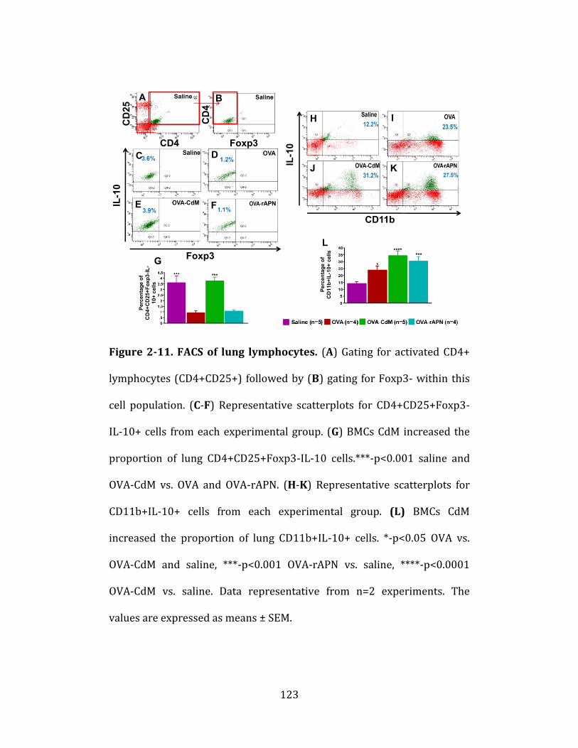
123
IL-1
0
Foxp3
CD11b
IL-1
0
CD
25
CD
4
CD4 Foxp3P
erc
en
tag
eo
f
CD
4+
CD
25+
Fo
xp
3-I
L-
10+
cells
Perc
en
tag
eo
f
CD
11b
+IL
-10+
cells
A
J
G
L
K
IH
B
E
DC
F
Figure 2-11. FACS of lung lymphocytes. (A) Gating for activated CD4+
lymphocytes (CD4+CD25+) followed by (B) gating for Foxp3- within this
cell population. (C-F) Representative scatterplots for CD4+CD25+Foxp3-
IL-10+ cells from each experimental group. (G) BMCs CdM increased the
proportion of lung CD4+CD25+Foxp3-IL-10 cells.***-p<0.001 saline and
OVA-CdM vs. OVA and OVA-rAPN. (H-K) Representative scatterplots for
CD11b+IL-10+ cells from each experimental group. (L) BMCs CdM
increased the proportion of lung CD11b+IL-10+ cells. *-p<0.05 OVA vs.
OVA-CdM and saline, ***-p<0.001 OVA-rAPN vs. saline, ****-p<0.0001
OVA-CdM vs. saline. Data representative from n=2 experiments. The
values are expressed as means ± SEM.

124
Figure 2-12. Flow-cytometric evaluation of CD45, Sca-1, CD29
expression by WT BMCs, CD45+ BMCs and BM-MSCs.
FL-H
WT BMCs WT BMCs WT BMCs
CD45+ BMCs CD45+ BMCs CD45+ BMCs
BM-MSCs BM-MSCs BM-MSCs
Surface Marker Expression - BALB/c BMCs

125
CHAPTER 3 - STEM CELL CONDITIONED MEDIUM PREVENTS ACUTE
LUNG INJURY IN MICE: IN VIVO EVIDENCE FOR STEM CELL
PARACRINE ACTION
A version of this chapter is being reviewed for publication.
Ionescu LI, Byrne RN, van Haaften TJ, Vadivel A, Alphonse RS, Rey-Parra
GJ, Weissmann G, Eaton F, Thébaud B. Stem Cell Conditioned Medium
Prevents Acute Lung Injury in Mice: In Vivo Evidence for Stem Cell
Paracrine Action. Am J Physiol Lung Cell Mol Biol
LII performed the majority of the experiments. RNB performed preliminary
experiments. TvH, AV, RSA, GJRP, GW and FE provided technical assistance. The
manuscript was written by LII and edited by BT.

126
3.1. Abstract
Mortality and morbidity of acute lung injury and acute respiratory
distress syndrome (ALI/ARDS) remain high in part because of the lack of
pharmacological therapies to prevent injury or promote repair. Stem cells
have been proposed to regenerate damaged organs. Mesenchymal stem
cells (MSCs) prevent lung injury in various experimental models, despite
a low proportion of donor-derived cell engraftment. Increasing evidence
suggest that MSCs exert their beneficial effects via a paracrine mechanism.
We hypothesized that soluble factors secreted by MSCs prevent lung
injury in part by modulating macrophage function. We tested the
protective effect of MSC-derived conditioned medium (CdM) compared to
whole MSCs, lung fibroblasts and fibroblast (Fib)-CdM in vivo and in vitro.
Intratracheal MSCs and MSC CdM significantly attenuated
lipopolysaccharide (LPS)-induced lung neutrophil influx, lung edema and
lung injury as assessed by an established lung injury score. MSC CdM
increased arginase-1 activity in LPS-exposed alveolar macrophages (AMs)
and enhanced expression of Ym1 while decreasing iNOS in AMs from LPS-
MSC and LPS-MSC CdM mice as compared to control mice, which suggests
the occurrence of MSC CdM-driven alternative macrophage activation to
an M2 “healer” phenotype. Comparative multiplex analysis between MSC
CdM and lung fibroblast-CdM demonstrated that MSC CdM contained a
127
range of proteins that may confer therapeutic benefit in lung injury. In
summary, MSCs exert their beneficial effects through a paracrine activity.
MSC CdM prevents LPS-induced lung injury by attenuating lung
inflammation and promoting a wound healing/anti-inflammatory M2
macrophage phenotype.

128
3.2. Introduction
Despite improvements in management, acute lung injury (ALI) and
acute respiratory distress syndrome (ARDS) remain major causes of
morbidity and mortality in critically ill patients of all ages [55]. The
incidence of ALI/ARDS in the US is 138:100,000 persons per year and is
anticipated to double in the next 25 years [55]. Treatment of ALI/ARDS
remains primarily supportive [34].
Bone-marrow derived mesenchymal stem cells (MSCs)
differentiate into cartilage, fat and bone, but also into muscle, liver, kidney,
heart and brain cells [50]. These properties have been harnessed for organ
regeneration [58]. In the lung, bone marrow derived cells, including MSCs,
engraft and adopt a lung epithelial cell phenotype [2, 17, 27, 28, 47, 54,
62]. MSCs can also suppress local immune responses and have the
capacity to avoid immune rejection in allotransplantation [40, 24, 29, 30,
51]. These immunomodulatory properties might be of therapeutic benefit
in ALI/ARDS and other lung diseases characterized by inflammation. Both
intratracheal and systemic MSC administrations improve
lipopolysaccharide (LPS)-induced ALI in mice [17, 37]. These findings are
congruent with the therapeutic benefits of MSCs described in other lung
injury models [2, 21, 31, 46, 54, 62]. In all these studies, the engraftment
rates of MSCs were low [26-28, 54, 62]. Together with the rapid

129
therapeutic benefits seen within 48 to 72 hours of MSC delivery in the ALI
models [17, 37], these findings suggest that beyond cell replacement,
MSCs may be releasing factors responsible for the beneficial effects of cell
therapy. In addition, since the use of MSCs as whole-cell therapy may hold
some risks to the patient [1, 32], we investigated the effects of MSC-
derived conditioned medium (CdM) in LPS-induced ALI in mice.

130
3.3. Materials and Methods
All procedures were approved by the Animal Welfare Committee of
the University of Alberta. Additional detail on all the methods is provided
in the online data supplement.
3.3.1. MSCs and Lung Fibroblasts Isolation, Culture and
Characterization
Bone marrow was harvested from adult (8-10 weeks) C57BL/6
mice (Charles River, ON, Canada), and MSCs were cultured and
characterized as previously described [21]. The femur and tibia were
excised and the marrow was flushed with Dulbecco’s Modified Eagle
Medium (DMEM; Invitrogen, Burlington, ON) containing 10% fetal bovine
serum and 1% PSF. The extracted marrow was dissociated and plated in a
tissue culture flask. After 24 hours, the media was aspirated, adherent
cells were rinsed three times with PBS and replenished with fresh media.
Cells were grown to ~80% confluency, trypsinized, and re-seeded at a
density of 105 cells/cm2. Differentiation of MSCs was performed over 21
days on passage 2-3 cells [21]. MSCs were evaluated for expression of a
panel of surface markers according to established criteria [12, 15, 37, 49,
60]. Antibodies against the following markers were obtained from Becton

131
Dickinson (BD, Mississauga, ON): stem cell antigen-1 (Sca-1, fluorescein
isothiocyanate [FITC]), CD31 (phycoerythrin [PE]), CD11b (FITC), CD45
(FITC), CD44 (FITC), CD73 (PE), CD14 (FITC), CD34 (FITC), c-kit (FITC),
Flk-1 (PE), CD106 (vascular cell adhesion molecule-1 [VCAM-1]), CD29
(PE) and from BioLegend (San Diego, CA): CD105 (PE). MSCs between
passages 7-11 were detached from culture surfaces, counted, and divided
into aliquots of ~ 0.5-1x10⁶ cells/sample in 12x75 mm polystyrene round-
bottom tubes (BD Falcon). Cells were washed twice with flow buffer
(0.05% sodium azide, 0.1% bovine serum albumin in PBS), incubated with
the respective antibodies at 4 °C with gentle shaking for 30 min, washed
twice, re-suspended in flow buffer and analyzed by flow cytometry
(FACSCalibur, BD). Cellquest (BD) and FlowJo (version 5.7.2) software
were used for analyses.
Lung fibroblasts were isolated from adult (8-10 weeks) C57BL/6
mice [59]. Fibroblast identity was confirmed by immunofluorescence
staining for the intermediate filament protein vimentin.
3.3.2. Conditioned Medium (CdM) Preparation
Passage 2- 8 MSCs and fibroblasts were grown to >80% confluency.
Medium (DMEM) was aspirated and cells were rinsed three times with
PBS. Cells were cultured with serum-free DMEM (+ PSF) for 24 hours. CdM

132
was collected and filtered through a 0.2 μm filter to remove cellular
debris. Adherent cells were trypsinized, stained with trypan blue, and
counted. The medium from 5 x 10⁶ cells yielded 15 mL of primary CdM
that was further de-salted and concentrated ~25-fold, yielding 600 μL
CdM, using ultrafiltration units with a 3 kDa molecular weight cut-off
(Amicon Ultra-PL 3, Millipore, Billerica, MA). Similar to work by others
[20], serum-free DMEM + PSF (de-salted and concentrated 25-fold) was
the vehicle control.
3.3.3. Murine LPS-Induced ALI
Eight to ten week-old male C57BL/6 mice were anaesthetized with
5% isoflurane and injected intratracheally (i.t.) with 4 mg/kg LPS (E. coli.
055:B5, Sigma-Aldrich, Oakville, ON). Four hours post-LPS, mice were re-
anesthetized and received a 30 μL i.t. instillation of MSCs, LFs, MSC CdM,
Fib CdM or DMEM. We ensured equivalence between cell-based and CdM-
based treatment by administering the same number of cells (250,000
cells/ 30 μL DMEM) that produced 30 μL concentrated CdM.
Mice (n>=5 per group per endpoint) were sacrificed via an
intraperitoneal injection of pentobarbital at 48 hours post-LPS for either
bronchoalveolar lavage fluid (BALF) or lung histological analysis.

133
3.3.4. BALF Analysis and Alveolar Macrophages (AM) Isolation
Lungs were lavaged with 2.5mL ice-cold phosphate buffered saline
(PBS) injected at 0.5mL increments via a 20 gauge catheter inserted in the
trachea. BALF was centrifuged for 10 minutes at 400 g and BALF cells
were enumerated using the Scepter automated cell counter (Millipore,
Billerica, MA). Differential cell counts were performed on cytospin
preparations (Thermo Shandon, Pittsburgh, PA) stained with Hema 3
Manual Staining System (Fisher Scientific, Nepean, ON) by counting 300
cells per cell-smear and multiplying by total cell number per mL.
For AMs isolation, an established protocol was followed [65].
Briefly, BALF was centrifuged at 300 g for 10 minutes and the cellular
pellet was washed with PBS, re-suspended in red blood cell lysis buffer
[8.3 g NH4Cl, 1 g KHCO3, 1.8 mL of 5% EDTA in 1 L of distilled water) for 5
minutes at room temperature and centrifuged again at 300 g for 10
minutes. The pellet was re-suspended in RPMI-1640 medium and plated
at a density of 600,000 cells/mL in a 24-well tissue culture plate. After 2
hours, medium was removed and adherent cells were washed three times
with PBS. AMs were stained with Hema 3 Manual Staining System for
morphological assessment. AMs were evaluated for the expression of
characteristic surface markers [18] using antibodies against CD11b (FITC)

134
and CD11c (allophycocyanin [APC]) (BD Biosciences) by flow cytometry
according to the same protocol described for MSC characterization.
3.3.5. Assessment of Lung Permeability
Lung edema due to LPS-induced increase in lung permeability was
measured using the wet / dry weight ratio of lung lobes as previously
described [17]. Briefly, lungs were weighed upon excision (wet weights),
homogenized in 1 mL of water and placed in a drying oven at 55 °C for 24
hours, dry weights recorded and wet / dry ratio calculated.
3.3.6. Lung Histological Analysis
Lungs were inflated and fixed with 4% formaldehyde solution
through a tracheal catheter at a constant pressure of 20 cm H2O [21].
Lungs were processed, paraffin-embedded and 4 μm thick serial sections
were stained with hematoxylin and eosin (H&E). Images were captured
(Openlab, Improvision, version 5.0.2.) with Leica CTRMIC microscope and
40 high-powered fields per lung were examined by a blinded investigator
to quantify the histopathology score. Parameters assessed were: alveolar
septal congestion, alveolar hemorrhage, intra-alveolar fibrin and intra-
alveolar infiltrates. A score from 0 to 3 was given for each criterion and a

135
total score was established, all according to a previously published
protocol [36, 37]. Briefly, lung injury score = [(alveolar hemorrhage
points/no. of fields) + 2 x (alveolar infiltrate points/no. of fields) + 3 x
(fibrin points/no. of fields) + (alveolar septal congestion/no. of
fields)]/total number of alveoli counted.
3.3.7. AM LPS Exposure and AM Phenotype Characterization
Freshly isolated AMs were cultured with RPMI-1640 media
containing 10% FBS in a 12-well plate. After overnight adherence, RPMI-
1640 media was replaced with either DMEM alone, DMEM+LPS (1 μg/ml),
or MSC CdM+LPS (1 μg/ml). After 48 hours in culture, media was
aspirated and cells were rinsed 3X with PBS. The monolayer of cells was
scraped, cells were spun down and pellets were frozen. Arginase activity
in AM was measured by determining the amount of urea generated by the
enzyme [9, 57]. Briefly, the frozen AM pellets were thawed and
homogenized in 100 μl per sample of lysis buffer (0.1 % Triton X-100 with
protease inhibitors: phenyl-methyl-sulphonyl fluoride [PMSF] (1 mM),
leupeptin (0.5 µg/ml), aprotinin (5 µg/ml), EDTA (2 mM)). The samples
were sonicated twice for 3-5 seconds and stored at -80 °C. Samples were
thawed and 50 μL of the homogenate was added to 50 μl Tris-HCl (25 mM,
pH 7.5) containing MnCl2 (5 mM) and incubated at 56 ºC for 10 min to

136
activate the enzyme. 50 μL of this heat-activated supernatant mixture was
incubated with 50 μL L-arginine (0.5 mol/L) at 37ºC for 60 min. The
reaction was stopped by adding 400 μL of an acid mixture (1 H2SO4: 3
H3PO4: 7 H2O). 25 μL of α-isonitrosopropiophenone (9 % dissolved in 100
% ethanol) was added to the above mixture and incubated at 100 ºC for 45
min for color development. The mixture was cooled at room temperature
in the dark for 10 min. A standard curve for urea (0-30 μg) was prepared.
Urea concentration in the homogenate was measured using a colorimeter
at 550/540 nm with 200 μL of the aliquot. Arginase activity was expressed
as units / mg protein / hour.
For further macrophage phenotype characterization by multicolor
flow cytometry, AMs were isolated from experimental animals and
allowed to adhere to plastic as described above. The following antibodies
were used: CD11b (Pacific Blue), CD11c (PE, both from Biolegend), rabbit
anti-mouse inducible nitric oxide synthase (iNOS, Abcam) and donkey
anti-rabbit secondary (FITC, Biomeda Corporation, Foster City, CA), goat
anti-mouse Ym1 (R&D Systems) and donkey anti-goat secondary (Cy5,
Biomeda). The staining was performed as described for MSC
characterization, with the addition of a 30 minute incubation of the cells at
4 ºC in Cytofix / Cytoperm Buffer (BD), followed by resuspension in
Permeabilization Buffer (BD) before incubation with primary antibodies.
Analysis was performed using FACSCanto with FACS Diva software (BD).

137
3.3.8. Antibody array of MSC CdM
MSC CdM and Fib CdM were prepared, concentrated and de-salted
as described above. Samples were analyzed for a panel of 96 factors using
an antibody array (RayBio Mouse Antibody Array G Series 1000,
RayBiotech Inc., Norcross, GA). Relative intensity of each factor in MSC
CdM and Fib CdM is reported compared to a positive and negative control,
as well as fold-change of specific factors in MSC CdM versus Fib CdM.
3.3.9. Statistical Analysis
Data are expressed as mean ± standard error of the mean (SEM).
Group comparisons were analyzed by one-way ANOVA with a Fisher’s
PLSD post hoc test. An unpaired t-test was used when appropriate. Data
was assessed with statistical software Statview (version 5.0.1, SAS
Institute, Cary, NC). Values were considered significant with p<0.05.
138
3.4. Results
3.4.1. MSC Display Functional Characteristics and Surface Marker
Phenotype of Murine MSCs
Bone marrow-derived MSCs differentiated into adipogenic,
osteogenic and chondrogenic mesenchymal lineages (Fig. 3-1). Cell
surface antigen phenotype was assessed by flow cytometry. MSCs
expressed high levels of Sca-1 (92.93% of cells) and CD29 (89.99%),
moderate levels of CD105 (33.92%), CD106 (14.36% ), CD11b (12.10%),
and CD45 (11.62%) and were considered negative for CD14 (0.30%), c-kit
(0.53%), CD34 (1.13%), CD73 (1.49%), Flk-1 (3.01%) and CD31 (4.08% of
cells) (Fig. 3-2).
3.4.2. MSC CdM Decreased Lung Inflammation and Lung Vascular
Permeability in LPS-Induced Lung Injury
LPS significantly increased the total cell and neutrophil count in the
BAL after 12 hours compared to uninjured controls (Fig. 3-3A). This
inflammatory influx was attenuated by MSCs and MSC CdM treatment, but
not by treatment with DMEM, the vehicle control, Fib or Fib CdM (Fig. 3-
3A). MSCs and MSC CdM, but not DMEM, Fib or Fib CdM prevented the

139
LPS-induced increase in lung vascular permeability, as assessed by lung
wet/dry ratio (Fig. 3-3B).
3.4.3. MSC CdM Failed to Prevent LPS-Induced Body Weight Loss
All mice given LPS were lethargic and had reduced activity and
decreased body weight over 48 hours compared to control mice (Fig. 3-
4A).
3.4.4. MSC CdM Improved LPS-Induced Lung Injury
Histological assessment of lung injury using a semi-quantitative
histopathology score revealed that mice treated with LPS had increased
septal thickening, alveolar hemorrhage, alveolar infiltrates, and fibrin
strands compared to controls, whereas treatment with MSCs or MSC CdM
significantly attenuated these features (Fig. 3-4B and C).
3.4.5. MSC CdM Determined Alternative Activation of AMs Following
LPS Exposure In Vitro and In Vivo
Isolated AMs were macroscopically (insert Fig. 3-5A) and
phenotypically (CD11c+CD11b-) (Fig. 3-5A) consistent with AMs. In vitro,

140
AMs were exposed to LPS in order to test the role of MSC CdM on the
induction and maintenance of the M2 phenotype in AMs. LPS increased
arginase activity in AMs as compared with non-LPS-exposed cells, and
MSC CdM further elevated these levels compared to LPS-exposed cells
cultured with DMEM (Fig. 3-5B).
In order to further investigate the effects of MSC CdM in vivo, AMs
were isolated from LPS-exposed animals that had received cell or CdM
treatment. MSC CdM induced an iNOS-Ym1+ phenotype in AMs from both
control (11.6% vs. 0.8%) and LPS-treated animals (69% vs. 6.8%). This
effect was enhanced in LPS-MSC recipients (79.9%). Fib and Fib CdM
markedly induced an iNOS+Ym1- phenotype (52% and 41.9%,
respectively) compared to control AMs (1.3%), similar to the proportion
found in LPS-DMEM AMs (41.8%) (Fig. 3-6).
3.4.6. MSC CdM Contains Soluble Factors That May Convey
Therapeutic Benefit
The levels of 96 different factors were compared between MSC
CdM and Fib CdM (Fig. 3-7). Cluster analysis of both CdMs revealed the
following distribution: chemokines (31%), binding proteins (27%),
cytokines (21%), growth factors (14%), cytokine antagonists (4%) and
molecules involved in extracellular matrix repair (3%) (Fig. 3-7).
141
Significant fold-variations of certain factors were found (Fig. 3-8). MSC
CdM contained higher concentrations of stem cell factor (SCF), IL-
12p40/p70, stromal-derived factor-1 alpha (SDF-1α) and osteoprotegerin
and lower concentrations of IL-6, macrophage inflammatory protein-3
alpha (MIP-3α) and thymus and activation regulated chemokine (TARC).

142
3.5. Discussion
We provide evidence that the therapeutic benefits of MSCs are
attributable to a paracrine mechanism. In vivo, administration of MSC CdM
alone attenuated lung neutrophil influx and improved lung histology. This
effect was comparable with administration of an equivalent number of
MSC and absent in mice treated with control cells (lung fibroblasts) or
control cell CdM. MSC CdM induced an M2 phenotype in AMs exposed to
LPS in vitro and in vivo in LPS-exposed animals. We also suggest that MSC
CdM contains soluble factors capable of attenuating lung injury.
Identification of these soluble factors secreted by MSCs may yield new
therapeutic options for ALI/ARDS.
Recent studies have shown that MSCs modulate immune cell
function [23, 40, 51] and have cell-protective effects through the release of
cytokines and growth factors [14, 20, 31]. MSC CdM decreased hypoxia-
induced cell death and improved tube formation of human aortic
endothelial cells [20]. CdM derived from MSCs engineered to over-express
the pro-survival gene Akt protected cardiomyocytes from hypoxia-
induced cell death and limited myocardial infarct size in vivo [13]. MSC
CdM improved healing in an excisional wound splinting model in mice [7],
oxygen-induced AT2 and pulmonary microvascular endothelial cell injury
in vitro [62], neonatal oxygen-induced lung injury in vivo [2] and reversed

143
hepatocyte death and increased survival in Gal-N induced fulminant
hepatic failure [41]. Moreover, human MSC CdM attenuated endotoxin-
induced lung injury in an ex vivo perfused human lung model [31] and
mouse MSC CdM prevented the development of murine asthma [21].
In the current study, the therapeutic benefit seen with CdM
mirrored the protective effects described with whole cell therapy. Similar
to previous findings with whole cell therapy after LPS injury [17, 37], we
observed that a single intratracheal injection of MSC CdM or MSCs 4 hours
after LPS administration decreased lung neutrophil influx, lung
permeability and improved the lung histopathology score as compared to
control media, lung fibroblasts or their CdM. These findings open new
therapeutic options by identifying potential healing molecules contained
in MSC-derived CdM and understanding their mechanism of action.
Our data suggest that MSC CdM promote a M2 “healer” macrophage
phenotype. In the development of ALI/ARDS, neutrophils and
macrophages are activated in order to eliminate pathogens, but also
contribute to tissue injury through the release of antimicrobial
compounds [63]. Macrophages are phenotypically heterogeneous because
they respond to stimuli in their microenvironment and they also differ
genetically [16]. Classically activated macrophages (M1) kill invading
microorganisms and tumors and promote type I immunity by secreting
high levels of pro-inflammatory cytokines and low levels of anti-

144
inflammatory cytokines [33]. In contrast, alternatively activated
macrophages (M2) secrete lower levels of pro-inflammatory cytokines
and higher levels of anti-inflammatory cytokines. M2 macrophages
promote type II immunity and are thought to dampen the immune
response and to promote wound healing, angiogenesis, and debris
scavenging [10]. M1 and M2 macrophages can be characterized by
receptor expression, effector function, cytokine and chemokine
production [33] or by a set of marker genes: M1 macrophages generate
nitric oxide (NO) by upregulation of inducible nitric oxide synthase
(iNOS), whereas M2 macrophages express Ym1 (also known as T-
lymphocyte-derived eosinophil chemotactic factor [ECF-L] and chitinase
3-like 3 [CHI3L3]), FIZZ1 (found in inflammatory zone-1, also known as
resistin-like molecule alpha [RELM-alpha]), and arginase-1 (Arg-1) [16,
43]. The upregulation of arginase-1 expression and activity is crucial in
the metabolic switch from M1 to M2 phenotype in mice [52]. In our LPS-
induced inflammation model, we screened for the effect of MSC CdM on
AM phenotype in vitro. AMs isolated from healthy mice and subsequently
exposed to LPS followed by MSC CdM had higher Arg-1 activity
characteristic of M2 macrophages. In order to more thoroughly test for the
occurrence of alternative macrophage activation in vivo, we isolated AMs
from experimental animals and found that LPS, as well as Fib and FibCdM,
increased the numbers of macrophages expressing iNOS in the absence of

145
Ym1 (a pattern corresponding with the M1 phenotype), while MSCs and
MSC CdM upregulated Ym1+ macrophages lacking iNOS expression (M2).
These data support the immunomodulatory capacity of MSC CdM
shifting the immune environment from pro- to anti-inflammatory via the
induction of a “healer” M2 AM phenotype from a “killer” M1 macrophage.
Our results are in line with several recent reports indicating that MSCs
exert anti-inflammatory properties via macrophage reprogramming [25,
42, 44]. However, the murine MSC populations differ amongst these
reports, mainly due to the current lack of consensus in establishing the
universal murine MSC phenotype [6, 49]. Comparison is also limited by
differences in experimental protocols and variations in CdM preparation
methods.
The paracrine mechanism of action of MSCs opens new therapeutic
perspectives. Indeed, several paracrine mediators that can mediate
restorative effects of MSCs have been identified, including interleukin-10,
IL-1 receptor antagonist (IL-1ra), and keratinocyte growth factor (KGF).
IL-1ra was identified as an MSC-derived paracrine factor that reduced the
severity of bleomycin-induced lung injury [46]. Allogeneic human MSCs or
their CdM attenuated endotoxin-induced lung injury in an ex vivo perfused
human lung model partly through KGF, a well-known growth factor to
reduce lung injury [31]. In our study, we found that MSC CdM contained
higher levels of KGF and HGF than Fib CdM. IL-10 was shown to be

146
responsible for the therapeutic benefits of exogenous MSCs in murine
sepsis [44]. Our group has recently reported that BMSC-derived
adiponectin contributes to the anti-asthmatic effect of BMSC CdM in a
murine ovalbumin-induced asthma model [21]. Human cord blood-
derived MSCs, but not fibroblasts, produce high levels of angiotensin
converting enzyme 2, an exopeptidase recently shown to be lung
protective [53]. Further identification of soluble factors may lead to the
development of critically missing pharmacological therapies for ALI/ARDS
and other inflammatory diseases [5].
In an attempt to broaden the search for candidate soluble factors,
we performed a multiplex analysis screening for 96 factors present in MSC
CdM and showed that their secretory profile differed from that of lung
fibroblasts. In a similar manner, Schinköthe et al. screened human MSCs to
attain the first large-scale description of factors they secrete and
categorized these into functional groups: anti-apoptotic,
immunosuppressive, pro-proliferative and angiogenic modulating [58].
Amongst factors that may account for the alternative macrophage
activation, we found that MSC CdM contained several known M2
activators. Interleukins (IL)-4, -10 and -13 were identified in our multiplex
screen of MSC CdM (Fig. 3-7). These cytokines can activate the JAK-STAT6
signaling pathway and promote STAT6 binding to the promoter region of
target genes, which ultimately drives the expression of M2-specific genes
147
[23]. Although the levels of these interleukins were comparable between
MSC CdM and Fib CdM, they may contribute to alternative macrophage
activation in an orchestrated fashion, together with other components of
MSC CdM. Fib CdM also contained higher amounts of the potent classical
(M1) activator IL-6, while MSC CdM had higher levels of factors that have
recently been shown to contribute to the promotion of an M2
environment. One of these factors is insulin-like growth factor-1 (IGF-1),
which has been found to aid in creating an M2-favorable environment [7].
Another factor, adiponectin, that may exert similar M2-activating effects
[43] was found in our MSCs CdM from both WT C57/BL6 and Balb/C
mouse strains, but was undetectable in Fib CdM by ELISA [21].
Of the 96 different cytokines screened, MSC CdM contained
markedly higher levels of potential protective factors compared to Fib
CdM, suggesting that additional mechanisms could explain the therapeutic
benefit of MSC CdM in this model. Stem cell factor (SCF) is increased
almost 3-fold in MSC CdM compared to Fib CdM. SCF improves survival,
proliferation, and differentiation of hematopoietic stem and progenitor
cells and anti-apoptotic effects reported in kidney tubular epithelial cells
[3, 12]. Another molecule, FcγRIIB, is increased 14-fold in MSC CdM
compared to Fib CdM. FcγRIIB mediates the anti-inflammatory benefits of
intravenous gammaglobulin (IVIG) in a murine model of immune
thrombocytopenia [56]. We also found that IL-12p40, a natural antagonist

148
of IL-12, which is a cytokine responsible for the production of IFN-γ, was
increased 7-fold in MSC CdM compared to Fib CdM. IL-12p40 has
protective effects in bacterial pneumonitis in mice [18] and selectively
inhibits airway hyperresponsiveness and peribronchial fibrosis in murine
asthma [46]. A two-fold increase in soluble TNF-α receptor II (sTNFRII),
an inhibitor of TNF-α and a possible regulator of inflammatory activity,
was observed in MSC CdM compared to Fib CdM. sTNFRII may contribute
to dampening of the activity of TNF-α in LPS injury [38].
In conclusion, our study provides direct in vivo evidence that MSCs
exert their therapeutic benefit through a paracrine activity. Identification
of pneumoprotective soluble factors in MSC CdM holds promise for the
discovery of new pharmacological therapies for lung diseases. While MSCs
may have a unique ability to monitor the microenvironment of injured
tissues and respond appropriately, therapies with the proteins or
cytokines produced by activated MSCs may be more practical than cell
therapies [1, 32]. In vitro priming of MSCs to optimize the production of
protective factors may combine the advantages of both cell and small
molecule therapy.

149
3.6. References
1. Amariglio N, Hirshberg A, Scheithauer BW, Cohen Y, Loewenthal R,
Trakhtenbrot L, Paz N, Koren-Michowitz M, Waldman D, Leider-
Trejo L, Toren A, Constantini S, and Rechavi G (2009) Donor-
derived brain tumor following neural stem cell transplantation in
an ataxia telangiectasia patient. PLoS Med 6: e1000029
2. Aslam M, Baveja R, Liang OD, Fernandez-Gonzalez A, Lee C,
Mitsialis SA, Kourembanas S (2009) Bone Marrow Stromal Cells
Attenuate Lung Injury in a Murine Model of Neonatal Chronic Lung
Disease. Am J Respir Crit Care Med, 180:1122-30
3. Bengatta S, Arnould C, Letavernier E, Monge M, de Preneuf HM,
Werb Z, Ronco P, and Lelongt B (2009) MMP9 and SCF protect from
apoptosis in acute kidney injury. J Am Soc Nephrol 20: 787-797
4. Bonfield TL, Koloze M, Lennon DP, Zuchowski B, Yang SE, Caplan AI
(2010) Human mesenchymal stem cells suppress chronic airway
inflammation in the murine ovalbumin asthma model. Am J Physiol
Lung Cell Mol Physiol 299:L760–L770
5. Cepkova M, Matthay MA (2006) Pharmacotherapy of acute lung
injury and the acute respiratory distress syndrome. J Intensive Care
Med 21: 119-143

150
6. Chamberlain G, Fox J, Ashton B, Middleton J (2007) Concise review:
mesenchymal stem cells: their phenotype, differentiation capacity,
immunological features, and potential for homing. Stem Cells
25:2739–2749
7. Chen F, Liu Z, Wu W, Rozo C, Bowdridge S, Millman A, Van Rooijen
N, Urban JF Jr, Wynn TA, Gause WC (2012) An essential role for
T(H)2-type responses in limiting acute tissue damage during
experimental helminth infection. Nat Med doi: 10.1038/nm.2628
8. Chen L, Tredget EE, Wu PY, Wu Y (2008) Paracrine factors of
mesenchymal stem cells recruit macrophages and endothelial
lineage cells and enhance wound healing. PLoS ONE 3: e1886
9. Corraliza IM, Campo ML, Soler G, Modolell M (1994) Determination
of arginase activity in macrophages: a micromethod. J Immunol
Methods 174: 231-235
10. Dal-Pizzol F (2004) Alternative activated macrophage: a new key
for systemic inflammatory response syndrome and sepsis
treatment? Crit Care Med 32: 1971-1972
11. Dominici M, Le Blanc K, Mueller I, Slaper-Cortenbach I, Marini F,
Krause D, Deans R, Keating A, Prockop D, Horwitz E (2006)
Minimal criteria for defining multipotent mesenchymal stromal
cells. The International Society for Cellular Therapy position
statement. Cytotherapy 8: 315-317
151
12. Galli MC, Giardina PJ, Migliaccio AR, Migliaccio G (1993) The
biology of stem cell factor, a new hematopoietic growth factor
involved in stem cell regulation. Int J Clin Lab Res 23: 70-77
13. Gnecchi M, He H, Liang OD, Melo LG, Morello F, Mu H, Noiseux N,
Zhang L, Pratt RE, Ingwall JS, Dzau VJ (2005) Paracrine action
accounts for marked protection of ischemic heart by Akt-modified
mesenchymal stem cells. Nat Med 11: 367-368
14. Gnecchi M, He H, Noiseux N, Liang OD, Zhang L, Morello F, Mu H,
Melo LG, Pratt RE, Ingwall JS, Dzau VJ (2006) Evidence supporting
paracrine hypothesis for Akt-modified mesenchymal stem cell-
mediated cardiac protection and functional improvement. FASEB J
20: 661-669
15. Gnecchi M, Melo LG. Bone marrow-derived mesenchymal stem
cells: isolation, expansion, characterization, viral transduction, and
production of conditioned medium. Methods Mol Biol 482: 281-294
16. Gordon S, Taylor PR (2005) Monocyte and macrophage
heterogeneity. Nat Rev Immunol 5: 953-964
17. Gupta N, Su X, Popov B, Lee JW, Serikov V, Matthay MA (2007)
Intrapulmonary delivery of bone marrow-derived mesenchymal
stem cells improves survival and attenuates endotoxin-induced
acute lung injury in mice. J Immunol 179: 1855-1863

152
18. Guth AM, Janssen WJ, Bosio CM, Crouch EC, Henson PM, and Dow
SW (2009) Lung environment determines unique phenotype of
alveolar macrophages. Am J Physiol Lung Cell Mol Physiol 296:
L936-946
19. Gutierrez JG, Valdez SR, Di Genaro S, Gomez NN (2008) Interleukin-
12p40 contributes to protection against lung injury after oral
Yersinia enterocolitica infection. Inflamm Res 57: 504-511
20. Hung SC, Pochampally RR, Chen SC, Hsu SC, Prockop DJ (2007)
Angiogenic effects of human multipotent stromal cell conditioned
medium activate the PI3K-Akt pathway in hypoxic endothelial cells
to inhibit apoptosis, increase survival, and stimulate angiogenesis.
Stem Cells 25: 2363-2370
21. Ionescu LI, Alphonse RS, Arizmendi N, Morgan B, Abel M, Eaton F,
Duszyk M, Vliagoftis H, Aprahamian TR, Walsh K, Thebaud B
(2012) Airway Delivery of Soluble Factors from Plastic-Adherent
Bone Marrow Cells Prevents Murine Asthma. Am J Respir Cell Mol
Biol 46(2):207-16
22. Ishii M, Wen H, Corsa CA, Liu T, Coelho AL, Allen RM, Carson WFt,
Cavassani KA, Li X, Lukacs NW, Hogaboam CM, Dou Y, Kunkel SL
(2009) Epigenetic regulation of the alternatively activated
macrophage phenotype. Blood 14:3244-54
153
23. Iyer SS, Rojas M (2008) Anti-inflammatory effects of mesenchymal
stem cells: novel concept for future therapies. Expert Opin Biol Ther
8:569–581
24. Jarvinen L, Badri L, Wettlaufer S, Ohtsuka T, Standiford TJ, Toews
GB, Pinsky DJ, Peters-Golden M, Lama VN (2008) Lung resident
mesenchymal stem cells isolated from human lung allografts
inhibit T cell proliferation via a soluble mediator. J Immunol
181:4389–4396
25. Kim J, Hematti P (2009) Mesenchymal stem cell-educated
macrophages: a novel type of alternatively activated macrophages.
Exp Hematol 37:1445–1453
26. Kotton DN, Fabian AJ, Mulligan RC (2005) Failure of bone marrow
to reconstitute lung epithelium. Am J Respir Cell Mol Biol 33: 328-
334
27. Kotton DN, Ma BY, Cardoso WV, Sanderson EA, Summer RS,
Williams MC, Fine A (2001) Bone marrow-derived cells as
progenitors of lung alveolar epithelium. Development 128: 5181-
5188
28. Krause DS, Theise ND, Collector MI, Henegariu O, Hwang S,
Gardner R, Neutzel S, Sharkis SJ (2001) Multi-organ, multi-lineage
engraftment by a single bone marrow-derived stem cell. Cell 105:
369-377
154
29. Le Blanc K, Ringden O (2007) Immunomodulation by
mesenchymal stem cells and clinical experience. J Intern Med
262:509–525.
30. Le Blanc K, Frassoni F, Ball L, Locatelli F, Roelofs H, Lewis I, Lanino
E, Sundberg B, Bernardo ME, Remberger M, Dini G, Egeler RM,
Bacigalupo A, Fibbe W, Ringdén O; Developmental Committee of
the European Group for Blood and Marrow Transplantation (2008)
Mesenchymal stem cells for treatment of steroid-resistant, severe,
acute graft-versus host disease: a phase II study. Lancet 371:1579–
1586
31. Lee JW, Fang X, Gupta N, Serikov V, Matthay MA (2009) Allogeneic
human mesenchymal stem cells for treatment of E. coli endotoxin-
induced acute lung injury in the ex vivo perfused human lung. Proc
Natl Acad Sci U S A 106: 16357-16362
32. Lepperdinger G, Brunauer R, Jamnig A, Laschober G, Kassem M
(2008) Controversial issue: is it safe to employ mesenchymal stem
cells in cell-based therapies? Exp Gerontol 43: 1018-1023
33. Mantovani A, Sica A, Sozzani S, Allavena P, Vecchi A, Locati M
(2004) The chemokine system in diverse forms of macrophage
activation and polarization. Trends Immunol 25: 677-686
34. Matthay MA, Zimmerman GA, Esmon C, Bhattacharya J, Coller B,
Doerschuk CM, Floros J, Gimbrone MA, Jr., Hoffman E, Hubmayr RD,

155
Leppert M, Matalon S, Munford R, Parsons P, Slutsky AS, Tracey KJ,
Ward P, Gail DB, Harabin AL (2003) Future research directions in
acute lung injury: summary of a National Heart, Lung, and Blood
Institute working group. Am J Respir Crit Care Med 167: 1027-1035
35. Matthay MA, Thompson BT, Read EJ, McKenna DH Jr, Liu KD, Calfee
CS, Lee JW (2010) Therapeutic potential of mesenchymal stem cells
for severe acute lung injury. Chest 138(4):965-72
36. Matute-Bello G, Winn RK, Jonas M, Chi EY, Martin TR, Liles WC
(2001) Fas (CD95) induces alveolar epithelial cell apoptosis in
vivo: implications for acute pulmonary inflammation. Am J Pathol
158: 153-161
37. Mei SH, McCarter SD, Deng Y, Parker CH, Liles WC, and Stewart DJ
(2007) Prevention of LPS-induced acute lung injury in mice by
mesenchymal stem cells overexpressing angiopoietin 1. PLoS Med
4: e269
38. Mohler KM, Torrance DS, Smith CA, Goodwin RG, Stremler KE, Fung
VP, Madani H, Widmer MB (1993) Soluble tumor necrosis factor
(TNF) receptors are effective therapeutic agents in lethal
endotoxemia and function simultaneously as both TNF carriers and
TNF antagonists. J Immunol 151: 1548-1561
39. Morrison AC, Correll PH (2002) Activation of the stem cell-derived
tyrosine kinase/RON receptor tyrosine kinase by macrophage-

156
stimulating protein results in the induction of arginase activity in
murine peritoneal macrophages. J Immunol 168(2):853-60
40. Nauta AJ, Fibbe WE (2007) Immunomodulatory properties of
mesenchymal stromal cells. Blood 110: 3499-3506
41. Nemeth K, Keane-Myers A, Brown JM, Metcalfe DD, Gorham JD,
Gorham JD, Bundoc VG, Bundoc VG, Hodges MG, Jelinek I, Madala S,
Karpati S, Mezey E (2010) Bone marrow stromal cells use TGF-beta
to suppress allergic responses in a mouse model of ragweed-
induced asthma. Proc Natl Acad Sci U S A 107(12):5652-5657
42. Németh K, Leelahavanichkul A, Yuen PST, Mayer B, Parmelee A, Doi
K, Robey PG, Leelahavanichkul K, Koller BH, Brown JM, Hu X,
Jelinek I, Star RA, Mezey E (2009) Bone marrow stromal cells
attenuate sepsis via prostaglandin E(2)-dependent reprogramming
of host macrophages to increase their interleukin-10 production.
Nat Med 15(1):42-49
43. Odegaard JI, Chawla A (2011) Alternative macrophage activation
and metabolism. Annu Rev Pathol 6:275-97
44. Ohtaki H, Ylostalo JH, Foraker JE, Robinson AP, Reger RL, Shioda S,
Prockop DJ (2008) Stem/progenitor cells from bone marrow
decrease neuronal death in global ischemia by modulation of

157
inflammatory/immune responses. Proc Natl Acad Sci U S A
105(38):14638-43
45. Onari Y, Yokoyama A, Haruta Y, Nakashima T, Iwamoto H, Hattori
N, Kohno N (2009) IL-12p40 is essential for the down-regulation of
airway hyperresponsiveness in a mouse model of bronchial asthma
with prolonged antigen exposure. Clin Exp Allergy 39: 290-298
46. Ortiz LA, Dutreil M, Fattman C, Pandey AC, Torres G, Go K, Phinney
DG (2007) Interleukin 1 receptor antagonist mediates the
antiinflammatory and antifibrotic effect of mesenchymal stem cells
during lung injury. Proc Natl Acad Sci U S A 104:11002-11007
47. Ortiz LA, Gambelli F, McBride C, Gaupp D, Baddoo M, Kaminski N,
Phinney DG (2003) Mesenchymal stem cell engraftment in lung is
enhanced in response to bleomycin exposure and ameliorates its
fibrotic effects. Proc Natl Acad Sci U S A 100: 8407-8411
48. Parekkadan B, van Poll D, Suganuma K, Carter EA, Berthiaume F,
Tilles AW, Yarmush ML (2007) Mesenchymal stem cell-derived
molecules reverse fulminant hepatic failure. PLoS ONE 2: e941
49. Peister A, Mellad JA, Larson BL, Hall BM, Gibson LF, Prockop DJ
(2004) Adult stem cells from bone marrow (MSCs) isolated from
different strains of inbred mice vary in surface epitopes, rates of
proliferation, and differentiation potential. Blood 103: 1662-1668

158
50. Prockop DJ (1997) Marrow stromal cells as stem cells for
nonhematopoietic tissues. Science 276: 71-74
51. Prockop DJ (2009) Repair of tissues by adult stem/progenitor cells
(MSCs): controversies, myths, and changing paradigms. Mol Ther
17:939–946
52. Rauh MJ, Ho V, Pereira C, Sham A, Sly LM, Lam V, Huxham L,
Minchinton AI, Mui A, Krystal G (2005) SHIP represses the
generation of alternatively activated macrophages. Immunity 23:
361-374
53. Rey-Parra GJ, Vadivel A, Coltan L, Hall A, Eaton F, Schuster M,
Loibner H, Penninger JM, Kassiri Z, Oudit GY, Thébaud B (2012)
Angiotensin converting enzyme 2 abrogates bleomycin-induced
lung injury. J Mol Med (Berl) 90(6):637-47
54. Rojas M, Xu J, Woods CR, Mora AL, Spears W, Roman J, Brigham KL
(2005) Bone marrow-derived mesenchymal stem cells in repair of
the injured lung. Am J Respir Cell Mol Biol 33: 145-152
55. Rubenfeld GD, Caldwell E, Peabody E, Weaver J, Martin DP, Neff M,
Stern EJ, Hudson LD (2005) Incidence and outcomes of acute lung
injury. N Engl J Med 353:1685-1693
56. Samuelsson A, Towers TL, Ravetch JV (2001) Anti-inflammatory
activity of IVIG mediated through the inhibitory Fc receptor.
Science 291: 484-486

159
57. Sankaralingam S, Xu H, Davidge ST (2010) Arginase contributes to
endothelial cell oxidative stress in response to plasma from women
with preeclampsia. Cardiovasc Res 85:194-203
58. Schinkothe T, Bloch W, Schmidt A. In vitro secreting profile of
human mesenchymal stem cells. Stem Cells Dev 17: 199-206
59. Sugiura H, Liu X, Duan F, Kawasaki S, Togo S, Kamio K, Wang XQ,
Mao L, Ahn Y, Ertl RF, Bargar TW, Berro A, Casale TB, Rennard SI
(2007) Cultured lung fibroblasts from ovalbumin-challenged
"asthmatic" mice differ functionally from normal. Am J Respir Cell
Mol Biol 37(4):424-30
60. Sung JH, Yang HM, Park JB, Choi GS, Joh JW, Kwon CH, Chun JM, Lee
SK, Kim SJ (2008) Isolation and characterization of mouse
mesenchymal stem cells. Transplant Proc 40:2649–2654
61. Uccelli A, Moretta L, Pistoia V (2008) Mesenchymal stem cells in
health and disease. Nat Rev Immunol 8:726-36
62. van Haaften T, Byrne R, Bonnet S, Rochefort GY, Akabutu J,
Bouchentouf M, Rey-Parra GJ, Galipeau J, Haromy A, Eaton F, Chen
M, Hashimoto K, Abley D, Korbutt G, Archer SL, Thébaud B (2009)
Airway Delivery of Mesenchymal Stem Cells Prevents Arrested
Alveolar Growth in Neonatal Lung Injury in Rats. Am J Respir Crit
Care Med 180:1131-42

160
63. Ware LB, Matthay MA (2000) The acute respiratory distress
syndrome. N Engl J Med 342: 1334-1349
64. Yan YJ, Li Y, Lou B, Wu MP (2006) Beneficial effects of ApoA-I on
LPS-induced acute lung injury and endotoxemia in mice. Life Sci 79:
210-215
65. Zhang C, Wang SH, Lasbury ME, Tschang D, Liao CP, Durant PJ, and
Lee CH (2006) Toll-like receptor 2 mediates alveolar macrophage
response to Pneumocystis murina. Infect Immun 74: 1857-1864
66. Zhang M, Mal N, Kiedrowski M, Chacko M, Askari AT, Popovic ZB,
Koc ON, Penn MS (2007) SDF-1 expression by mesenchymal stem
cells results in trophic support of cardiac myocytes after
myocardial infarction. FASEB J 21: 3197-3207

161
3.7. Figures
Figure 3-1. Mesenchymal lineage differentiation of C57BL/6 BM-
MSCs. MSCs differentiated along adipogenic, osteogenic and chondrogenic
lineages. Top row: differentiated MSCs; bottom row: control MSCs. Left to
right panels: Oil Red O staining (adipocytes), Alizarin Red (osteocytes),
Safranin O (chondrocytes). Size bar: 30 µm.

162
Figure 3-2. Flow-cytometric characterization of C57BL/6 BM-MSCs
surface marker profile. Representative flow-cytometry histograms (A)
and quantification of MSCs surface marker expression (B). Values are
expressed as mean ± SEM.

163
Figure 3-3. BALF and lung permeability analyses. (A) BALF total cell
and neutrophil numbers. LPS-DMEM mice (n=10) had significantly more
inflammatory cells than uninjured controls (n=5) and control-CdM (n=6)
mice. Treatment with MSCs (n=5) or MSC CdM (n=10), but not Fib (n=5)
or Fib CdM (n=5), significantly attenuated lung cells influx. BAL fluid of
mice treated with DMEM had 2 times more PMNs than those treated with

164
MSC CdM. There were no differences in MN number. *p<0.05 control,
control-CdM vs. LPS-MSC CdM; **-p<0.01 control, control-CdM vs. LPS-
DMEM, LPS-Fib, LPS-Fib CdM, LPS-MSC; LPS-MSC vs. LPS-DMEM, LPS-Fib,
LPS-Fib CdM. (B) Lung permeability evaluation. LPS-DMEM (n=5) lungs
had increased wet / dry weight ratios compared to control (n=5) and
control-CdM (n=5) lungs. LPS-MSCs (n=4) and LPS-MSC CdM (n=5), but
not LPS-Fib (n=4) or LPS-Fib CdM (n=5), had significantly decreased wet /
dry weight ratios compared to LPS-DMEM. *p<0.05 LPS-DMEM vs. LPS-
MSC; LPS-Fib and LPS-Fib CdM vs. LPS-MSC CdM; **-p<0.01 control,
control-CdM vs. LPS-DMEM, LPS-Fib, LPS-Fib CdM, LPS-MSC CdM; LPS-Fib
and LPS-Fib CdM vs. LPS-MSC.

165
Figure 3-4. LPS-induced body weight loss and lung histological
assessment. (A) Body weight loss following LPS-induced lung injury.
No effect of treatment on LPS-induced body weight loss. Control (n=13),
control-CdM (n=6), LPS-DMEM (n=36), LPS-Fib (n=9), LPS-Fib CdM
(n=10), LPS-MSC (n=8), LPS-MSC CdM (n=35). *p<0.01 control groups vs.
each LPS group. (B) Lung injury score. LPS-MSC (n=5) and LPS-MSC CdM
(n=8) lungs had improved lung injury score compared to LPS-DMEM
(n=8), LPS-Fib (n=5), LPS-Fib CdM (n=8). **p<0.01 control groups vs. each
LPS group, LPS-MSC and LPS-MSC CdM vs. LPS-DMEM, LPS-Fib and LPS-

166
Fib CdM. (C) Representative images of lungs from experimental animals.
Size bar: 130 µm.

167
Figure 3-5. AM polarization following in vitro LPS exposure. (A) AMs
were 98.2% CD11c+ and CD11b-. Insert: representative image of Hema3-
stained AMs. (B) AMs exposed to LPS for 24 h had greater arginase
activity compared to control AMs. MSC CdM enhanced arginase activity
compared to DMEM (n=4/group). *p<0.05 control vs. LPS-DMEM and LPS-
MSC CdM; LPS-DMEM vs. LPS-MSC CdM. *p<0.05.

168
Figure 3-6. AM polarization following in vivo LPS administration.
(A) Representative scatterplots of 4-color stained AMs from experimental
lungs. (B) AMs from LPS-DMEM, LPS-Fib, LPS-Fib CdM lungs displayed an
iNOS+Ym1- phenotype. AMs from LPS-MSC, LPS-MSC CdM showed an
iNOS-Ym1+ phenotype (n=5/group).

169
Figure 3-7. MSC secretome analysis. Antibody array-based comparison
between MSC CdM and Fib CdM.

170
Figure 3-8. Factors up- or downregulated in MSC compared to LF
secretome. Significant differences in MSC- vs. Fib CdM levels of factors
highlighted.

171
CHAPTER 4 – GENERAL DISCUSSION AND CONCLUSIONS
This chapter was written by LII and edited by BT. Fragments of this
chapter have been published as part of:
Coltan LI, Thébaud B. Chapter 30: Lung. in Regenerative Medicine,
Steinhoff, Gustav (Ed.), 1st Edition., 2011. ISBN 978-90-481-9074-4

172
4.1. Overview
Since the initial observations that stem cells engraft into the lung
[15, 16], stem cell-based therapies (using mostly whole BM-derived cells
or MSCs) have been studied extensively in various animal models of lung
diseases. An overwhelming proportion of these studies showed the ability
of these cells to prevent lung injury, despite a low rate of cell engraftment.
The current hypothesis is that stem cells act via a paracrine mechanism to
protect resident lung cells from injury, rather than through engraftment
and cell replacement. Recent insight is now constructing an emerging view
of cell-to-cell communication mediated by transfer of exosomes/
microvesicles or whole organelles. Microvesicles are small membrane
fragments enriched in bioactive molecules [29]. Since the observations
that ESCs can donate exosome-“packaged” mRNA and proteins to
reprogram hematopoietic progenitors [28], increasing evidence indicates
that MSCs donate microvesicles [27] and mitochondria [1, 6] in vitro and
in vivo to protect against ALI [11] and acute and chronic kidney injury
[10]. The microvesicles could contain a variety of molecules, including
microRNA [7] and factors involved in cellular processes such as
proliferation, migration, adhesion and morphogenesis [14]. The
identification of factors produced and released into the local
microenvironment or directly transferred to neighboring cells by MSCs
173
may yield new therapeutic avenues for lung diseases that currently lack
efficient treatment strategies, thereby alleviating the potential risks
associated with whole-cell delivery.
The studies presented highlight the ability of murine adult BM-
derived stromal cells to prevent:
(i) allergic airway inflammation, AHR and remodeling in an OVA-
induced mouse model of asthma;
(ii) lung inflammation in an LPS-induced mouse model of ALI
through paracrine-mediated actions on two immune cell effector types: a
“flexible” subset of regulatory T cells and lung macrophages.
The targeted approach used in the first study indicates that the
therapeutic effects of BALB/c BMCs CdM in Th2 allergic airway
inflammation are mediated by IL-10 and APN. IL-10 has been previously
shown to alleviate airway inflammation [23] and to contribute to anti-
inflammatory properties of BM-derived stromal cells [22]; the present
study proposes that exogenous IL-10 acts on IL-10-induced and –secreting
Tr1 to determine additional, endogenous production of IL-10; APN also
contributes to the generation of endogenous IL-10 through APN-
reprogrammed local macrophages. Overall, two anti-inflammatory factors
contribute through different effectors to amplify and sustain the local
production of one of these factors.

174
In the context of LPS-induced inflammation, a wider approach
(CdM screening) of C57Bl/6 BM-MSCs CdM was adopted in order to
investigate the CdM-mediated reprogramming of local macrophages.
Several factors with potentially relevant immunomodulatory activity were
detected in MSCs CdM at significantly different levels compared to Fib
CdM. Independent from the 96-factor screening, APN, which has also
recently been shown to determine the alternate (M2) activation of
macrophages [24], was also found to be present in C57Bl/6 MSCs CdM at
levels comparable to those in BALB/c BMCs CdM. APN was not detected in
Fib CdM. Fib CdM was used in both studies to determine whether the
observed effects of BMCs and MSCs CdM were indeed stem cell-specific;
minor improvements were observed following Fib CdM administration in
asthmatic mice, in line with previous similar findings [22].
The effects of BM-MSCs CdM in LPS-induced ALI also largely
reproduce the effects of whole-cell therapy. Although previous reports
indicate comparable effects of CdM and whole-cell therapy in other lung
injury models [4, 19], these findings may be considered at odds with a
previous report in which BM-MSCs CdM administration was insufficient
for macrophage reprogramming [22]. The method used here for preparing
CdM, however, differs from this report, in which cells were cultured in
complete medium for 3 days and the resulting supernatant was further
diluted 1:1 with fresh medium for experiments. The presented studies

175
employed serum-starvation of MSCs for 24 hours and the CdM was
prepared by concentrating the supernatants 25-fold for all the
experiments. This suggests that the concentrations of various factors
responsible in macrophage reprogramming in our CdM could be different
from (if not higher than) the concentrations in the study mentioned.
4.2. Study Limitations
The same method was used to isolate the BM-derived stromal cells
from experimental animals. Although the method used ensured minimal
cell population variability amongst batches within each mouse strain, the
innate differences amongst various mouse strains is well-documented [5,
11, 25, 26, 30]. This partially accounts for the current lack of consensus
with respect to defining mouse MSCs in a fashion that would mirror the
set of established criteria for human MSCs [8]; also, this accounts for one
of the inherent limitations of studies involving the use of mouse MSCs.
Both BALB/c BMCs and C57Bl/6 BM-MSCs met the plastic-adherence and
mesenchymal (tri-lineage) differentiation criteria, but differed in terms of
surface marker expression profile in a manner that has been previously
reported [26, 30]. For instance, the low amount of Sca-1 expression by
BALB/c BMCs compared to C57Bl/6 BM-MSCs has been described in
direct comparison studies [26]. Notably, whereas BALB/c mice are

176
traditionally considered “Th2-biased” [9, 11, 13, 32] and therefore widely
employed for generation of murine asthma models, the C57Bl/6 strain is
used on a larger scale and the BM-MSCs from this strain conform to a
greater extent to established MSCs criteria for human MSCs. These criteria
[8] have often been used as a substitute tool to compare the molecular
phenotype of different BM-derived stromal cell populations.
The “Th2 bias” of the BALB/c mouse strain and the more frequent
use of C57Bl/6 mice for the development of disease models also partially
justified a larger-scale approach to potential beneficial factors in C57Bl/6
MSCs CdM. One of the study-specific limitations derives from the relative
scarcity of commercially available multi-factor screening arrays, which
narrows the relevance of this array to a relatively limited number of
known anti-inflammatory factors.
Another general limitation stems from the fact that murine models
of asthma reproduce only a subset of features found in human asthma [18,
21].
The use of genetically engineered animals in which a gene has been
constitutively modified is another general limitation. The absence of a
single factor may often impact the levels of expression of numerous
related factors. Our study involving the use of APN KO BMCs sought to
partially account for possible additional consequences of innate lack of

177
APN by investigating the effects of recombinant APN administered alone
in OVA-exposed animals.
4.3. Conclusions and Future Perspectives on Lung Regenerative
Therapies
Recommendations for future directions for lung stem cell biology
have been previously summarized [31]. The future aims and perspectives
of this research fall within the general projections for the field. Stem cell
biology has been developing exponentially over a relatively short period
of time. The cell replacement paradigm was originally at the core of stem
cell research; this may still hold promise for genetic diseases like cystic
fibrosis, where a degree of CFTR functionality of no more than 5-10% of
normal would alleviate CF symptoms; it is reasonable to estimate that a
similar engraftment rate may be safely achievable and may ensure this
degree of improvement. Stem cells engineered to express the corrected
gene could be differentiated in vitro or in vivo and confer sufficient gene
function; however, whole-cell administration calls for careful
consideration of risks associated with cell transplantation. The dawning
insight into the intimate mechanisms of cellular communication based on
exosomal transfer indicates that preconditioning MSCs with lung-derived
microvesicles before transplantation may enhance acquisition of AT2

178
phenotype by transplanted MSCs [2]; however, further research is
mandatory, given that routine maintenance of MSCs in culture can also
expose these cells to foreign (rodent, bovine) microvesicles [17]. In recent
years, the change of focus toward the paracrine activity of stem cells has
partially attenuated concerns linked to whole-cell therapy; whereas it is
not impossible that the CdM by itself may still carry insufficiently
characterized pro-tumorigenic, pro-apoptotic and pro-inflammatory
factors that may become predominant in certain conditions,
determination of specific beneficial factors would eventually lead to
preparations containing only these factors.
The need to characterize the secretory profile of stem cells extends
to the need to link this profile to the molecular phenotype of these cells.
Definitive characterization of both endogenous and exogenous stem cell
population will certainly be a natural addition to the field. While much
more needs to be learned about the mechanisms of normal lung
development, injury and repair, stem cell biology and the long term
efficacy and safety of stem cell-based therapies, the promising animal and
sparse clinical observations suggest that it might be time to initiate clinical
trials using stem cell-based approaches for devastating lung diseases that
currently lack effective therapies. There is ample precedent in medicine of
established treatments for which the mechanism of action is still not fully
179
understood. Carefully conducted trials for patients in desperate need for
improvement may teach us more than additional pre-clinical studies.
The projected developments of iPSC research will facilitate further
investigation of cell-based approaches for therapeutic purposes but also
for understanding of disease processes and drug testing. The challenge of
lung regeneration, e.g. repair of established lung damage, relevant for lung
fibrosis and emphysema, for example, remains and may require additional
strategies in combination with stem cell-based approaches to “rebuild” the
lung. It is hoped that insight into lung stem cell biology will facilitate and
expand bioengineering approaches for lung regeneration.

180
4.4. References
1. Acquistapace A, Bru T, Lesault PF, Figeac F, Coudert AE, le Coz O,
Christov C, Baudin X, Auber F, Yiou R, Dubois-Randé JL, Rodriguez
AM (2011) Human mesenchymal stem cells reprogram adult
cardiomyocytes toward a progenitor-like state through partial cell
fusion and mitochondria transfer. Stem Cells 29(5):812-24. doi:
10.1002/stem.632
2. Aliotta JM, Sanchez-Guijo FM, Dooner GJ, Johnson KW, Dooner MS,
Greer KA, Greer D, Pimentel J, Kolankiewicz LM, Puente N,
Faradyan S, Ferland P, Bearer EL, Passero MA, Adedi M, Colvin GA,
Quesenberry PJ (2007) Alteration of marrow cell gene expression,
protein production, and engraftment into lung by lung-derived
microvesicles: a novel mechanism for phenotype modulation. Stem
Cells 25(9):2245-56
3. Amariglio N, Hirshberg A, Scheithauer BW, Cohen Y, Loewenthal R,
Trakhtenbrot L, Paz N, Koren-Michowitz M, Waldman D, Leider-
Trejo L, Toren A, Constantini S, Rechavi G (2009) Donor-derived
brain tumor following neural stem cell transplantation in an ataxia
telangiectasia patient. PLoS Med 6(2):e1000029
4. Aslam M, Baveja R, Liang OD, Fernandez-Gonzalez A, Lee C,
Mitsialis SA, Kourembanas S (2009) Bone marrow stromal cells

181
attenuate lung injury in a murine model of neonatal chronic lung
disease.. Am J Respir Crit Care Med 180(11):1122-30
5. Chamberlain G, Fox J, Ashton B, Middleton J (2007) Concise review:
mesenchymal stem cells: their phenotype, differentiation capacity,
immunological features, and potential for homing. Stem Cells
25(11):2739-49
6. Cho YM, Kim JH, Kim M, Park SJ, Koh SH, Ahn HS, Kang GH, Lee JB,
Park KS, Lee HK (2012) Mesenchymal stem cells transfer
mitochondria to the cells with virtually no mitochondrial function
but not with pathogenic mtDNA mutations. PLoS One 7(3):e32778
7. Collino F, Deregibus MC, Bruno S, Sterpone L, Aghemo G, Viltono L,
Tetta C, Camussi G (2010) Microvesicles derived from adult human
bone marrow and tissue specific mesenchymal stem cells shuttle
selected pattern of miRNAs. PLoS One 5(7):e11803
8. Dominici M, Le Blanc K, Mueller I, Slaper-Cortenbach I, Marini F,
Krause D, Deans R, Keating A, Prockop D, and Horwitz E (2006)
Minimal criteria for defining multipotent mesenchymal stromal
cells. The International Society for Cellular Therapy position
statement. Cytotherapy 8: 315-317
9. Duguet A, Biyah K, Minshall E, Gomes R, Wang CG, Taoudi-
Benchekroun M, Bates JH, Eidelman DH (2000) Bronchial

182
responsiveness among inbred mouse strains. Role of airway
smooth-muscle shortening velocity. Am J Respir Crit Care Med
161(3 Pt 1):839-48
10. Gatti S, Bruno S, Deregibus MC, Sordi A, Cantaluppi V, Tetta C,
Camussi G (2011) Microvesicles derived from human adult
mesenchymal stem cells protect against ischaemia-reperfusion-
induced acute and chronic kidney injury. Nephrol Dial Transplant
26(5):1474-83
11. Gueders MM, Paulissen G, Crahay C, Quesada-Calvo F, Hacha J, Van
Hove C, Tournoy K, Louis R, Foidart JM, Noël A, Cataldo DD (2009)
Mouse models of asthma: a comparison between C57BL/6 and
BALB/c strains regarding bronchial responsiveness, inflammation,
and cytokine production. Inflamm Res 58(12):845-54
12. Islam MN, Das SR, Emin MT, Wei M, Sun L, Westphalen K, Rowlands
DJ, Quadri SK, Bhattacharya S, Bhattacharya J (2012) Mitochondrial
transfer from bone-marrow-derived stromal cells to pulmonary
alveoli protects against acute lung injury. Nat Med 18(5):759-65.
doi: 10.1038/nm.2736
13. Jain AV, Zhang Y, Fields WB, McNamara DA, Choe MY, Chen GH, Erb-
Downward J, Osterholzer JJ, Toews GB, Huffnagle GB, Olszewski MA
(2009) Th2 but not Th1 immune bias results in altered lung
functions in a murine model of pulmonary Cryptococcus
183
neoformans infection. Infect Immun 77(12):5389-99
14. Kim HS, Choi DY, Yun SJ, Choi SM, Kang JW, Jung JW, Hwang D, Kim
KP, Kim DW (2012) Proteomic analysis of microvesicles derived
from human mesenchymal stem cells. J Proteome Res 11(2):839-49
15. Kotton DN, Ma BY, Cardoso WV, Sanderson EA, Summer RS,
Williams MC, Fine A (2001) Bone marrow-derived cells as
progenitors of lung alveolar epithelium. Development 128, 5181-
5188
16. Krause DS, Theise ND, Collector MI, Henegariu O, Hwang S, Gardner
R, Neutzel S, Sharkis SJ (2001) Multi-organ, multi-lineage
engraftment by a single bone marrow-derived stem cell. Cell 105,
369-377
17. Kubikova I, Konecna H, Sedo O, Zdrahal Z, Rehulka P, Hribkova H,
Rehulkova H, Hampl A, Chmelik J, Dvorak P (2009) Proteomic
profiling of human embryonic stem cell-derived microvesicles
reveals a risk of transfer of proteins of bovine and mouse origin.
Cytotherapy 11(3):330-40, 1 p following 340
18. Kumar RK, Foster PS (2002) Modeling allergic asthma in mice:
pitfalls and opportunities. Am J Respir Cell Mol Biol 27(3):267-72
19. Lee JW, Fang X, Gupta N, Serikov V, Matthay MA (2009) Allogeneic
human mesenchymal stem cells for treatment of E. coli endotoxin-
induced acute lung injury in the ex vivo perfused human lung. Proc

184
Natl Acad Sci U S A 106, 16357-16362
20. Lepperdinger G, Brunauer R, Jamnig A, Laschober G, and Kassem M
(2008) Controversial issue: is it safe to employ mesenchymal stem
cells in cell-based therapies? Exp Gerontol 43: 1018-1023
21. Meurs H, Gosens R, Zaagsma J (2008) Airway hyperresponsiveness
in asthma: lessons from in vitro model systems and animal models.
Eur Respir J 32(2):487-502
22. Németh K, Leelahavanichkul A, Yuen PST, Mayer B, Parmelee A, Doi
K, Robey PG, Leelahavanichkul K, Koller BH, Brown JM, Hu X,
Jelinek I, Star RA, Mezey E (2009) Bone marrow stromal cells
attenuate sepsis via prostaglandin E(2)-dependent reprogramming
of host macrophages to increase their interleukin-10 production.
Nat Med 15(1):42-49
23. Ogawa Y, Duru EA, Ameredes BT (2008) Role of IL-10 in the
resolution of airway inflammation. Curr Mol Med 8(5):437-445
24. Ohashi K, Parker JL, Ouchi N, Higuchi A, Vita JA, Gokce N, Pedersen
AA, Kalthoff C, Tullin S, Sams A, Summer R, Walsh K (2010)
Adiponectin promotes macrophage polarization toward an anti-
inflammatory phenotype. J Biol Chem 285(9):6153-6160
25. Peister A, Mellad JA, Larson BL, Hall BM, Gibson LF, Prockop DJ
(2004) Adult stem cells from bone marrow (MSCs) isolated from
185
different strains of inbred mice vary in surface epitopes, rates of
proliferation, and differentiation potential. Blood 103(5):1662-
1668
26. Phinney DG, Kopen G, Isaacson RL, Prockop DJ (1999) Plastic
adherent stromal cells from the bone marrow of commonly used
strains of inbred mice: variations in yield, growth, and
differentiation. J Cell Biochem 72(4):570-85
27. Plotnikov EY, Khryapenkova TG, Galkina SI, Sukhikh GT, Zorov DB
(2010) Cytoplasm and organelle transfer between mesenchymal
multipotent stromal cells and renal tubular cells in co-culture. Exp
Cell Res 316(15):2447-55
28. Ratajczak J, Miekus K, Kucia M, Zhang J, Reca R, Dvorak P, Ratajczak
MZ (2006) Embryonic stem cell-derived microvesicles reprogram
hematopoietic progenitors: evidence for horizontal transfer of
mRNA and protein delivery. Leukemia 20(5):847-56
29. Ratajczak J, Wysoczynski M, Hayek F, Janowska-Wieczorek A,
Ratajczak MZ (2006) Membrane-derived microvesicles: important
and underappreciated mediators of cell-to-cell communication.
Leukemia 20(9):1487-95

186
30. Sung JH, Yang HM, Park JB, Choi GS, Joh JW, Kwon CH, Chun JM, Lee
SK, Kim SJ (2008) Isolation and characterization of mouse
mesenchymal stem cells. Transplant Proc 40(8):2649-54
31. Weiss DJ, Bertoncello I, Borok Z, Kim C, Panoskaltsis-Mortari A,
Reynolds S, Rojas M, Stripp B, Warburton D, Prockop DJ (2011)
Stem cells and cell therapies in lung biology and lung diseases. Proc
Am Thorac Soc 8(3):223-72
32. Whitehead GS, Walker JK, Berman KG, Foster WM, Schwartz DA
(2003) Allergen-induced airway disease is mouse strain
dependent. Am J Physiol Lung Cell Mol Physiol 285(1):L32-42

187
APPENDIX 1 - INDUCED PLURIPOTENT STEM CELLS (IPSC)
DIFFERENTIATE INTO ALVEOLAR EPITHELIAL CELLS IN VITRO AND
PREVENT HYPEROXIA-INDUCED LUNG INJURY IN VIVO
Lavinia Iuliana Ionescu1, Arul Vadivel2, James Ellis3, Bernard Thébaud1,2
1 Department of Physiology, University of Alberta, Edmonton, Canada
2Department of Pediatrics, University of Alberta, Edmonton, Canada
3Hospital for Sick Children, Ontario iPS Facility, University of Toronto,
Toronto, Canada
Corresponding author:
Bernard Thébaud, MD, PhD

188
ABSTRACT
Bronchopulmonary dysplasia (BPD) is the chronic lung disease that
follows mechanical ventilation and oxygen therapy in about 30% of
premature infants with birth weight < 1000g. The delivery of
supplemental oxygen saves the lives of these infants, but arrests lung
development in the late canalicular stage, resulting in decreased
alveolarization and impaired angiogenesis characteristic of BPD. The lung
is a complex structure composed of more than 40 types of cells with
different embryologic origin. Hyperoxic injury of alveolar epithelial type 2
(AT2) cells, the putative distal lung progenitors and the sole source of
pulmonary surfactant, can lead to arrested lung development and
permanent respiratory dysfunction. Recent evidence indicates that
administration of embryonic stem cell (ESC)-derived AT2 may benefit
lung repair; ethical and immunological concerns that have so far limited
the clinical relevance of ESC may be alleviated with the use of induced
pluripotent stem cells (iPSC), “ESC-like” cells engineered from adult
somatic cells. Increasing evidence suggests that iPSC may differentiate in
vivo and restore the functionality of damaged organs such as heart and
liver.
We hypothesized that iPSC differentiate into AT2 cells in vitro and
alleviate hyperoxia-induced lung damage in vivo.

189
Mouse ESC and fibroblast-derived iPSC cultured as embryoid
bodies with activin A and small airway growth medium acquired lung cell-
specific markers: prosurfactant protein C (AT2) and aquaporin 5 (AT1), as
indicated by immunofluorescence and flow-cytometry.
In an in vivo model of BPD, newborn mice exposed to 95% O2 from
postnatal day (P) 4 to P14 received cells (ESC, iPSC or lung fibroblasts -
LF) intratracheally at P4 (prevention) or P14 (rescue), followed by lung
function testing and histological analysis at P28. ESC and iPSC could
prevent, but not rescue, hyperoxia-induced decreases in lung resistance
and elastance. ESC and, to a lesser extent, iPSC, prevented, but did not
rescue, arrested alveolar growth, while LF had no effect.
In vitro evaluation of potential paracrine actions revealed that ESC
and iPSC conditioned medium (CdM) significantly accelerated wound
closure rate in confluent AT2 compared to LF CdM. iPSC and ESC CdM also
protected against AT2 hyperoxic injury while LF CdM had no effect (MTT
assay).
In summary, mouse iPSC differentiate into alveolar epithelial cells,
accelerate wound healing and prevent hyperoxic injury in vitro, while
improving lung function in a mouse model of BPD. The possibility of using
readily available iPSC to generate AT2 holds great promise for the future
of cell-based therapy for lung injury.
190
INTRODUCTION
Preterm delivery is a major and growing health care problem [10],
affecting 10% of all births and accounting for more than 85% of all
perinatal complications and death [35]. Recent advances in perinatal care
favored the survival of premature infants born at increasingly earlier
stages of gestation [11], which is synonymous to earlier stages in lung
development. The immature lungs of extremely premature neonates (born
at < 28 weeks of gestation) require ventilator support and sustained
delivery of high concentrations of oxygen (O2); however, these infants are
at high risk for long-term injury to lung, brain and eyes [39] due to this
support in itself. Each year, ~10,000 babies suffer from
bronchopulmonary dysplasia (BPD) [5], a chronic lung disease that follows
ventilator and O2 therapy for acute respiratory failure after premature
birth [26]. BPD is characterized by an arrest in lung development, which
encompasses simplification of alveolar structure and blunting of lung
capillary growth. BPD has long term respiratory [1] and neuro-
developmental [6, 18] consequences that reach beyond childhood and
result in increased health care costs [23]. Structural abnormalities closely
mimicking human BPD (fewer and larger airspaces and decreased
capillary density) have been demonstrated in a newborn rat model of BPD

191
caused by exposure to hyperoxia during their first 14 days of postnatal life
[30, 33, 43].
Among the resident lung cell populations, type II alveolar epithelial
cells (AT2) are the sole source of pulmonary surfactant and differ both
phenotypically and functionally from their type I counterparts. AT2 cells
proliferate and generate AT1 cells following injury and have therefore
long been considered the putative AT1 progenitors [31]. AT1 have also
been shown to differentiate into AT2 in vitro [9], which suggests that AT1
and AT2 may be alternate progenitor cells depending on the type of lung
injury. Hyperoxic injury of AT2 can lead to permanent, life-threatening
respiratory dysfunction. Recent evidence indicates that administration of
embryonic stem cell (ESC)-derived AT2 may benefit lung repair; however,
ethical and immunological concerns limit the clinical relevance of ESC.
These concerns may be alleviated with the discovery of induced
pluripotent stem cells (iPSC) [36, 41], “ESC-like” cells engineered from
adult somatic cells. iPSC-derived AT2 may have therapeutic benefits in
diseases such as BPD and there is increasing evidence that iPSC may
differentiate in vivo and restore the functionality of damaged organs such
as heart [27], brain [42] and liver [7, 20]; moreover, there are reports
indicating that pluripotent stem cells cultured in specific directing
conditions can give rise to definitive endoderm lineages-committed cells

192
[4, 8, 12, 17, 32], including cells with liver- [16] and lung- and thyroid-
[21, 24] specifications.

193
MATERIALS AND METHODS
All procedures were approved by the Animal Welfare Committee of
the University of Alberta.
ESC and iPSC Culture Maintenance
iPSC were generated from CD-1 mouse embryonic fibroblasts using
the EOS lentiviral vector selection system [14, 15]. ESC and iPSC were
initially cultured on a feeder layer of irradiated mouse embryonic
fibroblasts (MEFs, The Jackson Laboratory) and gradually adapted to
feeder-free conditions in 6-well plates coated with a solution of 0.1%
gelatin in deionized water (Millipore, Billerica, MA). Cells were grown in
high-glucose Dulbecco’s Modified Eagle Medium (DMEM) supplemented
with 15% ES-qualified fetal bovine serum (FBS), L-glutamine, non-
essential amino-acids [NEAA], sodium pyruvate, beta-mercaptoethanol
(all Invitrogen, Burlington, ON) and leukemia inhibitory factor (LIF,
Millipore). Culture medium was replaced daily. For iPSC selection,
puromycin (1 µg/mL, Invitrogen) was added to the culture medium. ESC
and iPSC were grown on gelatin-coated plates for at least 4 weeks prior to
assessment of in vitro experimental endpoints or in vivo administration.
194
Conditioned Medium (CdM) Preparation
For in vitro studies CdM from ESC, iPSC, MSC and LF was prepared
by culturing the cells in serum-free medium for 24 hours. The resulting
CdM was stored at -80 °C until use.
Wound Closure (Scratch) Assay
Alveolar epithelial cells (RLE-6TN cell line, ATCC) were seeded at
25,000 cells / well in a 24-well plate and allowed to reach confluency. The
cell monolayer was scratched in a straight line using a P200 pipette tip
[19]. Cells were rinsed with PBS, complete culture medium (F12,
Invitrogen) as control or serum-free DMEM or cell-derived CdM were
added. The surface area of the wound was recorded at the time of scratch
(T0) and monitored at 4, 8 and 12 hours after scratch (OpenLab software).
Cell Viability (MTT Assay)
Alveolar epithelial cells (RLE-6TN) were seeded as specified above.
Upon reaching confluency, cells were exposed to 95% O2 for 48 hours
(XVivo, Biospherix). Following hyperoxic culture, cells were washed twice
with PBS and a solution of 3-(4,5-dimethylthiazol-2-yl)-2,5-
diphenyltetrazolium bromide (MTT, Invitrogen) (0.5 mg/mL in serum-
free DMEM) was added. Cells were incubated for 2 hours at 37°C. MTT
was removed and 200 μL of dimethyl sulfoxide (DMSO, Sigma) was used

195
to dissolve the resulting formazan crystals. Samples were transferred to a
96-well plate and a colorimetric plate reader was used to measure
absorbance at 550 nm, a direct indicator of cell number [22].
In Vivo Murine BPD Model and Cell Administration
Newborn CD-1 mice were exposed to 95% O2 from postnatal day
(P) 4 to P14. On P4 prevention protocol) or P14 (rescue protocol), the
animals received a single intratracheal injection of cells (iPSC, ESC, MSC or
lung fibroblasts, LF – 1.2 x 10⁵ cells / mouse in 25 µL DMEM). Control
animals received an equal volume of DMEM.
Lung Function Testing (LFT)
LFT was performed at P28 using a computer-controlled small
animal ventilator (flexiVent, Scireq, Montreal, QC) as previously described.
Lung Histological Analysis
Lung fixation and histological analysis were performed as
previously described [37].
In Vitro Differentiation Protocol
For differentiation, mouse ESC and fibroblast-derived iPSC were
transferred to low-adherence culture plates (Corning) on day 0 of

196
differentiation protocol and grown as embryoid bodies (EBs) for the
entire duration of the protocol. All EBs were initially cultured in EB
medium (high-glucose DMEM with 15% regular FBS, L-glutamine, NEAA,
sodium pyruvate and beta-mercaptoethanol); half of the EBs received
supplementation with 100 ng/mL activin A for 4 days. EBs were then
maintained in EB medium (control) or transferred to EB medium
supplemented with decellularized lung matrix powder (DLMP, 2 mg / mL)
or small airway growth medium (SAGM, Lonza) for an additional 14 days.
SAGM was supplemented with 30 ng/mL recombinant human
keratinocyte growth factor [38] (KGF, R&D Systems) between days 14-18.
On day 18, EBs were dissociated using 0.05% trypsin (Invitrogen) and
plated overnight for immunofluorescence staining or homogenized for
protein and RNA extraction.
Lung Decellularization and DLMP Preparation
Lung decellularization followed a previously published protocol
[28]. Briefly, adult (8-10 weeks) CD-1 mice were anesthetized using
sodium pentobarbital (65 mg/kg i.p.) and sternotomy was performed. The
trachea was cannulated, the cannula secured using surgical thread
(Prolene) and the lungs and heart were mobilized from the thorax.
Thoracic aorta and pulmonary artery were cannulated and successively
perfused at a constant pressure of 20 mm Hg with: heparinized PBS (15

197
min), 0.1% sodium dodecyl sulfate (SDS, Sigma-Aldrich, Oakville, ON) in
deionized water (120 min), deionized water alone (15 min), 1% Triton-X-
100 (Sigma) in deionized water (10 min), PBS supplemented with 100
U/ml penicillin G, 100 U/ml streptomycin, amphotericin B (Invitrogen)
(72 h). Complete decellularization was confirmed using hematoxylin &
eosin (H&E) staining and immunoblotting. Decellularized lungs were
lyophilized and the resulting DLMP reconstituted in EB medium at a
concentration of 2 mg/mL.
Immunofluorescence Staining
EBs were dissociated as a single-cell suspension by treatment with
trypsin (0.25%, Invitrogen) followed by passage through a 1000 µL
pipette tip and a 23-gauge syringe needle. The resulting cells were plated
onto poly-L-lysine (Sigma)-coated glass coverslips (Fisher) and allowed to
adhere overnight. Cells were stained with fluorescence-labeled antibodies
(Abcam) against murine prosurfactant protein C (pro-SP-C) and aquaporin
5 (AQP5). Slides were examined using an inverted microscope (Leica)
running Openlab software (Improvision, Coventry, UK).
Immunodetection (Western Blot)
EBs were homogenized using a Dounce homogenizer and the
protein content extracted by centrifugation at 14,000 g for 15 minutes.
198
Immunoblotting was performed using pro-SP-C, AQP5 and beta-actin
(Abcam) antibodies with corresponding secondary antibodies (Abcam).
Fluorescence-Activated Cell Sorting
EBs were dissociated into a single-cell suspension and the cells
stained with pro-SP-C or AQP5 antibody and corresponding secondary
antibodies. Data acquisition and analysis were carried out using the
FACSCanto II system running FACSDiva software (Becton-Dickinson).

199
RESULTS
iPSC CdM Accelerates In Vitro Wound Closure
ESC and iPSC CdM, as well as MSC CdM, significantly accelerated
wound (scratch) closure rate in confluent alveolar epithelial cells
compared to LF CdM (Fig. 1A).
iPSC CdM Protects Alveolar Epithelial Cells Against Hyperoxic Injury
ESC and iPSC CdM significantly improved cell viability (as assessed
by the MTT assay) following 48 hours of sustained hyperoxia compared to
LF CdM (Fig. 1B)
iPSC Prevent But Fail To Rescue O2-Induced Functional Lung Damage
LFT revealed that administration of iPSC at P4 restored O2-
impaired lung function, as assessed by measuring whole-lung resistance
and elastance at P28 (Fig. 2). iPSC prevented the significant decrease in
lung resistance and elastance observed in O2-exposed animals treated
with DMEM alone (O2-DMEM). Administration of iPSC at P14 failed to
rescue lung function (Fig. 3).
200
iPSC Prevent But Fail To Rescue O2-Induced Structural Lung Damage
Histological analysis indicated that iPSC administration at P4
preserves alveolar structure in O2-exposed animals (Fig. 4). The apparent
improvement in mice treated with iPSC at P14 compared to O2-DMEM or
O2-LF was not found significant by lung morphometric analysis (Fig. 5)
Tumor Development Following iPSC Administration
At 18 months post-cell administration at P14 (n=5), 3 iPSC
recipients developed abnormal growths with apparent infiltration in
kidney, spleen and colon (Fig. 6).
Lung Decellularization for DLMP-Medium Preparation
Complete lung decellularization was confirmed by histological
analysis and immunoblotting. H&E staining did not reveal remaining cell
nuclei or nuclear fragments. Immunoblotting confirmed absence of pro-
SP-C in decellularized constructs along with preservation of elastin (Fig.
8).
ESC and iPSC Express Pro-SP-C and AQP5 Following Culture in Lung
Cell-Directing Conditions
Immunofluorescence staining revealed that ESC (Fig. 9A) and iPSC
(Fig. 9B) can acquire pro-SP-C and AQP5 expression following culture as

201
EBs in lung lineage-specification conditions: AA (100 ng/mL) + SAGM.
Flow-cytometric analysis indicated that ASK yielded higher proportions of
pro-SP-C or AQP5-expressing iPSC (15% and 25%, respectively) than
DLMP-based medium (10% and 22%, respectively) (Fig. 10).

202
DISCUSSION
In the present study, we sought to assess the differentiation and
therapeutic potential of iPSC within the framework of BPD: impaired
alveolar structure [30, 33, 43] with a corresponding functional
impairment [13]. There is a large amount of evidence for the benefits of
MSC administration of in numerous organ injury models, including
hyperoxic lung injury [3, 38]; however, these benefits cannot be accounted
for by in situ cell differentiation and engraftment. There are only sparse
reports indicating the potential of ESC to acquire lung cell-specific
markers in vitro and to engraft into the lung to a very limited extent
following lung injury; a very recent study confirms the potential of iPSC to
give rise to lung- and thyroid-marker-expressing progenitor cells [21].
Another recent report indicates that intravenous administration of iPSC
prevents endotoxin-induced lung injury [44]; however, the therapeutic
impact of iPSC administration in hyperoxic lung injury has never been
investigated.
We tested the paracrine effects of iPSC in vitro by screening the
iPSC CdM against ESC, MSC and LF CdM in a wound closure assay and in an
in vitro hyperoxia exposure model. iPSC CdM (and generally stem cell-
derived CdM, but not LF CdM) accelerated wound closure and preserved
cell viability throughout 48-hour O2 exposure, suggesting that iPSC may

203
have beneficial properties relevant to our model that are mediated by
paracrine factors.
We assessed the therapeutic potential of iPSC in a mouse model of
BPD by administering a single i.t. injection of iPSC before (prevention) or
after (rescue) 10-day neonatal O2 exposure. We controlled for iPSC-
specific effects by comparing iPSC effects to LF, ESC and MSC
administration, as MSC have been previously shown to pose therapeutic
benefits in rodent models O2-induced lung injury [3, 38]. iPSC prevented
the development of BPD-reminiscent functional and structural
impairment, but were unable to rescue lung function or structure after
establishment of injury.
We also report a possible long-term effect of iPSC administration at
P14 in normoxic subjects (healthy). With the tumorigenic potential of
pluripotent stem cells in mind [3], we monitored ESC- and iPSC-treated
mice for two years after i.t. injection of cells. Three subjects that had
received iPSC and one ESC recipient developed tumors that were
macroscopically localized on the limbs. We show a characteristic synovial
sarcoma-like tumor (the earliest, developed by an iPSC recipient 18
months after cell administration). The tumor was accompanied by
infiltration of the spleen (with splenomegaly), kidney (with bilateral
nephromegaly) and colonic wall (Fig.6). It is important to note that
teratoma formation is usually employed to certify cell pluripotency in vivo;

204
although there are major differences in route of administration and timing
of tumor formation between our study and the usual cell administration -
teratoma formation test, possible reactivation of the proto-oncogene c-
myc used in generating the iPSC used in this study could form the basis for
malignant tumor (teratocarcinoma) formation in iPSC recipients [3].
Whereas methods for reprogramming somatic cells without the use of myc
have been proposed [25, 45], these tumorigenesis-related concerns may
be alleviated with the administration of pure iPSC-derived AT2.
We sought to differentiate iPSC into alveolar epithelial cells by
employing a few general differentiation principles: 1) removal of LIF from
the culture medium; 2) aggregation of iPSC as EBs and 3) culture in the
presence of Activin A (AA), a factor that promotes endodermal
differentiation [8, 12, 17], for 4 days. Following AA exposure, EBs were
transferred to SAGM or DLMP-based medium for two weeks. Previous
reports indicate the ability of SAGM, as part of a multi-step differentiation
protocol, to determine lung marker-expression in ESC. Differentiation of
iPSC was assessed using lung cell-specific markers: pro-SP-C (intracellular
AT2 marker) and AQP5 (AT1 surface marker). Immunofluorescent
staining-based screening of iPSC-derived EBs on day 18 indicated that
lung specification conditions (AA + SAGM) determined pro-SP-C and AQP5
expression.

205
In the quest to maximize the yield of iPSC-derived, pro-SP-C-
expressing cells, we treated the EBs with 30 ng/mL KGF during SAGM
exposure; recent evidence suggests that KGF promotes the proliferation of
ESC-derived alveolar epithelial cells in vitro [38]. However, the timing of
KGF supplementation (days 4-8, 9-13 or 14-18 of the differentiation
protocol) did not seem to impact the proportion of pro-SP-C- or AQP5-
expressing cells, as assessed by FACS (data not shown).
The successful decellularization of the lung, with preservation of
alveolar structure, has been described recently [28, 29]. We confirmed
lung decellularization with absence of cell residues, including pro-SP-C,
and prepared DLMP-based culture medium by addition of lyophilized lung
scaffolds (2 mg/mL) to the regular EBs culture medium. This preparation
was able to induce pro-SP-C and AQP5 expression in iPSC, but the yield
was lower when compared to iPSC cultured in ASK conditions.
In summary, we found that mouse iPSC differentiate into alveolar
epithelial cells, accelerate wound healing and prevent hyperoxic injury in
vitro, while improving lung function when administered preventively in an
oxygen-induced mouse model of BPD. Since the initial reports that three-
germ-layer-competent iPSC can be derived from terminally differentiated
cells, iPSC have often been referred to as “ESC-like” cells. However, the
biology of reprogramming is far from being understood. From the very
point of reprogramming, different approaches have already been used in

206
terms of gene “cocktails” or small molecules and transduction methods
[25, 34, 45]. Efficiency and “completeness” of reprogramming are still
subject to effervescent debate, as well as the risks of cell therapy in
general and iPSC in particular. While there is still much to be learned
about the biology of iPSC, the possibility of using readily available
pluripotent stem cells to generate AT2 holds great promise for the future
of cell-based therapy for lung injury.
Acknowledgements: Ramneek Kumar, Jenny Shi, Katherine Fu, Andrew
Qi, Alberta Diabetes Institute Histology Core.

207
REFERENCES
1. Allen J, Zwerdling R, Ehrenkranz R, Gaultier C, Geggel R, Greenough
A, Kleinman R, Klijanowicz A, Martinez F, Ozdemir A, Panitch HB,
Nickerson B, Stein MT, Tomezsko J, Van Der Anker J (2003)
Statement on the care of the child with chronic lung disease of
infancy and childhood. Am J Respir Crit Care Med 168:356-396
2. Aslam M, Baveja R, Liang OD, Fernandez-Gonzalez A, Lee C,
Mitsialis SA, Kourembanas S (2009) Bone marrow stromal cells
attenuate lung injury in a murine model of neonatal chronic lung
disease.. Am J Respir Crit Care Med 180(11):1122-30
3. Ben-David U, Benvenisty N (2011) The tumorigenicity of human
embryonic and induced pluripotent stem cells. Nat Rev Cancer
11(4):268-77
4. Borowiak M, Maehr R, Chen S, Chen AE, Tang W, Fox JL, Schreiber
SL, Melton DA (2009) Small molecules efficiently direct
endodermal differentiation of mouse and human embryonic stem
cells. Cell Stem Cell 4(4):348-58
5. Canadian Neonatal Network Annual Report (2003)
6. Chiriboga CA, Kuban KC, Durkin M, Hinton V, Kuhn L, Sanocka U,
Bellinger D (2003) Factors associated with microcephaly at school

208
age in a very-low-birthweight population. Dev Med Child Neurol
45:796-801
7. Choi SM, Kim Y, Liu H, Chaudhari P, Ye Z, Jang YY (2011) Liver
engraftment potential of hepatic cells derived from patient-specific
induced pluripotent stem cells. Cell Cycle 10(15):2423-7
8. D'Amour KA, Agulnick AD, Eliazer S, Kelly OG, Kroon E, Baetge EE
(2005) Efficient differentiation of human embryonic stem cells to
definitive endoderm. Nat Biotechnol 23(12):1534-41
9. Danto SI, Shannon JM, Borok Z, Zabski SM, Crandall ED (1995)
Reversible transdifferentiation of alveolar epithelial cells. Am J
Respir Cell Mol Biol 12(5):497-502
10. Goldenberg RL, Hauth JC, Andrews WW (2000) Intrauterine
infection and preterm delivery. N Engl J Med 342:1500-1507
11. Goldenberg RL, Jobe AH (2001) Prospects for research in
reproductive health and birth outcomes. JAMA 285:633-639
12. Gouon-Evans V, Boussemart L, Gadue P, Nierhoff D, Koehler CI,
Kubo A, Shafritz DA, Keller G (2006) BMP-4 is required for hepatic
specification of mouse embryonic stem cell-derived definitive
endoderm. Nat Biotechnol 24(11):1402-11
13. Hayes D Jr, Meadows JT Jr, Murphy BS, Feola DJ, Shook LA, Ballard
HO (2011) Pulmonary function outcomes in bronchopulmonary

209
dysplasia through childhood and into adulthood: implications for
primary care. Prim Care Respir J 20(2):128-33
14. Hotta A, Cheung AY, Farra N, Garcha K, Chang WY, Pasceri P,
Stanford WL, Ellis J (2009) EOS lentiviral vector selection system
for human induced pluripotent stem cells. Nat Protoc 4:1828-1844
15. Hotta A, Cheung AY, Farra N, Vijayaragavan K, Seguin CA, Draper JS,
Pasceri P, Maksakova IA, Mager DL, Rossant J, Bhatia M, Ellis J
(2009) Isolation of human iPS cells using EOS lentiviral vectors to
select for pluripotency, Nat Methods 6:370-376
16. Iwamuro M, Komaki T, Kubota Y, Seita M, Kawamoto H, Yuasa T,
Shahid JM, Hassan RA, Hassan WA, Nakaji S, Nishikawa Y, Kondo E,
Yamamoto K, Fox IJ, Kobayashi N (2010) Hepatic differentiation of
mouse iPS cells in vitro. Cell Transplant 19(6):841-7
17. Kubo A, Shinozaki K, Shannon JM, Kouskoff V, Kennedy M, Woo S,
Fehling HJ, Keller G (2004) Development of definitive endoderm
from embryonic stem cells in culture. Development 131: 1651–
1662
18. Lemons JA, Bauer CR, Oh W, Korones SB, Papile LA, Stoll BJ, Verter
J, Temprosa M, Wright LL, Ehrenkranz RA, Fanaroff AA, Stark A,
Carlo W, Tyson JE, Donovan EF, Shankaran S, Stevenson DK (2001)
Very low birth weight outcomes of the National Institute of Child
health and human development neonatal research network,

210
January 1995 through December 1996. NICHD Neonatal Research
Network, Pediatrics 107:E1
19. Liang CC, Park AY, Guan JL (2007) In vitro scratch assay: a
convenient and inexpensive method for analysis of cell migration in
vitro. Nat Protoc 2(2):329-33
20. Liu H, Kim Y, Sharkis S, Marchionni L, Jang YY (2011) In vivo liver
regeneration potential of human induced pluripotent stem cells
from diverse origins. Sci Transl Med 3(82):82ra39
21. Longmire TA, Ikonomou L, Hawkins F, Christodoulou C, Cao Y, Jean
JC, Kwok LW, Mou H, Rajagopal J, Shen SS, Dowton AA, Serra M,
Weiss DJ, Green MD, Snoeck HW, Ramirez MI, Kotton DN (2012)
Efficient derivation of purified lung and thyroid progenitors from
embryonic stem cells. Cell Stem Cell 10(4):398-411
22. Loveland BE, Johns TG, Mackay IR, Vaillant F, Wang ZX, Hertzog PJ
(1992) Validation of the MTT dye assay for enumeration of cells in
proliferative and antiproliferative assays. Biochem Int 27(3):501-
10
23. McLeod A, Ross P, Mitchell S, Tay D, Hunter L, Hall A, Paton J, Mutch
L (1996) Respiratory health in a total very low birthweight cohort
and their classroom controls. Arch Dis Child 74:188-194
24. Mou H, Zhao R, Sherwood R, Ahfeldt T, Lapey A, Wain J, Sicilian L,
Izvolsky K, Musunuru K, Cowan C, Rajagopal J (2012) Generation of

211
Multipotent Lung and Airway Progenitors from Mouse ESCs and
Patient-Specific Cystic Fibrosis iPSCs. Cell Stem Cell 10(4):385-97
25. Nakagawa M, Koyanagi M, Tanabe K, Takahashi K, Ichisaka T, Aoi T,
Okita K, Mochiduki Y, Takizawa N, Yamanaka S (2008) Generation
of induced pluripotent stem cells without Myc from mouse and
human fibroblasts. Nat Biotechnol 26(1):101-6
26. Northway WH, Jr., Rosan RC, Porter DY (1967) Pulmonary disease
following respirator therapy of hyaline-membrane disease.
Bronchopulmonary dysplasia. N Engl J Med 276:357-368
27. Oh Y, Wei H, Ma D, Sun X, Liew R (2012) Clinical applications of
patient-specific induced pluripotent stem cells in cardiovascular
medicine. Heart 98(6):443-9
28. Ott HC, Clippinger B, Conrad C, Schuetz C, Pomerantseva I,
Ikonomou L, Kotton D, Vacanti JP (2010) Regeneration and
orthotopic transplantation of a bioartificial lung.Nat Med 16:927-
933
29. Petersen TH, Calle EA, Zhao L, Lee EJ, Gui L, Raredon MB, Gavrilov K, Yi
T, Zhuang ZW, Breuer C, Herzog E, Niklason LE (2010) Tissue-
engineered lungs for in vivo implantation. Science 329(5991):538-41
30. Randell SH, Mercer RR, Young SL (1990) Neonatal hyperoxia alters
the pulmonary alveolar and capillary structure of 40-day-old rats.
Am J Pathol 136:1259-1266

212
31. Reddy R, Buckley S, Doerken M, Barsky L, Weinberg K, Anderson
KD, Warburton D, Driscoll B (2004) Isolation of a putative
progenitor subpopulation of alveolar epithelial type 2 cells. Am J
Physiol Lung Cell Mol Physiol 286, L658-667
32. Rippon HJ, Polak JM, Qin M, Bishop AE (2006) Derivation of distal
lung epithelial progenitors from murine embryonic stem cells using
a novel three-step differentiation protocol. Stem Cells 24(5):1389-
98
33. Shaffer SG, O'Neill D, Bradt SK, Thibeault DW (1987) Chronic
vascular pulmonary dysplasia associated with neonatal hyperoxia
exposure in the rat. Pediatr Res 21:14-20
34. Sidhu KS (2011) New approaches for the generation of induced
pluripotent stem cells. Expert Opin Biol Ther 11, 569–579
35. Statistics Canada, Cat. No. 84F0210XPB, Table 1.10, Births 1999
36. Takahashi K, Yamanaka S (2006) Induction of Pluripotent Stem
Cells from Mouse Embryonic and Adult Fibroblast Cultures by
Defined Factors. Cell 126, 663-676
37. Thebaud B, Ladha F, Michelakis ED, Sawicka M, Thurston G, Eaton
F, Hashimoto K, Harry G, Haromy A, Korbutt G, Archer SL (2005)
Vascular endothelial growth factor gene therapy increases survival,
promotes lung angiogenesis, and prevents alveolar damage in

213
hyperoxia-induced lung injury: evidence that angiogenesis
participates in alveolarization. Circulation 112:2477-2486
38. van Haaften T, Byrne R, Bonnet S, Rochefort GY, Akabutu J,
Bouchentouf M, Rey-Parra GJ, Galipeau J, Haromy A, Eaton F, Chen
M, Hashimoto K, Abley D, Korbutt G, Archer SL, Thébaud B (2009)
Airway Delivery of Mesenchymal Stem Cells Prevents Arrested
Alveolar Growth in Neonatal Lung Injury in Rats. Am J Respir Crit
Care Med 80(11):1131-42
39. Vanoirbeek JA, Rinaldi M, De Vooght V, Haenen S, Bobic S, Gayan-
Ramirez G, Hoet PH, Verbeken E, Decramer M, Nemery B, Janssens
W (2010) Noninvasive and invasive pulmonary function in mouse
models of obstructive and restrictive respiratory diseases. Am J
Respir Cell Mol Biol 42(1):96-104
40. Vohr BR, Wright LL, Dusick AM, Mele L, Verter J, Steichen JJ, Simon
NP, Wilson DC, Broyles S, Bauer CR, Delaney-Black V, Yolton KA,
Fleisher BE, Papile LA, Kaplan MD (2000) Neurodevelopmental and
functional outcomes of extremely low birth weight infants in the
National Institute of Child Health and Human Development
Neonatal Research Network, 1993-1994, Pediatrics 105:1216-1226
41. Wang D, Morales JE, Calame DG, Alcorn JL, Wetsel RA (2010)
Transplantation of human embryonic stem cell-derived alveolar

214
epithelial type II cells abrogates acute lung injury in mice, Mol Ther
18:625-634
42. Wernig M, Meissner A, Foreman R, Brambrink T, Ku M,
Hochedlinger K, Bernstein BE, Jaenisch R (2007) In vitro
reprogramming of fibroblasts into a pluripotent ES-cell-like state.
Nature 448(7151):318-24
43. Wernig M, Zhao JP, Pruszak J, Hedlund E, Fu D, Soldner F, Broccoli
V, Constantine-Paton M, Isacson O, Jaenisch R (2008) Neurons
derived from reprogrammed fibroblasts functionally integrate into
the fetal brain and improve symptoms of rats with Parkinson's
disease. Proc Natl Acad Sci U S A 105(15):5856-61
44. Wilson WL, Mullen M, Olley PM, Rabinovitch M (1985) Hyperoxia-
induced pulmonary vascular and lung abnormalities in young rats
and potential for recovery, Pediatr Res 19:1059-1067
45. Yang KY, Shih HC, How CK, Chen CY, Hsu HS, Yang CW, Lee YC,
Perng RP, Peng CH, Li HY, Chang CM, Mou CY, Chiou SH (2011)
Intravenous Delivery of Induced Pluripotent Stem Cells Attenuates
Endotoxin-induced Acute Lung Injury in Mice. Chest 140(5):1243-
53
46. Zhu S, Li W, Zhou H, Wei W, Ambasudhan R, Lin T, Kim J, Zhang K,
Ding S (2010) Reprogramming of Human Primary Somatic Cells by
OCT4 and Chemical Compounds. Cell Stem Cell 7(6), 651–655

215
FIGURES
Figure 1. iPSC CdM accelerates AT2 wound closure (upper panel) and
preserves AT2 viability during hyperoxic exposure (lower panel).

216
Figure 2. iPSC prevented lung function impairment in hyperoxia-induced
lung injury.

217
Figure 3. iPSC failed to rescue lung function in hyperoxia-induced lung
injury.

218
Figure 4. iPSC preserved lung structure in hyperoxia-induced lung injury
(prevention protocol).

219
Figure 5. iPSC did not rescue lung structure following hyperoxia-induced
lung injury (rescue protocol).

220
Figure 6. Potential long-term effects of iPSC administration.

221
Figure 7. Differentiation protocol.
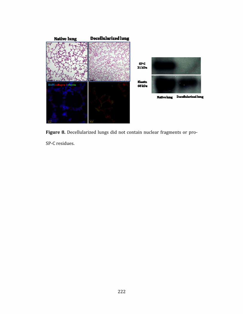
222
Figure 8. Decellularized lungs did not contain nuclear fragments or pro-
SP-C residues.
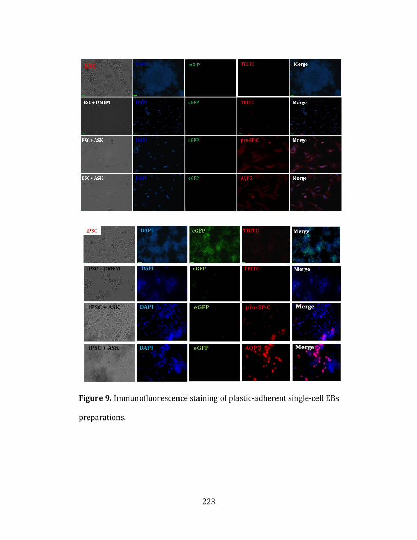
223
Figure 9. Immunofluorescence staining of plastic-adherent single-cell EBs
preparations.

224
Figure 10. FACS and immunoblotting indicated that ESC and iPSC
acquired alveolar epithelial cell markers.