dna repair
TRANSCRIPT

DNA REPAIR MECHANISM
PRAVEENA PRASAD. R
1ST M.SC-BIOTECHNLOGY
• DNA repair is a collection of processes by which a cell identifies and corrects damage to the DNA molecules that encode its genome.
• In human cells, both normal metabolic activities and environmental factors such as UV light and radiation can cause DNA damage, resulting in as many as 1 million individual molecular lesions per day.
• Many of these lesions cause structural damage to the DNA molecule and can alter or eliminate the cell's ability to transcribe the gene that the affected DNA encodes.

• As a consequence, the DNA repair process is constantly active as it responds
to damage in the DNA structure. When normal repair processes fail, and when cellular apoptosis does not occur, irreparable DNA damage may occur, including double-strand breaks and DNA cross-linkages.
• DNA damage can be subdivided into two main types:• Endogenous damage such as attack by reactive oxygen species produced
from normal metabolic byproducts (spontaneous mutation), especially the process of oxidative deamination– also includes replication errors
• Exogenous damage caused by external agents such as– ultraviolet [UV 200-400 nm] radiation from the sun– other radiation frequencies, including x-rays and gamma rays– hydrolysis or thermal disruption– certain plant toxins– human-made mutagenic chemicals, especially aromatic compounds that
act as DNA intercalating agents– viruses


Excision repair system :• Excision repair deals with a variety of structural defects in the
DNA. Mismatches between the strands of DNA are one of the major targets for the repair systems .
• In general , excision repair comprises 2 steps :1. Incision step: In this step , the damaged structure is recognized by
an endonuclease that cleaves the DNA strand on both sides of damage.
2. Excision step: In this step, a 5’to 3’ exonuclease removes a stretch of the damaged strand .Alternatively , a helicase displaces the damaged strand , which is subsequently degraded.

• Mismatches are usually corrected by excision repair . They are 2 types of excision repair :
• Base excision repair (BER)• Nucleotide excision repair (NER)

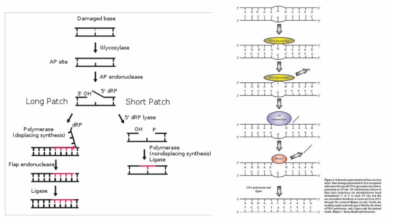
Base excision repair• This repair involve the direct removal of the damaged base
from DNA .• This serves as the trigger to activate the enzymes that excise
& replace a stretch of DNA , including the damaged site .• Enzymes that removes bases from DNA are called
glycosylases.• DNA glycosylases are responsible for initial recognition of
the lesion. They flip the damaged base out of the double helix and cleave the N-glycosidic bond of the damaged base, leaving an AP site.
• There are two categories of glycosylases: • monofunctional and bifunctional. • A wide variety of glycosylases have evolved to recognize different
damaged base.• The AP endonucleases cleave an AP site to yield a 3' hydroxyl
adjacent to a 5' deoxyribose phosphate (dRP).• AP endonucleases are divided into two families based on their
homology to bacterial AP endonuclease IV and exonuclease III.

DNA polymerase• Pol β is the main human polymerase that catalyzes short-patch
BER, with pol λ able to compensate in its absence.
• In addition to polymerase activity, these enzymes have a lyase domain that removes the 5' dRP left behind by AP endonuclease cleavage.
• These polymerases perform displacing synthesis, meaning that the downstream 5' DNA end is "displaced" to form a flap
Flap endonuclease• FEN1 removes the 5' flap generated during long patch BER. • This endonuclease shows a strong preference for a long 5' flap
adjacent to not 3' flap. In addition to its role in long-patch BER, FEN1 cleaves flaps with a similar structure during Okazaki fragment processing, an important step in lagging strand DNA replication.

DNA ligase• DNA ligase III catalyzes the nick-sealing step in short-patch BER
in humans. DNA ligase I ligates the break in long-patch BER.• In humans, polynucleotide kinase-phosphatase (PNKP) promotes
formation of these ends during BER. • This protein has a kinase domain, which phosphorylates 5' hydroxyl
ends, and a phosphatase domain, which removes phosphates from 3' ends. Together, these activities ready single-strand breaks with damaged termini for ligation.

Nucleotide excision repair:• The general principles of nucleotide excision repair is similar to
bacteria . • It has 2 major pathway • Global genome repair recognizes damage anywhere in genome .
Genes called XPA to XPG are involved . The XPC protein detects the damage and initiates the repair pathway .
• Transcription –coupled pathway is responsible for repairing lesions occurred in the transcribed strand of active genes . In this case , the damage is recognized by RNA polymerase 2.

• The 2 pathways eventually merge & use a common set of protein to bring about the repair .
• The strands of DNA are unwound by approximately 24bp around the damaged site by the helicase activity of the transcription factor TFII, which includes the products of 2 XP genes XPB & XPD .
• Cleavages are on either side of the lesion by endonuclease encoded by XPG & XPF genes .
• the single - stranded stretch including the damaged bases can then replaced by new synthesis and ligated by ligase-3 & ERCC1 complex.
• Mutations in TC-NER machinery are responsible for multiple genetic disorders including:
• Trichothiodystrophy (TTD): some individuals are photosensitive, mental/physical retardation
• Cockayne syndrome (CS): photosensitivity, mental retardation, progeria-like features.
SOS REPAIR / ERROR–PRONE REPAIR :
• The SOS response is a global response to DNA damage in which the cell cycle is arrested and DNA repair ,mutagenesis are induced.
• The system involves the Rec-A protein (Rad51 in eukaryotes). The RecA protein, stimulated by single-stranded DNA, is involved in the inactivation of the Lex-A repressor thereby inducing the response.
• It is an error-prone repair system that is attributed to mutagenesis.• The SOS response was discovered and named by Miroslav
Radman in 1975.

• The damaged DNA cause RecA to trigger the response & results in the auto cleavage of protein called LexA protein .
• RecA is activated on binding on a single-stranded DNA.• Lex A is a repressor that participate in DNA repair . RecA forms a
filament around these ssDNA regions in an ATP-dependent fashion, and becomes activated.
• The activated form of RecA interacts with the LexA repressor to facilitate the LexA repressor's self-cleavage from the operator.

• Once the pool of LexA decreases, repression of the SOS genes goes down according to the level of LexA affinity for the SOS boxes.
• Operators that bind LexA weakly are the first to be fully expressed.
• In this way LexA can sequentially activate different mechanisms of repair.
• Genes having a weak SOS box (such as uvrA, uvrB, and uvrD) are fully induced in response to even weak SOS-inducing treatments.

• Thus the first SOS repair mechanism to be induced is nucleotide excision repair(NER), whose aim is to fix DNA damage without commitment to a full-fledged SOS response.
• This causes filamentation, and the induction of UmuDC-dependent mutagenic repair.


Photo reactivation/direct repair :• Photolyases are DNA repair enzymes that repair damage caused by
exposure to ultraviolet light. This enzyme mechanism requires visible light(300-600 nm), preferentially from the violet/blue end of the spectrum, and is known as photo reactivation.
• Photolyase is a phylogenetically old enzyme which is present and functional in many species, from the bacteria to the fungi to plants and to the animals.
• Photolyase is particularly important in repairing UV induced damage in plants.
• The photolyase mechanism is no longer working in humans and other placental mammals who instead rely on the less efficient nucleotide excision repair mechanism

• Photolyases bind complementary DNA strands and break certain types of pyrimidine dimers that arise when a pair of thymine or cytosine bases on the same strand of DNA become covalently linked.
• These dimers result in a 'bulge' of the DNA structure, referred to as a lesion.
• Photolyases have a high affinity for these lesions and reversibly bind and convert them back to the original bases.
