dna-dependent protein kinase is not required for efficient
TRANSCRIPT

JOURNAL OF VIROLOGY,0022-538X/00/$04.0010
Dec. 2000, p. 11278–11285 Vol. 74, No. 23
Copyright © 2000, American Society for Microbiology. All Rights Reserved.
DNA-Dependent Protein Kinase Is Not Required for EfficientLentivirus Integration
VEERLE BAEKELANDT,1 ANJE CLAEYS,2 PETER CHEREPANOV,2 ERIK DE CLERCQ,2
BART DE STROOPER,3,4 BART NUTTIN,1 AND ZEGER DEBYSER2*
Rega Institute for Medical Research,2 Laboratory for Experimental Neurosurgery and Neuroanatomy,1
Center for Human Genetics,3 and Flanders Interuniversitary Institute for Biotechnology,4
Katholieke Universiteit Leuven, Leuven, Belgium
Received 26 April 2000/Accepted 26 July 2000
How DNA is repaired after retrovirus integration is not well understood. DNA-dependent protein kinase(DNA-PK) is known to play a central role in the repair of double-stranded DNA breaks. Recently, a role forDNA-PK in retroviral DNA integration has been proposed (R. Daniel, R. A. Katz, and A. M. Skalka, Science284:644–647, 1999). Reduced transduction efficiency and increased cell death by apoptosis were observed uponretrovirus infection of cultured scid cells. We have used a human immunodeficiency virus (HIV) type 1(HIV-1)-derived lentivirus vector system to further investigate the role of DNA-PK during integration. Wemeasured lentivirus transduction of scid mouse embryonic fibroblasts (MEF) and xrs-5 or xrs-6 cells. Thesecells are deficient in the catalytic subunit of DNA-PK and in Ku, the DNA-binding subunit of DNA-PK,respectively. At low vector titers, efficient and stable lentivirus transduction was obtained, excluding anessential role for DNA-PK in lentivirus integration. Likewise, the efficiency of transduction of HIV-derivedvectors in scid mouse brain was as efficient as that in control mice, without evidence of apoptosis. We observedincreased cell death in scid MEF and xrs-5 or xrs-6 cells, but only after transduction with high vector titers(multiplicity of infection [MOI], >1 transducing unit [TU]/cell) and subsequent passage of the transducedcells. At an MOI of <1 TU/cell, however, transduction efficiency was even higher in DNA-PK-deficient cellsthan in control cells. Taken together, the data suggest a protective role of DNA-PK against cellular toxicityinduced by high levels of retrovirus integrase or integration. Another candidate cellular enzyme that has beenclaimed to play an important role during retrovirus integration is poly(ADP-ribose) polymerase (PARP).However, no inhibition of lentivirus vector-mediated transduction or HIV-1 replication by 3-methoxybenz-amide, a known PARP inhibitor, was observed. In conclusion, DNA-PK and PARP are not essential forlentivirus integration.
Integration is an essential step in the retrovirus replicationcycle (8). The viral integrase catalyzes both the 39 processing ofthe viral DNA ends and the insertion of the viral DNA into thehost chromosome. This insertion is mediated by a coordinatednucleophilic attack by the hydroxyl groups of both processedends on both strands of the phosphodiester backbone of thehost DNA, followed by the ligation of the viral 39 ends tothe cellular DNA. The result is a gapped intermediate in whichthe viral 59 ends are not joined to the host DNA. Resolution ofthe integration intermediate leads to the chromosomal inser-tion of the proviral DNA trimmed of both terminal dinucle-otides and flanked by a duplicated host DNA fragment. Thesize of the DNA duplication is virus specific. For human im-munodeficiency virus (HIV), a 5-bp duplication is formed.Cellular DNA repair mechanisms are generally believed to fillin and ligate the remaining single-stranded DNA gaps, al-though the underlying mechanisms have not been character-ized. Alternatively, viral enzymes may be involved. Reversetranscriptase could fill in the gaps and, after removal of the twooverhanging nucleotides at the 59 end of the viral DNA, theDNA splicing activity of integrase could ligate the viral DNAto the target DNA (11, 35). A report that integrase would alsohave the required DNA polymerase activity awaits indepen-dent confirmation (2).
In eukaryotic cells, nonhomologous end joining representsthe major mechanism for the repair of double-stranded DNA(dsDNA) breaks (26, 30). In eukaryotes, dsDNA breaks occurduring V(D)J recombination and during meiotic recombina-tion and are also generated by ionizing radiation. Nonhomolo-gous end joining is mediated by DNA-dependent protein ki-nase (DNA-PK), a kinase activated by dsDNA ends (19, 24).DNA-PK is composed of a 450-kDa catalytic subunit (DNA-PKCS) and the heterodimeric protein Ku, composed of 70- and86-kDa subunits. Ku is the DNA-binding component ofDNA-PK required for the activation of the catalytic subunit.Ku binds strongly to dsDNA ends and, at least in vitro, togapped and nicked DNA molecules as well (5, 20). Mice withsevere combined immunodeficiency (scid) have a DNA-PKCStruncation mutation in both alleles (6, 14). This scid mutationaffects V(D)J rearrangement and double-strand break repair,resulting in the lack of mature B and T lymphocytes in scidmice (7). Primary cells derived from scid mice are deficient inDNA-PK activity (27, 30). Chinese hamster ovary (CHO) celllines deficient in Ku86 (xrs-5 and xrs-6) are also available (15,25). Like scid cells, these mutant cell lines are highly sensitiveto irradiation.
A role for nonhomologous end joining in general andDNA-PK in particular in repairing both DNA breaks gener-ated by retrovirus integration is certainly conceivable. Re-cently, DNA-PK was claimed to be required for retrovirusintegration (13). It was shown that integration efficiency wasreduced in DNA-PK-deficient murine scid cells and that high-titered virus stocks induced apoptosis in these cells.
* Corresponding author. Mailing address: Rega Institute for Medi-cal Research, Minderbroedersstr. 10, B-3000 Leuven, Belgium. Phone:32 16 33 21 82. Fax: 32 16 33 21 31. E-mail: [email protected].
11278
on April 5, 2018 by guest
http://jvi.asm.org/
Dow
nloaded from
Another candidate cellular enzyme that could play an im-portant role during retrovirus integration is poly(ADP-ribose)polymerase (PARP) (22). This nuclear enzyme (EC 2.4.2.30) isa zinc finger protein of 113 kDa that can bind to both single-stranded DNA and dsDNA breaks and that is believed toparticipate in a number of cellular processes involving DNAbreak formation and rejoining (9, 16). PARP activity is stim-ulated by a variety of DNA-damaging agents, including ioniz-ing irradiation, alkylating agents, and oxygen radicals. PARPcatalyzes the attachment of polymers of the ADP-ribose moi-ety of its substrate, NAD, to various proteins, including itself.After automodification, the protein dissociates from the DNA,providing access to other components of the DNA repair sys-tem (36). PARP is believed to protect the integrity of thegenome by preventing accidental homologous DNA recombi-nation (9). Excessive stimulation of PARP may deplete NADand lead to cell death (33). Selective PARP inhibitors, such as3-methoxybenzamide (3-MB) are known (34). Infection ofmammalian cells by recombinant retrovirus vectors is inhibitedby inhibition of PARP activity (22), suggesting a role duringretrovirus integration.
In the context of the establishment of a cellular integrationsystem (10), we have investigated the possible requirements ofDNA-PK and PARP for efficient lentivirus integration. To oursurprise and in contrast to published reports, we have found noevidence that these enzymes are essential for integration atmultiplicities of infection (MOI) below 1 transducing unit(TU)/cell.
MATERIALS AND METHODS
Cell cultures. HeLa, CHO-K1, xrs-5, and xrs-6 cells were obtained from theAmerican Type Culture Collection. By limiting dilution, individual clones of theKu-deficient CHO-K1-derived cell lines xrs-5 and xrs-6 (xrs-5/2 and xrs-6/1) inwhich the absence of the Ku protein was verified by Western blotting wereselected (data not shown). Human embryonic kidney 293T cells expressing sim-ian virus 40 (SV40) large T antigen, were obtained from O. Danos (Evry,France). Wild-type (WT) mouse embryonary fibroblasts (MEF) were obtainedfrom embryos of C57Black/6J and 129/OLA parents; scid MEF were obtainedfrom CB-17/scid 3 scid mice. Cells were grown in Dulbecco’s modified Eagle’smedium (DMEM) containing Glutamax-I and supplemented with 10% fetal calfserum (FCS) (Harlan Sera-Lab LTD) and 20 mg of gentamicin (Gibco BRL) perml at 37°C in a 5% CO2 humidified atmosphere. 293T cells were transfectedusing polyethyleneimine (PEI) (average molecular weight, 25,000; Sigma-Al-drich, Bornem, Belgium) (1); MEF cells were transfected using Lipofectamine(Gibco BRL). The continuous cell lines WT MEF-T and scid MEF-T wereobtained by transfecting WT and scid MEF at 50 to 70% confluence withpMSSVLT, encoding SV40 large T antigen. At 48 h after transfection, 500 mg ofGeneticin (G418) per ml was added to the culture medium to select stabletransfectants. The number of viable cells was determined by trypan blue exclu-sion. Anti-HIV activity and cytotoxicity measurements for MT-4 cells were basedon viability measurements using a tetrazolium-based colorimetric method (32).
Lentivirus vector production. HIV type 1 (HIV-1)-derived vector particles,pseudotyped with the envelope of vesicular stomatitis virus, were produced bytransfecting 293T cells with a packaging plasmid encoding viral Gag, Pol, andaccessory proteins (pCMVDR8.2), a plasmid encoding the envelope of vesicularstomatitis virus (pMDG), and a plasmid carrying a reporter gene flanked by twolong terminal repeats (pHR9-CMVLacZ) (31). A second-generation attenuatedpackaging plasmid, pCMVDR8.91, which lacks the vif, vpr, vpu, and nef genes(39), was used as well. All plasmids were kindly provided by O. Danos and D.Trono (Geneva, Switzerland).
For transfection of a 10-cm dish of 293T cells, 20 mg of vector plasmid, 10 mgof packaging construct, and 5 mg of envelope plasmid were mixed in 700 ml of 150mM NaCl. PEI solution (1.57 mM PEI in 150 mM NaCl) (700 ml) was addedslowly. The mixture was incubated at room temperature for 15 min and thenadded dropwise to 293T cells in DMEM containing 1% FCS. On the nextmorning, the medium was replaced with DMEM containing 10% FCS. Super-natants were collected from day 2 until day 5 posttransfection. The vectorparticles in the supernatants were sedimented by ultracentrifugation in a swing-ing-bucket rotor (SW27; Beckman, Palo Alto, Calif.) at 25,000 rpm for 2 h at 4°C.Pellets were redissolved in 300 ml of phosphate-buffered saline (PBS), resultingin a 100-fold concentration. p24 antigen content was determined using an HIV-1p24 core profile enzyme-linked immunosorbent assay (DuPont, Dreieich, Ger-many), and viral titers were determined by end-point dilution and 5-bromo-4-
chloro-3-indolyl-b-D-galactopyranoside (X-Gal) staining of transduced cells. p24content was used to normalize the different vector batches.
For transduction experiments, cells were seeded at 10,000/well in 96-wellplates. After 24 h, the medium was replaced with DMEM containing 1% FCS,and the cells were infected with different amounts of vector supplemented with2 mg of Polybrene per ml. On the next morning, the supernatant was replacedwith fresh medium containing 10% FCS. b-Galactosidase reporter gene activitywas measured 3 days after transduction. Cells were washed with PBS, fixed with2% formaldehyde–0.2% glutaraldehyde in PBS, and stained with freshly pre-pared X-Gal substrate (5 mM potassium ferrocyanide, 5 mM potassium ferri-cyanide, 2 mM MgCl2, and 400 mg of X-gal per ml [Biotech Trade & ServiceGmbH, St. Leon-Rot, Germany] in PBS) at 37°C overnight. Individual blue cellswere counted microscopically. Alternatively, total b-galactosidase activity in thecell lysate was determined. Medium was removed by gentle aspiration, and themonolayers were washed with PBS. Cells were lysed in 25 ml of 0.5% NonidetP-40. Five microliters of the extract was used to determine total protein contentaccording to the Bradford method (Bio-Rad, Hercules, Calif.). The remainingvolume was used to measure the conversion of o-nitrophenyl-b-D-galactopyrano-side (Sigma-Aldrich) in a colorimetric assay as described previously (3). Resultswere normalized to the total protein concentration. In experiments evaluatingtiter-dependent cellular toxicity, this normalization of reporter gene activity wasnot done.
In vivo analysis of lentivirus vector transduction. Adult CB-17/scid 3 scid andC57Black/6 mice of both sexes were used. All surgical procedures were per-formed under chloral hydrate anesthesia (400 mg/kg of body weight intraperi-toneally) using aseptic procedures. The mice were placed in a stereotactic headframe (Narishige); after midline incision of the skin, one or two small holes weredrilled in the skull in the appropriate location (AP 0.5, LAT 2.0, and DV 3.0,using bregma as a reference point [21]). Two microliters of highly concentratedvector (108 pg of p24/ml) supplemented with 4 mg of Polybrene per ml wasinjected at a rate of 0.5 ml/min with a 30-gauge needle connected by microdialysistubing to a 25-ml Hamilton syringe in a microinjection pump (CMA/microdialysisAB, Stockholm, Sweden). The needle was raised slowly 0.25 mm in the dorsaldirection every 60 s during the 4-min injection. After the injection, the needlewas left in place for an additional 4 min before being withdrawn slowly from thebrain (protocol adapted from that in reference 18).
To assess lentivirus transduction, the mice were deeply anesthetized withpentobarbital and perfused transcardially with saline followed by ice-cold 4%paraformaldehyde in PBS for 15 min. The brain was removed from the skull andpostfixed overnight in the same fixing solution. Coronal brain sections (50 mmthick) were cut with a vibratome and stored at 4°C in PBS containing 0.1%sodium azide. The sections were first screened for green fluorescent protein(GFP) expression with an inverted fluorescence microscope. More sensitivedetection was obtained by immunohistochemical analysis with a polyclonal an-tibody against GFP (Clontech, Palo Alto, Calif.). The sections were treated with3% hydrogen peroxide and preincubated in 5% normal swine serum–0.1% Tri-ton X-100 in PBS. The anti-GFP primary antibody (1:1,000) in 5% normal swineserum–0.1% Triton X-100 was added and incubated overnight at room temper-ature. The sections were then incubated with biotinylated swine anti-rabbitsecondary antibody, followed by Strept ABC-HRP complex (Dako, Glostrup,Denmark). Detection was accomplished with diaminobenzidine and H2O2 as asubstrate.
Astrocytes and neurons were identified using a polyclonal antibody againstglial fibrillary acidic protein (GFAP; Dako) and a monoclonal antibody againstNeuN (Chemicon, Temecula, Calif.), respectively. TUNEL (terminal de-oxynucleotidyltransferase [TdT]-mediated dUTP-biotin nick end labeling) stain-ing was performed according to a protocol adapted from that of Young et al.(38). Briefly, sections were pretreated with 1% H2O2 in methanol and thenincubated in 0.1% sodium citrate buffer containing 0.1% Triton X-100 on ice for2 min. The labeling reaction with TdT in the presence of biotin–16-dUTP wasperformed for 4 h at 37°C. Next, the sections were incubated with Strept ABC-HRP complex and stained with diaminobenzidine and H2O2. Sections treatedwith DNase I for 30 min at 37°C prior to TdT incubation were used as a positivecontrol.
Quantitative analysis of GFP expression was carried out by means of a Bio-quant image analyzing system (R&M Biometrics, Nashville, Tenn.). For eachanimal, serial sections (minimum of six) were analyzed over a distance of 2 mmcentered around the injection site. For each section, the relative density and thenumber of pixels above a threshold of intensity were measured in the area of thestriatum that was positive for GFP expression.
RESULTS
Efficient transduction of scid cells by lentivirus vectors. Therole of DNA-PK during lentivirus integration was investigatedusing amphotropic HIV-1-derived lentivirus vectors (31, 39)that encode b-galactosidase as a marker protein. The vectorsretrotranscribe and integrate their genomes into the host chro-mosome much like the parental virus, but they cannot replicatedue to the lack of viral coding sequences. This model system
VOL. 74, 2000 DNA-PK IS NOT REQUIRED FOR LENTIVIRUS INTEGRATION 11279
on April 5, 2018 by guest
http://jvi.asm.org/
Dow
nloaded from

can thus be used to study the requirements of lentivirus inte-gration. It was possible to transduce MEF using lentivirusvectors, although the efficiency of transduction in these cellswas lower than that in established cell lines, such as 293T,HeLa, or CHO. To verify whether DNA-PK was required forintegration, scid and WT MEF cells were transduced at lowMOI (,0.1 TU/cell) to prevent infection by more than oneparticle per cell. MEF derived from scid mice were as effi-ciently transduced as MEF derived from WT mice (Fig. 1).Both with WT and with attenuated vectors (the latter lackingthe four HIV accessory genes), scid MEF were transduced atleast as efficiently as WT MEF. Similar results were obtainedfor continuous cell lines (WT MEF-T and scid MEF-T) de-rived from MEF after transformation with SV40 T antigen.Since the transduction efficiency in these cell lines was repro-ducibly higher than that in primary cells, MEF-T were used forfurther experiments. Lentivirus transduction of cells at the lowtiters used in these experiments did not induce cell death. Toexclude the possibility that the need for DNA-PK-mediatedDNA repair is apparent only in cells undergoing mitosis, wepassaged the transduced cells prior to staining. After passage,the reporter gene activity remained equally high in WT MEF-Tand scid MEF-T (data not shown).
Efficient integration of lentivirus vectors in Ku-deficientcells. In a second set of experiments, we verified the role of Ku,the DNA-binding component of DNA-PK. The CHO-K1-de-rived cell lines xrs-5 and xrs-6 are deficient in Ku86 protein.The absence of Ku protein was verified by Western blotting(data not shown). The relative efficiency of lentivirus vectortransduction in these cell lines was compared with that in theparental CHO-K1 cell line (Fig. 2). The average transductionefficiencies were 70% in xrs-5 cells and 152% in xrs-6 cellsrelative to the transduction efficiency in CHO-K1 cells (Fig.2A). Transduction was not accompanied by cell death (Fig.2B). After passage of the transduced cells, the b-galactosidase-positive colonies were counted. Results similar to those ob-tained in transient transduction experiments were obtained(Fig. 2C). In mammalian cells, there is evidence that Ku playsa role in transcriptional regulation (23, 28, 29). This idea raisedthe possibility that in the absence of Ku, reduced transcriptionof integrated provirus could lead to an underestimation ofapparent transduction efficiency when total reporter gene ac-tivity is measured. We quantified the total b-galactosidase ac-tivity of the cell lysate photometrically. Again, at MOI of 0.04TU/cell, reporter gene activity was about 2-fold lower in xrs-5cells but 1.5-fold higher in xrs-6 cells relative to the activity inthe maternal cell line (data not shown). No significant reduc-tion in reporter gene activity could thus be demonstrated in theabsence of Ku.
Efficient lentivirus transduction of scid mouse brain. Wehave been studying the potential of HIV-1-derived lentivirusvectors for gene transfer into the murine brain as a step towardfuture gene therapy of neurodegenerative diseases (4, 31). Inthis context, we used scid mice in parallel with immunocom-petent C57Black/6 (C57BL) mice to investigate potential im-munological reactions to vector or transgene. Highly concen-trated vector (2 3 105 pg of p24) encoding GFP was injectedstereotactically into the striatum of adult mice, and the expres-sion of the transgene was evaluated by immunohistochemicalanalysis. The levels of GFP 2 and 11 weeks after injection wereas high in scid mice as in control C57BL mice (Fig. 3). GFPexpression was evident in both neurons and glial cells (data notshown). Thus, although deficient for DNA-PK, brain cells ofscid mice were efficiently and stably transduced by HIV-1-derived lentivirus vectors. Daniel et al. attributed the lowerretrovirus integration efficiency in scid cells to increased cell
death by apoptosis (13). Nissl staining of brain sections of scidand C57BL mice did not reveal extensive cell loss in the trans-duced area. We also performed TUNEL staining to visualizeapoptotic cells after lentivirus vector-mediated gene transfer(Fig. 4). Since reactive apoptosis is likely to occur as an early
FIG. 1. Lentivirus transduction efficiency in scid cells. Different amounts of asecond-generation HIV-1-derived lentivirus vector (devoid of Vpr, Nef, Vif, andVpu) encoding b-galactosidase were used to transduce subconfluent WT and scidMEF. At confluence, WT (A) and scid (B) MEF were stained with X-Gal, andpositive WT (F) or scid (E) MEF were counted microscopically (C). Means andstandard deviations of duplicate experiments are shown.
11280 BAEKELANDT ET AL. J. VIROL.
on April 5, 2018 by guest
http://jvi.asm.org/
Dow
nloaded from

event after transduction, we included a time point 2 days aftertransduction. GFP expression was indeed evident after 2 days,but TUNEL staining was negative for brain slices from bothscid and C57BL mice at all time points analyzed (Fig. 4). In
conclusion, the results obtained with our in vivo model argueagainst an essential role for DNA-PK during lentivirus inte-gration.
No inhibition of lentivirus integration by inhibitors of PARPactivity. We extended our study to a second candidate repairenzyme, PARP. This enzyme has been thought to play anessential role during retrovirus integration, based on the inhi-bition of retrovirus transduction by competitive inhibitors ofPARP (22). 3-MB is an inhibitor of PARP with a reported 50%inhibitory concentration (IC50) for lentivirus transduction orviral replication of approximately 0.8 mM, whereas 4-methoxy-benzamide (4-MB) is an inactive analogue. We performedlentivirus transduction experiments with CHO-K1 and xrs-5 orxrs-6 cells in the presence of various concentrations of 3-MB or4-MB. The cells were preincubated with the inhibitors for 24 hprior to infection. Lentivirus transduction was assessed bymeasuring b-galactosidase activity in fixed cells or in a celllysate. At nontoxic concentrations, 3-MB did not interfere withlentivirus transduction (Table 1). We also evaluated the effectsof 3-MB and 4-MB against HIV-1 (strain IIIB) replication inMT-4 cell cultures. Again, no inhibition was seen at the con-centrations tested (up to 5 mM).
FIG. 3. Efficient lentivirus transduction in scid mouse brain. (Top) Expres-sion of the GFP transgene in the striatum of C57BL and scid mice 2 weeks aftertransduction with the same batch of lentivirus vectors. Serial sections with aninterval of 250 mm showed equal transduction efficiency in both mouse strains.(Bottom) Quantification of GFP expression in serial sections over a distance of2 mm around the injection site in the striatum of scid and C57BL mice 11 weeksafter lentivirus vector transduction. The y axis indicates the area of positive GFPexpression. Although there was some interindividual variation in transgene ex-pression, no significant difference between scid and C57BL mice was observed.
FIG. 2. Lentivirus transduction efficiency in Ku-deficient cell lines. Subcon-fluent CHO-K1 (F), xrs-5/2 (ƒ), and xrs-6/1 ({) cells were transduced withdifferent amounts of a first-generation lentivirus vector and stained after 3 days.The number of b-galactosidase-positive cells (A) and the cell count (B) weredetermined microscopically. After passage (1/5) of the transduced CHO-K1 (F),xrs-5/2 (ƒ), and xrs-6/1 (h) cells, b-galactosidase-positive colonies ($10 bluecells) were counted at confluence (C). Means and standard deviations of dupli-cate experiments are shown.
VOL. 74, 2000 DNA-PK IS NOT REQUIRED FOR LENTIVIRUS INTEGRATION 11281
on April 5, 2018 by guest
http://jvi.asm.org/
Dow
nloaded from

Cellular toxicity induced in DNA-PK-deficient cells by hightiters of lentivirus vectors. In all of our experiments so far, lowMOI were used to investigate the requirement of DNA-PK forlentivirus integration. Recently, Daniel et al. proposed an es-sential role for DNA-PK during retrovirus integration basedon results obtained with high viral titers (13). To examine theeffect of high lentivirus vector titers, additional cell cultureexperiments were performed. We compared the lentivirus
transduction of WT versus scid MEF-T (Fig. 5) and ofCHO-K1 versus xrs-5 or xrs-6 cells (Fig. 6). Transduction ofsubconfluent cells was performed at different MOI. b-galacto-sidase activity was measured 3 days after transduction and atconfluence after passage of the transduced cells.
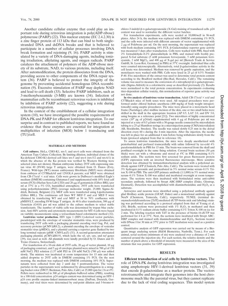
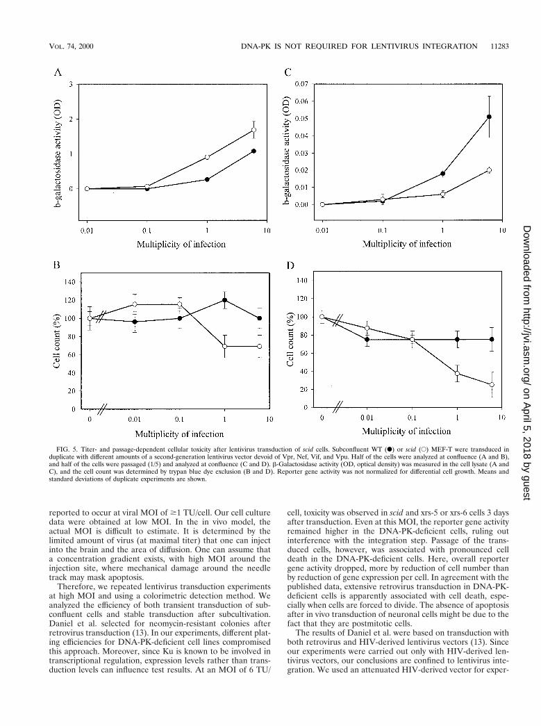
The reporter gene activity was always higher in scid than inWT MEF-T, even when higher MOI were used (Fig. 5A). AtMOI of 6 TU/cell, mild toxicity in scid but not in WT MEF-Twas evidenced by small reductions in cell count (Fig. 5B) andin total protein content (data not shown). Remarkably, scidMEF-T transduced with the highest titers of vector displayedmassive cell death after passage (Fig. 5D), resulting in reducedreporter gene activity (Fig. 5C). At low MOI, this toxicity wasnot observed. Analogous results were obtained with the Ku-deficient cell lines (Fig. 6). Lentivirus vector-mediated trans-duction efficiency for xrs-5 or xrs-6 cells was not below that ofthe parental CHO-K1 cells (Fig. 6A). At high vector titers,some cellular toxicity was observed for Ku-deficient cell lines(Fig. 6B). However, after subcultivation, cell death was pro-nounced in the Ku-deficient cells transduced with high titersbut not in the CHO-K1 cells (Fig. 6D). The cell death resultedin a reduction in total b-galactosidase activity in xrs-5 or xrs-6cells (Fig. 6C).
DISCUSSION
It remains unresolved to what extent the retroviral enzymesreverse transcriptase (gap filling) and integrase (DNA splicing[11]) or the eukaryotic host proteins, such as DNA-PK andPARP, contribute to the DNA repair of the single-strandedDNA gaps that remain after retrovirus integration. A role forPARP as a nick sensor during retrovirus integration has beenpostulated (22). In our experimental design, the PARP inhib-itor 3-MB was not capable of interfering with lentivirus vectortransduction or HIV-1 replication, arguing against an impor-tant role for this enzyme during lentivirus integration.
To investigate the role of DNA-PK during retrovirus inte-gration, we analyzed the transduction efficiency of HIV-1-de-rived lentivirus vectors in scid and xrs-5 or xrs-6 cells, which aredeficient in the DNA-PK pathway. We previously showed thata vector carrying the inactivating D64V mutation in the inte-grase gene gave rise to only 5% the transduced cells obtainedwith the WT vector (10). After passage of transduced cells, thispseudotransduction level even fell below 1%. Since transduc-tion by HIV vectors is thus largely dependent on the integra-tion step, the efficiency of transduction can be used to evaluatecofactors essential for integration. We reasoned that the use oflow vector titers would increase the likelihood that the require-ment for a potential host factor would become apparent. Sincelentivirus transduction was as efficient in scid, xrs-5, or xrs-6cells as in WT cells, an essential role for DNA-PK in integra-tion was ruled out. These results were confirmed with an invivo model. We have been investigating the potential of lenti-virus vectors for in vivo gene transfer after stereotactic injec-tion into the brain (4). scid mice were used to control for theimmunological response to the vector or transgene. In this invivo model, the transduction of neurons and glial cells was atleast as efficient in scid mice as in C57BL mice.
Our results with cell cultures and with the in vivo mousemodel do exclude an essential role for DNA-PK during lenti-virus integration. During the course of our experimental work,however, a study by Daniel et al. was published, wherein anessential role for DNA-PK during retrovirus integration wasclaimed (13). The claim was based on the observation thatretrovirus transduction was reduced in scid cells and accompa-nied by cell death due to apoptosis. Intriguingly, this effect was
FIG. 4. No evidence for increased apoptosis after lentivirus transduction inscid mouse brain. (A and D) GFP expression around the needle track in thestriatum of scid mice 2 days (A) and 2 weeks (D) after lentivirus transduction. (Band E) TUNEL staining of adjacent sections shows no apoptotic cells in the areaof the striatum positive for GFP expression 2 days (B) and 2 weeks (E) afterlentivirus transduction. (C) DNase treatment of a section like that in the positivecontrol results in numerous labeled nuclei. Scale bar, 100 mm.
TABLE 1. Lack of specific inhibition of lentivirus transduction andreplication by inhibitors of PARP
Virus and cell line3-MBa 4-MBa
IC50 CC50 IC50 CC50
HIV-1 vectorb
CHO-K1 .4 10 6 1 .4 20 6 5xrs-5 .4 8 6 2 .4 8 6 4xrs-6 .4 6 6 2 2 6 0.5 6 6 2
HIV-1 (strainIIIB)c, MT-4 cells
.3 3 6 1 .4 6 6 1
a The IC50 and the 50% cytotoxic concentration (CC50) are given as millimo-lar. Results reported as mean and standard deviation are based on two separateexperiments.
b CHO-derived cell lines were transduced with an HIV-1 vector (first gener-ation) encoding b-galactosidase in the absence or presence of different concen-trations of the known PARP inhibitor 3-MB or an inactive analogue, 4-MB. Twodays later, cells were fixed and stained with X-Gal, and blue cells were counted.Total protein content was used to determine cellular toxicity.
c Inhibition of HIV-1 replication and cellular toxicity in MT-4 cells weredetermined by the MTT method (32).
11282 BAEKELANDT ET AL. J. VIROL.
on April 5, 2018 by guest
http://jvi.asm.org/
Dow
nloaded from
reported to occur at viral MOI of $1 TU/cell. Our cell culturedata were obtained at low MOI. In the in vivo model, theactual MOI is difficult to estimate. It is determined by thelimited amount of virus (at maximal titer) that one can injectinto the brain and the area of diffusion. One can assume thata concentration gradient exists, with high MOI around theinjection site, where mechanical damage around the needletrack may mask apoptosis.
Therefore, we repeated lentivirus transduction experimentsat high MOI and using a colorimetric detection method. Weanalyzed the efficiency of both transient transduction of sub-confluent cells and stable transduction after subcultivation.Daniel et al. selected for neomycin-resistant colonies afterretrovirus transduction (13). In our experiments, different plat-ing efficiencies for DNA-PK-deficient cell lines compromisedthis approach. Moreover, since Ku is known to be involved intranscriptional regulation, expression levels rather than trans-duction levels can influence test results. At an MOI of 6 TU/
cell, toxicity was observed in scid and xrs-5 or xrs-6 cells 3 daysafter transduction. Even at this MOI, the reporter gene activityremained higher in the DNA-PK-deficient cells, ruling outinterference with the integration step. Passage of the trans-duced cells, however, was associated with pronounced celldeath in the DNA-PK-deficient cells. Here, overall reportergene activity dropped, more by reduction of cell number thanby reduction of gene expression per cell. In agreement with thepublished data, extensive retrovirus transduction in DNA-PK-deficient cells is apparently associated with cell death, espe-cially when cells are forced to divide. The absence of apoptosisafter in vivo transduction of neuronal cells might be due to thefact that they are postmitotic cells.
The results of Daniel et al. were based on transduction withboth retrovirus and HIV-derived lentivirus vectors (13). Sinceour experiments were carried out only with HIV-derived len-tivirus vectors, our conclusions are confined to lentivirus inte-gration. We used an attenuated HIV-derived vector for exper-
FIG. 5. Titer- and passage-dependent cellular toxicity after lentivirus transduction of scid cells. Subconfluent WT (F) or scid (E) MEF-T were transduced induplicate with different amounts of a second-generation lentivirus vector devoid of Vpr, Nef, Vif, and Vpu. Half of the cells were analyzed at confluence (A and B),and half of the cells were passaged (1/5) and analyzed at confluence (C and D). b-Galactosidase activity (OD, optical density) was measured in the cell lysate (A andC), and the cell count was determined by trypan blue dye exclusion (B and D). Reporter gene activity was not normalized for differential cell growth. Means andstandard deviations of duplicate experiments are shown.
VOL. 74, 2000 DNA-PK IS NOT REQUIRED FOR LENTIVIRUS INTEGRATION 11283
on April 5, 2018 by guest
http://jvi.asm.org/
Dow
nloaded from

iments at high MOI to avoid potential cellular toxicity due tothe presence of Vpr in the virus particle, as has been describedelsewhere (37).
The DNA-binding component of DNA-PK was recentlyshown to potentiate retrotransposition of the Ty element ofSaccharomyces cerevisiae in a galactose-inducible Ty1 system(17). However, the same authors reported that the level ofendogenous Ty1 retrotransposition was almost twofold higherin the absence of Ku, an effect that they attributed to thetranscriptional induction of genes responsive to DNA damage,such as Ty1 genes, in the abence of Ku. However, on the basisof our results with lentivirus transduction, it may be arguedthat the requirement of Ku for retrotransposition is linked tothe overexpression of the Ty1 element. Since Ty1 retrotrans-position was scored by selection of transduced colonies, thedistinction between lack of integration or integration-inducedcell death in the absence of Ku was not made. We hypothesize
that high levels of retrotransposition induce cell death in theabsence of Ku.
Our data do not exclude the possibility that an alternativecellular pathway can substitute for DNA-PK during lentivirusintegration. Nevertheless, our data argue against an essentialrole for this enzyme and question its suitability as an antiviraltarget. Scepticism toward a universal role for DNA-PK in ret-rovirus integration was already expressed by Coffin and Rosen-berg (12). It was pointed out that primary B cells from scidmice can be immortalized efficiently by infection with a retro-virus. Coffin and Rosenberg (12) suggested that, instead,DNA-PK might protect the cell from a side effect of integra-tion. Although we were able to obtain data analogous to thoseof Daniel et al. (13), namely, cell death induced by lentivirustransduction of DNA-PK-deficient cells, we observed this ef-fect only after transduction with excess vector and especiallywhen the transduced cells were forced to divide. Since trans-
FIG. 6. Titer- and passage-dependent cellular toxicity after lentivirus transduction of Ku-deficient cells. Subconfluent CHO-K1 (F), xrs-5/2 (ƒ), and xrs-6/1 ({) cellswere transduced in duplicate with different amounts of a second-generation lentivirus vector. Half of the cells were analyzed at confluence (A and B), and half of thecells were passaged (1/5) and analyzed at confluence (C and D). b-Galactosidase activity (OD, optical density) was measured in the cell lysate (A and C), and the cellcount was determined by trypan blue dye exclusion (B and D). Reporter gene activity was not normalized for differential cell growth. Means and standard deviationsof duplicate experiments are shown.
11284 BAEKELANDT ET AL. J. VIROL.
on April 5, 2018 by guest
http://jvi.asm.org/
Dow
nloaded from

duction was affected less than cell survival, the lack ofDNA-PK apparently does not prevent integration as such.Moreover, at low viral titers, lentivirus transduction was ap-parently more efficient in most DNA-PK-deficient cells. To-gether, our data tend to support the notion of a protective rolefor DNA-PK during excessive retrovirus integration. In thiscontext, DNA-PK may play a physiological role in protectionagainst superinfection. More research is required to determineexactly what triggers cell death after excessive lentivirus infec-tion of DNA-PK-deficient cells and what protective roleDNA-PK plays in this process.
In conclusion, no evidence was found that DNA-PK orPARP is essential for lentivirus integration when low virustiters are used. Another cellular or a virus-mediated gap repairmechanism may be involved.
ACKNOWLEDGMENTS
Veerle Baekelandt and Anje Claeys contributed equally to thiswork.
We thank K. Craessaerts, K. Eggermont, and M. Michiels for excel-lent technical assistance and the laboratory of F. Vandesande (Katho-lieke Universiteit Leuven [KUL]) for use of the photographic andimage analysis equipment. We are grateful to M. Witvrouw (KUL) foranti-HIV testing. The HIV-1-derived lentivirus vector system was akind gift from O. Danos (Evry, France) and D. Trono (University ofGeneva). We thank A. M. Skalka, R. Katz, and R. Daniel (Fox ChaseCancer Center, Philadelphia, Pa.) for helpful discussions.
V.B. and Z.D. are postdoctoral fellows of the Flemish Fund forScientific Research (FWO). This work was funded by IDO grant 98/006 from the Katholieke Universiteit Leuven Research Council andthe STWW Program of the Flemish Institute Supporting Scientific-Technological Research in Industry (IWT).
REFERENCES
1. Abdallah, B., A. Hassan, C. Benoist, D. Goula, J. P. Behr, and B. A. Deme-neix. 1997. A powerful nonviral vector for in vivo gene transfer into the adultmammalian brain: polyethylenimine. Hum. Gene Ther. 7:1947–1954.
2. Acel, A., B. E. Udashkin, M. A. Wainberg, and E. A. Faust. 1998. Efficientgap repair catalyzed in vitro by an intrinsic DNA polymerase activity ofhuman immunodeficiency virus type 1 integrase. J. Virol. 72:2062–2071.
3. Ausubel, F. M., R. Brent, R. E. Kingston, D. D. Moore, J. G. Seidman, J. A.Smith, and K. Struhl (ed.). 1987. Current protocols in molecular biology.John Wiley & Sons, Inc., New York, N.Y.
4. Baekelandt, V., B. De Strooper, B. Nuttin, and Z. Debyser. Gene therapeuticstrategies for neurodegenerative diseases. Curr. Opin. Mol. Ther., in press.
5. Blier, P. R., A. J. Griffith, J. Craft, and J. A. Hardin. 1993. Binding of Kuprotein to DNA. Measurement of affinity for ends and demonstration ofbinding to nicks. J. Biol. Chem. 268:7594–7601.
6. Blunt, T., N. J. Finnie, G. E. Taccioli, G. C. M. Smith, J. Demengeot, T. M.Gottlieb, R. Mizuta, A. J. Varghese, F. W. Alt, P. A. Jeggo, and S. P. Jackson.1995. Defective DNA-dependent protein kinase activity is linked to V(D)Jrecombination and DNA repair defects associated with the murine scidmutation. Cell 80:813–823.
7. Bosma, G. C., R. P. Custer, and M. J. Bosma. 1983. A severe combinedimmunodeficiency mutation in the mouse. Nature 301:527–530.
8. Brown, P. O. 1997. Integration, p. 161–203. In J. Coffin, S. Hughes, and H.Varmus (ed.), Retroviruses. Cold Spring Harbor Laboratory Press, ColdSpring Harbor, N.Y.
9. Chatterjee, S., S. Berger, and N. Berger. 1999. Poly(ADP-ribose)polymerase:a guardian of the genome that facilitates DNA repair by protecting againstDNA recombination. Mol. Cell. Biochem. 193:23–30.
10. Cherepanov, P., W. Pluymers, A. Claeys, P. Proost, E. De Clercq, and Z.Debyser. 2000. High-level expression of active HIV-1 integrase from a syn-thetic gene in human cells. FASEB J. 14:1389–1399.
11. Chow, S. A., K. A. Vincent, V. Ellison, and P. O. Brown. 1992. Reversal ofintegration and DNA splicing mediated by integrase. Science 255:723–726.
12. Coffin, J. M., and N. Rosenberg. 1999. Closing the joint. Nature 399:413–414.13. Daniel, R., R. A. Katz, and A. M. Skalka. 1999. A role for DNA-PK in
retroviral DNA integration. Science 284:644–647.14. Danska, J. S., D. P. Holland, S. Mariathasan, K. M. Williams, and C. J.
Guidos. 1996. Biochemical and genetic defects in the DNA-dependent pro-tein kinase in murine scid lymphocytes. Mol. Cell. Biol. 16:5507–5517.
15. Darroudi, F., and A. T. Natarajan. 1989. Cytogenetical characterization ofChinese hamster ovary X-ray-sensitive mutant cells, xrs 5 and xrs 6. IV. Studyof chromosomal aberrations and sister-chromatid exchanges by restrictionendonucleases and inhibitors of DNA topoisomerase II. Mutat. Res. 212:137–148.
16. de Murcia, G., and J. M. de Murcia. 1994. Poly(ADP-ribose)polymerase: amolecular nick-sensor. Trends Biochem. Sci. 19:172–176.
17. Downs, J. A., and S. P. Jackson. 1999. Involvement of DNA-end bindingprotein Ku in Ty element retrotransposition. Mol. Cell. Biol. 19:6260–6268.
18. Dull, T., R. Zufferey, M. Kelly, R. J. Mandel, M. Nguyen, D. Trono, and L.Naldini. 1998. A third-generation lentivirus vector with a conditional pack-aging system. J. Virol. 72:8463–8471.
19. Dvir, A., S. R. Peterson, M. W. Knuth, H. Lu, and W. S. Dynan. 1992. Kuautoantigen is the regulatory component of a template-associated proteinkinase that phosphorylates RNA polymerase II. Proc. Natl. Acad. Sci. USA89:11920–11924.
20. Falzon, M., J. W. Fewell, and E. L. Kuff. 1993. EBP-80, a transcription factorclosely resembling the human auto-antigen Ku, recognizes single- to double-strand transitions in DNA. J. Biol. Chem. 268:10546–10552.
21. Franklin, K. B. J., and G. Paxinos. 1997. The mouse brain in stereotaxiccoordinates. Academic Press, Inc., San Diego, Calif.
22. Gaken, J. A., M. Tavassoli, S.-U. Gan, S. Vallian, I. Giddings, D. C. Darling,J. Galea-Lauri, M. G. Thomas, H. Abedi, V. Schreiber, J. Menissier-deMurcia, M. K. L. Collins, S. Shall, and F. Farzaneh. 1996. Efficient retroviralinfection of mammalian cells is blocked by inhibition of poly(ADP-ribose)polymerase activity. J. Virol. 70:3992–4000.
23. Giffin, W., H. Torrance, D. J. Rodda, G. G. Prefontaine, L. Pope, and R. J. G.Hache. 1996. Sequence-specific DNA binding by Ku autoantigen and itseffects on transcription. Nature 380:265–268.
24. Gottlieb, T. M., and S. P. Jackson. 1993. The DNA-dependent proteinkinase: requirement of DNA ends and association with Ku antigen. Cell72:131–142.
25. Jeggo, P. A. 1990. Studies on mammalian mutants defective in rejoiningdouble-strand breaks in DNA. Mutat. Res. 239:1–16.
26. Jeggo, P. A. 1998. DNA breakage and repair. Adv. Genet. 38:185–219.27. Kirchgessner, C. U., C. K. Patil, J. W. Evans, C. A. Cuomo, L. M. Fried, T.
Carter, M. A. Oettinger, J. M. Brown, G. Iliakis, R. Mehta, and M. Jackson.1995. DNA-dependent protein kinase (p350) as a candidate gene for murineSCID defect. Science 267:1178–1183.
28. Kuhn, A., T. M. Gottleib, S. P. Jackson, and I. Grummt. 1995. DNA-dependent protein kinase—a potent inhibitor of transcription by RNA poly-merase I. Genes Dev. 9:193–203.
29. Labhart, P. 1995. DNA-dependent protein kinase specifically represses pro-moter-directed transcription initiation by RNA polymerase I. Proc. Natl.Acad. Sci. USA 92:2934–2938.
30. Lees-Miller, S. P., R. Godbout, D. W. Chan, M. Weinfeld, R. S. Day, G. M.Barron, and J. Allalunis-Turner. 1995. Absence of p350 subunit of DNA-activated protein kinase from a radiosensitive human cell line. Science 267:1183–1185.
31. Naldini, L., U. Blomer, P. Gallay, D. Ory, R. Mulligan, F. H. Gage, I. M.Verma, and D. Trono. 1996. In vivo gene delivery and stable transduction ofnondividing cells by a lentiviral vector. Science 272:263–267.
32. Pauwels, R., J. Balzarini, M. Baba, R. Snoeck, D. Schols, P. Herdewijn, J.Desmyter, and E. De Clercq. 1988. Rapid and automated tetrazolium-basedcolorimetric assay for the detection of anti-HIV compounds. J. Virol. Meth-ods 20:309–321.
33. Pieper, A. A., A. Verma, J. Zhang, and S. H. Snyder. 1999. Poly(ADP-ribose)polymerase, nitric oxide and cell death. Trends Pharmacol. Sci. 20:171–181.
34. Rankin, W. P., E. L. Jacobson, R. C. Benjamin, J. Moss, and M. K. Jacobson.1989. Quantitative studies of inhibitors of ADP-ribosylation in vitro and invivo. J. Biol. Chem. 264:4312–4317.
35. Roe, T., S. A. Chowand, and P. O. Brown. 1997. 39-End processing andkinetics of 59-end joining during retroviral integration in vivo. J. Virol.71:1334–1340.
36. Satoh, M. S., and T. Lindahl. 1992. Role of poly(ADP-ribose) formation inDNA repair. Nature (London) 356:356–358.
37. Stewart, S. A., B. Poon, J. Y. Song, and I. S. Y. Chen. 2000. Human immu-nodeficiency virus type 1 vpr induces apoptosis through caspase activation.J. Virol. 74:3105–3111.
38. Young, D., P. A. Lawlor, P. Leone, M. Dragunow, and M. J. During. 1999.Environmental enrichment inhibits spontaneous apoptosis, prevents seizuresand is neuroprotective. Nat. Med. 5:448–453.
39. Zufferey, R., D. Nagy, R. J. Mandel, L. Naldini, and D. Trono. 1997. Multiplyattenuated lentiviral vector achieves efficient gene delivery in vivo. Nat.Biotechnol. 15:871–875.
VOL. 74, 2000 DNA-PK IS NOT REQUIRED FOR LENTIVIRUS INTEGRATION 11285
on April 5, 2018 by guest
http://jvi.asm.org/
Dow
nloaded from