dna and protein (structure and functions)
TRANSCRIPT

Medical genetics
Safrina D. Ratnaningrum
Concepts:
Automosal recessive pattern
Carrier individual and obligate carrier
Chromosome aberration
Non-disjunction concepts (ex. Trisomy 21)
Chromosomal translocation inheritance
Recurrence risk
Laboratory testing for genetic mutation
Prenatal vs Postnatal
DNA testing vs Karyotiping

Contents:
• What is genetics and medical genetics?
• Genetic structures: DNA and chromosomes
• The Central Dogma of molecular biology and genetics
• Mutation (base level & chromosome level)
• Genetics and inheritance
• Genetics counseling

Genetics
Genetics is the study of the genes
Study of the genes and they role in: Cells organism population
Genes is DNA segments that have a functional role in the cell
and are responsible for inheritance of traits (gametes)

Medical genetics
Medical genetics
Application of principles and knowledge of human genetics in medicine
Areas of MG:
Clinical genetics, population genetics, pharmacogenetics,
immunogenetics, biochemical genetics
Clinical genetics: genetic disorders, incl.congenital anomalies
Do: Pre-postnatal dx; presymptomatic dx; genetic counseling;
care for patients and their families
Causes of genetic disorders
Monogenic (AD, AR, XL, Mt)
Chromosomal (numerical, structural)
Multifactorial/polygenic

Genetic structures
DNA consists of: sugar~phosphates~base
combined into complex double helix (sense~antisense)
the direction of the nucleotides in one strand is opposite to their direction in the other strand (antiparalel)
5’ ends: phosphate group
3’ ends: hydroxil group
5 molecule of bases:
Purines ~ pyrimidines:
Adenine (A) ~ Thymine (T)
Guanine (G) ~ Cytosine (C) more stable
Uracils (only found in RNA; take place of tymine)v
v

Chromosome packing
Oktamer histone
Spacer DNA plus
H1 histone
Double helix DNA

Cont’d…
Chromosome
46 chromosomes: 23 pairs autosomes; 2 sex chr. 46,xx or xy
Fig. Human karyotype

The interesting facts:
The human genome contains 3x10⁹ base pairs of DNA divided
into 23 chromosomes which if linked together would form a
thread of 1-2 meter with a diameter of 2 nm.
This DNA codes for about 105 different proteins.
Only about 2-4 % (about 20.000 genes) of the total coding
capacity in the human DNA is used for coding of different
genes, the rest of it probably has other more structural and
organizational functions.

The central dogma
DNA carries the genetic information of a cell and consists of
thousands of genes which serves as a recipe on how to build a
protein molecule.
Proteins perform important tasks for the cell functions.
The information from the genes determines the protein
composition and the functions of the cell.

RNA splicing

Translation
The transmission of genetic information from RNA to protein
Amino acids
tRNA
mRNAribosome/rRNA

Gene regulation
Gene activity can be turned on and off at the level of transcription by protein factors interaction with promoter that can prevent or allow transcription
Protein factor present in a certain type of cells
Human pancreatic cells insulin
Progenitor RBC hemoglobin
Progenitor RBC embryo embryonic hemoglobin

The genetic code Amino acids in protein are 20; DNA consists of 4 different bases
3 combined bases (triplet codon) 1 aa
The order of triplet codons = reading frame
43=64 codons, some aa are coded by more than one triplet codon
Sense DNA
Antisense DNA
mRNA
rRNA/ribosome
tRNA
Amino acids

Mutation
Is a change in the genetic material
Can occur in non-coding or coding sequences
Mutation in promoter regions harmful
somatic cells (not transmitted to offspring) ≠ gametes
Remember: MITOSIS vs MEIOSIS
Can occur in base level and chromosome level
Base level mutation, example:
Wild type: (tRNA) … AAA CUC CAC UUC UUC …
(protein) … phe glu val lys lys
Mutant … AAA CUC C ACU UCU UC… (deletion frameshift)
… phe glu (stop) xxx xx premature termination
Mutation C7del=V3Stop truncated protein

Types of mutations
Class Group Type Effect on protein product
Stable/
fixed
Synonymous Substitution Silent mutation/same aa no effect
Non-
synonymous
Substitution
- Missense
- Nonsense
Diff aa:
Chemically diff ≠ chemically similar (no
effect)
Stop codonloss of
function/activity/stability
Deletion/
insertion
Frameshift/premature termination
Dynamic/
unstable
Triple repeat Expansion Altered gene expression/transcription
The repeat will be increase in next
generation, ex: fragile X, Huntington
disease, myotonic dystrophy

Patterns of inheritanceThe purposes of family studies:
1. To provide a diagnosis
2. Necessary to determine the mode of inheritance of a trait or disorder and to give appropriate genetic counseling
3. Pedigree documentation of the family history
Most human genes are inherited in a Mendelian manner.
There are 2 types of chromosome: Autosome autosomal inheritance
Sex chromosome sex-linked inheritance
Mendelian inheritance
Autosomal dominant
Autosomal recessive
Sex-linked: dominant, recessive, sex influenced, sex limitation

Drawing pedigree
I, II, III, etc = Generations are numberered from the top of the pedigree
III1 , III2, III3, etc = Individuals in each generation

Estimation of Risk in Mendelian Disorders
Varies mechanisms in Mendelian law
New mutation
Anticipation
Reduced penetrance
Variable expressivity
Mosaicism
Uniparental disomy
Genomic imprinting
Mitochondrial inheritance
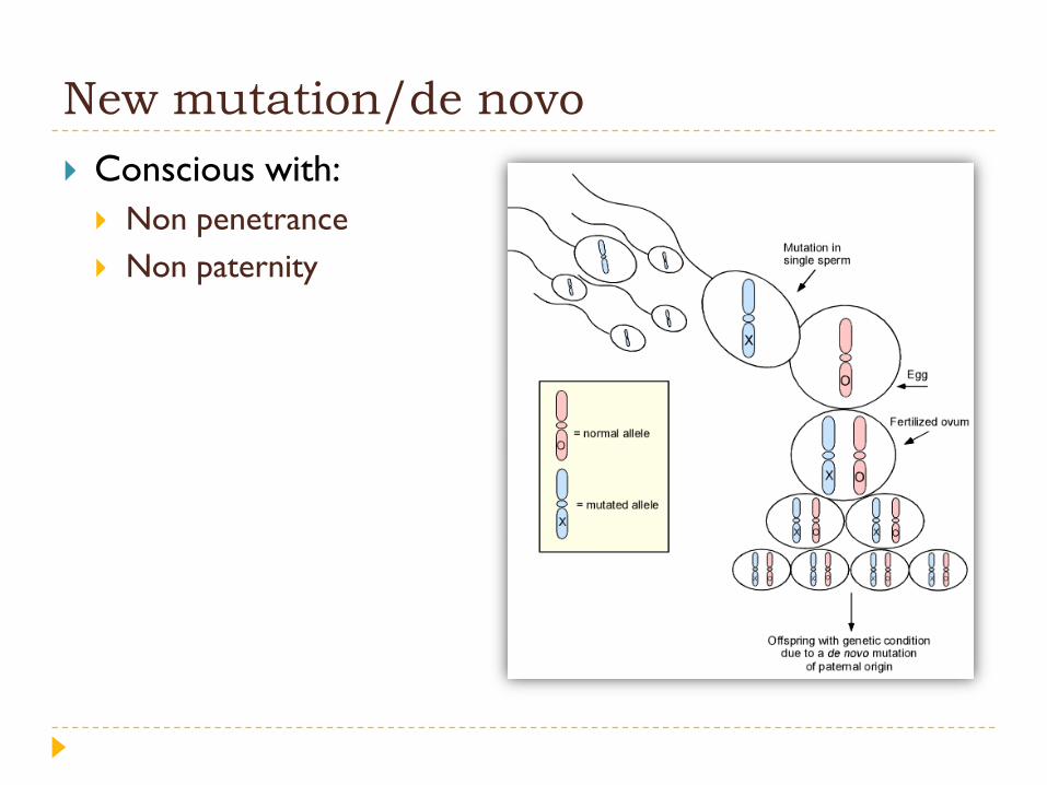
New mutation/de novo
Conscious with:
Non penetrance
Non paternity

Reduced penetrance
In dominant traits: if a condition is expressed is less than
100% of person who carry the mutant alelle
Note: red=not affected; pink=affected

Case:
the estimated penetrance for familial retinoblastoma
(RB) is approximately 80%. If a man has had familial RB
and mates with a woman who does not have a RB
mutation, what is the risk that their offspring will develop
RB?
Offspring probability=0,5
Thus, 0,5x0,8=0,4 or 40%

Variable expressivity
When the manifestation of a phenotype differs in people who have
the same genotype
As with reduced penetrance, variable expressivity is probably caused
by a combination of genetic, environmental, and lifestyle factors,
most of which have not been identified. If a genetic condition has
highly variable signs and symptoms, it may be challenging to
diagnose.

Mosaicism
Occur during mitosis after conception
Earlier more severe

Cont’d…
If parent(s) has:
Somatic mosaicism not inherited
Gonadal/germline mosaicism could be inherited

Cont’d…

Cytogenetics
Safrina D. Ratnaningrum

Structure of human chromosomes
Cytogenetics: the study of
chromosome and cell
division
Chromosome can be seen
clearly during metaphase,
maximally condensed

Type of chromosome
Telocentric
Absence in human
Some authors denotes extreme acrocentric
chr in human as telocentric, such as: 21, 22, Y
Acrocentric: 13, 14, 15, 21, 22
Submetacentric
Metacentric
p/short
arm
q/long
arm
Chromatid; double/
sister chromatid

Chromosome analysis (karyotyping)
Chromosome staining for identification and detection if any
abnormality >4Mbp
The light bands on chromosome regions rich in GC and genes.
Dark bands rich in AT and few on genes. Ex: Chromosome 19, dense with
genes, has few dark bands.
Metaphase spread Ideogram
Fig. normal male karyotyping

Chromosome level mutation
Abnormalities of chromosome:
1. Numerical (~heteroploid): the most common mechanism is
in meiotic nondisjunction (during M1/M2, usually during M1)
Euploidy/polyploidy: addition one or more complete haploid chr. Triploidy (3n); tetraploidy (4n)
Aneuploidy: loss or gain of one or more chromosomes
Monosomy (lethal), exception monosomy X chromosome (Turner syndrome, 45,x); Trisomy:
Autosomal chromosome: trisomy 13, trisomy 18, trisomy 21.
Sex chromosome: klinefelter syndrome (47,xxy), xyy syndrome (47,xyy), trisomy X (47,xxx)
2. Structural

Abnormalities of chromosome structure Result from chromosome breakage with subsequent reunion in different
configuration.
Remember: Mendelian laws (1. segregasi; 2. independent assortment)
Unbalanced rearrangements (abnormal phenotype, chromosomal complement contains an incorrect amount of chromosome material)
Balanced rearrangements (normal phenotype because all the genetic information is present eventhough it is packaged differently)
Normal Balanced Unbalanced
Fertilization
Parent: gametogenesis Offspring

1. Translocations
Reciprocal
Robertsonian
2. Deletion
3. Insertion
4. Invertion
Pericentric
Paracentric
5. Ring chromosome
6. Isochromosome

Translocations
Reciprocal:
involves breakage of at
least 2 chromosomes
with exchange of the
fragments; common in
chr. 11 and 22
Chromosomes involved
in the translocations
cannot pair normally
with their homolog to
form bivalent during M1
(profase) then form a
cluster called pachytene
quadrivalent

Cont’d…
Robertsonian
Results from breakage of
two acrocentric
chromosomes
(13,14,15,21,22) at or close
to their centromeres with
subsequent fusion of their
long arms (the short
arms/satellite of each
chromosome are lost).
Total chromosomes
number is reduced to 45.
Common in chromosomes
13 and 14.

Example: karyotyping result of DS caused by
translocation
46,XX,t(14;21)(q10;q10)
Both are result of Robertsonian translocation with different
consequence of the recurrence risk for next pregnancy

Deletions The loss of part of a
chromosome and results in monosomy for that segment of the chromosomes.
Cri du chat, del(5p); PraderWilli syndrome and Angelmannsyndrome, del(15q11-q13)
Insertions A segment of one chromosome
becomes inserted into another chromosome.
Duplication
Ring chromosome
Isochromosome

Inversions A two-break rearrangement involving a single chromosome in which
a segment is reversed in position.
Pericentric
Paracentric Mutation
in parents
Gametogenesis/Meiosis

Genetic counseling
Definition:
a process of communication and education which addresses concerns relating to the development and/or transmission of a hereditary disorder
For whom genetic counseling:
Parents with a previous child with a (possible) genetic disorder
One of the parents has a (possible) genetic disorder
Patient(s) in the family with a (possible) genetic disorder
Consanguinity of parents
Exposition to teratogenic/mutagenic drug

Consultand is provided with these following informations:
The medical dx, its prognosis and possible treatment
The mode of inheritance of the disorder and the risk of developing
and/or transmitting it
The choices or options available for dealing with the risks
Steps of genetic counseling:
1. Establishing the dx
Taking a family history; examination
2. Calculating and presenting the risk
3. Discussing the options
4. Communication and support
Cont’d…

Pedigree drawing and terminology
I, II, III, etc = Generations are numberered from the top of the pedigree
III1 , III2, III3, etc = Individuals in each generation
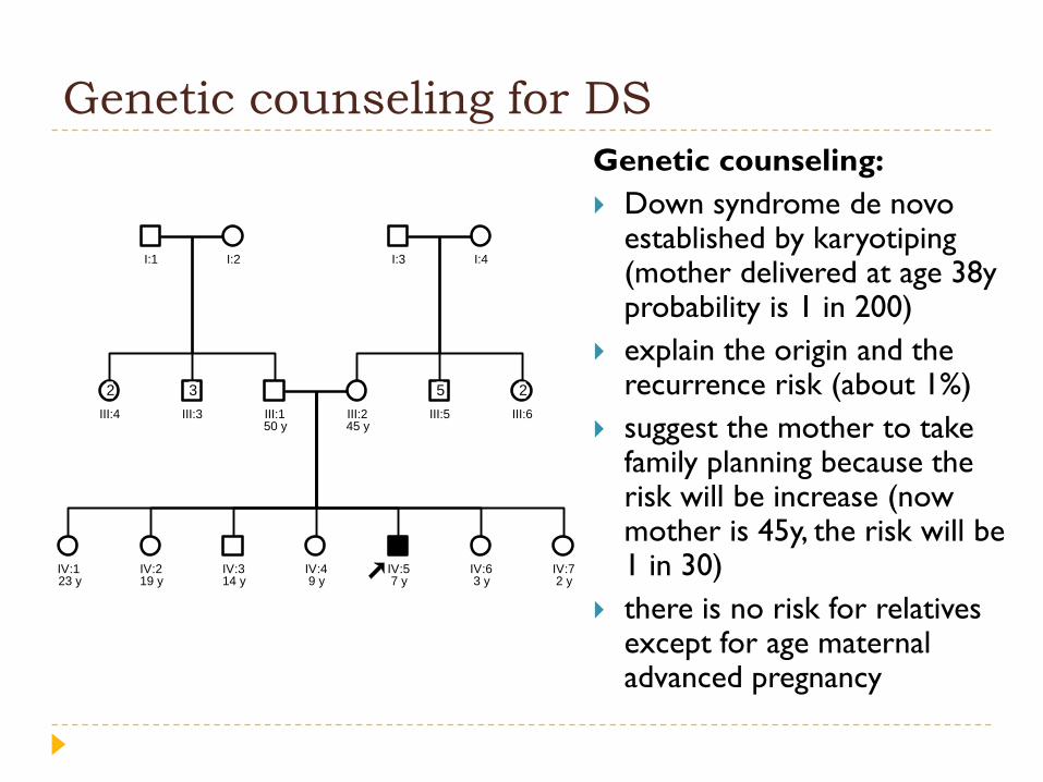
Genetic counseling for DS
Genetic counseling:
Down syndrome de novo established by karyotiping(mother delivered at age 38y probability is 1 in 200)
explain the origin and the recurrence risk (about 1%)
suggest the mother to take family planning because the risk will be increase (now mother is 45y, the risk will be 1 in 30)
there is no risk for relatives except for age maternal advanced pregnancy
IV:1
III:1 III:2
IV:2 IV:3 IV:4 IV:5 IV:6 IV:7
I:1 I:2
III:3
3
III:4
2
I:3 I:4
III:5
5
III:6
2
50 y 45 y
23 y 19 y 14 y 9 y 7 y 3 y 2 y

Discussion
Mrs A has a brother with a severe haemophilia A. He died due to a cerebral bleeding at the age of 8 years. She has also 3 healthy brothers. There is no other affected family member.
X-linked recessive; Skipping generation and not all male affected
The chance for Mrs A to be a carrier for haemophilia A: 50%
The possibility of the son to affected hemophilia if his mother is carrieris 50%
I:1 I:2
II:1 II:2 II:3 II:4 II:6II:5
?
III:1
Mrs.A

Autosomal recessive pattern:
