dissertation - katie maloney - final submission
TRANSCRIPT

ABSTRACT
MALONEY, KATHERINE PATTERSON. Sweetpotato Peel Proteins: Extraction Optimization and Susceptibility to Digestive Enzymes. (Under the direction of Dr. Jonathan Allen.)
North Carolina is the leading producer of sweetpotatoes in the United States. In 2009, 21%
of the 679,780,240 pounds of sweetpotatoes that were produced in North Carolina were
processed before entering the market. Generally, one of the first steps in processing is
removal of the peel. The peel is considered a waste product, but contains potentially
valuable compounds, such as proteins. The first objective of this research was to optimize a
process for extracting proteins from industrial peel waste. Material from the primary peeling
of raw roots and from a secondary peeling after blanching was obtained from a local
sweetpotato processing facility. Peel was mixed with saline solvent to dissolve proteins, and
then the mixture was centrifuged to separate non-soluble material. After the proteins were
precipitated from the supernatant with CaCl2, centrifugation was used to obtain a protein
pellet. Linear segmented and quadratic models were used to optimize peel to solvent ratio,
NaCl concentration, and CaCl2 concentration. More proteins could be extracted from
secondary peelings than primary peelings. The highest recovery, 32.0%, was obtained by
mixing 1 g of secondary peelings with 59.7 mL of 0.025 mM NaCl and then precipitating
with 6.8 mM CaCl2. The protein banding pattern and glycosylation characteristics of the
extract were similar to Caiapo, a commercial anti-diabetic supplement containing
sweetpotato proteins.
In order to exhibit systemic effects, a protein must survive gastric and duodenal digestion,
and then be absorbed. Sweetpotato proteins have been reported to possess anti-diabetic,

antioxidant, and anti-proliferative properties, but the mechanism by which the proteins evade
digestion is unknown. The second objective of this research was to determine the
susceptibility of sweetpotato proteins to digestive enzymes. Caiapo, an extract from primary
peelings, and an extract from secondary peelings were incubated with pepsin, trypsin, and
chymotrypsin. Samples were removed throughout the digestion procedure and protein
breakdown was visualized with SDS-PAGE. Samples were also assayed for amylase activity
and amylase inhibitory activity after incubation with pepsin. Proteins were present in all of
the samples that were resistant to digestion by pepsin, trypsin, and chymotrypsin. The
extract from secondary peelings exhibited lower resistance to pepsin than Caiapo and the
extract from primary peelings. Compact structure is most likely responsible for the noted
resistance to digestion, since the amino acid sequence of the major storage protein in
sweetpotatoes, sporamin, showed numerous potential cleavage sites. In addition, heat
treatment, which would cause denaturation, increased susceptibility of the protein to
digestion. Trypsin inhibitors remained active after simulated gastric digestion, with the
Caiapo and extract from primary peelings exhibiting higher inhibitory activity compared to
the extract from secondary peelings. Active amylase and chymotrypsin inhibitors were not
found in any of the samples after digestion. Modified glucose tolerance tests in rats
confirmed the lack of digestion-resistant amylase inhibitors, and showed that sweetpotato
proteins do not alter maltose digestion or glucose absorption.

Sweetpotato Peel Protein: Extraction Optimization and Susceptibility to Digestive Enzymes
by Katherine Patterson Maloney
A dissertation submitted to the Graduate Faculty of North Carolina State University
in partial fulfillment of the requirements for the degree of
Doctor of Philosophy
Food Science
Raleigh, North Carolina
2011
APPROVED BY:
_______________________________ ______________________________ Dr. Jonathan Allen Dr. Van-Den Truong Committee Chair ________________________________ ________________________________ Dr. Gisele Passador-Gurgel Dr. Sophia Kathariou

ii
DEDICATION
To my parents and my husband.

iii
BIOGRAPHY
Katherine Patterson Maloney was born and raised in Raleigh, NC. She received her B.S. in
Biological Sciences and M.S. in Nutrition and Food Science from NCSU. Upon completion
of her Ph.D. in Food Science, she will begin work at Novozymes.

iv
ACKNOWLEDGMENTS
I would like to thank the following people:
Dr. Jonathan Allen: for his continuous support and guidance throughout my time at NCSU.
Dr. Van-Den Truong, Dr. Sophia Kathariou, and Dr. Gisele Passador-Gurgel: for
serving on my committee and providing guidance.
Jack Canady and Gary Cartwright: for their help in the pilot plant.
Dr. Gary Gilleskie, John Taylor, Jessica Weaver, Greg Worsley, and Jacob Bullard: for
their help in the scale up of the protein extraction process.
Bill Heafy and the Yamco staff: for providing the raw material for this work.
Barbara Welker and Umair Arshad: for their help with the animal study.
Heather Hickman and Ruth Watkins: for keeping the lab running smoothly.
Dr. Jason Osborne: for his help with statistical analysis.
April Fogleman, Caroline Summers, Erica Story, and Elizabeth Dixon: for being
wonderful lab mates and friends.

v
TABLE OF CONTENTS
List of Tables ................................................................................................................... viii
List of Figures .................................................................................................................... ix
Chapter 1. Literature Review ..............................................................................................1
1.1 Sweetpotato production in North Carolina ................................................................2
1.2 Processing of sweetpotatoes ......................................................................................4
1.3 Sweetpotato protein quantity .....................................................................................5
1.4 Sweetpotato protein quality .......................................................................................6
1.5 Effect of agricultural practices on sweetpotato protein content .................................7
1.6 Changes during storage of sweetpotato roots ............................................................8
1.7 Characterization of sweetpotato proteins ...................................................................9
1.8 Sweetpotato trypsin inhibitors ...................................................................................9
1.9 Sweetpotato amylase inhibitors ...............................................................................11
1.10 Antioxidant activity of sweetpotato protein ...........................................................12
1.11 Anti-diabetic properties of sweetpotatoes ..............................................................13
1.12 References ..............................................................................................................20
Chapter 2. Chemical Optimization of Protein Extraction from Sweetpotato Peel ...........25
2.1 Abstract ....................................................................................................................26
2.2 Introduction ..............................................................................................................27
2.3 Materials and Methods .............................................................................................29
2.3.1 Pilot Plant Trial .................................................................................................29
2.3.1.1 Chemicals and raw material .......................................................................29

vi
2.3.1.2 Protein extraction process ..........................................................................29
2.3.1.3 Protein precipitation process ......................................................................30
2.3.1.4 Fraction analysis ........................................................................................30
2.3.2. Bench-top Process Optimization ......................................................................31
2.3.2.1 Chemicals and raw material .......................................................................31
2.3.2.2 Protein extraction process ..........................................................................31
2.3.2.3 Protein precipitation process ......................................................................32
2.3.2.4 Determining optimum conditions for protein extraction ...........................32
2.3.2.5 Determining optimum conditions for protein precipitation .......................33
2.3.2.6 Comparison of Caiapo and peel extract with gel electrophoresis ..............34
2.4 Results and Discussion ............................................................................................34
2.4.1 Pilot plant trial ..................................................................................................34
2.4.2 Bench-top optimization of the extraction process ............................................36
2.4.3 Bench-top optimization of the precipitation process ........................................39
2.4.4 Comparison of protein extraction from peel and blanched peel .......................42
2.4.5 Comparison of protein quantification methods .................................................43
2.4.6 Comparison of Caiapo and peel extract with gel electrophoresis .....................44
2.5 Conclusions ..............................................................................................................45
2.6 References ................................................................................................................57
Chapter 3. Susceptibility of Sweetpotato Protein to Digestive Enzymes .........................60
3.1 Abstract ....................................................................................................................61
3.2 Introduction ..............................................................................................................62

vii
3.3 Materials and Methods .............................................................................................64
3.3.1 Enzymes ............................................................................................................64
3.3.2 Raw material .....................................................................................................65
3.3.3 In vitro gastric digestion ...................................................................................65
3.3.4 In vitro duodenal digestion ...............................................................................66
3.3.5 Amylase activity assay ......................................................................................66
3.3.6 Gel electrophoresis ............................................................................................67
3.3.7 Modified glucose tolerance tests in rats ............................................................68
3.3.8 Statistical analysis .............................................................................................68
3.4 Results and Discussion ............................................................................................68
3.4.1 Susceptibility of sporamin to digestive enzymes ..............................................68
3.4.2 Trypsin and chymotrypsin inhibitory activity...................................................71
3.4.3 Amylase activity and amylase inhibitory activity .............................................72
3.4.4 Modified glucose tolerance tests in rats ............................................................74
3.5 Conclusions ..............................................................................................................75
3.6 References ................................................................................................................85
Chapter 4. Conclusions .....................................................................................................90

viii
LIST OF TABLES
Table 2.1. Compositional comparison of Caiapo and pilot plant trial peel extract ..........48
Table 3.1. Modified glucose tolerance tests in rats ...........................................................84

ix
LIST OF FIGURES
Figure 1.1. Percentage harvested by top 9 states of total sweetpotato acres in the
US in 2008 ........................................................................................................2
Figure 1.2. Percentage produced by top 10 counties of total sweetpotatoes
produced in NC ................................................................................................3
Figure 1.3. Sweetpotato cultivars grown in North Carolina ...............................................3
Figure 1.4. Isolation of WSSP anti-diabetic component by Kusano and others ...............17
Figure 1.5. Isolation of WSSP anti-diabetic component by Ozaki and others .................18
Figure 2.1. Mass balance during pilot plant extraction of protein from
sweetpotato peel ..............................................................................................47
Figure 2.2. Linear segmented models fit to extraction data for peel ................................49
Figure 2.3. Linear segmented models fit to extraction data for blanched peel .................50
Figure 2.4. Optimizing NaCl concentration and solvent to peel ratio for protein
extraction from peel using quadratic models ..................................................51
Figure 2.5. Optimizing NaCl concentration and solvent to peel ratio for protein
extraction from blanched peel using quadratic models ..................................52
Figure 2.6. Linear segmented models fit to peel precipitation data ..................................53
Figure 2.7. Linear segmented model fit to blanched peel precipitation data all
temperatures combined ...................................................................................54
Figure 2.8. Comparison of Caiapo and peel extract protein banding patterns .................55
Figure 2.9. Comparison of Caiapo and peel extract glycoprotein banding
patterns ............................................................................................................56

x
Figure 3.1. SDS-PAGE of Caiapo digested with pepsin, trypsin, and
chymotrypsin ..................................................................................................76
Figure 3.2. SDS-PAGE of sweetpotato peel digested with pepsin, trypsin, and
chymotrypsin ..................................................................................................77
Figure 3.3. SDS-PAGE of blanched sweetpotato peel digested with pepsin,
trypsin, and chymotrypsin ..............................................................................78
Figure 3.4. Theoretical pepsin cleavage sites of sporamin determined by
inputting sporamin protein sequence from GenBank accession
AAB52550 into ExPASy PeptideCutter .........................................................79
Figure 3.5. Theoretical trypsin cleavage sites of sporamin determined by
inputting sporamin protein sequence from GenBank accession
AAB52550 into ExPASy PeptideCutter .........................................................80
Figure 3.6. Theoretical chymotrypsin cleavage sites of sporamin determined by
inputting sporamin protein sequence from GenBank accession
AAB52550 into ExPASy PeptideCutter .........................................................81
Figure 3.7. Amylase activity of Caiapo, peel, and blanched peel before and after
incubation with pepsin for 1 hour at pH=2 and 37°C .....................................82
Figure 3.8. Activity of porcine pancreatic amylase (% retained) in the presence
of Caiapo, peel, and blanched peel previously incubated with pepsin
for 1 hour at pH=2 and 37°C ..........................................................................83

1
CHAPTER 1
Literature Review
2
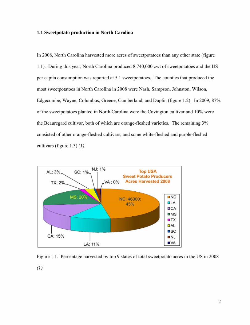
1.1 Sweetpotato production in North Carolina
In 2008, North Carolina harvested more acres of sweetpotatoes than any other state (figure
1.1). During this year, North Carolina produced 8,740,000 cwt of sweetpotatoes and the US
per capita consumption was reported at 5.1 sweetpotatoes. The counties that produced the
most sweetpotatoes in North Carolina in 2008 were Nash, Sampson, Johnston, Wilson,
Edgecombe, Wayne, Columbus, Greene, Cumberland, and Duplin (figure 1.2). In 2009, 87%
of the sweetpotatoes planted in North Carolina were the Covington cultivar and 10% were
the Beauregard cultivar, both of which are orange-fleshed varieties. The remaining 3%
consisted of other orange-fleshed cultivars, and some white-fleshed and purple-fleshed
cultivars (figure 1.3) (1).
Figure 1.1. Percentage harvested by top 9 states of total sweetpotato acres in the US in 2008
(1).

3
Figure 1.2. Percentage produced by top 10 counties of total sweetpotatoes produced in NC
(1).
Figure 1.3. Sweetpotato cultivars grown in North Carolina (1).

4
In 2009, a total of 537,821,840 pounds of North Carolina sweetpotatoes were distributed to
retail establishments (70%), food service establishments (18%), and the international market
(5%). Another 141,958,400 pounds of sweetpotatoes were processed into frozen product
(23%), canned product (21%), chips (14%), baby food (14%), or other processed product
(28%), before entering the market (2).
1.2 Processing of sweetpotatoes
Sweetpotatoes are traditionally viewed as holiday cuisine, with much higher sales in
November and December than any other time of year (3). In recent years, however, there has
been a focus on developing processing techniques that maintain flavor and texture to increase
year-round consumption of sweetpotatoes. Restructured sweetpotato fries have been
improved by adding alginate-calcium to increase textural consistency (4), as well as, adding
sodium tripolyphosphate during blanching and sweetpotato flour during mixing to increase
product firmness and dry matter content (5). New methods for preserving sweetpotato puree
and new uses for sweetpotato puree have also been investigated. Coronel and others (6) used
continuous flow microwave processing on sweetpotato puree from orange-fleshed varieties to
obtain a shelf-stable product. This technology was licensed to, and is currently being used by
Yamco, LLC in Snow Hill, NC to create orange-fleshed sweetpotato purees of superior
flavor and texture, with significant storage energy savings, compared to those previously
available. Continuous flow microwave processing has also been applied to sweetpotato
puree from purple-fleshed varieties (7). Grabowski and others (8) optimized the process for

5
spray drying sweetpotato puree to create a powder that can, compared to traditional drying
methods, be more easily incorporated into other foods, further expanding the sweetpotato
market. These technologies have been reviewed by Truong and Ramesh (9). US per capita
sweetpotato consumption data is, currently, only available up to 2008, and shows a relatively
stable market (1). However, recent events, such as the opening of a new plant in Delhi, LA
focused on producing various processed sweetpotato products, point to consumption being on
the rise (10).
1.3 Sweetpotato protein quantity
The protein content of sweetpotatoes can vary substantially depending on both cultivar and
growing conditions. Analysis of various cultivars grown in North Carolina showed protein
content to vary from 1.73% to 9.14% on a dry weight basis (11). Similar results were
obtained by a study conducted in Nigeria on 49 different cultivars of sweetpotato. Protein
content was found to range from 1.39% to 9.4% on a dry weight basis (12). Purcell and
others (13) found that even within a cultivar, significant differences in protein content could
exist. Sweetpotatoes of the cultivar Jewel were found to vary in protein content from 3.99%
to 8.81% on a dry weight basis and sweetpotatoes of the cultivar Centennial were found to
vary in protein content from 5.27% to 7.24% on a dry weight basis.
Bradbury and others (14) found that the peel of the sweetpotato was 50-80% higher in crude
protein than the flesh. Makki and others (15) compared the peel and pulp of two varieties of

6
Egyptian sweetpotatoes and found that the peel was also higher in protein than the pulp, but
the difference between the two values was smaller than that found by Bradbury and others
(14). The peel of the cultivar Abees contained 6.7% crude protein compared to 5.0% in the
pulp, and the peel of the cultivar Giza 69 contained 7.4% protein compared to 4.9% in the
pulp. Purcell and others (16), when comparing the distribution of protein within the
sweetpotato, also noted that the peel was higher in protein than the flesh.
1.4 Sweetpotato protein quality
Tryptophan, sulfur amino acids, and lysine have been identified as limiting amino acids in
different sweetpotato cultivars. Purcell and others (11) compared the amino acid
composition of six sweetpotato cultivars grown in North Carolina to the FAO reference
protein and found that tryptophan and total sulfur amino acids were limiting. An excess of
other essential amino acids was present, however. Bradford and others (14) found sulfur
amino acids to be limiting in 65% of the cultivars tested from the Highlands of Papua New
Guinea. Mu and others (17) found lysine and tryptophan to be limiting compared to the FAO
reference in the Chinese cultivar 55-2 while many of the other essential amino acids were
present in excess. Walter and others (18) compared protein extracts from the cultivars Jewel
and Centennial to casein and found that the extracts were higher in total sulfur amino acids
than casein, however, both casein and the extracts were still below the FAO reference. The
protein efficiency ratio of the extracts was equal to that of casein. Ravindran and others (19)
compared the in vitro digestibility of sweetpotato protein from 16 cultivars and found an

7
average digestibility of 75.8% with a range of 71.4% to 79.5%. Apparent metabolizable
energy for poultry was then determined and found to be similar to values for corn, 14.54
MJ/kg versus 14.45 MJ/kg.
1.5 Effect of agricultural practices on sweetpotato protein content
The effect of nitrogen fertilization on protein content of sweetpotato roots has been
extensively studied. Constantin and others (20) found that as nitrogen levels increased,
protein content increased. The study, conducted in Louisiana, used nitrogen application
levels of 0 kg/ha, 33.64 kg/ha, 67.28 kg/ha, and 100.92 kg/ha. Purcell and others (21), in a
study conducted in North Carolina, also found that nitrogen fertilization increased protein
content of roots. Application levels were similar with 0 kg/ha, 56 kg/ha, and 112 kg/ha being
applied during the 1977 trial and 0 kg/ha, 51 kg/ha, and 101 kg/ha being applied during the
1978 trial. Nitrogen fertilization did not change the ratio of non-protein nitrogen to total
nitrogen. A more recent study (22), conducted in Nigeria, also reported an increase in
protein content with nitrogen fertilization up to 80 kg/ha. Application levels tested included
0 kg/ha, 40 kg/ha, 80 kg/ha, and 120 kg/ha. Ukom and others (22) found that 40-80 kg/ha
was sufficient for maximum protein yield in most cultivars. Application above these levels
did not further increase protein content. The highest protein yield obtained was 9.84% on a
dry weight basis with a nitrogen application level of 80 kg/ha, a protein content value on the
higher end of the ranges reported by other groups (11, 12).

8
The effect of irrigation and fertilization with other minerals on root protein yield has also
been studied. Constantin and others (20) found that irrigation negatively impacted protein
content. Sweetpotatoes from plots maintained at 25% and 50% soil moisture had reduced
protein content (6.5% and 6.7% on a dry weight basis, respectively) compared to those from
the control plot (7.9% on a dry weight basis) in which only rainfall provided moisture to the
soil. Purcell and others (21) found that potassium and sulfur fertilization did not alter the
protein content of individual roots, however, potassium application did increase total root
yield and thus total protein yield.
1.6 Changes during storage of sweetpotato roots
Purcell and others (23) found that protein content of roots decreases with storage; however,
percent protein increases because dry matter is lost at a rate twice that of protein. Zhang and
others (24) found that dry matter and starch content of sweetpotatoes were correlated (r =
0.92) and that starch content decreased during storage with rates of decrease differing among
cultivar. Alpha-amylase activity was found to increase during the first 2 months of storage
and then decrease with continued storage back to levels observed at harvest with cultivar
again affecting rate. Trypsin inhibitor activity decreased slightly in some cultivars with
extended storage (> 60 days).

9
1.7 Characterization of sweetpotato proteins
Sporamin A and sporamin B account for 80% of the total protein content of sweetpotato
roots. Both proteins migrate to a position corresponding to a molecular weight of 25 kDa
under denaturing SDS-PAGE. Under non-denaturing SDS-PAGE, however, sporamin A
migrates to a position corresponding to a molecular weight of 31 kDa and sporamin B
migrates to a position corresponding to a molecular weight of 22 kDa. The amino acid
composition and immunological properties of the sporamins are very similar but not
identical. Antibodies raised against sporamin A also recognized sporamin B. Neither protein
was adsorbed on a concanavalin A-Sepharose column or stained by periodic acid-Schiff
staining of a gel, thus it was concluded that neither was a glycoprotein (25).
1.8 Sweetpotato trypsin inhibitors
Trypsin inhibitors in sweetpotatoes have been extensively studied. Bradbury and others (14)
found a 67-fold range in trypsin inhibitor activity, from 0.33 to 22.1 trypsin inhibitory units
(TIU), among sweetpotato cultivars from the Highlands of Papua New Guinea. Results of
this study failed to find a significant correlation between crude protein content and trypsin
inhibitory activity (r = 0.057). Later work (26) confirmed the lack of a significant correlation
between crude protein content and trypsin inhibitory activity across cultivars; however,
Bradbury and others found that within cultivar, crude protein content and trypsin inhibitory
activity were correlated.

10
Sweetpotatoes contain multiple proteins that exhibit trypsin inhibitor activity. Sugiura and
others (27) identified trypsin inhibitors with molecular weights of 23 and 24 kDa. Obidairo
and Akpochafo (28) isolated 10 different trypsin inhibitors from sweetpotatoes, with the most
active inhibitors having molecular weights of 12, 10, and 9.3 kDa. Hou and others (29)
identified proteins with molecular weights of 73, 38, and 22 kDa as trypsin inhibitors. Jaw
and others (30) purified a trypsin inhibitor with a molecular weight of 23 kDa. Trypsin
inhibitors had no effect on chymotrypsin and pepsin activity (27). Obidairo and others (28)
showed that maximum inhibitor activity was attained with pH values between 7.5 and 8.5,
and Sugiura and others (27) found that sweetpotato trypsin inhibitors are stable over a wide
pH range. Incubation at pH values between 2 and 11 for 12 hours before assaying for
inhibitor activity at pH=8 did not result in significant loss of activity. Hou and others (31)
identified a native 64 kDa aspartic protease that was capable of degrading trypsin inhibitors.
Processing of sweetpotatoes can greatly affect the activity of trypsin inhibitors. Obidairo and
Akpochafo (28) found that boiling sweetpotatoes for 40 minutes resulted in complete
inactivation of trypsin inhibitors. Kiran and others (32) found microwave baking to be the
most effective method for inactivating trypsin inhibitors in sweetpotatoes followed by boiling
and then oven drying. Microwave baking for 180 seconds resulted in complete inactivation
of trypsin inhibitors while boiling for 30 minutes resulted in 17-31% residual activity.
Trypsin inhibitors in oven dried sweetpotato chips were relatively stable for 2 hours at 70°C,
with 80-90% residual activity. After 2 hours, however, inactivation progressed at a more

11
rapid rate, with less than 20% activity remaining after 24 hours. Higher temperatures also
led to more rapid inactivation, with inactivation complete after 4 hours at 100°C. Minor
variations in thermostability were seen for different cultivars. The researchers also compared
the effect of different flour preparation methods on residual trypsin inhibitor activity. Flour
was prepared by drying either sweetpotato chips, grated sweetpotato pieces, or ground
sweetpotato pieces at 70°C for 24 hours and then powdering. All methods resulted in 5-12%
residual activity with no statistically significant differences between methods being noted.
Zhang and Corke (33) found that moist heat treatment provided better inactivation of trypsin
inhibitors than dry heat treatment. Dry heat treatment at 60, 80, and 100°C for 15 minutes
resulted in average residual activities of 92, 84, and 71%, respectively, where as moist heat
treatment at 60, 80, and 100°C for 15 minutes resulted in average residual activities of 71,
26, and 5%, respectively. Similarly to Kiran and others, Zhang and Corke found that the
trypsin inhibitors of some cultivars were more heat resistant than others.
1.9 Sweetpotato amylase inhibitors
Shivaraj and others (34) found that sweetpotato protein exhibited high amylase activity with
an average of 480 units/mg protein and a range of 274-758 units/mg protein. After heating
the extract for 10 minutes at 80°C to destroy native amylase activity, amylase inhibitory
activity was assayed via the dinitrosalycylic acid method. No amylase inhibitors were found
in the sweetpotato extract. Rekha and others (35) found that native amylase activity
remained after heating for 10 minutes at 80°C and thus used trichloroacetic acid to

12
selectively precipitate the amylases before assaying the extract for amylase inhibitory activity
via the iodine binding method. Of the 100 cultivars studied, amylase inhibitors were found
in 79.
Rekha and others (36) later studied the effect of processing on native amylase inhibitors in
sweetpotatoes and found that cultivar played a significant role in the stability of the
inhibitors. Boiling sweetpotato pieces in water for 30 minutes resulted in residual amylase
inhibitor activities of 29.3 ± 1.1% (cultivar RS III), 29.1 ± 1.1% (cultivar S 62), 44.6 ± 1.9%
(cultivar S 56-2), and 58.9 ± 0.7% (cultivar S 1195). Microwave baking resulted in complete
amylase inhibitor inactivation in the S 62 cultivar after 120 seconds and the S 1195 cultivar
after 180 seconds. Residual amylase inhibitor activities of 29.1 ± 1.1% and 19.2 ± 0.6%
remained after 180 seconds in the cultivars RS III and S 56-2, respectively. Grating or
blending, oven drying at 70°C for 24 hours, and then powdering to obtain flour resulted in
complete inactivation of amylase inhibitors in all cultivars tested.
1.10 Antioxidant activity of sweetpotato proteins
Various antioxidant functions have been reported for sweetpotato proteins. Trypsin
inhibitors isolated from sweetpotatoes were shown to possess DHA reductase and MDA
reductase activities (37). A 33 kDa trypsin inhibitor was isolated with preparative SDS-
PAGE and found to possess scavenging activity against 1,1-diphenyl-2-picrylhydrazyl
(DPPH) and hydroxyl radical (38). This 33 kDa trypsin inhibitor was later shown to possess

13
glutathione peroxidase-like activity (39). Further studies showed that the trypsin inhibitors
were able to prevent Cu2+-induced human LDL peroxidation and hydroxyl radical-induced
DNA damage of calf thymus in vitro. Degree of protection was dependent on the cultivar
from which the trypsin inhibitors were isolated. Trypsin inhibitors isolated from the cultivar
Tainong 65 had 3-fold greater activity against LDL peroxidation but trypsin inhibitors
isolated from the cultivar Tainong 57 showed 10-fold greater hydroxyl radical scavenging
ability. Hydrolysis with pepsin and chymotrypsin increased the scavenging activity of the
trypsin inhibitors against DPPH, indicating that digestion would not eliminate the antioxidant
properties (40).
1.11 Anti-diabetic properties of sweetpotatoes
Proteins isolated from a white-skinned sweetpotato cultivar have been extensively studied for
their anti-diabetic properties. Kusano and others (41) compared the anti-diabetic efficacy of
white-skinned sweetpotato (WSSP) extract to troglitazone using obese Zucker fatty rats. The
WSSP was administered at 100 mg/kg/day for 8 weeks. After 8 weeks, blood insulin levels
were significantly lower in the WSSP group (384 ± 97 µU/mL) and troglitazone group (394
± 94 µU/mL) compared to the control group (753 ± 214 µU/mL). Blood triglycerides and
free fatty acid (FFA) levels were reduced in both the WSSP and troglitazone groups
compared to the control group. Cholesterol was not significantly different in any of the
groups, however. Blood glucose and blood insulin levels after glucose loading were also

14
reduced in the WSSP and troglitazone groups compared to the control group. Body weight
was significantly higher in the troglitazone group compared to the WSSP and control groups.
In another animal study, Kusano and others (42) found that oral administration of 400
mg/kg/day of WSSP to KK-Ay mice significantly reduced blood glucose levels and increased
glucose tolerance compared to the control. No significant differences in body weight were
found between the group receiving WSSP and the control group. After 3 weeks of treatment,
the group receiving WSSP had significantly higher expression of ACRP30, indicating that
the anti-diabetic mechanism of action of WSSP may be increasing secretion of adiponectin, a
hormone associated with insulin sensitivity.
Ludvik and others have performed several human trials with the WSSP extract, Caiapo (43-
46). In their first trial (43), Ludvik and others randomly divided 18 male type II diabetic
patients into 3 groups, control, 2 g Caiapo per day, and 4 g Caiapo per day. The double-
blind, placebo-controlled study lasted 6 weeks. A significant decrease in fasting blood
glucose (from 8.3 ± 0.6 to 7.2 ± 0.4 mmol/L), total cholesterol (from 4.97 ± 0.21 to
4.45 ± 0.18 mmol/L), and LDL cholesterol (3.12 ± 0.16 to 2.72 ± 0.16 mmol/L) was noted in
the 4 g treatment group after 6 weeks. No significant changes were seen in body weight or
blood pressure in any of the groups. Insulin sensitivity, measured by frequently sampled
intravenous glucose tolerance test (FSIGT), increased in both treatment groups. A 37%

15
increase was noted in the 2 g treatment group and a 42% increase was noted in the 4 g
treatment group. In a second trial, Ludvik and others (44) again randomly divided 18 male
type II diabetic patients into 3 groups, control, 2 g Caiapo per day, or 4 g Caiapo per day. In
this trial, oral glucose tolerance test (OGTT) was performed in addition to FSIGT. After 6
weeks of treatment, insulin sensitivity, measured by both FSIGT and OGTT, increased
significantly in the 4 g treatment group. In the 2 g treatment group, only insulin sensitivity
measured by FSIGT increased significantly. Ludvik and others (45) then performed a larger
study with 61 type II diabetic patients randomly assigned to receive either placebo or 4 g per
day of Caiapo. The study lasted 12 weeks. Significant decreases were seen in fasting blood
glucose (from 143.7 ± 1.9 to 128.5 ± 1.7 mg/dl), HbA1c (from 7.21 ± 0.15 to 6.68 ± 0.14%),
and blood glucose 2 hours after OGTT (from 193.3 ± 10.4 to 162.8 ± 8.2 mg/dl) in the
treatment group while no significant changes in these parameters were seen in the control
group.
In a later trial, Ludvik and others (46) found similar results to Kusano and others (42)
relating to the mechanism of action of WSSP on increasing insulin sensitivity. Previous
results concerning Caiapo’s ability to increase insulin sensitivity, lower fasting blood
glucose, and lower HbA1c were also confirmed by this 5 month study involving 61 type II
diabetic patients randomized to receive either 4 g Caiapo per day or placebo. After 5 months,
oral glucose insulin sensitivity, measured by OGTT, increased from 293 ± 15 to 321 ± 12
mL/m2/min, fasting blood glucose decreased from 138 ± 4 to 128 ± 5 mg/dl, and HbA1c

16
decreased from 6.46 ± 0.12 to 6.25 ± 0.11%. Additional parameters that were measured
included plasma adiponectin and fibrinogen. Significant increases in plasma adiponectin
(from 5.97 ± 0.65 to 6.63 ± 0.70 µg/mL) and significant decreases in fibrinogen (from 3.83 ±
0.16 to 3.64 ± 0.18 mg/mL) were seen in the treatment group. Thus, increasing adiponectin
secretion appears to be the mechanism by which Caiapo exerts its anti-diabetic effects. The
observed decrease in fibrinogen may indicate that Caiapo possesses other nutraceutical
properties and may be useful as an anti-atherosclerotic supplement, as well.
Kusano and others (47) attempted to isolate the component responsible for the anti-diabetic
effect of WSSP. Figure 1.4 shows the steps of the isolation procedure. The anti-diabetic
component remained in the inner solution during dialysis, exhibited solubility in 85%
ethanol, remained in solution upon the addition of 15% trichloroacetic acid, and passed
through a filter with a 30 kDa molecular weight cut-off during ultrafiltration. The molecular
weight of the active component was estimated at 22 kDa with gel filtration chromatography.
The component was found to be acidic with ion exchange chromatography and contained
both protein and sugar, so was assumed to be a glycoprotein. Oral administration of the
isolated component to db/db mice for 2 weeks resulted in decreased blood glucose and blood
insulin levels compared to the control.

Figur
A dif
later
The a
comp
consi
isolat
levels
re 1.4. Isola
fferent anti-d
isolated by O
anti-diabetic
ponent was f
isting of 95%
ted compone
s compared t
ation of WSS
diabetic com
Ozaki and ot
component
found to be a
% (w/w) carb
ent to KK-A
to the contro
SP anti-diabe
mponent than
thers (48). F
was not pre
an arabinoga
bohydrate an
Ay mice resul
ol group.
etic compone
that isolated
Figure 1.5 sh
ecipitated by
alactan-prote
nd 5% (w/w)
lted in signif
ent by Kusan
d by Kusano
hows the step
ammonium
ein with a mo
) protein. Or
ficantly lowe
no and other
o and others
ps in the isol
sulfate. Th
olecular wei
ral administr
er fasting pla
rs (47).
from WSSP
lation proced
he isolated
ight of 126.8
ration of the
asma glucos
17
P was
dure.
8 kDa
e
e

Figur
Zakir
cultiv
grow
simil
blood
levels
the gl
mg/d
216.2
re 1.5. Isola
r and others
var common
wn in the Uni
ar. The rese
d glucose low
s 2 hours aft
lucose contr
dl, in the Wh
29 ± 93.16 m
ation of WSS
(49) compar
ly grown in
ted States, B
earchers also
wering effica
ter feeding w
rol. Blood g
ite Star grou
mg/dl, and in
SP anti-diabe
red the prote
Pakistan, W
Beauregard.
o conducted
acy of Caiap
were lower in
lucose level
up were 246
n the Beaureg
etic compone
ein banding p
White Star, an
The protein
a feeding tri
po, White St
n the White
s in the gluc
± 87.07 mg/
gard group w
ent by Ozak
pattern of Ca
nd a sweetpo
n banding pat
ial with type
ar, and Beau
Star and Bea
cose control g
/dl, in the W
were 257 ± 9
ki and others
aiapo to a sw
otato cultivar
tterns of all
II diabetics
uregard. Blo
auregard gro
group were
White Star (sk
98.90 mg/dl.
(48).
weetpotato
r commonly
samples wer
to compare
ood glucose
oups compar
296 ± 111.3
kin) group w
The additio
18
re
the
red to
4
were
on of

19
Caiapo to glucose resulted in 2 hour blood glucose levels similar to the control, 301 ± 113.91
mg/dl. Corbitt (50) compared the glycemic index of Beauregard sweetpotatoes prepared by
various methods. Steamed, baked and microwaved sweetpotato flesh were found to be
medium glycemic index foods, with glycemic indices of 63 ± 8.4, 64 ± 10.0 and 66 ± 13.3,
respectively. Dehydrated and raw sweet potato flesh were found to be low glycemic index
foods, with glycemic indices of 40 ± 8.2 and 28 ± 7.3, respectively.

20
1.12 REFERENCES
1. North Carolina SweetPotato Commission Sweet Potatoes - The Vegetable with Super Food Powers. <http://www.ncsweetpotatoes.com>
2. North Carolina Department of Agriculture & Consumer Services. N.C. Annual Sweetpotato Summary. <http://www.ncagr.gov/markets/mktnews/swpotsum.pdf>
3. Economic Research Service, U.S. Department of Agriculture. U.S. Sweet Potato Statistics. <http://usda.mannlib.cornell.edu/MannUsda/viewDocumentInfo.do?document ID=1492>
4. Walter, W.M.; Truong, V.D.; Espinel, K.R. Textural Measurements and Product Quality of Restructured Sweetpotato French Fries. Lebensmittel-Wissenschaft und-Technologie 2002, 35, 209-215.
5. Utomo, J.S.; Che Man, Y.B.; Rahman, R.A.; Said Saad, M. The effect of shape, blanching methods and flour on characteristics of restructured sweetpotato stick. Int. J. Food Sci. Tech. 2008, 43, 1896-1900.
6. Coronel, P.; Truong, V.; Simunovic, J.; Sandeep, K.P.; Cartwright, G.D. Aseptic Processing of Sweetpotato Purees Using a Continuous Flow Microwave System. J. Food Sci. 2005, 70, E531-E536.
7. Steed, L.E.; Truong, V.D.; Simunovic, J.; Sandeep, K.P.; Kumar, P.; Cartwright, G.D.; Swartzel, K.R. Continuous flow microwave-assisted processing and aseptic packaging of purple-fleshed sweetpotato purees. J. Food Sci. 2008, 73, E455-62.
8. Grabowski, J.A.; Truong, V.D.; Daubert, C.R. Spray-Drying of Amylase Hydrolyzed Sweetpotato Puree and Physicochemical Properties of Powder. J. Food Sci. 2006, 71, E209-E217.
9. Truong, V.D.; Ramesh, A.Y. Sweet Potato Purees and Powders for Functional Food Ingredients. Sweet Potato: Post Harvest Aspects in Food. New York: Nova Science Publishers, Inc., 2010, 117-161.
10. The Times-Picayune. ConAgra Foods officially opens sweet potato processing plant in Delhi, La. <http://www.nola.com/business/index.ssf/2010/11/conagra_foods_officially_ opens.html>
11. Purcell, A.E.; Swaisgood, H.E.; Pope, D.T. Protein and amino acid content of sweetpotato cultivars. J. Amer. Soc. Hort. Sci. 1972, 97, 30-33.

21
12. Oboh, S.; Ologhobo, A.; Tewe, O. Some aspects of the biochemistry and nutritional value of the sweet potato (Ipomea batatas). Food Chem. 1989, 31, 9-18.
13. Purcell, A.E.; Walter, W.M.,Jr; Giesbrecht, F.G. Root, hill, and field variance in protein content of North Carolina sweet potatoes. J. Agric. Food Chem. 1978, 26, 362-364.
14. Bradbury, J.H.; Baines, J.; Hammer, B.; Anders, M.; Millar, J.S. Analysis of sweet potato (Ipomoea batatas) from the highlands of Papua New Guinea: relevance to the incidence of Enteritis necroticans. J. Agric. Food Chem. 1984, 32, 469-473.
15. Makki, H.M.; Abdel-Rahman, A.Y.; Khalil, M.K.M.; Mohamed, M.S. Chemical composition of Egyptian sweet potatoes. Food Chem. 1986, 20, 39-44.
16. Purcell, A.E.; Walter, W.M.; Giesbrecht, F.G. Distribution of protein within sweet potato roots (Ipomea batatas L.). J. Agric. Food Chem. 1976, 24, 64-66.
17. Mu, T.; Tan, S.; Xue, Y. The amino acid composition, solubility and emulsifying properties of sweet potato protein. Food Chem. 2009, 112, 1002-1005.
18. Walter, W.M.; Catignani, G.L. Biological quality and composition of sweet potato protein fractions. J. Agric. Food Chem. 1981, 29, 797-799.
19. Ravindran, V.; Ravindran, G.; Sivakanesan, R.; Rajaguru, S.B. Biochemical and nutritional assessment of tubers from 16 cultivars of sweet potato (Ipomoea batatas L.). J. Agric. Food Chem. 1995, 43, 2646-2651.
20. Constantin, R.J.; Hernandez, T.P.; Jones, L.G. Effects of irrigation and nitrogen fertilization on quality of sweet potatoes. J. Amer. Soc. Hort. Sci. 1974, 99, 308-310.
21. Purcell, A.E.; Walter, W.M.,Jr; Nicholaides, J.J.; Collins, W.W.; Chancy, H. Nitrogen, potassium, sulfur fertilization, and protein content of sweet potato roots. J. Amer. Soc. Hort. Sci. 1982, 107, 425-427.
22. Ukom, A.N.; Ojimelukwe, P.C.; Okpara, D.A. Nutrient Composition of Selected Sweet Potato [Ipomea batatas (L) Lam] Varieties as Influenced by Different Levels of Nitrogen Fertilizer Application. Pakistan Journal of Nutrition 2009, 8, 1791-1795.
23. Purcell, A.E.; Walter, W.M.,Jr; Giesbrecht, F.G. Changes in dry matter, protein and non-protein nitrogen during storage of sweet potatoes. J. Amer. Soc. Hort. Sci. 1978, 103, 190-192.
24. Zhang, Z.; Wheatley, C.C.; Corke, H. Biochemical changes during storage of sweet potato roots differing in dry matter content. Postharvest Biol. Technol. 2002, 24, 317-325.

22
25. Maeshima, M.; Sasaki, T.; Asahi, T. Characterization of major proteins in sweet potato tuberous roots. Phytochemistry 1985, 24, 1899-1902.
26. Bradbury, J.H.; Hammer, B.; Nguyen, T.; Anders, M.; Millar, J.S. Protein quantity and quality and trypsin inhibitor content of sweet potato cultivars from the highlands of Papua New Guinea. J. Agric. Food Chem. 1985, 33, 281-285.
27. Sugiura, M.; Ogiso, T.; Takeuti, K.; Tamura, S.; Ito, A. Studies on trypsin inhibitors in sweet potato I. Purification and some properties. Biochimica et Biophysica Acta (BBA) - Protein Structure 1973, 328, 407-417.
28. Obidairo, T.K.; Akpochafo, O.M. Isolation and characterization of some proteolytic enzyme inhibitors in sweet potato (Ipomoea batatas). Enzyme Microb. Technol. 1984, 6, 132-134.
29. Hou, W.; Lin, Y. Polyamine-bound trypsin inhibitors in sweet potato (Ipomoea batatas [L.] Lam cv. Tainong 57) storage roots, sprouted roots and sprouts. Plant Science 1997, 126, 11-19.
30. Jaw, K.S.; Chou, L.H.; Chang, S.M.; Duan, K.J. Purification of a trypsin inhibitor from sweet potato in an aqueous two phase system Biotechnol. Lett. 2007, 29, 137-140.
31. Hou, W.; Huang, D.; Lin, Y. An aspartic type protease degrades trypsin inhibitors, the major root storage proteins of sweet potato (Ipomoea batatas (L.) Lam cv. Tainong 57). Bot. Bull. Acad. Sin. 2002, 43, 271-276.
32. Kiran, K.S.; Padmaja, G. Inactivation of trypsin inhibitors in sweet potato and taro tubers during processing Plant Foods Hum. Nutr. 2003, 58, 153-163.
33. Zhang, Z.; Corke, H. Trypsin inhibitor activity in vegetative tissue of sweet potato plants and its response to heat treatment. J. Sci. Food Agric. 2001, 81, 1358-1363.
34. Shivaraj, B.; Sharma, K.K.; Pattabiraman, T.N. Natural plant enzyme inhibitors: Part VII--Alpha amylase inhibitors & amylases in plant tubers Indian J. Biochem. Biophys. 1979, 16, 52-55.
35. Rekha, M.R.; Padmaja, G.; Easwari Amma, C.S.; Sheela, M.N. Cultivar differences in the alpha-amylase inhibitor activity of sweet potato and yam tubers. J. Root Crops 1999, 25, 185-191.
36. Rekha, M.R.; Padmaja, G. Alpha-amylase inhibitor changes during processing of sweet potato and taro tubers. Plant Foods for Human Nutrition 2002, 57, 285-294.

23
37. Hou, W.; Lin, Y. Dehydroascorbate reductase and monodehydroascorbate reductase activities of trypsin inhibitors, the major sweet potato (Ipomoea batatas [L.] Lam) root storage protein. Plant Science 1997, 128, 151-158.
38. Hou, W.C.; Chen, Y.C.; Chen, H.J.; Lin, Y.H.; Yang, L.L.; Lee, M.H. Antioxidant activities of trypsin inhibitor, a 33 KDa root storage protein of sweet potato (Ipomoea batatas (L.) Lam cv. Tainong 57). J. Agric. Food Chem. 2001, 49, 2978-2981.
39. Hou, W.; Chen, H.; Han, C.; Lin, C.; Lin, Y. Glutathione peroxidase-like activity of 33 kDa trypsin inhibitor from roots of sweet potato (Ipomoea batatas [L.] Lam 'Tainong 57'). Plant Science 2004, 166, 1541-1546.
40. Hou, W.; Han, C.; Chen, H.; Wen, C.; Lin, Y. Storage proteins of two cultivars of sweet potato (Ipomoea batatas L.) and their protease hydrolysates exhibited antioxidant activity in vitro. Plant Science 2005, 168, 449-456.
41. Kusano, S.; Abe, H. Antidiabetic activity of white skinned sweet potato (Ipomoea batatas L.) in obese Zucker fatty rats. Biol. Pharm. Bull. 2000, 23, 23-26.
42. Kusano, S.; Tamasu, S.; Nakatsugawa, S. Effects of the White-Skinned Sweet Potato (Ipomoea batatas L.) on the Expression of Adipocytokine in Adipose Tissue of Genetic Type 2 Diabetic Mice. Food Science and Technology Research 2005, 11, 369-372.
43. Ludvik, B.H.; Mahdjoobian, K.; Waldhaeusl, W.; Hofer, A.; Prager, R.; Kautzky-Willer, A.; Pacini, G. The Effect of Ipomoea batatas (Caiapo) on Glucose Metabolism and Serum Cholesterol in Patients With Type 2 Diabetes. Diabetes Care 2002, 25, 239-240.
44. Ludvik, B.; Waldhausl, W.; Prager, R.; Kautzky-Willer, A.; Pacini, G. Mode of action of ipomoea batatas (Caiapo) in type 2 diabetic patients. Metabolism 2003, 52, 875-880.
45. Ludvik, B.; Neuffer, B.; Pacini, G. Efficacy of Ipomoea batatas (Caiapo) on diabetes control in type 2 diabetic subjects treated with diet. Diabetes Care 2004, 27, 436-440.
46. Ludvik, B.; Hanefeld, M.; Pacini, G. Improved metabolic control by Ipomoea batatas (Caiapo) is associated with increased adiponectin and decreased fibrinogen levels in type 2 diabetic subjects. Diabetes Obes. Metab. 2008, 10, 586-592.
47. Kusano, S.; Abe, H.; Tamura, H. Isolation of antidiabetic components from white-skinned sweet potato (Ipomoea batatas L.). Biosci. Biotechnol. Biochem. 2001, 65, 109-114.
48. Ozaki, S.; Oki, N.; Suzuki, S.; Kitamura, S. Structural characterization and hypoglycemic effects of arabinogalactan-protein from the tuberous cortex of the white-skinned sweet potato (Ipomoea batatas L.). J. Agric. Food Chem. 2010, 58, 11593-11599.

24
49. Zakir, S.; Sarwar, M.; Allen, J.; Khan, M.N.; Butt, M.S. Variation in physio-chemical characteristics of some cultivars of sweet potato. Pak. J. Bot. 2006, 38, 283-291.
50. Corbitt, A. Characterization of the Glycemic Index of Raw and Thermally Processed Sweet Potatoes (Ipomea batatas L.). Master's Thesis. 2008. <http://repository.lib.ncsu.edu/ir/handle/1840.16/786>

25
CHAPTER 2
Chemical Optimization of Protein Extraction from Sweetpotato Peel

26
2.1 ABSTRACT
Proteins isolated from sweetpotato have been shown to possess anti-diabetic, antioxidant, and
anti-proliferative properties. Sweetpotato peel generated during the processing of
sweetpotatoes for puree currently has little market value and is a good source for protein
extraction. The objective of this study was to chemically optimize the process for extracting
protein from sweetpotato peel. The extraction procedure involved mixing peel with saline
solvent to dissolve protein and then precipitating with CaCl2. Quadratic and segmented
models were used to determine the optimum NaCl concentration and peel to solvent ratio to
maximize protein solubility while minimizing solvent usage. A segmented model was also
used to optimize the concentration of CaCl2 used for precipitation. The highest yield was
obtained by mixing secondary peelings with 59.7 mL of 0.025 mM NaCl per g peel and then
precipitating with 6.8 mM CaCl2. SDS-PAGE comparison of the peel extract to Caiapo, a
sweetpotato protein extract currently being marketed as an anti-diabetic supplement, revealed
only minor differences. The results of this study show that potentially valuable protein can
be extracted from peel generated during processing of sweetpotatoes and industrial costs can
be minimized by using these optimum conditions.

27
2.2 INTRODUCTION
Proteins isolated from sweetpotato have been shown to possess many properties beneficial to
human health. Caiapo, a protein extract from a white-skinned sweetpotato cultivar, has been
shown to lower fasting blood glucose levels and increase insulin sensitivity in type II
diabetics (1-4). A 22 kDa acidic glycoprotein (5) and a 126.8 kDa arabinogalactan-protein
(6) have been proposed as the active components of Caiapo. Sweetpotato trypsin inhibitor
proteins have been shown to possess antioxidant properties with scavenging abilities against
2,2-diphenyl-1-picrylhydrazyl (DPPH), hydroxyl radical (7), and reactive nitrogen species
(8) in vitro. Feeding sweetpotato trypsin inhibitor proteins to mice increased serum
superoxide dismutase, catalase, and glutathione peroxidase activity (9). Sweetpotato trypsin
inhibitor proteins have also been shown to possess anti-proliferative properties, inhibiting the
growth of NB4 promyeolcytic leukemia cells in vitro (10). In addition to these nutritional
benefits, sweetpotato proteins have been shown to possess the desirable functional properties
of high water solubility and ability to stabilize emulsions over a wide pH range (11).
Various solvents have been used to extract proteins from sweetpotatoes. Rekha and others
(12) found that of the 5 buffers tested, homogenization with 0.02 M sodium phosphate buffer
containing 0.3 M NaCl maximized extraction of alpha-amylase inhibitory proteins from taro.
These results were then transferred to extraction of alpha-amylase inhibitory proteins from
sweetpotatoes (13). Shivaraj and others (14), however, used distilled water to extract alpha-
amylase inhibitory proteins from sweetpotatoes, and Purcell and others (15) and Walter and

28
Catignani (16) started their protein extractions by blending with water. Kusano and others
(5) and Ozaki and others (6) also began their process to isolate the protein responsible for the
anti-diabetic properties of sweetpotatoes by mixing with distilled water. Thus, the most
efficient solvent for extraction of proteins from sweetpotatoes is unclear.
Various techniques for precipitating sweetpotato protein from solution once extracted have
also been used. Purcell and others (15) employed both heat treatment and 0.5% calcium
chloride while Walter and Catignani (16) used heat treatment and 0.1% calcium chloride to
precipitate sweetpotato proteins. Peters (17) compared the efficiency of precipitation of
sweetpotato proteins from solution with calcium chloride and ammonium sulfate and found
that both methods worked equally well. The optimum concentration of calcium chloride to
precipitate the maximum amount of protein while minimizing chemical usage, however,
appears to be unknown.
Sweetpotato peel generated during the processing of sweetpotatoes for puree currently has
little market value, but is a good source for protein extraction. The objective of this research
was to chemically optimize the process for extracting proteins from sweetpotato peel.
Proteins were dissolved by mixing peel with saline solvent and then precipitated with CaCl2.
Response surface methodology was investigated as an optimization technique; however,
initial trials indicated that a linear segmented model fit the solvent to peel ratio data better
than a second order polynomial model, and would thus give a better estimate for the
optimum. A quadratic model was used to determine the optimum NaCl concentration to

29
maximize protein solubility. A linear segmented model was also used to optimize the
concentration of CaCl2 used for precipitation. After the optimum conditions for extraction
were determined, SDS-PAGE was used to visualize the proteins present in the extract and
compare the extract to Caiapo.
2.3 MATERIALS AND METHODS
2.3.1 Pilot Plant Trial
2.3.1.1 Chemicals and raw material
Calcium chloride was obtained from Sigma-Aldrich (St. Louis, MO). Sweetpotato peel was
obtained from Yamco, LLC (Snow Hill, NC). Peel was from a mixture of orange-flesh
cultivars including Beauregard, Jewel, and Covington. Peel was kept at -20°C until five days
before use, at which time it was thawed at 4°C. Caiapo was obtained from Fuji-Sangyo
Company (Japan).
2.3.1.2 Protein extraction process
Proteins were extracted by mixing 24 kg peel (wet weight) with 120 L deionized water in a
pilot plant scale mixer in Schaub Hall (NCSU, Raleigh, NC). Insoluble material was
removed from the mixture by pumping through a 1 mm mesh screen. The solution was

30
transferred to the Biomanufacturing Training and Education Center (BTEC) (NCSU,
Raleigh, NC), where it was pumped into a pressurized holding tank and run through a
Westfalia production-scale disc stack centrifuge (GEA Mechanical Equipment USA, Inc.,
Northvale, NJ) at a flow rate of 1 L per minute. The supernatant and insoluble material were
retained for further analysis.
2.3.1.3 Protein precipitation process
Proteins were precipitated from the supernatant by adding 0.4% (w/v) calcium chloride under
slight agitation and then centrifuging in a bench-top scale disc stack centrifuge (Alfa Laval
United States, Richmond, VA) at a flow rate of 0.5 L per minute. The supernatant and
precipitate were retained for further analysis.
2.3.1.4 Fraction analysis
Protein in liquid samples was determined using BCA protein assay kit (Thermo Fisher
Scientific, Rockford, IL) and protein in solid samples was determined using the Kjeldahl
method with 6.25 as the conversion factor. Atomic absorption was used to measure calcium
content of the precipitate and the iodine-binding method was used to determine if starch was
present in the precipitate.

31
2.3.2 Bench-top Process Optimization
2.3.2.1 Chemicals and raw material
Sodium chloride and calcium chloride were obtained from Sigma-Aldrich (St. Louis, MO).
Solvents were made by adding the appropriate amount of NaCl to deionized water.
Sweetpotato peel was obtained from a local processing plant (Yamco, LLC, Snow Hill, NC).
Peel was from a mixture of orange flesh cultivars including Beauregard, Jewel, and
Covington. Peel was obtained from two different points along the processing line, the initial
peeling of the sweetpotatoes before any further processing, from here after referred to as
“peel,” and a secondary peeling after blanching of the sweetpotatoes, from here after referred
to as “blanched peel.” Upon receipt, the peel and blanched peel were freeze dried and stored
at -20°C.
2.3.2.2 Protein extraction process
Proteins were extracted by mixing peel or blanched peel with saline solvent, centrifuging the
mixture at 1,000 g for 5 minutes, and then vacuum filtering the supernatant through
Whattman 4 filter paper. Insoluble material was discarded and protein in solution was
determined using Bradford reagent (Thermo Fisher Scientific, Rockford, IL).

32
2.3.2.3 Protein precipitation process
Proteins were precipitated by adding calcium chloride, vortexing for 10 seconds, incubating
for 15 minutes at either 25, 65, or 95°C, and then centrifuging at 1,000 g for 10 minutes to
obtain a protein pellet. The supernatants were retained and protein in solution was
determined using Bradford reagent (Thermo Fisher Scientific, Rockford, IL). The quantity
of protein precipitated was calculated by subtracting protein in the supernatant after
precipitation from protein in the supernatant before precipitation. After protein in solution
was determined, the supernatants were discarded. The protein pellet was retained for further
testing.
2.3.2.4 Determining optimum conditions for protein extraction
The results from each concentration of NaCl were fit to a linear segmented model using SAS
(SAS, Cary, NC). The equation (1) was,
y= m1*x + b1 x < θ
y = m2*θ + b2 x > θ
where y = protein extracted (mg), m = slope, x = solvent (mL), b = y-intercept, and θ = join
point. The values obtained for protein extracted and join point were then fit to a quadratic
model using SAS (SAS, Cary, NC). The equation (2) was,

33
y = β1*x2 + β2*x + β3
where y= protein extracted (mg) per g peel (dry weight) or join point and x = log NaCl (M).
The NaCl concentration for maximum protein extraction was determined from the model
with y = protein extracted (mg) per g peel (wet weight) and then join point at this NaCl
concentration was determined from the model with y = join point.
2.3.2.5 Determining optimum conditions for protein precipitation
The percentage protein precipitated based on the initial protein in solution at each level of
CaCl2 addition was fit to a linear segmented model with SAS (SAS, Cary, NC). The
equation (3) was,
y= m1*x + b1 x < θ
y = m2*θ + b2 x > θ
where y = protein precipitated (%), m = slope, x = CaCl2 (mM), b = y-intercept, and θ = join
point. Optimum level of CaCl2 addition was the join point since further addition beyond the
join point would not increase % protein precipitated.

34
2.3.2.6 Comparison of Caiapo and peel extract with gel electrophoresis
Reducing SDS-PAGE was performed to compare the peel protein extract to Caiapo.
Laemmli sample buffer and 10x Tris/Glycine/SDS running buffer were obtained from Bio-
Rad (Hercules, CA), β-mercaptoethanol was obtained from Sigma-Aldrich (St. Louis, MO),
and BenchMark pre-stained protein ladder was obtained from Invitrogen (Carlsbad, CA).
Samples were run on a 15% Tris-HCl Ready Gel (Bio-Rad Laboratories, Hercules, CA) at a
constant voltage of 200 V. One gel was stained with Imperial Protein Stain (Thermo Fisher
Scientific, Rockford, IL) to view the total protein banding pattern and one gel was stained
with Glycoprotein Staining Kit (Thermo Fisher Scientific, Rockford, IL) to view the
glycoprotein banding pattern.
2.4 RESULTS AND DISCUSSION
2.4.1 Pilot plant trial
The mass balance for the pilot plant trial can be seen in figure 2.1. The starting material
contained approximately 6.6% protein on a dry weight basis. This value is consistent with
Makki and others (18) who found that the peel of the cultivar Abees contained 6.7% crude
protein and the peel of the cultivar Giza 69 contained 7.4% crude protein. The total dry
weight was 1479 ± 46 g and 98 ± 3 g of this was protein. Slightly more protein (66 ± 1 g)
was lost in the insoluble material after mixing and centrifuging than was retained in the

35
supernatant (45 ± 4 g). Precipitation of proteins with calcium chloride from this solution
followed by centrifugation resulted in a dry weight recovery of 324 ± 64 g containing 10 ±
0.4 g protein. The supernatant after precipitation contained more total protein (31 ± 3 g) than
the precipitate; however, it was not in a concentrated form since the leftover liquid
constituted 130 ± 2 L. The peel extract contained 3.3 ± 0.75% protein. This was lower than
Caiapo, which contained 6.4% protein (table 2.1).
The method of Purcell and others (15) for extracting protein from whole sweetpotatoes was
applied to protein extraction from sweetpotato peel by Makki and others (18) on the bench-
top scale. Purity of the protein extract was lower for the peel than the pulp. One cultivar,
Abees, yielded a product containing 33.5% protein when extraction was performed on the
peel compared to a product containing 76.5% protein when extraction was performed on the
pulp. Another cultivar, Giza 69, yielded a product containing 53.3% protein when extraction
was performed on the peel compared to a product containing 80.9% protein when extraction
was performed on the pulp. The percentage protein of the extracts produced in our pilot
plant trial was much lower (3.27 ± 0.75%) than that of Makki and others (18). Differences in
the amount of pulp included in the peel samples could account for this discrepancy. Prior to
partnering with an industrial sweetpotato processor, bench-top trials were performed with
sweetpotatoes peeled by hand in our lab. Using this method, results similar to Makki and
others (18) were obtained; extracts contained 40-60% protein (data not shown). The layer of
pulp that remained attached to the peel during hand peeling was not present in the peelings
obtained from the industrial peeler. Since extract protein purity has been shown to be higher

36
in pulp samples than peel samples (18), the decrease in purity could be related to the lower
amount of pulp in the industrial peelings.
Makki and others (18) found that the majority of the non-protein material in their peel
extracts was carbohydrate, calculated by difference after determining crude fat and ash.
Since iodine binding assay did not detect any starch in our extract, we assume that the
majority of the non-protein material was non-starch polysaccharides. Due to the
disappointing yield and product purity obtained from the pilot plant trial, bench-top work was
undertaken to chemically optimize the process.
2.4.2 Bench-top optimization of the extraction process
The method chosen to optimize the extraction process involved fitting data to linear
segmented and quadratic models despite the recent popularity of the response surface method
to optimize protein extraction processes (19-25). The response surface method is often
chosen for optimization experiments because of the ability to obtain a vast amount of
information from a small number of experiments and the ability to determine how the
interaction of variables affects the response. However, a major limitation of the response
surface method is that the data must be fit to a second order polynomial (26). In this
research, the fit limitation outweighed the advantages of using the response surface method.
A second order polynomial fit for NaCl concentration versus protein extracted was able to be
obtained by using a log transformation, however, solvent to solute ratio could not be

37
adequately fit to a second order polynomial. At a certain point, protein extracted remained
constant despite continued addition of solvent, and determination of this point was important
because it represented the most efficient solvent to solute ratio, i.e. protein extraction was
maximized while solvent usage, and thus cost, was minimized. While the trend of protein
recovery from sweetpotatoes at different ratios of solvent to solute could not be well
explained by a second order polynomial model, the effect of solvent to solute ratio on protein
recovery in other processes may be explained by a second order polynomial. For example,
some protein extraction raw materials contain compounds that dissolve as solvent is
increased, bind with protein, and lower total protein recovery. In this case, solvent to solute
ratio versus protein recovery may fit well to a second order polynomial because as solvent is
first increased, protein recovery will increase, a maximum will be reached, and then protein
recovery will start to decrease as additional solvent is added and other compounds are
extracted that bind with the protein and make it unavailable (20, 27).
The results for proteins extracted over different solvent to peel and blanched peel ratios were
fit to a linear segmented model for each of the NaCl concentrations tested. Figures 2.2 and
2.3 show the models fit to the data obtained for raw peel mixed with 0.001, 0.01, 0.1, and 1
mM NaCl and blanched peel mixed with 0.001, 0.01, 0.1, and 1 mM NaCl, respectively. A
point was seen for each solvent in which increasing solvent volume no longer increased
protein extracted. This point was termed the join point because it is the point where the two
lines of the segmented model cross. The join point was important in this research because it

38
represents the optimum ratio of solvent to peel since addition of solvent beyond this point
does not increase yield but adds to cost.
In order to determine the true optimum NaCl concentration rather than just the optimum of
the NaCl concentrations tested, a quadratic model was fit to protein extracted versus log
[NaCl]. A salting in and salting out effect was observed for both peel and blanched peel
mixed with saline solvent. Figure 2.4 shows the quadratic model fit to log [NaCl] versus
protein extracted for peel and figure 2.5 shows the quadratic model fit to log [NaCl] versus
protein for blanched peel. The optimum NaCl concentration was log [NaCl] = -4.7 for peel
and log [NaCl] = -4.6 for blanched peel. A quadratic model was also fit to join point versus
log [NaCl] in order to find the minimum solvent required to extract maximum protein.
Figures 2.4 and 2.5 show the quadratic models fit to log [NaCl] versus join point for peel and
blanched peel, respectively. Once the optimum NaCl concentration had been determined,
this value was substituted into the join point quadratic equation. The predicted join point for
peel at log [NaCl] = -4.7 was 63.0 mL solvent per g peel (dry weight) and the predicted join
point for blanched peel at log [NaCl] = -4.6 was 59.7 mL solvent per g blanched peel (dry
weight). A trial was run using the optimum conditions predicted by the models, 63.0 mL of
0.02 mM NaCl per g peel and 59.7 mL of 0.025 mM NaCl per g blanched peel. The
predicted protein extracted from peel was 2.30 mg per g peel (dry weight) and the
experimental protein extracted was 2.36 ± 0.26 mg per g peel (dry weight). The predicted
protein extracted from blanched peel was 4.37 mg per g blanched peel (dry weight) and the
experimental protein extracted was 4.35 ± 0.06 mg per g blanched peel (dry weight).

39
Samples were freeze dried prior to the start of the optimization trial to increase weighing
accuracy; however, this step would not be practical in an industrial setting, so the dry weight
to wet weight conversion becomes important. A 1 g portion of dried material was equal to
about 17.5 g wet material. Since previous researchers used wet weight to report starting
material, this conversion also allows for comparison of our results to previous work. Purcell
and others (15) extracted protein by combining 500 g sweetpotatoes with 1500 mL water.
We found a slightly higher solvent to solute ratio to be necessary for maximum extraction of
protein. It is important to note, however, that the starting materials were different. Purcell
and others (15) used whole sweetpotatoes while we used only sweetpotato peel.
2.4.3 Bench-top optimization of the precipitation process
The optimum concentration of CaCl2 for precipitation of the extracted proteins was
determined using a linear segmented model, as well. The join point was again important
because it represented the concentration above which additional CaCl2 would no longer
increase yield but would increase cost. The effect of temperature was also investigated.
Incubation of peel solution at 95°C greatly reduced the concentration of CaCl2 required for
maximum precipitation of protein. The join point at 95°C was 9.3 mM CaCl2 where as the
join point at 65°C was 27.6 mM CaCl2 and at 25°C was 32.7 mM CaCl2 (figure 2.6).
Incubation temperature of blanched peel solution, however, did not significantly affect

40
concentration of CaCl2 required for maximum precipitation of protein. The join point when
the model was fit to points from all temperatures was 6.8 mM CaCl2 (figure 2.7).
Purcell and others (15) heated sweetpotato protein solution to 65°C and then added 0.5%
CaCl2 to precipitate the chromoplast proteins. After removing this fraction, the researchers
heated the solution to 95°C to coagulate and precipitate the remaining proteins. In contrast to
these findings, we did not observe an increase in total proteins precipitated when the protein
solution was incubated at 95°C. We did, however, observe a drastic decrease in the amount
of CaCl2 required for maximum precipitation of proteins from the peel solution when
temperature was increased to 95°C. Makki and others (18) applied the procedure of Purcell
and others (15) to sweetpotato peel and found that protein concentrates from sweetpotato
peel were lower in purity than protein concentrates from sweetpotato pulp. We observed that
CaCl2 addition beyond the point of maximum protein precipitation led to dilution of the
protein by precipitation of non-protein material, presumably soluble fiber, which could
explain the low protein concentration in peel extracts compared to flesh extracts.
Sweetpotato peel is higher in fiber than sweetpotato flesh (18), so the dilution would
presumably be greater in samples prepared with peel. Walter and Catignani (16) modified
the procedure of Purcell and others (15) for their studies on the nutritional quality of
sweetpotato protein extracts. One of the changes was a reduction in the amount of CaCl2
used for protein precipitation. Instead of using 34 mM (0.5%), the authors used 6.8 mM
(0.1%). This CaCl2 concentration is lower than the amount we found to be required for
maximum precipitation of proteins from sweetpotato peel solutions. The optimum CaCl2

41
concentration for precipitation of proteins from solutions made from whole sweetpotatoes
was not investigated in this study. It may be that the optimum for peel proteins is different
than the optimum for flesh proteins.
The lack of reduction in the amount of CaCl2 required for maximum precipitation from
blanched peel solution with incubation at 95°C was most likely due to heat sensitive proteins
having already been denatured during blanching and being able to more readily interact with
the added ions even at low temperatures. When peel solution was heated to 95°C, the
amount of CaCl2 required for maximum precipitation (9.3 mM) began to approach the
amount required for maximum precipitation from blanched peel solution at any temperature
(6.8 mM), indicating that denaturation of proteins was likely responsible for the change.
For peel, the optimum conditions for protein precipitation resulted in 14.4% of the protein in
the starting material being recovered in the peel extract, which consisted of 4.8% protein,
calculated from % nitrogen with a conversion factor of 6.25. In the case of blanched peel,
the optimum conditions for protein precipitation resulted in 32.0% of the protein in the
starting material being recovered in the blanched peel extract, which consisted of 41.3%
protein, calculated from % nitrogen with a conversion factor of 6.25. The higher purity of
the blanched peel extract may be due to either the lower amount of CaCl2 added for protein
precipitation or the difference in composition between the blanched peel and the peel. It was
noted that CaCl2 also precipitated non-protein material, presumably soluble fiber. The rate of
precipitation of non-protein material increased more quickly compared to the rate of

42
precipitation of additional protein, as the concentration of CaCl2 was increased. In regards to
the differences in composition between the blanched peel and peel, the blanched peel
contained more pulp than the peel because the blanched peel was obtained from a secondary
peeling step. As was previously mentioned, extract protein purity from pulp samples has
been shown to be higher than extract protein purity from peel samples when prepared by
CaCl2 precipitation (18). The higher fiber content of the peel compared to the pulp (18) is
likely a major contributing factor to this difference in extract protein purity.
2.4.4 Comparison of protein extraction from peel and blanched peel
Blanched peel appears to be a better starting material for protein extraction and
concentration. More protein was present in blanched peel than the peel (8.2% versus 6.4%
protein), more protein could be recovered from blanched peel than peel (32.0% versus
14.4%), and the extract from blanched peel was more concentrated than the extract from peel
(41.3% versus 4.8% protein). The proteins extracted from blanched peel may be of less use,
however, due to possible loss of the desirable nutraceutical properties from heat treatment.
Kusano and others (28) reported that boiling in water inactivated the anti-diabetic component
of white-skinned sweetpotato extract. Other activities of sweetpotato proteins have also been
reported to be heat labile. Trypsin inhibitors and amylase inhibitors in sweetpotatoes can be
inactivated by heat treatment, with the degree of inactivation varying based on time,
temperature, and type of heat treatment (29-32). For the same reason, even though heating

43
the peel solution decreased the amount of CaCl2 required for maximum protein precipitation,
doing this may be undesirable because nutraceutical activities may be lost.
2.4.5 Comparison of protein quantification methods
Protein in the starting material and the final product was estimated by nitrogen analysis with
a conversion factor of 6.25. It is likely that protein was overestimated in the starting material
due to the presence of non-protein nitrogen. Up to 30% of the nitrogen in some cultivars of
sweetpotatoes may be classified as non-protein nitrogen, depending on storage time (33). At
107 days of storage, Purcell and others (34) found that the non-protein nitrogen fraction of
the Jewel cultivar was composed mostly of amino acids, asparagine made up 61%, aspartic
acid made up 11%, glutamic acid made up 4%, serine made up 4%, and threonine made up
3%. It is unlikely that these small peptides or free amino acids would be precipitated with
CaCl2 if they were not precipitated with trichloroacetic acid, and thus protein recovery may
be underestimated due to overestimation of protein in the starting material when based on
nitrogen.
BCA assay was used to quantify protein in solution during the pilot plant trial while Bradford
assay was used to quantify protein in solution during the bench-top optimization. Bovine
serum albumin was used to prepare a standard curve for each assay. It was discovered that
Bradford assay and BCA assay yield very different protein concentrations for the same
solution. Performing BCA assay with a representative peel solution gave 0.90 ± 0.02 mg/mL

44
protein while Bradford assay on the same solution gave 0.04 ± 0.01 mg/mL. A similar trend
was seen with a blanched peel solution. BCA assay gave 1.91 ± 0.03 mg/mL while Bradford
assay gave 0.07 ± 0.01 mg/mL. Similar results for the optimum NaCl concentration and peel
to solvent ratio were obtained during a trial using BCA assay for protein quantification
instead of Bradford assay, so the proposed optimum conditions do not appear to be affected
by the lack of inter-assay consistency. Bradford assay most likely underestimated protein in
solution. BCA assay is largely based on the peptide bond while the presence of specific
amino acid residues, namely arginine, lysine and histidine, is important in Bradford assay.
Mu and others (11) found that sweetpotato protein contained 5.30% arginine, 5.20% lysine,
and 1.55% histidine. Similarly, Walter and Catignani (16) found that one fraction of
sweetpotato protein from the Jewel cultivar contained 5.90%, 5.16%, and 2.70% and another
fraction contained 6.33%, 7.03%, and 3.21% arginine, lysine, and histidine, respectively.
Another cultivar, Cenntenial, contained 5.91%, 5.21%, and 2.88% in one fraction and 6.34%,
6.43%, and 2.96% in another fraction of arginine, lysine, and histidine, respectively. The
standard curve that was used to determine sweetpotato protein in solution was prepared using
BSA, which is much higher in these amino acids than sweetpotato protein. BSA contains
11.8% arginine, 15.3% lysine, and 6.75% histidine (35).
2.4.6 Comparison of Caiapo and peel extract with gel electrophoresis
SDS-PAGE showed major protein bands present in Caiapo at 22 kDa and 58 kDa. These
bands were also present in the peel extract; however, the intensity of the 58 kDa band was

45
reduced in the peel extract compared to Caiapo (figure 2.18). Despite the absence of any
apparent protein bands above 58 kDa with total protein staining, a high molecular weight
glycoprotein was found in both the extract and Caiapo after glycoprotein staining. A 22 kDa
glycoprotein was also present in both the peel extract and Caiapo (figure 2.19).
The proteins extracted appear to be similar, at least in molecular weight, to proteins known to
have unique nutritional benefits. Glycoprotein staining (figure 2.19) revealed that the 22 kDa
protein of both the extract and Caiapo contained sugar, indicating that the active anti-diabetic
component of Caiapo isolated by Kusano and others (5) may also be present in the extract. A
higher molecular weight glycoprotein was also seen in both the extract and Caiapo, which
may correspond to the active anti-diabetic component isolated by Ozaki and others (6). The
protein was not detected by the Imperial protein stain; however, a very intense colored band
was seen with glycoprotein staining, which would fit with a product containing 95% sugar
and 5% protein, the composition reported by Ozaki and others (6).
2.5 CONCLUSIONS
Primary industrial peeling of sweetpotatoes yields a material in which concentration of the
protein is difficult, due to co-precipitation of non-protein material. Much better protein
concentration results can be attained using material from the secondary peeling after
blanching. The optimum conditions for extracting and precipitating protein from the
sweetpotato peel were presented. Using the optimum conditions, 32.0% of the protein

46
present in blanched peel was recovered in the extract, which consisted of 41.3% protein.
Preliminary work with gel electrophoresis and glycoprotein staining suggests that the anti-
diabetic proteins in Caiapo, an extract of a white-skinned sweetpotato cultivar, may also be
present in extracts from orange-fleshed sweetpotatoes.

Figur
re 2.1. Masss balance durring pilot pla
ant extractioon of proteinn from sweetppotato peel.
47

48
Table 2.1. Compositional comparison of Caiapo and pilot plant trial peel extract. Protein was determined by Kjeldahl nitrogen analysis with a conversion factor of 6.25, calcium was determined by atomic absorption, and starch was determined by iodine binding.
Component Caiapo Peel Extract Protein 6.38% 3.27 ± 0.75% Calcium None detected. 0.14% Starch None detected. None detected.

Figur
re 2.2. Lineaar segmentedd models fit
to extractionn data for peeel.
49

Figur
re 2.3. Lineaar segmentedd models fit
to extractionn data for bllanched peell.
50

51
Figure 2.4. Optimizing NaCl concentration and solvent to peel ratio for protein extraction from peel using quadratic models.
R² = 0.91
R² = 0.83
54
56
58
60
62
64
66
2.1
2.2
2.3
2.4
2.5
2.6
‐7 ‐6 ‐5 ‐4 ‐3 ‐2 ‐1 0
minim
um solvent re
quire
d (m
L) per g peel
protein extracted (m
g) per g peel
log [NaCl]

52
Figure 2.5. Optimizing NaCl concentration and solvent to peel ratio for protein extraction from blanched peel using quadratic models.
R² = 0.89
R² = 0.98
57
57.5
58
58.5
59
59.5
60
4
4.1
4.2
4.3
4.4
4.5
4.6
‐7 ‐6 ‐5 ‐4 ‐3 ‐2 ‐1 0
minim
um solvent re
quire
d (m
L) per g peel
protein extracted (m
g) per g peel
log [NaCl]

Figur
re 2.6. Lineaar segmentedd models fit
to peel preccipitation datta.
53

Figurcomb
re 2.7. Lineabined.
ar segmentedd model fit tto blanched ppeel precipittation data a
ll temperatu
54
ures

FigurproteImpe
re 2.8. Comein ladder (larial protein s
mparison of Cane 1), Caiapstain.
Caiapo and ppo (lane 2), a
eel extract pand peel extr
protein bandiract (lane 3).
ing patterns.. Gel was st
. SDS-PAGtained with
55
E of

FigurPAGEperoxGel w
re 2.9. ComE of protein xidase positiwas stained w
mparison of Cladder (laneve control (lwith glycopr
Caiapo and pe 1), Caiapo lane 4), and rotein stainin
eel extract g(lane 2), peesoybean tryp
ng kit.
glycoprotein el extract (lapsin inhibito
banding patane 3), horseor negative c
tterns. SDS-eradish control (lane
56
-
5).

57
2.5 REFERENCES
1. Ludvik, B.; Waldhausl, W.; Prager, R.; Kautzky-Willer, A.; Pacini, G. Mode of action of ipomoea batatas (Caiapo) in type 2 diabetic patients. Metabolism 2003, 52, 875-880.
2. Ludvik, B.; Neuffer, B.; Pacini, G. Efficacy of Ipomoea batatas (Caiapo) on diabetes control in type 2 diabetic subjects treated with diet. Diabetes Care 2004, 27, 436-440.
3. Ludvik, B.; Hanefeld, M.; Pacini, G. Improved metabolic control by Ipomoea batatas (Caiapo) is associated with increased adiponectin and decreased fibrinogen levels in type 2 diabetic subjects. Diabetes Obes. Metab. 2008, 10, 586-592.
4. Kusano, S.; Tamasu, S.; Nakatsugawa, S. Effects of the White-Skinned Sweet Potato (Ipomoea batatas L.) on the Expression of Adipocytokine in Adipose Tissue of Genetic Type 2 Diabetic Mice. Food Science and Technology Research 2005, 11, 369-372.
5. Kusano, S.; Abe, H.; Tamura, H. Isolation of antidiabetic components from white-skinned sweet potato (Ipomoea batatas L.). Biosci. Biotechnol. Biochem. 2001, 65, 109-114.
6. Ozaki, S.; Oki, N.; Suzuki, S.; Kitamura, S. Structural characterization and hypoglycemic effects of arabinogalactan-protein from the tuberous cortex of the white-skinned sweet potato (Ipomoea batatas L.). J. Agric. Food Chem. 2010, 58, 11593-11599.
7. Hou, W.C.; Chen, Y.C.; Chen, H.J.; Lin, Y.H.; Yang, L.L.; Lee, M.H. Antioxidant activities of trypsin inhibitor, a 33 KDa root storage protein of sweet potato (Ipomoea batatas (L.) Lam cv. Tainong 57). J. Agric. Food Chem. 2001, 49, 2978-2981.
8. Huang, G.J.; Sheu, M.J.; Chen, H.J.; Chang, Y.S.; Lin, Y.H. Inhibition of reactive nitrogen species in vitro and ex vivo by trypsin inhibitor from sweet potato 'Tainong 57' storage roots. J. Agric. Food Chem. 2007, 55, 6000-6006.
9. Huang, G.; Chang, H.; Chen, H.; Lu, T.; Chang, Y.; Sheu, M.; Lin, Y. Effects of trypsin inhibitor on plasma antioxidant activity and lipid levels in mice from sweet potato roots. J. Sci. Food Agric. 2008, 88, 2556-2562.
10. Huang, G.J.; Sheu, M.J.; Chen, H.J.; Chang, Y.S.; Lin, Y.H. Growth inhibition and induction of apoptosis in NB4 promyelocytic leukemia cells by trypsin inhibitor from sweet potato storage roots. J. Agric. Food Chem. 2007, 55, 2548-2553.
11. Mu, T.; Tan, S.; Xue, Y. The amino acid composition, solubility and emulsifying properties of sweet potato protein. Food Chem. 2009, 112, 1002-1005.

58
12. Rekha, M.R.; Padmaja, G.; Thankamma Pillai, P.K. Cultivar differences in the apparent amylase inhibitory activity of taro tubers. J. Root Crops 1997, 23, 39-44.
13. Rekha, M.R.; Padmaja, G.; Easwari Amma, C.S.; Sheela, M.N. Cultivar differences in the alpha-amylase inhibitor activity of sweet potato and yam tubers. J. Root Crops 1999, 25, 185-191.
14. Shivaraj, B.; Sharma, K.K.; Pattabiraman, T.N. Natural plant enzyme inhibitors: Part VII--Alpha amylase inhibitors & amylases in plant tubers Indian J. Biochem. Biophys. 1979, 16, 52-55.
15. Purcell, A.E.; Walter, W.M.; Giesbrecht, F.G. Protein and amino acids of sweet potato (Ipomoea batatas (L.) Lam.) fractions. J. Agric. Food Chem. 1978, 26, 699-701.
16. Walter, W.M.; Catignani, G.L. Biological quality and composition of sweet potato protein fractions. J. Agric. Food Chem. 1981, 29, 797-799.
17. Peters, T.B. Isolation of Ipomoea batatas protein and characterization of amylase inhibitory activity. Master's Thesis. 2007. <http://www.lib.ncsu.edu/resolver/1840.16/1640>
18. Makki, H.M.; Abdel-Rahman, A.Y.; Khalil, M.K.M.; Mohamed, M.S. Chemical composition of Egyptian sweet potatoes. Food Chem. 1986, 20, 39-44.
19. Arifin, B.; Bono, A.; Farm, Y.Y.; Ling, A.L.L.; Fui, S.Y. Protein Extraction from Palm Kernel Meal. J. Applied Sci. 2009, 9, 2996-3004.
20. Eromosele, C.O.; Arogundade, L.A.; Eromosele, I.C.; Ademuyiwa, O. Extractability of African yam bean (Sphenostylis stenocarpa) protein in acid, salt and alkaline aqueous media. Food Hydrocoll. 2008, 22, 1622-1628.
21. Guan, X.; Yao, H. Optimization of Viscozyme L-assisted extraction of oat bran protein using response surface methodology. Food Chem. 2008, 106, 345-351.
22. Kanu, P.J.; Zhou, H.M.; Kanu, J.B.; Zhu, K.X.; Zhu, K.R.; Qian, H. The use of Response Surface Methodology in Predicting Sesame (Sesamum indicum L.) Protein Extractability with Water and the Analysis of the Protein Extracted for it Amino Acid Profile. Biotechnology 2007, 6, 447-455.
23. Nurdiyana, H.; Mazlina, M.K.S. Optimization of Protein Extraction from Fish Waste using Response Surface Methodology. J. Applied Sci. 2009, 9, 3121-3125.
24. Peričin, D.; Radulović, L.; Trivić, S.; Dimić, E. Evaluation of solubility of pumpkin seed globulins by response surface method. J. Food Eng. 2008, 84, 591-594.

59
25. Quanhong, L.; Caili, F. Application of response surface methodology for extraction optimization of germinant pumpkin seeds protein. Food Chem. 2005, 92, 701-706.
26. Baş, D.; Boyacı, İ.H. Modeling and optimization I: Usability of response surface methodology. J. Food Eng. 2007, 78, 836-845.
27. Rivas R., N.; Dench, J.E.; Caygill, J.C. Nitrogen extractability of sesame (Sesamum indicum L.) seed and the preparation of two protein isolates. J. Sci. Food Agric. 1981, 32, 565-571.
28. Kusano, S.; Abe, H. Antidiabetic activity of white skinned sweet potato (Ipomoea batatas L.) in obese Zucker fatty rats. Biol. Pharm. Bull. 2000, 23, 23-26.
29. Sasi Kiran, K.; Padmaja, G. Inactivation of trypsin inhibitors in sweet potato and taro tubers during processing. Plant Foods for Human Nutrition 2003, 58, 153-163.
30. Rekha, M.R.; Padmaja, G. Alpha-amylase inhibitor changes during processing of sweet potato and taro tubers. Plant Foods for Human Nutrition 2002, 57, 285-294.
31. Obidairo, T.K.; Akpochafo, O.M. Isolation and characterization of some proteolytic enzyme inhibitors in sweet potato (Ipomoea batatas). Enzyme Microb. Technol. 1984, 6, 132-134.
32. Kiran, K.S.; Padmaja, G. Inactivation of trypsin inhibitors in sweet potato and taro tubers during processing Plant Foods Hum. Nutr. 2003, 58, 153-163.
33. Purcell, A.E.; Walter, W.M.,Jr; Giesbrecht, F.G. Changes in dry matter, protein and non-protein nitrogen during storage of sweet potatoes. J. Amer. Soc. Hort. Sci. 1978, 103, 190-192.
34. Purcell, A.E.; Walter, W.M. Changes in composition of the nonprotein-nitrogen fraction of "jewel" sweet potatoes (Ipomcea batatas (Lam.)) during storage. J. Agric. Food Chem. 1980, 28, 842-844.
35. Stein, W.H.; Moore, S. Amino acid composition of β-lactoglobulin and bovine serum albumin. Journal of Biological Chemistry 1949, 178, 79-91.

60
CHAPTER 3
Susceptibility of sweetpotato proteins to digestive enzymes

61
3.1 ABSTRACT
Sweetpotato proteins have been shown to possess antioxidant properties in vitro and in
animals, anti-proliferative properties in vitro, and anti-diabetic properties in animals and
humans. The ability of a protein to exhibit systemic effects is somewhat unusual since
proteins are typically susceptible to digestive enzymes. This study was undertaken to better
understand how digestive enzymes affect sweetpotato proteins. Sweetpotato proteins from 3
different sources, Caiapo, an extract from a white-skinned sweetpotato cultivar currently sold
as an anti-diabetic supplement, an extract from the peel of orange-fleshed sweetpotatoes, and
an extract from blanched peel of orange-flesh sweetpotatoes, was incubated with pepsin,
trypsin, and chymotrypsin. Aliquots were removed and proteins were visualized with SDS-
PAGE as the reaction progressed. Aliquots removed after pepsin incubation was complete
were also assayed for amylase inhibitory activity. Sporamin, the major storage protein in
sweetpotatoes, which functions as a trypsin inhibitor as well, exhibited resistance to pepsin,
trypsin, and chymotrypsin. Sporamin from blanched peel was less resistant to pepsin
digestion than sporamin from Caiapo and peel. Trypsin inhibitory activity remained after
simulated gastric digestion, with the Caiapo and peel samples exhibiting higher inhibitory
activity compared to the blanched peel sample. Amylase and chymotrypsin inhibitory
activity was not present in any of the samples after digestion. Lack of amylase inhibitory
activity was confirmed by modified glucose tolerance test in rats.

62
3.2 INTRODUCTION
Sweetpotato proteins possess numerous nutraceutical properties. Caiapo, a protein extract
from a white-skinned sweetpotato cultivar, is currently being marketed as an anti-diabetic
supplement and has been shown to lower fasting blood glucose levels and increase insulin
sensitivity in type II diabetics (1-4). In vitro, sweetpotato trypsin inhibitor proteins have
been shown to possess antioxidant properties with scavenging abilities against 2,2-diphenyl-
1-picrylhydrazyl (DPPH), hydroxyl radical (5, 6), and reactive nitrogen species (7) and in
mice, have been shown to increase serum superoxide dismutase, catalase, and glutathione
peroxidase activity (8). Anti-proliferative properties (NB4 promyeolcytic leukemia cells in
vitro) have also been reported (9).
Multiple proteins isolated from sweetpotatoes have been shown to possess trypsin inhibitory
activity (10-13); however, the protein found in the greatest quantity that possesses trypsin
inhibitory activity is the 25 kDa storage protein, sporamin. Sporamin makes up over 80% of
the total protein found in sweetpotatoes (14). Trypsin inhibitory activity has been shown to
vary by cultivar. Bradford and others (15) found a 67-fold range in trypsin inhibitory
activity, from 0.33 TIU to 22.1 TIU, among sweetpotato cultivars from the Highlands of
Papua New Guinea. Nutraceutical properties of the trypsin inhibitors have also been shown
to vary by cultivar. Hou and others (6) found that trypsin inhibitors isolated from the cultivar
Tainong 65 exhibited a higher degree (3-fold) of protection against Cu2+-induced human
LDL peroxidation than trypsin inhibitors isolated from the cultivar Tainong 57. Trypsin

63
inhibitors from Tainong 57, however, exhibited a higher degree (10-fold) of protection
against hydroxyl radical-induced DNA damage of calf thymus compared to trypsin inhibitors
isolated from Tainong 65. Trypsin inhibitors are stable over a wide pH range (11), but their
activity can be reduced by processing (10, 16, 17).
While less studied, amylase inhibitors have also been identified in sweetpotatoes with
amylase inhibitory activity varying by cultivar (18). The stability of the amylase inhibitors to
processing has been found to vary by cultivar (19). Amylase inhibitors, in general, have been
investigated for their potentially positive impact on diabetes management and weight control,
due to their ability to slow and/or reduce starch digestion, thus slowing and/or reducing
glucose absorption, lowering glycemic response and possibly lowering caloric absorption. In
order to function as an inhibitor of intestinal digestive enzymes in vivo, a protein must
survive gastric digestion. Several studies found high in vitro inhibitory activities, but then
failed to find the same inhibitory activities in vivo (20, 21), possibly due to susceptibility of
the inhibitor to digestion. Therefore, in this study, extracts were subjected to in vitro gastric
digestion prior to assaying for amylase inhibitory activity.
In order to have systemic effects, such as the effects seen in Caiapo administration, proteins
must survive both gastric and intestinal digestion, so that they will have the chance to be
absorbed. A first step in determining if protein extracts from orange-flesh cultivars of
sweetpotato may be useful as an anti-diabetic supplement, like the extract from white-
skinned sweetpotatoes, Caiapo, is to ensure proteins would be able to be absorbed intact by

64
the body, i.e. are resistant to digestion. A limitation to the use of proteins for therapeutic
treatment is their susceptibility to digestive enzymes (22); however, if the protein can remain
intact in the presence of digestive enzymes, absorption may occur. Several proteins have
been shown to pass through the intestinal barrier intact (23-25). The objectives of this study
were to determine if proteins were present in sweetpotato extracts that were resistant to
digestive enzymes and if activities were retained after in vitro digestion, in order to better
understand how sweetpotato proteins could exhibit systemic effects in the body. Caiapo, a
sweetpotato protein supplement already on the market, was compared to extracts from two
other potential sources for sweetpotato protein supplements.
3.3 MATERIALS AND METHODS
3.3.1 Enzymes and chemicals
Enzymes were obtained from Sigma-Aldrich (St. Louis, MO). The reported activity for
pepsin from porcine gastric mucosa was 3802 units/mg protein, α-chymotrypsin from bovine
pancreas was 59.3 units/mg protein, and trypsin from porcine pancreas was 14476 BAEE
units/mg protein. Soluble starch was obtained from Sigma-Aldrich (St. Louis, MO). Caiapo
was obtained from Fuji-Sangyo Company (Japan).

65
3.3.2 Raw material
Sweetpotato peel was obtained from a local processing plant (Yamco, LLC, Snow Hill, NC).
Peel was from a mixture of orange-flesh cultivars including Beauregard, Jewel, and
Covington. Peel was obtained from two different points along the processing line. Material
was obtained from the initial peeling of the sweetpotatoes before any further processing,
from here after referred as “peel,” and material was obtained from a secondary peeling after
blanching of the sweetpotatoes, from here after referred to as “blanched peel.” Upon receipt,
the peel and blanched peel were freeze dried and stored at -20°C.
3.3.3 In vitro gastric digestion
For in vitro gastric digestion, the low-protease digestion assay of Mandalari and others (26)
was followed with some modifications. Saturated solutions of peel, blanched peel, and
Caiapo in 150 mM NaCl, pH=2 were incubated at 37°C for 10 minutes. A 100 µL portion of
pepsin solution (5 mg/mL, pH=2) was then added to 10 mL of each protein solution.
Solutions were incubated at 37°C for 1 hour with 100 µL portions removed at 0, 1, 2, 5, 10,
30, and 60 minutes. The reactions were stopped by addition of 20 µL 0.5 M NaOH.

66
3.3.4 In vitro duodenal digestion
For in vitro duodenal digestion, the low-protease digestion assay of Mandalari and others
(26) was again followed with some modifications. After simulated gastric digestion for 60
minutes, the pH of the protein solutions was adjusted to 7 using 0.5 M NaOH. A 100 µL
portion of either trypsin solution (25 µg/mL, pH=7) or chymotrypsin solution (0.5 mg/mL,
pH=7) was then added to 5 mL of each protein solution. Solutions were incubated at 37°C
for 1 hour with 100 µL portions removed at 0, 1, 2, 5, 10, 30, and 60 minutes. The reactions
were stopped by addition of 20 µL SigmaFAST protease inhibitor tablet solution (1 tablet
dissolved in 10 mL DI water).
3.3.5 Amylase activity assay
The method of Bernfeld (27) with some modifications was used to determine the amylase
activity of the peel, blanched peel, and Caiapo before and after digestion, and to determine if
amylase inhibitors were present in the extracts after digestion. A 1% soluble starch solution
was prepared by dissolving 1 g soluble starch in 100 mL warm 20 mM sodium phosphate
buffer containing 150 mM NaCl, pH=7. For the assay of amylase activity, a 250 µL portion
of each protein solution before and after in vitro gastric digestion for 60 minutes was
combined with 250 µL sodium phosphate buffer, so that the final reaction buffer consisted of
20 mM sodium phosphate buffer containing 150 mM NaCl, pH=7. For the assay of amylase
inhibitory activity after gastric digestion, a 250 µL portion of each protein solution after in

67
vitro gastric digestion for 60 minutes was combined with 250 µL sodium phosphate buffer
containing porcine pancreatic amylase, so that the final reaction buffer consisted of 20 mM
sodium phosphate buffer containing 150 mM NaCl, pH=7. Amylase activity of the samples
containing digested peel, blanched peel, or Caiapo in combination with porcine pancreatic
amylase were compared to a sample containing only porcine pancreatic amylase. All
solutions were incubated at 37°C for 15 minutes and then 1 mL of soluble starch solution
was added to the protein solutions to start the reaction. Samples were incubated at 37°C for
3 minutes and then the reaction was stopped by the addition of 2 mL of 3,5-dinitrosalycylic
acid reagent. Absorbance was measured at 540 nm. One unit of amylase activity was
defined as the amount required to liberate 1 µmol maltose per minute under the conditions of
the assay.
3.3.6 Gel electrophoresis
Changes in proteins over time during incubation with pepsin, trypsin, and chymotrypsin were
visualized with reducing SDS-PAGE. Laemmli sample buffer and 10x Tris/Glycine/SDS
running buffer were obtained from Bio-Rad (Hercules, CA), β-mercaptoethanol was obtained
from Sigma-Aldrich (St. Louis, MO), and SeeBlue Plus2 protein standard was obtained from
Invitrogen (Carlsbad, CA). Samples were run on a 15% Tris-HCl Ready Gel (Bio-Rad
Laboratories, Hercules, CA) at constant voltage (200 V). Gels were stained with Imperial
Protein Stain (Thermo Fisher Scientific, Rockford, IL). Band density was quantified with
UN-SCAN-IT gel analysis software (Silk Scientific, Inc., Orem, UT).

68
3.3.7 Modified glucose tolerance tests in rats
Male Wistar rats (n=12) were fasted for 6 hours prior to treatment, but were given free access
to water. Treatments, consisting of 300 mg carbohydrate (glucose, maltose, or starch) and 16
mg protein (Caiapo, peel extract, or flesh extract) dissolved in saline solution, were
administered via gastric intubation. Controls containing only carbohydrate were also
administered. Blood was collected from the tail vein immediately before intubation, and at 1
and 2 hours after intubation. Blood glucose was determined with a glucometer. Each rat
received every treatment in random order, with at least 48 hours between treatments.
3.3.8 Statistical analysis
Data were analyzed by ANOVA in JMP (SAS, Inc., Cary, NC). Mean differences were
determined with Tukey-Kramer test (P < 0.05).
3.4 RESULTS AND DISCUSSION
3.4.1 Susceptibility of sporamin to digestive enzymes
Visualization of proteins with SDS-PAGE at various points during digestion of Caiapo, peel
extract, and blanched peel extract with pepsin, trypsin, and chymotrypsin revealed that
sporamin exhibits resistance to cleavage by these enzymes. The density of the sporamin

69
band in Caiapo and peel extract remained unchanged despite incubation with pepsin, trypsin,
and chymotrypsin (figures 3.1-3.2). The density of the sporamin band in blanched peel
extract decreased as incubation time increased, however, resistance to digestive enzymes was
still noted (figure 3.3). After 1 minute of incubation with pepsin, sporamin band density in
the blanched peel sample decreased to 55% of the original band density. After 60 minutes,
the band density decreased to 23% of the original density. The sporamin that remained after
60 minutes of incubation with pepsin was resistant to digestion by trypsin and chymotrypsin,
indicated by the lack of difference between the sporamin band density at the start of
incubation and after 60 minutes of incubation. Generally, protein resistance to digestion is
due to either a unique amino acid sequence, causing digestive enzymes to not recognize
cleavage sites, or compact structure, that hinders the ability of the digestive enzymes to reach
the cleavage sites. ExPASy PeptideCutter was used to predict potential cleavage sites on
sporamin for pepsin, trypsin, and chymotrypsin, in order to determine if resistance was due to
a unique amino acid sequence. The sequence used for sporamin was GenBank accession
AAB52550, determined by Yeh and others (28). Sporamin contained numerous potential
cleavage sites for pepsin, trypsin, and chymotrypsin (figures 3.4-3.6), indicating that a unique
amino acid sequence is not likely the mechanism for the resistance of sporamin to digestion.
The compact structure of the protein is more likely responsible for the lack of cleavage by
digestive enzymes. Sugiura and others (11) found that sweetpotato trypsin inhibitors with
molecular weights of 23 and 24 kDa (probably sporamin) were stable at pH=2 and 37°C.
Since this is within the pH range and temperature of the stomach, it is likely that potential
cleavage sites were inaccessible to pepsin due to the structural stability of sporamin.

70
Compact structure has been found to play a role in reducing the digestibility of several
proteins, including chickpea albumin (29), lupin γ-conglutin (30), and many allergenic
albumins (31).
In contrast to our findings, Hou and others (6) reported that free amino ends increased during
incubation of the major root storage protein isolated from the sweetpotato cultivars Tainong
57 and Tainong 65 with pepsin and chymotrypsin. The digestion procedure used by Hou and
others was more exhaustive than our procedure (incubation with pepsin for 8 hours and then
chymotrypsin for 4, 8, or 12 hours), however, which could account for the difference in
results. Hou and others noted that the susceptibility to digestion of the protein did not hinder
its antioxidant properties. In fact, scavenging ability against DPPH radical increased upon
hydrolysis.
An additional band around 60 kDa was seen in the Caiapo sample that was also resistant to
pepsin, trypsin, and chymotrypsin digestion. This may be the same protein that Chen and
others (32) found exhibited glutathione reductase activity and was resistant to trypsin and
chymotrypsin digestion. A 60 kDa protein was not seen in the peel and blanched peel
samples, but this was likely due to the concentration being below the staining threshold, not
the absence of the protein. Caiapo exhibited higher solubility than the peel and blanched peel
samples at pH=2, and thus more proteins were able to be visualized.

71
3.4.2 Trypsin and chymotrypsin inhibitory activity
Trypsin and chymotrypsin inhibitory activity was determined by monitoring the degradation
of the pepsin band during incubation with trypsin and chymotrypsin. Trypsin inhibitors were
present in the Caiapo, peel extract, and blanched peel extract after digestion with pepsin.
Pepsin band density remained unchanged in the Caiapo and peel samples during incubation
with trypsin (figures 3.1-3.2). Trypsin inhibitory activity was lower in the blanched peel
sample than the Caiapo and peel samples. After 1 minute of incubation with trypsin, pepsin
band density was reduced to 90% of the original band density, and after 60 minutes, pepsin
band density was reduced to 18% of the original density (figure 3.3). These results are
consistent with previous studies that found heat treatment reduced the activity of sweetpotato
trypsin inhibitors (10, 16, 17). Trypsin inhibitors isolated from other sources have also been
shown to exhibit resistance to pepsin digestion that is reduced by heat treatment. Liao and
others (33) found that a trypsin inhibitor isolated from Cassia obtusifolia seeds, completely
resistant to pepsin digestion before heating, became susceptible to pepsin digestion upon
heating. Band density did not change in the non-heated sample after 60 minutes of
incubation with pepsin; however, band density was reduced by 40% after 20 minutes of
incubation with pepsin in the heated sample.
Chymotrypsin inhibitory activity did not appear to be present in any of the samples. The
pepsin band was completely eliminated after 1 minute of incubation with chymotrypsin in all
samples and degradation products were visible (figures 3.1-3.3). While dual trypsin-

72
chymotrypsin inhibitors have been identified in some plants (34-38), the sweetpotato trypsin
inhibitors do not appear to fall into this category, since trypsin inhibitory activity was present
and chymotrypsin inhibitory activity was not present in the same samples. These results are
consistent with Sugiura and others (11) who found that sweetpotato trypsin inhibitors did not
affect chymotrypsin activity.
3.4.3 Amylase activity and amylase inhibitory activity
Caiapo and peel extract exhibited amylase activity; however, blanched peel extract did not
exhibit amylase activity. These findings are in agreement with Hagenimana and others (39)
who showed that sweetpotato α-amylase and β-amylase were rapidly inactivated at high
temperatures, such as those that would be used for blanching. The amylase activity of
Caiapo and peel extract was eliminated by pepsin digestion (figure 3.7). The activity of
porcine pancreatic amylase was not significantly changed by the presence of pepsin digested
Caiapo, peel extract, or blanched peel extract, indicating that amylase inhibitors were not
present after pepsin digestion (figure 3.8).
Amylase inhibitors have been isolated from numerous plants, including legumes (40), grains
(41, 42), and tubers (18, 37, 43), as well as several types of seeds (44-46), but their effects in
vivo have been inconsistent. Carlson (21) found that despite high in vitro activity of a bean
amylase inhibitor, in humans, glycemic response was not reduced by addition of the amylase
inhibitor supplement to a high starch meal. Similarly, Bo-Linn and others (20) found that

73
calories absorbed were not reduced by amylase inhibitor supplements given in conjunction
with a high starch meal. Lack of stability to digestion was cited as a possible reason for the
ineffectiveness of these supplements in vivo. Gibbs and Alli (40) later found that an amylase
inhibitor isolated from white kidney bean was resistant to pepsin digestion, but was readily
hydrolyzed by trypsin and chymotrypsin. If amylase inhibitors were present in the
sweetpotato samples, they are likely susceptible to pepsin digestion, and thus would result in
the same type of situation described above when consumed as a dietary supplement, i.e.
would not exhibited amylase inhibitory activity in vivo.
It is possible, however, that amylase inhibitors were not present in the sweetpotato samples
prior to digestion. Shivaraj and others (47) did not find amylase inhibitors in the
sweetpotatoes they tested and Rekha and others (18) only found amylase inhibitors in 79 of
the 100 accessions they tested. The presence of native amylase activity in sweetpotato
protein complicates assays for amylase inhibitors. Shivaraj and others (47) overcame this
issue by heating the extract for 10 minutes at 80°C to eliminate native amylase activity while
Rekha and others (18) selectively precipitated the amylases with trichloroacetic acid prior to
assaying for amylase inhibitory activity. Since both of these methods could lead to
inactivation or removal of amylase inhibitors as well as amylases, we attempted to take into
account the native amylase activity in our calculations of amylase inhibitory activity by
assaying for native amylase activity and porcine pancreatic amylase activity separately and
then together, instead of inactivating or removing the native amylases. Expected activity was
calculated by adding the native amylase activity and the porcine pancreatic amylase activity

74
assayed separately, and then amylase inhibitory activity was determined by subtracting
experimental amylase activity of the sweetpotato protein and the porcine pancreatic amylase
assayed together from the expected amylase activity. No amylase inhibitory activity was
found in any of the extracts (data not shown) using this method. We decided to proceed with
the digestion and then assay for amylase inhibition again after amylase activity was removed
by digestion, since it has been suggested that the activities of α-amylase (like the porcine
pancreatic amylase we used in the assay) and β-amylase (found in sweetpotatoes) may not be
additive (48), which could have masked the presence of amylase inhibitors. This digestion
method for eliminating native amylase activity was well-suited for our study because only
the presence of digestion-resistant amylase inhibitors was important, since amylase inhibitors
susceptible to digestion would be unlikely to exhibit nutraceutical effects in the body.
3.4.4 Modified glucose tolerance tests in rats
Lack of amylase inhibitory activity was confirmed in vivo by a modified glucose tolerance
test with starch as the carbohydrate source. No statistically significant differences in blood
glucose at 1 hour and 2 hours after intubation were seen between the control (starch) and the
treatment (starch + Caiapo, starch + peel extract, and starch + flesh extract) groups. In
addition, tests were performed with glucose and maltose as the carbohydrate source to
determine if sweetpotato proteins altered glucose absorption or possessed maltase inhibitory
activity, respectively. No statistically significant differences in blood glucose at 1 hour and 2
hours after intubation were seen between the control and the treatment groups with these

75
carbohydrate sources either (table 3.1). Short-term blood glucose lowering effects have been
reported for sweetpotatoes (49), but based on the results of this study, the effect does not
appear to be due to the protein fraction. Short-term blood glucose reduction could be due to
other sweetpotato components, such as fiber.
3.5 CONCLUSIONS
Sporamin, in Caiapo and extracts from orange-fleshed sweetpotato peel, was resistant to
pepsin digestion in vitro. This resistance was most likely due to the compact structure of the
protein because the amino acid sequence of the protein suggested that numerous pepsin
cleavage sites were present, and blanching, which would cause denaturation of the protein,
increased susceptibility to pepsin. Sporamin that was not digested by pepsin was also
resistant to trypsin and chymotrypsin digestion. Again, this was most likely due to the
compact structure of the protein because the amino acid sequence showed that cleavage sites
for trypsin and chymotrypsin were present. Trypsin inhibitors persisted through in vitro
gastric and duodenal digestion; however, chymotrypsin and amylase inhibitors were not
present after digestion. This work is a first step in understanding how proteins isolated from
sweetpotatoes can exhibit systemic effects.

Figurchymcontarespe
re 3.1. SDS-motrypsin (boain samples iectively.
-PAGE of Cottom). Lanin which the
Caiapo digeste 1 contains
e reaction wa
ted with pep SeeBlue Plu
as stopped at
sin (top), tryus2 Pre-Stait 0, 1, 2, 5, 1
ypsin (middlned Standar0, 30, and 6
le), and d. Lanes 2-0 minutes,
76
8

Figurchymcontarespe
re 3.2. SDS-motrypsin (boain samples iectively.
-PAGE of swottom). Lanin which the
weetpotato pe 1 contains
e reaction wa
peel digested SeeBlue Plu
as stopped at
d with pepsinus2 Pre-Stait 0, 1, 2, 5, 1
n (top), trypsned Standar0, 30, and 6
sin (middle),d. Lanes 2-0 minutes,
77
, and 8

Figur(middLaneminu
re 3.3. SDS-dle), and chys 2-8 contain
utes, respectiv
-PAGE of blymotrypsin (n samples invely.
lanched swe(bottom). Lan which the r
eetpotato peeane 1 containreaction was
el digested wns SeeBlue P stopped at 0
with pepsin (tPlus2 Pre-St0, 1, 2, 5, 10
top), trypsintained Stand
0, 30, and 60
78
n dard.

79
Figure 3.4. Theoretical pepsin cleavage sites of sporamin determined by inputting sporamin protein sequence from GenBank accession AAB52550 into ExPASy PeptideCutter.

80
Figure 3.5. Theoretical trypsin cleavage sites of sporamin determined by inputting sporamin protein sequence from GenBank accession AAB52550 into ExPASy PeptideCutter. Likelihood of cleavage is shown in parenthesis. Only sites with a likelihood of cleavage greater than 50% are shown.

81
Figure 3.6. Theoretical chymotrypsin cleavage sites of sporamin determined by inputting sporamin protein sequence from GenBank accession AAB52550 into ExPASy PeptideCutter. Likelihood of cleavage is shown in parenthesis. Only sites with a likelihood of cleavage greater than 50% are shown.

Figurwith differ
re 3.7. Amypepsin for 1rences (P < 0
ylase activity hour at pH=0.05).
y of Caiapo, =2 and 37°C
peel, and blaC. Different
anched peel letters repre
before and asent statistic
after incubatcally signific
82
tion cant

Figurpeel, statis
re 3.8. Activand blanchetically signi
vity of porcined peel previficant differe
ne pancreatiiously incubences were o
ic amylase (%ated with peobserved (P
% retained) iepsin for 1 ho< 0.05).
in the presenour at pH=2
nce of Caiap and 37°C. N
83
po, No

84
Table 3.1. Modified glucose tolerance tests in rats. Carbohydrate (300 mg) and protein (16 mg) were administered via gastric intubation. Average blood glucose values followed by standard deviation are given. No statistically significant differences in blood glucose were observed between treatments at 0, 1, or 2 hours (P < 0.05).
Blood Glucose (mg/dl) Treatment 0 h 1 h 2 h
Starch + Casein 102 ± 26 128 ± 37 118 ± 24 Starch + Caiapo 96 ± 15 136 ± 29 121 ± 30
Starch + Peel Extract 110 ± 28 126 ± 20 123 ± 19 Starch + Flesh Extract 93 ± 12 122 ± 23 111 ± 22
Maltose + Casein 96 ± 34 120 ± 37 125 ± 32 Maltose + Caiapo 105 ± 36 127 ± 40 122 ± 22
Maltose + Peel Extract 111 ± 17 136 ± 18 130 ± 24 Maltose + Flesh Extract 96 ± 32 135 ± 34 106 ± 31
Glucose + Casein 101 ± 36 123 ± 36 124 ± 25 Glucose + Caiapo 101 ± 26 125 ± 21 128 ± 20
Glucose + Peel Extract 110 ± 23 125 ± 30 116 ± 22 Glucose + Flesh Extract 90 ± 18 118 ± 31 112 ± 36

85
3.5 REFERENCES
1. Ludvik, B.; Waldhausl, W.; Prager, R.; Kautzky-Willer, A.; Pacini, G. Mode of action of ipomoea batatas (Caiapo) in type 2 diabetic patients. Metabolism 2003, 52, 875-880.
2. Ludvik, B.; Neuffer, B.; Pacini, G. Efficacy of Ipomoea batatas (Caiapo) on diabetes control in type 2 diabetic subjects treated with diet. Diabetes Care 2004, 27, 436-440.
3. Ludvik, B.; Hanefeld, M.; Pacini, G. Improved metabolic control by Ipomoea batatas (Caiapo) is associated with increased adiponectin and decreased fibrinogen levels in type 2 diabetic subjects. Diabetes Obes. Metab. 2008, 10, 586-592.
4. Kusano, S.; Tamasu, S.; Nakatsugawa, S. Effects of the White-Skinned Sweet Potato (Ipomoea batatas L.) on the Expression of Adipocytokine in Adipose Tissue of Genetic Type 2 Diabetic Mice. Food Science and Technology Research 2005, 11, 369-372.
5. Hou, W.C.; Chen, Y.C.; Chen, H.J.; Lin, Y.H.; Yang, L.L.; Lee, M.H. Antioxidant activities of trypsin inhibitor, a 33 KDa root storage protein of sweet potato (Ipomoea batatas (L.) Lam cv. Tainong 57). J. Agric. Food Chem. 2001, 49, 2978-2981.
6. Hou, W.; Han, C.; Chen, H.; Wen, C.; Lin, Y. Storage proteins of two cultivars of sweet potato (Ipomoea batatas L.) and their protease hydrolysates exhibited antioxidant activity in vitro. Plant Science 2005, 168, 449-456.
7. Huang, G.J.; Sheu, M.J.; Chen, H.J.; Chang, Y.S.; Lin, Y.H. Inhibition of reactive nitrogen species in vitro and ex vivo by trypsin inhibitor from sweet potato 'Tainong 57' storage roots. J. Agric. Food Chem. 2007, 55, 6000-6006.
8. Huang, G.; Chang, H.; Chen, H.; Lu, T.; Chang, Y.; Sheu, M.; Lin, Y. Effects of trypsin inhibitor on plasma antioxidant activity and lipid levels in mice from sweet potato roots. J. Sci. Food Agric. 2008, 88, 2556-2562.
9. Huang, G.J.; Sheu, M.J.; Chen, H.J.; Chang, Y.S.; Lin, Y.H. Growth inhibition and induction of apoptosis in NB4 promyelocytic leukemia cells by trypsin inhibitor from sweet potato storage roots. J. Agric. Food Chem. 2007, 55, 2548-2553.
10. Obidairo, T.K.; Akpochafo, O.M. Isolation and characterization of some proteolytic enzyme inhibitors in sweet potato (Ipomoea batatas). Enzyme Microb. Technol. 1984, 6, 132-134.

86
11. Sugiura, M.; Ogiso, T.; Takeuti, K.; Tamura, S.; Ito, A. Studies on trypsin inhibitors in sweet potato I. Purification and some properties. Biochimica et Biophysica Acta (BBA) - Protein Structure 1973, 328, 407-417.
12. Hou, W.; Lin, Y. Polyamine-bound trypsin inhibitors in sweet potato (Ipomoea batatas [L.] Lam cv. Tainong 57) storage roots, sprouted roots and sprouts. Plant Science 1997, 126, 11-19.
13. Jaw, K.S.; Chou, L.H.; Chang, S.M.; Duan, K.J. Purification of a trypsin inhibitor from sweet potato in an aqueous two phase system Biotechnol. Lett. 2007, 29, 137-140.
14. Maeshima, M.; Sasaki, T.; Asahi, T. Characterization of major proteins in sweet potato tuberous roots. Phytochemistry 1985, 24, 1899-1902.
15. Bradbury, J.H.; Baines, J.; Hammer, B.; Anders, M.; Millar, J.S. Analysis of sweet potato (Ipomoea batatas) from the highlands of Papua New Guinea: relevance to the incidence of Enteritis necroticans. J. Agric. Food Chem. 1984, 32, 469-473.
16. Kiran, K.S.; Padmaja, G. Inactivation of trypsin inhibitors in sweet potato and taro tubers during processing Plant Foods Hum. Nutr. 2003, 58, 153-163.
17. Zhang, Z.; Corke, H. Trypsin inhibitor activity in vegetative tissue of sweet potato plants and its response to heat treatment. J. Sci. Food Agric. 2001, 81, 1358-1363.
18. Rekha, M.R.; Padmaja, G.; Easwari Amma, C.S.; Sheela, M.N. Cultivar differences in the alpha-amylase inhibitor activity of sweet potato and yam tubers. J. Root Crops 1999, 25, 185-191.
19. Rekha, M.R.; Padmaja, G. Alpha-amylase inhibitor changes during processing of sweet potato and taro tubers. Plant Foods for Human Nutrition 2002, 57, 285-294.
20. Bo-Linn, G.W.; Santa Ana, C.A.; Morawski, S.G.; Fordtran, J.S. Starch blockers--their effect on calorie absorption from a high-starch meal. N. Engl. J. Med. 1982, 307, 1413-1416.
21. Carlson, G.L.; Li, B.U.K.; Bass, P.; Olsen, W.A. A Bean α-Amylase Inhibitor Formulation (Starch Blocker) is Ineffective in Man. Science 1983, 219, pp. 393-395.
22. Goldberg, M.; Gomez-Orellana, I. Challenges for the oral delivery of macromolecules. Nat. Rev. Drug Discov. 2003, 2, 289-295.
23. Castell, J.V.; Friedrich, G.; Kuhn, C.S.; Poppe, G.E. Intestinal absorption of undegraded proteins in men: presence of bromelain in plasma after oral intake. Am. J. Physiol. 1997, 273, G139-46.

87
24. Imai, T.; Inoue, S.; Tomono, Y.; Yamamoto, A.; Seno, S. Studies on the intestinal absorption of pollen proteins, observations on rat. Acta Histochem. Cytochem. 2002, 35, 295-303.
25. Seno, S.; Inoue, S.; Akita, M.; Setsu, K.; Tsugaru, Y.; Furuhata, Y. Intestinal absorption of lysozyme molecules and their destination, an immunohistochemical study on rat. Acta Histochem. Cytochem. 1998, 31, 329-334.
26. Mandalari, G.; Adel-Patient, K.; Barkholt, V.; Baro, C.; Bennett, L.; Bublin, M.; Gaier, S.; Graser, G.; Ladics, G.S.; Mierzejewska, D.; Vassilopoulou, E.; Vissers, Y.M.; Zuidmeer, L.; Rigby, N.M.; Salt, L.J.; Defernez, M.; Mulholland, F.; Mackie, A.R.; Wickham, M.S.; Mills, E.N. In vitro digestibility of beta-casein and beta-lactoglobulin under simulated human gastric and duodenal conditions: a multi-laboratory evaluation. Regul. Toxicol. Pharmacol. 2009, 55, 372-381.
27. Bernfeld, P. [17] Amylases, α and β, In Methods in Enzymology, 1955, 1, 149-158.
28. Yeh, K.W.; Chen, J.C.; Lin, M.I.; Chen, Y.M.; Lin, C.Y. Functional activity of sporamin from sweet potato (Ipomoea batatas Lam.): a tuber storage protein with trypsin inhibitory activity Plant Mol. Biol. 1997, 33, 565-570.
29. Clemente, A.; Vioque, J.; Sánchez-Vioque, R.; Pedroche, J.; Bautista, J.; Millán, F. Factors affecting the in vitro protein digestibility of chickpea albumins. J. Sci. Food Agric. 2000, 80, 79-84.
30. Capraro, J.; Magni, C.; Scarafoni, A.; Duranti, M. Susceptibility of lupin gamma-conglutin, the plasma glucose-lowering protein of lupin seeds, to proteolytic enzymes. J. Agric. Food Chem. 2009, 57, 8612-8616.
31. Moreno, F.J.; Clemente, A. 2S Albumin Storage Proteins: What Makes them Food Allergens? Open Biochem. J. 2008, 2, 16-28.
32. Chen, C.J.; Huang, C.Y.; Huang, J.K.; Lin, C.Y.; Lin, C.T. Cloning, Expression, and Purification of a Functional Glutathione Reductase from Sweet Potato (Ipomoea batatas [L.] Lam): Kinetic Studies and Characterization. J. Agric. Food Chem. 2009, 57, 4403-4408.
33. Liao, H.; Ren, W.; Kang, Z.; Jiang, J.; Zhao, X.; Du, L. A trypsin inhibitor from Cassia obtusifolia seeds: isolation, characterization and activity against Pieris rapae. Biotechnology Letters 2007, 29, 653-658.
34. Gennis, L.S.; Cantor, C.R. Double-headed protease inhibitors from black-eyed peas. I. Purification of two new protease inhibitors and the endogenous protease by affinity chromatography. J. Biol. Chem. 1976, 251, 734-740.

88
35. Tasneem, M.; Cornford, C.A.; Laing, W.A.; McManus, M.T. Two dual trypsin/chymotrypsin iso-inhibitors purified from Festuca arundinacea seed. Phytochemistry 1996, 43, 983-988.
36. Tian, W.; Hu, Z.; Chen, Y.; Shi, M.; Wang, X.; Peng, S. Vegetative Storage Protein in Litchi chinensis, a Subtropical Evergreen Fruit Tree, Possesses Trypsin Inhibitor Activity. Ann Bot. 2007, 100, 1199–1208.
37. Sasikiran K., Rekha M.R., Padmaja G. Proteinase and alpha-amylase inhibitors of sweet potato: Changes during growth phase, sprouting, and wound induced alterations. Bot. Bull. Acad. Sin. 2002, 43, 291-298.
38. Sasikiran, K.; Rekha, M.R.; Padmaja, G. Purification and Partial Characterization of Proteinase and α-Amylase Inhibitors from Lesser Yam (Dioscorea esculenta). Int. J. Food Prop. 2004, 7, 185-199.
39. Hagenimana, V.; Vézina, L.P.; Simard, R.E. Sweetpotato Alpha- and Beta-Amylases: Characterization and Kinetic Studies with Endogenous Inhibitors. J. Food Sci. 1994, 59, 373-376.
40. Gibbs, B.F.; Alli, I. Characterization of a purified α-amylase inhibitor from white kidney beans (Phaseolus vulgaris). Food Res. Int. 1998, 31, 217-225.
41. Burgos-Hernández, A.; Rosas-Burgos, C.; Ramírez-Wong, B.; Carbonell-Barrachina, A.A.; Cinco-Moroyoqui, F.J. Identification of α-amylase inhibitors in triticale grain. J. Sci. Food Agric. 1999, 79, 1671-1675.
42. Islamov, R.A.; Fursov, O.V. Bifunctional inhibitor of a-amylase/trypsin from wheat grain. Appl. Biochem. Microbiol. 2007, 43, 379-382.
43. Rekha, M.R.; Padmaja, G.; Thankamma Pillai, P.K. Cultivar differences in the apparent amylase inhibitory activity of taro tubers. J. Root Crops 1997, 23, 39-44.
44. Giri, A.P.; Kachole, M.S. Amylase inhibitors of pigeonpea (Cajanus cajan) seeds. Phytochemistry 1998, 47, 197-202.
45. Guzman-Partida, A.M.; Jatomea-Fino, O.; Robles-Burgueño, M.R.; Ortega-Nieblas, M.; Vazquez-Moreno, L. Characterization of α-amylase inhibitor from Palo Fierro seeds. Plant Physiology and Biochemistry 2007, 45, 711-715.
46. Kotaru, M.; Iwami, K.; Yeh, H.; Ibuki, F. Occurrence in leguminous seeds, resistance to protease digestion and antigenicity of an α-amylase inhibitor. Food Chem. 1992, 43, 47-50.

89
47. Shivaraj, B.; Sharma, K.K.; Pattabiraman, T.N. Natural plant enzyme inhibitors: Part VII--Alpha amylase inhibitors & amylases in plant tubers Indian J. Biochem. Biophys. 1979, 16, 52-55.
48. Delcour, J.A.; Verschaeve, S.G. Malt diastatic activity. II. A modified EBC diastatic power assay for the selective estimation of β-amylase activity. Time and temperature dependence of the release of reducing sugars. J. Inst. Brew. 1987, 93, 296-301.
49. Zakir, S.; Sarwar, M.; Allen, J.; Khan, M.N.; Butt, M.S. Variation in physio-chemical characteristics of some cultivars of sweet potato. Pak. J. Bot. 2006, 38, 283-291.

90
CHAPTER 4
Conclusions

91
Waste streams of the sweetpotato processing industry, such as peelings, contain valuable
compounds, and could serve as an additional revenue stream provided these compounds
could be efficiently extracted. This research, conducted in partnership with Yamco, LLC, a
sweetpotato puree processing facility, showed that protein can be more efficiently extracted
and concentrated from the secondary peeling after blanching of sweetpotatoes compared to
the primary peeling before blanching. Using the determined optimum extraction (59.7 mL of
0.025 mM NaCl per g blanched peel) and precipitation (6.8 mM CaCl2) conditions, 32.0% of
the protein present in blanched peel was recovered in the extract, which consisted of 41.3%
protein. Examination of activities during simulated gastric and duodenal digestions revealed
that blanched peel retained some trypsin inhibitory activity after digestion, indicating that the
protein responsible for the effect was not completely denatured by the blanching process and
was resistant to digestive enzymes. Since sweetpotato trypsin inhibitors have been shown to
possess antioxidant and anti-proliferative effects, their retained activity implies that a protein
supplement made from blanched peel may provide nutraceutical benefits. In addition,
proteins of the same molecular weight and glycosylation characteristics as those identified as
anti-diabetic agents in white-skinned sweetpotatoes were seen in the extracts produced from
orange-fleshed sweetpotatoes. Future research should focus on confirming the nutraceutical
properties of the protein extracted from the peel of orange-flesh cultivars of sweetpotato.