disruption of canalicular function in isolated rat hepatocyte couplets caused by cyclosporin a
TRANSCRIPT
~ P e r g a m o n Biochemical Pharmacology, Vol. 48, No. 12, pp. 2181-2188, 1994.
Copyright (~ 1994 Elsevier Science Ltd Printed in Great Britain. All fights reserved
0006-2952/94 $7.00 + 0.00
110116-2952(94)110394-7
DISRUPTION OF CANALICULAR FUNCTION IN ISOLATED RAT HEPATOCYTE COUPLETS CAUSED BY
CYCLOSPORIN A
IRENE D. ROM./~N and R O G E R COLEMAN* School of Biochemistry, The University of Birmingham, Edgbaston, Birmingham B15 2Tr, U.K.
(Received 29 March 1994; accepted 24 August 1994)
Abstract--Isolated rat hepatocyte couplets were used to study the effects of different concentrations of cyclosporin A in relation to canalicular function. Canalicular function was assessed by counting the percentage of couplets which were able to accumulate the fluorescent cholephile cholyl lysyl fluorescein (CLF) into the canalicular vacuole between the two cells, i.e. canalicular vacuole accumulation (CVA). At lower doses, the immunosuppressor increased the CVA, reaching 121 --- 3.86% of control at 25 nM cyclosporin A. However, higher doses of cyclosporin A induced a concentration-dependent inhibition of CVA to 64.0 --- 3.51% of control at 100 nM. Modifications in canalicular area (as % couplet area) were also observed. Image analysis of the fluorescent image showed that cyclosporin A (25 nM) increased canalicular area by 25% (of control); however, this parameter decreased to 36% of control at 100 nM cyclosporin A. In addition, at 100 nM, cyclosporin A reduced the proportion of couplets retaining CLF within the canaliculus to 75.0 -+ 6.59% of control. Treatment of couplets with cyclosporin A (0-2/tM) for 15 min revealed that reduced glutathione (GSH) intracellular content does not change significantly at these doses. However, alteration in pericanalicular F-actin at 100 nM cyclosporin A may be an important factor in the disruption of the canalicular function induced by higher doses of the immunosuppressor.
Key words: canaliculus; cholyl lysyl fluorescein; glutathione; pericanalicular cytoskeleton; cholestasis; in vitro
C y A , t a powerful immunosuppressor drug, is widely used in transplantation procedures and in the treatment of several autoimmune diseases. However, its therapy is associated with numerous side- effects, especially dose-related nephrotoxicity and hepatotoxicity [1,2]. The most common abnor- malities related to hepatotoxicity are increases of serum bile acid levels, hyperbilirubinemia and cholestasis [1, 3, 4]. Several hepatobiliary mech- anisms relating to the cholestasis have being proposed as being affected by this immunosuppressor: (a) reduction of the B A D F of bile flow due to an inhibition of uptake, synthesis and/or ATP- dependent canalicular transport of bile acid in the liver [3, 5-7], (b) reduction of the BAIF of bile flow [4, 8], (c) impairment of the synthesis of cholesterol into bile salts or of the conjugation of bile salts [9], (d) inhibition of hepatocytic vesicular transport [4], and/or (e) decreased bile canalicular membrane fluidity [10].
Hepatocyte couplets constitute an in vitro technique for the study of bile canalicular function. Within 4-5 hr of couplet isolation, the canalicular membrane reorganizes to establish a biliary pole
* Corresponding author. Tel. 021 414 5447; FAX 021 414 3982.
t Abbreviations: CyA, cyclosporin A; CVA, canalicular vacuole accumulation; CLF, cholyl lysyl fluorescein; GSH, reduced glutathione; BADF, bile acid-dependent fraction; BAIF, bile acid-independent fraction; FITC, fluorescein isothiocyanate; HPTLC, high-performance thin-layer chromatography.
directly between the two cells surrounded by tight junctions and the cells can secrete biliary components ("primary bile") across their canalicular membranes into its vacuole [11, 12]. Preparations of hepatocyte couplets have been employed as an appropriate tool in the assessment of canalicular structure and function [11, 13], electrophysiological studies [14, 15], and gap junction function [16, 17].
We have chosen this experimental model to study the effect(s) of cyclosporin A on canalicular function directly, in an attempt to look for new and complementary information on CyA-induced cho- lestasis. CLF has been used to assess canalicular function; Fentem et al. [18] have shown that the selective accumulation of this compound in the canalicular vacuoles exhibits kinetics similar to those in the whole animal. When the effect of CyA, at a range of doses, on canalicular function is charac- terized in this model, it will be possible to evaluate (i) whether intracellular GSH depletion might be involved in the side effects of CyA on liver, and (ii) whether the integrity of the pericanalicular cytoskeleton is affected, since its alteration has been proposed as a marker of hepatocellular cholestasis [19-26].
MATERIALS AND METHODS
Materials. CLF was synthesized and its purity confirmed by Dr C. O. Mills [27]; the synthetic procedure gave a high yield of CLF which appeared as a single spot after HPTLC. Collagenase (type
2181
"° I 120
I. D. ROMAN and R. COLEMAN
o
8
u
.<
100
80
60 I I I I
0 25 50 75 100
2182
C o n c e n t r a t i o n o f c y c l o s p o r i n A (nM)
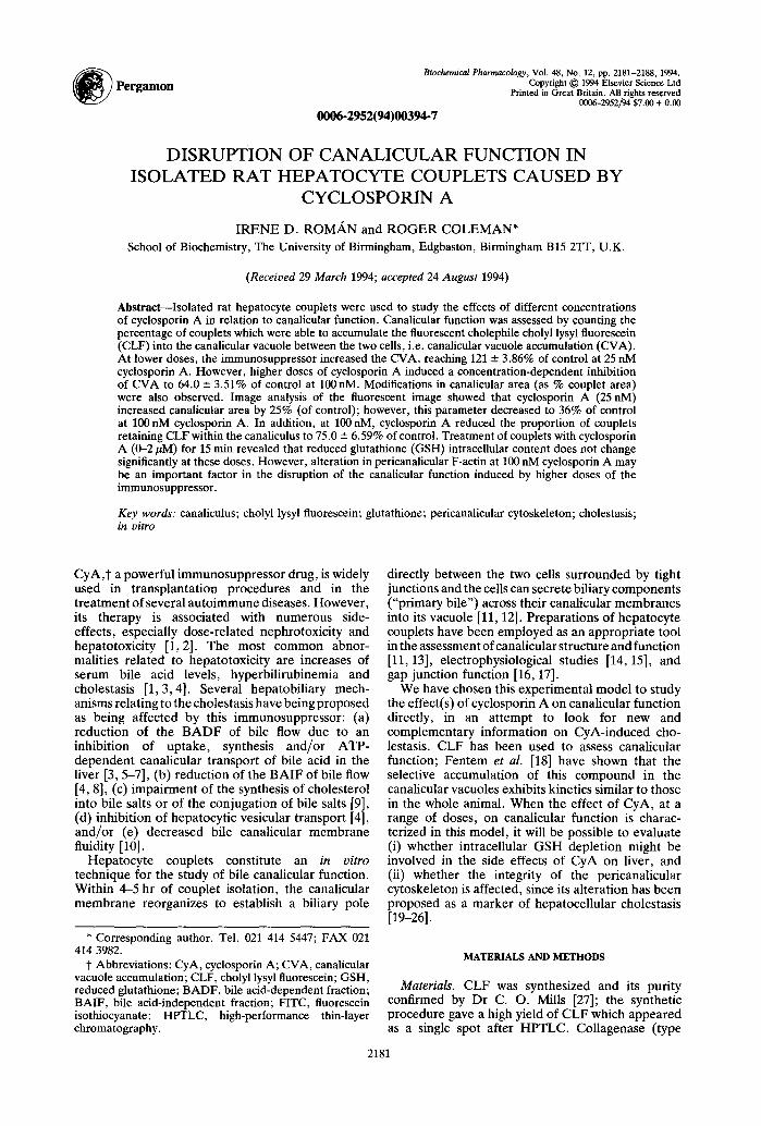
Fig. 1. Couplet canalicular vacuole accumulation (CVA) of CLF during simultaneous treatment with CyA (0- 100nM) for 15 min. Each value is the mean +-SEM
(N = 9).
A) was obtained from Clostridium hystolyticum (Boehringer Mannheim, Lewes, Sussex, U.K.). Cyclosporin A, in powder form (Sandoz A.G., Basel, Switzerland), and Leibovitz-15 (L-15) tissue culture medium (Gibco, Paisley, U.K.) were used. Bovine serum albumin (fraction V) was purchased from Winlab (Maidenhead, Berkshire, U.K.). Other fine chemicals were obtained from Sigma (Poole, Dorset, U.K.) or BDH (Poole, Dorset, U.K.).
The elutriation buffer consisting of Krebs- Henseleit buffer (pH 7.4), containing 0.1 g/100 mL BSA, 0 .1g/100mL glucose, gassed with 95% oxygen/5% carbon dioxide, was kept at room temperature.
Animals. Male Wistar rats (230-240 g), bred in the University of Birmingham, were allowed free access to food (standard laboratory chow: 41B maintenance diet, Pilsbury, Birmingham, U.K.) and tap water ad libitum before surgery, which was performed between 8 a.m. and 9 a.m. The rats were housed in an environment of constant temperature and humidity with alternating 12 hr light and dark cycles. Anaesthesia was achieved using Sagatal (pentobarbitone 6 mg/100 g body weight, i.p., RMB Animal Health Ltd, Dagenham, U.K.).
Preparation of hepatocyte couplets. Rat hepatocyte couplets were isolated according to a method adapted from Gautam et al. [28], involving an incomplete digestion of liver by limited collagenase perfusion in situ, which was modified by reducing the collagenase perfusion to 0.03% (w/v) collagenase for 4.5 min [291.
Tissue remaining from the initial digest was reincubated in collagenase solution at 37 ° for 7 min to yield a second population of cells with a high viability used for all experiments; after filtration of
these cells through 150/an nylon gauze, they were washed three times with L-15 [29].
Centrifugal elutriation. To perform image analysis of canalicular size and for the analyses of GSH content, couplets were enriched (80.2%---2.5 couplet frequency) by centrifugal elutriation using a standard elutriation chamber in a JE-6B elutriation rotor (Beckman RIIC Ltd, High Wycombe, U.K.) incorporated to a Beckman J6-ME centrifuge [29].
Cell counting and cell viability. Quantification of hepatocyte preparations was expressed in terms of units using an improved Neubauer hemocytometer. A unit could consist of a singlet, couplet, triplet or larger multiple [29]. Their viability (97.6% +-- 0.30) was estimated by Trypan blue exclusion [30]. If any cell within the unit failed to exclude Trypan blue, the whole unit was considered dead.
Culture and treatment of hepatocyte couplets. Hepatocytes were incubated at densities of 1 x 10⁵
5 units/2 mL and 4 × 10 units/2 mL of L-15 medium to assess the canalicular activity and to perform biochemical analysis, respectively. The cells were incubated in plastic tissue culture dishes and maintained at 37 ° in an air atmosphere for 4.5 hr.
Cyclosporin A was added as 10/tL doses dissolved in DMSO (at the final concentration in culture dish of 0.5%) to give 0 nM, 5 nM, 25 nM, 50 nM, 75 nM and 100 nM final concentrations in the culture dishes. GSH analyses were performed with the above concentrations, and in addition, with 200nM, 500 nM, 1000 nM and 2000 nM final doses of CyA in the culture dishes. In all experimental groups, each CyA-treated dish was always accompanied by its own control.
Although in hepatocyte studies DMSO is very frequently used by many authors working on CyA [31-35] or on other compounds [26, 36, 37], we have carried out controls with DMSO in each experimental group, which have demonstrated that the solvent does not affect the various experimental parameters measured (data not shown).
Assessment of canalicular activity. Counting of couplets able to undergo CVA of CLF, expressed as a percentage of control couplets exhibiting this phenomenon, has been used to assess canalicular function. CLF (5/~M final concentration) was added to each plate and incubated at 37 ° for 15 min before washing twice with 2 mL of L-15 and observing. Cyclosporin A was incorporated and incubated for 15 min at 37 ° either simultaneous with, or subsequent to, CLF addition. Cultures were examined using an inverted fluorescence microscope (Olympus IMT2- RFL) equipped with a 100 W mercury light source and an incubator to maintain the cells at 37 ° .
We have chosen a 15 min incubation period in accumulation and retention studies in couplets, since (i) CLF is rapidly taken into the canalicular vesicles within 10-15 min [18, 29, 38], and (ii) CyA uptake was found to be concentration independent, reaching an apparent steady state within 5 min [31].
In order to computate the fluorescent images, an image analysis system (Applied Imaging, Sunderland, U.K.) was used to measure canalicular area and breadth. Light microscopy was employed to assess blebbing of the cell membrane [39].
Study of phalloidin-FITC stained actin with

Cyclosporin A disrupts canalicular function
Table 1. Effect of CyA treatment (15 min) on couplet canalicular area (% of couplet area)
Concentration of CyA Canalicular area
(nM) (% couplet area)
0 4.28 -+ 0.21 25 5.35 -+ 0.38
100 2.76 -+ 0.48
Values are expressed as mean % control -+ SEM (N = 13 animals in each group).
2183
Table 2. Effect of CyA treatment (15 min) on plasma membrane blebbing (% of control)
Concentration of CyA Plasma membrane blebbing
(nM) (% of control)
5 107 -+ 10.9 75 127 - 10.3
100 148 -+ 5.23
Values are expressed as mean % control -+ SEM (N = 6--10).
confocal microscopy. After plating on to glass coverslips, hepatocyte couplets were incubated in L- 15 at 37 ° for 4.5 hr. Cells were then (i) treated with CyA (100 nM) for 15 min, (ii) fixed with 3% formalin in PBS, (iii) stored at 4 ° until permeabilized with 0.1% Triton X-100 in PBS, and (iv) labelled with phal loidin-FITC according to the method of Knutton et al. [40]. Coverslips were inverted on to citifluor mounting solution, to observe the stained cells.
To obtain xy images of the z-axis through the specimen, we used a Bio Rad 500 confocal laser scanning system attached to a Leitz SM-LUX microscope, as described by Stone et al. [37].
Analysis of GSH. GSH was determined flu- orometrically as described by Hissin and Hilf [41]. Its fluorescence was quantified using a Perkin Elmer Luminescence Spectrometer (LS 50B; Buck- inghamshire, U.K.) .
Statistical analysis. Results are expressed as mean -+ SEM. Analysis was performed using Student's t-test.
RESULTS
Effect of CyA on uptake of CLF Cyclosporin A-induced cholestasis and hyper-
bilirubinaemia has been often related to an inhibition of the hepatic uptake of bile acids and bilirubin [3, 35, 42--44]. At 100 nM CyA, CLF did not appear to have been taken up into the cytoplasm, suggesting that, at this concentration, CyA may inhibit the bile acid uptake mechanism in the sinusoidal membrane of liver cells in rat.
Effect of CyA on canalicular vacuole accumulation of CLF
Concurrent incubation of CyA and CLF for 15 min shows that, at low doses, the immunosuppressor increased the CVA reaching 121 + 3.86% of control at 25 nM cyclosporin A. However, higher doses of cyclosporin A induced a concentration-dependent inhibition of CVA to 64.0-+ 3.51% of control at 100 nM (Fig. 1).
In addition, we also could observe modifications in the canalicular area (as % of couplet area). Image analysis of the fluorescent images showed that cyclosporin A (25 nM) increased the canalicular area by 25% (of control); however, this parameter decreased to 36% of control at 100 nM CyA (Table 1). Other authors have described previously the very
BP 48:12-C
.-, 140 I
120
g
,.~ Ioo
.~ 80
60 I t I . I
25 50 75 100
C o n c e n t r a t i o n o f c y c l o s p o r i n A (nM) Fig, 2. Retention of CLF (15min preincubation and washing) within the couplet canalicular vacuole after subsequent treatment with CyA (0-100 nM) for 15 min.
Each value is the mean - SEM (N = 7).
close link between the two physiological functions: (i) secretion, and (ii) contraction of the bile canaliculus. Gautam et al. [28] found, during acute bile acid choleresis, a correlation between the flow rate and the canalicular surface area, being the expansion of the canalicular space as the result of canalicular stretching. Watanabe and Phillips [45] showed that after phalloidin treatment the canalicular lumen did not widen progressively, which may reflect that the decrease in bile secretion had induced a reduction in canalicular area (as % of couplet area).
Plasma membrane blebbing increased to above control at 75 nM and 100 nM CyA (Table 2), showing a parallelism between alteration of canalicular accumulation and appearance of plasma membrane blebbing. This effect was insignificant at lower doses. Bleb formation appears to be a prominent feature of most forms of liver injury, regardless of origin, and has already been described previously in cholestatic hepatic injury [46].
Effect of CyA on the retention of accumulated CLF After incubation of couplets with CLF for 15 min,
2184 I. D. ROMAN and R. COLEMAN
Table 3. Couplet GSH intracellular content after treatment with cyclosporin A (0-2 #M) for 15 min
Concentration GSH intracellular content of CyA (/zmol/million units)
0 nM 1.76 -!-- 0.12 5 nM 1.73 -+ 0.21
25 nM 1.71 -+ 0.18 50 nM 1.89 -+ 0.16 75 nM 1.95 -+ 0.19
100 nM 1.69 +-- 0.16 200 nM 1.91 --- 0.10 500 nM 1.73 --- 0.21
1/zM 1.78 -+ 0.20 2/~M 1.70 -+ 0.19
Values are given as means-+ SEM for six animals in each group.
washing twice with 2 m L of L-15 and following treatment with CyA (100 nM) for another 15 min, the proportion of couplets retaining CLF within the canaliculus was reduced by the drug to 75.0 -+ 6.59% of control (Fig. 2). However, use of this experimental method cannot establish a direct relationship between tight junction function after CyA treatment and loss of CLF retention, as we have ignored the route by which CLF has left the canaliculus, although alterations in tight junctional integrity could contribute in part or totally to this phenomenon.
Recovery of canalicular secretory function Removal of CyA (75 nM and 100 nM) by washing
twice with 2 mL of fresh L-15 medium (37 °) and addition of CLF for 15 rain prior to observation indicated that only the couplets treated with 75 nM CyA were able to recover their CVA (to 96.8 --- 17.0% of control) within 3 hr of drug removal; in addition, plasma membrane blebbing returned to control levels 90 rain and 120 min after treatment with these concentrations of CyA (75nM and 100 nM, respectively). This is in agreement with the previously described transitory cholestatic effect for CyA [4, 42] and with the observation that the restoration of plasma membrane integrity preceded the improved canalicular function.
The effect of CyA on couplet GSH content Treatment of couplets with CyA (0-100 nM) for
15 min revealed that the GSH intracellular content does not change significantly at these doses; even when the concentration of CyA was increased to 2/~M, no significant differences in total GSH content were observed (Table 3).
Pericanalicular actin disposition after CyA treatment
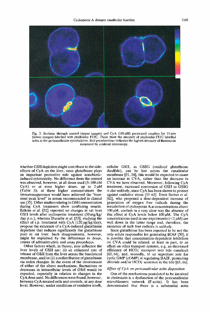
Phalloidin-FITC labelling of fixed cells allows visualization of the F-actin cytoskeleton [26]. Image analysis of the confocal images permits the characterization of the distribution of the peri- canalicular cytoskeleton, by quantitation of fluor- escence intensity. F-actin distribution was assessed for CyA treatment (100nM) of 20 couplets from
each of five separate isolations and compared with untreated couplets (Fig. 3).
After CyA treatment, the pericanalicular cyto- skeletal fluorescence/gin 2 was reduced significantly by 23.3% of the control (P < 0.05).
DISCUSSION
Concentration range of CyA effect Since the use of CyA is associated with side-effects
that are dose related in humans and nearly always improve after reduction of its dose, we have chosen to work in hepatocyte couplets at 5-100 nM CyA because most of these doses are included in the recommended "trough level" of the immuno- suppressor in serum for clinical use [35]. Our findings show that CyA affects the ability of couplets to accumulate CLF in the canalicular vacuole: lower doses of CyA favour CVA, whereas higher ones inhibit it in a dose-dependent manner (Fig. 1). These results are consistent with those described by others both in in vivo and in vitro experimental models in the rat [3, 47]. However, the CyA doses in the present study that inhibit CVA are lower than those cholestatic doses found in the literature [3, 4, 7, 8, 42, 44]. This observation seems to be a feature of the experimental model, since Stone et al. [37] have reported a similar relative sensitivity in couplets in the case of menadione.
The most sensitive hepatic in vitro systems for the inhibitory effect of CyA, so far described, are (i) that of inhibition of the canalicular ATP-dependent bile acid transporter, in human hepatic plasma membrane vesicles, with a Ki of 200 nM for CyA [34], and (ii) that of inhibition of the multidrug efflux pump, in rat couplets, being its half-maximal inhibitory concentration 0.6/~M [35]. The higher doses of CyA used in the present experiments are, however, below all of these and whilst inhibition in these systems may make a contribution at higher concentrations of CyA, at the concentrations used in the present experiments < 100 nM, a further more sensitive system must be affected prior to, or accompanying, these other systems.
Role of glutathione The concept of BAIF was introduced to explain
the observation that a significant portion of canalicular bile production was independent of bile acid excretion [48, 49]. Recent studies provide direct evidence for glutathione as one of the solutes responsible for BAIF formation. Ballatory and Truong [50] found a significant positive correlation between increased BAIF and increased glutathione excretion. This tripeptide, secreted into canalicular bile at relatively high concentrations via a specific carrier-mediated mechanism [50], constitutes an osmotic driving force for the secretion of water and electrolytes into the canaliculus [51].
Impairment of bile secretion in CyA-treated rats has been suggested to be related to a reduction in both BADF and BAIF [4, 8]. We have evaluated the intracefiular content of GSH after CyA treatment: (i) to determine the role that GSH can play in the concentration-dependent inhibition of CyA on CLF eanalicular vacuole accumulation, and (ii) to evaluate
Cyclosporin A disrupts canalicular function 2185
Fig. 3. Sections through control (upper images) and CyA (100 nM) pretreated couplets for 15 min (lower images) labelled with phalloidin-FITC. These show the intensity of phalloidin-FITC labelled actin at the pericanalicular cytoskeleton. Red pseudocolour indicates the highest intensity of fluorescein
measured by confocal microscopy.
whether GSH depletion might contribute to the side- effects of CyA on the liver, since glutathione plays an important protective role against xenobiotic- induced cytotoxicity. No difference from the control was observed, however, at all doses used (5-100 nM CyA) or at even higher doses, up to 2/tM (Table 3); at these higher concentrations the immunosuppressor would have achieved the "tran- sient peak level" in serum recommended in clinical use [35]. Other studies relating to GSH concentration during CyA treatment show conflicting results. Edkins et al. [52] reported no changes in rat liver GSH levels after cyclosporin treatment (50 mg/kg/ day p.o.), whereas Duruibe et al. [53], studying the effect of i.p. treatment with CyA (120/~g/kg/day), propose the existence of a CyA-induced glutathione depletion that reduces significantly the glutathione pool in rat liver. Such disagreements, however, might be explained by the differences in doses, routes of administration and assay procedures.
Other factors which, in theory, may influence the liver levels of GSH are: (i) an enhanced rate of release of GSH from the liver across the basolateral membrane, and/or (ii) a redistribution of glutathione via redox changes. In the event of the involvement of either of the above mechanisms, fluctuations or decreases in intracellular levels of GSH would be expected, especially in relation to changes in the CyA dose used. No differences were found, however, between CyA-treated cells and controls, at any dose level. However, under conditions of oxidative streW's,
cellular GSH, as GSSG (oxidized glutathione disulfide), can be lost across the canalicular membrane [51, 54]; this would be expected to cause an increase in CVA, rather than the decrease in CVA we have observed. Moreover, following CyA treatment, increased conversion of GSH to GSSG is also unlikely, since CyA has been shown to protect against oxidative stress [55~1]. Even Serino et al. [62], who proposed a dose-dependent increase of generation of oxygen free radicals during the metabolism of cyclosporin A at concentrations above 100/tM, confirm in a very clear way the absence of this effect at CyA levels below 100/~M. The CyA concentrations used in our experiments (<2/tM) are well down in the latter range and, therefore, the existence of such free radicals is unlikely.
Since glutathione has been reported to be not the only solute responsible for generating BIAF [50], it is possible that concentration-dependent inhibition on CVA could be related, at least in part, to an effect on other transport systems, e.g. an decreased efficiency of HCO~ transport into bile canaliculi [63, 64], and, secondly, to an important role for cyclic GMP (cGMP) in regulating BAIF, promoting chloride and/or HCO~ secretion in the bile [65, 66].
Effect o f CyA on pericanalicular actin disposition
One of the mechanisms postulated to be involved in cholestasis is a dysfunction of the pericanalicular microfilament network (F-actin). It has been demonstrated that there is a substantial actin
2186 I. D. ROMA, N and R. COLEMAN
microfilament network around the bile canaliculus and that imicrotubules, ar ranged throughout the cytoplasm, are specially well organized around the pericanalicular sheath of cytokeratin [21-25, 67]. Pericanalicular F-actin abnormalities can lead to cholestasis b.y impairing canalicular contractions or, as in addition, by vesicular transport impairment or increased tight junction permeability [19, 20, 24,26]. Thibault et al. [26] showed that cholestatic and hepatotoXlc, b u t ' n o t non-cholestatic, compounds affected F-actin distribution in couplets, probably as a consequence o f an inhibition of F-ac t in depolymerization or of some higher organization of actin (redistribution, bundling or reorientation). All ~ of these observations have led to the concept that pericanalicular F-actin alteration appears to be a specific marker of hepatocellular cholestasis [26], resulting in impaired canalicular accumulation a n d contractility, proposed to be necessary f o r bile propulsion within the biliary tree [68].
Confocal microscopy and image analysis showed that Fza~tin labelling (with phalloidin-FITC) was always higher: in ~ the pericanalicular cyt0skeleton than in the remainder of the couplet (Fig. 3) /Pr ior treatment of the couplets with CyA (100 nM) caused a significant reduction in this pericanalicular fluorescence. This alteration, indicative of F-actin disruption, may explain the negative effect of CyA on the accumulation and/or retention of CLF within the canaliculus (Figs 1 and 2). In addition, the observation that preaccumulated CLF within the canaliculus can be released by subsequent exposure to higher CyA doses (Fig. 2), when the processes of sinusoidal uptake, transcellular movement and canalicular secretion had already occurred, can be related to increasing tight junction permeability. Such change would dissipate the osmotic gradient needed for both BAIF and B A D F [20, 69].
In summary, our data show a reduction in canalicular vacuole accumulation and retention of CLF, at low concentrations of CyA (<100 nM), in a dose-dependent fashion. At these concentrations, we observe essentially no changes in couplet GSH intracellular content. However, the disruption observed on canalicular function in CyA-treated cells (100 nM) may explain these phenomena by impairing canalicular contractions and/or increasing tight junction permeability. This finding can offer a new perspective on the evaluation and study of CyA- induced cholestatic syndrome, since, independent of effects of CyA on cellular uptake and transport processes, the canalicular membrane constitutes the rate-limiting step of the overall secretory process. Moreover, the high sensitivity of these concentration- dependent effects, and the good correlation existing between isolated rat hepatocyte couplets and other experimental models in the study of cyclosporin A, make the couplet model a good potential tool for the study of hepatoprotective agents against the side- effects related to cyclosporin A therapy.
Acknowledgements--This work has been supported by E1 Ministerio Espafiol de Educaci6n y Ciencia and The Wellcome Trust. The scientific suggestions of Dr R. Jim6nez (Departamento de Fsiologia y Farmacologia, Universidad de Salamanca, Spain) are greatly acknowl-
edged. We thank Dr G. Johnson, Department of Immunology for the confocal microscopy. We also thank Dr J. Wilton and V. Stone, D. Lankester and M. Guppy for their support.
~ REFERENCES
1. Kahan BB, Cyclosporine. N Engl J Med 321: 1725- 1738, 1989.
2, Wiesner RH, Ludwig J, Lindor KD, Jorgensen RA and Baldus WP, A controlled trial of cyclosporine in the treatment of primary bil'iary cirrhosis. N Engl J Med 20: 1419-1424, 1990.
3. Rotolo FS, Branum GD, Bowers BA and Meyers WC, Effect of cyclosporine on bile secretion in rats. Am J Surg 151: 35--39, 1986.
4. Romfin ID, Monte MJ, Gonz~dez-Buitrago JM, Esteller A and Jim6nez R, Inhibition of hepatocytary vesicular transport by cyclosporin A in the rat: relationship with ch01estasis and hyperbilirubinemia. Hepatology 12: 83- 91, 1990.
5. Ziegler K and Frimmer M, Cyclosporine A and a diaziridine derivate inhibit the hepatocellular uptake of cholate, phalloidin and rifampicine. Biochim Biophys Acta 855: 136, 1986.
6. Ballantyne CM, Podet EJ, Putsch WP, Harati Y, Appel V, Gotto AM and Young JB, Effects of cyclosporine therapy on plasma iipoprotein levels. J Am Med Assoc 262: 53, 1989.
7. Boelsterli UA, Bouis P, Donatsch P, Effects of cyclosporin A on bile acid uptake, conjugation and release in cultured rat hepatocytes. In: Liver Cells and Drugs (Ed. Gillouzo A), p. 335. Colloque INSERM, John Libbey Eurotext, Paris, 1989.
8. Stone BG, Udani M, Sanghyi A, Warty V, Plocki K, Bedetti CD and Vanthiel DH, Cyclosporin A- induced cholestasis. The mechanism in a rat model. Gastroenterology 93: 344-351, 1987.
9. Chanussot F, Botta-Fridlund D, Lechene de La Porte P, Sbarra V, Portugal H, Pauli AM, Hauton J, Gauthier A and Lafont H, Effects of cyclosporine and corticoids on bile secretion in the rat. Transplantation 54: 226- 231, 1992.
10. Whitington PF, Dudeja P, Hecht JR, Whitington SH and Brasitus TA. Lipid alterations in the hepatocyte basolateral sinusoidal membrane (BLM) domain may explain cyclosporine (CsA) induced cholestasis in the rat (Abstract). Hepatology 8: 1363, 1988.
11. Gautam A, Ng O-C and Boyer JL, Isolated rat hepatocyte couplets in short-term culture: Structural characteristics and plasma membrane reorganisation. Hepatology 7: 216-223, 1987.
12. Graf J and Boyer JL, The use of isolated rat hepatocyte couplets in hepatobiliary physiology. J Hepatol 10: 387-394, 1990.
13. Weinman SA, Graf J and Boyer JL, Voltage driven taurocholate dependent secretion in isolated hepatocyte couplets. Am J Physiol 256: G826-G832, 1989.
14. Graf J, Gautam A and Boyer JL, Isolated rat hepatocyte couplets: A primary secretary unit for electrophysiologicai studies of bile secretory function. Proc Natl Acad Sci USA 81: 6516-6520, 1984.
15. Henderson RM, GrafJ and Boyer JL, Na÷-H + exchange regulates intraceUular pH in isolated rat hepatocyte couplets. Am J Physiol 252: G109-G113, 1987.
16. Spray DC, Ginzberg RD, Morales EA, Gatmaitan Z and Arias IM, Electrophysiological properties of gap junctions between associated pairs of rat hepatocytes. J Cell Biol 103: 135-144, 1986.
17. Revendin EC and Weingart R, Electrical properties of the gap junctional membrane studied in rat liver cell pairs. Am J Physiol 254: C226-C234, 1988.
18. Fentem JH, Foster B, Mills CO, Coleman R

Cyclosporin A disrupts canalicular function 2187
and Chipman JK, Biliary excretion of fluorescent cholephiles in hepatic couplets: an in vitro model for hepatobiliary and hepatotoxicity studies. Toxicol In Vitro 4: 452-457, 1990.
19. Dubin M, Maurice M, Feldman G and Erlinger S, Phalloidin-induced cholestasis in the rat: Relation to changes in microfilaments. Gastroenterology 75: 450- 455, 1978.
20. Elias E, Hruban Z, Wade JB and Boyer JL, Phalloidin- induced cholestasis: A microfilament-mediated change in junctional complex permeability. Proc Natl Acad Sci USA 77: 2229-2233, 1980.
21. Low RB, Low ES, Chaponnier C, Mitchell JW and Gabbiani G, Effect of phalloidin on liver actin distribution, content and turnover. J Cell Biochem 20: 393--407, 1982.
22. Phillips M J, Oshio C, Miyiari M and Smith CR, Intrahepatic cholestasis as a canalicular motility disorder. Evidence using cytochalasin. Lab Invest 48: 205-211, 1983.
23. Phillips MJ, Oshio C, Miyiari M, Watanabe S and Smith CR, What is actin doing in liver cells? Hepatology 3: 433-436, 1983.
24. Watanabe S, Miyiari M, Oshio C, Smith CR and Phillips MJ, Phalloidin alters bile canalicular contractibility in primary monolayer cultures of rat liver. Gastroenterology 85: 245-253, 1983.
25. Nickola I and Frimmer M, Effects of phalloidin and cytochalasin B on cytoskeletal structures in cultured rat hepatocytes. Cell Tissue Res 245: 635-641, 1986.
26. Thibault N, Claude JR and Ballet F, Actin filament alteration as a potential marker for cholestasis: a study in isolated rat hepatocyte couplets. Toxicology 73: 269- 279, 1992.
27. Mills CO, Rahman K, Coleman R and Elias E, Cholyllysyl fluorescein synthesis, biliary excretion in vivo and during single pass perfusion in isolated rat liver. Biochem Biophys Acta 1115: 151-156, 1991.
28. Gautam A, Ng OC, Strazzabosco M and Boyer JL, Quantitative assessment of canalicular bile formation in isolated hepatocyte couplets using microscopic optical planimetry. J Clin Invest 83: 565-573, 1989.
29. Wilton J, Williams DE, Strain AJ, Parslow RA, Chipman JK and Coleman R, Purification of hepatocyte couplets by centrifugal elutriation. Hepatology 14: 180- 183, 1991.
30. Moldeus P, Hogberg J and Orrenius S, Isolation and use of liver cells. Methods Enzymol 52: 60-71, 1978.
31. Prueksaritanont T, Koike M, Hoener BA and Benet LZ, Transport and metabolism of cyclosporine in isolated rat hepatocytes. Biochem Pharmaco143: 1997- 2006, 1992.
32. Azer SA and Stacey NH, Differential effects of cyclosporin A on the transport of bile acids by human hepatocytes. Biochern Pharrnacol 46: 813-819, 1993.
33. Henke W and Jung K, Comparison of the effects of the immunosuppressive agents FK 506 and cyclosporin A on rat kidney mitochondria. Biochem Pharmacol 46: 829-832, 1993.
34. Kadmon M, Kliinemann C, B6hme M, Ishikawa T, Gorgas K, Otto G, Herfarth C and Keppler D, Inhibition by cyclosporin A of the adenosine triphosphate-dependent transport from the hepatocyte into bile. Gastroenterology 104: 1507-1514, 1993.
35. Takeguchi N, Ichimura K, Koike M, Matsui W, Kasbiwagura T and Kawahara K, Inhibition of the multidrug efflux pump in isolated hepatocyte couplets by immunosuppressants FK506 and cyclosporine. Transplantation 55: 646--650, 1993.
36. Thibault N, Maurice M, Maratrat M, Cordier A, Feldmann G and Ballet F, Effect of taurourso- deoxycholate on actin filament alteration induced by
cholestatic agents. A study in isolated rat hepatocyte couplets. J Hepatol 19: 367-376, 1993.
37. Stone V, Johnson GD, Wilton JC, Coleman R and Chipman JK, Effect of oxidative stress and disruption of Ca 2÷ homeostasis on hepatocyte canalicular function in vitro. Biochem Pharmacol 47: 6254532, 1994.
38. E1-Seaidy AZ, Mills CO, Elias E and Crawford JM, Intracellular distribution of fluorescent bile salts: studies with confocal microscopy (AASLD Abstract). Hepatology 18: 136A, 1993.
39. Nicotera P, Hartzell P, Davis G and Orrenius S, The formation of plasma membrane blebs in hepatocytes exposed to agents that increase cytosolic Ca 2÷ is mediated by the activation of a non-lysosomal proteolytic system. FEBS Lett 209: 139-144, 1986.
40. Knutton S, Baldwin TJ, Williams PH and McNeish AS, Actin accumulation at sites of vacterial adhesion to tissue culture cells: Basis of a new diagnostic test for enteropathogenic and enterohemorrhagic Escherichia coli. Infect Immuno157: 1290-1298, 1989.
41. Hissin PJ and Hilf R, A fluometric method for determination of oxidised and reduced glutathione in tissues. Anal Biochern 74: 214-225, 1976.
42. Kenny RP, Piccoli DA, Mailer ES, Vanderslice RR, Shaw LM and Watkins JB, Cyclosporine inhibits bile acid uptake and secretion in the isolated perfused rat liver. Gastroenterology 90: 1738, 1986.
43. Kukonquiriyapan U and Stacey NH, Inhibition of taurocholate transport by cyclosporin A in cultured rat hepatocytes. J Pharmacol Exp Ther 247: 6854589, 1988.
44. Moseley RH, Johnson TR and Morrissette JM, Inhibition of bile acid transport by cyclosporine A in rat liver plasma membrane vesicles. J Pharmacol Exp Ther 253: 974-980, 1990.
45. Watanabe S and Phillips J, Acute phalloidin toxicity in living hepatocytes. A m J Pathol 122: 101-111, 1986.
46. Vital A, Bioulac-Sage P, Bedin C and Balabaud C, Bile canaliculi morphology after bile duct ligation in the rat with portacaval shunt: a time-course study. Liver 2: 230-235, 1982.
47. Princen HMG, Meijer P, Hofstee B, Havekes LM, Kuipers F and Vonk RJ, Effects of cyclosporin A (CsA) on LDL-receptor activity and bile acid synthesis in hepatocyte monolayer cultures and in vivo in rat (Abstract). Hepatology 7: 1109, 1987.
48. Forker EL, Two sites of bile formation as determined by mannitol and erythritol clearance in the guinea pig. J Clin Invest 46: 1189-1195, 1967.
49. Erlinger S, Dhumeaux D, Berthelot P and Dumont M, Effect of inhibitors of sodium transport on bile formation in the rabbit. A m J Physiol 219: 416--422, 1970.
50. Ballatori N and Truong T, Relation between biliary glutathione excretion and bile acid-independent bile flow. Am J Physiol 256: G22-G30, 1989.
51. Ballatori N and Truong T, Glutathione as a primary osmotic driving force in hepatic bile formation. A m J Physiol 263: G617-G624, 1992.
52. Edkins'RD, Rybak LP and Hoffman DW, Comparison of cyclosporine and FK506 effects on glutathione levels in rat cochlea, brain, liver and kidney. Biochem Pharmacol 43: 911-913, 1992.
53. Duruibe VA, Okonmah A and Blyden GT, Effect of cyclosporin on rat liver and kidney glutathione content. Pharmacology 39: 205-212, 1989.
54. Chang M, Shi M and Forman H J, Exogenous glutathione protects endothelial cells from menadione toxicity. A m J Physiol (Lung Cell Mol Physiol) 6: L637-L643, 1992.
55. Nazareth W, Yafei N and Crompton M, Inhibition of anoxia-induced injury in heart myocytes by cyclosporin A. J Mol Cell Cardiol 23: 1351-1354, 1991.

2188 I. D. ROMAN and R. COLEMAN
56. Schlegel J, Meier P, Kass GEN and Richter C, Inhibition by cyclosporine A of the prooxidant-induced but not of the sodium-induced calcium release from rat kidney mitochondria. Biochem Pharmaco142: 2193- 2197, 1991.
57. Broekemeier KM, Carpenter-Deyo L, Reed DJ and Pfeiffer R, Cyclosporin A protects hepatocytes subjected to high Ca 2+ and oxidative stress. FEBS Lett 304: 192-194, 1992.
58. Crompton M, McGuinness O and Nazareth W, The involvement of cyclosporin A binding proteins in regulating and uncoupling mitochondrial energy transduction. Biochim Biophys Acta 1101: 214-217,
• 1992. 59. Kass GEN, Juedes MJ and Orrenius S, Cyclosporin A
protects hepatocytes against prooxidant-induced cell killing. Biochern Pharmacol 44: 1995-2003, 1992.
60. Katz A, Coran AG, Oldham KT and Guice KS, Decreased oxidized glutathione with aerosolized cyclosporine delivery. J Surg Res 54: 597-602, 1993.
61. Reichman N, Porteous CM and Murphy MP, Cyclosporin A blocks 6-hydroxydopamine-induced efflux of Ca 2+ from mitochondria without inactivating the mitochondrial inner-membrane pore. Biochem J 297: 151-155, 1994.
62. Serino F, Grevel J, Napoli KL, Kahan BD and Strobel HW, Generation of oxygen free radicals during the metabolism of cyclosporine A: a cause-effect
relationship with metabolism inhibition. Mol Cell Biochem 122: 101-112, 1993.
63. Hardison WGM and Wood GA, Importance of bicarbonate in bile salt independent fraction on bile flow. A m J Physiol 235: E158-E164, 1978.
64. Nathanson MH and Boyer JL, Mechanisms and regulation of bile secretion. Hepatology 14: 551-566, 1991.
65. Myers NC, Griine S and Anwer MS, Cyclic GMP stimulates bile acid independent bile formation (AASLD Abstract). Hepatology 18: 135A, 1993.
66. St-Pierre MV, Dufour JF and Arias IM, Cyclic GMP modulates bile acid independent bile flow (AASLD Abstract). Hepatology 18: 135A, 1993.
67. Prentki M, Chaponnier C, Jeanrenaud B and Gabbiani G, Actin microfilaments, cell shape and secretory processes in isolated rat hepatocytes. J Cell Biol 81: 592-607, 1979.
68. Watanabe N, Tsukada N, Smith CR, Edwards V and Phillips M J, Permeabilised hepatocyte couplets. Adenosine triphosphate-dependent bile canalicular contractions and a circumferential pericanalicular microfilament belt demonstrated. Lab Invest 65: 203- 213, 1991.
69. Duc Vu D, Tuchweber B, Raymond P and Yousef I, Tight junction permeability and liver plasma membrane fluidity in lithocholate-induced cholestasis. Exp Mol Pathol 57: 47-61, 1992.