diseases of economic importance in small ruminants in … · diseases of economic importance in...
TRANSCRIPT

jJN~!JC.h1-;;l.7YDiseases of EconomicImportance in SmallRuminants in sub-Saharan Africa
ILRI Slide Series 3
H. IbrahimS. TembelyF. Roger
(.-~ InternatIonal Uvestock Research InstItute~ '?I P.o. Box 30709, NaIrobI, Kenya
I Ir
/
ISBN 92-914lHJ52-4
Correct citation: Ibtahim H., Tembely S. andRoger F. 1999. Diseases of &onomic Importance inSmall Ruminants in sub-Saharan Africa. ILRI SlideSeries 3. ILRI (International Livestock ResearchInstitute), Nairobi, Kenya. 44 pp.
I{

Table of ContentsPreface vParr A: Diagnosis ofdiseases 1ParrB: Posr-morremexaminarion 21Recommended reading •.................. 37
iii
PrefaceDiseases of small ruminants affect the incomes ofsmallholder farmers in sub-Saharan Africa by reducingproductivity or through loss of the animal. This slides,eries aims at making available to animal scientists in
national agricultural research systems (NARS) acollection of slides and accompanying text that will
~ help identify symptoms of the diseases. This seties isdivided into two parts. The symptoms of the diseasesare covered in part A while post-mortem examination
is covered in part B. The two parts complement eachother. In most cases the symptoms ofa disease. can onlybe identified through examining the internal organs of
a dead animal. In the absence of a veterinarian, it isimportant that livestock scientists perform post~
mortem examinations when animals die on theirresearch farms to ascertain the cause before anepid~micbreaks out.
This series is directed atyoung animal scientists withBSc or MSc degrees. It can also serve senior animalscientists by providing,slides for seminars or lectUres ineducational institutions. The series is made up of slidesand a booklet that contains pictures and text.
I would like to acknowledge the assistance of themanager ofAddis Ababa Abattoir for allowing ILRI tophotograph diseased organs of sheep and goats. I am
. also grateful to the management and staff of the ILRIResearch Station at Debre Birhan, Ethiopia. Iacknowledge Woizero Menbere W/Giorgis for thephotographic WOtk. I acknowledge Ms Anne MarieNyamu fot editing the text of the booklet and the staffin the ILRI Publications Section for design, layout andprinting.
Habib IbrahimTraining Materials Specialist
v
Part A: Diagnosis of diseasesThis is a slide set on IDiseases of EconomicImportance in Small Ruminants in sub-SaharanAfrica' which is part of a series produced by theInternational Livestock Research Institute (ILRI).
Diseases of small ruminants cause economic lossesto smallholder farmers. These diseases must beidentified correctly so farmers can take propercontrol measures. This slide series will enable youto:
• identify small ruminant diseases of economicimportance in sub-Saharan Africa
• conduct post-mortem examinations
• dispose of carcasses.
1
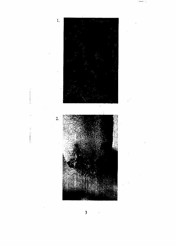
1. Disease: Contagious caprinepleuropneumonia (CCPP)Small ruminant, GoarLocation, Debre Zeit, EthiopiaYear, 1996Symptoms: Abundant and purulent nasaldischarge
2. Disease: Contagious caprinepleuropneumonia (CCPP)Small ruminant: GoatLocation: National Veterinary Institute,Debre Zeir, EthiopiaYear: 1996Symptoms: Artificially infected goatshowing purulent nasal discharge
2
1.
2.
3
3.
4.
5.
4
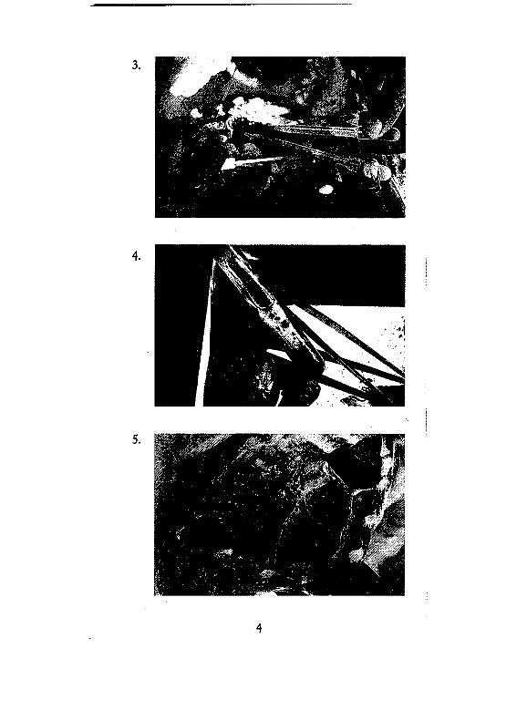
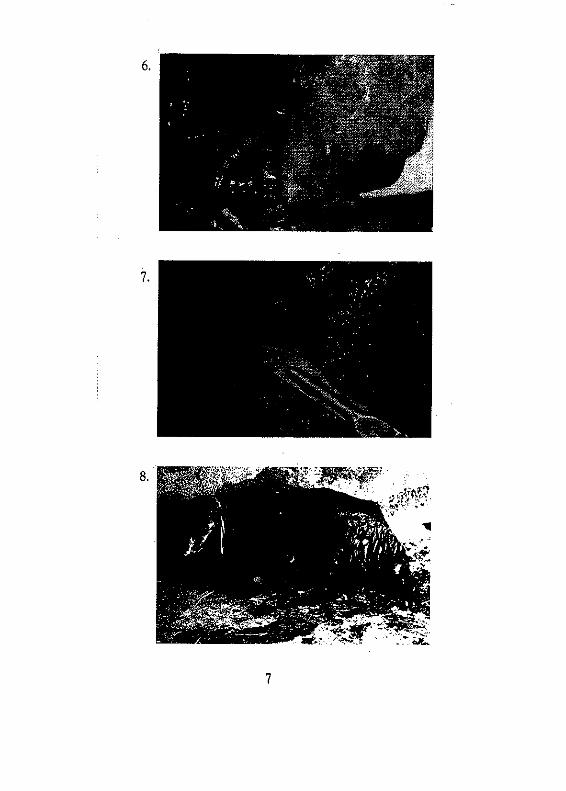
3. Disease: Contagiouscaprinepleuropneumonia (CCPP)Small ruminant: GoatLocation: Yabello (Borana), EthiopiaYear: 1995Symptoms: Straw-eoloured thoracic fluid(drawn in the pipette)
4. Disease: Contagious caprinepleuropneumonia (CCPP)Small ruminant: GoatLocation: National Vererinary Institute,Debre Zeit, Ethiopia·Year: 1995Symptoms: Straw-eoloured thoracic fluid(In the test tube) taken from artificiallyinfected goat
5. DiseaSe: Contagious caprinepleuropneumonia (CCPP)Small ruminant GoatLocation: Yabello (Borana), Ethiopia
. Year: 1995Symptoms: Pleuritic adhesions (chroniccase). Parts of the lungs stick to the thoraciccavity
5

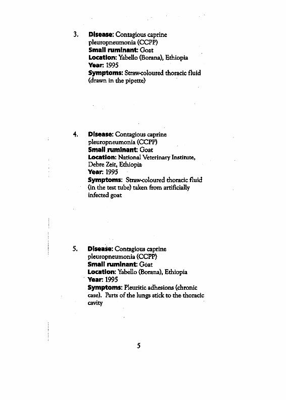
6. Disease: Contagious caprinepleuropneumonia (CCPP)Small ruminant: GoatLocation: Debre Zeit, EthiopiaYear: 1995Symptoms: Artificially infected goatshowing omelette of fibrine on the lung(yellow mass on the right)
7. Disease: Contagious caprinepleuropneumonia (CCPP)Smalr ruminant: GoatLocation: National Veterinary Institute,Debre Zeit, EthiopiaYear: 1996Sympt~ms: Lung hepatisation in anartificially infected goat. Section: granularspot, wine-coloured
8. Disease: Peste des petits ruminants (PPR)Small ruminantl SheepLocation: National Veterinary Institute,Debre Zeit, EthiopiaYear: 1995Symptoms: Peracute case, depressedanimal; haemorrhagic diarrhoea
6
6.
7.
8.
7
9.
10.
8

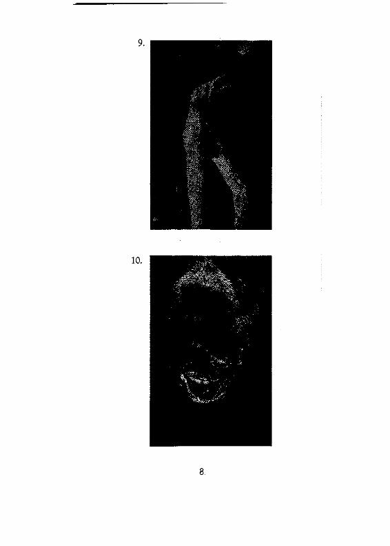
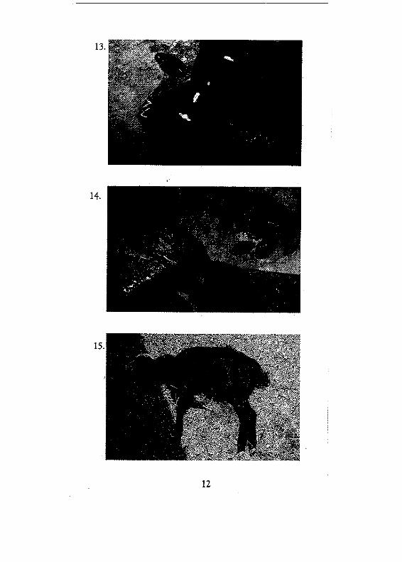
9. Disease: Peste des petits ruminants (PPR)Small ruminant: GoatLocation: Shoa Robit (North Shoal,EthiopiaYear: 1995Symptoms: Hemorrhagic diarrhoea
10. Disease: Peste des petits ruminants (PPR)Small ruminant: GoatLocation: Shoa Robit (North Shoal,EthiopiaYear: 1995Symptoms: White necrotic spots on thetongue
9
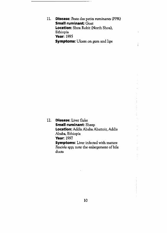
11. Disease: Peste des petits ruminants (PPR)Small ruminant: GoatLocation: Shoa Robit (North Shoal,ErhiopiaYear: 1995Symptoms: Ulcers on gum and lips
12. Disease: Liver flukeSmall ruminant: SheepLocation: Addis Ababa Abattoir, AddisAbaba, EthiopiaYear: 1997Symptoms: Liver infected with matureFasciola spp; note the enlargement of bileducts
10
11.
12.
11
13.
14.
12
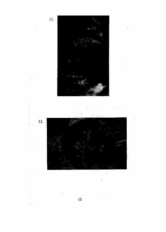
13. Disease: Liver flukeSmall ruminant: SheepLocation: Addis Ababa Abattoir, AddisAbaba, EthiopiaYear: 1997Symptoms: Enlarged gall bladder (notethe livet fluke extracted from the bile ductshown on the outside)
14. Disease: Liver flukeSmall ruminant: SheepLocation: Addis Ababa Abattoir, AddisAbaba, EthiopiaYear: 1997Symptoms: Liver flukes extracted from aninfected liver
15. Disease: HaemonchosisSmall ruminant: SheepLocation: ILRI Debre Birhan ResearchStation, Debre Birhan, EthiopiaYear: 1997Symptoms: A 3.month-old lambartificially infected with Haemonchuscontortus (a blood sucking parasite in sheep)showing weakness and emaciation
13
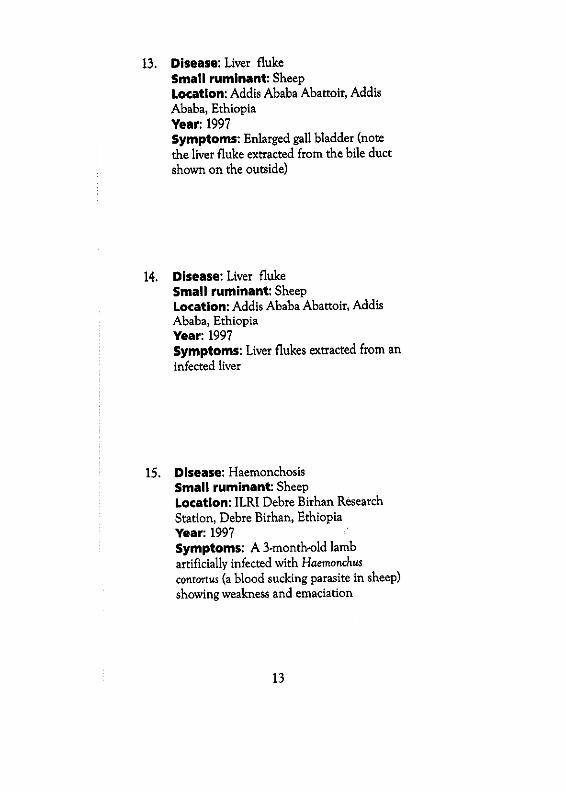
16. Disease: HaemonchosisSmall ruminant: SheepLocation: ILRI Debre Birhan ResearchStation, Debre Birhan, EthiopiaYear: 1997Symptoms: Open abomasum showingHaemonchus contortus
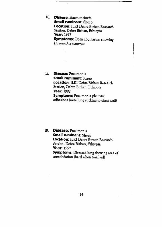
17. Disease: PneumoniaSmall ruminant: SheepLocation: ILRI Debre Birhan ResearchSrarion, Debre Birhan, ErhiopiaYear: 1997Symptoms: Pneumonia pleuriticadhesions (nore lung sricking to chesr wall)
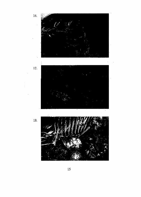
18. Disease: PneumoniaSmall ruminant: SheepLocatIon: ILRl Debre Birhan ResearchStation, Debre Birhan, EthiopiaYear: 1997Symptoms: Diseased lung showing area ofconsolidation (hard when rouched)
14
16.
17.
18.
15
19.
20.
21.
16
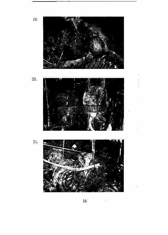
19. Disease: PneumoniaSmall ruminant: SheepLocation: lLRI Debre Birhan ResearchSration, Debre Birhan, EthiopiaYear: 1997Symptoms: Lung with hepatisation (hardand looks like liver in structure)
20. Disease: PneumoniaSmall ruminant: SheepLocation: ILRI Debre Birhan ResearchStation, Debre Birhan, EthiopiaYear: 1997Symptoms: Normal lung on the left,diseased lung on the right; size and colourof the two lungs are different; diseased lungis congested
21. Disease: Tapeworm (Moniezia expansa)Small ruminant: SheepLocation: ILRI Debre Birhan ResearchStation, Debre Birhan, EthiopiaYear: 1997Symptoms: Tapeworm in the smallintestine
17
18
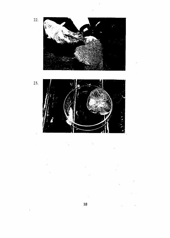
22. Disease: Rumen flukes (Paramphistoma spp)Small ruminant: SheepLocation: ILRI Debre Birhan ResearchStation, Debre Birhan, EthiopiaYear: 1997Symptoms: Rumen showing adult rumenflukes attached to the wall (red in colour)
23. Disease: Gid (coenurosis, staggers, sturdy)Small ruminant: SheepLocation: ILRI Debre Birhan ResearchStation, Debre Birhan, EthiopiaYear: 1997Symptoms: Cystic larva (Coenu1UScerebralis), the intermediate stage of the adultdog parasite Taenia multiceps
19
Part B: Post-mortem examinationWe have learned the symptoms of diseases ofsmall ruminants in part A. As we have seen,many of these symptoms ate visible in internalotgans. Animals die and it is important to knowthe cause. Knowing this infotmation helps us toptotect other animals. When you need todiagnose the cause of death of an animal, seek theassistance ofaveterinarian. A simple postmortem examination is helpful to detetmine thepossible cause of the death. Ifyou cannot find aveterinarian, you may perfotm the post-mortemexamination yourself. It is safe to perfQrtn postmortem examinations in the field buttake carenot to contaminate the area surrounding the post..mortem site. In case of obvious diseases that canbe transmitted from animals to humans, such asanthrax, the carcass should not be opened underany circumstance. The following are the stepsinvolved in perfotming a post-mortemexamination.
PREVIOUS PAGE BLANK
21
24.
25.
26.
22
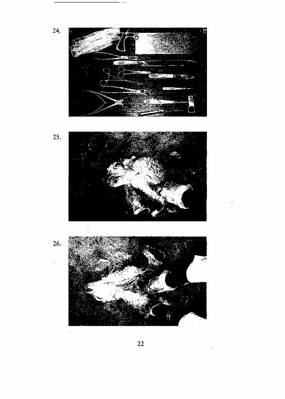
24. The basic equipment you need to perform apost-mortem includes knife, axe. saw, pair ofscissors, screw-eapped jars, rubber gloves andgum boots. You may need other materialsuch as formalin, water and disinfectant.
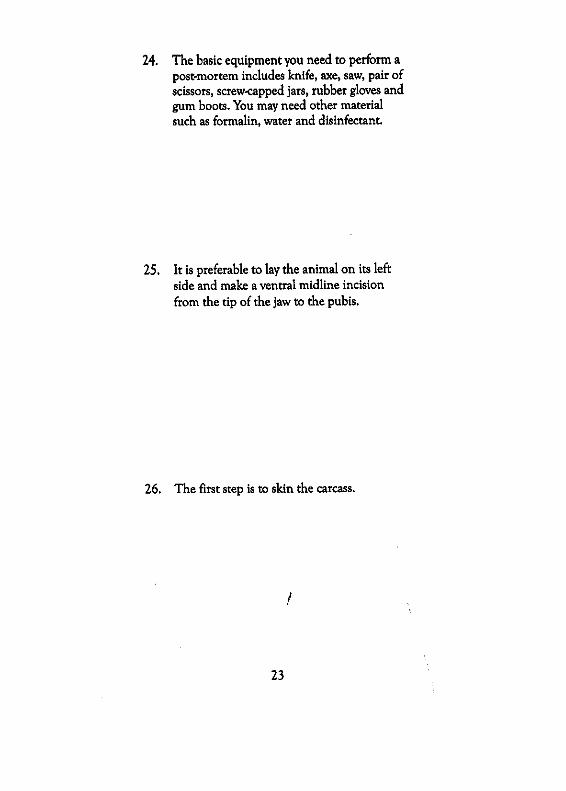
25. It is preferable to lay the animal on its leftside and make aventral midline incisionfrom the tip of the jaw to the pubis.
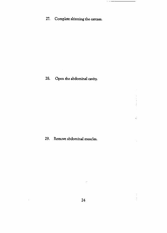
26. The first step is to skin the carcass.
I
23
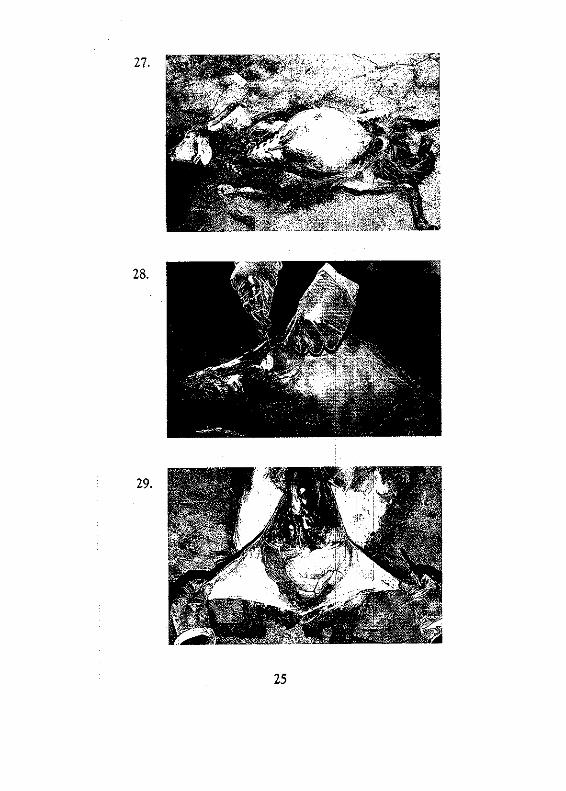
27. Complete skinning the catcass.
28. Open the abdominal cavity.
29. Remove abdominal muscles.
24
27.
28.
29.
25
30.
31.
32.
26
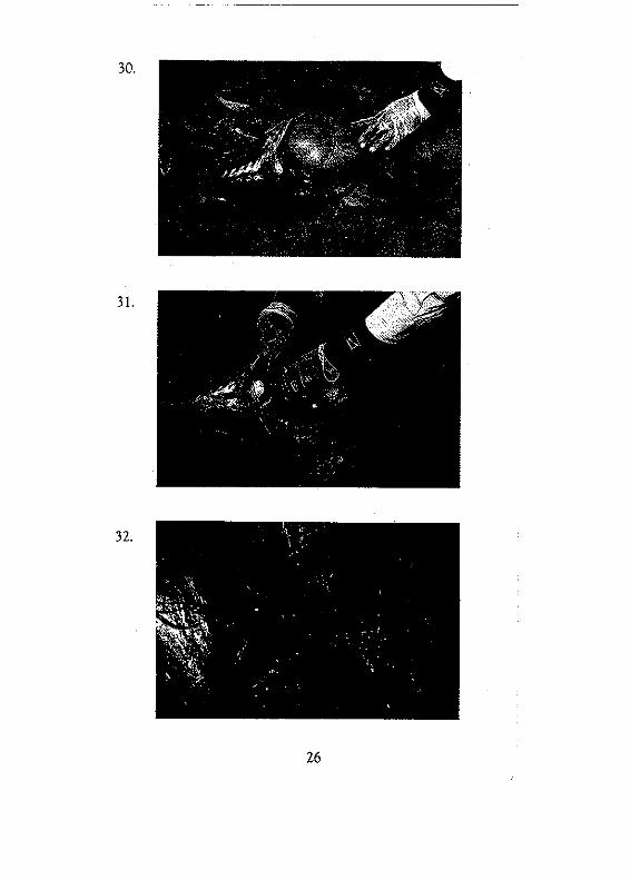
30. When you open the abdominal cavity. all ofthe viscera should be visible. and each pattmust be examined in detail.
31. Remove the otgans from the abdominalcavity.
32. Examine both kidneys in the abdominalcavity.
27
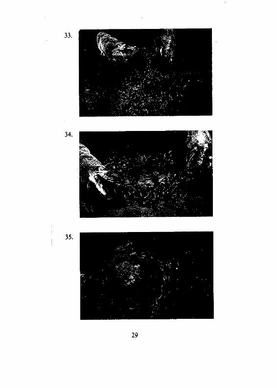
33. Tie up the first patt of intestine andseparate it from the abomasum.
34. Examine the small and large intestines fotdiseases..
35. Examine all other stomach parts fotdiseases.
28
33.
34.
35.
29
36.
37.
38.
30
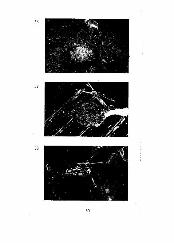
36. Empty the rumen and look for flukes which,if presenr, are attached ro the walls.
37. Open the abomasum and examine it fordiseases.
38. Open rhe rhoracic cavity using forceps robreak the bones.
31
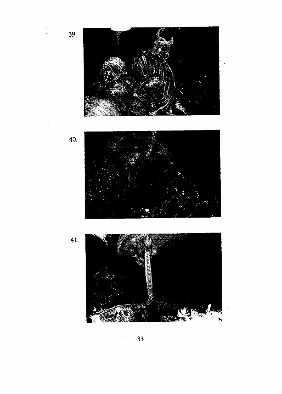
39. When you open the thotacic cavity, examinethe lungs and heart for diseases.
40. Remove the lungs from the thoracic cavityfor closer examination.
41. Open the trachea and examine it fordiseases.
32
39.
40.
41.
33
42.
43.
34
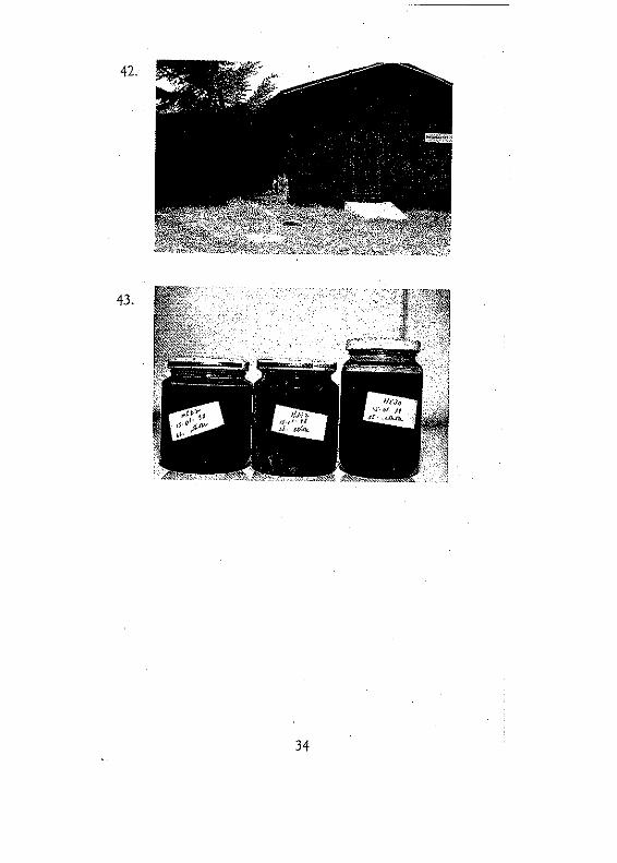
42. After you finish the post.mortemexamination, the dead animals must bedestroyed because they are carriers. Bury orbum the wastes of these diseased animals(bedding, manure, contaminated feed andwater). An incinerating room would be theideal place to perform this operation.
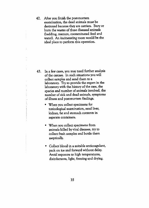
43. In a few cases, you may need further analysisof the carcass. In such situations you willcollect samples and send them to alaboratory. Try to provide the ~ert in thelaboratory with the history of the case, thespecies and number of animals involved, thenumber of sick and dead animals, symptomsof illness and post·mortem findings.
• When you collect specimens fortoxicological examination, send liver,kidney, fat and stomach contents inseparate containers.
• When you collect specimens ftomanimals killed by viral diseases, try tocollect ftesh samples and bottle themaseptically.
• Collect blood in a suitable anticoagulant,pack on ice and forward without delay.Avoid exposure to high temperatures,disinfectants, light, freering afld drying.
35
• Collect fresh specimens for bacterial andfungal examination and preserve on ice.You can also preserve the specimens in37% formalin for histopathologicalexamination.
• Use sterile instruments to collectspecimens for bacteriological andvirological examination and place themin sterile containers.
• To avoid contamination, use a differentset of instruments and containers foreach organ.
• Collect fresh faeces specimens forparasitological examination.
36
Recommended readingHansen J. and Perry B. 1990. The Epidemiology, Diagno.sis
and Control of Gastro-intestinal Parasites of Ruminantsin Ajrico.. A Handbook. URAD (InternationalLaboratory for Research on Animal Diseases),Nairobi, Kenya. 121 pp.
37