discovery of another fern-feeding group of moths: the ... · hoploscopini (insecta: lepidoptera:...
TRANSCRIPT
100
Mally et al.: Fern-feeding Hoploscopini larvae
Discovery of another fern-feeding group of moths: the larvae of Hoploscopini (Insecta: Lepidoptera: Pyraloidea) from Borneo
Richard Mally1*, Théo Léger2, Charles S. Vairappan3, Stephen Sutton3 & Matthias Nuss2
Abstract. We report the discovery of Hoploscopini larvae (Lepidoptera: Crambidae: Heliothelinae) on ferns at the southern slopes of Mount Kinabalu (Sabah, Borneo). The COI barcode of the larvae assigns them to the genus Hoploscopa. We provide the first detailed description of the larval stage for this tribe. Among Crambidae, these larvae are most similar to Crambinae larvae but differ in the presence of two L setae on A9, a character state that is present in Acentropinae and Schoenobiinae. We discuss the presence and distribution of L setae on A9 in Crambidae. Our observations of these larvae on this host plant and published host plant data support our hypothesis that larvae of the entire tribe Hoploscopini may be fern-feeders.
Key words. larva, chaetotaxy, Hoploscopa, fern, DNA barcoding
RAFFLES BULLETIN OF ZOOLOGY 65: 100–108Date of publication: 24 May 2017http://zoobank.org/urn:lsid:zoobank.org:pub:9B3F66A2-2939-48AB-B8E8-FBF63C8235CF
© National University of SingaporeISSN 2345-7600 (electronic) | ISSN 0217-2445 (print)
1University Museum of Bergen, Natural History Collections, Realfagbygget, Allégaten 41, 5007 Bergen, Norway; Email: [email protected] (*corresponding author)2Senckenberg Naturhistorische Sammlungen Dresden, Museum für Tierkunde, Königsbrücker Landstraße 159, 01109 Dresden, Germany; Email: [email protected] (TL), [email protected] (MN)3Institute for Tropical Biology and Conservation, Universiti Malaysia Sabah, Jalan UMS, 88400 Kota Kinabalu, Sabah, Malaysia; Email: [email protected] (CSV); [email protected] (SS)
INTRODUCTION
Hoploscopini is comprised of two genera, Hoploscopa Meyrick, 1886 and Perimeceta Turner, 1915, with a total of 20 described and more than 60 undescribed species in the Oriental Region, Wallacea, New Guinea and northern Queensland (Robinson et al., 1994; Nuss, 1998, 1999; Nuss et al., 2003–2016). Hoploscopini are still poorly studied, and preimaginal stages as well as larval food plants were unknown until very recently (Miller et al., 2015), but had not been assigned to this tribe. The nocturnal adults are recorded from mountainous habitats and are attracted by artificial light. The forewing length of the moths ranges from 7 to 10 mm in Hoploscopa and from 11 to 13 mm in Perimeceta. Forewing colouration is reddish-brown with various markings of diagonal stripes, ellipses, or silvery spots (Robinson et al., 1994; Nuss, 1998).
The classification of the group is somewhat controversial. Robinson et al. (1994) established the Hoploscopini within Scopariinae without an explanation for doing so, but Nuss (1998) pointed to the lack of synapomorphies supporting this grouping. Later, Hoploscopini were included in Heliothelinae based on the conspicuous, inwardly directed spine in the corpus bursae of the female genitalia (Nuss, 1998, 1999).
The Heliothelinae, originally established as a tribe within Scopariinae (Amsel, 1961) and later elevated to subfamily rank by Minet (1982), were subsequently synonymised with Scopariinae by Munroe & Solis (1998) and retained in synonymy by Solis & Maes (2003). Future phylogenetic analyses may show whether one of these or even another classification might be supported.
The objective of this paper is to record the discovery of five hoploscopine larvae on fern fronds at Mount Kinabalu (Sabah, Borneo) and to compare these findings with the available information of food plants of Hoploscopini.
MATERIAL & METHODS
Five larvae were found on Mount Kinabalu at an altitude of 1,680 m during the night of 13 June 2015 sitting on the undersides of fern fronds, which were unfolded and unwebbed. They were taken with the plants on which they were found for rearing purposes down to Kota Kinabalu at sea level, where rearing efforts were continued using fern species from the lowlands. None of the larvae accepted this alteration in food, climate, and elevation, and all the larvae died. Two of the larvae were kept in 96% natural ethanol for subsequent morphological and genetic analyses and are deposited at the University Museum of Bergen, Norway.
Genetic analysis was performed by extraction of DNA from the whole body of one of the larvae using Qiagen’s DNEasy blood & tissue kit. PCR amplification of the DNA barcoding region of the mitochondrial cytochrome C oxidase subunit I (COI) gene was done using the primers LCO (Folmer et al., 1994) and Nancy (Wahlberg & Wheat, 2008) in combination with a universal T7/T3 tail (Wahlberg & Wheat, 2008). We used 25 μl of PCR volume containing 0.75u TaKaRa Ex Taq Hot-Start DNA polymerase, 2.5 μl 10 × buffer, 400 nM of each primer, 800 nM dNTP mix and 2 μl DNA extract.
Taxonomy & Systematics

101
RAFFLES BULLETIN OF ZOOLOGY 2017
Cycling conditions were as follows: initial denaturation for 5 min at 95°C, 40 cycles with (1) 30 s at 95°C, (2) 30 s at 48°C, (3) 90 s at 70°C, final extension of 10 min at 70°C. PCR success was evaluated via gel electrophoresis on a 1% agarose gel using GelRed (Biotium). For clean-up of the successfully amplified PCR products, 0.5u of each the Exonuclease (Exo) and Shrimp Alkaline Phosphatase (SAP) enzymes were added to 8 µl of PCR product and the mixture was incubated in a thermocycler for 15 min at 37°C before inactivating the enzymes for 30 min at 80°C. The sequencing PCR was performed with BigDye, using 160 nM of T7/T3 sequencing primers and 0.5–2 μl PCR product. Sequencing was done at the Sequencing Facility, University of Bergen.
The alignment of the DNA sequence data was done with PhyDE version 0.9971 (Müller et al., 2010). MEGA 7 (Kumar et al., 2016) was used to find the best-fitting DNA model, which resulted in the GTR+G+I model. A Maximum Likelihood (ML) analysis of the sequence data was done with RAxML 7.4.2 (Stamatakis, 2006), using the raxmlGUI 1.3 interface (Silvestro & Michalak, 2012). The ML analysis included a bootstrap test with 1,000 replications. The resulting ML tree was edited in TreeGraph version 2.11.1-654 beta (Stöver & Müller, 2010). We used the BIN numbers of the Barcode of Life Database (BOLD, http://v4.boldsystems.org; Ratnasingham & Hebert, 2007) as DNA-barcoded taxa may not have been identified to species level.
In addition to the "BC MTD" Barcode samples provided by MN, BOLD was mined for further relevant records. Due to the different opinions regarding the classification of Hoploscopini we included Scopariinae in our search.
After DNA extraction, the exoskeleton of the larva was cut laterally, flattened, and preserved together with the head capsule in Euparal on a microscopic slide for further examination. The second larva was left intact in order to study the length and direction of the setae. Terminology of larval morphology, especially chaetotaxy, follows Hasenfuss (1963). Thoracic segments are abbreviated as T1–T3, abdominal segments as A1–A10. Drawings were done using Adobe Illustrator CS6, version 16.0.3.
The larval food plant was identified using Raciborski (1898) and Beaman & Edwards (2007).
RESULTS
A total of five individuals of larvae were found on fern fronds at 1,680 m altitude on the southern slope of Mount Kinabalu in Mesilau (see Fig. 1 for two of the larvae). Weather conditions were cloudy, but not rainy, with high humidity and temperature at about 20°C.
Material examined. Two larvae: Malaysia, Sabah, Mount Kinabalu National Park, Mesilau, western edge of Mount Kinabalu Golf Club, 6°01′38″N 116°35′32″E, 1,680 m, 13.vi.2015, leg. Théo Léger & Richard Mally (University Museum of Bergen, Norway).
Molecular identification of the larva. Sequencing of one of the larvae yielded a 655 bp fragment of the 5′ part of the COI gene. A search for similar sequences with the NCBI nucleotide blast tool resulted in a closest match with three specimens of “Scopariinae sp.” originating from Papua New Guinea (Miller et al., 2015). The corresponding images of these specimens on BOLD allowed us to identify these three and another seven specimens, altogether forming four barcode-species, as belonging to the genus Hoploscopa, and the information was corrected accordingly in the BOLD database.
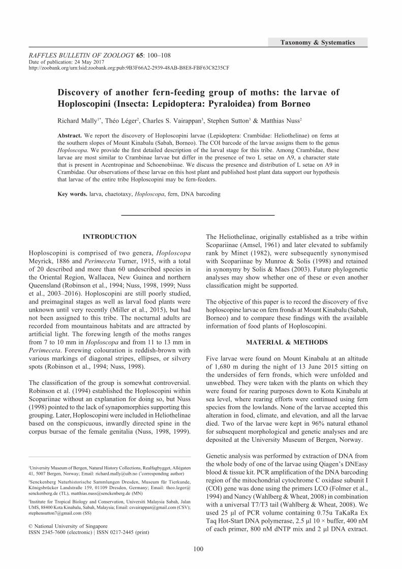
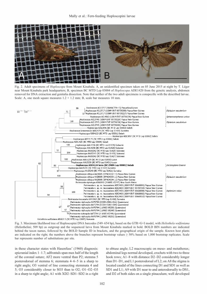
Subsequently, we analysed all sequences available for Hoploscopa and Perimeceta available to us on BOLD in a ML analysis. The species whose larva we describe here is sister to the species pair Hoploscopa AAU5240 + Hoploscopa AAU5241, both from North Sumatra (Sumatera Utara), in the ML tree (Fig. 3). The adult Hoploscopa specimens that we collected in Mount Kinabalu National Park (see Fig. 2 for representatives) appear as five separate species (BOLD BINs ADE1420, ADE3896, ADE3897, ADE4123 and ADE4125 in Table 1 and Fig. 3). The larvae could not be matched to any of the adults of the 15 DNA-barcoded species of Hoploscopa (Fig. 3).
Perimeceta, the other genus of Hoploscopini with four species included in this analysis, is sister to Hoploscopa in the ML tree (Fig. 3).
All included COI barcode sequences (Table 1) are publicly available on NCBI’s GenBank and the European Nucleotide Archive (ENA) via the accession numbers as well as on BOLD.
Morphological description of the larvae. The larvae are identified as Pyraloidea by the presence of two setae in the prespiracular group of the prothorax and three subventral setae on abdominal segments 3 to 6 as well as by the crochets forming a complete circle (Solis, 2006).
In the larval key on European Pyraloidea by Hasenfuss (1960), our larvae are identified as Crambidae (“Crambinae” sensu Hasenfuss, 1960), but they match none of the treated subgroups in this family. The closest similarity is found with Crambinae (“Crambini” sensu Hasenfuss, 1960), agreeing
Fig. 1. Two Hoploscopa larvae (centre and right) on the underside of a fragment of their food plant, Dicranopteris linearis (Burman, 1768) Underwood, 1907. Scale: one square measures 5 mm.
102
Mally et al.: Fern-feeding Hoploscopini larvae
Fig. 2. Adult specimens of Hoploscopa from Mount Kinabalu. A, an unidentified specimen taken on 05 June 2015 at night by T. Léger near Mount Kinabalu park headquarters; B, specimen BC MTD Lep 03004 of Hoploscopa ADE1420 from the genetic analysis, abdomen removed for DNA extraction and genitalia dissection. Note that neither of the two adult specimens is conspecific with the described larvae. Scale: A, one mesh square measures 1.2 × 1.2 mm; B, scale bar measures 10 mm.
Fig. 3. Maximum likelihood tree of Hoploscopini DNA barcodes (549–654 bp), based on the GTR+G+I model, with Heliothela wulfeniana (Heliothelini; 509 bp) as outgroup and the sequenced larva from Mount Kinabalu marked in bold. BOLD BIN numbers are indicated behind the taxon names, followed by the BOLD Sample ID in brackets, and the geographical origin of the sample. Known host plants are indicated on the right; the numbers above the branches represent bootstrap values ≥ 50% based on 1,000 bootstrap replicates. Scale bar represents number of substitutions per site.
in these character states with Hasenfuss’ (1960) diagnosis: epicranial index 1–1.7; adfrontals span max half of the length of the coronal suture; AF2 more ventral than P2; stemma 5 posterodorsal of stemma 6; stemmata 4–6–5 in a sharp to right angle; O3 ventral of line connecting stemmata 4 and 5; O3 considerably closer to SO3 than to O2; O1–O2–O3 in a sharp to right angle; A1 with XD2–SD1–SD2 in a right
to obtuse angle; L2 macroscopic on meso- and metathorax; abdominal legs normal developed, crochets with two to three hook rows; A1–8 with distance D2–D2 considerably longer than D1–D1, and L1 posterodorsal of L2; on A8 the stigma is located caudal of the lines connecting D1 and SD1 as well as SD1 and L1; A9 with D1 near to and anterodorsally to DS1, and D2 of both sides on a single pinaculum; well developed

103
RAFFLES BULLETIN OF ZOOLOGY 2017
Table 1. Samples of DNA barcoded specimens included in this study. For taxa without species identification the respective BOLD BIN number was used as species epithet.
TaxonBOLD BIN
(No. of Sampled Specimens)
Geographical Origin
GenBank Accession Number BOLD Sample ID
Heliothela wulfeniana ABU6313(n = 1)
Romania KY080439 BC MTD Lep 03005
Hoploscopa luteomacula AAX2479(n = 1)
Indonesia, Sumatra KX843698 BC MTD Lep 01419
Hoploscopa obliqua AAZ8005(n = 3)
Papua New Guinea KX783025KX783026KX783027
USNM ENT 00665932USNM ENT 00514750USNM ENT 00514731
Hoploscopa ADE4124 ADE4124(n = 1)
Malaysia, Sabah KY080442 BC ZMBN Lep 00081
Hoploscopa AAU5242 AAU5242(n = 3)
Indonesia, Sulawesi JN272552JN272553JN272554
BC MTD Lep 01425BC MTD Lep 01426BC MTD Lep 01427
Hoploscopa AAU5239 AAU5239(n = 2)
Philippines, Luzon JN272557JN272558
BC MTD Lep 01430BC MTD Lep 01431
Hoploscopa AAU4238 AAU4238(n = 2)
Indonesia, Sulawesi JN272555JN272556
BC MTD Lep 01428BC MTD Lep 01429
Hoploscopa ADE4125 ADE4125(n = 1)
Malaysia, Sabah KY080444 BC MTD Lep 03006
Hoploscopa ADE1420 ADE1420(n = 1)
Malaysia, Sabah KY080440 BC MTD Lep 03004
Hoploscopa AAU5240 AAU5240(n = 1)
Indonesia, Sumatra JN272550 BC MTD Lep 01421
Hoploscopa AAU5241 AAU5241(n = 1)
Indonesia, Sumatra JN272551 BC MTD Lep 01422
Hoploscopa ADE4123 ADE4123(n = 1)
Malaysia, Sabah KY080441 BC MTD Lep 03001
Hoploscopa ADE3896 ADE3896(n = 1)
Malaysia, Sabah KY080445 BC MTD Lep 03003
Hoploscopa ACL3745 ACL3745(n = 3)
Papua New Guinea KP850086KP850401KP850535
USNM ENT 00739216USNM ENT 00739238USNM ENT 00739200
Hoploscopa ACL4063 ACL4063(n = 1)
Papua New Guinea KP850867 USNM ENT 00739239
Hoploscopa ACL3717 ACL3717(n = 3)
Papua New Guinea KP850187KP850609KX842727
USNM ENT 00739208USNM ENT 00739227
YAWCATCR0759
Hoploscopa ADE3897 ADE3897(n = 1)
Malaysia, Sabah KY080443 BC MTD Lep 03002
Perimeceta incrustalis AAY1912(n = 1)
Indonesia, Sumatra KX843699 BC MTD Lep 01418
Perimeceta niphospila AAF5794(n = 6)
Australia, Queensland
KF388782KF391745JN272547JN272548KF390107KF392415
11ANIC-0524811ANIC-0524911ANIC-0525011ANIC-05251
CCDB-15861-D09CCDB-15861-D11

104
Mally et al.: Fern-feeding Hoploscopini larvae
Fig. 4. Chaetotaxy of the postcranial body, sinistral view. Chaetal terminology after Hasenfuss (1963). Abbreviations: s.pp., seta paraproctalis.
anal plate; A10 with distance II–II smaller than or equal to II–III, IIIa macroscopic. Our larvae differ from Hasenfuss’ (1960) diagnosis of Crambinae in these character states: AF2 lateral of bifurcation of epicranial suture (instead of more dorsal than bifurcation); A1 with only two SV setae (instead of three); A9 with L2 present (instead of absent); distance V1–V1 on A10 larger than on A9 (instead of smaller); and on A10 distance V1–VIId smaller than VIIc–VIIb (instead of larger).
Head. (Fig. 4) Orthognathous, brown; epicranial suture present; vertex with microsetae V1–3 in a line; pore Va variable in position, slightly lateral between V2 and V3 or lateral of V3; front with P1 close to AF1, P2 between P1 and V1, pore Pb ventral of P2, pore Pa in the centre of P1, L1 and A3; AF1 slightly dorsal of the centre of adfrontal
area, AF2 at level of lower end of central suture, pore AFa between AF1 and AF2, closer to AF2; F1 ventral of AFa halfway of dorsoventral expanse of the frontal area, pore Fa medioventral of F1; ventral clypeus margin slightly undulated, C1 on lateral end, C2 halfway between C1 and sagittal plane dorsal of slight ventrad protrusion; A1, A2 and A3 in an arched line, distance A2–A3 approximately twice the distance A1–A2; L1 central on lateral head, pore La posterodorsal of L1; microseta G1 at level of P1 and L1, pore Ga anteroventral of G1; six stemmata in an oval semicircle, O1 in its centre, O2 posterior of stemma 1, O3 well posterior of stemma 6, pore Oa posteroventral of stemma 6; SO1 ventral of stemma 5 posterior of antennal socket, SO2 ventral of stemma 6, SO3 ventral of pore Oa, pore SOa anterior of SO3.
TaxonBOLD BIN
(No. of Sampled Specimens)
Geographical Origin
GenBank Accession Number BOLD Sample ID
Perimeceta niphotypa ABA0010(n = 1)
Australia, New South Wales
KF391291 11ANIC-05247
Perimeceta sp. near leucoselene ADE2403(n = 5)
Papua New Guinea KY034067KY034068KY034066KY034070KY034069
USNM ENT 00668001USNM ENT 00668002USNM ENT 00668008USNM ENT 00668033USNM ENT 00700334

105
RAFFLES BULLETIN OF ZOOLOGY 2017
Thorax. (Fig. 5) Prothoracic and prespiracular shield as well as dorsal, subdorsal and lateral pinacula strongly sclerotised, black, subventral and ventral pinacula less so. Prothoracic shield with roughly equally long D1 and D2, XD1, XD2, SD1 and SD2, three pores (a–c) present, pore a posterodorsal and pore b posterior of XD1, pore c dorsal of XD2, microseta MXD1 at posterior edge of prothoracic shield between D1 and D2; prespiracular shield anterior to spiracle bisetose (L1–2), extending posteroventrally around spiracle; subventral pinaculum bisetose (SV1–2); coxa of prothoracic leg with five setae, MV2 microseta on anterodorsal end of coxal band, MV3 microseta between the two anterior coxal macrosetae, a third microseta posterodorsally close to posterior coxal macroseta; V1 posteroventral of leg. T2 and T3 identical: bisetose dorsal and subdorsal pinacula (D1+D2, SD1+SD2), D2 about twice the length of D1; MD1 microseta anterior of D2, MSD1–2 microsetae anteroventral of subdorsal pinaculum; two lateral pinacula with anterior bisetose (L1+L2) and the posterior unisetose (L3), posterior lateral pinaculum including L3 is missing on one body side in one of the two investigated specimens; subventral pinacula unisetose (SV1); coxal band with MV2 and MV3 as in prothorax, plus MV1 microseta anterodorsally of MV2; V1 posteroventral of leg.
Abdomen. (Fig. 5) A1–8: Two unisetose (D1, D2) dorsal pinacula and the unisetose (SD1) subdorsal pinaculum strongly sclerotised, subventral pinacula less so; D1, D2 and SD1 about the same length; MD1 microseta anterior to D2 on anterior segment edge; SD2 microseta anterior of spiracle; two lateral pinacula below spiracle, anterodorsal one bisetose (L1–2), the posteroventral unisetose (L3), setae of approximately equal length; subventral group bisetose (SV1, SV3) in A1 and A7, trisetose (SV1–3) in A2 and A3–6 (at anteriodorsal base of prolegs), and unisetose (SV1) in A8 and A9, SV1–2 are absent on left side of A4 in one larva; V1 at posteromedial end of abdominal segments; MV3 microseta on anterior segment edge between subventral group and V1. A9 with a large unpaired dorsal pinaculum covering the dorsum, bearing one long seta (D2) on each side; subdorsal pinaculum trisetose (D1, SD1, L2), with SD1 in a less strongly sclerotised semicircular part of pinaculum;
MD1 on anterior segment edge between dorsal and subdorsal pinaculum; lateral pinaculum unisetose (L1); one subventral (SV1) and one ventral (V1) seta, with MV3 on anterior segment edge between SV1 and V1. Unpaired anal shield (segment A10) with four setae (I, II, III, IIIa) on each side, seta I being the shortest; short Seta paraproctalis (S.ppr.) posteroventral of anal shield on dorsal tip of a lanceolate, weakly sclerotised area; V1 close to ventral base of terminal legs. Prolegs on A3–6 with crochets in a complete circle of three concentric rows of outward-directed hooks, the outer circle bearing the shortest hooks and the inner circle the longest; prolegs on A10 forming a semicircle.
Food plant records. The larvae were found on Dicranopteris linearis (Burman, 1768) Underwood, 1907 (Gleicheniaceae; Fig. 6). The larvae were sitting on the underside of intact fronds, which did not show any traces of frass. In captivity, they fed on this plant species from the outer edge of the pinnate leaves towards the mid-ribs (Fig. 1). After we left the collecting area at 1,680 m we continued to feed the caterpillars with non-Gleicheniaceae ferns growing in the lowland of Kota Kinabalu, but these were rejected and the larvae died.
The search for related species on BOLD resulted in the recognition of five barcoded Hoploscopinae species collected in Yawan, Papua New Guinea which are found to feed on ferns as well: Hoploscopa ACL3745, Hoploscopa ACL3717 and Hoploscopa obliqua (Rothschild, 1915) feed on Diplazium esculentum (Retzius in Retzius & König, 1791) Swartz, 1803 (Athyriaceae), Hoploscopa ACL4063 feeds on Sphaerostephanos unitus (Linnaeus, 1759) Holttum, 1794 (Thelypteridaceae), and Perimeceta sp. near leucoselene (Hampson, 1919) feeds on Asplenium nidus Linnaeus, 1753 (Aspleniaceae) (S. Miller, C. Redmond & T. Whitfield, pers. comm.; Botanical Research Institute of Texas, 2003–2009).
DISCUSSION
All known larval host plant records for Hoploscopinae belong to six species, and all are included in our analysis (Fig. 3). The larvae are feeding on fern species of
Fig. 5. Chaetotaxy of the postcranial body, sinistral view. Chaetal terminology after Hasenfuss (1963). Abbreviations: s.pp., seta paraproctalis.

106
Mally et al.: Fern-feeding Hoploscopini larvae
Gleicheniaceae (Gleicheniales), Aspleniaceae, Athyriaceae or Thelypteridaceae (all three Polypodiales). These repeated findings from ferns as food plants suggest that Hoploscopini are fern feeders. This fern-feeding habit contrasts with the feeding habit of the supposed sister-group Heliothelini. For one of its species, Heliothela wulfeniana (Scopoli, 1763), the angiosperm genera Viola Linnaeus, 1753 (Violaceae) and Mentha Linnaeus, 1753 (Lamiaceae) are recorded as larval food plants (Nuss, 2005). Other fern-feeders among Crambidae are known in Musotiminae (e.g. Munroe, 1972; Kirk, 1978; Phillips & Solis, 1996; Solis et al., 2004, 2005a, 2005b; Yen et al., 2004), in Phenacodes Turner, 1937, treated in Scopariinae by Munroe (1958) and Munroe & Solis (1998), and provisionally placed in Cybalomiinae by Nuss (1999), and in the Spilomelinae Herpetogramma sphingealis Handfield & Handfield, 2011, H. aeglealis (Walker, 1859) and H. platycapna (Meyrick, 1897) (Kirk, 1978; Solis, 2008; Handfield & Handfield, 2011), Diasemiopsis ramburialis (Duponchel, 1833) (Farahpour-Haghani et al., 2016) and Udea decrepitalis (Herrich-Schäffer, 1848) (Lhomme, 1935).
Based on the diagnostic characters of the chaetotaxy provided by Hasenfuss (1960), the Crambinae are the group that shares the most larval similarities with Hoploscopa. One character that differs in Hoploscopa is the presence of an L2 seta on A9, grouped on a pinaculum together with D1 and SD1. Among Crambidae, a second L seta on A9 is yet only known to occur in Acentropinae (Hasenfuss, 1960, 1963) and Schoenobiinae (Passoa & Habeck, 1987; Passoa, 1988). This character state may turn out to be a synapomorphy for a group comprising Acentropinae, Schoenobiinae and Hoploscopini. A comprehensive comparison of the chaetotaxy of all crambid subfamilies is currently not possible, as descriptions of larvae of Cathariinae, Lathrotelinae, Linostinae, Midilinae, and Heliothelini are still lacking.
The molecular data analysis reveals 16 DNA barcode-species for Hoploscopa. This is precisely the number of species so far described in the genus, but some known species are recorded
from other geographical places (Nuss et al., 2003–2016) and therefore at least some of them are probably not conspecific with those included in our analysis. This, as well as the long terminal branches of some taxa in our analysis, point to a large proportion of still undescribed species in Hoploscopa, and in fact Robinson et al. (1994) already report more than 50 undescribed species of Hoploscopa based on material in entomological collections. The inability to link the larvae from Mount Kinabalu with any of the collected adults during the same field trip suggests that different Hoploscopa species on Mount Kinabalu may have different flight times, or species may not be attracted by artificial light. Future study of altitudinal and chronological occurrence as well as of food plant usage in Hoploscopini will contribute to our understanding of speciation in this still poorly studied group.
ACKNOWLEDGEMENTS
We are thankful for the enormous help and support that we experienced in preparation of and during our work in Sabah from Chung (Sabah Biodiversity Centre) and the staff of Sabah Parks and of Universiti Malaysia Sabah, Kota Kinabalu. Sabah Parks and the Sabah Biodiversity Centre provided permits for conducting research and collecting, and for export of part of the material. We are grateful to Peter Achleitner for his generous hospitality during the first and second author’s stay in his house in Mesilau near which we found the larvae. Papua New Guinea data come from a Binatang Research Center program led by Vojtech Novotny (Institute of Entomology, Czech Academy of Sciences, České Budějovice, Czech Republic), George Weiblen (Bell Museum of Natural History, University of Minnesota, U.S.A.), Yves Basset (Smithsonian Tropical Research Institute, Panama), and Scott Miller (National Museum of Natural History, Smithsonian Institution, Washington, D.C., U.S.A.), and supported by the US National Science Foundation (grants DEB-0515678, 0841885 and others) and the Czech Science Foundation (grants 206/09/0115, 14-04258S and others). DNA barcodes for specimens from PNG were provided by Paul Hebert (Department of Integrative Biology, University of Guelph, Ontario, Canada) through a grant from Genome Canada and the Ontario Genomics Institute in support of the iBOL project. George Weiblen, Timothy Whitfeld (Department of Ecology and Evolutionary Biology, Brown University, Rhode Island, U.S.A.), and Conor Redmond (Institute of Entomology, Czech Academy of Sciences, České Budějovice, Czech Republic) provided host plant identifications, and Scott Miller provided insect identifications. Francesca Vegliante (Senckenberg Museum für Tierkunde Dresden, Germany) contributed with valuable discussions and comments on the chaetotaxy. Two reviewers, Alma Solis (U.S. Department of Agriculture, Washington, D.C., U.S.A.) and Christian Schulze (University of Vienna, Austria), provided helpful comments on the manuscript. Collecting on Borneo was supported through a travel grant of the Royal Entomological Society and through the Norwegian Meltzer Research Grant.
Fig. 6. Dicranopteris linearis (Burman, 1768) Underwood, 1907, the food plant of the investigated Hoploscopa larvae; photo taken at the collecting site of the larvae.
107
RAFFLES BULLETIN OF ZOOLOGY 2017
LITERATURE CITED
Amsel HG (1961) Die Microlepidopteren der Brandt’schen Iran-Ausbeute 5. Teil. Arkiv för Zoologi Stockholm, 13: 323–445, pls. 1–9.
Beaman JH & Edwards PJ (2007) Ferns of Kinabalu: An Introduction. Natural History Publications (Borneo), Malaysia, 6 + 198 pp.
Botanical Research Institute of Texas (2003–2009) Digital Flora of New Guinea. http://ng.atrium-biodiversity.org/atrium (Accessed 26 July 2016).
Burman NL (1768) Flora Indica: Cui Accedit Series Zoophytorum Indicorum, Nec Non Prodromus Florae Capensis. Apud Cornelium Haek & Johannem Schreuderum, Leiden & Amsterdam, 242 pp.
Duponchel PAJ (1831–1834 [“1831”]) Nocturnes 5(2). Histoire naturelle des Lépidoptères ou Papillons de France, Paris, 8(2): pp. 5–402, pls. 211–236.
Farahpour Haghani A, Jalaeian M & Landry B (2016) Diasemiopsis ramburialis (Duponchel) (Lepidoptera, Pyralidae s. l., Spilomelinae) in Iran: first record for the country and first host plant report on water fern (Azolla filiculoides Lam., Azollaceae). Nota Lepidopterologica, 39(1): 1–11.
Folmer O, Black M, Hoeh W, Lutz R & Vrijenhoek R (1994) DNA primers for amplification of mitochondrial cytochrome c oxidase subunit I from diverse metazoan invertebrates. Molecular Marine Biology and Biotechnology, 3(5): 294–299.
Hampson GF (1919) Descriptions of new Pyralidae of the subfamilies Crambinae and Siginae. Annals and Magazine of Natural History, including Zoology, Botany and Geology, London, 9(3): 275–292, 437–457, 533–547.
Handfield L & Handfield D (2011) A new species of Herpetogramma (Lepidoptera, Crambidae, Spilomelinae) from eastern North America. ZooKeys, 149: 5–15.
Hasenfuss I (1960) Die Larvalsystematik der Zünsler (Pyralidae). Abhandlungen zur Larvalsystematik der Insekten, 5: 1–263.
Hasenfuss I (1963) Eine Vergleichend-morphologische Analyse der regulären Borstenmuster der Lepidopterenlarven. Studien zur Methodik der vergleichenden Morphologie der Borstenmuster und zur phylogenetischen Deutung der Abwandlungen der regulären Borstenmuster der Lepidopterenlarven. Zeitschrift für Morphologie und Ökologie der Tiere, 52: 197–364.
Herrich-Schäffer GAW (1847–1855, imprint “1849”) Systematische Bearbeitung der Schmetterlinge von Europa, zugleich als Text, Revision und Supplement zu Jakob Hübner’s Sammlung europäischer Schmetterlinge. 4: Die Zünsler und Wickler. G. J. Manz, Regensburg, pp. [1]–2–288, (Index) [1]–2–48, pls. 1–23 (Pyralidides) + 1–59 (Tortricides).
Holttum RE (1974) Thelypteridaceae of Africa and adjacent islands (Thelypteridaceae van Afrika en omliggende eilande). Journal of South African Botany, 40(2): 123–168.
Kirk AA (1978) The insect fauna of the weed Pteridium aquilinum (L.) Kuhn (Polypodiaceae) in Papua New Guinea: A potential source of biological control agents. Journal of the Australian Entomological Society, 16: 403–409.
Kumar S, Stecher G & Tamura K (2016) MEGA7: Molecular Evolutionary Genetics Analysis version 7.0 for bigger datasets. Molecular Biology and Evolution 33(7): 1870–1874.
Lhomme L (1935) Catalogue des Lépidoptères de France et de Belgique. Microlépidoptères. Le Carriol, Lot, 1–172.
Linnaeus C (1753) Species Plantarum, Tomus 2. Impensis Laurentii Salvii, Holmiae, 561–1231.
Linnaeus C (1759) Systema naturae per regna tria naturae, secundum classes, ordines, genera, species, cum characteribus, differentiis, synonymis, locis. Salvius, Holmiae 10(2), 1–4, 825–1384.
Meyrick E (1886) Descriptions of Lepidoptera from the South Pacific. Transactions of the Entomological Society of London, 1886: 189–296.
Meyrick E (1897) On Lepidoptera from the Malay Archipelago. Transactions of the Entomological Society of London, 1897: 69–92.
Miller SE, Rosati ME, Gewa B, Novotny V, Weiblen GD & Hebert PDN (2015) DNA barcodes of Lepidoptera reared from Yawan, Papua New Guinea. Proceedings of the Entomological Society of Washington, 117(2): 247–250.
Minet J (1982) Les Pyraloidea et leurs principales divisions systématiques (Lep. Ditrysia). Bulletin de la Société entomologique de France, Paris, 86(1981): 262–280.
Müller J, Müller K, Neinhuis C & Quandt D (2010) PhyDE – Phylogenetic Data Editor, version 0.9971. http://www.phyde.de (Accessed 31 July 2016).
Munroe EG (1958) The geographic distribution of the Scopariinae (Lepidoptera: Pyralidae). Proceedings of the Tenth International Congress on Entomology, 1956, Montreal, 1: 831–837.
Munroe EG (1972) Pyraloidea, Pyralidae comprising subfamilies Scopariinae, Nymphulinae. In: Dominick RB et al. (eds.), The Moths of America North of Mexico 13(1A).( E. W. Classey and R. B. D. Publications Inc., London, pp. 1–134.
Munroe EG & Solis MA (1998) The Pyraloidea. In: Kristensen NP (ed.), Lepidoptera, Moths and Butterflies, Volume 1: Evolution, systematics, and biogeography. In: Fischer M (ed.), Handbook of Zoology. Volume IV Arthropoda: Insecta, Part 35. Walter de Gruyter, Berlin, pp. 233–256.
Nuss M (1998) The Scopariinae and Heliothelinae stat. rev. (Lepidoptera: Pyraloidea, Crambidae) of the Oriental Region – a revisional synopsis with descriptions of new species from the Philippines and Sumatra. Nachrichten des entomologischen Vereins Apollo, Supplement 17: 475–528.
Nuss M (1999) Revision der Gattungen der Scopariinae (Lepidoptera: Pyraloidea, Crambidae). Nova Supplementa Entomologica, 13: 3–151.
Nuss M (2005) Heliothelinae. In: Goater B, Nuss M & Speidel W, Pyraloidea I. In: Huemer P & Karsholt O (eds.), Microlepidoptera of Europe 4. Apollo Books, Stenstrup, pp. 109–111, 182, 223, 257, pl. 3 fig. 53.
Nuss M, Landry B, Mally R, Vegliante F, Tränkner A, Bauer F, Hayden J, Segerer A, Schouten R, Li H, Trofimova T, Solis MA, De Prins J & Speidel W (2003–2016) Global Information System on Pyraloidea. www.pyraloidea.org (Accessed 04 August 2016).
Passoa S (1988) Systematic positions of Acentria ephemerella (Denis & Schiffermüller), Nymphulinae, and Schoenobiinae based on morphology of immature stages (Pyralidae). Journal of the Lepidopterists’ Society 42(4): 247–262.
Passoa S & Habeck DH (1987) A description of the larva and pupa of Rupela albinella, a pest of rice in Latin America (Lepidoptera: Pyralidae: Schoenobiinae). Florida Entomologist, 70(3): 368–375.
Phillips E & Solis MA (1996) Neurophyseta (Lepidoptera: Crambidae) de Costa Rica. Revista de Biología Tropical, 44 (2): 693–717.
Raciborski M (1898) Die Pteridophyten der Flora von Buitenzorg. Flore de Buitenzorg, Volume 1. E. J. Brill, Leiden, pp. I–XII, 1–255.
Ratnasingham S & Hebert PDN (2007) BOLD: The Barcode of Life Data System (http://www.barcodinglife.org). Molecular Ecology Notes, 7(3): 355–364.
Retzius AJ & König JG (1791) Observationes botanicae sex fascicvlis comprehensae. Siegfried Lebrecht Crusium, Lipsiae, 15–40.

108
Mally et al.: Fern-feeding Hoploscopini larvae
Robinson GS, Tuck KR & Shaffer M (1994) A field guide to the smaller moths of South-East Asia. Malaysian Nature Society, Kuala Lumpur, 309 pp.
Rothschild LW (1915) Macrolepidoptera. In: Rothschild W & Durrant JH (eds.), Lepidoptera of the British Ornithologists’ Union and Wollaston expeditions in the Snow Mountains, southern Dutch New Guinea. Zoological Museum, Tring, pp. 1–146, pls. 1–2.
Scopoli JA (1763) Entomologia Carniolica exhibens insecta Carnioliae indigena et distributa in ordines, genera, species, varietates, methodo Linneana. Joannis Thomae Trattner, Vienna, 420 pp.
Silvestro D & Michalak I (2012) raxmlGUI: A graphical front-end for RAxML. Organisms Diversity & Evolution, 12: 335–337.
Solis MA (2006) Key to selected Pyraloidea (Lepidoptera) larvae intercepted at U.S. ports of entry: Revision of Pyraloidea in “Keys to some frequently intercepted lepidopterous larvae” by Weisman 1986. USDA Systematic Entomology Laboratory, Lincoln, NE, USA: 1–58.
Solis MA (2008) Pyraloidea and their known hosts (Insecta: Lepidoptera) of Plummers Island, Maryland. Bulletin of the Biological Society of Washington 15: 88–106.
Solis MA & Maes KVN (2003) Preliminary phylogenetic analysis of the subfamilies of Crambidae (Pyraloidea Lepidoptera). Belgian Journal of Entomology, 4(2002): 53–95.
Solis MA, Yen SH & Goolsby JA (2004) Species of Lygomusotima new genus and Neomusotima Yoshiyasu (Lepidoptera: Crambidae) from Australia and Southeastern Asia feeding on Lygodium microphyllum (Schizaeceae). Annals of the Entomological Society of America, 97(1): 64–76.
Solis MA, Davis DR & Nishida K (2005a) Life history and systematics of Albusambia elaphoglossumae (Lepidoptera: Crambidae): A new genus and species of musotimine with leaf-mining biology from Costa Rica. International Journal of Tropical Biology, 53 (3–4): 487–501.
Solis MA, Yen SH, Goolsby JH, Wright T, Pemberton R, Winotal A, Chattrukul U, Thagong A & Rimbut S (2005b) Siamusotima aranea, a new stem-boring musotimine (Lepidoptera: Crambidae) from Thailand feeding on Lygodium flexuosum (Schizaeaceae). Annals of the Entomological Society of America, Washington, 98(6): 887–895.
Stamatakis A (2006) RAxML-VI-HPC: maximum likelihood-based phylogenetic analyses with thousands of taxa and mixed models. Bioinformatics, 22(21): 2688–2690.
Stöver BC & Müller KF (2010) TreeGraph 2: Combining and visualizing evidence from different phylogenetic analyses. BMC Bioinformatics, 11: 7.
Swartz OP (1803) Observationes botanicae genera et species filicum illustrantes. Journal für die Botanik, 1801(2): 273–312.
Turner AJ (1915) Studies in Australian Lepidoptera. Proceedings of the Royal Society of Queensland, Brisbane, 27: 11–57.
Turner AJ (1937) New Australian Pyraloidea (Lepidoptera). Proceedings of the Royal Society of Queensland, Brisbane, 48(10): 61–88.
Underwood LM (1907) American ferns – VIII. A preliminary review of the North American Gleicheniaceae. Bulletin of the Torrey Botanical Club, 34(5): 243–262.
Wahlberg N & Wheat CW (2008) Genomic outposts serve the phylogenomic pioneers: designing novel nuclear markers for genomic DNA extractions of Lepidoptera. Systematic Biology, 57(2): 231–242.
Walker F (1859) Pyralides. List of the Specimens of Lepidopterous Insects in the Collection of the British Museum, 18: 509–798.
Yen SH, Solis MA & Goolsby JA (2004) Austromusotima, a new musotimine genus (Lepidoptera: Crambidae) feeding on Old World climbing fern, Lygodium microphyllum (Schizaeaceae). Annals of the Entomological Society of America, Washington, 97(3): 397–410.