discover your future in research - monash university...discover your future in research 3 monash...
TRANSCRIPT
Discover your future in research PROJECT OPPORTUNITIES FOR:
• UNDERGRADUATE
• HONOURS
• MASTERS
• PHD
monash.edu/discovery-institute
CONTENTS
Acknowledgement
We acknowledge the traditional lands of Indigenous peoples.
The Faculty of Medicine, Nursing and Health Sciences incorporates the Aboriginal and Torres Strait Islander Curriculum Framework in educating future health professionals. You will learn skills in respect, communication, safety and quality, advocacy and reflection to improve Indigenous health.
Monash is committed to facilitating the entry of Indigenous students into courses. There are a range of pathways, entry points, bursaries, scholarships, accommodation, tutorial support and cadetships. To learn more about entry requirements and our Indigenous Access Interview, contact Gukwonderuk Indigenous Health staff via email at [email protected] or 03 9905 3828.
Discover your future in research 3
Monash Biomedicine Discovery Institute (BDI) 4
Our Discovery Programs 6
Targeting the intersection of disease areas 7
Why study at the Monash BDI? 8
Interested in research? Your future starts here 10
Undergraduate programs 12
Honours programs 14
Postgraduate programs 17
Monash BDI Group Leaders and their Discovery Program affiliations 19
Cancer Program Group Leaders 23
Cardiovascular Disease Program Group Leaders 39
Development and Stem Cells Program Group Leaders 47
Infection and Immunity Program Group Leaders 69
Metabolism, Diabetes and Obesity Program Group Leaders 122
Neuroscience Program Group Leaders 133
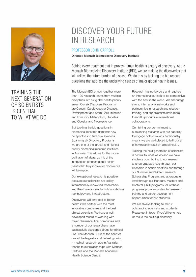
Behind every treatment that improves human health is a story of discovery. At the Monash Biomedicine Discovery Institute (BDI), we are making the discoveries that will relieve the future burden of disease. We do this by tackling the big research questions that address the underlying causes of major global health issues.
DISCOVER YOUR FUTURE IN RESEARCHPROFESSOR JOHN CARROLLDirector, Monash Biomedicine Discovery Institute
The Monash BDI brings together more than 120 research teams from multiple disciplines into six global health priority areas. Our six Discovery Programs are Cancer, Cardiovascular Disease, Development and Stem Cells, Infection and Immunity, Metabolism, Diabetes and Obesity, and Neuroscience.
But tackling the big questions in biomedical research demands new perspectives to find new solutions. Spanning six Discovery Programs, we are one of the largest and highest quality biomedical research institutes in Australia. This allows for the cross-pollination of ideas, as it is at the intersection of these global health issues that truly innovative discoveries will be made.
Our exceptional research is possible because our scientists are led by internationally-renowned researchers and they have access to truly world-class technology and infrastructure.
Discoveries will only lead to better health if we partner with the most innovative companies and the best clinical scientists. We have a well-developed record of working with major pharmaceutical companies and a number of our researchers have successfully developed drugs for clinical use. The Monash BDI is at the heart of one of the largest – and fastest growing – medical research hubs in Australia thanks to our relationships with Monash Partners and the Monash Academic Health Science Centre.
TRAINING THE NEXT GENERATION OF SCIENTISTS IS CENTRAL TO WHAT WE DO.
Research has no borders and requires an international outlook to be competitive with the best in the world. We encourage strong international networks and partnerships in research and research training, and our scientists have more than 200 productive international collaborations.
Combining our commitment to outstanding research with our capacity to engage both clinicians and industry means we are well placed to fulfil our aim of having an impact on global health.
Training the next generation of scientists is central to what we do and we have students contributing to our research at undergraduate level through our Research in Action electives and through our Summer and Winter Research Scholarship Program, and at graduate level through our Honours, Masters and Doctoral (PhD) programs. All of these programs provide outstanding research training and career development opportunities for our students.
We are always looking to recruit outstanding scientists and students. Please get in touch if you’d like to help us make the next big discovery.
3www.monash.edu/discovery-institute
OUR STRATEGIC PRIORITIES
DISCOVERPursue excellence in discovery research within and across these six global health priority areas:
• Cancer • Cardiovascular Disease • Development and Stem Cells • Infection and Immunity • Metabolism, Diabetes and Obesity • Neuroscience
TRANSLATETranslate and commercialise our research to impact on health outcomes.
COLLABORATE AND CONNECTCollaborate globally to conduct outstanding research.
DEVELOP OUR PEOPLEAttract, support and develop the science leaders of the future in a diverse and supportive environment.
ENGAGEEngage with the wider community in all that we do, to inform, influence and advocate.
AT A GLANCE
WHO WE AREAn institute with the scale and scope to tackle major research questionsWe are one of the largest and highest-quality biomedical research institutes in Australia. With more than 120 internationally-renowned research teams, we work with national and international collaborators on global health priorities.
WHAT WE DODiscover and innovate to enhance livesOur scientists are passionate about discovery research and committed to establishing a culture of excellence and collaboration to enable the most important research questions to be addressed.
With strong international networks and partnerships with researchers, health precincts and industry, together with access to unparalleled, world-leading research infrastructure, our discoveries accelerate the ability to prevent, diagnose and treat disease.
TACKLING THE BIG QUESTIONS IN BIOMEDICAL RESEARCH IS NO LONGER THE DOMAIN OF INDIVIDUAL SCIENTIFIC DISCIPLINES
– IT DEMANDS A CROSS-DISCIPLINARY APPROACH
MONASH BIOMEDICINE DISCOVERY INSTITUTE
4 www.monash.edu/discovery-institute
MIC
ROBI
OLOGY
PHARMACOLOGY
PHYSIOLOGY
BIOCHEMISTRY & MOLECULAR BIOLOGY
ANATOMY & DEVELOPMENTA
L BI
OLOG
Y
Cancer
Development& Stem Cells
Metabolism,Diabetes & Obesity
CardiovascularDisease
Neuroscience
Infection & Immunity
THERAPEU
TICSPL
ATFO
RM T
ECHNOLOGIES
COMPUTATIONAL BIOLOGY
RESEARCHEDUCATION
ENGAGEMENT
FAST FACTS
DISCOVERY TO IMPACT: THE WIDER CONTEXTA NEW FRAMEWORK FOR RESEARCH EXCELLENCE
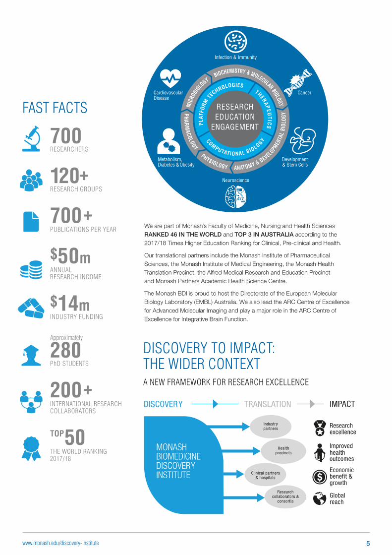
We are part of Monash’s Faculty of Medicine, Nursing and Health Sciences RANKED 46 IN THE WORLD and TOP 3 IN AUSTRALIA according to the 2017/18 Times Higher Education Ranking for Clinical, Pre-clinical and Health.
Our translational partners include the Monash Institute of Pharmaceutical Sciences, the Monash Institute of Medical Engineering, the Monash Health Translation Precinct, the Alfred Medical Research and Education Precinct and Monash Partners Academic Health Science Centre.
The Monash BDI is proud to host the Directorate of the European Molecular Biology Laboratory (EMBL) Australia. We also lead the ARC Centre of Excellence for Advanced Molecular Imaging and play a major role in the ARC Centre of Excellence for Integrative Brain Function.
700 +PUBLICATIONS PER YEAR
200 +INTERNATIONAL RESEARCH COLLABORATORS
TOP 50
THE WORLD RANKING 2017/18
$50m ANNUAL RESEARCH INCOME
700 RESEARCHERS
120+ RESEARCH GROUPS
Approximately
280 PhD STUDENTS
$14m INDUSTRY FUNDING
Research excellence
Improved healthoutcomes
Economic benefit & growth
Global reach
DISCOVERY IMPACT
Research collaborators &
consortia
Industrypartners
Health precincts
Clinical partners & hospitals
TRANSLATION
MONASHBIOMEDICINEDISCOVERYINSTITUTE
5www.monash.edu/discovery-institute
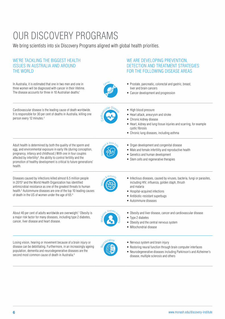
WE’RE TACKLING THE BIGGEST HEALTH ISSUES IN AUSTRALIA AND AROUND THE WORLD
WE ARE DEVELOPING PREVENTION, DETECTION AND TREATMENT STRATEGIES FOR THE FOLLOWING DISEASE AREAS
In Australia, it is estimated that one in two men and one in three women will be diagnosed with cancer in their lifetime. The disease accounts for three in 10 Australian deaths.1
Cancer• Prostate, pancreatic, colorectal and gastric, breast,
liver and brain cancers• Cancer development and progression
Cardiovascular disease is the leading cause of death worldwide. It is responsible for 30 per cent of deaths in Australia, killing one person every 12 minutes.2
Card
iova
scular Disease • High blood pressure
• Heart attack, aneurysm and stroke • Chronic kidney disease• Heart, kidney and lung tissue injuries and scarring, for example
cystic fibrosis• Chronic lung diseases, including asthma
Adult health is determined by both the quality of the sperm and egg, and environmental exposure in early life (during conception, pregnancy, infancy and childhood.) With one in four couples affected by infertility3, the ability to control fertility and the promotion of healthy development is critical to future generations’ health.
Deve
lopm
ent & Stem Cells • Organ development and congenital disease• Male and female infertility and reproductive health• Genetics and human development• Stem cells and regenerative therapies
Diseases caused by infections killed almost 6.5 million people in 20154 and the World Health Organization has identified antimicrobial resistance as one of the greatest threats to human health.5 Autoimmune diseases are one of the top 10 leading causes of death in the US of women under the age of 65.6
Infe
ction
& Immunity • Infectious diseases, caused by viruses, bacteria, fungi or parasites, including HIV, influenza, golden staph, thrush and malaria
• Hospital-acquired infections• Antibiotic-resistant superbugs• Autoimmune diseases
About 40 per cent of adults worldwide are overweight.7 Obesity is a major risk factor for many diseases, including type 2 diabetes, cancer, liver disease and heart disease. M
etab
oli
sm, Diabetes and Obesity
• Obesity and liver disease, cancer and cardiovascular disease• Type 2 diabetes• Obesity and the central nervous system• Mitochondrial disease
Losing vision, hearing or movement because of a brain injury or disease can be debilitating. Furthermore, in an increasingly ageing population, dementia and neurodegenerative diseases are the second most common cause of death in Australia.8
Neuro
science• Nervous system and brain injury • Restoring neural function through brain computer interfaces• Neurodegenerative diseases including Parkinson’s and Alzheimer’s
disease, multiple sclerosis and others
OUR DISCOVERY PROGRAMSWe bring scientists into six Discovery Programs aligned with global health priorities.
6 www.monash.edu/discovery-institute
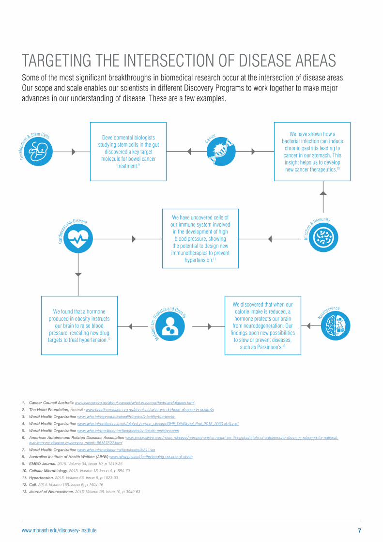
TARGETING THE INTERSECTION OF DISEASE AREASSome of the most significant breakthroughs in biomedical research occur at the intersection of disease areas. Our scope and scale enables our scientists in different Discovery Programs to work together to make major advances in our understanding of disease. These are a few examples.
1. Cancer Council Australia www.cancer.org.au/about-cancer/what-is-cancer/facts-and-figures.html
2. The Heart Foundation, Australia www.heartfoundation.org.au/about-us/what-we-do/heart-disease-in-australia
3. World Health Organization www.who.int/reproductivehealth/topics/infertility/burden/en
4. World Health Organization www.who.int/entity/healthinfo/global_burden_disease/GHE_DthGlobal_Proj_2015_2030.xls?ua=1
5. World Health Organization www.who.int/mediacentre/factsheets/antibiotic-resistance/en
6. American Autoimmune Related Diseases Association www.prnewswire.com/news-releases/comprehensive-report-on-the-global-state-of-autoimmune-diseases-released-for-national-autoimmune-disease-awareness-month-86187622.html
7. World Health Organization www.who.int/mediacentre/factsheets/fs311/en
8. Australian Institute of Health Welfare (AIHW) www.aihw.gov.au/deaths/leading-causes-of-death
9. EMBO Journal. 2015. Volume 34, Issue 10, p 1319-35
10. Cellular Microbiology. 2013. Volume 15, Issue 4, p 554-70
11. Hypertension. 2015. Volume 66, Issue 5, p 1023-33
12. Cell. 2014. Volume 159, Issue 6, p 1404-16
13. Journal of Neuroscience. 2016. Volume 36, Issue 10, p 3049-63
We have shown how a bacterial infection can induce
chronic gastritis leading to cancer in our stomach. This insight helps us to develop new cancer therapeutics.10
We have uncovered cells of our immune system involved in the development of high blood pressure, showing
the potential to design new immunotherapies to prevent
hypertension.11
Developmental biologists studying stem cells in the gut
discovered a key target molecule for bowel cancer
treatment.9
We found that a hormone produced in obesity instructs
our brain to raise blood pressure, revealing new drug targets to treat hypertension.12
Card
iova
sc
ular Disease
Cancer
Metabo
lism
, Diab
etes and Obesity
Infe
ctio
n & Immunity
Neuro
science
Deve
lopm
en
t & Stem Cells
We discovered that when our calorie intake is reduced, a hormone protects our brain
from neurodegeneration. Our findings open new possibilities
to slow or prevent diseases, such as Parkinson’s.13
7www.monash.edu/discovery-institute
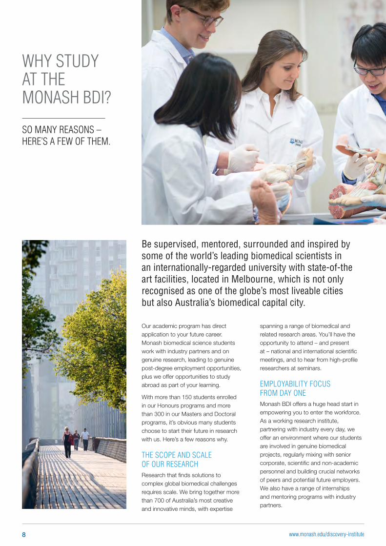
Be supervised, mentored, surrounded and inspired by some of the world’s leading biomedical scientists in an internationally-regarded university with state-of-the art facilities, located in Melbourne, which is not only recognised as one of the globe’s most liveable cities but also Australia’s biomedical capital city.
Our academic program has direct application to your future career. Monash biomedical science students work with industry partners and on genuine research, leading to genuine post-degree employment opportunities, plus we offer opportunities to study abroad as part of your learning.
With more than 150 students enrolled in our Honours programs and more than 300 in our Masters and Doctoral programs, it’s obvious many students choose to start their future in research with us. Here’s a few reasons why.
THE SCOPE AND SCALE OF OUR RESEARCHResearch that finds solutions to complex global biomedical challenges requires scale. We bring together more than 700 of Australia’s most creative and innovative minds, with expertise
WHY STUDY AT THE MONASH BDI?
SO MANY REASONS – HERE’S A FEW OF THEM.
spanning a range of biomedical and related research areas. You’ll have the opportunity to attend – and present at – national and international scientific meetings, and to hear from high-profile researchers at seminars.
EMPLOYABILITY FOCUS FROM DAY ONEMonash BDI offers a huge head start in empowering you to enter the workforce. As a working research institute, partnering with industry every day, we offer an environment where our students are involved in genuine biomedical projects, regularly mixing with senior corporate, scientific and non-academic personnel and building crucial networks of peers and potential future employers. We also have a range of internships and mentoring programs with industry partners.
8 www.monash.edu/discovery-institute
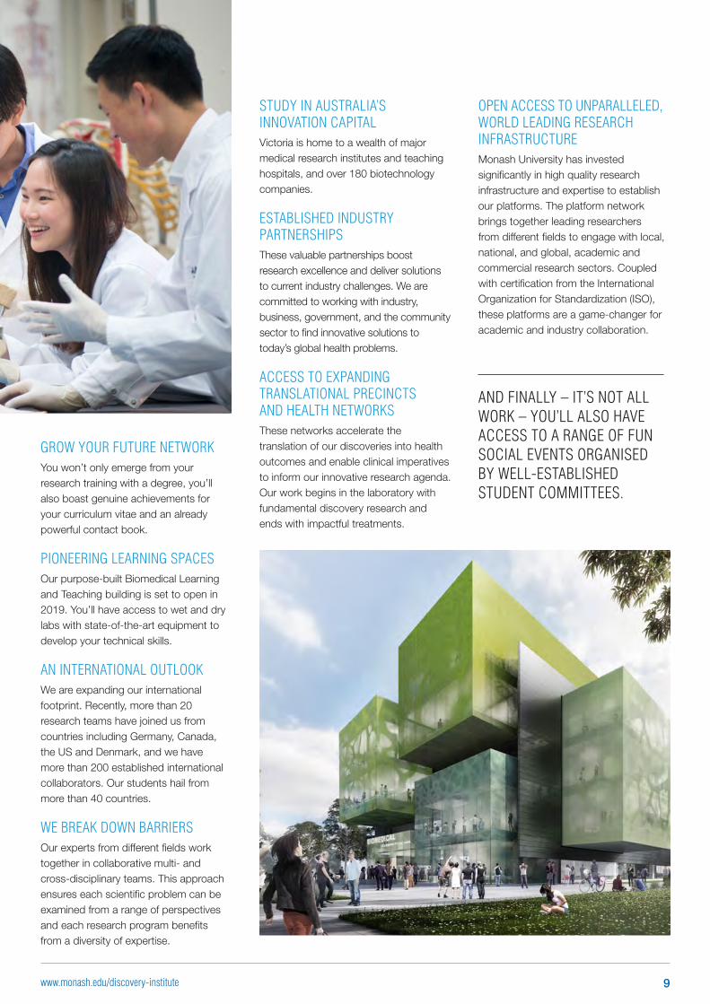
GROW YOUR FUTURE NETWORKYou won’t only emerge from your research training with a degree, you’ll also boast genuine achievements for your curriculum vitae and an already powerful contact book.
PIONEERING LEARNING SPACESOur purpose-built Biomedical Learning and Teaching building is set to open in 2019. You’ll have access to wet and dry labs with state-of-the-art equipment to develop your technical skills.
AN INTERNATIONAL OUTLOOKWe are expanding our international footprint. Recently, more than 20 research teams have joined us from countries including Germany, Canada, the US and Denmark, and we have more than 200 established international collaborators. Our students hail from more than 40 countries.
WE BREAK DOWN BARRIERSOur experts from different fields work together in collaborative multi- and cross-disciplinary teams. This approach ensures each scientific problem can be examined from a range of perspectives and each research program benefits from a diversity of expertise.
STUDY IN AUSTRALIA’S INNOVATION CAPITALVictoria is home to a wealth of major medical research institutes and teaching hospitals, and over 180 biotechnology companies.
ESTABLISHED INDUSTRY PARTNERSHIPSThese valuable partnerships boost research excellence and deliver solutions to current industry challenges. We are committed to working with industry, business, government, and the community sector to find innovative solutions to today’s global health problems.
ACCESS TO EXPANDING TRANSLATIONAL PRECINCTS AND HEALTH NETWORKSThese networks accelerate the translation of our discoveries into health outcomes and enable clinical imperatives to inform our innovative research agenda. Our work begins in the laboratory with fundamental discovery research and ends with impactful treatments.
OPEN ACCESS TO UNPARALLELED, WORLD LEADING RESEARCH INFRASTRUCTUREMonash University has invested significantly in high quality research infrastructure and expertise to establish our platforms. The platform network brings together leading researchers from different fields to engage with local, national, and global, academic and commercial research sectors. Coupled with certification from the International Organization for Standardization (ISO), these platforms are a game-changer for academic and industry collaboration.
AND FINALLY – IT’S NOT ALL WORK – YOU’LL ALSO HAVE ACCESS TO A RANGE OF FUN SOCIAL EVENTS ORGANISED BY WELL-ESTABLISHED STUDENT COMMITTEES.
9www.monash.edu/discovery-institute
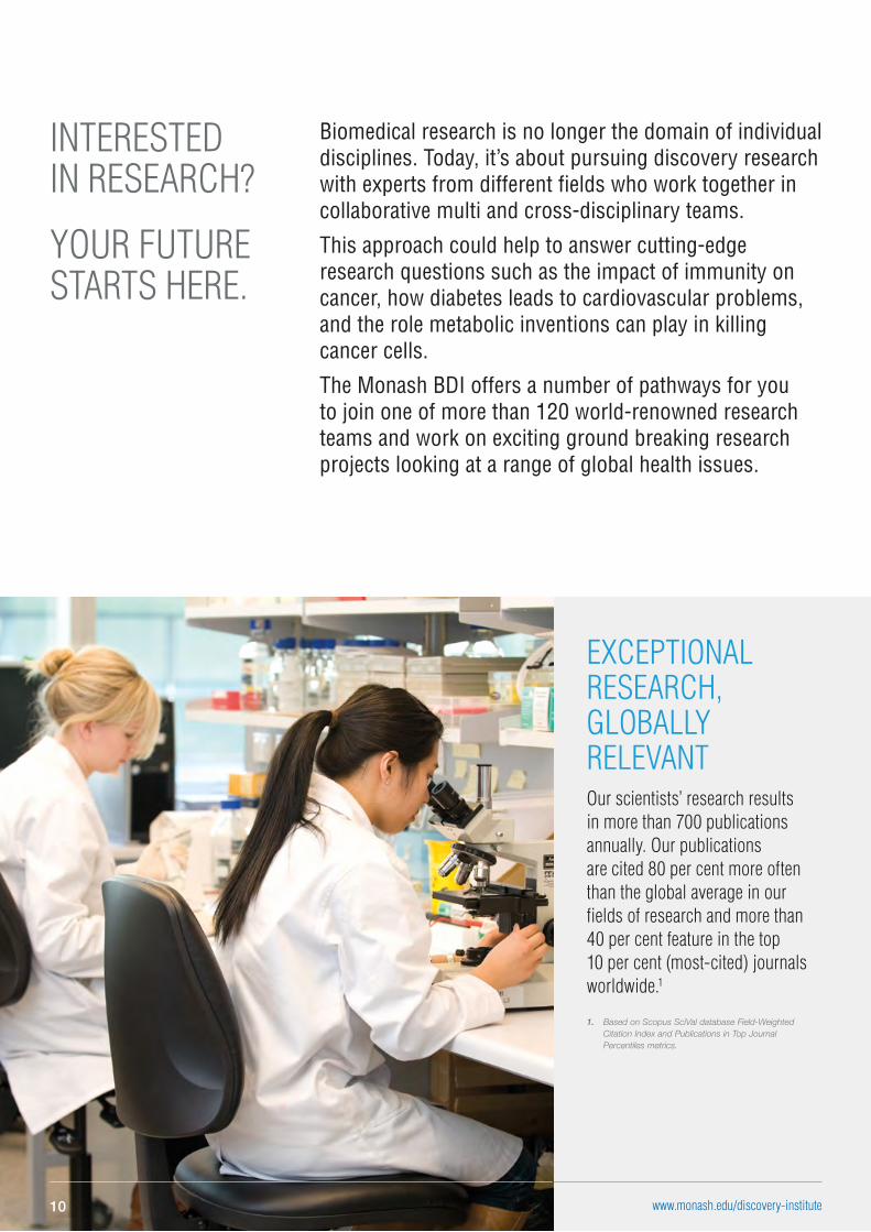
Biomedical research is no longer the domain of individual disciplines. Today, it’s about pursuing discovery research with experts from different fields who work together in collaborative multi and cross-disciplinary teams.
This approach could help to answer cutting-edge research questions such as the impact of immunity on cancer, how diabetes leads to cardiovascular problems, and the role metabolic inventions can play in killing cancer cells.
The Monash BDI offers a number of pathways for you to join one of more than 120 world-renowned research teams and work on exciting ground breaking research projects looking at a range of global health issues.
INTERESTED IN RESEARCH?
YOUR FUTURE STARTS HERE.
EXCEPTIONAL RESEARCH, GLOBALLY RELEVANT Our scientists’ research results in more than 700 publications annually. Our publications are cited 80 per cent more often than the global average in our fields of research and more than 40 per cent feature in the top 10 per cent (most-cited) journals worldwide.1
1. Based on Scopus SciVal database Field-Weighted Citation Index and Publications in Top Journal Percentiles metrics.
www.monash.edu/discovery-institute10
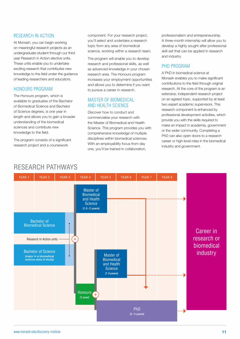
RESEARCH IN ACTIONAt Monash, you can begin working on meaningful research projects as an undergraduate student through our third year Research in Action elective units. These units enable you to undertake exciting research that contributes new knowledge to the field under the guidance of leading researchers and educators.
HONOURS PROGRAMThe Honours program, which is available to graduates of the Bachelor of Biomedical Science and Bachelor of Science degrees, is one year in length and allows you to gain a broader understanding of the biomedical sciences and contribute new knowledge to the field.
The program consists of a significant research project and a coursework
professionalism and entrepreneurship. A three-month internship will allow you to develop a highly sought after professional skill-set that can be applied in research and industry.
PHD PROGRAMA PhD in biomedical science at Monash enables you to make significant contributions to the field through original research. At the core of the program is an extensive, independent research project on an agreed topic, supported by at least two expert academic supervisors. This research component is enhanced by professional development activities, which provide you with the skills required to make an impact in academia, government or the wider community. Completing a PhD can also open doors to a research career or high-level roles in the biomedical industry and government.
component. For your research project, you’ll select and undertake a research topic from any area of biomedical science, working within a research team.
The program will enable you to develop research and professional skills, as well as advanced knowledge in your chosen research area. The Honours program increases your employment opportunities and allows you to determine if you want to pursue a career in research.
MASTER OF BIOMEDICAL AND HEALTH SCIENCEDiscover how to conduct and commercialise your research with the Master of Biomedical and Health Science. This program provides you with comprehensive knowledge of multiple disciplines within biomedical sciences. With an employability focus from day one, you’ll be trained in collaboration,
RESEARCH PATHWAYSYEAR 1 YEAR 2 YEAR 3 YEAR 4 YEAR 5 YEAR 6 YEAR 7 YEAR 8
Master of Biomedical and Health
Science(1.5 years)
Master of Biomedical and Health
Science(1.5 - 2 years)
PhD(3 - 4 years)
Honours(1 year)
Career in research or biomedical
industry
Bachelor of Biomedical Science
Bachelor of Science(major in a biomedical science area of study)
Research in Action units
11www.monash.edu/discovery-institute
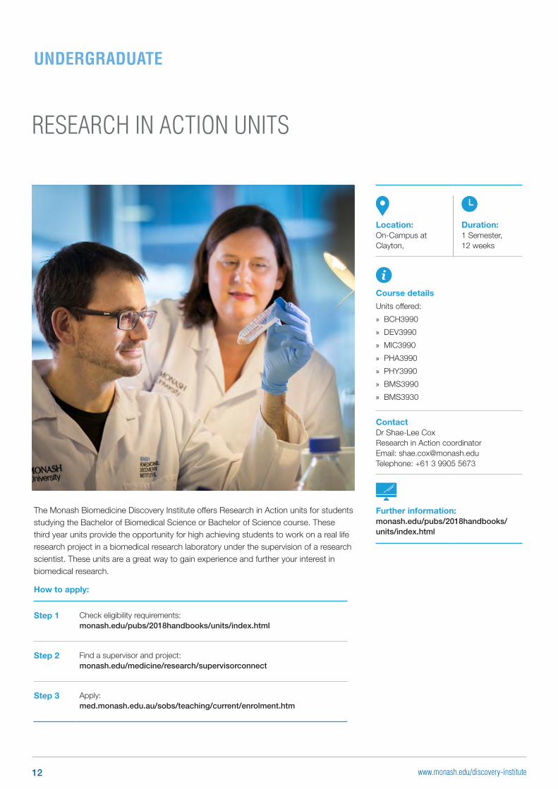
RESEARCH IN ACTION UNITS
The Monash Biomedicine Discovery Institute offers Research in Action units for students studying the Bachelor of Biomedical Science or Bachelor of Science course. These third year units provide the opportunity for high achieving students to work on a real life research project in a biomedical research laboratory under the supervision of a research scientist. These units are a great way to gain experience and further your interest in biomedical research.
How to apply:
Step 1 Check eligibility requirements: monash.edu/pubs/2018handbooks/units/index.html
Step 2 Find a supervisor and project: monash.edu/medicine/research/supervisorconnect
Step 3 Apply: med.monash.edu.au/sobs/teaching/current/enrolment.htm
UNDERGRADUATE
Location:On-Campus at Clayton,
Duration:1 Semester, 12 weeks
Course details
Units offered:
» BCH3990
» DEV3990
» MIC3990
» PHA3990
» PHY3990
» BMS3990
» BMS3930
ContactDr Shae-Lee CoxResearch in Action coordinatorEmail: [email protected] Telephone: +61 3 9905 5673
Further information:monash.edu/pubs/2018handbooks/units/index.html
12 www.monash.edu/discovery-institute
SUMMER AND WINTER RESEARCH SCHOLARSHIP PROGRAM
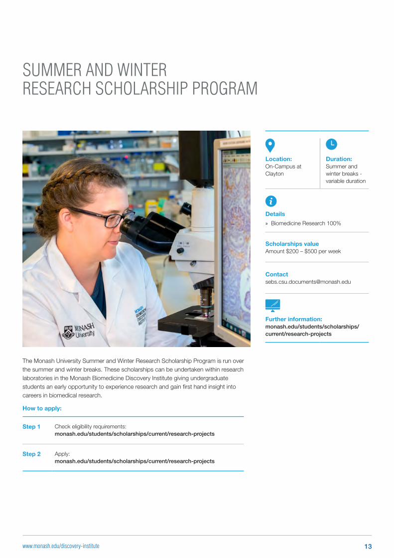
The Monash University Summer and Winter Research Scholarship Program is run over the summer and winter breaks. These scholarships can be undertaken within research laboratories in the Monash Biomedicine Discovery Institute giving undergraduate students an early opportunity to experience research and gain first hand insight into careers in biomedical research.
How to apply:
Step 1 Check eligibility requirements: monash.edu/students/scholarships/current/research-projects
Step 2 Apply:monash.edu/students/scholarships/current/research-projects
Location:On-Campus at Clayton
Duration:Summer and winter breaks - variable duration
Details
» Biomedicine Research 100%
Scholarships valueAmount $200 – $500 per week
Further information:monash.edu/students/scholarships/current/research-projects
13www.monash.edu/discovery-institute
BACHELOR OF BIOMEDICAL SCIENCE HONOURS (BBiomedSc (Hons))
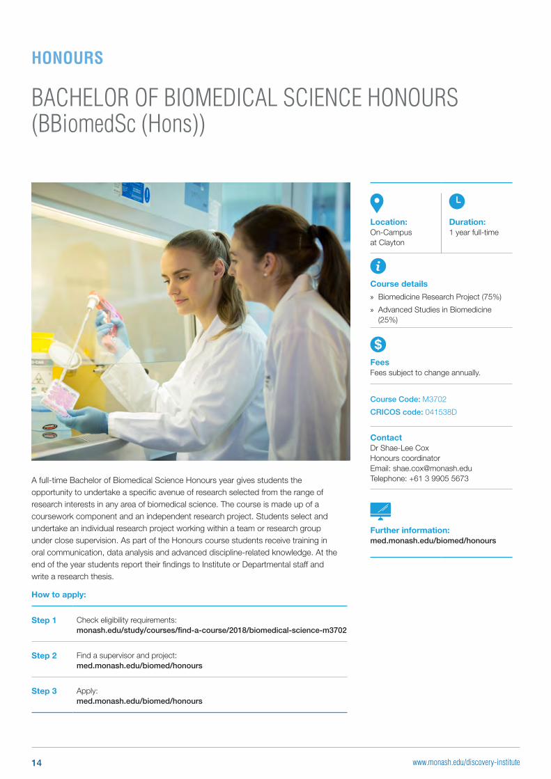
A full-time Bachelor of Biomedical Science Honours year gives students the opportunity to undertake a specific avenue of research selected from the range of research interests in any area of biomedical science. The course is made up of a coursework component and an independent research project. Students select and undertake an individual research project working within a team or research group under close supervision. As part of the Honours course students receive training in oral communication, data analysis and advanced discipline-related knowledge. At the end of the year students report their findings to Institute or Departmental staff and write a research thesis.
How to apply:
Step 1 Check eligibility requirements: monash.edu/study/courses/find-a-course/2018/biomedical-science-m3702
Step 2 Find a supervisor and project: med.monash.edu/biomed/honours
Step 3 Apply: med.monash.edu/biomed/honours
Location:On-Campus at Clayton
Duration:1 year full-time
Course details
» Biomedicine Research Project (75%)
» Advanced Studies in Biomedicine (25%)
FeesFees subject to change annually.
Course Code: M3702
CRICOS code: 041538D
ContactDr Shae-Lee Cox Honours coordinator Email: [email protected] Telephone: +61 3 9905 5673
Further information:med.monash.edu/biomed/honours
HONOURS
14 www.monash.edu/discovery-institute
BACHELOR OF SCIENCE HONOURS (BSc (Hons))
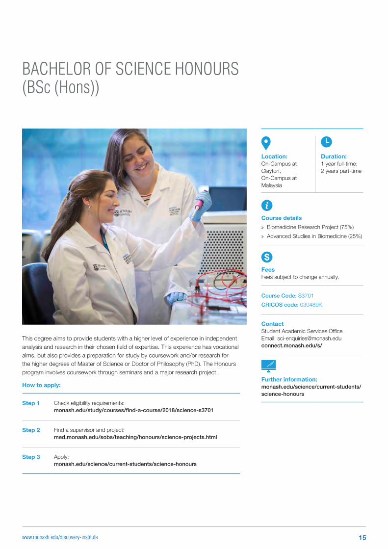
This degree aims to provide students with a higher level of experience in independent analysis and research in their chosen field of expertise. This experience has vocational aims, but also provides a preparation for study by coursework and/or research for the higher degrees of Master of Science or Doctor of Philosophy (PhD). The Honours program involves coursework through seminars and a major research project.
How to apply:
Step 1 Check eligibility requirements: monash.edu/study/courses/find-a-course/2018/science-s3701
Step 2 Find a supervisor and project: med.monash.edu/sobs/teaching/honours/science-projects.html
Step 3 Apply: monash.edu/science/current-students/science-honours
Location:On-Campus at Clayton, On-Campus at Malaysia
Duration:1 year full-time; 2 years part-time
Course details
» Biomedicine Research Project (75%)
» Advanced Studies in Biomedicine (25%)
FeesFees subject to change annually.
Course Code: S3701
CRICOS code: 030489K
ContactStudent Academic Services Office Email: [email protected]/s/
Further information:monash.edu/science/current-students/science-honours
15www.monash.edu/discovery-institute
BACHELOR OF MEDICAL SCIENCE (HONOURS) (BMedSc (Hons))
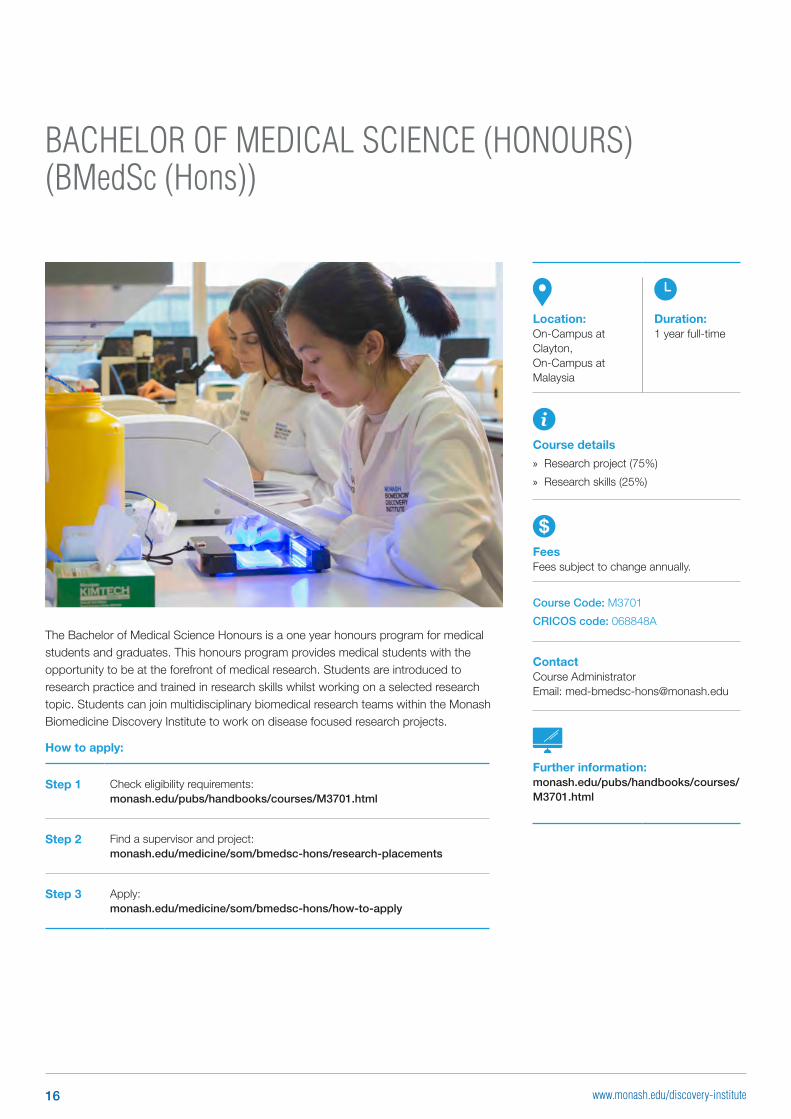
The Bachelor of Medical Science Honours is a one year honours program for medical students and graduates. This honours program provides medical students with the opportunity to be at the forefront of medical research. Students are introduced to research practice and trained in research skills whilst working on a selected research topic. Students can join multidisciplinary biomedical research teams within the Monash Biomedicine Discovery Institute to work on disease focused research projects.
How to apply:
Step 1 Check eligibility requirements: monash.edu/pubs/handbooks/courses/M3701.html
Step 2 Find a supervisor and project: monash.edu/medicine/som/bmedsc-hons/research-placements
Step 3 Apply: monash.edu/medicine/som/bmedsc-hons/how-to-apply
Location:On-Campus at Clayton, On-Campus at Malaysia
Duration:1 year full-time
Course details
» Research project (75%)
» Research skills (25%)
FeesFees subject to change annually.
Course Code: M3701
CRICOS code: 068848A
ContactCourse AdministratorEmail: [email protected]
Further information:monash.edu/pubs/handbooks/courses/M3701.html
16 www.monash.edu/discovery-institute
MASTER OF BIOMEDICAL AND HEALTH SCIENCE (MBiomedHlthSc)
How to apply:
Step 1 Check eligibility requirements for this degree: monash.edu/pubs/handbooks/courses/M6003.html
Step 2 Check eligibility for scholarships: study.monash/fees-scholarships/scholarships
Step 3 Apply: monash.edu/admissions/apply/online
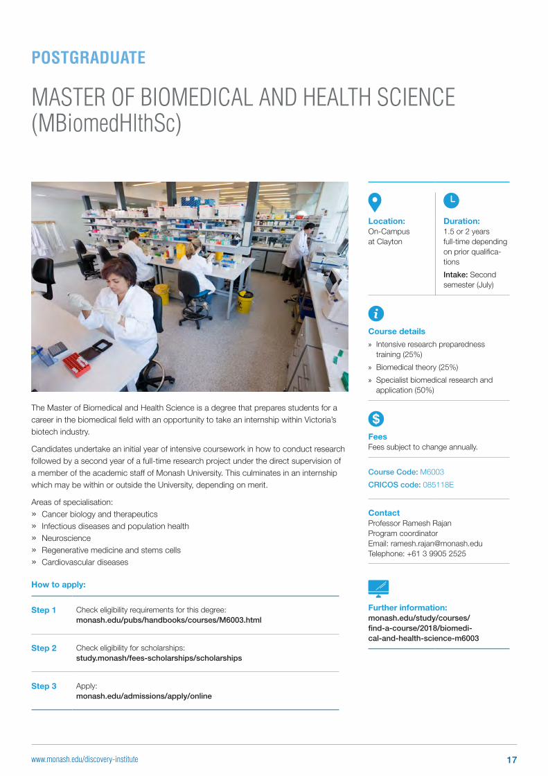
Location:On-Campus at Clayton
Duration:1.5 or 2 years full-time depending on prior qualifica-tions
Intake: Second semester (July)
Course details
» Intensive research preparedness training (25%)
» Biomedical theory (25%)
» Specialist biomedical research and application (50%)
FeesFees subject to change annually.
Course Code: M6003
CRICOS code: 085118E
ContactProfessor Ramesh Rajan Program coordinator Email: [email protected] Telephone: +61 3 9905 2525
Further information:monash.edu/study/courses/ find-a-course/2018/biomedi-cal-and-health-science-m6003
The Master of Biomedical and Health Science is a degree that prepares students for a career in the biomedical field with an opportunity to take an internship within Victoria’s biotech industry.
Candidates undertake an initial year of intensive coursework in how to conduct research followed by a second year of a full-time research project under the direct supervision of a member of the academic staff of Monash University. This culminates in an internship which may be within or outside the University, depending on merit.
Areas of specialisation:
» Cancer biology and therapeutics
» Infectious diseases and population health
» Neuroscience
» Regenerative medicine and stems cells
» Cardiovascular diseases
POSTGRADUATE
17www.monash.edu/discovery-institute
DOCTORAL PROGRAM IN BIOMEDICAL SCIENCES (PHD)
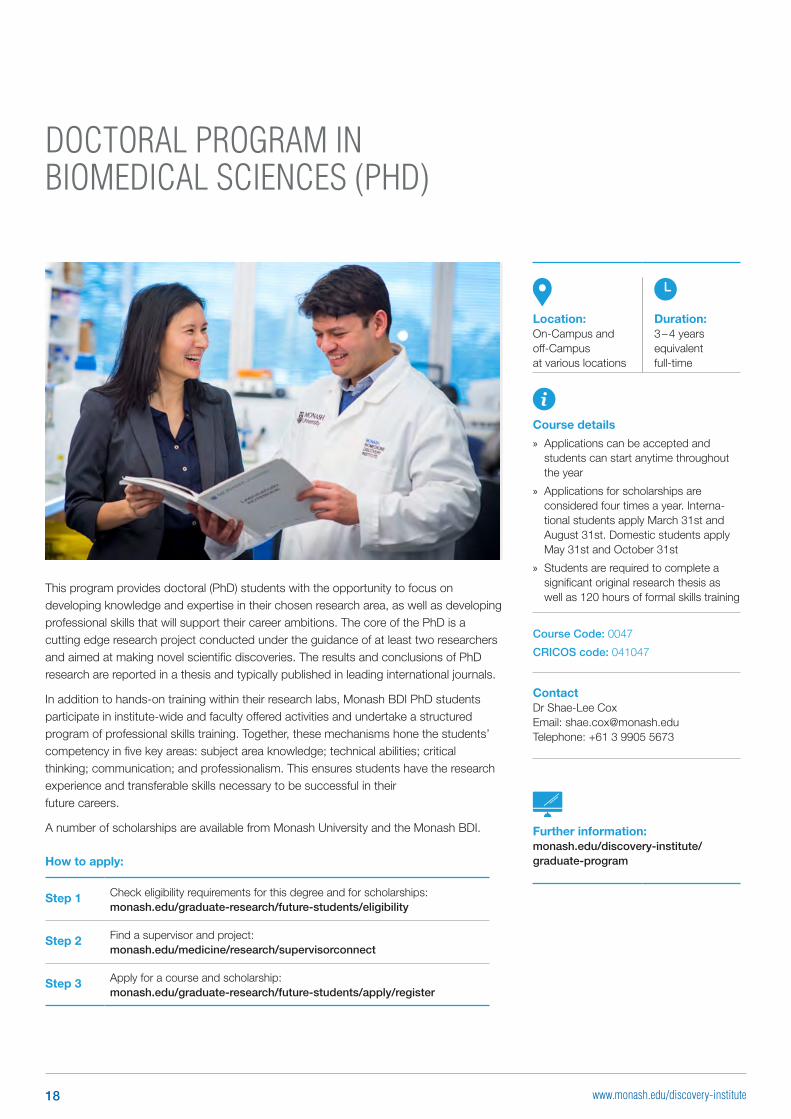
This program provides doctoral (PhD) students with the opportunity to focus on developing knowledge and expertise in their chosen research area, as well as developing professional skills that will support their career ambitions. The core of the PhD is a cutting edge research project conducted under the guidance of at least two researchers and aimed at making novel scientific discoveries. The results and conclusions of PhD research are reported in a thesis and typically published in leading international journals.
In addition to hands-on training within their research labs, Monash BDI PhD students participate in institute-wide and faculty offered activities and undertake a structured program of professional skills training. Together, these mechanisms hone the students’ competency in five key areas: subject area knowledge; technical abilities; critical thinking; communication; and professionalism. This ensures students have the research experience and transferable skills necessary to be successful in their future careers.
A number of scholarships are available from Monash University and the Monash BDI.
How to apply:
Step 1 Check eligibility requirements for this degree and for scholarships: monash.edu/graduate-research/future-students/eligibility
Step 2 Find a supervisor and project: monash.edu/medicine/research/supervisorconnect
Step 3 Apply for a course and scholarship: monash.edu/graduate-research/future-students/apply/register
Location:On-Campus and off-Campus at various locations
Duration:3 – 4 years equivalent full-time
Course details
» Applications can be accepted and students can start anytime throughout the year
» Applications for scholarships are considered four times a year. Interna-tional students apply March 31st and August 31st. Domestic students apply May 31st and October 31st
» Students are required to complete a significant original research thesis as well as 120 hours of formal skills training
Course Code: 0047
CRICOS code: 041047
ContactDr Shae-Lee Cox Email: [email protected] Telephone: +61 3 9905 5673
Further information:monash.edu/discovery-institute/ graduate-program
18 www.monash.edu/discovery-institute

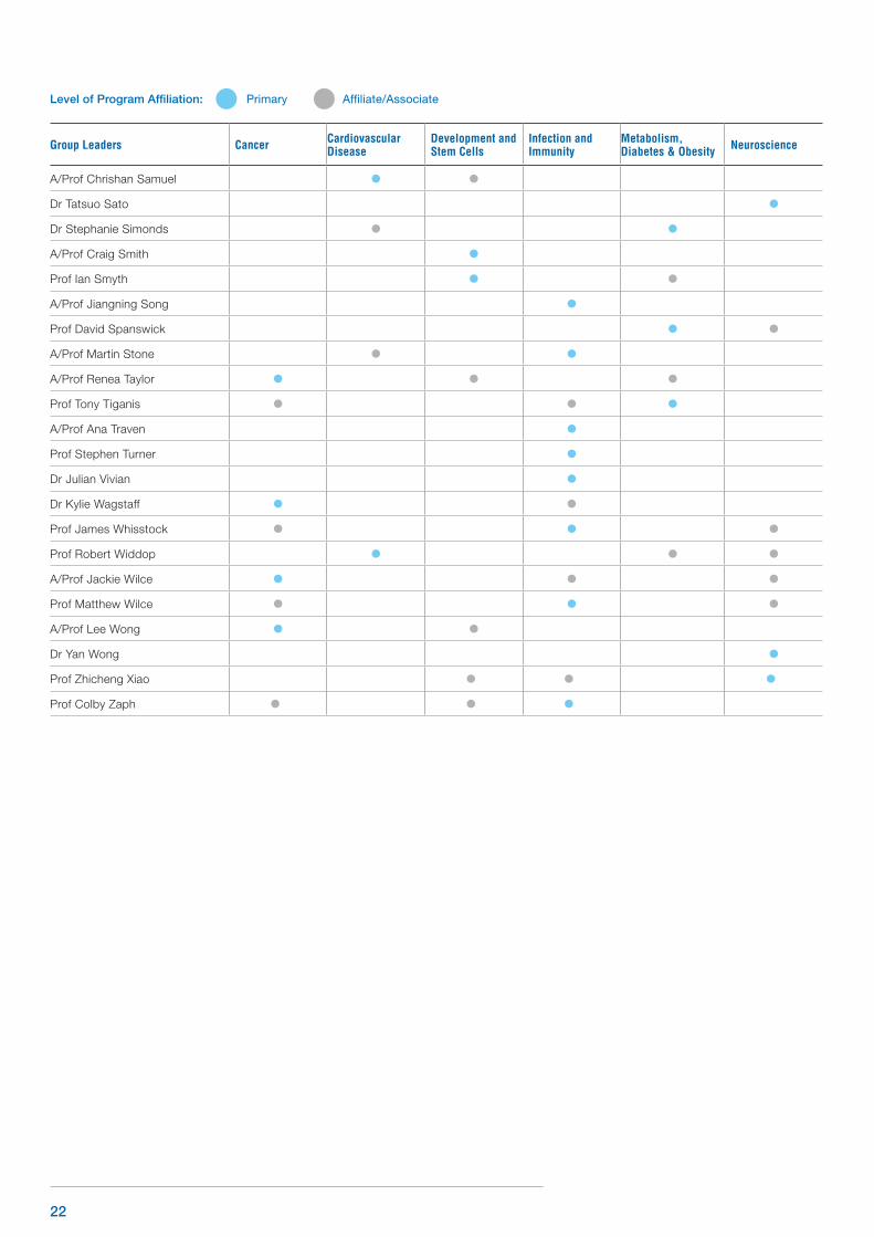
MONASH BDI GROUP LEADERS AND THEIR DISCOVERY PROGRAM AFFILIATIONSFor more information, please visit monash.edu/discovery-institute/our-people
Group Leaders Cancer Cardiovascular Disease
Development and Stem Cells
Infection and Immunity
Metabolism, Diabetes & Obesity Neuroscience
A/Prof Helen Abud
Dr Justin Adams
Prof Mibel Aguilar
A/Prof Zane Andrews
A/Prof Traude Beilharz
Dr Richard Berry
Prof John Bertram
Prof Phil Bird
Prof Jane (Mary) Black
Dr Peter Boag
Dr Natalie Borg
Dr Jane Bourke
A/Prof John Boyce
Dr Bradley Broughton
A/Prof Ashley Buckle
A/Prof Irina Caminschi
Prof John Carroll
A/Prof Siew Chai
A/Prof Ann Chidgey
Prof Iain Clarke AM
A/Prof Tim Cole
Prof Brian Cooke
A/Prof Fasseli Coulibaly
Prof Michael Cowley
A/Prof Max Cryle
Dr Stephen Daley
Prof Roger Daly
Dr Partha Pratim Das
A/Prof Chen Davidovich
Prof Mariapia Degli-Esposti
Prof David de Kretser
A/Prof Alex de Marco
Level of Program Affiliation: Primary Affiliate/Associate
19www.monash.edu/discovery-institute
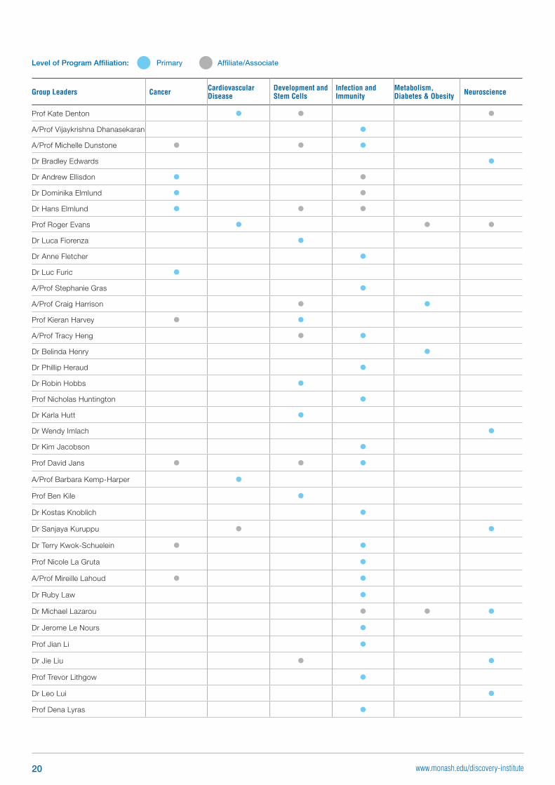
Group Leaders Cancer Cardiovascular Disease
Development and Stem Cells
Infection and Immunity
Metabolism, Diabetes & Obesity Neuroscience
Prof Kate Denton
A/Prof Vijaykrishna Dhanasekaran
A/Prof Michelle Dunstone
Dr Bradley Edwards
Dr Andrew Ellisdon
Dr Dominika Elmlund
Dr Hans Elmlund
Prof Roger Evans
Dr Luca Fiorenza
Dr Anne Fletcher
Dr Luc Furic
A/Prof Stephanie Gras
A/Prof Craig Harrison
Prof Kieran Harvey
A/Prof Tracy Heng
Dr Belinda Henry
Dr Phillip Heraud
Dr Robin Hobbs
Prof Nicholas Huntington
Dr Karla Hutt
Dr Wendy Imlach
Dr Kim Jacobson
Prof David Jans
A/Prof Barbara Kemp-Harper
Prof Ben Kile
Dr Kostas Knoblich
Dr Sanjaya Kuruppu
Dr Terry Kwok-Schuelein
Prof Nicole La Gruta
A/Prof Mireille Lahoud
Dr Ruby Law
Dr Michael Lazarou
Dr Jerome Le Nours
Prof Jian Li
Dr Jie Liu
Prof Trevor Lithgow
Dr Leo Lui
Prof Dena Lyras
Level of Program Affiliation: Primary Affiliate/Associate
20 www.monash.edu/discovery-institute
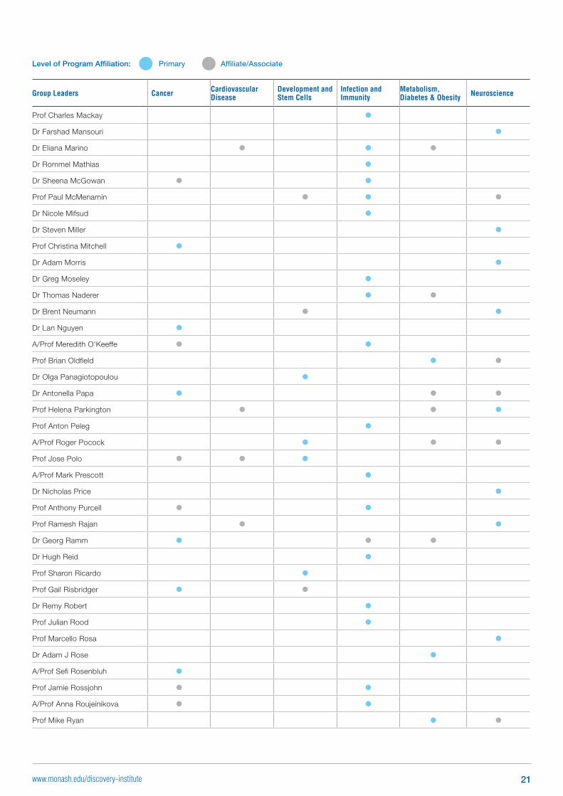
Group Leaders Cancer Cardiovascular Disease
Development and Stem Cells
Infection and Immunity
Metabolism, Diabetes & Obesity Neuroscience
Prof Charles Mackay
Dr Farshad Mansouri
Dr Eliana Marino
Dr Rommel Mathias
Dr Sheena McGowan
Prof Paul McMenamin
Dr Nicole Mifsud
Dr Steven Miller
Prof Christina Mitchell
Dr Adam Morris
Dr Greg Moseley
Dr Thomas Naderer
Dr Brent Neumann
Dr Lan Nguyen
A/Prof Meredith O'Keeffe
Prof Brian Oldfield
Dr Olga Panagiotopoulou
Dr Antonella Papa
Prof Helena Parkington
Prof Anton Peleg
A/Prof Roger Pocock
Prof Jose Polo
A/Prof Mark Prescott
Dr Nicholas Price
Prof Anthony Purcell
Prof Ramesh Rajan
Dr Georg Ramm
Dr Hugh Reid
Prof Sharon Ricardo
Prof Gail Risbridger
Dr Remy Robert
Prof Julian Rood
Prof Marcello Rosa
Dr Adam J Rose
A/Prof Sefi Rosenbluh
Prof Jamie Rossjohn
A/Prof Anna Roujeinikova
Prof Mike Ryan
Level of Program Affiliation: Primary Affiliate/Associate
21www.monash.edu/discovery-institute
Group Leaders Cancer Cardiovascular Disease
Development and Stem Cells
Infection and Immunity
Metabolism, Diabetes & Obesity Neuroscience
A/Prof Chrishan Samuel
Dr Tatsuo Sato
Dr Stephanie Simonds
A/Prof Craig Smith
Prof Ian Smyth
A/Prof Jiangning Song
Prof David Spanswick
A/Prof Martin Stone
A/Prof Renea Taylor
Prof Tony Tiganis
A/Prof Ana Traven
Prof Stephen Turner
Dr Julian Vivian
Dr Kylie Wagstaff
Prof James Whisstock
Prof Robert Widdop
A/Prof Jackie Wilce
Prof Matthew Wilce
A/Prof Lee Wong
Dr Yan Wong
Prof Zhicheng Xiao
Prof Colby Zaph
Level of Program Affiliation: Primary Affiliate/Associate
22
Cancer Program Group Leaders
23www.monash.edu/discovery-institute
Selected significant publications: 1. Fleuren EDG, Vlenterie M, van der
Graaf WTA, Hillebrandt-Roeffen MHS, Blackburn J, Ma X, Chan H, Magias MC, van Erp A, van Houdt L, Cebeci SAS, van de Ven A, Flucke UE, Heyer EE, Thomas D, Lord CJ, Marini KD, Vaghjiani V, Mercer TR, Cain JE, Wu J, Versleijen-Jonkers YMH and Daly RJ. 2017. Phosphoproteomic profiling across sarcoma subtypes reveals ALK and MET as novel actionable targets in synovial sarcomas. Cancer Research (In-press)
2. Fleuren ED, Zhang L, Wu J, Daly RJ. 2016. The kinome ‘at large’ in cancer. Nat Rev Cancer 16(2):83-98
3. Croucher DR, Hochgräfe F, Zhang L, Liu L, Lyons RJ, Rickwood D, Tactacan CM, Browne BC, Ali N, Chan H, Shearer R, Gallego-Ortega D, Saunders DN, Swarbrick A, Daly RJ. 2013. Involvement of Lyn and the atypical kinase SgK269/PEAK1 in a basal breast cancer signaling pathway. Cancer Res 73(6):1969-80
4. Zheng Y, Zhang C, Croucher DR, Soliman MA, St-Denis N, Pasculescu A, Taylor L, Tate SA, Hardy WR, Colwill K, Dai AY, Bagshaw R, Dennis JW, Gingras AC, Daly RJ, Pawson T. 2013. Temporal regulation of EGF signalling networks by the scaffold protein Shc1. Nature. 499(7457):166-71.
5. Hochgrafe F, Zhang L, O’Toole SA, Browne BC, Pinese M, Porta Cubas A, Lehrbach GM, Croucher DR, Rickwood D, Boulghourjian A, Shearer R, Nair R, Swarbrick A, Faratian D, Mullen P, Harrison DJ, Biankin AV, Sutherland RL, Raftery MJ, Daly RJ. 2010. Tyrosine phosphorylation profiling reveals the signaling network characteristics of Basal breast cancer cells. Cancer Res 70(22):9391-401.
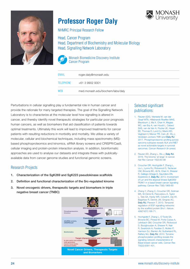
Perturbations in cellular signalling play a fundamental role in human cancer and
provide the rationale for many targeted therapies. The goal of the Signalling Network
Laboratory is to characterize at the molecular level how signalling is altered in
cancer, and thereby identify novel therapeutic strategies for particular poor prognosis
human cancers, as well as biomarkers that aid classification of patients towards
optimal treatments. Ultimately this work will lead to improved treatments for cancer
patients with resulting reductions in morbidity and mortality. We utilise a variety of
molecular, cellular and biochemical techniques, including mass spectrometry (MS)-
based phosphoproteomics and kinomics, siRNA library screens and CRISPR/Cas9,
cellular imaging and protein-protein interaction analysis. In addition, bioinformatic
approaches are used to analyse our datasets and integrate these with publically-
available data from cancer genome studies and functional genomic screens.
Research Projects
1. Characterization of the SgK269 and SgK223 pseudokinase scaffolds
2. Definition and functional characterization of the Src-regulated kinome
3. Novel oncogenic drivers, therapeutic targets and biomarkers in triple
negative breast cancer (TNBC)
Professor Roger DalyNHMRC Principal Research Fellow
Head, Cancer Program Head, Department of Biochemistry and Molecular Biology Head, Signalling Network Laboratory
Monash Biomedicine Discovery Institute Cancer Program
EMAIL [email protected]
TELEPHONE +61 3 9902 9301
WEB med.monash.edu/biochem/labs/daly
MS-based proteomicsPhosphoproteomicsKinomics
Novel Cancer Drivers, Therapeutic Targets and Biomarkers
Data integration
Bioinformatics
Target characterization and validationIn vitro and in vivo modelsmAb generationCancer tissue banksStructural studies
Functional genomicssh/siRNA screens, CRISPR/Cas9
TNS3 PLCG1
IRS4
CRKLMET
ARHGEF2
MAPK8
NEDD9
PTK2 TGFB1I1BCAR1
DDX39A
GAB1
HNRNPA2B1
ERBB3PIK3R2
24 www.monash.edu/discovery-institute
Selected significant publications: 1. Wang X, Goodrich KJ, Gooding AR,
Naeem H, Archer S, Paucek RD, Youmans DT, Cech TR, Davidovich C. 2017. Targeting of Polycomb repressive complex 2 to RNA by short repeats of consecutive guanines. Mol Cell 65(6)1056-1067.
2. Lu Z, Zhang QC, Lee B, Flynn RA, Smith MA, Robinson JT, Davidovich C, Gooding AR, Goodrich KJ, Mattick JS, Mesirov JP, Cech TR, Chang HY. 2016. RNA Duplex Map in Living Cells Reveals Higher-Order Transcriptome Structure. Cell 165(5):1267-79.
3. Davidovich C, Wang X, Cifuentes-Rojas C, Goodrich KJ, Gooding AR, Lee JT, Cech TR. 2015. Toward a consensus on the binding specificity and promiscuity of PRC2 for RNA. Mol Cell. 57(3),552-8.
4. Davidovich C, Zheng L, Goodrich KJ, Cech TR. 2013. Promiscuous RNA binding by Polycomb repressive complex 2. Nat Struct Mol Biol. 20(11),1250-7.
5. Davidovich C, Bashan A, Yonath A. 2008. Structural basis for cross-resistance to ribosomal PTC antibiotics. Proc Natl Acad Sci USA.105(52), 20665-70.
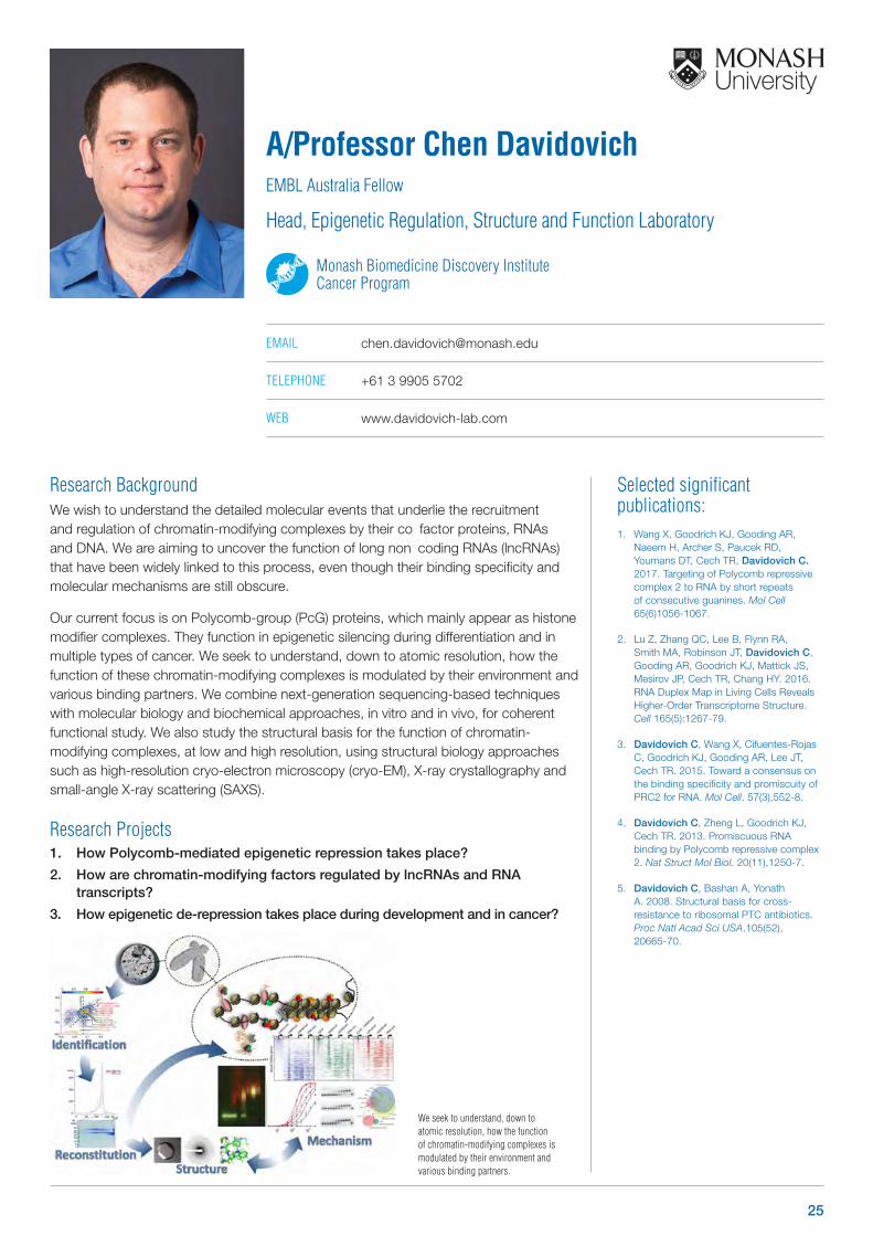
Research BackgroundWe wish to understand the detailed molecular events that underlie the recruitment and regulation of chromatin-modifying complexes by their co‐factor proteins, RNAs and DNA. We are aiming to uncover the function of long non‐coding RNAs (lncRNAs) that have been widely linked to this process, even though their binding specificity and molecular mechanisms are still obscure.
Our current focus is on Polycomb-group (PcG) proteins, which mainly appear as histone modifier complexes. They function in epigenetic silencing during differentiation and in multiple types of cancer. We seek to understand, down to atomic resolution, how the function of these chromatin-modifying complexes is modulated by their environment and various binding partners. We combine next-generation sequencing-based techniques with molecular biology and biochemical approaches, in vitro and in vivo, for coherent functional study. We also study the structural basis for the function of chromatin-modifying complexes, at low and high resolution, using structural biology approaches such as high-resolution cryo-electron microscopy (cryo-EM), X-ray crystallography and small-angle X-ray scattering (SAXS).
Research Projects1. How Polycomb-mediated epigenetic repression takes place?
2. How are chromatin-modifying factors regulated by lncRNAs and RNA transcripts?
3. How epigenetic de-repression takes place during development and in cancer?
A/Professor Chen DavidovichEMBL Australia Fellow
Head, Epigenetic Regulation, Structure and Function Laboratory
Monash Biomedicine Discovery Institute Cancer Program
EMAIL [email protected]
TELEPHONE +61 3 9905 5702
WEB www.davidovich-lab.com
We seek to understand, down to atomic resolution, how the function of chromatin-modifying complexes is modulated by their environment and various binding partners.
25
OTHER PROGRAM AFFILIATIONS
Infection and Immunity
Selected significant publications: 1. Ellisdon A, Nold-Petry C, D’Andrea
L, Cho S, Lao J, Rudloff I, Ngo D, Lo C, Soares da Costa T, Perugini M, Conroy P, Whisstock J, Nold M. 2017. Homodimerization attenuates the anti-inflammatory activity of interleukin-37. Sci Immunol. Feb 10;2(8).
2. Ellisdon A, Reboul C, Panjikar S, Huynh K, Oellig CA, Winter K, Dunstone M, Hodgson W, Seymour J, Dearden P, Tweten R, Whisstock J, McGowan S. 2016. Stonefish toxin defines an ancient branch of the perforin-like superfamily. PNAS USA. Dec 15;112(50):15360-5.
3. Lucato C, Halls ML Ooms L, Liu H, Mitchell C, Whisstock J, Ellisdon A. 2015. The Phosphatidylinositol (3,4,5)-Trisphosphate-dependent Rac Exchanger 1·Ras-related C3 Botulinum Toxin Substrate 1 (PREX1·Rac1) Complex Reveals the Basis of Rac1 Activation in Breast Cancer Cells. JBC. Aug 21;290(34):20827-40.
4. Lupton C, Steer D, Wintrode P, Bottomley S, Hughes V, Ellisdon A. 2015. Enhanced molecular mobility of ordinarily structured regions drives polyglutamine disease. JBC. Oct 2;290(40):24190-200.
5. Ellisdon A, Dimitrova L, Hurt E, Stewart M. 2012. Structural basis for the assembly and nucleic acid binding of the TREX-2 transcription-export complex. Nature Structural and Molecular Biology. Feb 19;19(3):328-36.
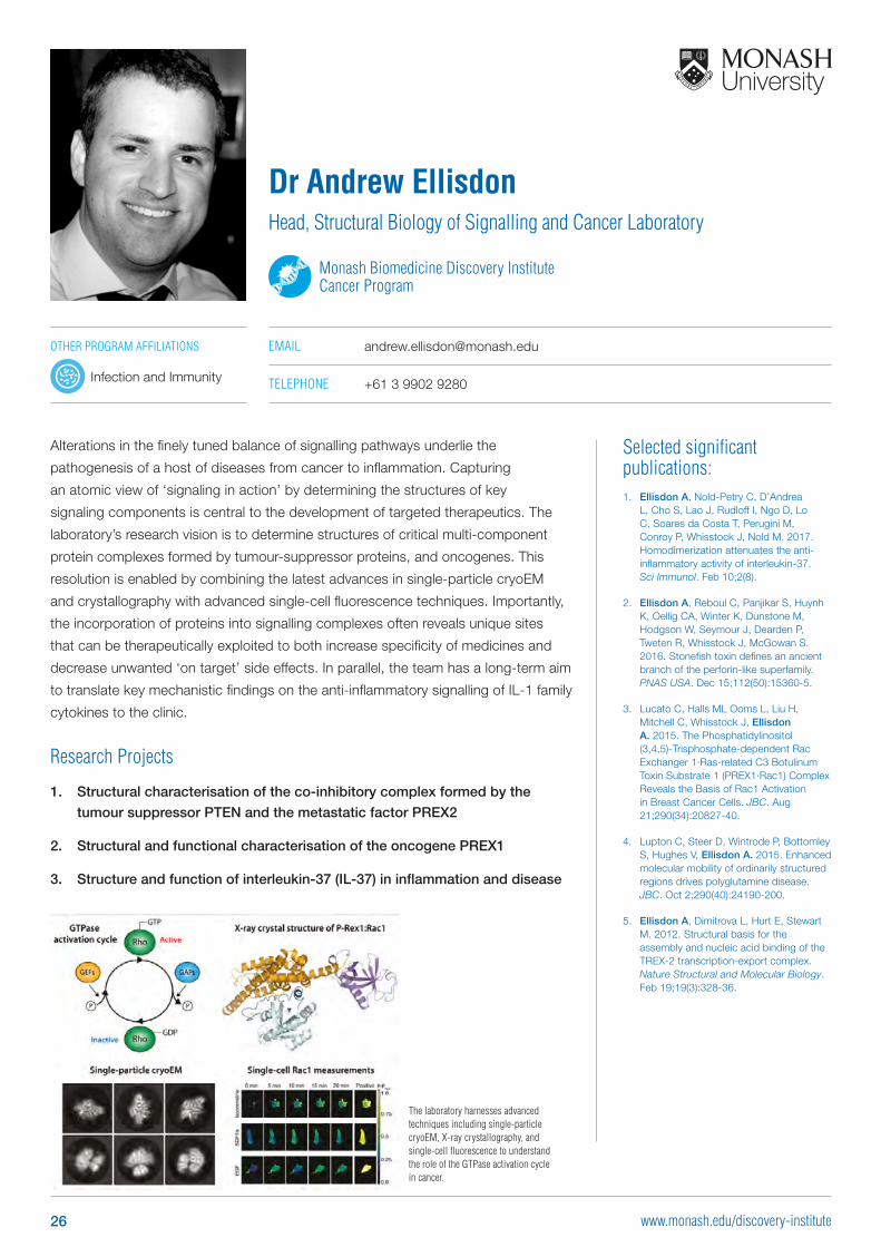
Alterations in the finely tuned balance of signalling pathways underlie the
pathogenesis of a host of diseases from cancer to inflammation. Capturing
an atomic view of ‘signaling in action’ by determining the structures of key
signaling components is central to the development of targeted therapeutics. The
laboratory’s research vision is to determine structures of critical multi-component
protein complexes formed by tumour-suppressor proteins, and oncogenes. This
resolution is enabled by combining the latest advances in single-particle cryoEM
and crystallography with advanced single-cell fluorescence techniques. Importantly,
the incorporation of proteins into signalling complexes often reveals unique sites
that can be therapeutically exploited to both increase specificity of medicines and
decrease unwanted ‘on target’ side effects. In parallel, the team has a long-term aim
to translate key mechanistic findings on the anti-inflammatory signalling of IL-1 family
cytokines to the clinic.
Research Projects
1. Structural characterisation of the co-inhibitory complex formed by the
tumour suppressor PTEN and the metastatic factor PREX2
2. Structural and functional characterisation of the oncogene PREX1
3. Structure and function of interleukin-37 (IL-37) in inflammation and disease
Dr Andrew Ellisdon Head, Structural Biology of Signalling and Cancer Laboratory
Monash Biomedicine Discovery Institute Cancer Program
EMAIL [email protected]
TELEPHONE +61 3 9902 9280
The laboratory harnesses advanced techniques including single-particle cryoEM, X-ray crystallography, and single-cell fluorescence to understand the role of the GTPase activation cycle in cancer.
26 www.monash.edu/discovery-institute
Selected significant publications: 1. Elmlund D, Elmlund H. 2015. Cryogenic
electron microscopy and single-particle analysis. Annu Rev Biochem. 84:499-517.
2. Park J*, Elmlund H*, Ercius P* et al. 2015. 3D structure of individual nanocrystals in solution by electron microscopy. Science. 349 (6245): 290-5. *equal contribution
3. Murakami K*, Elmlund H*, et al. 2013. Architechture of an RNA polymerase II transcription pre-initiation complex. Science. 342(6159):1238724. *equal contribution
4. Elmlund H, Elmlund D, Bengio S. 2013. PRIME: probabilistic initial 3D model generation for single-particle cryo-electron microscopy. Structure. 21(8): 1299-306.
5. Elmlund D, Elmlund H. 2012. SIMPLE: Software for ab initio reconstruction of heterogeneous single-particles. J Struct Biol. 180(3):420-7.
6. Elmlund D, Davis R, Elmlund H. 2010. Ab initio structure determination from electron microscopic images of single molecules coexisting in different functional states. Structure. 18(3):354-65.
7. Elmlund D, Elmlund H. 2009. High-resolution single-particle orientation refinement based on spectrally self-adapting common lines. J Struct Biol. 167(1):83-94.
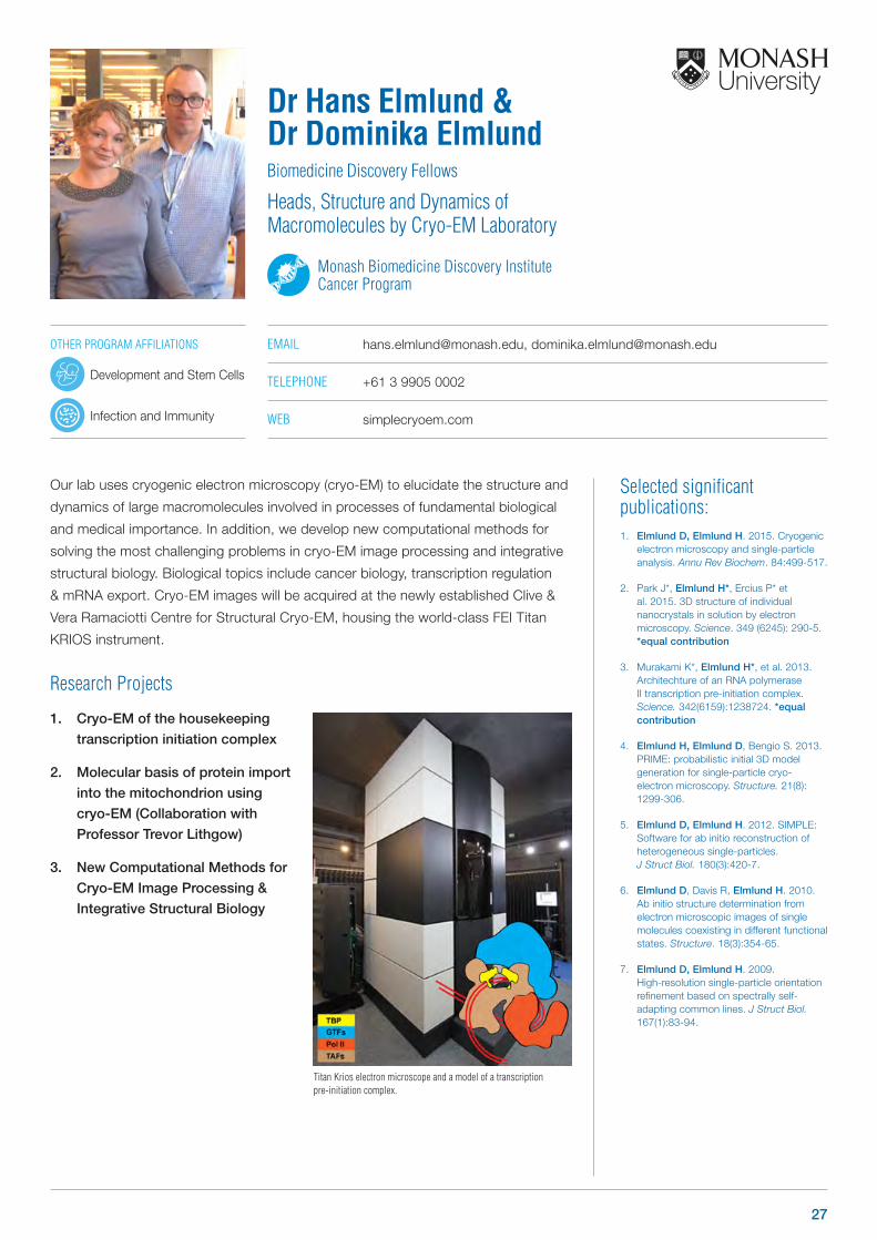
Our lab uses cryogenic electron microscopy (cryo-EM) to elucidate the structure and
dynamics of large macromolecules involved in processes of fundamental biological
and medical importance. In addition, we develop new computational methods for
solving the most challenging problems in cryo-EM image processing and integrative
structural biology. Biological topics include cancer biology, transcription regulation
& mRNA export. Cryo-EM images will be acquired at the newly established Clive &
Vera Ramaciotti Centre for Structural Cryo-EM, housing the world-class FEI Titan
KRIOS instrument.
Research Projects
1. Cryo-EM of the housekeeping
transcription initiation complex
2. Molecular basis of protein import
into the mitochondrion using
cryo-EM (Collaboration with
Professor Trevor Lithgow)
3. New Computational Methods for
Cryo-EM Image Processing &
Integrative Structural Biology
Dr Hans Elmlund & Dr Dominika ElmlundBiomedicine Discovery Fellows
Heads, Structure and Dynamics of Macromolecules by Cryo-EM Laboratory
Monash Biomedicine Discovery Institute Cancer Program
EMAIL [email protected], [email protected]
TELEPHONE +61 3 9905 0002
WEB simplecryoem.com
Titan Krios electron microscope and a model of a transcription pre-initiation complex.
OTHER PROGRAM AFFILIATIONS
Development and Stem Cells
Infection and Immunity
27
Selected significant publications: 1. Rebello RJ, Kusnadi E, Cameron DP,
Pearson HB, Lesmana A, Devlin JR, Drygin D, Clark AK, Porter L, Pedersen J, Sandhu S, Risbridger GP, Pearson RB, Hannan RD, Furic L. 2016. The Dual Inhibition of RNA Pol I Transcription and PIM Kinase as a New Therapeutic Approach to Treat Advanced Prostate Cancer. Clin Cancer Res 22: 5539-5552
2. Takizawa I, Lawrence MG, Balanathan P, Rebello R, Pearson HB, Garg E, Pedersen J, Pouliot N, Nadon R, Watt MJ, Taylor RA, Humbert P, Topisirovic I, Larsson O, Risbridger GP, Furic L. 2015. Estrogen receptor alpha drives proliferation in PTEN-deficient prostate carcinoma by stimulating survival signaling, MYC expression and altering glucose sensitivity. Oncotarget 6: 604-16
3. Furic L, Rong L, Larsson O, Koumakpayi IH, Yoshida K, Brueschke A, Petroulakis E, Robichaud N, Pollak M, Gaboury LA, Pandolfi PP, Saad F, Sonenberg N. 2010. eIF4E phosphorylation promotes tumorigenesis and is associated with prostate cancer progression. Proc Natl Acad Sci U S A 107: 14134-9
4. Kim YK, Furic L, Parisien M, Major F, DesGroseillers L, Maquat LE. 2007. Staufen1 regulates diverse classes of mammalian transcripts. EMBO J 26: 2670-81
5. Kim YK, Furic L, Desgroseillers L, Maquat LE. 2005. Mammalian Staufen1 recruits Upf1 to specific mRNA 3’UTRs so as to elicit mRNA decay. Cell 120: 195-208
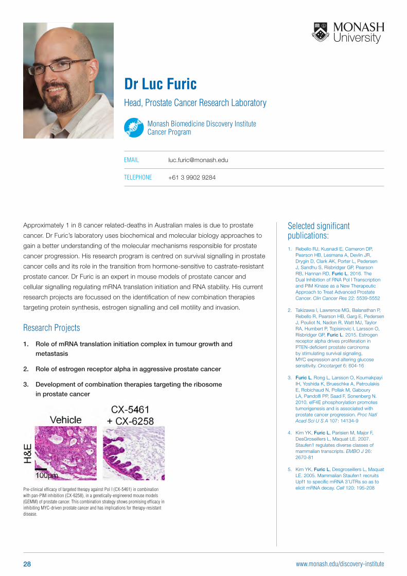
Approximately 1 in 8 cancer related-deaths in Australian males is due to prostate
cancer. Dr Furic’s laboratory uses biochemical and molecular biology approaches to
gain a better understanding of the molecular mechanisms responsible for prostate
cancer progression. His research program is centred on survival signalling in prostate
cancer cells and its role in the transition from hormone-sensitive to castrate-resistant
prostate cancer. Dr Furic is an expert in mouse models of prostate cancer and
cellular signalling regulating mRNA translation initiation and RNA stability. His current
research projects are focussed on the identification of new combination therapies
targeting protein synthesis, estrogen signalling and cell motility and invasion.
Research Projects
1. Role of mRNA translation initiation complex in tumour growth and
metastasis
2. Role of estrogen receptor alpha in aggressive prostate cancer
3. Development of combination therapies targeting the ribosome
in prostate cancer
Dr Luc FuricHead, Prostate Cancer Research Laboratory
Monash Biomedicine Discovery Institute Cancer Program
EMAIL [email protected]
TELEPHONE +61 3 9902 9284
Pre-clinical efficacy of targeted therapy against Pol I (CX-5461) in combination with pan-PIM inhibition (CX-6258), in a genetically-engineered mouse models (GEMM) of prostate cancer. This combination strategy shows promising efficacy in inhibiting MYC-driven prostate cancer and has implications for therapy-resistant disease.
28 www.monash.edu/discovery-institute
Selected significant publications: 1. Conduit SE, Ramaswamy V, Remke M,
Watkins DN, Wainwright BJ, Taylor MD, Mitchell CA and Dyson JM. 2017. A compartmentalized phosphoinositide signaling axis at cilia is regulated by INPP5E to maintain cilia and promote Sonic Hedgehog medulloblastoma. Oncogene (accepted)
2. Dyson JM, Conduit SE, Feeney SJ, Hakim S, DiTommaso T, Fulcher AJ, Sriratana A, Ramm G, Horan KA, Gurung R, Wicking C, Smyth I, Mitchell CA. 2017. INPP5E regulates phosphoinositide-dependent cilia transition zone function. Journal of Cell Biology 216(1):247-263
3. Ooms LM, Binge LC, Davies EM, Rahman P, Conway JR, Gurung R, Ferguson DT, Papa A, Fedele CG, Vieusseux JL, Chai RC, Koentgen F, Price JT, Tiganis T, Timpson P, McLean CA and Mitchell CA. 2015. The inositol polyphosphate 5-phosphatase PIPP regulates AKT1-dependent breast cancer growth and metastasis. Cancer Cell 28(2):155-169.
4. McGrath MJ, Binge LC, Sriratana A, Wang H, Robinson PA, Pook D, Fedele CG, Brown S, Dyson JM, Cottle DL, Cowling BS, Niranjan B, Risbridger GP, Mitchell CA. 2013. Regulation of the transcriptional coactivator FHL2 licenses activation of the androgen receptor in castrate-resistant prostate cancer. Cancer Research 73(16):5066-5079.
5. Fedele CG, Ooms LM, Ho M, Vieusseux J, O’Toole SA, Millar EK, Lopez-Knowles E, Sriratana A, Gurung R, Baglietto L, Giles GG, Bailey CG, Rasko JE, Shields BJ, Price JT, Majerus PW, Sutherland RL, Tiganis T, McLean CA, Mitchell CA. 2010. Inositol polyphosphate 4-phosphatase II regulates PI3K/Akt signaling and is lost in human basal-like breast cancers. Proc. Nat. Acad. Sci. USA 107(51): 22231-36.
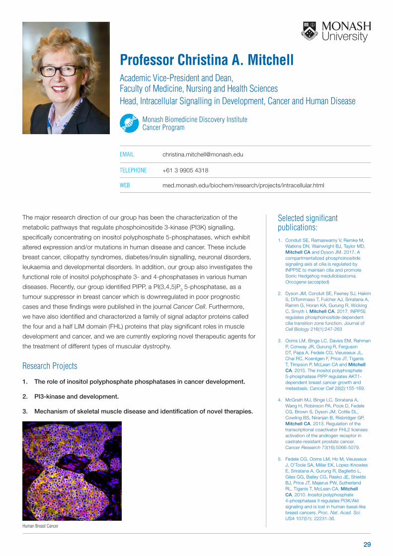
The major research direction of our group has been the characterization of the
metabolic pathways that regulate phosphoinositide 3-kinase (PI3K) signalling,
specifically concentrating on inositol polyphosphate 5-phosphatases, which exhibit
altered expression and/or mutations in human disease and cancer. These include
breast cancer, ciliopathy syndromes, diabetes/insulin signalling, neuronal disorders,
leukaemia and developmental disorders. In addition, our group also investigates the
functional role of inositol polyphosphate 3- and 4-phosphatases in various human
diseases. Recently, our group identified PIPP, a PI(3,4,5)P3 5-phosphatase, as a
tumour suppressor in breast cancer which is downregulated in poor prognostic
cases and these findings were published in the journal Cancer Cell. Furthermore,
we have also identified and characterized a family of signal adaptor proteins called
the four and a half LIM domain (FHL) proteins that play significant roles in muscle
development and cancer, and we are currently exploring novel therapeutic agents for
the treatment of different types of muscular dystrophy.
Research Projects
1. The role of inositol polyphosphate phosphatases in cancer development.
2. PI3-kinase and development.
3. Mechanism of skeletal muscle disease and identification of novel therapies.
Professor Christina A. MitchellAcademic Vice-President and Dean, Faculty of Medicine, Nursing and Health Sciences Head, Intracellular Signalling in Development, Cancer and Human Disease
Monash Biomedicine Discovery Institute Cancer Program
EMAIL [email protected]
TELEPHONE +61 3 9905 4318
WEB med.monash.edu/biochem/research/projects/intracellular.html
Human Breast Cancer
29
Selected significant publications: 1. Varusai TM, Kolch W, Kholodenko BK
and Nguyen LK*. 2015. Protein-protein interactions generate hidden feedback and feed-forward loops to trigger bistable switches, oscillations and biphasic dose-responses. Molecular Biosystems (in press) (* Correspondence)
2. Nguyen LK*, Degasperi A, Cotter P & Kholodenko BK. 2015. DYVIPAC: an integrated analysis and visualisation framework to probe multi-dimensional biological networks. Scientific Reports 5, Article number: 12569 doi:10.1038/srep12569 (*Correspondence)
3. Romano D, Nguyen LK*, Matallanas D, Halasz M, Doherty C, Kholodenko BN, Kolch W. 2014. Protein interaction switches coordinate oncogenic and apoptotic signaling. Nature Cell Biology doi: 10.1038/ncb2986. *Lead modeller.
4. Nguyen LK, Cavadas MAS, Scholz CC, Fitzpatrick SF, Bruning U, Cummins EP, Tambuwala MT, Manresa MC, Kholodenko BN, Taylor CT, & Cheong A. 2013. A dynamic model of the hypoxia-inducible factor (HIF) network. Journal of Cell Science. doi: 10.1242/jcs.119974. Epub 2013 Feb 6. (Most read paper of JCS, February 2013).
5. Nguyen LK, Muñoz-García J, Maccario H, Ciechanover A, Kolch W & Kholodenko BK. 2011. Switches, excitable responses and oscillations in the Ring1B/Bmi1 ubiquitination system. PLoS Computational Biology 7(12): p. e1002317. (Highlighted in the Conway Research Focus)
The advent of modern -omics technologies has revolutionised biology and has led us
to view biological processes as interconnected networks rather than as assemblies
of isolated molecules. This paradigm shift has instigated efforts to analyse cellular
networks through computational models, which in turn has revealed new insights
into the mechanisms of fundamental biological processes and their malfunctioning
in disease states. However, the translation of the computational modelling of
cellular networks into medical applications remains limited. In my lab, we ask the
following questions: “How can we harness network biology and modelling to better
understand diseases such as cancer? And can we turn network modelling into
new diagnostic and therapeutic applications?”. We propose to address these using
integrated systems approaches, which combine predictive computational network
modelling with cutting-edge experimental technologies. Our main focus is to exploit
developed and tested mathematical models of cancer-related signalling networks to
rationally: (i) understand the mechanism of drug resistance which arise from network
structures; (ii) find effective anti-cancer drug combinations which either avoid or
overcome developed drug resistance; and (iii) design therapies tailored to patients’
mutational profiles. This model-based approach is applicable to multiple signalling
pathways and cancer types.
Research Projects
1. Predictive modelling of the
mTOR network to discover
novel therapies
2. Systems analysis of the ErbB
interaction network in Breast
Cancer (Collaboration with
Professor Roger Daly)
3. Mathematical modelling to
understand network dynamics
and cell-fate decisions
Dr Lan NguyenVictoria Cancer Agency Mid-Career Research Fellow
Head, Network Modelling Laboratory
Monash Biomedicine Discovery Institute Cancer Program
EMAIL [email protected]
TELEPHONE +61 3 9902 9365
WEB med.monash.edu/biochem/staff/lan-nguyen.html
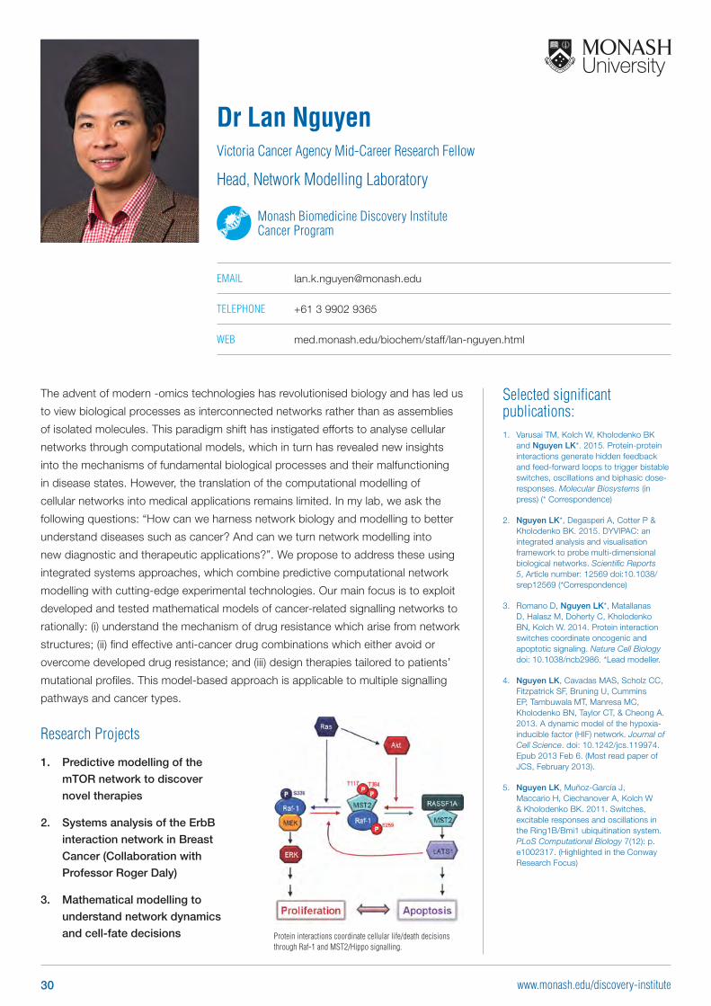
Protein interactions coordinate cellular life/death decisions through Raf-1 and MST2/Hippo signalling.
30 www.monash.edu/discovery-institute
Selected significant publications: 1. Papa A, Wan L, Bonora M, Salmena L,
Song MS, Hobbs RM, Lunardi A, Webster K, Ng C, Newton RH, et al. 2014. Cancer-associated PTEN mutants act in a dominant-negative manner to suppress PTEN protein function. Cell 157, 595-610.
2. Liu P, Begley M, Michowski W, Inuzuka H, Ginzberg M, Gao D, Tsou P, Gan W, Papa A, Kim BM, et al. 2014. Cell-cycle-regulated activation of Akt kinase by phosphorylation at its carboxyl terminus. Nature 508, 541-545.
3. Juvekar A, Burga LN, Hu H, Lunsford EP, Ibrahim YH, Balmana J, Rajendran A, Papa A, Spencer K, Lyssiotis CA, et al. 2012. Combining a PI3K inhibitor with a PARP inhibitor provides an effective therapy for BRCA1-related breast cancer. Cancer Discovery 2, 1048-1063.
4. Bernardi R, Papa A, Egia A, Coltella N, Teruya-Feldstein J, Signoretti S, and Pandolfi PP. 2011. Pml represses tumour progression through inhibition of mTOR. EMBO Molecular Medicine 3, 249-257.
5. Iraci N, Diolaiti D, Papa A, Porro A, Valli E, Gherardi S, Herold S, Eilers M, Bernardoni R, Della Valle G, et al. 2011. A SP1/MIZ1/MYCN repression complex recruits HDAC1 at the TRKA and p75NTR promoters and affects neuroblastoma malignancy by inhibiting the cell response to NGF. Cancer Research 71, 404-412.
Dr Antonella PapaNational Breast Cancer Foundation (NBCF) Career Development Fellow
Head, Cancer Biology Laboratory
Monash Biomedicine Discovery Institute Cancer Program
EMAIL [email protected]
TELEPHONE +61 3 9902 9330
OTHER PROGRAM AFFILIATIONS
Metabolism, Diabetes and Obesity
Neuroscience
OncogenicMutation
Tumour
Intrinsic/AcquiredMutations
NormalEpithelial Cells Cancer
Initiation Progression
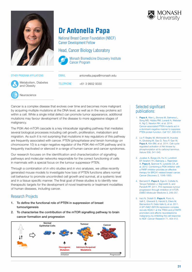
Cancer is a complex disease that evolves over time and becomes more malignant by acquiring multiple mutations at the DNA level, as well as in the way proteins act within a cell. While a single initial defect can promote tumor appearance, additional mutations may favour development of the disease to more aggressive stages of malignancy.
The PI3K-Akt-mTOR cascade is a key intracellular signalling pathway that mediates several biological processes including cell growth, proliferation, metabolism and migration. As such it is not surprising that mutations in key regulators of this pathway are frequently associated with cancer. PTEN (phosphatase and tensin homology on chromosome 10) is a major negative regulator of the PI3K-Akt-mTOR pathway and is frequently inactivated or silenced in a range of human cancer and cancer syndromes.
Our research focuses on the identification and characterization of signalling pathways and molecular networks responsible for the correct functioning of cells in mammals with a special focus on the tumour suppressor PTEN.
Through a combination of in vitro studies and in vivo analyses, we utilise recently generated mouse models to investigate how loss of PTEN functions alters normal cell behaviour to promote uncontrolled cell growth and survival, at a systemic level and in a tissue specific manner. The final goal of these studies is to identify new therapeutic targets for the development of novel treatments or treatment modalities of human diseases, including cancer.
Research Projects1. To define the functional role of PTEN in suppression of breast
tumourigenesis
2. To characterise the contribution of the mTOR signalling pathway to brain
cancer formation and progression
31
Selected significant publications: 1. Oorschot VMJ, Sztal TE, Bryson-
Richardson RJ, Ramm G. 2014. Immunocorrelative Light and Electron Microscopy on Tokuyasu Cryosections. Methods in Cell Biology 124, 241-257.
2. Padman BS, Bach M, Lucarelli G, Prescott M, Ramm G. 2013. The protonophore CCCP interferes with lysosomal degradation of autophagic cargo in yeast and mammalian cells. Autophagy 9,1862-75.
3. Bach M, Larance M, James DE, Ramm G. 2011. The serine/threonine kinase ULK1 is a target of multiple phosphorylation events. Biochem J 440, 283-91.
4. Yip FMF, Ramm G, Larance M, Wagner MC, Guilhaus M, James DE. 2008. Phosphorylation of the Myosin Motor Myo1c Is Required For Insulin-Stimulated GLUT4 Translocation in Adipocytes. Cell Metab 8, 384-98. (Cover story & Preview Cell Metab 8, 344-6).
5. Ng Y, Ramm G, Lopez JA, James DE. 2008. Rapid activation of Akt2 is sufficient to stimulate GLUT4 translocation in 3T3-L1 adipocytes. Cell Metab 7, 348-56.
Our lab is focused on the regulation of autophagy, a major intracellular degradation
process. In cancer, autophagy plays complex roles and can suppress tumours,
but also helps tumour cells survive in other cases. Autophagy delivers cellular and
cytoplasmic structures to the lysosome, where they are degraded. This process
is tightly linked to cellular metabolism and is an evolutionary conserved survival
mechanism that helps cells cope with nutrient starvation. We have recently
discovered a link between metabolic control and the Serine/Threonine kinase ULK1,
a key regulator of autophagy. We aim to develop a detailed understanding of how
these regulatory networks are causing changes in intracellular membrane trafficking
during autophagy.
Research Projects
1. High-resolution imaging of the mitophagy pathway
2. Autophagy and Cancer
Dr Georg RammHead, Organelle Biology/ Advanced Cellular Imaging Laboratory
Monash Biomedicine Discovery Institute Cancer Program
EMAIL [email protected]
TELEPHONE +61 3 9905 1280
WEB med.monash.edu/biochem/georg-ramm-homepage.html
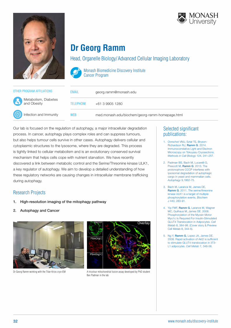
A tricolour mitochondrial fusion assay developed by PhD student Ben Padman in the lab
Dr Georg Ramm working with the Titan Krios cryo-EM
OTHER PROGRAM AFFILIATIONS
Metabolism, Diabetes and Obesity
Infection and Immunity
32 www.monash.edu/discovery-institute
Selected significant publications: 1. Taylor RA*, Fraser M*, Livingstone J*,
Espiritu SMG*, Thorne H*, Huang V, Lo W, Shiah Y-J, Yamaguchi TN, Sliwinski A, Horsburgh S, Meng A, Heisler LE, Yu N, Yousif F, Papargiris M, Lawrence MG, Timms L, Murphy DG, Frydenberg M, Hopkins JF, Bolton D, Clouston D, McPherson JD, van der Kwast T, Boutros PC**, Risbridger GP**, Bristow RG**. 2017. Germline BRCA2 Mutations Drive Prostate Cancers with Distinct Evolutionary Trajectories. Nature Communications 8: 13671
2. Alsop K. et al. 2016. A community-based model of rapid autopsy in end-stage cancer patients. Nat Biotechnol 34: 1010-1014
3. GP Risbridger, RA Taylor, D Clouston, A Sliwinski, H Thorne, S Hunter, JLi, kConFab, GWE Mitchell, Murphy DGM, M Frydenberg, D Pook, J Pedersen, R Toivanen, H Wang, M Papargiris, MG Lawrence, DM Bolton. 2015. Patient-derived xenografts reveal that intraductal carcinoma of the prostate is a prominent pathology in BRCA2 mutation carriers with prostate cancer and correlates with poor prognosis. European Urology. 67(3):496-503.
4. Toivanen R, Frydenberg M, Murphy D, Pedersen J, Ryan A, Pook D, Berman DM, Australian Prostate Cancer BioResource, Taylor RA, Risbridger GP. 2013. A pre-clinical model identifies castration-tolerant cancer repopulating cells in localized prostate tumors. Science Translational Medicine 5(187):187ra71.
5. Clark AK, Taubenberger AV, Taylor RA, Niranjan B, Chea ZY, Zotenko E, Sieh S, Pedersen J, Norden S, Frydenberg M, Grummet J, Pook DW, Australian Prostate Cancer BioResourse, Stirzaker C, Clark SJ, Lawrence MG, Ellem SJ, Hutmacher DW, Risbridger GP. 2013. A bioengineered microenvironment to quantitatively measure the tumorigenic properties of cancer-associated fibroblasts in human prostate cancer. Biomaterials 34(20):4777-4785.
Prostate cancer is one of the most common forms of cancer in men, affecting 1:6
men throughout their lifetime. Despite all our efforts to find a cure, prostate cancer
remains a lethal disease and in Australia, about 60 men die from prostate cancer
each week.
Working as a multidisciplinary team, we aim to improve patient treatment and
outcome through a better understanding of the mechanisms that drive prostate
cancer. Our research utilises state of the art techniques (eg. xenografting,
bioengineered in vitro modelling, and transgenic animal models) that allow us to
examine the mechanisms that contribute to disease development and progression.
Research Projects
1. Patient derived xenograft models of prostate cancer for preclinical studies
2. Defining the features of familial and high risk prostate cancer
3. Novel combination therapies for prostate cancer that target the ribosome
4. Targeting the eukaryotic translation initiation factor 4E in prostate cancer
5. In vitro modelling of the human prostate cancer microenvironment
6. Estrogen signalling and metabolism in prostate cancer
7. Epigenetic regulation of the tumour microenvironment
8.
Professor Gail RisbridgerNHMRC Senior Principal Research Fellow
Head, Prostate Cancer Research Group
Monash Biomedicine Discovery Institute Cancer Program
EMAIL [email protected]
TELEPHONE +61 3 9902 9558
WEB med.monash.edu/anatomy/research/prostate-research.html
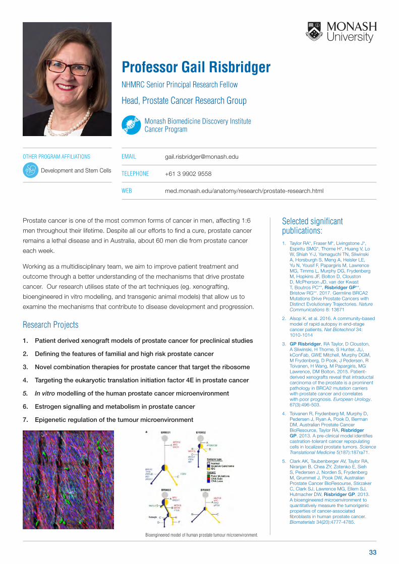
Bioengineered model of human prostate tumour microenvironment.
OTHER PROGRAM AFFILIATIONS
Development and Stem Cells
33
Selected significant publications: 1. Rosenbluh J, Xu H, Harrington W, Gill S,
Wang X, Vazquez F, Root DE, Tsherniak A, Hahn WC. 2017. Complementary information derived from CRISPR Cas9 mediated gene deletion and suppression. Nature Communications, Accepted.
2. Rosenbluh J, Mercer J, Shrestha Y, Oliver R, Tamayo P, Doench JG, Piccioni F, Horn H, Fagbami L, Yang-Zho D, Perrimon N, Jaffe J, Lage K, Boehm JS, Hahn WC. 2016. Integrated genetic and proteomic interrogation of WNT/β-catenin cancer dependencies. Cell Systems, 3(3):302-316.
3. Shao DD, Xue W, Krall, EB, Bhutkar A, Piccioni F, Wang X, Schinzel AC, Sood S, Rosenbluh J, Kim WJ, Zwang Y, Root DE, Jacks T, Hahn WC. 2014. KRAS and YAP1 converge to regulate EMT and tumor survival. Cell, 158(1):171-184.
4. Rosenbluh J, Nijhawan D, Cox AG, Li X, Neal JT, Schafer EJ, Zack TI, Wang X, Tsherniak T, Schinzel AC, Shao DD, Schumacher SE, Weir BA, Vazquez F, Cowley GS, Root DE, Mesirov JP, Beroukhim R, Kuo CJ, Goessling W, Hahn WC. 2012. β-catenin driven cancers require a YAP1 transcriptional complex for survival and tumorigenesis. Cell, 2012 151(7):1457-1473
5. Rosenbluh J, Wang X, Hahn WC. 2014. Genomic insights into WNT/β-catenin signaling. Trends in Pharmacological Science, 35(2):103-9.
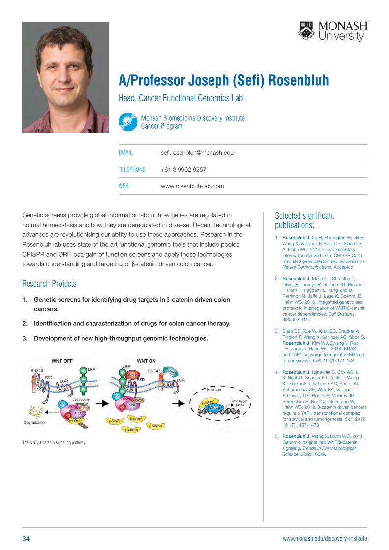
Genetic screens provide global information about how genes are regulated in
normal homeostasis and how they are deregulated in disease. Recent technological
advances are revolutionising our ability to use these approaches. Research in the
Rosenbluh lab uses state of the art functional genomic tools that include pooled
CRISPR and ORF loss/gain of function screens and apply these technologies
towards understanding and targeting of β-catenin driven colon cancer.
Research Projects
1. Genetic screens for identifying drug targets in β-catenin driven colon
cancers.
2. Identification and characterization of drugs for colon cancer therapy.
3. Development of new high-throughput genomic technologies.
A/Professor Joseph (Sefi) RosenbluhHead, Cancer Functional Genomics Lab
Monash Biomedicine Discovery Institute Cancer Program
EMAIL [email protected]
TELEPHONE +61 3 9902 9257
WEB www.rosenbluh-lab.com
The WNT/β-catenin signaling pathway.
34 www.monash.edu/discovery-institute
Selected significant publications: 1. Taylor RA*, Risbridger GP*, Clouston
D, Sliwinski A, Thorne H, Hunter S, Li J, Mitchell G, Murphy D, Frydenberg M, Pook D, Pedersen J, Toivanen R, Wang H, Papargiris M, Lawrence MG, Bolton DM. 2014. Patient-derived xenografts reveal that intraductal carcinoma of the prostate is a prominent pathology in BRCA2 mutation carriers with prostate cancer and correlates with poor prognosis. European Urology. doi: 10.1016/j.eururo.2014.08.007. *Joint authors
2. Toivanen R, Frydenberg M, Murphy D, Pedersen J, Ryan A, Pook A, Berman DM, Australian Prostate Cancer BioResource, Risbridger GP, Taylor RA. 2013. A pre-clinical model to identify castrate-resistant cancer repopulating cells in localized prostate tumors. Science Translational Medicine. 5(187):187ra71.
3. Lawrence MG, Taylor RA, Toivanen R, Pedersen J, Norden S, Pook DW, Frydenberg M, Australian Prostate Cancer Bioresource, Papargiris MM, Niranjan B, Richards MG, Wang H, Collins AT, Maitland NJ, Risbridger GP. 2013. A preclinical xenograft model of prostate cancer using human tumours. Nature Protocols. 8(5):836-48.
4. Toivanen R, Berman DM, Wang H, Frydenberg M, Pedersen J, Meeker AK, Ellem SJ, Risbridger GP, Taylor RA. 2011. Bioassays for cancer repopulating cells. Stem Cells. 29(8):1310-4.
5. Taylor RA, Cowin PA, Cunha GR, Trounson AO, Pedersen J, Risbridger GP. 2006. Formation of human prostate tissue from embryonic stem cells. Nature Methods 3(3):179-181.
Prostate cancer is an androgen dependent disease. Advanced (castrate-sensitive)
prostate cancers are managed with hormonal therapy. Inevitably, these tumours
adapt to low serum levels of androgens and become castrate-resistant prostate
cancers. In both castrate-sensitive and castrate-resistant disease, the Androgen
Receptor (AR) plays a major role in driving tumour progression. Additionally, castrate-
resistant tumours metastasise to different parts of the body. The most common
location is the bone, but more recently soft tissue metastases such as liver, lung and
brain have emerged. The origins of each metastatic tumour and the genetic drivers
responsible for their growth are currently unknown.
Research Projects
1. Characterising the androgen receptor in castrate-sensitive
prostate cancer cells
2. Investigating the origins of metastatic prostate cancer
A/Professor Renea TaylorVictorian Cancer Agency – Mid Career Research Fellow
Head, Reproductive Physiology Laboratory
Monash Biomedicine Discovery Institute Cancer Program
EMAIL [email protected]
TELEPHONE +61 3 9594 7130
OTHER PROGRAM AFFILIATIONS
Development and Stem Cells
Metabolism, Diabetes and Obesity
35
Selected significant publications: 1. Fatima S, Wagstaff KM, Lieu KG, Davies
RG, Tanaka SS, Yamaguchi YL, Loveland KL, Tam PP, Jans DA. 2017. Interactome of the inhibitory isoform of the nuclear transporter Importin 13. Biochim Biophys Acta 1864: 546-561
2. Chandrasekaran R, Lee AS, Yap LW, Jans DA, Wagstaff KM, Cheng W. 2016. Tumor cell-specific photothermal killing by SELEX-derived DNA aptamer-targeted gold nanorods. Nanoscale 8: 187-96
3. Nastasie MS, Thissen H, Jans DA, Wagstaff KM. 2015. Enhanced tumour cell nuclear targeting in a tumour progression model. BMC Cancer 15: 76
4. Wagstaff KM, Sivakumaran H, Heaton SM, Harrich D, Jans DA. 2012. Ivermectin is a specific inhibitor of importin alpha/beta-mediated nuclear import able to inhibit replication of HIV-1 and dengue virus. Biochem J 443: 851-6
5. Wagstaff KM, Rawlinson SM, Hearps AC, Jans DA. 2011. An AlphaScreen(R)-based assay for high-throughput screening for specific inhibitors of nuclear import. J Biomol Screen 16: 192-200
Dr Kylie WagstaffHead, Cancer Targeting and Nuclear Therapeutics Laboratory
Monash Biomedicine Discovery Institute Cancer Program
EMAIL [email protected]
TELEPHONE +61 3 9902 9348
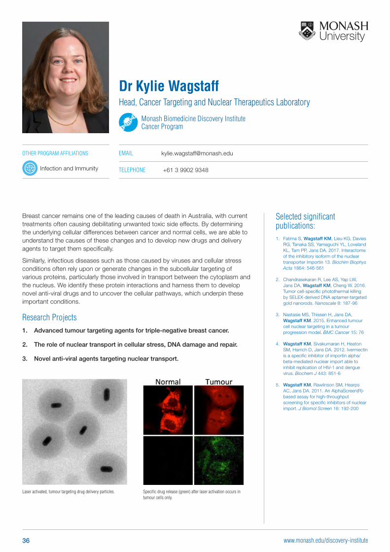
Breast cancer remains one of the leading causes of death in Australia, with current treatments often causing debilitating unwanted toxic side effects. By determining the underlying cellular differences between cancer and normal cells, we are able to understand the causes of these changes and to develop new drugs and delivery agents to target them specifically.
Similarly, infectious diseases such as those caused by viruses and cellular stress conditions often rely upon or generate changes in the subcellular targeting of various proteins, particularly those involved in transport between the cytoplasm and the nucleus. We identify these protein interactions and harness them to develop novel anti-viral drugs and to uncover the cellular pathways, which underpin these important conditions.
Research Projects1. Advanced tumour targeting agents for triple-negative breast cancer.
2. The role of nuclear transport in cellular stress, DNA damage and repair.
3. Novel anti-viral agents targeting nuclear transport.
Laser activated, tumour targeting drug delivery particles. Specific drug release (green) after laser activation occurs in tumour cells only.
OTHER PROGRAM AFFILIATIONS
Infection and Immunity
36 www.monash.edu/discovery-institute
Selected significant publications: 1. Waris S, García-Mauriño SM,
Sivakumaran A, Beckham SA, Loughlin FE, Gorospe M, Díaz-Moreno I, Wilce MC, Wilce JA. 2017. TIA-1 RRM23 binding and recognition of target oligonucleotides. Nucleic Acids Res. 2017 Feb 10. doi: 10.1093/nar/gkx102
2. Gunzburg MJ, Kulkarni K, Watson GM, Ambaye ND, Del Borgo MP, Brandt R, Pero SC, Perlmutter P, Wilce MC, Wilce JA. 2016. Unexpected involvement of staple leads to redesign of selective bicyclic peptide inhibitor of Grb7. Sci Rep. 6:27060.
3. Watson GM, Gunzburg MJ, Ambaye ND, Lucas, WA, Traore DA, Kulkarni K, Cergol KM, Payne RJ, Panjikar S, Pero SC, Perlmutter P, Wilce MCJ and Wilce JA. 2015 Cyclic peptides incorporating phosphotyrosine mimetics as potent and specific inhibitors of the Grb7 breast cancer target. Journal of Medicinal Chemistry 58, 7707-7718.
4. Kim HS, Wilce MCJ, Yoga YMK, Pendini NR, Gunzburg MJ, Cowieson NP, Wilson GM, Williams BR, Gorospe M and Wilce JA. 2011. Different modes of interaction by TIAR and HuR with target RNA and DNA. Nucleic Acids Research 39, 1–14.
5. Ambaye ND, Pero SC, Gunzburg MJ, Yap M-Y, Clayton DJ, Del Borgo MP, Perlmutter P, Aguilar M-I, Shukla GS, Peletskaya E, Cookson MM, Krag DN, Wilce MCJ and Wilce JA. 2011 Structural basis of binding by cyclic non-phosphorylated peptide antagonists of Grb7 implicated in breast cancer progression. Journal of Molecular Biology. 412, 397-411.
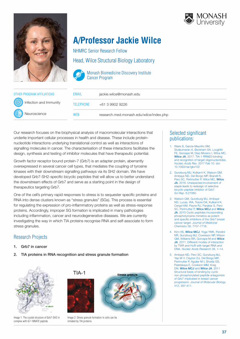
Our research focuses on the biophysical analysis of macromolecular interactions that underlie important cellular processes in health and disease. These include protein-nucleotide interactions underlying translational control as well as interactions of signalling molecules in cancer. The characterisation of these interactions facilitates the design, synthesis and testing of inhibitor molecules that have therapeutic potential.
Growth factor receptor bound protein-7 (Grb7) is an adapter protein, aberrantly overexpressed in several cancer cell types, that mediates the coupling of tyrosine kinases with their downstream signalling pathways via its SH2 domain. We have developed Grb7-SH2-specific bicyclic peptides that will allow us to better understand the downstream effects of Grb7 and serve as a starting point in the design of therapeutics targeting Grb7.
One of the cell’s primary rapid responses to stress is to sequester specific proteins and RNA into dense clusters known as “stress granules” (SGs). This process is essential for regulating the expression of pro-inflammatory proteins as well as stress-response proteins. Accordingly, improper SG formation is implicated in many pathologies including inflammation, cancer and neurodegenerative diseases. We are currently investigating the way in which TIA proteins recognise RNA and self-associate to form stress granules.
Research Projects
1. Grb7 in cancer
2. TIA proteins in RNA recognition and stress granule formation
A/Professor Jackie WilceNHMRC Senior Research Fellow
Head, Wilce Structural Biology Laboratory
Monash Biomedicine Discovery Institute Cancer Program
EMAIL [email protected]
TELEPHONE +61 3 9902 9226
WEB research.med.monash.edu/wilce/index.php
Image 1: The crystal structure of Grb7-SH2 in complex with G7-18NATE peptide.
Image 2: Stress granule formation in cells can be initiated by TIA proteins.
OTHER PROGRAM AFFILIATIONS
Infection and Immunity
Neuroscience
37

Selected significant publications: 1. Udugama M, FT MC, Chan FL, Tang
MC, Pickett HA, JD RM, Mayne L, Collas P, Mann JR, Wong LH. 2015. Histone variant H3.3 provides the heterochromatic H3 lysine 9 tri-methylation mark at telomeres. Nucleic Acids Res 43: 10227-37
2. Voon HPJ, Collas P, Wong LH. 2016. Compromised Telomeric Heterochromatin Promotes ALTernative Lengthening of Telomeres. Trends in Cancer 2: 114-116
3. Chang FTM, Chan FL, McGhie JDR, Udugama M, Mayne L, Collas P, Mann JR, Wong LH, et al. 2015. CHK1-driven histone H3.3 serine 31 phosphorylation is important for chromatin maintenance and cell survival in human ALT cancer cells. Nucleic Acids Res, 43 (5): 2603-2614.
4. Chan FL, Marshall OJ, Kim B, Saffery R, Choo KHA and Wong LH. 2012. Novel RNA polymerase II transcriptional activity at the kinetochore during mitosis. Proc Natl Acad Sci USA. 109(6): 1979 – 1984.
5. Wong LH, McGhie JD, Sim M, Anderson MA, Ahn S, Hannan RD, George A, Morgan K, Mann JR and Choo KHA. 2010. ATRX interacts with H3.3 in maintaining telomere structural integrity in pluripotent embryonic stem cells. Genome Res. 20: 351-360.
Our research interest is to identify new chromatin factors that control chromosome
stability and genetic transmission. We aim to uncover fundamental epigenetic
mechanisms that regulate transcriptional silencing at repetitive DNA sequences in
the genome including telomeres and centromeres. Recent studies have identified
the frequent mutations of histone variant H3.3 and its chaperone ATRX in human
cancers, including the brain and bone cancers. We use CRISPR CAS9 gene editing
system, highly advanced cellular imaging and high throughput DNA and RNA
sequencing to investigate the genome-wide epigenetic defects associated with H3.3
and ATRX mutations in cancers.
Research Projects:
1. Investigate how chromatin defects cause brain tumours
2. Investigate epigenetic mechanisms that control transcriptional silencing
DNA repeats
3. Investigate chromatin defects associated with Alternative Lengthening of
Telomeres, a mechanism used for telomere elongation in cancers
A/Professor Lee WongARC Future Fellow
Head, Epigenetics and Chromatin Research Laboratory
Monash Biomedicine Discovery Institute Cancer Program
EMAIL [email protected]
TELEPHONE +61 3 9902 4925
WEB med.monash.edu/biochem/staff/wong.html
Staining of trimethylated H4K20, a repressive histone mark-trimethylated H4K20 at the centromeres and telomeres in mouse embryonic stem cells.
OTHER PROGRAM AFFILIATIONS
Development and Stem Cells
38 www.monash.edu/discovery-institute

Cardiovascular Disease Program Group Leaders
39www.monash.edu/discovery-institute
Selected significant publications: 1. Luder K, Kulkarni K, Lee HW, Widdop
R, Del Borgo MP* & Aguilar MI*. 2016. Decorated Self-Assembling β3- Tripeptides Form Cell Adhesive Scaffolds. Chem Commun 52:4549-4552.
2. Hirst D, Lee TH, Pattenden LK, Thomas WG and Aguilar MI. 2015. Helix 8 of the angiotensin-1a receptor interacts with phosphatidylinositol phosphates and modulates membrane insertion”. Scientific Reports 5:09972.
3. Andreu-Fernández V, Genoves A*, Lee TH, Stellato M, Lucantoni F, Orzáez, M, Mingarro,I, Aguilar MI* & Perez-Payá, E. 2014. Peptides derived from Transmembrane (TM) domains of diverse Bcl-2 proteins exert disruptive behavior in mitochondrial membranes. ACS Chem Biol. 9, 1799-1811.
4. Hall, K, Lee TH, Mechler A, Swann MJ and Aguilar MI. 2014. Real-time Measurement of Multiple Membrane Conformational States During Antimicrobial Peptide Binding: The Balance Between Recovery and Lysis. Scientific Reports, June 27 4:5479.
5. Del Borgo MP, Mechler AI, Traore D, Forsyth C, Wilce JA, Wilce MCJ, Aguilar MI* and Perlmutter* P. 2013. Supramolecular Self-Assembly of N-Acetyl capped β-Peptides Leads to Nano-to Macroscale Fibre Formation’. Angewandte Chemie Int Ed., 52 8266-8270. Selected for Front Piece Highlights.
Our group focuses on peptide-based drug design and biomembrane
nanotechnology. We are developing novel compounds that allow us to exploit the
potential of peptides as drugs. We are currently applying our technology to the
development of new compounds for treatments of cardiovascular disease and new
biomaterials for regenerative medicine. Our membrane nanotechnology projects
probes the role of membranes in the mechanism of Alzheimer’s disease, G protein-
coupled receptor function, apoptosis and antimicrobial peptide function. The long-
term aim of these studies is to increase our understanding of the molecular basis
of peptide and protein function and allow the rational design of peptide and protein
based therapeutics.
Research Projects
1. Peptide-Based Biomaterials
2. Role of the Mitochondrial Membrane in Apoptosis
3. New Ligands for Cardiovascular Disease
Professor Mibel AguilarHead, Biomaterials & Drug Design Laboratory
Monash Biomedicine Discovery Institute Cardiovascular Disease Program
EMAIL [email protected]
TELEPHONE +61 3 9905 3723
WEB med.monash.edu/biochem/research/projects/peptide.html
Peptide-Based Nanomaterials
OTHER PROGRAM AFFILIATIONS
Neuroscience
40 www.monash.edu/discovery-institute
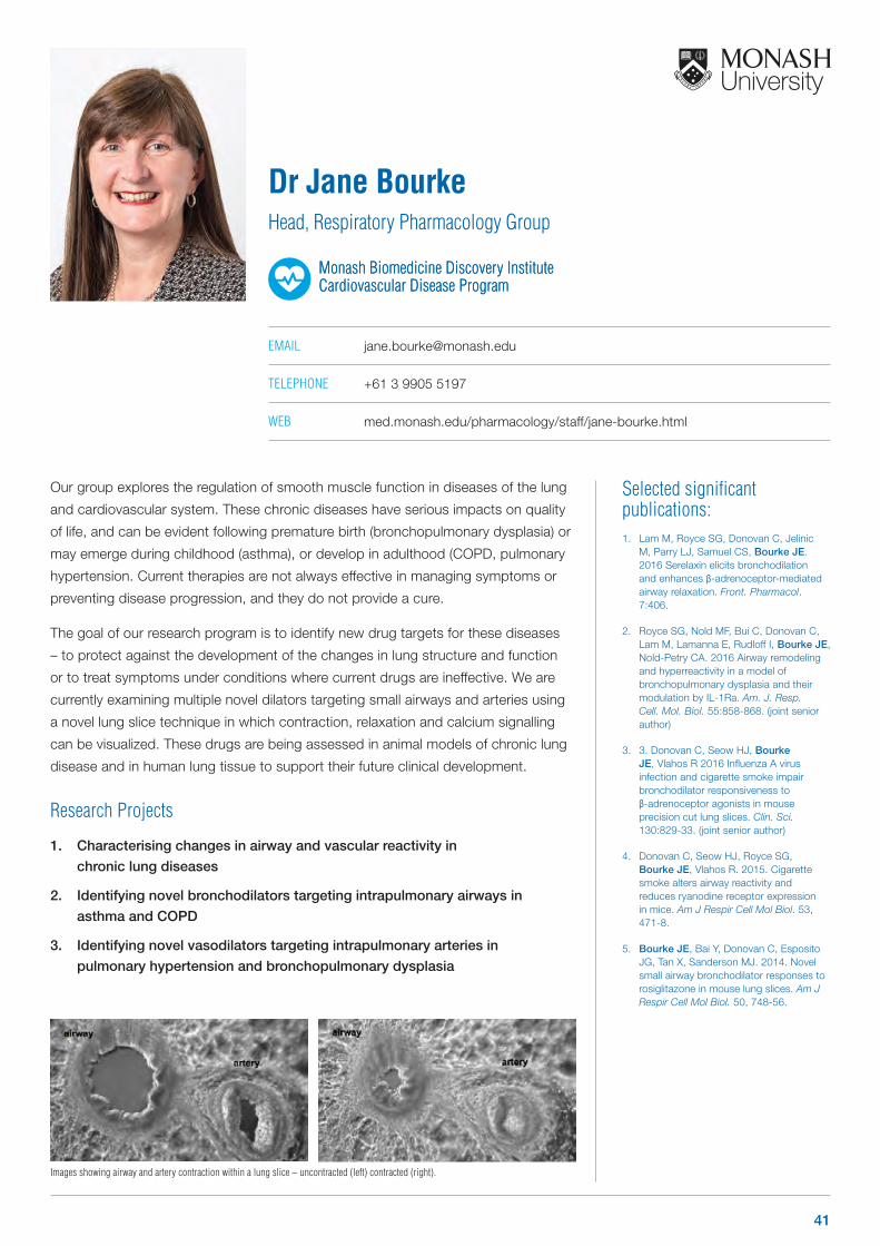
Selected significant publications: 1. Lam M, Royce SG, Donovan C, Jelinic
M, Parry LJ, Samuel CS, Bourke JE. 2016 Serelaxin elicits bronchodilation and enhances β-adrenoceptor-mediated airway relaxation. Front. Pharmacol. 7:406.
2. Royce SG, Nold MF, Bui C, Donovan C, Lam M, Lamanna E, Rudloff I, Bourke JE, Nold-Petry CA. 2016 Airway remodeling and hyperreactivity in a model of bronchopulmonary dysplasia and their modulation by IL-1Ra. Am. J. Resp. Cell. Mol. Biol. 55:858-868. (joint senior author)
3. 3. Donovan C, Seow HJ, Bourke JE, Vlahos R 2016 Influenza A virus infection and cigarette smoke impair bronchodilator responsiveness to β-adrenoceptor agonists in mouse precision cut lung slices. Clin. Sci. 130:829-33. (joint senior author)
4. Donovan C, Seow HJ, Royce SG, Bourke JE, Vlahos R. 2015. Cigarette smoke alters airway reactivity and reduces ryanodine receptor expression in mice. Am J Respir Cell Mol Biol. 53, 471-8.
5. Bourke JE, Bai Y, Donovan C, Esposito JG, Tan X, Sanderson MJ. 2014. Novel small airway bronchodilator responses to rosiglitazone in mouse lung slices. Am J Respir Cell Mol Biol. 50, 748-56.
Our group explores the regulation of smooth muscle function in diseases of the lung
and cardiovascular system. These chronic diseases have serious impacts on quality
of life, and can be evident following premature birth (bronchopulmonary dysplasia) or
may emerge during childhood (asthma), or develop in adulthood (COPD, pulmonary
hypertension. Current therapies are not always effective in managing symptoms or
preventing disease progression, and they do not provide a cure.
The goal of our research program is to identify new drug targets for these diseases
– to protect against the development of the changes in lung structure and function
or to treat symptoms under conditions where current drugs are ineffective. We are
currently examining multiple novel dilators targeting small airways and arteries using
a novel lung slice technique in which contraction, relaxation and calcium signalling
can be visualized. These drugs are being assessed in animal models of chronic lung
disease and in human lung tissue to support their future clinical development.
Research Projects
1. Characterising changes in airway and vascular reactivity in
chronic lung diseases
2. Identifying novel bronchodilators targeting intrapulmonary airways in
asthma and COPD
3. Identifying novel vasodilators targeting intrapulmonary arteries in
pulmonary hypertension and bronchopulmonary dysplasia
Dr Jane BourkeHead, Respiratory Pharmacology Group
Monash Biomedicine Discovery Institute Cardiovascular Disease ProgramMonash Biomedicine Discovery Institute Cardiovascular Disease Program
EMAIL [email protected]
TELEPHONE +61 3 9905 5197
WEB med.monash.edu/pharmacology/staff/jane-bourke.html
Images showing airway and artery contraction within a lung slice – uncontracted (left) contracted (right).
41
Selected significant publications: 1. Singh RR, Sajeesh V, Booth LC, McArdle
ZM, May CN, Head GA, Moritz KM, Sclaich MP, Denton KM. 2017. Catheter-based renal denervation exacerbates blood pressure fall during hemorrhage. Journal of the American College of Cardiology 69:951-96.
2. Hilliard LM, Colefella KM, Bulmer LL, Puelles VG, Singh RR, Ow CP, Drummond G, Evans RG, Denton KM. 2016. Chronic recurrent dehydration associated with periodic water intake exacerbates hypertension and promotes renal damage in male spontaneously hypertensive rats. Science reports 6:33855.
3. Lankadeva YR, Singh RR, Moritz KM, Parkington HC, *Denton KM, *Tare M. 2015. Renal dysfunction is associated with a reduced contribution of nitric oxide and enhanced vasoconstriction following a congenital renal mass reduction in sheep. Circulation 131:280-8. * Equal Authorship
4. Mirabito KM, Hilliard LM, Wei Z, Tikellis C, Widdop RE, Vinh A, Denton KM. 2014. Role of inflammation and the Angiotensin type 2 receptor in the regulation of arterial pressure during pregnancy in mice. Hypertens. 64:626-31.
5. Hilliard LM, Jones, ES, Stekelings UM, Unger T, Widdop RE, Denton KM. 2012. Sex-specific influence of angiotensin type 2 receptor stimulation on renal function: a novel therapeutic target for hypertension. Hypertens. 59(2):409-14
Hypertension is the world’s leading risk factor for disease. It strongly correlates
with adverse outcomes such as heart disease, stroke and kidney failure. The
challenges of managing and preventing the development of hypertension are
increasing as it is predicted that 60% of the population by 2025 will be hypertensive.
Greater understanding of the mechanisms causing increased blood pressure, and
identification of new therapies to prevent hypertensive tissue injury are pivotal in
meeting this challenge.
Research Projects
1. The path to hypertension and cardiovascular disease has it origins in early
life. Current work is directed at identifying prognostic indicators of disease
and developing intervention strategies in the very young.
2. Women prior to menopause are protected against hypertension and
cardiovascular disease. Ongoing research focuses on understanding sex-
difference in the regulation of blood pressure. Novel treatments to prevent
post-menopausal hypertension are being examined.
3. Our work also examines the efficacy and safety of renal artery denervation,
an emerging treatment for hypertension.
Professor Kate DentonNHMRC Principal Research Fellow
Head, Cardiovascular Disease Program Head, Hypertension Research Group
Monash Biomedicine Discovery Institute Cardiovascular Disease Program
EMAIL [email protected]
TELEPHONE +61 3 9905 9553
WEB med.monash.edu/physiology/staff/denton.html
OTHER PROGRAM AFFILIATIONS
Development and Stem Cells
Neuroscience
42 www.monash.edu/discovery-institute
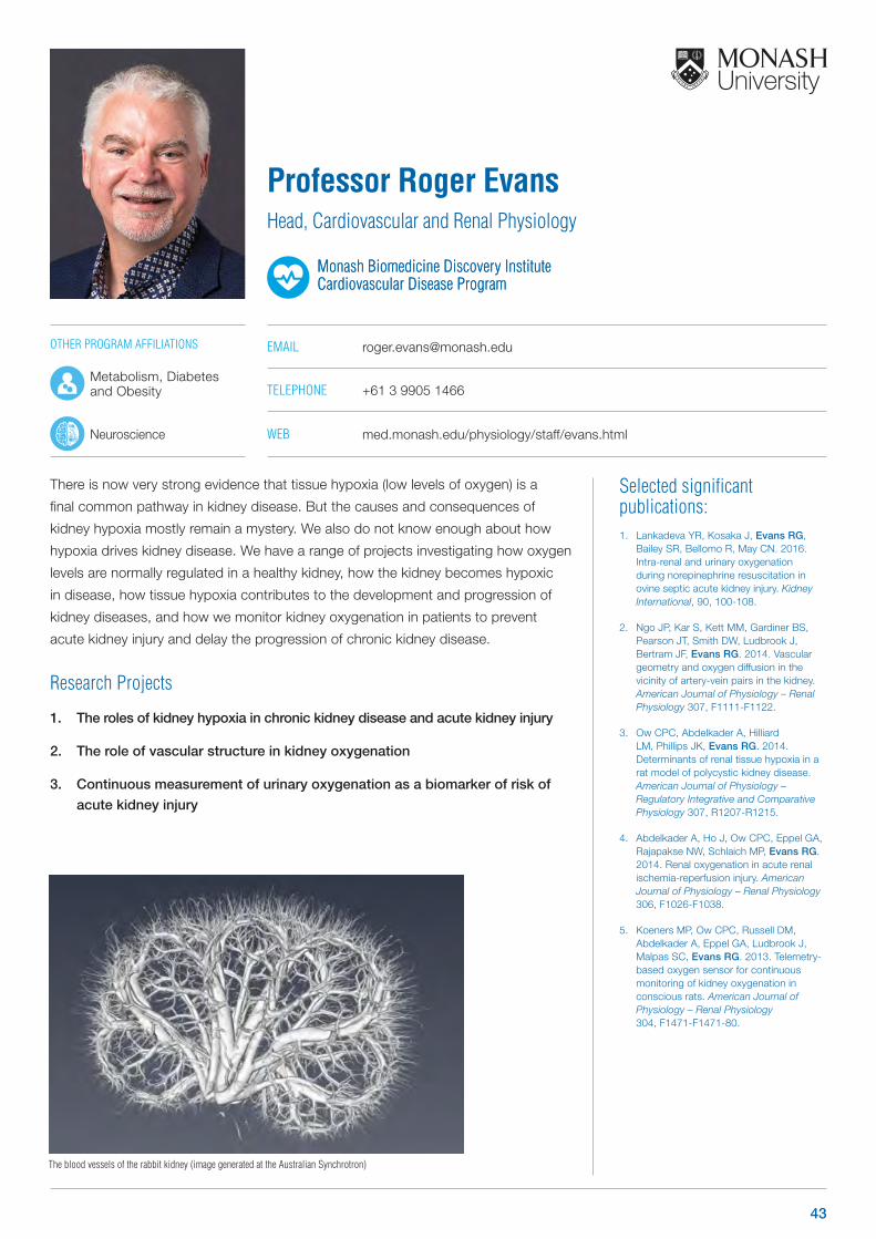
Selected significant publications: 1. Lankadeva YR, Kosaka J, Evans RG,
Bailey SR, Bellomo R, May CN. 2016. Intra-renal and urinary oxygenation during norepinephrine resuscitation in ovine septic acute kidney injury. Kidney International, 90, 100-108.
2. Ngo JP, Kar S, Kett MM, Gardiner BS, Pearson JT, Smith DW, Ludbrook J, Bertram JF, Evans RG. 2014. Vascular geometry and oxygen diffusion in the vicinity of artery-vein pairs in the kidney. American Journal of Physiology – Renal Physiology 307, F1111-F1122.
3. Ow CPC, Abdelkader A, Hilliard LM, Phillips JK, Evans RG. 2014. Determinants of renal tissue hypoxia in a rat model of polycystic kidney disease. American Journal of Physiology – Regulatory Integrative and Comparative Physiology 307, R1207-R1215.
4. Abdelkader A, Ho J, Ow CPC, Eppel GA, Rajapakse NW, Schlaich MP, Evans RG. 2014. Renal oxygenation in acute renal ischemia-reperfusion injury. American Journal of Physiology – Renal Physiology 306, F1026-F1038.
5. Koeners MP, Ow CPC, Russell DM, Abdelkader A, Eppel GA, Ludbrook J, Malpas SC, Evans RG. 2013. Telemetry-based oxygen sensor for continuous monitoring of kidney oxygenation in conscious rats. American Journal of Physiology – Renal Physiology 304, F1471-F1471-80.
There is now very strong evidence that tissue hypoxia (low levels of oxygen) is a
final common pathway in kidney disease. But the causes and consequences of
kidney hypoxia mostly remain a mystery. We also do not know enough about how
hypoxia drives kidney disease. We have a range of projects investigating how oxygen
levels are normally regulated in a healthy kidney, how the kidney becomes hypoxic
in disease, how tissue hypoxia contributes to the development and progression of
kidney diseases, and how we monitor kidney oxygenation in patients to prevent
acute kidney injury and delay the progression of chronic kidney disease.
Research Projects
1. The roles of kidney hypoxia in chronic kidney disease and acute kidney injury
2. The role of vascular structure in kidney oxygenation
3. Continuous measurement of urinary oxygenation as a biomarker of risk of
acute kidney injury
Professor Roger EvansHead, Cardiovascular and Renal Physiology
Monash Biomedicine Discovery Institute Cardiovascular Disease ProgramMonash Biomedicine Discovery Institute Cardiovascular Disease Program
EMAIL [email protected]
TELEPHONE +61 3 9905 1466
WEB med.monash.edu/physiology/staff/evans.html
The blood vessels of the rabbit kidney (image generated at the Australian Synchrotron)
OTHER PROGRAM AFFILIATIONS
Metabolism, Diabetes and Obesity
Neuroscience
43
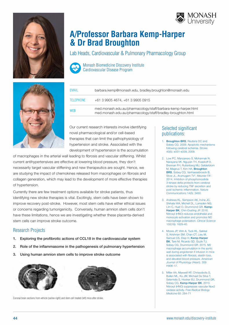
Selected significant publications: 1. Broughton BRS, Reutens DC and
Sobey CG. 2009. Apoptotic mechanisms following cerebral ischemia. Stroke. 40(5): e331-e339, 2009.
2. Low PC, Manzanero S, Mohannak N, Narayana VK, Nguyen TH, Kvaskoff D, Brennan FH, Ruitenberg MJ, Gelderblom M, Magnus T, Kim HA, Broughton BRS, Sobey CG, Vanhaesebroeck B, Stow JL, Arumugam TV*, Meunier FA*. 2014. Inhibition of phosphoinositide 3-kinase delta protects from cerebral stroke by reducing TNF secretion and post-ischemic inflammation. Nature Communications 14(5): 3450.
3. Andrews KL, Sampson AK, Irvine JC, Shihata WA, Michell DL, Lumsden NG, Lim C, Huet O, Drummond GR, Kemp-Harper BK, Chin-Dusting JP. 2016. Nitroxyl (HNO) reduces endothelial and monocyte activation and promotes M2 macrophage polarisation. Clinical Science 130(18): 1629-40.
4. Moore JP, Vinh A, Tuck KL, Sakkal S, Krishnan SM, Chan CT, Lieu M, Samuel CS, Diep H, Kemp-Harper BK, Tare M, Ricardo SD, Guzik TJ, Sobey CG, Drummond GR. 2015. M2 macrophage accumulation in the aortic wall during angiotensin II infusion in mice is associated with fibrosis, elastin loss, and elevated blood pressure. American Journal of Physiology (Heart), 309: H906-17.
5. Miller AA, Maxwell KF, Chrissobolis S, Bullen ML, Ku JM, Michael De Silva T, Selemidis S, Hooker EU, Drummond GR, Sobey CG, Kemp-Harper BK. 2013. Nitroxyl (HNO) suppresses vascular Nox2 oxidase activity. Free Radical Biology Medicine 60: 264-71
Our current research interests involve identifying
novel pharmacological and/or cell-based
therapies that can limit the pathophysiology of
hypertension and stroke. Associated with the
development of hypertension is the accumulation
of macrophages in the arterial wall leading to fibrosis and vascular stiffening. Whilst
current antihypertensives are effective at lowering blood pressure, they don’t
necessarily target vascular stiffening and new therapeutics are sought. Hence, we
are studying the impact of chemokines released from macrophages on fibrosis and
collagen generation, which may lead to the development of more effective therapies
of hypertension.
Currently there are few treatment options available for stroke patients, thus
identifying new stroke therapies is vital. Excitingly, stem cells have been shown to
improve recovery post-stroke. However, most stem cells have either ethical issues
or concerns regarding tumorigenicity. Conversely, human amnion stem cells don’t
have these limitations, hence we are investigating whether these placenta-derived
stem cells can improve stroke outcome.
Research Projects
1. Exploring the profibrotic actions of CCL18 in the cardiovascular system
2. Role of the inflammasome in the pathogenesis of pulmonary hypertension
3. Using human amnion stem cells to improve stroke outcome
A/Professor Barbara Kemp-Harper & Dr Brad BroughtonLab Heads, Cardiovascular & Pulmonary Pharmacology Group
Monash Biomedicine Discovery Institute Cardiovascular Disease Program
EMAIL [email protected], [email protected]
TELEPHONE +61 3 9905 4674, +61 3 9905 0915
WEB med.monash.edu.au/pharmacology/staff/barbara-kemp-harper.html med.monash.edu.au/pharmacology/staff/bradley-broughton.html
Coronal brain sections from vehicle (saline-right) and stem cell treated (left) mice after stroke.
44 www.monash.edu/discovery-institute
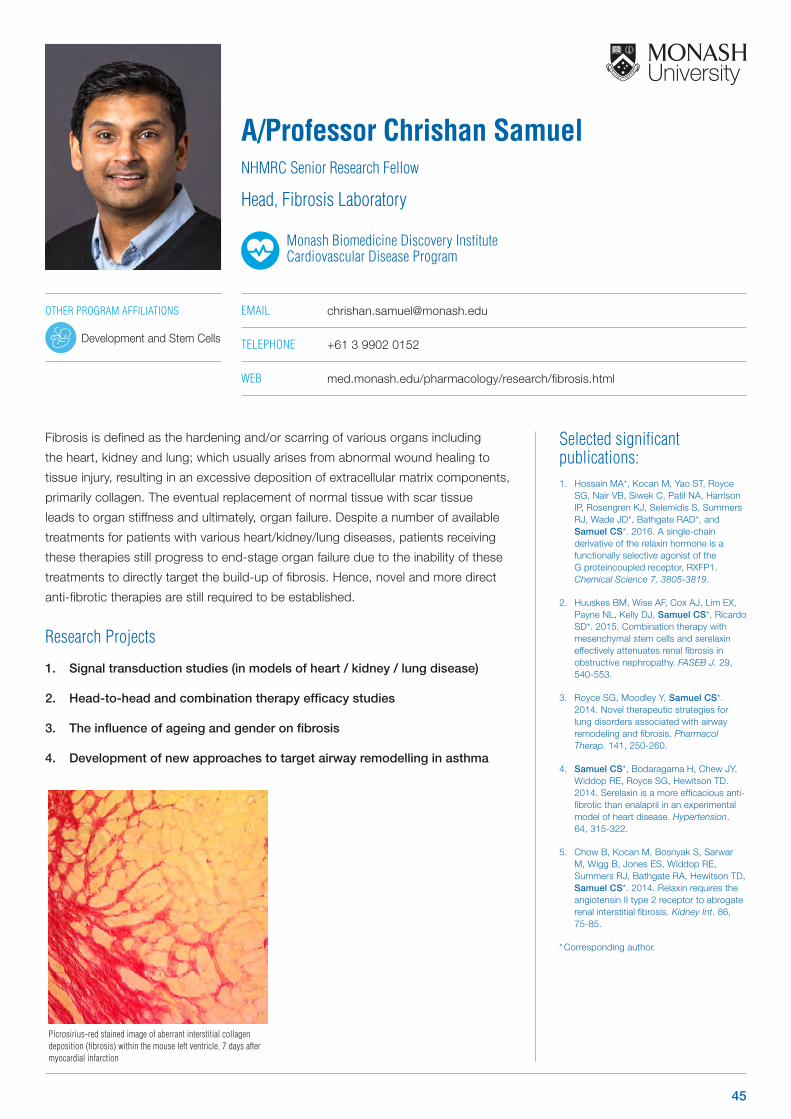
Selected significant publications: 1. Hossain MA*, Kocan M, Yao ST, Royce
SG, Nair VB, Siwek C, Patil NA, Harrison IP, Rosengren KJ, Selemidis S, Summers RJ, Wade JD*, Bathgate RAD*, and Samuel CS*. 2016. A single-chain derivative of the relaxin hormone is a functionally selective agonist of the G proteincoupled receptor, RXFP1. Chemical Science 7, 3805-3819.
2. Huuskes BM, Wise AF, Cox AJ, Lim EX, Payne NL, Kelly DJ, Samuel CS*, Ricardo SD*. 2015. Combination therapy with mesenchymal stem cells and serelaxin effectively attenuates renal fibrosis in obstructive nephropathy. FASEB J. 29, 540-553.
3. Royce SG, Moodley Y, Samuel CS*. 2014. Novel therapeutic strategies for lung disorders associated with airway remodeling and fibrosis. Pharmacol Therap. 141, 250-260.
4. Samuel CS*, Bodaragama H, Chew JY, Widdop RE, Royce SG, Hewitson TD. 2014. Serelaxin is a more efficacious anti-fibrotic than enalapril in an experimental model of heart disease. Hypertension. 64, 315-322.
5. Chow B, Kocan M, Bosnyak S, Sarwar M, Wigg B, Jones ES, Widdop RE, Summers RJ, Bathgate RA, Hewitson TD, Samuel CS*. 2014. Relaxin requires the angiotensin II type 2 receptor to abrogate renal interstitial fibrosis. Kidney Int. 86, 75-85.
* Corresponding author.
Fibrosis is defined as the hardening and/or scarring of various organs including
the heart, kidney and lung; which usually arises from abnormal wound healing to
tissue injury, resulting in an excessive deposition of extracellular matrix components,
primarily collagen. The eventual replacement of normal tissue with scar tissue
leads to organ stiffness and ultimately, organ failure. Despite a number of available
treatments for patients with various heart/kidney/lung diseases, patients receiving
these therapies still progress to end-stage organ failure due to the inability of these
treatments to directly target the build-up of fibrosis. Hence, novel and more direct
anti-fibrotic therapies are still required to be established.
Research Projects
1. Signal transduction studies (in models of heart / kidney / lung disease)
2. Head-to-head and combination therapy efficacy studies
3. The influence of ageing and gender on fibrosis
4. Development of new approaches to target airway remodelling in asthma
A/Professor Chrishan SamuelNHMRC Senior Research Fellow
Head, Fibrosis Laboratory
Monash Biomedicine Discovery Institute Cardiovascular Disease Program
EMAIL [email protected]
TELEPHONE +61 3 9902 0152
WEB med.monash.edu/pharmacology/research/fibrosis.html
Picrosirius-red stained image of aberrant interstitial collagen deposition (fibrosis) within the mouse left ventricle, 7 days after myocardial infarction
OTHER PROGRAM AFFILIATIONS
Development and Stem Cells
45
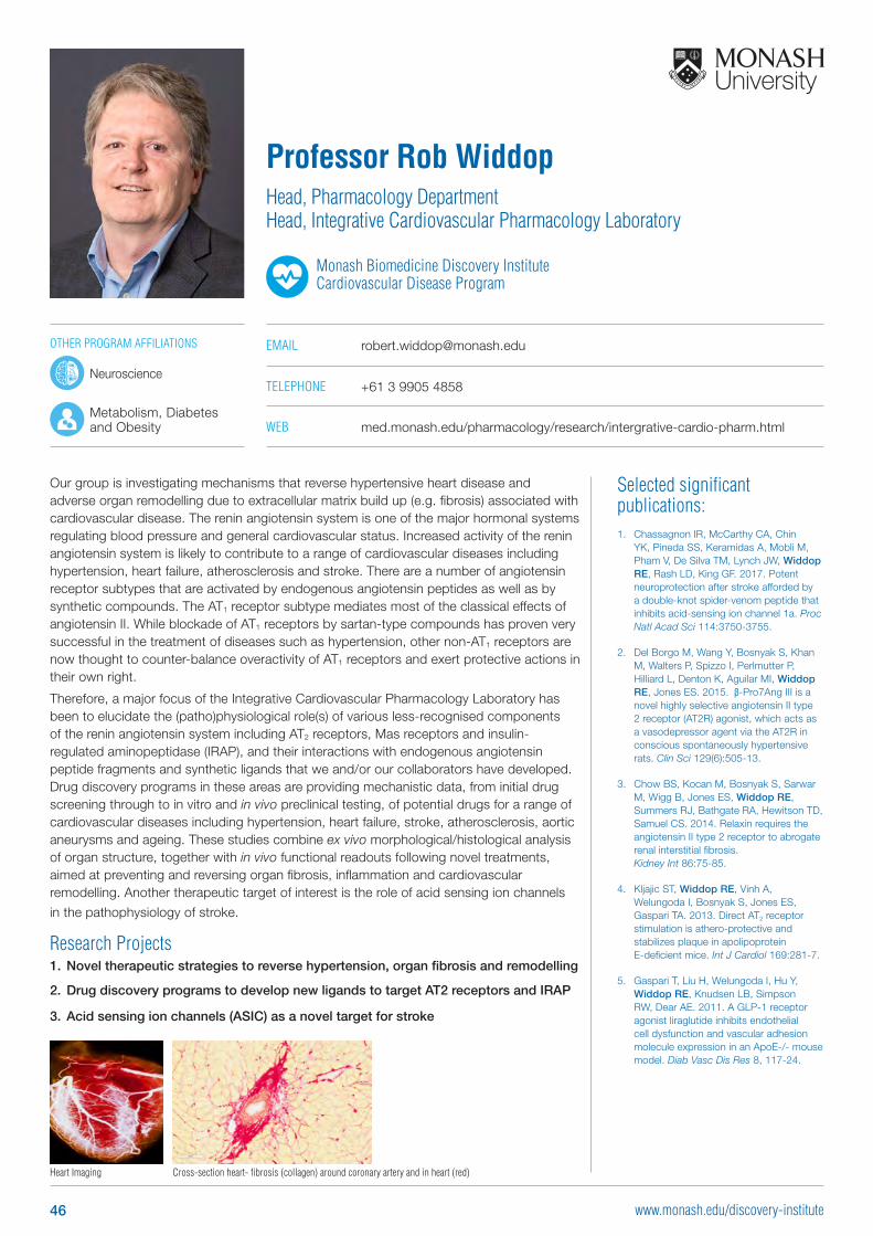
Selected significant publications: 1. Chassagnon IR, McCarthy CA, Chin
YK, Pineda SS, Keramidas A, Mobli M, Pham V, De Silva TM, Lynch JW, Widdop RE, Rash LD, King GF. 2017. Potent neuroprotection after stroke afforded by a double-knot spider-venom peptide that inhibits acid-sensing ion channel 1a. Proc Natl Acad Sci 114:3750-3755.
2. Del Borgo M, Wang Y, Bosnyak S, Khan M, Walters P, Spizzo I, Perlmutter P, Hilliard L, Denton K, Aguilar MI, Widdop RE, Jones ES. 2015. β-Pro7Ang III is a novel highly selective angiotensin II type 2 receptor (AT2R) agonist, which acts as a vasodepressor agent via the AT2R in conscious spontaneously hypertensive rats. Clin Sci 129(6):505-13.
3. Chow BS, Kocan M, Bosnyak S, Sarwar M, Wigg B, Jones ES, Widdop RE, Summers RJ, Bathgate RA, Hewitson TD, Samuel CS. 2014. Relaxin requires the angiotensin II type 2 receptor to abrogate renal interstitial fibrosis. Kidney Int 86:75-85.
4. Kljajic ST, Widdop RE, Vinh A, Welungoda I, Bosnyak S, Jones ES, Gaspari TA. 2013. Direct AT2 receptor stimulation is athero-protective and stabilizes plaque in apolipoprotein E-deficient mice. Int J Cardiol 169:281-7.
5. Gaspari T, Liu H, Welungoda I, Hu Y, Widdop RE, Knudsen LB, Simpson RW, Dear AE. 2011. A GLP-1 receptor agonist liraglutide inhibits endothelial cell dysfunction and vascular adhesion molecule expression in an ApoE-/- mouse model. Diab Vasc Dis Res 8, 117-24.
Our group is investigating mechanisms that reverse hypertensive heart disease and adverse organ remodelling due to extracellular matrix build up (e.g. fibrosis) associated with cardiovascular disease. The renin angiotensin system is one of the major hormonal systems regulating blood pressure and general cardiovascular status. Increased activity of the renin angiotensin system is likely to contribute to a range of cardiovascular diseases including hypertension, heart failure, atherosclerosis and stroke. There are a number of angiotensin receptor subtypes that are activated by endogenous angiotensin peptides as well as by synthetic compounds. The AT1 receptor subtype mediates most of the classical effects of angiotensin II. While blockade of AT1 receptors by sartan-type compounds has proven very successful in the treatment of diseases such as hypertension, other non-AT1 receptors are now thought to counter-balance overactivity of AT1 receptors and exert protective actions in their own right.
Therefore, a major focus of the Integrative Cardiovascular Pharmacology Laboratory has been to elucidate the (patho)physiological role(s) of various less-recognised components of the renin angiotensin system including AT2 receptors, Mas receptors and insulin-regulated aminopeptidase (IRAP), and their interactions with endogenous angiotensin peptide fragments and synthetic ligands that we and/or our collaborators have developed. Drug discovery programs in these areas are providing mechanistic data, from initial drug screening through to in vitro and in vivo preclinical testing, of potential drugs for a range of cardiovascular diseases including hypertension, heart failure, stroke, atherosclerosis, aortic aneurysms and ageing. These studies combine ex vivo morphological/histological analysis of organ structure, together with in vivo functional readouts following novel treatments, aimed at preventing and reversing organ fibrosis, inflammation and cardiovascular remodelling. Another therapeutic target of interest is the role of acid sensing ion channels
in the pathophysiology of stroke.
Research Projects1. Novel therapeutic strategies to reverse hypertension, organ fibrosis and remodelling
2. Drug discovery programs to develop new ligands to target AT2 receptors and IRAP
3. Acid sensing ion channels (ASIC) as a novel target for stroke
Professor Rob WiddopHead, Pharmacology Department Head, Integrative Cardiovascular Pharmacology Laboratory
Monash Biomedicine Discovery Institute Cardiovascular Disease Program
EMAIL [email protected]
TELEPHONE +61 3 9905 4858
WEB med.monash.edu/pharmacology/research/intergrative-cardio-pharm.html
Heart Imaging Cross-section heart- fibrosis (collagen) around coronary artery and in heart (red)
OTHER PROGRAM AFFILIATIONS
Neuroscience
Metabolism, Diabetes and Obesity
46 www.monash.edu/discovery-institute
Development and Stem Cells Program Group Leaders
47www.monash.edu/discovery-institute
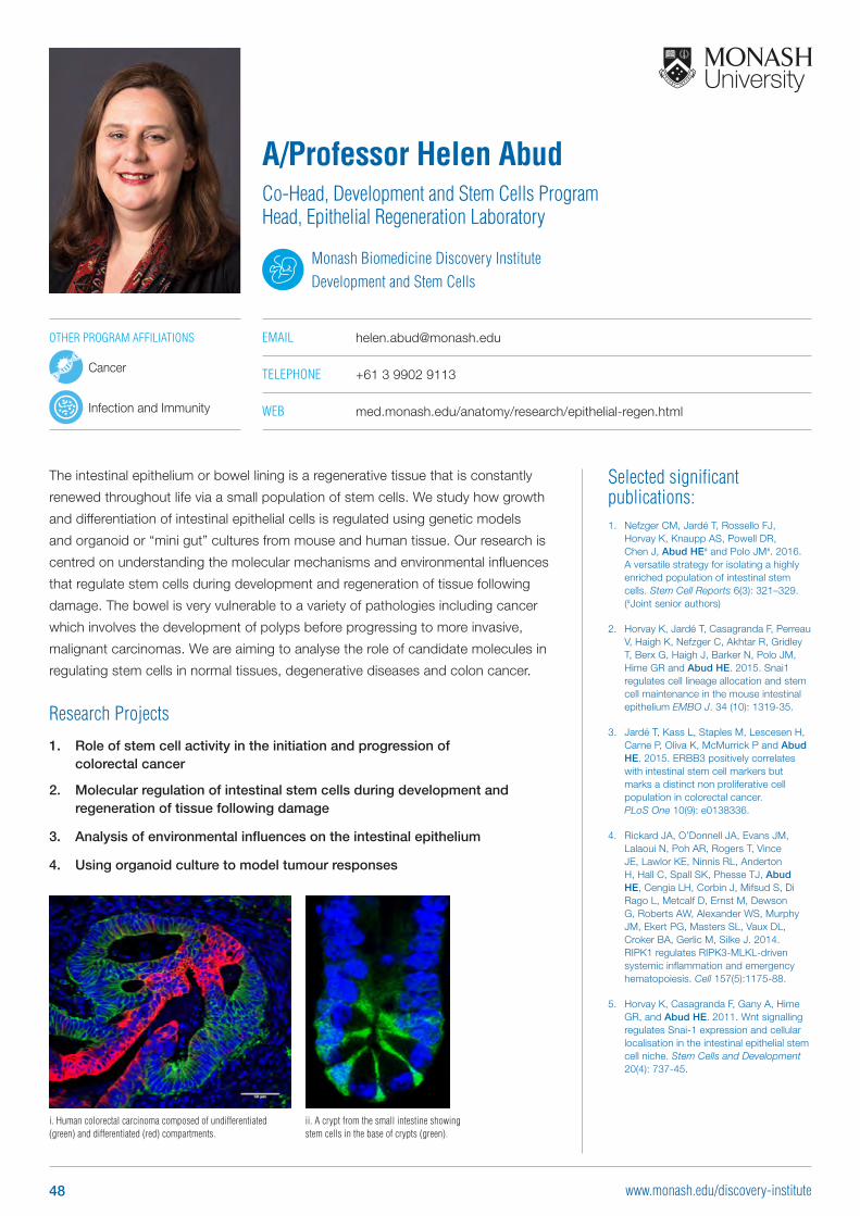
Selected significant publications: 1. Nefzger CM, Jardé T, Rossello FJ,
Horvay K, Knaupp AS, Powell DR, Chen J, Abud HE# and Polo JM#. 2016. A versatile strategy for isolating a highly enriched population of intestinal stem cells. Stem Cell Reports 6(3): 321–329. (#Joint senior authors)
2. Horvay K, Jardé T, Casagranda F, Perreau V, Haigh K, Nefzger C, Akhtar R, Gridley T, Berx G, Haigh J, Barker N, Polo JM, Hime GR and Abud HE. 2015. Snai1 regulates cell lineage allocation and stem cell maintenance in the mouse intestinal epithelium EMBO J. 34 (10): 1319-35.
3. Jardé T, Kass L, Staples M, Lescesen H, Carne P, Oliva K, McMurrick P and Abud HE. 2015. ERBB3 positively correlates with intestinal stem cell markers but marks a distinct non proliferative cell population in colorectal cancer. PLoS One 10(9): e0138336.
4. Rickard JA, O’Donnell JA, Evans JM, Lalaoui N, Poh AR, Rogers T, Vince JE, Lawlor KE, Ninnis RL, Anderton H, Hall C, Spall SK, Phesse TJ, Abud HE, Cengia LH, Corbin J, Mifsud S, Di Rago L, Metcalf D, Ernst M, Dewson G, Roberts AW, Alexander WS, Murphy JM, Ekert PG, Masters SL, Vaux DL, Croker BA, Gerlic M, Silke J. 2014. RIPK1 regulates RIPK3-MLKL-driven systemic inflammation and emergency hematopoiesis. Cell 157(5):1175-88.
5. Horvay K, Casagranda F, Gany A, Hime GR, and Abud HE. 2011. Wnt signalling regulates Snai-1 expression and cellular localisation in the intestinal epithelial stem cell niche. Stem Cells and Development 20(4): 737-45.
The intestinal epithelium or bowel lining is a regenerative tissue that is constantly
renewed throughout life via a small population of stem cells. We study how growth
and differentiation of intestinal epithelial cells is regulated using genetic models
and organoid or “mini gut” cultures from mouse and human tissue. Our research is
centred on understanding the molecular mechanisms and environmental influences
that regulate stem cells during development and regeneration of tissue following
damage. The bowel is very vulnerable to a variety of pathologies including cancer
which involves the development of polyps before progressing to more invasive,
malignant carcinomas. We are aiming to analyse the role of candidate molecules in
regulating stem cells in normal tissues, degenerative diseases and colon cancer.
Research Projects
1. Role of stem cell activity in the initiation and progression of colorectal cancer
2. Molecular regulation of intestinal stem cells during development and regeneration of tissue following damage
3. Analysis of environmental influences on the intestinal epithelium
4. Using organoid culture to model tumour responses
A/Professor Helen AbudCo-Head, Development and Stem Cells Program Head, Epithelial Regeneration Laboratory
Monash Biomedicine Discovery InstituteDevelopment and Stem Cells
EMAIL [email protected]
TELEPHONE +61 3 9902 9113
WEB med.monash.edu/anatomy/research/epithelial-regen.html
OTHER PROGRAM AFFILIATIONS
Cancer
Infection and Immunity
i. Human colorectal carcinoma composed of undifferentiated (green) and differentiated (red) compartments.
ii. A crypt from the small intestine showing stem cells in the base of crypts (green).
48 www.monash.edu/discovery-institute
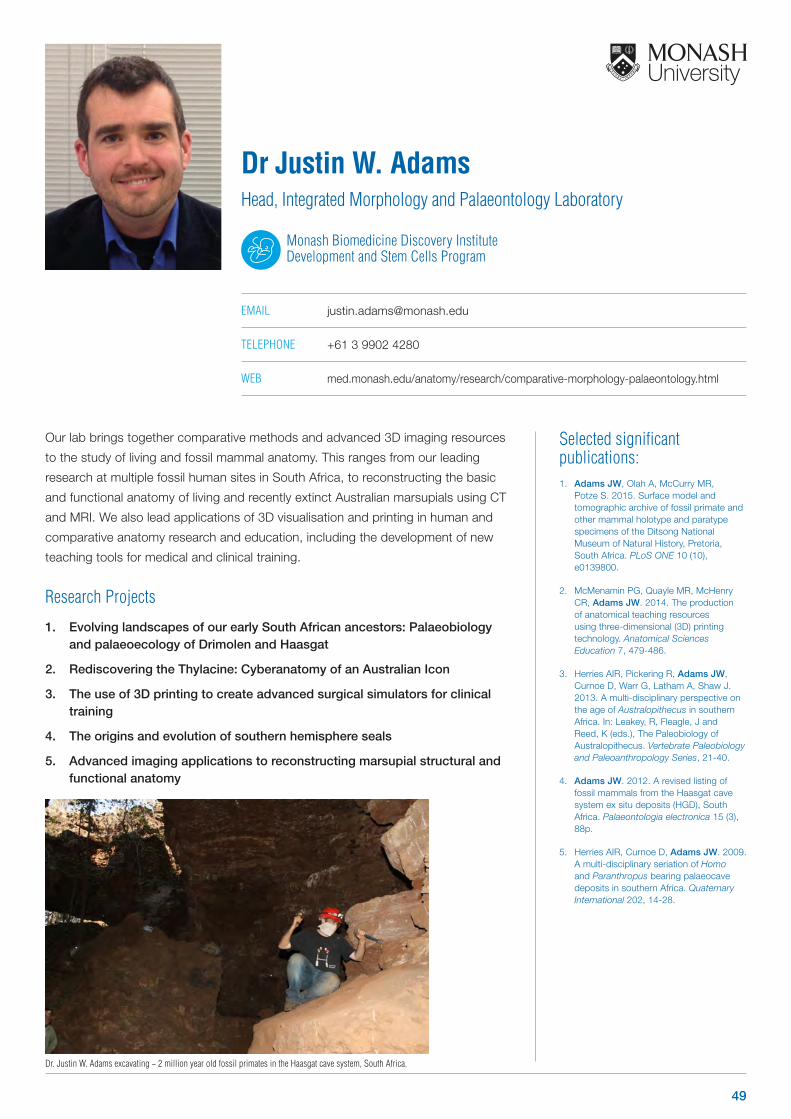
Selected significant publications: 1. Adams JW, Olah A, McCurry MR,
Potze S. 2015. Surface model and tomographic archive of fossil primate and other mammal holotype and paratype specimens of the Ditsong National Museum of Natural History, Pretoria, South Africa. PLoS ONE 10 (10), e0139800.
2. McMenamin PG, Quayle MR, McHenry CR, Adams JW. 2014. The production of anatomical teaching resources using three-dimensional (3D) printing technology. Anatomical Sciences Education 7, 479-486.
3. Herries AIR, Pickering R, Adams JW, Curnoe D, Warr G, Latham A, Shaw J. 2013. A multi-disciplinary perspective on the age of Australopithecus in southern Africa. In: Leakey, R, Fleagle, J and Reed, K (eds.), The Paleobiology of Australopithecus. Vertebrate Paleobiology and Paleoanthropology Series, 21-40.
4. Adams JW. 2012. A revised listing of fossil mammals from the Haasgat cave system ex situ deposits (HGD), South Africa. Palaeontologia electronica 15 (3), 88p.
5. Herries AIR, Curnoe D, Adams JW. 2009. A multi-disciplinary seriation of Homo and Paranthropus bearing palaeocave deposits in southern Africa. Quaternary International 202, 14-28.
Our lab brings together comparative methods and advanced 3D imaging resources
to the study of living and fossil mammal anatomy. This ranges from our leading
research at multiple fossil human sites in South Africa, to reconstructing the basic
and functional anatomy of living and recently extinct Australian marsupials using CT
and MRI. We also lead applications of 3D visualisation and printing in human and
comparative anatomy research and education, including the development of new
teaching tools for medical and clinical training.
Research Projects
1. Evolving landscapes of our early South African ancestors: Palaeobiology and palaeoecology of Drimolen and Haasgat
2. Rediscovering the Thylacine: Cyberanatomy of an Australian Icon
3. The use of 3D printing to create advanced surgical simulators for clinical training
4. The origins and evolution of southern hemisphere seals
5. Advanced imaging applications to reconstructing marsupial structural and functional anatomy
Dr Justin W. AdamsHead, Integrated Morphology and Palaeontology Laboratory
Monash Biomedicine Discovery Institute Development and Stem Cells Program
EMAIL [email protected]
TELEPHONE +61 3 9902 4280
WEB med.monash.edu/anatomy/research/comparative-morphology-palaeontology.html
Dr. Justin W. Adams excavating ~ 2 million year old fossil primates in the Haasgat cave system, South Africa.
49
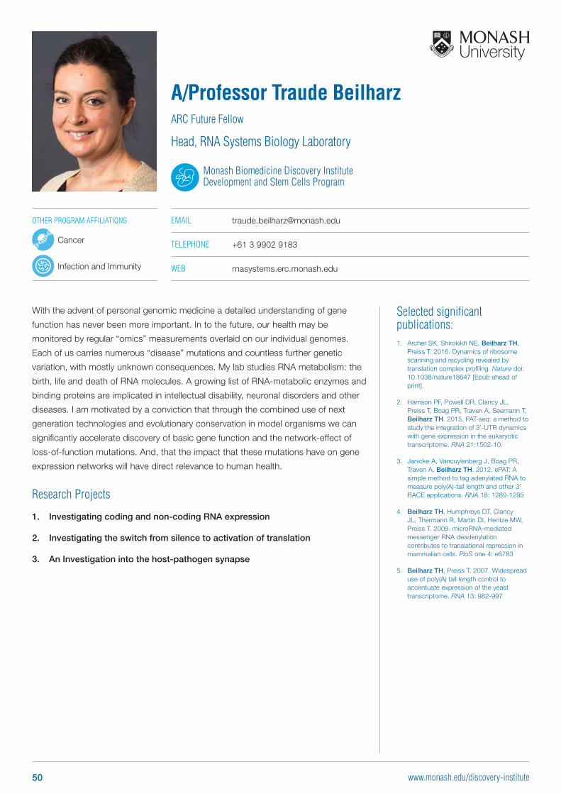
Selected significant publications: 1. Archer SK, Shirokikh NE, Beilharz TH,
Preiss T. 2016. Dynamics of ribosome scanning and recycling revealed by translation complex profiling. Nature doi: 10.1038/nature18647 [Epub ahead of print].
2. Harrison PF, Powell DR, Clancy JL, Preiss T, Boag PR, Traven A, Seemann T, Beilharz TH. 2015. PAT-seq: a method to study the integration of 3’-UTR dynamics with gene expression in the eukaryotic transcriptome. RNA 21:1502-10.
3. Janicke A, Vancuylenberg J, Boag PR, Traven A, Beilharz TH. 2012. ePAT: A simple method to tag adenylated RNA to measure poly(A)-tail length and other 3’ RACE applications. RNA 18: 1289-1295
4. Beilharz TH, Humphreys DT, Clancy JL, Thermann R, Martin DI, Hentze MW, Preiss T. 2009. microRNA-mediated messenger RNA deadenylation contributes to translational repression in mammalian cells. PloS one 4: e6783
5. Beilharz TH, Preiss T. 2007. Widespread use of poly(A) tail length control to accentuate expression of the yeast transcriptome. RNA 13: 982-997
With the advent of personal genomic medicine a detailed understanding of gene
function has never been more important. In to the future, our health may be
monitored by regular “omics” measurements overlaid on our individual genomes.
Each of us carries numerous “disease” mutations and countless further genetic
variation, with mostly unknown consequences. My lab studies RNA metabolism: the
birth, life and death of RNA molecules. A growing list of RNA-metabolic enzymes and
binding proteins are implicated in intellectual disability, neuronal disorders and other
diseases. I am motivated by a conviction that through the combined use of next
generation technologies and evolutionary conservation in model organisms we can
significantly accelerate discovery of basic gene function and the network-effect of
loss-of-function mutations. And, that the impact that these mutations have on gene
expression networks will have direct relevance to human health.
Research Projects
1. Investigating coding and non-coding RNA expression
2. Investigating the switch from silence to activation of translation
3. An Investigation into the host-pathogen synapse
A/Professor Traude BeilharzARC Future Fellow
Head, RNA Systems Biology Laboratory
Monash Biomedicine Discovery Institute Development and Stem Cells Program
EMAIL [email protected]
TELEPHONE +61 3 9902 9183
WEB rnasystems.erc.monash.edu
OTHER PROGRAM AFFILIATIONS
Cancer
Infection and Immunity
50 www.monash.edu/discovery-institute
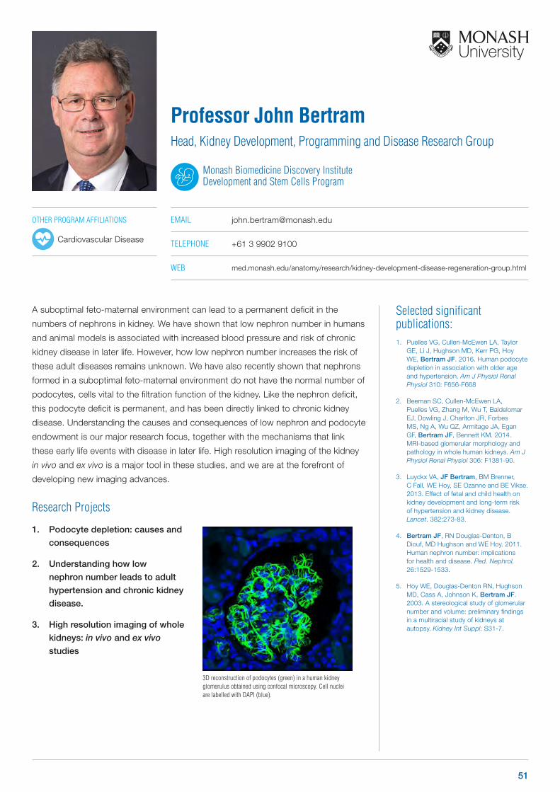
Selected significant publications: 1. Puelles VG, Cullen-McEwen LA, Taylor
GE, Li J, Hughson MD, Kerr PG, Hoy WE, Bertram JF. 2016. Human podocyte depletion in association with older age and hypertension. Am J Physiol Renal Physiol 310: F656-F668
2. Beeman SC, Cullen-McEwen LA, Puelles VG, Zhang M, Wu T, Baldelomar EJ, Dowling J, Charlton JR, Forbes MS, Ng A, Wu QZ, Armitage JA, Egan GF, Bertram JF, Bennett KM. 2014. MRI-based glomerular morphology and pathology in whole human kidneys. Am J Physiol Renal Physiol 306: F1381-90.
3. Luyckx VA, JF Bertram, BM Brenner, C Fall, WE Hoy, SE Ozanne and BE Vikse. 2013. Effect of fetal and child health on kidney development and long-term risk of hypertension and kidney disease. Lancet. 382:273-83.
4. Bertram JF, RN Douglas-Denton, B Diouf, MD Hughson and WE Hoy. 2011. Human nephron number: implications for health and disease. Ped. Nephrol. 26:1529-1533.
5. Hoy WE, Douglas-Denton RN, Hughson MD, Cass A, Johnson K, Bertram JF. 2003. A stereological study of glomerular number and volume: preliminary findings in a multiracial study of kidneys at autopsy. Kidney Int Suppl: S31-7.
A suboptimal feto-maternal environment can lead to a permanent deficit in the
numbers of nephrons in kidney. We have shown that low nephron number in humans
and animal models is associated with increased blood pressure and risk of chronic
kidney disease in later life. However, how low nephron number increases the risk of
these adult diseases remains unknown. We have also recently shown that nephrons
formed in a suboptimal feto-maternal environment do not have the normal number of
podocytes, cells vital to the filtration function of the kidney. Like the nephron deficit,
this podocyte deficit is permanent, and has been directly linked to chronic kidney
disease. Understanding the causes and consequences of low nephron and podocyte
endowment is our major research focus, together with the mechanisms that link
these early life events with disease in later life. High resolution imaging of the kidney
in vivo and ex vivo is a major tool in these studies, and we are at the forefront of
developing new imaging advances.
Research Projects
1. Podocyte depletion: causes and
consequences
2. Understanding how low
nephron number leads to adult
hypertension and chronic kidney
disease.
3. High resolution imaging of whole
kidneys: in vivo and ex vivo
studies
Professor John BertramHead, Kidney Development, Programming and Disease Research Group
Monash Biomedicine Discovery Institute Development and Stem Cells Program
EMAIL [email protected]
TELEPHONE +61 3 9902 9100
WEB med.monash.edu/anatomy/research/kidney-development-disease-regeneration-group.html
3D reconstruction of podocytes (green) in a human kidney glomerulus obtained using confocal microscopy. Cell nuclei are labelled with DAPI (blue).
OTHER PROGRAM AFFILIATIONS
Cardiovascular Disease
51
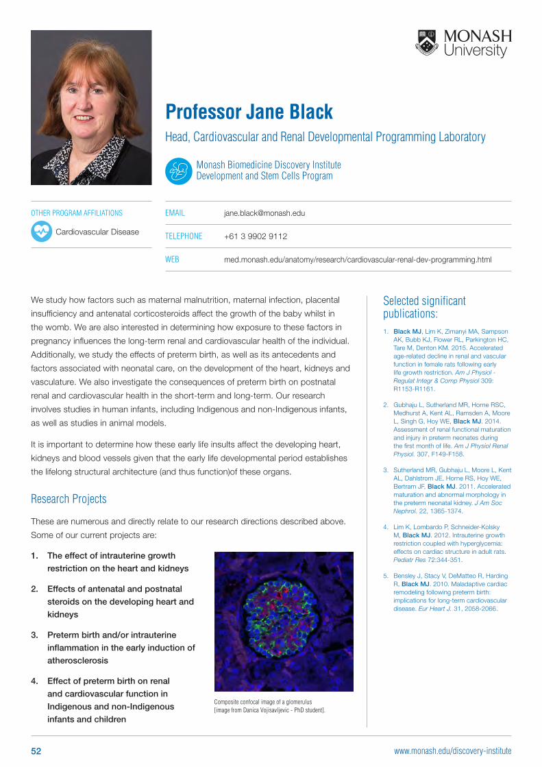
Selected significant publications: 1. Black MJ, Lim K, Zimanyi MA, Sampson
AK, Bubb KJ, Flower RL, Parkington HC, Tare M, Denton KM. 2015. Accelerated age-related decline in renal and vascular function in female rats following early life growth restriction. Am J Physiol - Regulat Integr & Comp Physiol 309: R1153-R1161.
2. Gubhaju L, Sutherland MR, Horne RSC, Medhurst A, Kent AL, Ramsden A, Moore L, Singh G, Hoy WE, Black MJ. 2014. Assessment of renal functional maturation and injury in preterm neonates during the first month of life. Am J Physiol Renal Physiol. 307, F149-F158.
3. Sutherland MR, Gubhaju L, Moore L, Kent AL, Dahlstrom JE, Horne RS, Hoy WE, Bertram JF, Black MJ. 2011. Accelerated maturation and abnormal morphology in the preterm neonatal kidney. J Am Soc Nephrol. 22, 1365-1374.
4. Lim K, Lombardo P, Schneider-Kolsky M, Black MJ. 2012. Intrauterine growth restriction coupled with hyperglycemia: effects on cardiac structure in adult rats. Pediatr Res 72:344-351.
5. Bensley J, Stacy V, DeMatteo R, Harding R, Black MJ. 2010. Maladaptive cardiac remodeling following preterm birth: implications for long-term cardiovascular disease. Eur Heart J. 31, 2058-2066.
We study how factors such as maternal malnutrition, maternal infection, placental
insufficiency and antenatal corticosteroids affect the growth of the baby whilst in
the womb. We are also interested in determining how exposure to these factors in
pregnancy influences the long-term renal and cardiovascular health of the individual.
Additionally, we study the effects of preterm birth, as well as its antecedents and
factors associated with neonatal care, on the development of the heart, kidneys and
vasculature. We also investigate the consequences of preterm birth on postnatal
renal and cardiovascular health in the short-term and long-term. Our research
involves studies in human infants, including Indigenous and non-Indigenous infants,
as well as studies in animal models.
It is important to determine how these early life insults affect the developing heart,
kidneys and blood vessels given that the early life developmental period establishes
the lifelong structural architecture (and thus function)of these organs.
Research Projects
These are numerous and directly relate to our research directions described above.
Some of our current projects are:
1. The effect of intrauterine growth
restriction on the heart and kidneys
2. Effects of antenatal and postnatal
steroids on the developing heart and
kidneys
3. Preterm birth and/or intrauterine
inflammation in the early induction of
atherosclerosis
4. Effect of preterm birth on renal
and cardiovascular function in
Indigenous and non-Indigenous
infants and children
Professor Jane BlackHead, Cardiovascular and Renal Developmental Programming Laboratory
Monash Biomedicine Discovery Institute Development and Stem Cells Program
EMAIL [email protected]
TELEPHONE +61 3 9902 9112
WEB med.monash.edu/anatomy/research/cardiovascular-renal-dev-programming.html
OTHER PROGRAM AFFILIATIONS
Cardiovascular Disease
Composite confocal image of a glomerulus [image from Danica Vojisavljevic - PhD student].
52 www.monash.edu/discovery-institute
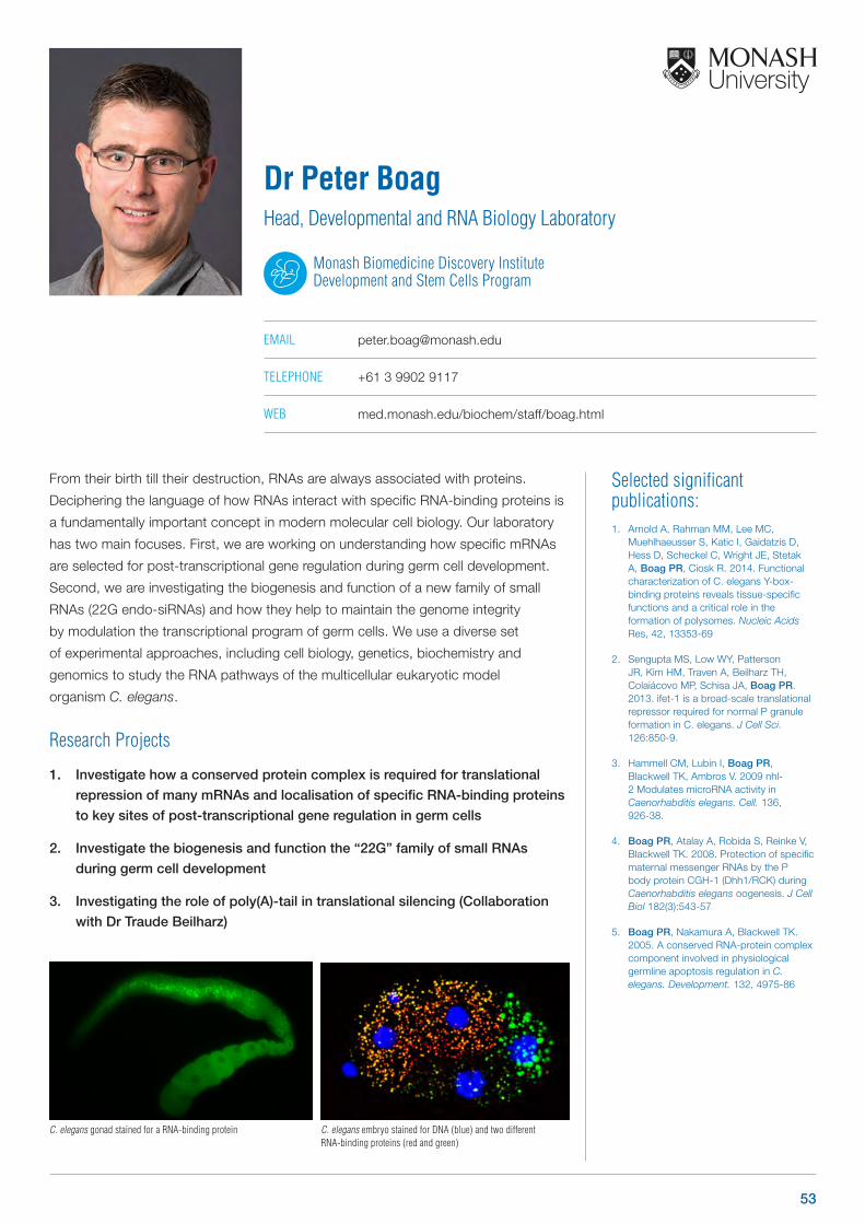
Selected significant publications: 1. Arnold A, Rahman MM, Lee MC,
Muehlhaeusser S, Katic I, Gaidatzis D, Hess D, Scheckel C, Wright JE, Stetak A, Boag PR, Ciosk R. 2014. Functional characterization of C. elegans Y-box-binding proteins reveals tissue-specific functions and a critical role in the formation of polysomes. Nucleic Acids Res, 42, 13353-69
2. Sengupta MS, Low WY, Patterson JR, Kim HM, Traven A, Beilharz TH, Colaiácovo MP, Schisa JA, Boag PR. 2013. ifet-1 is a broad-scale translational repressor required for normal P granule formation in C. elegans. J Cell Sci. 126:850-9.
3. Hammell CM, Lubin I, Boag PR, Blackwell TK, Ambros V. 2009 nhl-2 Modulates microRNA activity in Caenorhabditis elegans. Cell. 136, 926-38.
4. Boag PR, Atalay A, Robida S, Reinke V, Blackwell TK. 2008. Protection of specific maternal messenger RNAs by the P body protein CGH-1 (Dhh1/RCK) during Caenorhabditis elegans oogenesis. J Cell Biol 182(3):543-57
5. Boag PR, Nakamura A, Blackwell TK. 2005. A conserved RNA-protein complex component involved in physiological germline apoptosis regulation in C. elegans. Development. 132, 4975-86
From their birth till their destruction, RNAs are always associated with proteins.
Deciphering the language of how RNAs interact with specific RNA-binding proteins is
a fundamentally important concept in modern molecular cell biology. Our laboratory
has two main focuses. First, we are working on understanding how specific mRNAs
are selected for post-transcriptional gene regulation during germ cell development.
Second, we are investigating the biogenesis and function of a new family of small
RNAs (22G endo-siRNAs) and how they help to maintain the genome integrity
by modulation the transcriptional program of germ cells. We use a diverse set
of experimental approaches, including cell biology, genetics, biochemistry and
genomics to study the RNA pathways of the multicellular eukaryotic model
organism C. elegans.
Research Projects
1. Investigate how a conserved protein complex is required for translational
repression of many mRNAs and localisation of specific RNA-binding proteins
to key sites of post-transcriptional gene regulation in germ cells
2. Investigate the biogenesis and function the “22G” family of small RNAs
during germ cell development
3. Investigating the role of poly(A)-tail in translational silencing (Collaboration
with Dr Traude Beilharz)
Dr Peter BoagHead, Developmental and RNA Biology Laboratory
Monash Biomedicine Discovery Institute Development and Stem Cells Program
EMAIL [email protected]
TELEPHONE +61 3 9902 9117
WEB med.monash.edu/biochem/staff/boag.html
C. elegans gonad stained for a RNA-binding protein C. elegans embryo stained for DNA (blue) and two different RNA-binding proteins (red and green)
53

Selected significant publications: 1. Nabti I, Grimes R, Sarna H, Marangos P,
Carroll J. 2017. Maternal age-dependent APC/C-mediated decrease in securin causes premature sister chromatid separation in meiosis II. Nat Commun 8: 15346
2. Marangos P, Stevense M, Niaka K, Lagoudaki M, Nabti I, Jessberger R, Carroll J. 2015. DNA damage-induced metaphase I arrest is mediated by the spindle assembly checkpoint and maternal age. Nat Commun 6:8706.
3. Nabti I, Marangos P, Bormann J, Kudo N, Carroll J. 2014 Dual-mode regulation of the APC/C by CDK1 and MAPK controls meiosis I progression and fidelity. J Cell Biol. 204, 891-900
4. Dalton CM, Carroll J. 2013. Biased inheritance of mitochondria during asymmetric cell division in the mouse oocyte. J Cell Sci. 126, 2955-64
5. Homer H, Gui L, Carroll J. 2009. A spindle assembly checkpoint protein functions in prophase I arrest and prometaphase progression. Science. 326, 991-994
The mammalian oocyte is the largest cell in the body and undergoes two highly
specialised asymmetric meiotic cell divisions. Coordination of organelle inheritance,
polarity and meiotic progression is essential for the production of an oocyte capable
of undergoing fertilization and development to term. We use molecular and genetic
approaches combined with live cell imaging to investigate the cell biology of
these processes in mice and humans. Investigating these questions allows us to
understand how oocytes make the transition into a healthy embryo and why it
goes wrong in cases such as maternal ageing.
Research Projects
1. Role of mitochondria in controlling
meiosis in oocytes
2. Coordinating polarity and cell cycle
progression in the meiotic divisions
3. Impact of maternal age and
obesity on chromosome dynamics
and oocyte quality (in collaboration with
Rebecca Robker, University of Adelaide)
Professor John CarrollDirector, Monash Biomedicine Discovery Institute
Head, Oocyte and Embryo Development Laboratory
Monash Biomedicine Discovery Institute Development and Stem Cells Program
EMAIL [email protected]
TELEPHONE +61 3 9902 4381
WEB med.monash.edu/anatomy/staff/carroll-john.html
An oocyte arrested in metaphase of meiosis II just before fertilisation.
Cytoplasmic architecture in a maturing oocyte. The developing first meiotic spindle is surrounded by ER (red) and mitochondria (green).
OTHER PROGRAM AFFILIATIONS
Cancer
Metabolism, Diabetes and Obesity
54 www.monash.edu/discovery-institute
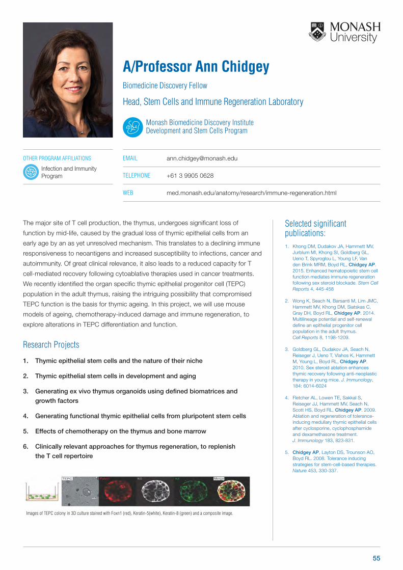
Selected significant publications: 1. Khong DM, Dudakov JA, Hammett MV,
Jurblum MI, Khong SI, Goldberg GL, Ueno T, Spyroglou L, Young LF, Van den Brink MRM, Boyd RL, Chidgey AP. 2015. Enhanced hematopoietic stem cell function mediates immune regeneration following sex steroid blockade. Stem Cell Reports 4, 445-458
2. Wong K, Seach N, Barsanti M, Lim JMC, Hammett MV, Khong DM, Siatskas C, Gray DH, Boyd RL, Chidgey AP. 2014. Multilineage potential and self-renewal define an epithelial progenitor cell population in the adult thymus. Cell Reports 8, 1198-1209.
3. Goldberg GL, Dudakov JA, Seach N, Reiseger J, Ueno T, Vlahos K, Hammett M, Young L, Boyd RL, Chidgey AP. 2010. Sex steroid ablation enhances thymic recovery following anti-neoplastic therapy in young mice. J. Immunology, 184: 6014-6024
4. Fletcher AL, Lowen TE, Sakkal S, Reiseger JJ, Hammett MV, Seach N, Scott HS, Boyd RL, Chidgey AP. 2009. Ablation and regeneration of tolerance-inducing medullary thymic epithelial cells after cyclosporine, cyclophosphamide and dexamethasone treatment. J. Immunology 183, 823-831.
5. Chidgey AP, Layton DS, Trounson AO, Boyd RL. 2008. Tolerance inducing strategies for stem-cell-based therapies. Nature 453, 330-337.
The major site of T cell production, the thymus, undergoes significant loss of
function by mid-life, caused by the gradual loss of thymic epithelial cells from an
early age by an as yet unresolved mechanism. This translates to a declining immune
responsiveness to neoantigens and increased susceptibility to infections, cancer and
autoimmunity. Of great clinical relevance, it also leads to a reduced capacity for T
cell-mediated recovery following cytoablative therapies used in cancer treatments.
We recently identified the organ specific thymic epithelial progenitor cell (TEPC)
population in the adult thymus, raising the intriguing possibility that compromised
TEPC function is the basis for thymic ageing. In this project, we will use mouse
models of ageing, chemotherapy-induced damage and immune regeneration, to
explore alterations in TEPC differentiation and function.
Research Projects
1. Thymic epithelial stem cells and the nature of their niche
2. Thymic epithelial stem cells in development and aging
3. Generating ex vivo thymus organoids using defined biomatrices and
growth factors
4. Generating functional thymic epithelial cells from pluripotent stem cells
5. Effects of chemotherapy on the thymus and bone marrow
6. Clinically relevant approaches for thymus regeneration, to replenish
the T cell repertoire
A/Professor Ann ChidgeyBiomedicine Discovery Fellow
Head, Stem Cells and Immune Regeneration Laboratory
Monash Biomedicine Discovery Institute Development and Stem Cells Program
EMAIL [email protected]
TELEPHONE +61 3 9905 0628
WEB med.monash.edu/anatomy/research/immune-regeneration.html
Images of TEPC colony in 3D culture stained with Foxn1 (red), Keratin-5(white), Keratin-8 (green) and a composite image.
OTHER PROGRAM AFFILIATIONS
Infection and Immunity Program
55
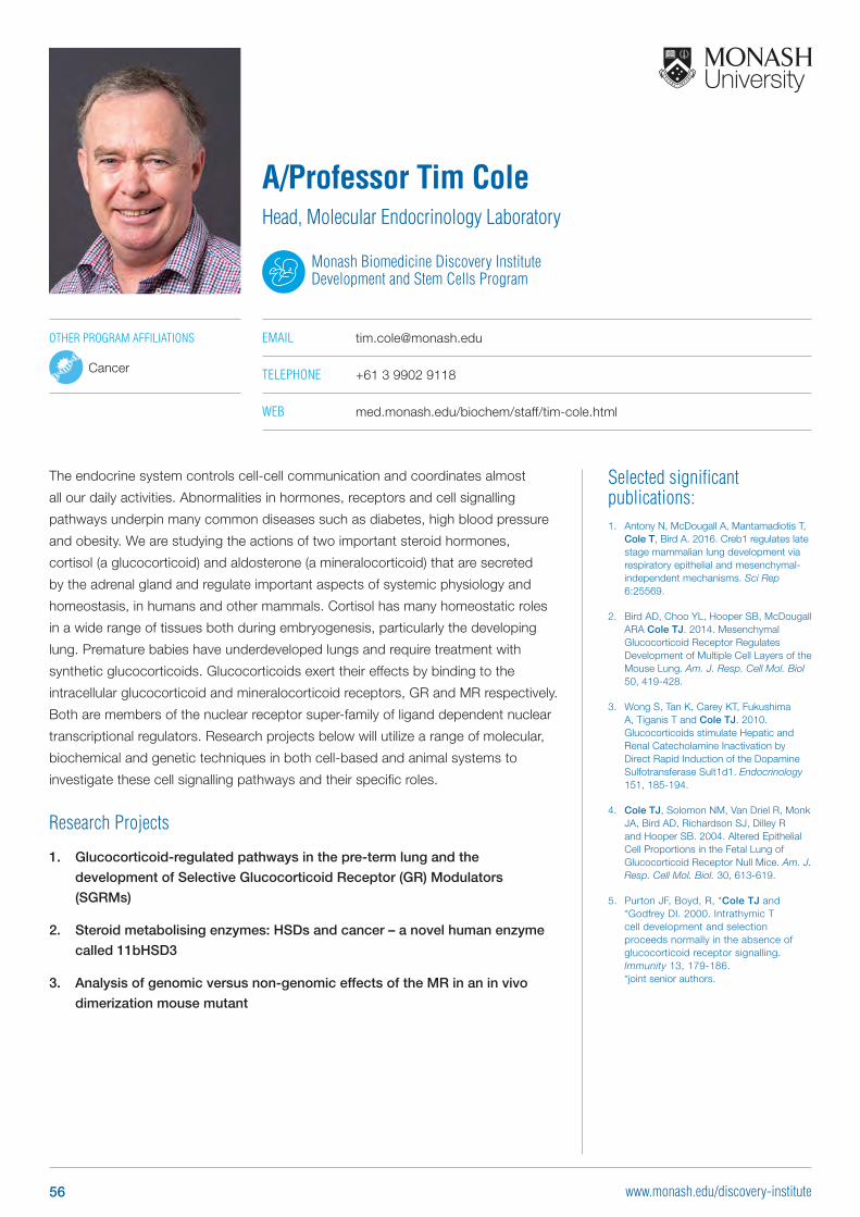
Selected significant publications: 1. Antony N, McDougall A, Mantamadiotis T,
Cole T, Bird A. 2016. Creb1 regulates late stage mammalian lung development via respiratory epithelial and mesenchymal-independent mechanisms. Sci Rep 6:25569.
2. Bird AD, Choo YL, Hooper SB, McDougall ARA Cole TJ. 2014. Mesenchymal Glucocorticoid Receptor Regulates Development of Multiple Cell Layers of the Mouse Lung. Am. J. Resp. Cell Mol. Biol 50, 419-428.
3. Wong S, Tan K, Carey KT, Fukushima A, Tiganis T and Cole TJ. 2010. Glucocorticoids stimulate Hepatic and Renal Catecholamine Inactivation by Direct Rapid Induction of the Dopamine Sulfotransferase Sult1d1. Endocrinology 151, 185-194.
4. Cole TJ, Solomon NM, Van Driel R, Monk JA, Bird AD, Richardson SJ, Dilley R and Hooper SB. 2004. Altered Epithelial Cell Proportions in the Fetal Lung of Glucocorticoid Receptor Null Mice. Am. J. Resp. Cell Mol. Biol. 30, 613-619.
5. Purton JF, Boyd, R, *Cole TJ and *Godfrey DI. 2000. Intrathymic T cell development and selection proceeds normally in the absence of glucocorticoid receptor signalling. Immunity 13, 179-186. *joint senior authors.
The endocrine system controls cell-cell communication and coordinates almost
all our daily activities. Abnormalities in hormones, receptors and cell signalling
pathways underpin many common diseases such as diabetes, high blood pressure
and obesity. We are studying the actions of two important steroid hormones,
cortisol (a glucocorticoid) and aldosterone (a mineralocorticoid) that are secreted
by the adrenal gland and regulate important aspects of systemic physiology and
homeostasis, in humans and other mammals. Cortisol has many homeostatic roles
in a wide range of tissues both during embryogenesis, particularly the developing
lung. Premature babies have underdeveloped lungs and require treatment with
synthetic glucocorticoids. Glucocorticoids exert their effects by binding to the
intracellular glucocorticoid and mineralocorticoid receptors, GR and MR respectively.
Both are members of the nuclear receptor super-family of ligand dependent nuclear
transcriptional regulators. Research projects below will utilize a range of molecular,
biochemical and genetic techniques in both cell-based and animal systems to
investigate these cell signalling pathways and their specific roles.
Research Projects
1. Glucocorticoid-regulated pathways in the pre-term lung and the
development of Selective Glucocorticoid Receptor (GR) Modulators
(SGRMs)
2. Steroid metabolising enzymes: HSDs and cancer – a novel human enzyme
called 11bHSD3
3. Analysis of genomic versus non-genomic effects of the MR in an in vivo
dimerization mouse mutant
A/Professor Tim ColeHead, Molecular Endocrinology Laboratory
Monash Biomedicine Discovery Institute Development and Stem Cells Program
EMAIL [email protected]
TELEPHONE +61 3 9902 9118
WEB med.monash.edu/biochem/staff/tim-cole.html
OTHER PROGRAM AFFILIATIONS
Cancer
56 www.monash.edu/discovery-institute
Selected significant publications: 1. Xie H, Peng C, Huang J, Li BE, Kim W,
Smith EC, Fujiwara Y, Qi J, Cheloni G, Das PP, Nguyen M, Li S, Bradner JE, Orkin SH. 2016. Chronic myelogenous leukemia initiating cells require Polycomb group protein EZH2. Cancer Discovery pii: CD-15-1439
2. Das PP, Hendrix DA, Apostolou E, Buchner AH, Canver MC, Beyaz S, Ljuboja D, Kuintzle R, Kim W, Karnik R, Shao Z, Xie H, Xu J, De Los Angeles A, Zhang Y, Choe J, Jun DL, Shen X, Gregory RI, Daley GQ, Meissner A, Kellis M, Hochedlinger K, Kim J, Orkin SH. 2015. PRC2 is required to maintain expression of the maternal Gtl2-Rian-Mirg locus by preventing de novo DNA methylation in mouse embryonic stem cells. Cell Reports 12(9):1456-70.
3. Das PP, Shao Z, Beyaz S, Apostolou E, Pinello L, Los Angeles AD, O’Brien K, Atsma JM, Fujiwara Y, Nguyen M, Ljuboja D, Guo G, Woo A, Yuan GC, Onder T, Daley GQ, Hochedlinger K, Kim J, Orkin SH. 2014. Distinct and combinatorial functions of Jmjd2b/Kdm4b and Jmjd2c/Kdm4c in mouse embryonic stem cells identity. Molecular Cell 53(1): 32-48.
4. Guo G, Luc S, Marco E, Lin TW, Peng C, Kerenyi MA, Beyaz S, Kim W, Xu J, Das PP, Neff T, Zou K, Yuan GC, Orkin SH. 2013. Mapping the hematopoietic hierarchy by single cell analysis of the cell surface repertoire. Cell Stem Cell 13(4): 492-505.
5. Das PP, Bagijn MP, Goldstein LD, Woolford JR, Lehrbach NJ, Sapetschnig A, Buhecha HR, Gilchrist MJ, Howe KL, Stark R, Matthews N, Berezikov E, Ketting RF, Tavare S, Miska EA. 2008. Piwi and piRNAs act upstream of an endogenous siRNA pathway to suppress Tc3 transposon mobility in the Caenorhabditis elegans germline. Molecular Cell 31(1): 79-90.
Our laboratory research interest focuses on how transcription factors (TFs)
and epigenetic regulators, along with small RNAs and long non-coding RNAs
(lncRNAs) regulate gene expression programs in embryonic stem cells (ESCs),
neural stem cells and differentiated cells under normal and pathological conditions,
such as, cancers and neurodegenerative diseases. We use various experimental
approaches and cutting edge techniques/technologies including – Cell and Molecular
Biology, Biochemistry, CRISPRs, CRISPR screens (using sgRNAs to target all the
genes in the genome, epigenetic regulators and regulatory elements), ChIPs,
ChIP-sequencing, RNA-seq, WGS, ATAC-seq, RRBS, ChIA-PET, 3C, 4C, Hi-C,
proteomics, bioinformatics and computational biology to understand the gene
regulation under physiological conditions and diseases.
Research Projects1. Dissect the role of histone demethylases (HDMs) in transcriptional regulatory
network in mouse ESCs
2. Investigating substrate specificity and redundancy of HDMs, and their role in controlling gene expression programs in mouse ESCs and development
3. Examining the functions of regulatory elements (Enhancers and Super-enhancers) in ESCs
4. Investigating the role of regulatory elements in human medulloblastoma, a paediatric brain cancer
5. Examining the role of genetic and epigenetic regulation in neural stem cells, brain development and neurodegenerative diseases
Dr Partha Pratim DasHead, Epigenetics and Gene Regulation Laboratory
Monash Biomedicine Discovery Institute Development and Stem Cells
EMAIL [email protected]
TELEPHONE +61 3 9902 4009
WEB med.monash.edu/anatomy/research/epigenetics-and-gene-regulation.html
Differential expression of miRNAs (chromosome wise) from Ezh2-/-, Eed-/- and Jarid2-/- mESCs of PRC2 complex (small RNA-seq)
RianGtl2 Mirg
-505
Log
Fold
-505
Log
Fold
-505
Log
Fold
Ezh2-/- / Wild-type
Chromosomes:
miRNA expression:
1 2 3 4 5 6 7 8 9 10 11 12 13 14 15 16 17 18 19 X
-505
-505
-505
Eed-/- / Wild-type
Jarid2-/- / Wild-type
Log
Fold
Log
Fold
Log
Fold
Gtl2-Rian-Mirg locus
Ezh2-/- / Wild-type
Eed-/- / Wild-type
Jarid2-/- / Wild-type
57
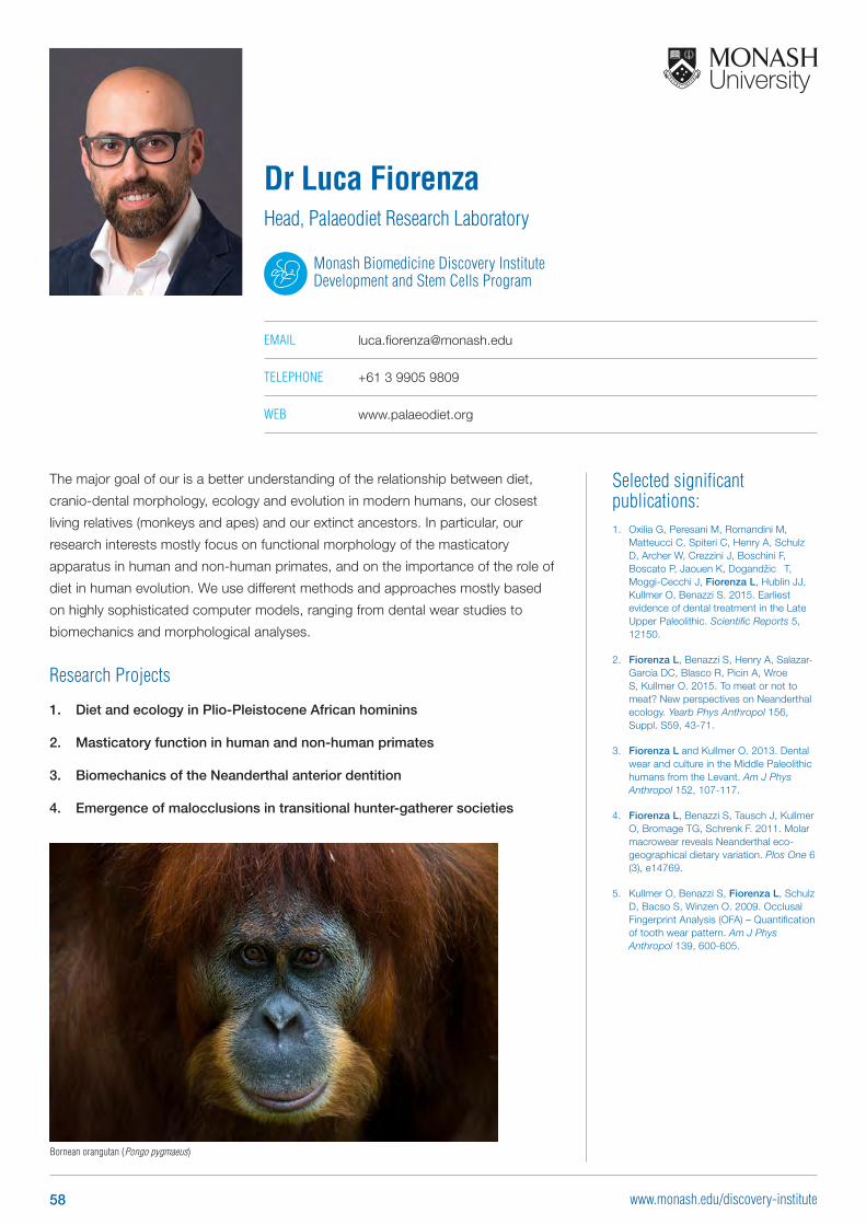
The major goal of our is a better understanding of the relationship between diet,
cranio-dental morphology, ecology and evolution in modern humans, our closest
living relatives (monkeys and apes) and our extinct ancestors. In particular, our
research interests mostly focus on functional morphology of the masticatory
apparatus in human and non-human primates, and on the importance of the role of
diet in human evolution. We use different methods and approaches mostly based
on highly sophisticated computer models, ranging from dental wear studies to
biomechanics and morphological analyses.
Research Projects
1. Diet and ecology in Plio-Pleistocene African hominins
2. Masticatory function in human and non-human primates
3. Biomechanics of the Neanderthal anterior dentition
4. Emergence of malocclusions in transitional hunter-gatherer societies
Selected significant publications: 1. Oxilia G, Peresani M, Romandini M,
Matteucci C, Spiteri C, Henry A, Schulz D, Archer W, Crezzini J, Boschini F, Boscato P, Jaouen K, Dogandžic‐ T, Moggi-Cecchi J, Fiorenza L, Hublin JJ, Kullmer O, Benazzi S. 2015. Earliest evidence of dental treatment in the Late Upper Paleolithic. Scientific Reports 5, 12150.
2. Fiorenza L, Benazzi S, Henry A, Salazar-García DC, Blasco R, Picin A, Wroe S, Kullmer O. 2015. To meat or not to meat? New perspectives on Neanderthal ecology. Yearb Phys Anthropol 156, Suppl. S59, 43-71.
3. Fiorenza L and Kullmer O. 2013. Dental wear and culture in the Middle Paleolithic humans from the Levant. Am J Phys Anthropol 152, 107-117.
4. Fiorenza L, Benazzi S, Tausch J, Kullmer O, Bromage TG, Schrenk F. 2011. Molar macrowear reveals Neanderthal eco-geographical dietary variation. Plos One 6 (3), e14769.
5. Kullmer O, Benazzi S, Fiorenza L, Schulz D, Bacso S, Winzen O. 2009. Occlusal Fingerprint Analysis (OFA) – Quantification of tooth wear pattern. Am J Phys Anthropol 139, 600-605.
Dr Luca FiorenzaHead, Palaeodiet Research Laboratory
Monash Biomedicine Discovery Institute Development and Stem Cells Program
EMAIL [email protected]
TELEPHONE +61 3 9905 9809
WEB www.palaeodiet.org
Bornean orangutan (Pongo pygmaeus)
58 www.monash.edu/discovery-institute
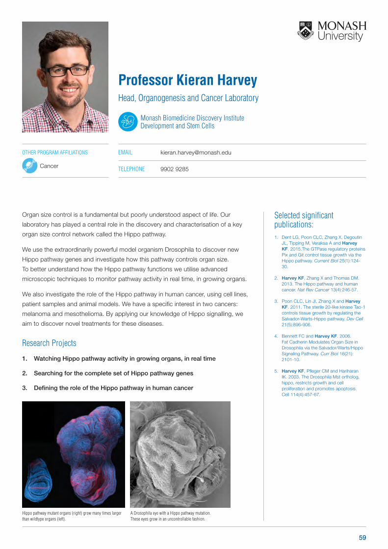
Selected significant publications: 1. Dent LG, Poon CLC, Zhang X, Degoutin
JL, Tipping M, Veraksa A and Harvey KF. 2015.The GTPase regulatory proteins Pix and Git control tissue growth via the Hippo pathway. Current Biol 25(1):124-30.
2. Harvey KF, Zhang X and Thomas DM. 2013. The Hippo pathway and human cancer. Nat Rev Cancer 13(4):246-57.
3. Poon CLC, Lin JI, Zhang X and Harvey KF. 2011. The sterile 20-like kinase Tao-1 controls tissue growth by regulating the Salvador-Warts-Hippo pathway. Dev Cell 21(5):896-906.
4. Bennett FC and Harvey KF. 2006. Fat Cadherin Modulates Organ Size in Drosophila via the Salvador/Warts/Hippo Signaling Pathway. Curr Biol 16(21): 2101-10.
5. Harvey KF, Pfleger CM and Hariharan IK. 2003. The Drosophila Mst ortholog, hippo, restricts growth and cell proliferation and promotes apoptosis. Cell 114(4):457-67.
Organ size control is a fundamental but poorly understood aspect of life. Our
laboratory has played a central role in the discovery and characterisation of a key
organ size control network called the Hippo pathway.
We use the extraordinarily powerful model organism Drosophila to discover new
Hippo pathway genes and investigate how this pathway controls organ size.
To better understand how the Hippo pathway functions we utilise advanced
microscopic techniques to monitor pathway activity in real time, in growing organs.
We also investigate the role of the Hippo pathway in human cancer, using cell lines,
patient samples and animal models. We have a specific interest in two cancers:
melanoma and mesothelioma. By applying our knowledge of Hippo signalling, we
aim to discover novel treatments for these diseases.
Research Projects
1. Watching Hippo pathway activity in growing organs, in real time
2. Searching for the complete set of Hippo pathway genes
3. Defining the role of the Hippo pathway in human cancer
Professor Kieran HarveyHead, Organogenesis and Cancer Laboratory
Monash Biomedicine Discovery Institute Development and Stem Cells
EMAIL [email protected]
TELEPHONE 9902 9285
OTHER PROGRAM AFFILIATIONS
Cancer
A Drosophila eye with a Hippo pathway mutation. These eyes grow in an uncontrollable fashion.
Hippo pathway mutant organs (right) grow many times larger than wildtype organs (left).
59
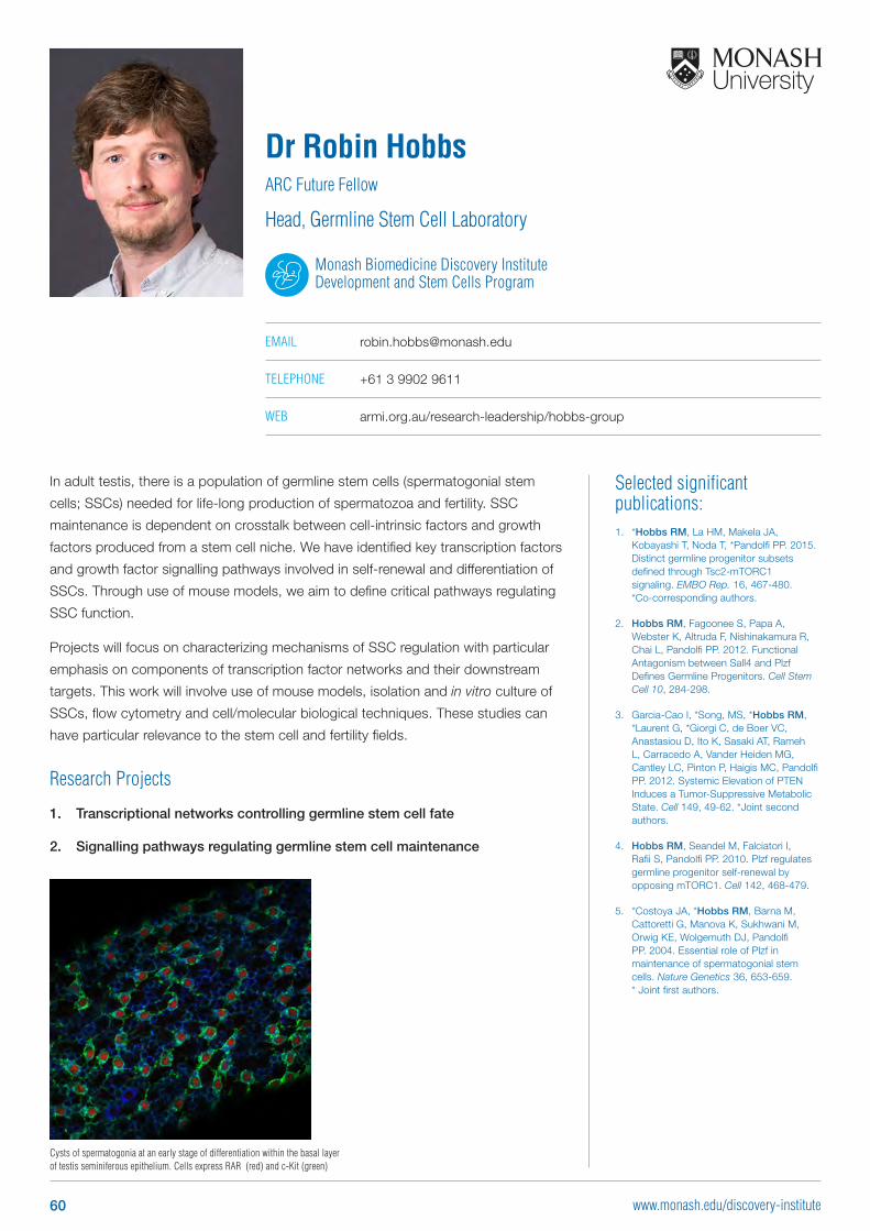
Selected significant publications: 1. *Hobbs RM, La HM, Makela JA,
Kobayashi T, Noda T, *Pandolfi PP. 2015. Distinct germline progenitor subsets defined through Tsc2-mTORC1 signaling. EMBO Rep. 16, 467-480. *Co-corresponding authors.
2. Hobbs RM, Fagoonee S, Papa A, Webster K, Altruda F, Nishinakamura R, Chai L, Pandolfi PP. 2012. Functional Antagonism between Sall4 and Plzf Defines Germline Progenitors. Cell Stem Cell 10, 284-298.
3. Garcia-Cao I, *Song, MS, *Hobbs RM, *Laurent G, *Giorgi C, de Boer VC, Anastasiou D, Ito K, Sasaki AT, Rameh L, Carracedo A, Vander Heiden MG, Cantley LC, Pinton P, Haigis MC, Pandolfi PP. 2012. Systemic Elevation of PTEN Induces a Tumor-Suppressive Metabolic State. Cell 149, 49-62. *Joint second authors.
4. Hobbs RM, Seandel M, Falciatori I, Rafii S, Pandolfi PP. 2010. Plzf regulates germline progenitor self-renewal by opposing mTORC1. Cell 142, 468-479.
5. *Costoya JA, *Hobbs RM, Barna M, Cattoretti G, Manova K, Sukhwani M, Orwig KE, Wolgemuth DJ, Pandolfi PP. 2004. Essential role of Plzf in maintenance of spermatogonial stem cells. Nature Genetics 36, 653-659. * Joint first authors.
In adult testis, there is a population of germline stem cells (spermatogonial stem
cells; SSCs) needed for life-long production of spermatozoa and fertility. SSC
maintenance is dependent on crosstalk between cell-intrinsic factors and growth
factors produced from a stem cell niche. We have identified key transcription factors
and growth factor signalling pathways involved in self-renewal and differentiation of
SSCs. Through use of mouse models, we aim to define critical pathways regulating
SSC function.
Projects will focus on characterizing mechanisms of SSC regulation with particular
emphasis on components of transcription factor networks and their downstream
targets. This work will involve use of mouse models, isolation and in vitro culture of
SSCs, flow cytometry and cell/molecular biological techniques. These studies can
have particular relevance to the stem cell and fertility fields.
Research Projects
1. Transcriptional networks controlling germline stem cell fate
2. Signalling pathways regulating germline stem cell maintenance
Dr Robin HobbsARC Future Fellow
Head, Germline Stem Cell Laboratory
Monash Biomedicine Discovery Institute Development and Stem Cells Program
EMAIL [email protected]
TELEPHONE +61 3 9902 9611
WEB armi.org.au/research-leadership/hobbs-group
Cysts of spermatogonia at an early stage of differentiation within the basal layer of testis seminiferous epithelium. Cells express RAR (red) and c-Kit (green)
60 www.monash.edu/discovery-institute
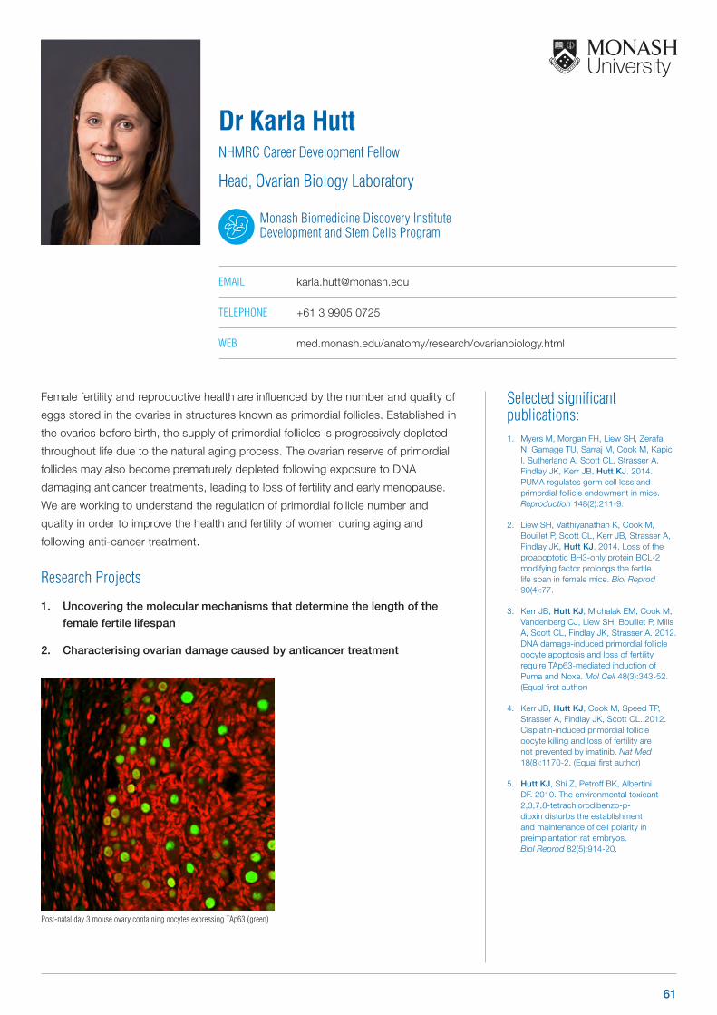
Selected significant publications: 1. Myers M, Morgan FH, Liew SH, Zerafa
N, Gamage TU, Sarraj M, Cook M, Kapic I, Sutherland A, Scott CL, Strasser A, Findlay JK, Kerr JB, Hutt KJ. 2014. PUMA regulates germ cell loss and primordial follicle endowment in mice. Reproduction 148(2):211-9.
2. Liew SH, Vaithiyanathan K, Cook M, Bouillet P, Scott CL, Kerr JB, Strasser A, Findlay JK, Hutt KJ. 2014. Loss of the proapoptotic BH3-only protein BCL-2 modifying factor prolongs the fertile life span in female mice. Biol Reprod 90(4):77.
3. Kerr JB, Hutt KJ, Michalak EM, Cook M, Vandenberg CJ, Liew SH, Bouillet P, Mills A, Scott CL, Findlay JK, Strasser A. 2012. DNA damage-induced primordial follicle oocyte apoptosis and loss of fertility require TAp63-mediated induction of Puma and Noxa. Mol Cell 48(3):343-52. (Equal first author)
4. Kerr JB, Hutt KJ, Cook M, Speed TP, Strasser A, Findlay JK, Scott CL. 2012. Cisplatin-induced primordial follicle oocyte killing and loss of fertility are not prevented by imatinib. Nat Med 18(8):1170-2. (Equal first author)
5. Hutt KJ, Shi Z, Petroff BK, Albertini DF. 2010. The environmental toxicant 2,3,7,8-tetrachlorodibenzo-p-dioxin disturbs the establishment and maintenance of cell polarity in preimplantation rat embryos. Biol Reprod 82(5):914-20.
Female fertility and reproductive health are influenced by the number and quality of
eggs stored in the ovaries in structures known as primordial follicles. Established in
the ovaries before birth, the supply of primordial follicles is progressively depleted
throughout life due to the natural aging process. The ovarian reserve of primordial
follicles may also become prematurely depleted following exposure to DNA
damaging anticancer treatments, leading to loss of fertility and early menopause.
We are working to understand the regulation of primordial follicle number and
quality in order to improve the health and fertility of women during aging and
following anti-cancer treatment.
Research Projects
1. Uncovering the molecular mechanisms that determine the length of the
female fertile lifespan
2. Characterising ovarian damage caused by anticancer treatment
Dr Karla HuttNHMRC Career Development Fellow
Head, Ovarian Biology Laboratory
Monash Biomedicine Discovery Institute Development and Stem Cells Program
EMAIL [email protected]
TELEPHONE +61 3 9905 0725
WEB med.monash.edu/anatomy/research/ovarianbiology.html
Post-natal day 3 mouse ovary containing oocytes expressing TAp63 (green)
61
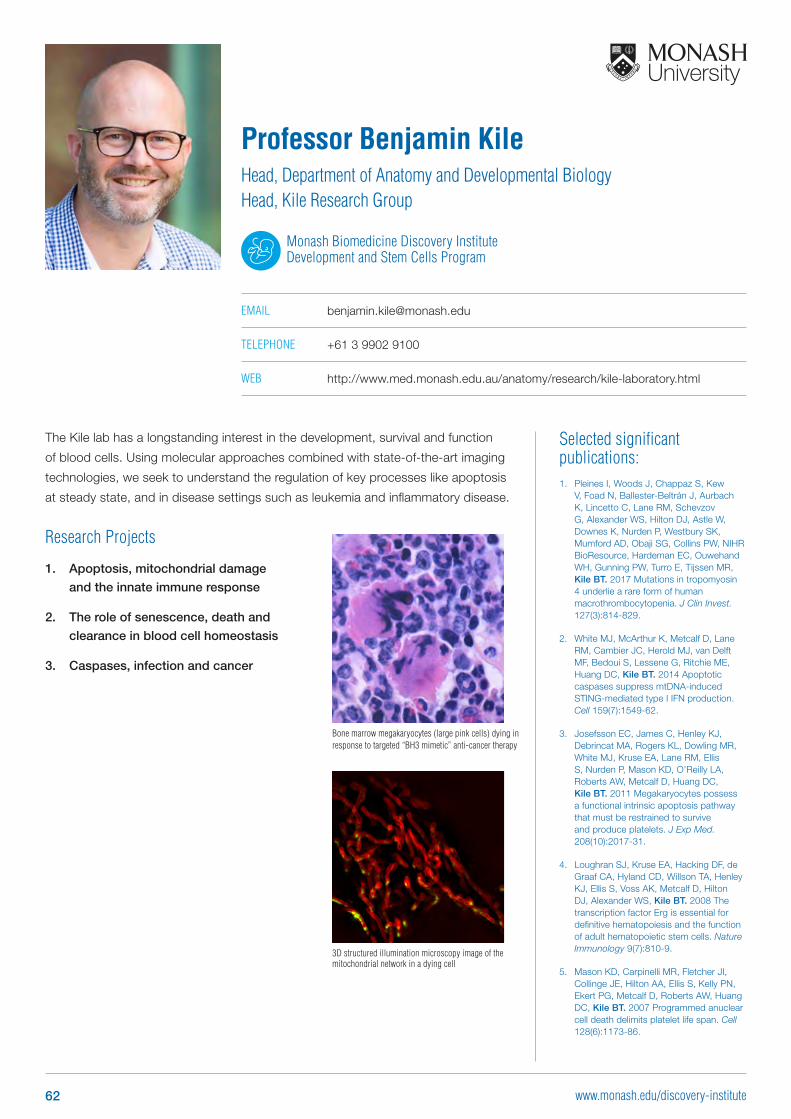
Selected significant publications: 1. Pleines I, Woods J, Chappaz S, Kew
V, Foad N, Ballester-Beltrán J, Aurbach K, Lincetto C, Lane RM, Schevzov G, Alexander WS, Hilton DJ, Astle W, Downes K, Nurden P, Westbury SK, Mumford AD, Obaji SG, Collins PW, NIHR BioResource, Hardeman EC, Ouwehand WH, Gunning PW, Turro E, Tijssen MR, Kile BT. 2017 Mutations in tropomyosin 4 underlie a rare form of human macrothrombocytopenia. J Clin Invest. 127(3):814-829.
2. White MJ, McArthur K, Metcalf D, Lane RM, Cambier JC, Herold MJ, van Delft MF, Bedoui S, Lessene G, Ritchie ME, Huang DC, Kile BT. 2014 Apoptotic caspases suppress mtDNA-induced STING-mediated type I IFN production. Cell 159(7):1549-62.
3. Josefsson EC, James C, Henley KJ, Debrincat MA, Rogers KL, Dowling MR, White MJ, Kruse EA, Lane RM, Ellis S, Nurden P, Mason KD, O’Reilly LA, Roberts AW, Metcalf D, Huang DC, Kile BT. 2011 Megakaryocytes possess a functional intrinsic apoptosis pathway that must be restrained to survive and produce platelets. J Exp Med. 208(10):2017-31.
4. Loughran SJ, Kruse EA, Hacking DF, de Graaf CA, Hyland CD, Willson TA, Henley KJ, Ellis S, Voss AK, Metcalf D, Hilton DJ, Alexander WS, Kile BT. 2008 The transcription factor Erg is essential for definitive hematopoiesis and the function of adult hematopoietic stem cells. Nature Immunology 9(7):810-9.
5. Mason KD, Carpinelli MR, Fletcher JI, Collinge JE, Hilton AA, Ellis S, Kelly PN, Ekert PG, Metcalf D, Roberts AW, Huang DC, Kile BT. 2007 Programmed anuclear cell death delimits platelet life span. Cell 128(6):1173-86.
The Kile lab has a longstanding interest in the development, survival and function
of blood cells. Using molecular approaches combined with state-of-the-art imaging
technologies, we seek to understand the regulation of key processes like apoptosis
at steady state, and in disease settings such as leukemia and inflammatory disease.
Research Projects
1. Apoptosis, mitochondrial damage
and the innate immune response
2. The role of senescence, death and
clearance in blood cell homeostasis
3. Caspases, infection and cancer
Professor Benjamin KileHead, Department of Anatomy and Developmental BiologyHead, Kile Research Group
Monash Biomedicine Discovery Institute Development and Stem Cells Program
EMAIL [email protected]
TELEPHONE +61 3 9902 9100
WEB http://www.med.monash.edu.au/anatomy/research/kile-laboratory.html
3D structured illumination microscopy image of the mitochondrial network in a dying cell
Bone marrow megakaryocytes (large pink cells) dying in response to targeted “BH3 mimetic” anti-cancer therapy
62 www.monash.edu/discovery-institute
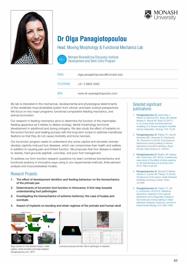
Steps involved in Finite Element Analysis: model creation, model simulation and model validation (Panagiotopoulou et al., 2017)
Locomotor pressures and their links to pathologies in elephants.
Selected significant publications: 1. Panagiotopoulou O, Iriarte-Diaz J,
Wilshin S, Dechow PC, Taylor AB, Mehari Abraha H, Aljunid SF, Ross CF (2017). In vivo bone strain and finite element modeling of a rhesus macaque mandible during mastication. Zoology 124: 13-29
2. Panagiotopoulou O, Pataky TC, Day M, Hensman MC, Hensman S, Hutchinson JR, Clemente CJ (2016). Foot pressure distributions during walking in African elephants (Loxodonta africana). Royal Society Open Science 3: 160203
3. Panagiotopoulou O, Rankin JW, Gatesy SM, Hutchinson JR^ (2016). A preliminary case study of the effect of shoe-wearing on the biomechanics of a horse’s foot. 2016. PeerJ 4:e2164
4. Panagiotopoulou O, Spyridis P, Mehari Abraha H, Carrier DR, Pataky TC (2016). Architecture of the sperm whale forehead facilitates ramming combat. PeerJ 4:e1895
5. Panagiotopoulou O, Pataky TC, Hill Z, Hutchinson JR (2012). Statistical parametric mapping of the regional distribution and ontogenetic scaling of foot pressures during walking in Asian elephants (Elephas maximus). Journal of Experimental Biology 215:1584-1593
My lab is interested in the mechanical, developmental and physiological determinants of the vertebrate musculoskeletal system from clinical, and basic science perspectives. We focus on two major programs: functional comparative feeding mechanics, and animal locomotion.
Our research in feeding mechanics aims to determine the function of the mammalian feeding apparatus as it relates to dietary ecology, dental morphology and bone development in adulthood and during ontogeny. We also study the effect of implants on the bone’s function and healing process with the long-term scope to optimise mandibular fixations so that they do not cause morbidity and bone necrosis.
Our locomotor program seeks to understand why some captive and domestic animals develop captivity-induced foot diseases, which can compromise their health and welfare, in addition to causing pain and limited function. We proposes that foot disease is related to obesity, hard grounds (asphalt, concrete), and poor foot management.
To address our form-function research questions my team combines biomechanics and functional anatomy in innovative ways using in vivo experimental methods, finite element analysis and musculoskeletal models.
Research Projects1. The effect of development dentition and feeding behaviour on the biomechanics
of the primate jaw
2. Determinants of locomotor foot function in rhinoceros: A first step towards understanding foot pathologies
3. Investigating the biomechanics of extreme herbivory: the case of koalas and wombats
4. Impact of implants on bending and strain regimes of the primate and human skull
Dr Olga PanagiotopoulouHead, Moving Morphology & Functional Mechanics Lab
Monash Biomedicine Discovery Institute Development and Stem Cells Program
EMAIL [email protected]
TELEPHONE +61 3 9905 0262
WEB www.dr-opanagiotopoulou.com
63
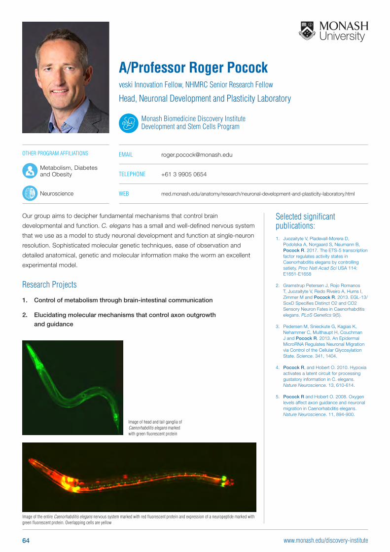
Selected significant publications: 1. Juozaityte V, Pladevall-Morera D,
Podolska A, Norgaard S, Neumann B, Pocock R. 2017. The ETS-5 transcription factor regulates activity states in Caenorhabditis elegans by controlling satiety. Proc Natl Acad Sci USA 114: E1651-E1658
2. Gramstrup Petersen J, Rojo Romanos T, Juozaityte V, Redo Riveiro A, Hums I, Zimmer M and Pocock R. 2013. EGL-13/SoxD Specifies Distinct O2 and CO2 Sensory Neuron Fates in Caenorhabditis elegans. PLoS Genetics 9(5).
3. Pedersen M, Snieckute G, Kagias K, Nehammer C, Multhaupt H, Couchman J and Pocock R. 2013. An Epidermal MicroRNA Regulates Neuronal Migration via Control of the Cellular Glycosylation State. Science. 341, 1404.
4. Pocock R, and Hobert O. 2010. Hypoxia activates a latent circuit for processing gustatory information in C. elegans. Nature Neuroscience. 13, 610-614.
5. Pocock R and Hobert O. 2008. Oxygen levels affect axon guidance and neuronal migration in Caenorhabditis elegans. Nature Neuroscience. 11, 894-900.
Our group aims to decipher fundamental mechanisms that control brain
developmental and function. C. elegans has a small and well-defined nervous system
that we use as a model to study neuronal development and function at single-neuron
resolution. Sophisticated molecular genetic techniques, ease of observation and
detailed anatomical, genetic and molecular information make the worm an excellent
experimental model.
Research Projects
1. Control of metabolism through brain-intestinal communication
2. Elucidating molecular mechanisms that control axon outgrowth
and guidance
A/Professor Roger Pocockveski Innovation Fellow, NHMRC Senior Research Fellow
Head, Neuronal Development and Plasticity Laboratory
Monash Biomedicine Discovery Institute Development and Stem Cells Program
EMAIL [email protected]
TELEPHONE +61 3 9905 0654
WEB med.monash.edu/anatomy/research/neuronal-development-and-plasticity-laboratory.html
Image of head and tail ganglia of Caenorhabditis elegans marked with green fluorescent protein
Image of the entire Caenorhabditis elegans nervous system marked with red fluorescent protein and expression of a neuropeptide marked with green fluorescent protein. Overlapping cells are yellow
OTHER PROGRAM AFFILIATIONS
Metabolism, Diabetes and Obesity
Neuroscience
64 www.monash.edu/discovery-institute
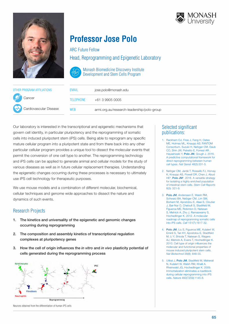
Neurons obtained from the differentiation of human iPS cells
Our laboratory is interested in the transcriptional and epigenetic mechanisms that
govern cell identity, in particular pluripotency and the reprogramming of somatic
cells into induced pluripotent stem (iPS) cells. Being able to reprogram any specific
mature cellular program into a pluripotent state and from there back into any other
particular cellular program provides a unique tool to dissect the molecular events that
permit the conversion of one cell type to another. The reprogramming technology
and iPS cells can be applied to generate animal and cellular models for the study of
various diseases as well as in future cellular replacement therapies. Understanding
the epigenetic changes occurring during these processes is necessary to ultimately
use iPS cell technology for therapeutic purposes.
We use mouse models and a combination of different molecular, biochemical,
cellular techniques and genome wide approaches to dissect the nature and
dynamics of such events.
Research Projects
1. The kinetics and universality of the epigenetic and genomic changes
occurring during reprogramming
2. The composition and assembly kinetics of transcriptional regulation
complexes at pluripotency genes
3. How the cell of origin influences the in vitro and in vivo plasticity potential of
cells generated during the reprogramming process
Professor Jose PoloARC Future Fellow
Head, Reprogramming and Epigenetic Laboratory
Monash Biomedicine Discovery Institute Development and Stem Cells Program
EMAIL [email protected]
TELEPHONE +61 3 9905 0005
WEB armi.org.au/research-leadership/polo-group
Selected significant publications: 1. Rackham OJ, Firas J, Fang H, Oates
ME, Holmes ML, Knaupp AS; FANTOM Consortium, Suzuki H, Nefzger CM, Daub CO, Shin JW, Petretto E, Forrest AR , Hayashizaki Y, Polo JM, Gough J. 2016. A predictive computational framework for direct reprogramming between human cell types. Nat Genet 48(3):331-5.
2. Nefzger CM, Jardé T, Rossello FJ, Horvay K, Knaupp AS, Powell DR, Chen J, Abud HE#, Polo JM#. 2016. A versatile strategy for isolating a highly enriched population of intestinal stem cells. Stem Cell Reports 6(3): 321-9.
3. Polo JM, Anderssen E, Walsh RM, Schwarz BA, Nefzger CM, Lim SM, Borkent M, Apostolou E, Alaei S, Cloutier J, Bar-Nur O, Cheloufi S, Stadtfeld M, Figueroa ME, Robinton D, Natesan S, Melnick A, Zhu J, Ramaswamy S, Hochedlinger K. 2012. A molecular roadmap of reprogramming somatic cells into iPS cells. Cell 151(7):1617-32.
4. Polo JM, Liu S, Figueroa ME, Kulalert W, Eminli S, Tan KY, Apostolou E, Stadtfeld M, Li Y, Shioda T, Natesan S, Wagers AJ, Melnick A, Evans T, Hochedlinger K. 2010. Cell type of origin influences the molecular and functional properties of mouse induced pluripotent stem cells. Nat Biotechnol 28(8): 848-55.
5. Utikal J, Polo JM, Stadtfeld M, Maherali N, Kulalert W, Walsh RM, Khalil A, Rheinwald JG, Hochedlinger K. 2009. Immortalization eliminates a roadblock during cellular reprogramming into iPS cells. Nature 460(7259):1145-8.
OTHER PROGRAM AFFILIATIONS
Cancer
Cardiovascular Disease
65
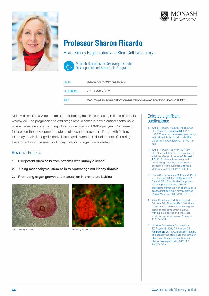
Selected significant publications: 1. Wang B, Yao K, Wise AF, Lau R, Shen
HH, Tesch GH, Ricardo SD. 2017. miR-378 reduces mesangial hypertrophy and kidney tubular fibrosis via MAPK signalling. Clinical Science. 131(5):411-423.
2. Wang B, Yao K, Huuskes BM, Shen HH, Zhuang J, Godson C, Brennan EP, Wilkinson-Berka JL, Wise AF, Ricardo SD. 2016. Mesenchymal stem cells deliver exogenous Microrna-let7c via exosomes to attenuate renal fibrosis. Molecular Therapy 24(7):1290-301.
3. Royce SG, Tominaga AM, Shen M, Patel KP, Huuskes BM, Lim R, Ricardo SD, Samuel CS. 2016. Serelaxin improves the therapeutic efficacy of RXFP1-expressing human amnion epithelial cells in experimental allergic airway disease. Clinical Science 130(23):2151-2165.
4. Wise AF, Williams TM, Rudd S, Wells CA, Kerr PG, Ricardo SD. 2016. Human mesenchymal stem cells alter the gene profile of monocytes from patients with Type 2 diabetes and end-stage renal disease. Regenerative Medicine 11(2):145-58.
5. Huuskes BM, Wise AF, Cox AJ, Lim EX, Payne NL, Kelly DJ, Samuel CS, Ricardo SD. 2015. Combination therapy of mesenchymal stem cells and serelaxin effectively attenuates renal fibrosis in obstructive nephropathy. FASEB J 29(2):540-53.
Kidney disease is a widespread and debilitating health issue facing millions of people
worldwide. The progression to end-stage renal disease is now a critical health issue
where the incidence is rising rapidly at a rate of around 6-8% per year. Our research
focuses on the development of stem cell-based therapies and/or growth factors
that may repair damaged kidney tissues and reverse the development of scarring,
thereby reducing the need for kidney dialysis or organ transplantation.
Research Projects
1. Pluripotent stem cells from patients with kidney disease
2. Using mesenchymal stem cells to protect against kidney fibrosis
3. Promoting organ growth and maturation in premature babies
Professor Sharon RicardoHead, Kidney Regeneration and Stem Cell Laboratory
Monash Biomedicine Discovery Institute Development and Stem Cells Program
EMAIL [email protected]
TELEPHONE +61 3 9905 0671
WEB med.monash.edu/anatomy/research/kidney-regeneration-stem-cell.html
iPS cell colony in culture Mesenchymal stem cells
66 www.monash.edu/discovery-institute
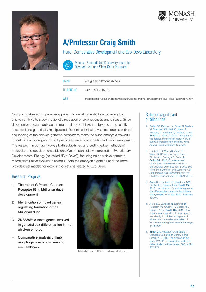
Selected significant publications: 1. Farlie, PG, Davidon, N, Baker, N, Raabus,
M, Roeszler, KN, Hirst, C, Major, A, Mariette, M, Lambert D, Oshlack, A and Smith CA. 2017. A novel 1 co-option of the cardiac transcription factor Nkx2.5 during development of the emu wing. Nature Communications (in press)
2. Lambeth LS, Morris K, Ayers KL, Wise TG, O’Neil T, Wilson S, Cao Y, Sinclair AH, Cutting AD, Doran TJ, Smith CA. 2016. Overexpression of Anti-Müllerian Hormone Disrupts Gonadal Sex Differentiation, Blocks Sex Hormone Synthesis, and Supports Cell Autonomous Sex Development in the Chicken. Endocrinology 157(3):1258-75.
3. Ayers KL, Lambeth LS, Davidson, NM, Sinclair AH, Oshlack A and Smith CA. 2015. Identification of candidate gonadal sex differentiation genes in the chicken embryo using RNA-seq. BMC Genomics 16:704.
4. Ayers KL, Davidson N, Demiyah D, Roeszler KN, Grützner F, Sinclair AH, Oshlack A and Smith CA. 2013. RNA sequencing supports cell autonomous sex identity in chicken embryos and allows comprehensive annotation of W-chromosome genes. Genome Biology 14 (3):R26.
5. Smith CA, Roeszler K, Onhesorg T , Cummins, D, Farlie, P, Doran, T and Sinclair AH. 2009. The avian Z-linked gene, DMRT1, is required for male sex determination in the chicken. Nature 461, 267- 271.
Our group takes a comparative approach to developmental biology, using the
chicken embryo to study the genetic regulation of organogenesis and disease. Since
development occurs outside the maternal body, chicken embryos can be readily
accessed and genetically manipulated. Recent technical advances coupled with the
sequencing of the chicken genome combine to make the avian embryo a powerful
model for functional genomics. Specifically, we study gonadal and limb development.
The research in our lab involves both established and cutting edge methods of
molecular and developmental biology. We are particularly interested in Evolutionary
Developmental Biology (so-called “Evo-Devo”), focusing on how developmental
mechanisms have evolved in animals. Both the embryonic gonads and the limbs
provide ideal models for exploring questions related to Evo-Devo.
Research Projects
1. The role of G Protein Coupled
Receptor 56 in Müllerian duct
development
2. Identification of novel genes
regulating formation of the
Müllerian duct
3. ZNF385B: A novel genes involved
in gonadal sex differentiation in the
chicken embryo
4. Comparative analysis of limb
morphogenesis in chicken and
emu embryos
A/Professor Craig Smith Head, Comparative Development and Evo-Devo Laboratory
Monash Biomedicine Discovery Institute Development and Stem Cells Program
EMAIL [email protected]
TELEPHONE +61 3 9905 0203
WEB med.monash.edu/anatomy/research/comparative-development-evo-devo-laboratory.html
Unilateral delivery of GFP into an embryonic chicken gonad.
67
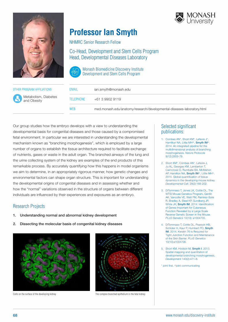
Selected significant publications: 1. Combes AN*, Short KM*, Lefevre J*,
Hamilton NA, Little MH^, Smyth IM^. 2014. An integrated pipeline for the multidimensional analysis of branching morphogenesis. Nature Protocols 9(12):2859-79.
2. Short KM*, Combes AN*, Lefevre J, Ju AL, Georgas KM, Lamberton T, Cairncross O, Rumballe BA, McMahon AP, Hamilton NA, Smyth IM^, Little MH^. 2014. Global quantification of tissue dynamics in the developing mouse kidney. Developmental Cell, 29(2):188-202.
3. DiTommaso T, Jones LK, Cottle DL, The WTSI Mouse Genetics Program, Gerdin AK, Vancollie VE, Watt FM, Ramirez-Solis R, Bradley A, Steel KP, Sundberg JP, White JK, Smyth IM. 2014. Identification of Genes Important for Cutaneous Function Revealed by a Large Scale Reverse Genetic Screen in the Mouse. PLoS Genetics 10(10): e1004705.
4. DiTommaso T, Cottle DL, Pearson HB, Schlüter H, Kaur P, Humbert PO, Smyth IM. 2014. Keratin 76 Is Required for Tight Junction Function and Maintenance of the Skin Barrier. PLoS Genetics 10(10):e1004706.
5. Short KM, Hodson M, Smyth I. 2013. Spatial mapping and quantitation of developmental branching morphogenesis. Development 140(2):471-8.
* joint first, ^joint communicating
Our group studies how the embryo develops with a view to understanding the
developmental basis for congenital diseases and those caused by a compromised
fetal environment. In particular we are interested in understanding the developmental
mechanism known as “branching morphogenesis”, which is employed by a large
number of organs to establish the tissue architecture required to facilitate exchange
of nutrients, gases or waste in the adult organ. The branched airways of the lung and
the urine collecting system of the kidney are examples of the end products of this
remarkable process. By accurately quantifying how this happens in model organisms
we aim to determine, in an appropriately rigorous manner, how genetic changes and
environmental factors can shape organ structure. This is important for understanding
the developmental origins of congenital diseases and in assessing whether and
how the “normal” variations observed in the structure of organs between different
individuals are influenced by their experiences and exposures as an embryo.
Research Projects
1. Understanding normal and abnormal kidney development
2. Dissecting the molecular basis of congenital kidney diseases
Professor Ian SmythNHMRC Senior Research Fellow
Co-Head, Development and Stem Cells ProgramHead, Developmental Diseases Laboratory
Monash Biomedicine Discovery Institute Development and Stem Cells Program
EMAIL [email protected]
TELEPHONE +61 3 9902 9119
WEB med.monash.edu/anatomy/research/developmental-diseases-laboratory.html
Cells on the surface of the developing kidney The complex branched epithelium in the fetal kidney
OTHER PROGRAM AFFILIATIONS
Metabolism, Diabetes and Obesity
68 www.monash.edu/discovery-institute
Infection and Immunity Program Group Leaders
69www.monash.edu/discovery-institute
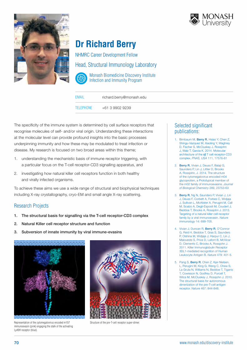
Selected significant publications: 1. Birnbaum M, Berry R, Hsiao Y, Chen Z,
Shingu-Vazquez M, Xiaoling Y, Waghrey D, Fischer S, McCluskey J, Rossjohn J, Walz T, Garcia K. 2014. Molecular architecture of the αβ T cell receptor-CD3 complex. PNAS, USA 111, 17576-81
2. Berry R, Vivian J, Deuss F, Balaji G, Saunders P, Lin J, Littler D, Brooks A, Rossjohn, J. 2014. The structure of the cytomegalovirus encoded m04 glycoprotien, a Prototypical member of the m02 family of immunoevasins. Journal of Biological Chemistry 289, 23753-63
3. Berry R, Ng N, Saunders P, Vivian J, Lin J, Deuss F, Corbett A, Forbes C, Widjaja J, Sullivan L, McAlister A, Perugini M, Call M, Scalzo A, Degli-Esposti M, Coudert J, Beddoe T, Brooks A, Rossjohn J. 2013. Targeting of a natural killer cell receptor family by a viral immunoevasin. Nature Immunology 14: 699-705.
4. Vivian J, Duncan R, Berry R, O’Connor G, Reid H, Beddoe T, Gras S, Saunders P, Olshina M, Widjaja J, Harpur C, Lin J, Maloveste S, Price D, Lafont B, McVicar D, Clements C, Brooks A, Rossjohn J. 2011. Killer Immunoglobulin Receptor 3DL1-mediated recognition of Human Leukocyte Antigen B. Nature 479: 401-5.
5. Pang S, Berry R, Chen Z, Kjer-Nielsen, L, Perugini M, King G, Wang C, Chew S, La Gruta N, Williams N, Beddoe T, Tiganis T, Cowieson N, Godfrey D, Purcell T, Wilce M, McCluskey J, Rossjohn J. 2010. The structural basis for autonomous dimerization of the pre-T-cell antigen receptor. Nature 467: 844-848.
The specificity of the immune system is determined by cell surface receptors that
recognise molecules of self- and/or viral origin. Understanding these interactions
at the molecular level can provide profound insights into the basic processes
underpinning immunity and how these may be modulated to treat infection or
disease. My research is focused on two broad areas within this theme;
1. understanding the mechanistic basis of immune receptor triggering, with
a particular focus on the T-cell receptor-CD3 signalling apparatus, and
2. investigating how natural killer cell receptors function in both healthy
and virally infected organisms.
To achieve these aims we use a wide range of structural and biophysical techniques
including X-ray crystallography, cryo-EM and small angle X-ray scattering.
Research Projects
1. The structural basis for signalling via the T-cell receptor-CD3 complex
2. Natural Killer cell receptor structure and function
3. Subversion of innate immunity by viral immune-evasins
Dr Richard BerryNHMRC Career Development Fellow
Head, Structural Immunology Laboratory
Monash Biomedicine Discovery Institute Infection and Immunity Program
EMAIL [email protected]
TELEPHONE +61 3 9902 9239
Representation of the cytomegalovirus encoded m157 immunoevasin (pink) engaging the stalk of the activating Ly49H receptor (blue).
Structure of the pre-T-cell receptor super-dimer.
70 www.monash.edu/discovery-institute
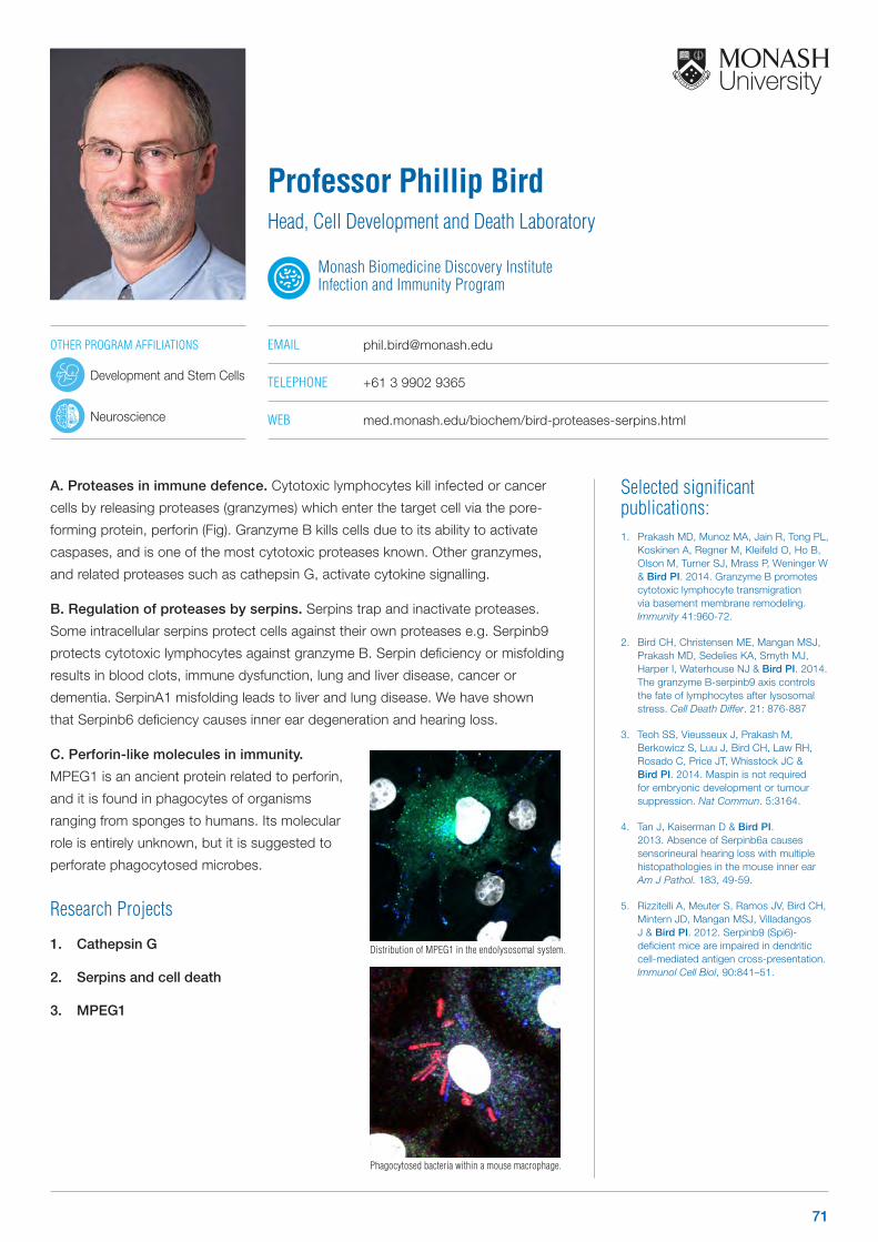
Selected significant publications: 1. Prakash MD, Munoz MA, Jain R, Tong PL,
Koskinen A, Regner M, Kleifeld O, Ho B, Olson M, Turner SJ, Mrass P, Weninger W & Bird PI. 2014. Granzyme B promotes cytotoxic lymphocyte transmigration via basement membrane remodeling. Immunity 41:960-72.
2. Bird CH, Christensen ME, Mangan MSJ, Prakash MD, Sedelies KA, Smyth MJ, Harper I, Waterhouse NJ & Bird PI. 2014. The granzyme B-serpinb9 axis controls the fate of lymphocytes after lysosomal stress. Cell Death Differ. 21: 876-887
3. Teoh SS, Vieusseux J, Prakash M, Berkowicz S, Luu J, Bird CH, Law RH, Rosado C, Price JT, Whisstock JC & Bird PI. 2014. Maspin is not required for embryonic development or tumour suppression. Nat Commun. 5:3164.
4. Tan J, Kaiserman D & Bird PI. 2013. Absence of Serpinb6a causes sensorineural hearing loss with multiple histopathologies in the mouse inner ear Am J Pathol. 183, 49-59.
5. Rizzitelli A, Meuter S, Ramos JV, Bird CH, Mintern JD, Mangan MSJ, Villadangos J & Bird PI. 2012. Serpinb9 (Spi6)-deficient mice are impaired in dendritic cell-mediated antigen cross-presentation. Immunol Cell Biol, 90:841–51.
A. Proteases in immune defence. Cytotoxic lymphocytes kill infected or cancer
cells by releasing proteases (granzymes) which enter the target cell via the pore-
forming protein, perforin (Fig). Granzyme B kills cells due to its ability to activate
caspases, and is one of the most cytotoxic proteases known. Other granzymes,
and related proteases such as cathepsin G, activate cytokine signalling.
B. Regulation of proteases by serpins. Serpins trap and inactivate proteases.
Some intracellular serpins protect cells against their own proteases e.g. Serpinb9
protects cytotoxic lymphocytes against granzyme B. Serpin deficiency or misfolding
results in blood clots, immune dysfunction, lung and liver disease, cancer or
dementia. SerpinA1 misfolding leads to liver and lung disease. We have shown
that Serpinb6 deficiency causes inner ear degeneration and hearing loss.
C. Perforin-like molecules in immunity.
MPEG1 is an ancient protein related to perforin,
and it is found in phagocytes of organisms
ranging from sponges to humans. Its molecular
role is entirely unknown, but it is suggested to
perforate phagocytosed microbes.
Research Projects
1. Cathepsin G
2. Serpins and cell death
3. MPEG1
Professor Phillip BirdHead, Cell Development and Death Laboratory
Monash Biomedicine Discovery Institute Infection and Immunity Program
EMAIL [email protected]
TELEPHONE +61 3 9902 9365
WEB med.monash.edu/biochem/bird-proteases-serpins.html
Phagocytosed bacteria within a mouse macrophage.
Distribution of MPEG1 in the endolysosomal system.
OTHER PROGRAM AFFILIATIONS
Development and Stem Cells
Neuroscience
71
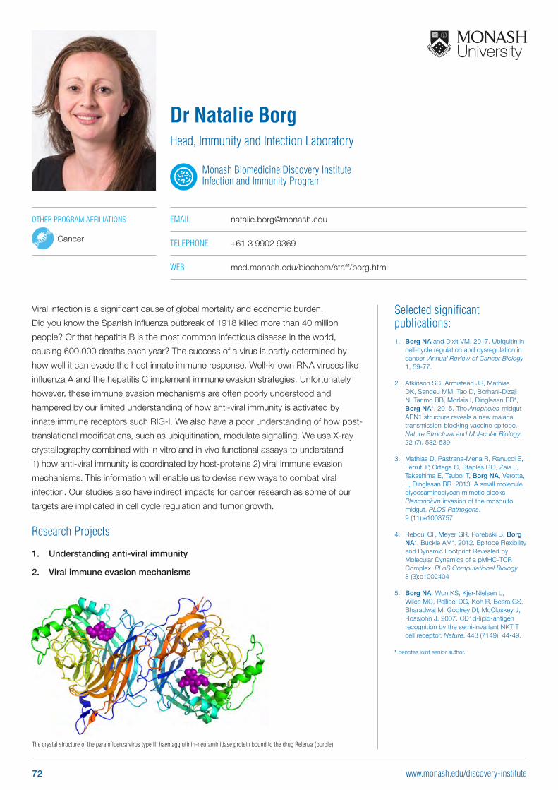
Selected significant publications: 1. Borg NA and Dixit VM. 2017. Ubiquitin in
cell-cycle regulation and dysregulation in cancer. Annual Review of Cancer Biology 1, 59-77.
2. Atkinson SC, Armistead JS, Mathias DK, Sandeu MM, Tao D, Borhani-Dizaji N, Tarimo BB, Morlais I, Dinglasan RR*, Borg NA*. 2015. The Anopheles-midgut APN1 structure reveals a new malaria transmission-blocking vaccine epitope. Nature Structural and Molecular Biology. 22 (7), 532-539.
3. Mathias D, Pastrana-Mena R, Ranucci E, Ferruti P, Ortega C, Staples GO, Zaia J, Takashima E, Tsuboi T, Borg NA, Verotta, L, Dinglasan RR. 2013. A small molecule glycosaminoglycan mimetic blocks Plasmodium invasion of the mosquito midgut. PLOS Pathogens. 9 (11):e1003757
4. Reboul CF, Meyer GR, Porebski B, Borg NA*, Buckle AM*. 2012. Epitope Flexibility and Dynamic Footprint Revealed by Molecular Dynamics of a pMHC-TCR Complex. PLoS Computational Biology. 8 (3):e1002404
5. Borg NA, Wun KS, Kjer-Nielsen L, Wilce MC, Pellicci DG, Koh R, Besra GS, Bharadwaj M, Godfrey DI, McCluskey J, Rossjohn J. 2007. CD1d-lipid-antigen recognition by the semi-invariant NKT T cell receptor. Nature. 448 (7149), 44-49.
* denotes joint senior author.
Dr Natalie BorgHead, Immunity and Infection Laboratory
Monash Biomedicine Discovery Institute Infection and Immunity Program
EMAIL [email protected]
TELEPHONE +61 3 9902 9369
WEB med.monash.edu/biochem/staff/borg.html
The crystal structure of the parainfluenza virus type III haemagglutinin-neuraminidase protein bound to the drug Relenza (purple)
OTHER PROGRAM AFFILIATIONS
Cancer
Viral infection is a significant cause of global mortality and economic burden.
Did you know the Spanish influenza outbreak of 1918 killed more than 40 million
people? Or that hepatitis B is the most common infectious disease in the world,
causing 600,000 deaths each year? The success of a virus is partly determined by
how well it can evade the host innate immune response. Well-known RNA viruses like
influenza A and the hepatitis C implement immune evasion strategies. Unfortunately
however, these immune evasion mechanisms are often poorly understood and
hampered by our limited understanding of how anti-viral immunity is activated by
innate immune receptors such RIG-I. We also have a poor understanding of how post-
translational modifications, such as ubiquitination, modulate signalling. We use X-ray
crystallography combined with in vitro and in vivo functional assays to understand
1) how anti-viral immunity is coordinated by host-proteins 2) viral immune evasion
mechanisms. This information will enable us to devise new ways to combat viral
infection. Our studies also have indirect impacts for cancer research as some of our
targets are implicated in cell cycle regulation and tumor growth.
Research Projects
1. Understanding anti-viral immunity
2. Viral immune evasion mechanisms
72 www.monash.edu/discovery-institute
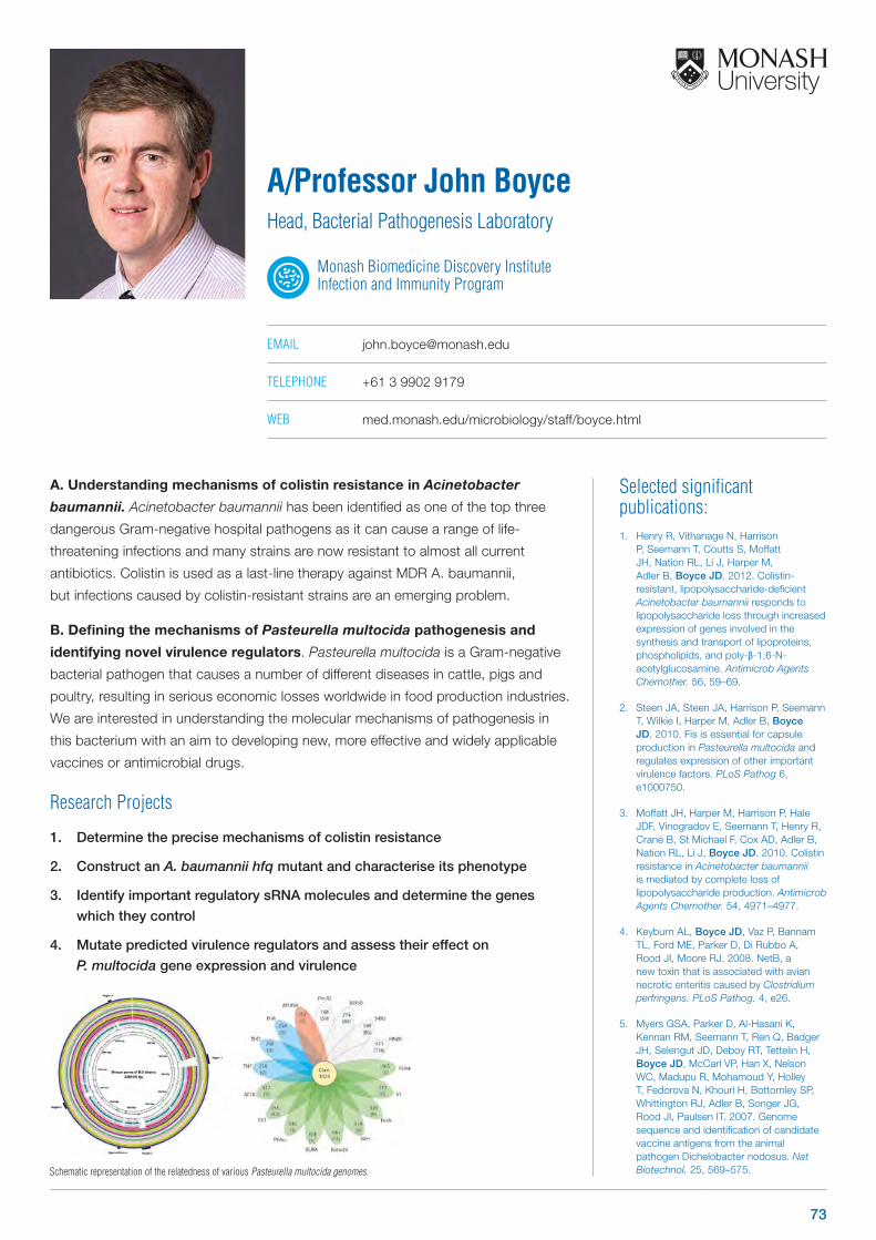
Selected significant publications: 1. Henry R, Vithanage N, Harrison
P, Seemann T, Coutts S, Moffatt JH, Nation RL, Li J, Harper M, Adler B, Boyce JD. 2012. Colistin-resistant, lipopolysaccharide-deficient Acinetobacter baumannii responds to lipopolysaccharide loss through increased expression of genes involved in the synthesis and transport of lipoproteins, phospholipids, and poly-β-1,6-N-acetylglucosamine. Antimicrob Agents Chemother. 56, 59–69.
2. Steen JA, Steen JA, Harrison P, Seemann T, Wilkie I, Harper M, Adler B, Boyce JD. 2010. Fis is essential for capsule production in Pasteurella multocida and regulates expression of other important virulence factors. PLoS Pathog 6, e1000750.
3. Moffatt JH, Harper M, Harrison P, Hale JDF, Vinogradov E, Seemann T, Henry R, Crane B, St Michael F, Cox AD, Adler B, Nation RL, Li J, Boyce JD. 2010. Colistin resistance in Acinetobacter baumannii is mediated by complete loss of lipopolysaccharide production. Antimicrob Agents Chemother. 54, 4971–4977.
4. Keyburn AL, Boyce JD, Vaz P, Bannam TL, Ford ME, Parker D, Di Rubbo A, Rood JI, Moore RJ. 2008. NetB, a new toxin that is associated with avian necrotic enteritis caused by Clostridium perfringens. PLoS Pathog. 4, e26.
5. Myers GSA, Parker D, Al-Hasani K, Kennan RM, Seemann T, Ren Q, Badger JH, Selengut JD, Deboy RT, Tettelin H, Boyce JD, McCarl VP, Han X, Nelson WC, Madupu R, Mohamoud Y, Holley T, Fedorova N, Khouri H, Bottomley SP, Whittington RJ, Adler B, Songer JG, Rood JI, Paulsen IT. 2007. Genome sequence and identification of candidate vaccine antigens from the animal pathogen Dichelobacter nodosus. Nat Biotechnol. 25, 569–575.
A. Understanding mechanisms of colistin resistance in Acinetobacter
baumannii. Acinetobacter baumannii has been identified as one of the top three
dangerous Gram-negative hospital pathogens as it can cause a range of life-
threatening infections and many strains are now resistant to almost all current
antibiotics. Colistin is used as a last-line therapy against MDR A. baumannii,
but infections caused by colistin-resistant strains are an emerging problem.
B. Defining the mechanisms of Pasteurella multocida pathogenesis and
identifying novel virulence regulators. Pasteurella multocida is a Gram-negative
bacterial pathogen that causes a number of different diseases in cattle, pigs and
poultry, resulting in serious economic losses worldwide in food production industries.
We are interested in understanding the molecular mechanisms of pathogenesis in
this bacterium with an aim to developing new, more effective and widely applicable
vaccines or antimicrobial drugs.
Research Projects
1. Determine the precise mechanisms of colistin resistance
2. Construct an A. baumannii hfq mutant and characterise its phenotype
3. Identify important regulatory sRNA molecules and determine the genes
which they control
4. Mutate predicted virulence regulators and assess their effect on
P. multocida gene expression and virulence
A/Professor John BoyceHead, Bacterial Pathogenesis Laboratory
Monash Biomedicine Discovery Institute Infection and Immunity Program
EMAIL [email protected]
TELEPHONE +61 3 9902 9179
WEB med.monash.edu/microbiology/staff/boyce.html
Schematic representation of the relatedness of various Pasteurella multocida genomes.
73
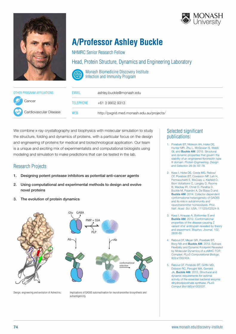
Selected significant publications: 1. Porebski BT, Nickson AA, Hoke DE,
Hunter MR, Zhu L, McGowan S, Webb GI, and Buckle AM. 2015. Structural and dynamic properties that govern the stability of an engineered fibronectin type III domain. Protein Engineering, Design and Selection 28 (3): 67-78.
2. Kass I, Hoke DE, Costa MG, Reboul CF, Porebski BT, Cowieson NP, Leh H, Pennacchietti E, McCoey J, Kleifeld O, Borri Voltattorni C, Langley D, Roome B, Mackay IR, Christ D, Perahia D, Buckle M, Paiardini A, De Biase D and Buckle AM. 2014. Cofactor-dependent conformational heterogeneity of GAD65 and its role in autoimmunity and neurotransmitter homeostasis. Proc. Natl. Acad. Sci. USA, 111(25):E2524-9.
3. Kass I, Knaupp A, Bottomley S and Buckle AM. 2012. Conformational properties of the disease-causing Z variant of α1 antitrypsin revealed by theory and experiment. Biophys. Journal, 102, 2856-65
4. Reboul CF, Meyer GR, Porebski BT, Borg NA and Buckle AM. 2012. Epitope Flexibility and Dynamic Footprint Revealed by Molecular Dynamics of a pMHC-TCR Complex. PLoS Computational Biology, 8(3):e1002404.
5. Reboul CF, Porebski BT, Griffin MD, Dobson RC, Perugini MA, Gerrard JA, Buckle AM. 2012. Structural and dynamic requirements for optimal activity of the essential bacterial enzyme dihydrodipicolinate synthase. PLoS Comput Biol 8(6):e1002537.
We combine x-ray crystallography and biophysics with molecular simulation to study
the structure, folding and dynamics of proteins, with a particular focus on the design
and engineering of proteins for medical and biotechnological application. Our team
is a unique and exciting mix of experimentalists and computational biologists using
modeling and simulation to make predictions that can be tested in the lab.
Research Projects
1. Designing potent protease inhibitors as potential anti-cancer agents
2. Using computational and experimental methods to design and evolve
novel proteins
3. The evolution of protein dynamics
A/Professor Ashley BuckleNHMRC Senior Research Fellow
Head, Protein Structure, Dynamics and Engineering Laboratory
Monash Biomedicine Discovery Institute Infection and Immunity Program
EMAIL [email protected]
TELEPHONE +61 3 9902 9313
WEB http://pxgrid.med.monash.edu.au/projects/
Implications of GAD65 autoinactivation for neurotransmitter biosynthesis and autoantigenicity.
Design, engineering and evolution of Adnectins.
OTHER PROGRAM AFFILIATIONS
Cancer
Cardiovascular Disease
Glu GABA
PMP + SSA
PLP
AbAb
conformationalselection
74 www.monash.edu/discovery-institute
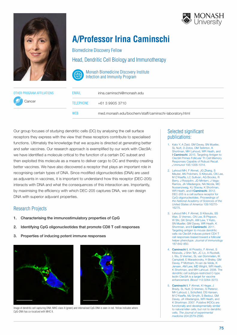
Selected significant publications: 1. Kato Y, A Zaid, GM Davey, SN Mueller,
SL Nutt, D Zotos, DM Tarlinton, K Shortman, MH Lahoud, WR Heath, and I Caminschi. 2015. Targeting Antigen to Clec9A Primes Follicular Th Cell Memory Responses Capable of Robust Recall. J Immunol 195:1006-1014.
2. Lahoud MH, F Ahmet, JG Zhang, S Meuter, AN Policheni, S Kitsoulis, CN Lee, M O’Keeffe, LC Sullivan, AG Brooks, R Berry, J Rossjohn, JD Mintern, J Vega-Ramos, JA Villadangos, NA Nicola, MC Nussenzweig, KJ Stacey, K Shortman, WR Heath, and I Caminschi. 2012. DEC-205 is a cell surface receptor for CpG oligonucleotides. Proceedings of the National Academy of Sciences of the United States of America 109:16270-16275.
3. Lahoud MH, F Ahmet, S Kitsoulis, SS Wan, D Vremec, CN Lee, B Phipson, W Shi, GK Smyth, AM Lew, Y Kato, SN Mueller, GM Davey, WR Heath, K Shortman, and I Caminschi. 2011. Targeting antigen to mouse dendritic cells via Clec9A induces potent CD4 T cell responses biased toward a follicular helper phenotype. Journal of immunology 187:842-850.
4. Caminschi I, AI Proietto, F Ahmet, S Kitsoulis, J Shin Teh, JC Lo, A Rizzitelli, L Wu, D Vremec, SL van Dommelen, IK Campbell, E Maraskovsky, H Braley, GM Davey, P Mottram, N van de Velde, K Jensen, AM Lew, MD Wright, WR Heath, K Shortman, and MH Lahoud. 2008. The dendritic cell subtype-restricted C-type lectin Clec9A is a target for vaccine enhancement. Blood 112:3264-3273.
5. Caminschi I, F Ahmet, K Heger, J Brady, SL Nutt, D Vremec, S Pietersz, MH Lahoud, L Schofield, DS Hansen, M O’Keeffe, MJ Smyth, S Bedoui, GM Davey, JA Villadangos, WR Heath, and K Shortman. 2007. Putative IKDCs are functionally and developmentally similar to natural killer cells, but not to dendritic cells. The Journal of experimental medicine 204:2579-2590.
Our group focuses of studying dendritic cells (DC) by analysing the cell surface
receptors they express with the view that these receptors contribute to specialised
functions. Ultimately the knowledge that we acquire is directed at generating better
and safer vaccines. Our research approach is exemplified by our work with Clec9A:
we have identified a molecule critical to the function of a certain DC subset and
then exploited this molecule as a means to deliver cargo to DC and thereby creating
better vaccines. We have also discovered a receptor that plays an important role in
recognising certain types of DNA. Since modified oligonucleotides (DNA) are used
as adjuvants in vaccines, it is important to understand how this receptor (DEC-205)
interacts with DNA and what the consequences of this interaction are. Importantly,
by maximising the efficiency with which DEC-205 captures DNA, we can design
DNA with superior adjuvant properties.
Research Projects
1. Characterising the immunostimulatory properties of CpG
2. Identifying CpG oligonucleotides that promote CD8 T cell responses
3. Properties of inducing potent immune responses
A/Professor Irina CaminschiBiomedicine Discovery Fellow
Head, Dendritic Cell Biology and Immunotherapy
Monash Biomedicine Discovery Institute Infection and Immunity Program
EMAIL [email protected]
TELEPHONE +61 3 9905 3710
WEB med.monash.edu/biochem/staff/caminschi-laboratory.html
Image of dendritic cell capturing DNA: MHC class II (green) and internalised CpG DNA is seen in red. Yellow indicates where CpG-DNA has co-localized with MHC II.
OTHER PROGRAM AFFILIATIONS
Cancer
75
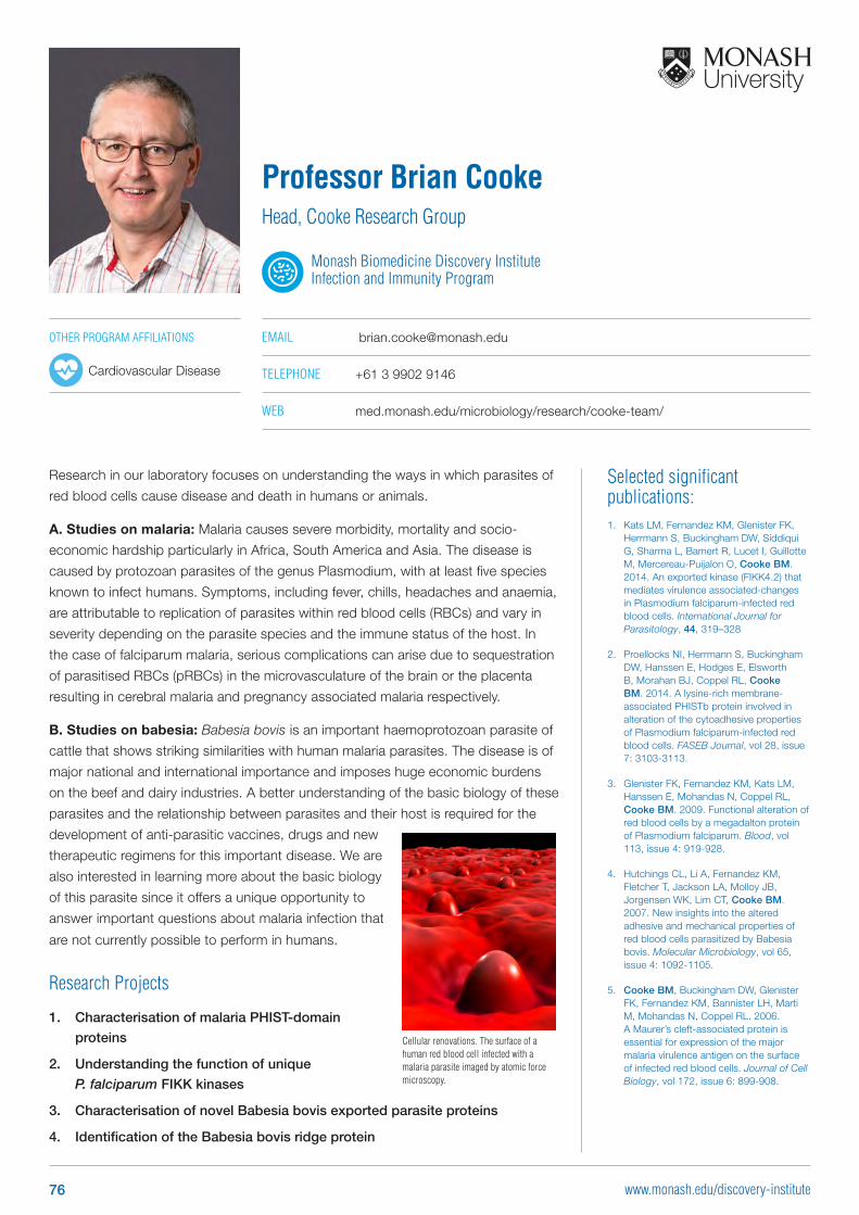
Selected significant publications: 1. Kats LM, Fernandez KM, Glenister FK,
Herrmann S, Buckingham DW, Siddiqui G, Sharma L, Bamert R, Lucet I, Guillotte M, Mercereau-Puijalon O, Cooke BM. 2014. An exported kinase (FIKK4.2) that mediates virulence associated-changes in Plasmodium falciparum-infected red blood cells. International Journal for Parasitology, 44, 319–328
2. Proellocks NI, Herrmann S, Buckingham DW, Hanssen E, Hodges E, Elsworth B, Morahan BJ, Coppel RL, Cooke BM. 2014. A lysine-rich membrane-associated PHISTb protein involved in alteration of the cytoadhesive properties of Plasmodium falciparum-infected red blood cells. FASEB Journal, vol 28, issue 7: 3103-3113.
3. Glenister FK, Fernandez KM, Kats LM, Hanssen E, Mohandas N, Coppel RL, Cooke BM. 2009. Functional alteration of red blood cells by a megadalton protein of Plasmodium falciparum. Blood, vol 113, issue 4: 919-928.
4. Hutchings CL, Li A, Fernandez KM, Fletcher T, Jackson LA, Molloy JB, Jorgensen WK, Lim CT, Cooke BM. 2007. New insights into the altered adhesive and mechanical properties of red blood cells parasitized by Babesia bovis. Molecular Microbiology, vol 65, issue 4: 1092-1105.
5. Cooke BM, Buckingham DW, Glenister FK, Fernandez KM, Bannister LH, Marti M, Mohandas N, Coppel RL. 2006. A Maurer’s cleft-associated protein is essential for expression of the major malaria virulence antigen on the surface of infected red blood cells. Journal of Cell Biology, vol 172, issue 6: 899-908.
Research in our laboratory focuses on understanding the ways in which parasites of
red blood cells cause disease and death in humans or animals.
A. Studies on malaria: Malaria causes severe morbidity, mortality and socio-
economic hardship particularly in Africa, South America and Asia. The disease is
caused by protozoan parasites of the genus Plasmodium, with at least five species
known to infect humans. Symptoms, including fever, chills, headaches and anaemia,
are attributable to replication of parasites within red blood cells (RBCs) and vary in
severity depending on the parasite species and the immune status of the host. In
the case of falciparum malaria, serious complications can arise due to sequestration
of parasitised RBCs (pRBCs) in the microvasculature of the brain or the placenta
resulting in cerebral malaria and pregnancy associated malaria respectively.
B. Studies on babesia: Babesia bovis is an important haemoprotozoan parasite of
cattle that shows striking similarities with human malaria parasites. The disease is of
major national and international importance and imposes huge economic burdens
on the beef and dairy industries. A better understanding of the basic biology of these
parasites and the relationship between parasites and their host is required for the
development of anti-parasitic vaccines, drugs and new
therapeutic regimens for this important disease. We are
also interested in learning more about the basic biology
of this parasite since it offers a unique opportunity to
answer important questions about malaria infection that
are not currently possible to perform in humans.
Research Projects
1. Characterisation of malaria PHIST-domain
proteins
2. Understanding the function of unique
P. falciparum FIKK kinases
3. Characterisation of novel Babesia bovis exported parasite proteins
4. Identification of the Babesia bovis ridge protein
Professor Brian CookeHead, Cooke Research Group
Monash Biomedicine Discovery Institute Infection and Immunity Program
EMAIL [email protected]
TELEPHONE +61 3 9902 9146
WEB med.monash.edu/microbiology/research/cooke-team/
Cellular renovations. The surface of a human red blood cell infected with a malaria parasite imaged by atomic force microscopy.
OTHER PROGRAM AFFILIATIONS
Cardiovascular Disease
76 www.monash.edu/discovery-institute
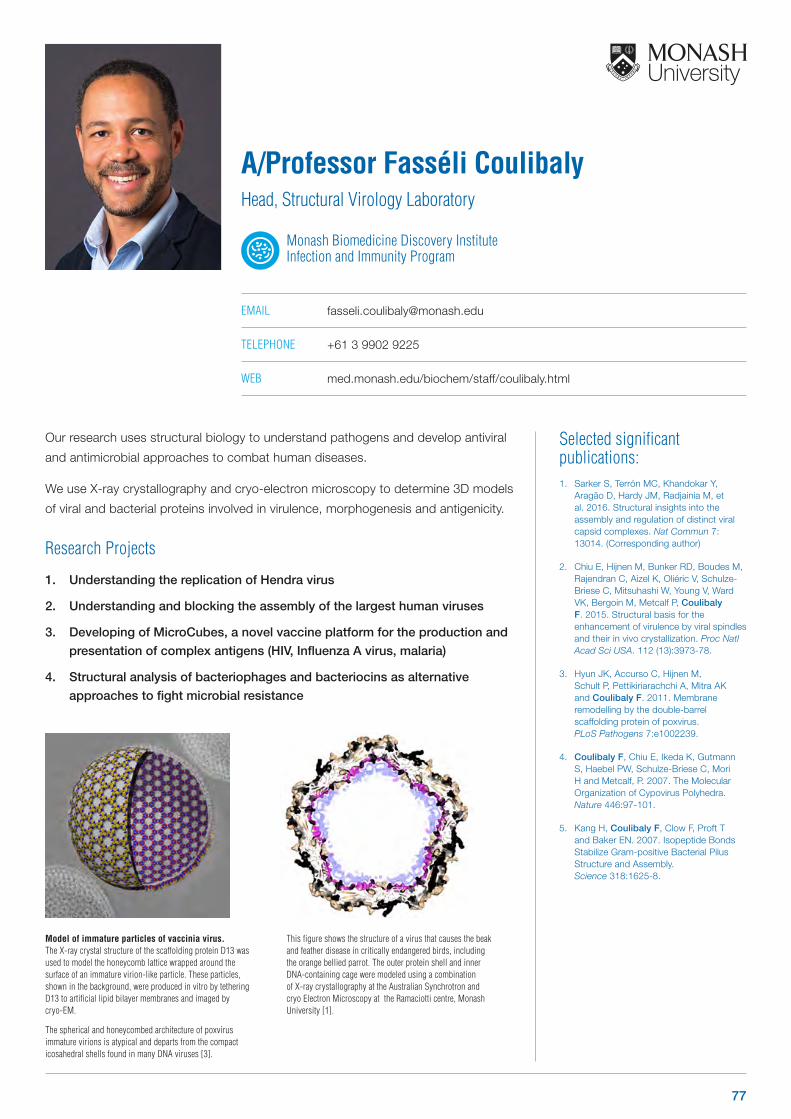
Model of immature particles of vaccinia virus. The X-ray crystal structure of the scaffolding protein D13 was used to model the honeycomb lattice wrapped around the surface of an immature virion-like particle. These particles, shown in the background, were produced in vitro by tethering D13 to artificial lipid bilayer membranes and imaged by cryo-EM.
The spherical and honeycombed architecture of poxvirus immature virions is atypical and departs from the compact icosahedral shells found in many DNA viruses [3].
This figure shows the structure of a virus that causes the beak and feather disease in critically endangered birds, including the orange bellied parrot. The outer protein shell and inner DNA-containing cage were modeled using a combination of X-ray crystallography at the Australian Synchrotron and cryo Electron Microscopy at the Ramaciotti centre, Monash University [1].
Selected significant publications: 1. Sarker S, Terrón MC, Khandokar Y,
Aragão D, Hardy JM, Radjainia M, et al. 2016. Structural insights into the assembly and regulation of distinct viral capsid complexes. Nat Commun 7: 13014. (Corresponding author)
2. Chiu E, Hijnen M, Bunker RD, Boudes M, Rajendran C, Aizel K, Oliéric V, Schulze-Briese C, Mitsuhashi W, Young V, Ward VK, Bergoin M, Metcalf P, Coulibaly F. 2015. Structural basis for the enhancement of virulence by viral spindles and their in vivo crystallization. Proc Natl Acad Sci USA. 112 (13):3973-78.
3. Hyun JK, Accurso C, Hijnen M, Schult P, Pettikiriarachchi A, Mitra AK and Coulibaly F. 2011. Membrane remodelling by the double-barrel scaffolding protein of poxvirus. PLoS Pathogens 7:e1002239.
4. Coulibaly F, Chiu E, Ikeda K, Gutmann S, Haebel PW, Schulze-Briese C, Mori H and Metcalf, P. 2007. The Molecular Organization of Cypovirus Polyhedra. Nature 446:97-101.
5. Kang H, Coulibaly F, Clow F, Proft T and Baker EN. 2007. Isopeptide Bonds Stabilize Gram-positive Bacterial Pilus Structure and Assembly. Science 318:1625-8.
Our research uses structural biology to understand pathogens and develop antiviral
and antimicrobial approaches to combat human diseases.
We use X-ray crystallography and cryo-electron microscopy to determine 3D models
of viral and bacterial proteins involved in virulence, morphogenesis and antigenicity.
Research Projects
1. Understanding the replication of Hendra virus
2. Understanding and blocking the assembly of the largest human viruses
3. Developing of MicroCubes, a novel vaccine platform for the production and
presentation of complex antigens (HIV, Influenza A virus, malaria)
4. Structural analysis of bacteriophages and bacteriocins as alternative
approaches to fight microbial resistance
A/Professor Fasséli CoulibalyHead, Structural Virology Laboratory
Monash Biomedicine Discovery Institute Infection and Immunity Program
EMAIL [email protected]
TELEPHONE +61 3 9902 9225
WEB med.monash.edu/biochem/staff/coulibaly.html
77
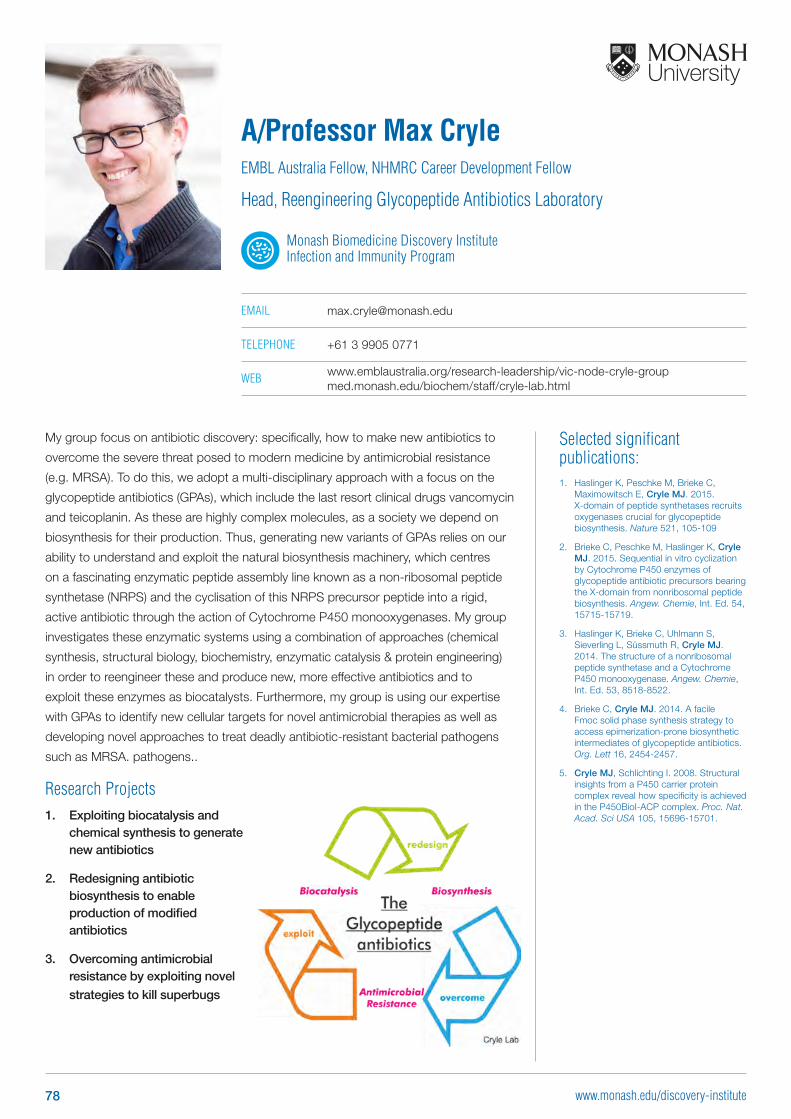
Selected significant publications: 1. Haslinger K, Peschke M, Brieke C,
Maximowitsch E, Cryle MJ. 2015. X-domain of peptide synthetases recruits oxygenases crucial for glycopeptide biosynthesis. Nature 521, 105-109
2. Brieke C, Peschke M, Haslinger K, Cryle MJ. 2015. Sequential in vitro cyclization by Cytochrome P450 enzymes of glycopeptide antibiotic precursors bearing the X-domain from nonribosomal peptide biosynthesis. Angew. Chemie, Int. Ed. 54, 15715-15719.
3. Haslinger K, Brieke C, Uhlmann S, Sieverling L, Süssmuth R, Cryle MJ. 2014. The structure of a nonribosomal peptide synthetase and a Cytochrome P450 monooxygenase. Angew. Chemie, Int. Ed. 53, 8518-8522.
4. Brieke C, Cryle MJ. 2014. A facile Fmoc solid phase synthesis strategy to access epimerization-prone biosynthetic intermediates of glycopeptide antibiotics. Org. Lett 16, 2454-2457.
5. Cryle MJ, Schlichting I. 2008. Structural insights from a P450 carrier protein complex reveal how specificity is achieved in the P450BioI-ACP complex. Proc. Nat. Acad. Sci USA 105, 15696-15701.
My group focus on antibiotic discovery: specifically, how to make new antibiotics to
overcome the severe threat posed to modern medicine by antimicrobial resistance
(e.g. MRSA). To do this, we adopt a multi-disciplinary approach with a focus on the
glycopeptide antibiotics (GPAs), which include the last resort clinical drugs vancomycin
and teicoplanin. As these are highly complex molecules, as a society we depend on
biosynthesis for their production. Thus, generating new variants of GPAs relies on our
ability to understand and exploit the natural biosynthesis machinery, which centres
on a fascinating enzymatic peptide assembly line known as a non-ribosomal peptide
synthetase (NRPS) and the cyclisation of this NRPS precursor peptide into a rigid,
active antibiotic through the action of Cytochrome P450 monooxygenases. My group
investigates these enzymatic systems using a combination of approaches (chemical
synthesis, structural biology, biochemistry, enzymatic catalysis & protein engineering)
in order to reengineer these and produce new, more effective antibiotics and to
exploit these enzymes as biocatalysts. Furthermore, my group is using our expertise
with GPAs to identify new cellular targets for novel antimicrobial therapies as well as
developing novel approaches to treat deadly antibiotic-resistant bacterial pathogens
such as MRSA. pathogens..
Research Projects1. Exploiting biocatalysis and
chemical synthesis to generate new antibiotics
2. Redesigning antibiotic biosynthesis to enable production of modified antibiotics
3. Overcoming antimicrobial resistance by exploiting novel
strategies to kill superbugs
A/Professor Max CryleEMBL Australia Fellow, NHMRC Career Development Fellow
Head, Reengineering Glycopeptide Antibiotics Laboratory
Monash Biomedicine Discovery Institute Infection and Immunity Program
EMAIL [email protected]
TELEPHONE +61 3 9905 0771
WEB www.emblaustralia.org/research-leadership/vic-node-cryle-group med.monash.edu/biochem/staff/cryle-lab.html
78 www.monash.edu/discovery-institute
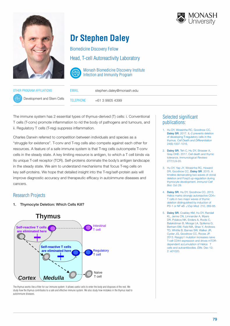
Selected significant publications: 1. Hu DY, Wirasinha RC, Goodnow CC,
Daley SR. 2017. IL-2 prevents deletion of developing T-regulatory cells in the thymus. Cell Death and Differentiation 24(6):1007-1016.
2. Daley SR, Teh C, Hu DY, Strasser A, Gray DHD. 2017. Cell death and thymic tolerance. Immunological Reviews 277(1):9-20.
3. Hu DY, Yap JY, Wirasinha RC, Howard DR, Goodnow CC, Daley SR. 2015. A timeline demarcating two waves of clonal deletion and Foxp3 up-regulation during thymocyte development. Immunol Cell Biol. Oct 29.
4. Daley SR, Hu DY, Goodnow CC. 2013. Helios marks strongly autoreactive CD4+ T cells in two major waves of thymic deletion distinguished by induction of PD-1 or NF-κB. J Exp Med. 210, 269-85.
5. Daley SR, Coakley KM, Hu DY, Randall KL, Jenne CN, Limnander A, Myers DR, Polakos NK, Enders A, Roots C, Balakishnan B, Miosge LA, Sjollema G, Bertram EM, Field MA, Shao Y, Andrews TD, Whittle B, Barnes SW, Walker JR, Cyster JG, Goodnow CC, Roose JP. 2013. Rasgrp1 mutation increases naive T-cell CD44 expression and drives mTOR-dependent accumulation of Helios‐ T cells and autoantibodies. Elife. Dec 12; 2: e01020.
The immune system has 2 essential types of thymus-derived (T) cells: i. Conventional
T cells (T-conv) promote inflammation to rid the body of pathogens and tumours, and
ii. Regulatory T cells (T-reg) suppress inflammation.
Charles Darwin referred to competition between individuals and species as a
“struggle for existence”. T-conv and T-reg cells also compete against each other for
resources. A feature of a safe immune system is that T-reg cells outcompete T-conv
cells in the steady state. A key limiting resource is antigen, to which a T cell binds via
its unique T-cell receptor (TCR). Self-proteins dominate the body’s antigen landscape
in the steady state. We aim to understand mechanisms that focus T-reg cells on
key self-proteins. We hope that detailed insight into the T-reg/self-protein axis will
improve diagnostic accuracy and therapeutic efficacy in autoimmune diseases and
cancers.
Research Projects
1. Thymocyte Deletion: Which Cells Kill?
Dr Stephen DaleyBiomedicine Discovery Fellow
Head, T-cell Autoreactivity Laboratory
Monash Biomedicine Discovery Institute Infection and Immunity Program
EMAIL [email protected]
TELEPHONE +61 3 9905 4399
OTHER PROGRAM AFFILIATIONS
Development and Stem Cells
The thymus works like a filter for our immune system: it allows useful cells to enter the body and disposes of the rest. We study how the thymus contributes to a safe and effective immune system. We also study how mistakes in the thymus lead to autoimmune diseases.
Cortex Medulla
Thymus
RegulatoryT cell
IntestinalT cell
Naïve T cell
Self-reactive T cells are eliminated here
The thymus works like a filter for our immune system: it allows usefulcells to enter the body and disposes of the rest. We study how thethymus contributes to a safe and effective immune system. We alsostudy how mistakes in the thymus lead to autoimmune diseases.
Self-reactive T cells are eliminated here
79
Selected significant publications: 1. Szymborska A, de Marco A, Cordes VC,
Briggs JAG, Ellenberg J. 2013. Structural organization of the nuclear pore scaffold revealed by super-resolution microscopy and particle averaging. Science 341(6146):655-8.
2. Schur FKM, Hagen W, de Marco A, Briggs JAG. 2013. Determination of protein structure at 8.5 Å resolution using cryo-electron tomography and subtomogram averaging. J Struct Biol 184(3):394-400.
3. de Marco A, Hauser AM, Glass B, Kräusslich HG, Mueller B, Briggs JAG. 2012. The role of the SP2 domain and its proteolytic cleavage in HIV-1 structural maturation and infectivity. J Virol 86(24):13708-16.
4. de Marco A, Müller B, Glass B, Riches J D, Kräusslich HG, Briggs JAG. 2010. Structural analysis of HIV-1 maturation using cryo-electron tomography. PLoS Pathog 6(11):e1001215.
5. Carlson LA, de Marco A, Oberwinkler H, Habermann A, Briggs JAG, Kräusslich HG, Grünewald K. 2010. Cryo-Electron Tomography of Native HIV-1 Budding Sites. PLoS Pathog 6(11): e1001173.
My laboratory has worked on the development of Cryo-Electron Tomography and
Subtomogram averaging on large scale in order to study various stages of the
replication cycle in HIV-1 as well as on the development of hardware and software
solutions for Correlative Light and Electron Microscopy (CLEM). Currently we are
focusing on the development of hardware and software to extend the use of cryo-
CLEM to in situ structural biology and biochemistry. The goal of our research is to
bring full automation and high throughput in the sample preparation, data collection
and analyses for cryo-Electron Microscopy performed directly in cells or in embryos
at early stages of development.
A/Professor Alex de MarcoMonash-Warwick Alliance Fellow
Head, de Marco Research Group
Monash Biomedicine Discovery Institute Infection and Immunity Program
EMAIL [email protected]
TELEPHONE +61 3 9905 3791
80 www.monash.edu/discovery-institute
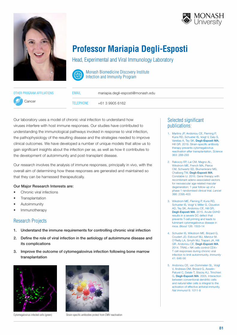
Selected significant publications: 1. Martins JP, Andoniou CE, Fleming P,
Kuns RD, Schuster IS, Voigt V, Daly S, Varelias A, Tey SK, Degli-Esposti MA, Hill GR. 2019. Strain-specific antibody therapy prevents cytomegalovirus reactivation after transplantation. Science 363: 288-293
2. Rakoczy EP, Lai CM, Magno AL, Wikstrom ME, French MA, Pierce CM, Schwartz SD, Blumenkranz MS, Chalberg TW, Degli-Esposti MA, Constable IJ. 2015. Gene therapy with recombinant adeno-associated vectors for neovascular age-related macular degeneration: 1 year follow-up of a phase 1 randomised clinical trial. Lancet 386: 2395-403
3. Wikstrom ME, Fleming P, Kuns RD, Schuster IS, Voigt V, Miller G, Clouston AD, Tey SK, Andoniou CE, Hill GR, Degli-Esposti MA. 2015. Acute GVHD results in a severe DC defect that prevents T-cell priming and leads to fulminant cytomegalovirus disease in mice. Blood 126: 1503-14
4. Schuster IS, Wikstrom ME, Brizard G, Coudert JD, Estcourt MJ, Manzur M, O’Reilly LA, Smyth MJ, Trapani JA, Hill GR, Andoniou CE, Degli-Esposti MA. 2014. TRAIL+ NK cells control CD4+ T cell responses during chronic viral infection to limit autoimmunity. Immunity 41: 646-56
5. Andoniou CE, van Dommelen SL, Voigt V, Andrews DM, Brizard G, Asselin-Paturel C, Delale T, Stacey KJ, Trinchieri G, Degli-Esposti MA. 2005. Interaction between conventional dendritic cells and natural killer cells is integral to the activation of effective antiviral immunity. Nat Immunol 6: 1011-9
Our laboratory uses a model of chronic viral infection to understand how
viruses interfere with host immune responses. Our studies have contributed to
understanding the immunological pathways invoked in response to viral infection,
the pathophysiology of the resulting disease and the strategies needed to improve
clinical outcomes. We have developed a number of unique models that allow us to
gain significant insights about the infection per se, as well as how it contributes to
the development of autoimmunity and post-transplant disease.
Our research involves the analysis of immune responses, principally in vivo, with the
overall aim of determining how these responses are generated and maintained so
that they can be harnessed therapeutically.
Our Major Research Interests are:
• Chronic viral infections
• Transplantation
• Autoimmunity
• Immunotherapy
Research Projects
1. Understand the immune requirements for controlling chronic viral infection
2. Define the role of viral infection in the aetiology of autoimmune disease and
its complications
3. Improve the outcome of cytomegalovirus infection following bone marrow
transplantation
Professor Mariapia Degli-EspostiHead, Experimental and Viral Immunology Laboratory
Monash Biomedicine Discovery Institute Infection and Immunity Program
EMAIL [email protected]
TELEPHONE +61 3 9905 6162
OTHER PROGRAM AFFILIATIONS
Cancer
Cytomegalovirus infected cells (green) Strain-specific antibodies protect from CMV reactivation
81
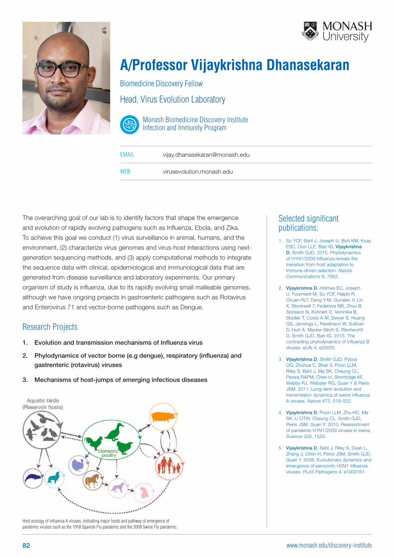
Selected significant publications: 1. Su YCF, Bahl J, Joseph U, Butt KM, Koay
ESC, Oon LLE, Barr IG, Vijaykrishna D, Smith GJD. 2015. Phylodynamics of H1N1/2009 influenza reveals the transition from host adaptation to immune-driven selection. Nature Communications 6, 7952.
2. Vijaykrishna D, Holmes EC, Joseph U, Fourment M, Su YCF, Halpin R, Chuen RLT, Deng Y-M, Gunalan V, Lin X, Stockwell T, Federova NB, Zhou B, Spirason N, Kühnert D, Veronika B, Stadler T, Costa A-M, Dwyer S, Huang QS, Jennings L, Rawlinson W, Sullivan S, Hurt A, Maurer-Stroh S, Wentworth D, Smith GJD, Barr IG. 2015. The contrasting phylodynamics of influenza B viruses. eLife 4, e05055.
3. Vijaykrishna D, Smith GJD, Pybus OG, Zhuhua C, Bhat S, Poon LLM, Riley S, Bahl J, Ma SK, Cheung CL, Perera RAPM, Chen H, Shortridge KF, Webby RJ, Webster RG, Guan Y & Peiris JSM. 2011. Long-term evolution and transmission dynamics of swine influenza A viruses. Nature 473, 519–522.
4. Vijaykrishna D, Poon LLM, Zhu HC, Ma SK, Li OTW, Cheung CL, Smith GJD, Peiris JSM, Guan Y. 2010. Reassortment of pandemic H1N1/2009 viruses in swine. Science 328, 1529.
5. Vijaykrishna D, Bahl J, Riley S, Duan L, Zhang J, Chen H, Peiris JSM, Smith GJD, Guan Y. 2008. Evolutionary dynamics and emergence of panzootic H5N1 influenza viruses. PLoS Pathogens 4, e1000161.
The overarching goal of our lab is to identify factors that shape the emergence
and evolution of rapidly evolving pathogens such as Influenza, Ebola, and Zika.
To achieve this goal we conduct (1) virus surveillance in animal, humans, and the
environment, (2) characterize virus genomes and virus-host interactions using next-
generation sequencing methods, and (3) apply computational methods to integrate
the sequence data with clinical, epidemiological and immunological data that are
generated from disease surveillance and laboratory experiments. Our primary
organism of study is influenza, due to its rapidly evolving small malleable genomes,
although we have ongoing projects in gastroenteric pathogens such as Rotavirus
and Enterovirus 71 and vector-borne pathogens such as Dengue.
Research Projects
1. Evolution and transmission mechanisms of Influenza virus
2. Phylodynamics of vector borne (e.g dengue), respiratory (influenza) and
gastrenteric (rotavirus) viruses
3. Mechanisms of host-jumps of emerging infectious diseases
A/Professor Vijaykrishna DhanasekaranBiomedicine Discovery Fellow
Head, Virus Evolution Laboratory
Monash Biomedicine Discovery Institute Infection and Immunity Program
EMAIL [email protected]
WEB virusevolution.monash.edu
Host ecology of influenza A viruses, indicating major hosts and pathway of emergence of pandemic viruses such as the 1918 Spanish Flu pandemic and the 2009 Swine Flu pandemic.
82 www.monash.edu/discovery-institute
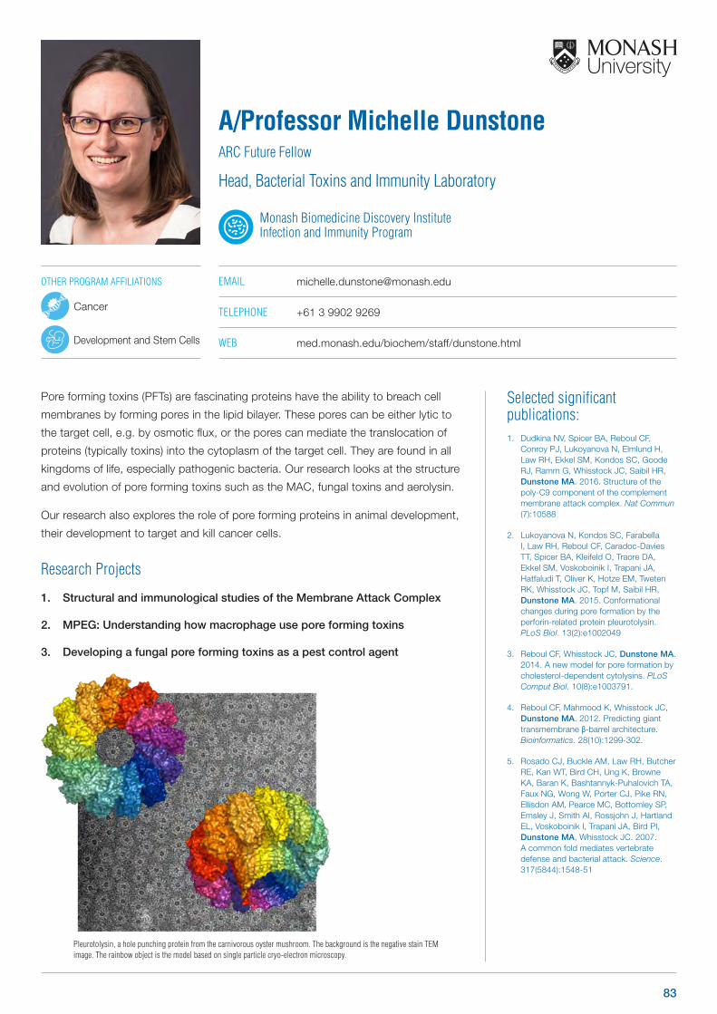
Selected significant publications: 1. Dudkina NV, Spicer BA, Reboul CF,
Conroy PJ, Lukoyanova N, Elmlund H, Law RH, Ekkel SM, Kondos SC, Goode RJ, Ramm G, Whisstock JC, Saibil HR, Dunstone MA. 2016. Structure of the poly-C9 component of the complement membrane attack complex. Nat Commun (7):10588
2. Lukoyanova N, Kondos SC, Farabella I, Law RH, Reboul CF, Caradoc-Davies TT, Spicer BA, Kleifeld O, Traore DA, Ekkel SM, Voskoboinik I, Trapani JA, Hatfaludi T, Oliver K, Hotze EM, Tweten RK, Whisstock JC, Topf M, Saibil HR, Dunstone MA. 2015. Conformational changes during pore formation by the perforin-related protein pleurotolysin. PLoS Biol. 13(2):e1002049
3. Reboul CF, Whisstock JC, Dunstone MA. 2014. A new model for pore formation by cholesterol-dependent cytolysins. PLoS Comput Biol. 10(8):e1003791.
4. Reboul CF, Mahmood K, Whisstock JC, Dunstone MA. 2012. Predicting giant transmembrane β-barrel architecture. Bioinformatics. 28(10):1299-302.
5. Rosado CJ, Buckle AM, Law RH, Butcher RE, Kan WT, Bird CH, Ung K, Browne KA, Baran K, Bashtannyk-Puhalovich TA, Faux NG, Wong W, Porter CJ, Pike RN, Ellisdon AM, Pearce MC, Bottomley SP, Emsley J, Smith AI, Rossjohn J, Hartland EL, Voskoboinik I, Trapani JA, Bird PI, Dunstone MA, Whisstock JC. 2007. A common fold mediates vertebrate defense and bacterial attack. Science. 317(5844):1548-51
Pore forming toxins (PFTs) are fascinating proteins have the ability to breach cell
membranes by forming pores in the lipid bilayer. These pores can be either lytic to
the target cell, e.g. by osmotic flux, or the pores can mediate the translocation of
proteins (typically toxins) into the cytoplasm of the target cell. They are found in all
kingdoms of life, especially pathogenic bacteria. Our research looks at the structure
and evolution of pore forming toxins such as the MAC, fungal toxins and aerolysin.
Our research also explores the role of pore forming proteins in animal development,
their development to target and kill cancer cells.
Research Projects
1. Structural and immunological studies of the Membrane Attack Complex
2. MPEG: Understanding how macrophage use pore forming toxins
3. Developing a fungal pore forming toxins as a pest control agent
A/Professor Michelle DunstoneARC Future Fellow
Head, Bacterial Toxins and Immunity Laboratory
Monash Biomedicine Discovery Institute Infection and Immunity Program
EMAIL [email protected]
TELEPHONE +61 3 9902 9269
WEB med.monash.edu/biochem/staff/dunstone.html
Pleurotolysin, a hole punching protein from the carnivorous oyster mushroom. The background is the negative stain TEM image. The rainbow object is the model based on single particle cryo-electron microscopy.
OTHER PROGRAM AFFILIATIONS
Cancer
Development and Stem Cells
83
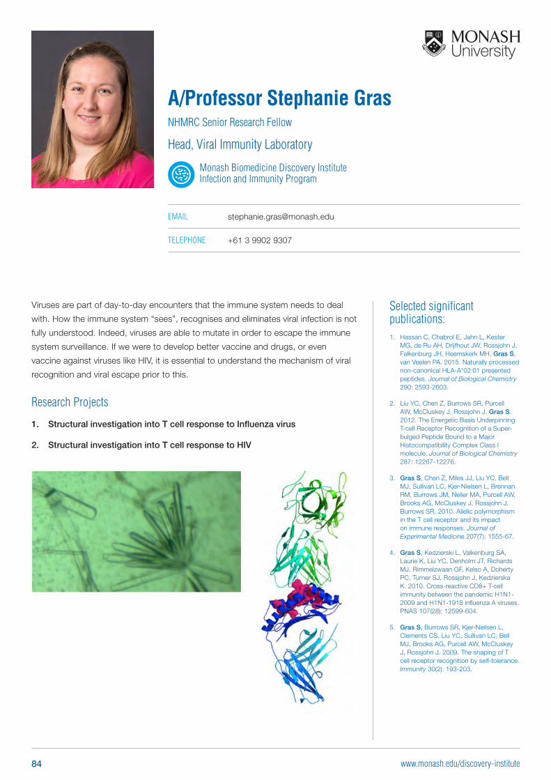
Selected significant publications: 1. Hassan C, Chabrol E, Jahn L, Kester
MG, de Ru AH, Drijfhout JW, Rossjohn J, Falkenburg JH, Heemskerk MH, Gras S, van Veelen PA. 2015. Naturally processed non-canonical HLA-A*02:01 presented peptides. Journal of Biological Chemistry 290: 2593-2603.
2. Liu YC, Chen Z, Burrows SR, Purcell AW, McCluskey J, Rossjohn J, Gras S. 2012. The Energetic Basis Underpinning T-cell Receptor Recognition of a Super- bulged Peptide Bound to a Major Histocompatibility Complex Class I molecule. Journal of Biological Chemistry 287: 12267-12276.
3. Gras S, Chen Z, Miles JJ, Liu YC, Bell MJ, Sullivan LC, Kjer-Nielsen L, Brennan RM, Burrows JM, Neller MA, Purcell AW, Brooks AG, McCluskey J, Rossjohn J, Burrows SR. 2010. Allelic polymorphism in the T cell receptor and its impact on immune responses. Journal of Experimental Medicine 207(7): 1555-67.
4. Gras S, Kedzierski L, Valkenburg SA, Laurie K, Liu YC, Denholm JT, Richards MJ, Rimmelzwaan GF, Kelso A, Doherty PC, Turner SJ, Rossjohn J, Kedzierska K. 2010. Cross-reactive CD8+ T-cell immunity between the pandemic H1N1-2009 and H1N1-1918 influenza A viruses. PNAS 107(28): 12599-604.
5. Gras S, Burrows SR, Kjer-Nielsen L, Clements CS, Liu YC, Sullivan LC, Bell MJ, Brooks AG, Purcell AW, McCluskey J, Rossjohn J. 2009. The shaping of T cell receptor recognition by self-tolerance. Immunity 30(2): 193-203.
Viruses are part of day-to-day encounters that the immune system needs to deal
with. How the immune system “sees”, recognises and eliminates viral infection is not
fully understood. Indeed, viruses are able to mutate in order to escape the immune
system surveillance. If we were to develop better vaccine and drugs, or even
vaccine against viruses like HIV, it is essential to understand the mechanism of viral
recognition and viral escape prior to this.
Research Projects
1. Structural investigation into T cell response to Influenza virus
2. Structural investigation into T cell response to HIV
A/Professor Stephanie GrasNHMRC Senior Research Fellow
Head, Viral Immunity Laboratory
Monash Biomedicine Discovery Institute Infection and Immunity Program
EMAIL [email protected]
TELEPHONE +61 3 9902 9307
84 www.monash.edu/discovery-institute
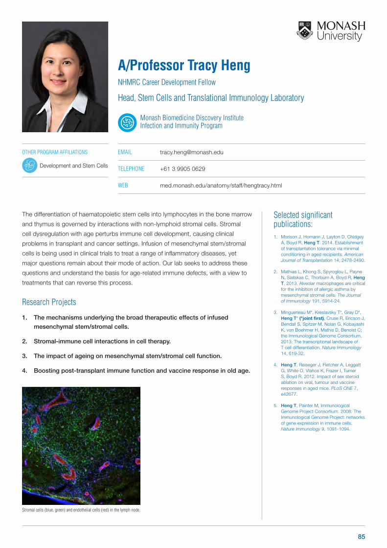
Selected significant publications: 1. Morison J, Homann J, Layton D, Chidgey
A, Boyd R, Heng T. 2014. Establishment of transplantation tolerance via minimal conditioning in aged recipients. American Journal of Transplantation 14, 2478-2490.
2. Mathias L, Khong S, Spyroglou L, Payne N, Siatskas C, Thorburn A, Boyd R, Heng T. 2013. Alveolar macrophages are critical for the inhibition of allergic asthma by mesenchymal stromal cells. The Journal of Immunology 191, 5914-24.
3. Mingueneau M*, Kreslavsky T*, Gray D*, Heng T* (*joint first), Cruse R, Ericson J, Bendall S, Spitzer M, Nolan G, Kobayashi K, von Boehmer H, Mathis D, Benoist C; the Immunological Genome Consortium. 2013. The transcriptional landscape of ‐‐ T cell differentiation. Nature Immunology 14, 619-32.
4. Heng T, Reiseger J, Fletcher A, Leggatt G, White O, Vlahos K, Frazer I, Turner S, Boyd R. 2012. Impact of sex steroid ablation on viral, tumour and vaccine responses in aged mice. PLoS ONE 7, e42677.
5. Heng T, Painter M, Immunological Genome Project Consortium. 2008. The Immunological Genome Project: networks of gene expression in immune cells. Nature Immunology 9, 1091-1094.
The differentiation of haematopoietic stem cells into lymphocytes in the bone marrow
and thymus is governed by interactions with non-lymphoid stromal cells. Stromal
cell dysregulation with age perturbs immune cell development, causing clinical
problems in transplant and cancer settings. Infusion of mesenchymal stem/stromal
cells is being used in clinical trials to treat a range of inflammatory diseases, yet
major questions remain about their mode of action. Our lab seeks to address these
questions and understand the basis for age-related immune defects, with a view to
treatments that can reverse this process.
Research Projects
1. The mechanisms underlying the broad therapeutic effects of infused
mesenchymal stem/stromal cells.
2. Stromal-immune cell interactions in cell therapy.
3. The impact of ageing on mesenchymal stem/stromal cell function.
4. Boosting post-transplant immune function and vaccine response in old age.
A/Professor Tracy HengNHMRC Career Development Fellow
Head, Stem Cells and Translational Immunology Laboratory
Monash Biomedicine Discovery Institute Infection and Immunity Program
EMAIL [email protected]
TELEPHONE +61 3 9905 0629
WEB med.monash.edu/anatomy/staff/hengtracy.html
Stromal cells (blue, green) and endothelial cells (red) in the lymph node.
OTHER PROGRAM AFFILIATIONS
Development and Stem Cells
85
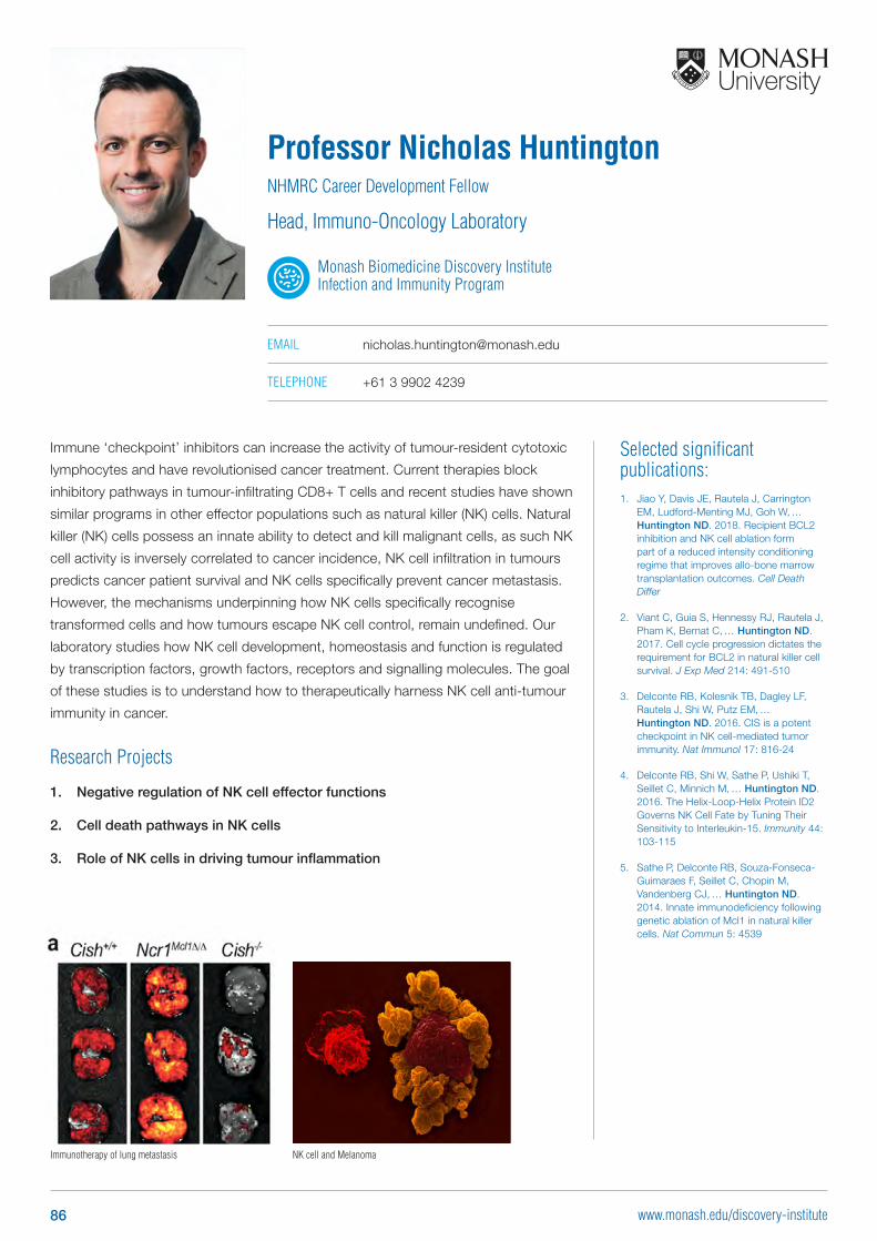
Selected significant publications: 1. Jiao Y, Davis JE, Rautela J, Carrington
EM, Ludford-Menting MJ, Goh W, ...Huntington ND. 2018. Recipient BCL2 inhibition and NK cell ablation form part of a reduced intensity conditioning regime that improves allo-bone marrow transplantation outcomes. Cell Death Differ
2. Viant C, Guia S, Hennessy RJ, Rautela J, Pham K, Bernat C, ... Huntington ND. 2017. Cell cycle progression dictates the requirement for BCL2 in natural killer cell survival. J Exp Med 214: 491-510
3. Delconte RB, Kolesnik TB, Dagley LF, Rautela J, Shi W, Putz EM, ... Huntington ND. 2016. CIS is a potent checkpoint in NK cell-mediated tumor immunity. Nat Immunol 17: 816-24
4. Delconte RB, Shi W, Sathe P, Ushiki T, Seillet C, Minnich M, ... Huntington ND. 2016. The Helix-Loop-Helix Protein ID2 Governs NK Cell Fate by Tuning Their Sensitivity to Interleukin-15. Immunity 44: 103-115
5. Sathe P, Delconte RB, Souza-Fonseca-Guimaraes F, Seillet C, Chopin M, Vandenberg CJ, ... Huntington ND. 2014. Innate immunodeficiency following genetic ablation of Mcl1 in natural killer cells. Nat Commun 5: 4539
Immune ‘checkpoint’ inhibitors can increase the activity of tumour-resident cytotoxic
lymphocytes and have revolutionised cancer treatment. Current therapies block
inhibitory pathways in tumour-infiltrating CD8+ T cells and recent studies have shown
similar programs in other effector populations such as natural killer (NK) cells. Natural
killer (NK) cells possess an innate ability to detect and kill malignant cells, as such NK
cell activity is inversely correlated to cancer incidence, NK cell infiltration in tumours
predicts cancer patient survival and NK cells specifically prevent cancer metastasis.
However, the mechanisms underpinning how NK cells specifically recognise
transformed cells and how tumours escape NK cell control, remain undefined. Our
laboratory studies how NK cell development, homeostasis and function is regulated
by transcription factors, growth factors, receptors and signalling molecules. The goal
of these studies is to understand how to therapeutically harness NK cell anti-tumour
immunity in cancer.
Research Projects
1. Negative regulation of NK cell effector functions
2. Cell death pathways in NK cells
3. Role of NK cells in driving tumour inflammation
Professor Nicholas HuntingtonNHMRC Career Development Fellow
Head, Immuno-Oncology Laboratory
Monash Biomedicine Discovery Institute Infection and Immunity Program
EMAIL [email protected]
TELEPHONE +61 3 9902 4239
Immunotherapy of lung metastasis NK cell and Melanoma
86 www.monash.edu/discovery-institute
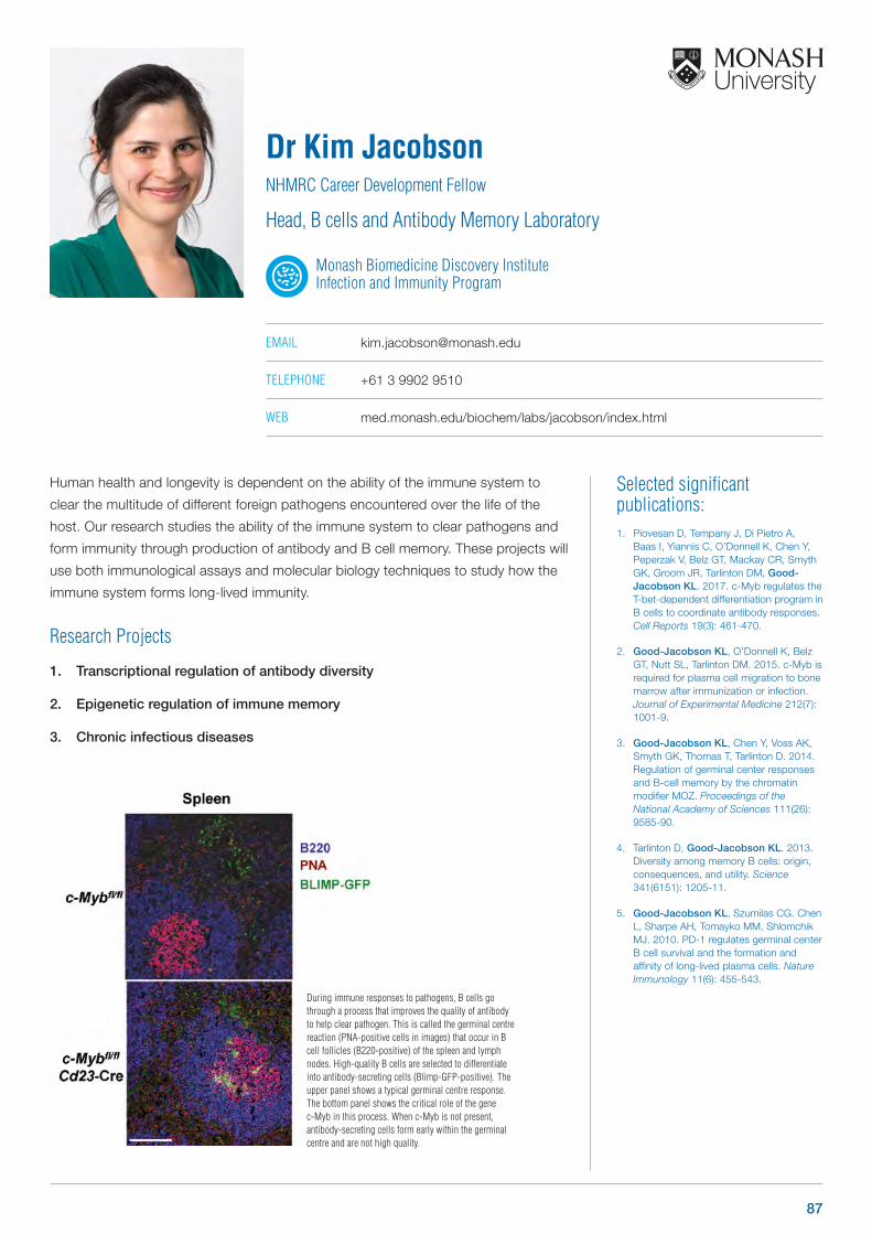
Selected significant publications: 1. Piovesan D, Tempany J, Di Pietro A,
Baas I, Yiannis C, O’Donnell K, Chen Y, Peperzak V, Belz GT, Mackay CR, Smyth GK, Groom JR, Tarlinton DM, Good-Jacobson KL. 2017. c-Myb regulates the T-bet-dependent differentiation program in B cells to coordinate antibody responses. Cell Reports 19(3): 461-470.
2. Good-Jacobson KL, O’Donnell K, Belz GT, Nutt SL, Tarlinton DM. 2015. c-Myb is required for plasma cell migration to bone marrow after immunization or infection. Journal of Experimental Medicine 212(7): 1001-9.
3. Good-Jacobson KL, Chen Y, Voss AK, Smyth GK, Thomas T, Tarlinton D. 2014. Regulation of germinal center responses and B-cell memory by the chromatin modifier MOZ. Proceedings of the National Academy of Sciences 111(26): 9585-90.
4. Tarlinton D, Good-Jacobson KL. 2013. Diversity among memory B cells: origin, consequences, and utility. Science 341(6151): 1205-11.
5. Good-Jacobson KL, Szumilas CG. Chen L, Sharpe AH, Tomayko MM, Shlomchik MJ. 2010. PD-1 regulates germinal center B cell survival and the formation and affinity of long-lived plasma cells. Nature Immunology 11(6): 455-543.
Human health and longevity is dependent on the ability of the immune system to
clear the multitude of different foreign pathogens encountered over the life of the
host. Our research studies the ability of the immune system to clear pathogens and
form immunity through production of antibody and B cell memory. These projects will
use both immunological assays and molecular biology techniques to study how the
immune system forms long-lived immunity.
Research Projects
1. Transcriptional regulation of antibody diversity
2. Epigenetic regulation of immune memory
3. Chronic infectious diseases
Dr Kim JacobsonNHMRC Career Development Fellow
Head, B cells and Antibody Memory Laboratory
Monash Biomedicine Discovery Institute Infection and Immunity Program
EMAIL [email protected]
TELEPHONE +61 3 9902 9510
WEB med.monash.edu/biochem/labs/jacobson/index.html
During immune responses to pathogens, B cells go through a process that improves the quality of antibody to help clear pathogen. This is called the germinal centre reaction (PNA-positive cells in images) that occur in B cell follicles (B220-positive) of the spleen and lymph nodes. High-quality B cells are selected to differentiate into antibody-secreting cells (Blimp-GFP-positive). The upper panel shows a typical germinal centre response. The bottom panel shows the critical role of the gene c-Myb in this process. When c-Myb is not present, antibody-secreting cells form early within the germinal centre and are not high quality.
EMAIL [email protected]
TELEPHONE +61 3 9902 4239
87
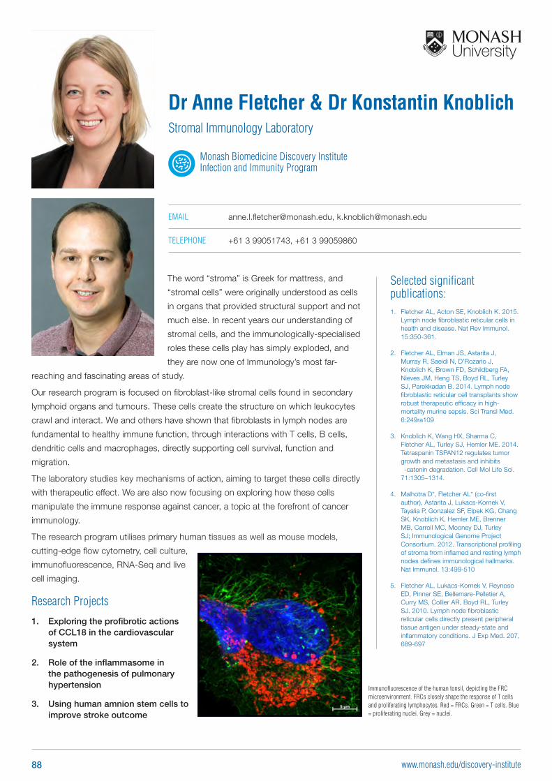
Selected significant publications: 1. Fletcher AL, Acton SE, Knoblich K. 2015.
Lymph node fibroblastic reticular cells in health and disease. Nat Rev Immunol. 15:350-361.
2. Fletcher AL, Elman JS, Astarita J, Murray R, Saeidi N, D’Rozario J, Knoblich K, Brown FD, Schildberg FA, Nieves JM, Heng TS, Boyd RL, Turley SJ, Parekkadan B. 2014. Lymph node fibroblastic reticular cell transplants show robust therapeutic efficacy in high-mortality murine sepsis. Sci Transl Med. 6:249ra109
3. Knoblich K, Wang HX, Sharma C, Fletcher AL, Turley SJ, Hemler ME. 2014. Tetraspanin TSPAN12 regulates tumor growth and metastasis and inhibits ‐-catenin degradation. Cell Mol Life Sci. 71:1305–1314.
4. Malhotra D*, Fletcher AL* (co-first author), Astarita J, Lukacs-Kornek V, Tayalia P, Gonzalez SF, Elpek KG, Chang SK, Knoblich K, Hemler ME, Brenner MB, Carroll MC, Mooney DJ, Turley SJ; Immunological Genome Project Consortium. 2012. Transcriptional profiling of stroma from inflamed and resting lymph nodes defines immunological hallmarks. Nat Immunol. 13:499-510
5. Fletcher AL, Lukacs-Kornek V, Reynoso ED, Pinner SE, Bellemare-Pelletier A, Curry MS, Collier AR, Boyd RL, Turley SJ. 2010. Lymph node fibroblastic reticular cells directly present peripheral tissue antigen under steady-state and inflammatory conditions. J Exp Med. 207, 689-697
The word “stroma” is Greek for mattress, and
“stromal cells” were originally understood as cells
in organs that provided structural support and not
much else. In recent years our understanding of
stromal cells, and the immunologically-specialised
roles these cells play has simply exploded, and
they are now one of Immunology’s most far-
reaching and fascinating areas of study.
Our research program is focused on fibroblast-like stromal cells found in secondary
lymphoid organs and tumours. These cells create the structure on which leukocytes
crawl and interact. We and others have shown that fibroblasts in lymph nodes are
fundamental to healthy immune function, through interactions with T cells, B cells,
dendritic cells and macrophages, directly supporting cell survival, function and
migration.
The laboratory studies key mechanisms of action, aiming to target these cells directly
with therapeutic effect. We are also now focusing on exploring how these cells
manipulate the immune response against cancer, a topic at the forefront of cancer
immunology.
The research program utilises primary human tissues as well as mouse models,
cutting-edge flow cytometry, cell culture,
immunofluorescence, RNA-Seq and live
cell imaging.
Research Projects
1. Exploring the profibrotic actions of CCL18 in the cardiovascular system
2. Role of the inflammasome in the pathogenesis of pulmonary hypertension
3. Using human amnion stem cells to improve stroke outcome
Dr Anne Fletcher & Dr Konstantin KnoblichStromal Immunology Laboratory
Monash Biomedicine Discovery Institute Infection and Immunity Program
EMAIL [email protected], [email protected]
TELEPHONE +61 3 99051743, +61 3 99059860
Immunofluorescence of the human tonsil, depicting the FRC microenvironment. FRCs closely shape the response of T cells and proliferating lymphocytes. Red = FRCs. Green = T cells. Blue = proliferating nuclei. Grey = nuclei.
88 www.monash.edu/discovery-institute
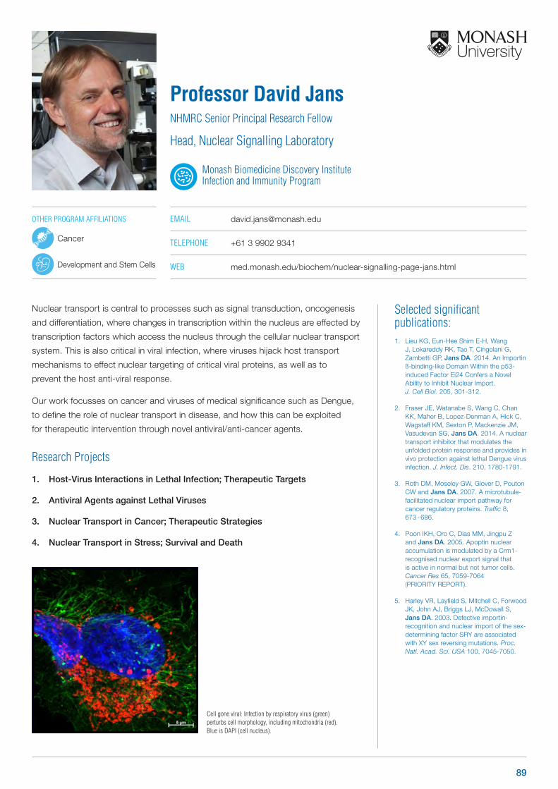
Selected significant publications: 1. Lieu KG, Eun-Hee Shim E-H, Wang
J, Lokareddy RK, Tao T, Cingolani G, Zambetti GP, Jans DA. 2014. An Importin ß-binding-like Domain Within the p53-induced Factor Ei24 Confers a Novel Ability to Inhibit Nuclear Import. J. Cell Biol. 205, 301-312.
2. Fraser JE, Watanabe S, Wang C, Chan KK, Maher B, Lopez-Denman A, Hick C, Wagstaff KM, Sexton P, Mackenzie JM, Vasudevan SG, Jans DA. 2014. A nuclear transport inhibitor that modulates the unfolded protein response and provides in vivo protection against lethal Dengue virus infection. J. Infect. Dis. 210, 1780-1791.
3. Roth DM, Moseley GW, Glover D, Pouton CW and Jans DA. 2007. A microtubule-facilitated nuclear import pathway for cancer regulatory proteins. Traffic 8, 673 - 686.
4. Poon IKH, Oro C, Dias MM, Jingpu Z and Jans DA. 2005. Apoptin nuclear accumulation is modulated by a Crm1-recognised nuclear export signal that is active in normal but not tumor cells. Cancer Res 65, 7059-7064 (PRIORITY REPORT).
5. Harley VR, Layfield S, Mitchell C, Forwood JK, John AJ, Briggs LJ, McDowall S, Jans DA. 2003. Defective importin-‐ recognition and nuclear import of the sex-determining factor SRY are associated with XY sex reversing mutations. Proc. Natl. Acad. Sci. USA 100, 7045-7050.
Nuclear transport is central to processes such as signal transduction, oncogenesis
and differentiation, where changes in transcription within the nucleus are effected by
transcription factors which access the nucleus through the cellular nuclear transport
system. This is also critical in viral infection, where viruses hijack host transport
mechanisms to effect nuclear targeting of critical viral proteins, as well as to
prevent the host anti-viral response.
Our work focusses on cancer and viruses of medical significance such as Dengue,
to define the role of nuclear transport in disease, and how this can be exploited
for therapeutic intervention through novel antiviral/anti-cancer agents.
Research Projects
1. Host-Virus Interactions in Lethal Infection; Therapeutic Targets
2. Antiviral Agents against Lethal Viruses
3. Nuclear Transport in Cancer; Therapeutic Strategies
4. Nuclear Transport in Stress; Survival and Death
Professor David JansNHMRC Senior Principal Research Fellow
Head, Nuclear Signalling Laboratory
Monash Biomedicine Discovery Institute Infection and Immunity Program
EMAIL [email protected]
TELEPHONE +61 3 9902 9341
WEB med.monash.edu/biochem/nuclear-signalling-page-jans.html
Cell gone viral: Infection by respiratory virus (green) perturbs cell morphology, including mitochondria (red). Blue is DAPI (cell nucleus).
OTHER PROGRAM AFFILIATIONS
Cancer
Development and Stem Cells
89
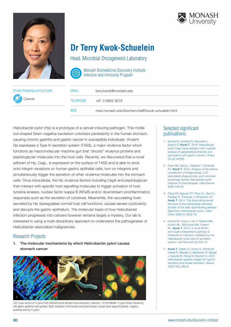
Selected significant publications: 1. Gorrell RJ, Zwickel N, Reynolds J,
Bulach D, Kwok T. 2016. Helicobacter pylori CagL hypervariable motif: a global analysis of geographical diversity and association with gastric cancer. J Infect Dis pii: jiw060.
2. Deen NS, Gong L, Naderer T, Devenish RJ, Kwok T. 2015. Analysis of the relative contribution of phagocytosis, LC3-associated phagocytosis, and canonical autophagy during Helicobacter pylori infection of macrophages. Helicobacter 20(6):449-59.
3. Pang SS, Nguyen ST, Perry AJ, Day CJ, Panjikar S, Tiralongo J, Whisstock JC, Kwok T. 2014. The three-dimensional structure of the extracellular adhesion domain of the sialic acid-binding adhesin SabA from Helicobacter pylori. J Biol Chem 289(10): 6332-40.
4. Gorrell RJ, Guan J, Xin Y, Tafreshi MA, Hutton ML, McGuckin MA, Ferrero RL, Kwok T. 2013. A novel NOD1- and CagA-independent pathway of interleukin-8 induction mediated by the Helicobacter pylori type IV secretion system. Cell Microbiol (4):554-70.
5. Kwok T, Zabler D, Urman S, Rohde M, Hartig R, Wessler S, Misselwitz R, Berger J, Sewald N, König W, Backert S. 2007. Helicobacter exploits integrin for type IV secretion and kinase activation. Nature 449(7164): 862-6.
Helicobacter pylori (Hp) is a prototype of a cancer-inducing pathogen. This motile
rod-shaped Gram negative bacterium colonises persistently in the human stomach,
causing chronic gastritis and gastric cancer in susceptible individuals. Virulent
Hp expresses a Type IV secretion system (T4SS), a major virulence factor which
functions as macromolecular machine gun that “shoots” virulence proteins and
peptidoglycan molecules into the host cells. Recently, we discovered that a novel
adhesin of Hp, CagL, is expressed on the surface of T4SS and is able to dock
onto integrin receptors on human gastric epithelial cells, turn on integrins and
simultaneously trigger the secretion of other virulence molecules into the stomach
cells. Once intracellular, the Hp virulence factors including CagA and peptidoglycan
then interact with specific host signalling molecules to trigger activation of host
tyrosine kinases, nuclear factor kappa B (NFκB) and/or downstream proinflammatory
responses such as the secretion of cytokines. Meanwhile, the vacuolating toxin
secreted by Hp dysregulates normal host cell functions, causes severe cytotoxicity
and disrupts the gastric epithelium. The molecular basis of how Helicobacter
infection progresses into cancers however remains largely a mystery. Our lab is
interested in using a multi-disciplinary approach to understand the pathogenesis of
Helicobacter-associated malignancies.
Research Projects1. The molecular mechanisms by which Helicobacter pylori causes
stomach cancer
Dr Terry Kwok-SchueleinHead, Microbial Oncogenesis Laboratory
Monash Biomedicine Discovery Institute Infection and Immunity Program
EMAIL [email protected]
TELEPHONE +61 3 9902 9216
WEB med.monash.edu/biochem/staff/kwok-schuelein.html
Left: Cross-section of H. pylori (red)-infected mouse stomach (blue and green); scale bar = 10 mm Middle: H. pylori (blue) interacting with gastric epithelial cells (yellow). Right: Activation of the human transcription factor, nuclear factor kappa B (yellow), in gastric epithelial cells by H. pylori.
OTHER PROGRAM AFFILIATIONS
Cancer
90 www.monash.edu/discovery-institute
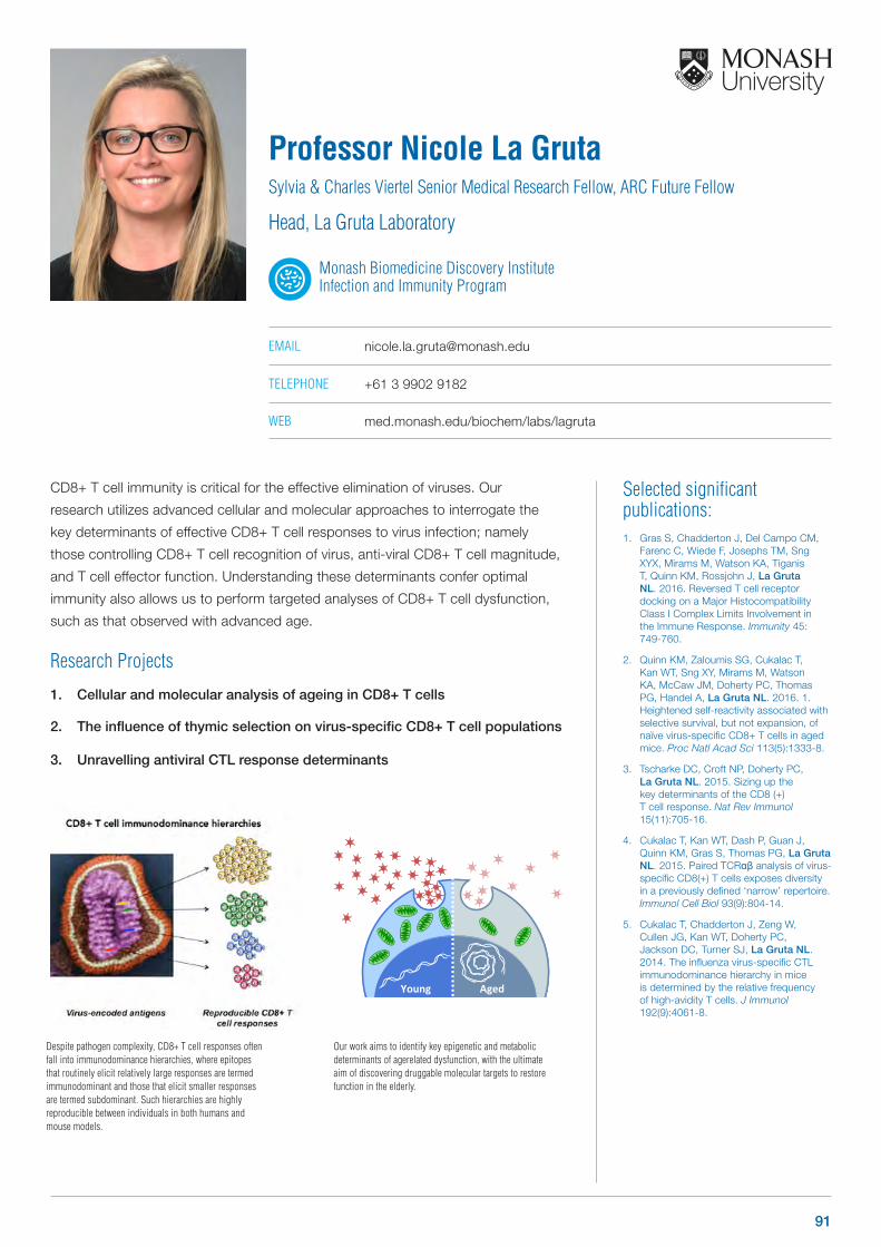
Selected significant publications: 1. Gras S, Chadderton J, Del Campo CM,
Farenc C, Wiede F, Josephs TM, Sng XYX, Mirams M, Watson KA, Tiganis T, Quinn KM, Rossjohn J, La Gruta NL. 2016. Reversed T cell receptor docking on a Major Histocompatibility Class I Complex Limits Involvement in the Immune Response. Immunity 45: 749-760.
2. Quinn KM, Zaloumis SG, Cukalac T, Kan WT, Sng XY, Mirams M, Watson KA, McCaw JM, Doherty PC, Thomas PG, Handel A, La Gruta NL. 2016. 1. Heightened self-reactivity associated with selective survival, but not expansion, of naïve virus-specific CD8+ T cells in aged mice. Proc Natl Acad Sci 113(5):1333-8.
3. Tscharke DC, Croft NP, Doherty PC, La Gruta NL. 2015. Sizing up the key determinants of the CD8 (+) T cell response. Nat Rev Immunol 15(11):705-16.
4. Cukalac T, Kan WT, Dash P, Guan J, Quinn KM, Gras S, Thomas PG, La Gruta NL. 2015. Paired TCRαβ analysis of virus-specific CD8(+) T cells exposes diversity in a previously defined ‘narrow’ repertoire. Immunol Cell Biol 93(9):804-14.
5. Cukalac T, Chadderton J, Zeng W, Cullen JG, Kan WT, Doherty PC, Jackson DC, Turner SJ, La Gruta NL. 2014. The influenza virus-specific CTL immunodominance hierarchy in mice is determined by the relative frequency of high-avidity T cells. J Immunol 192(9):4061-8.
CD8+ T cell immunity is critical for the effective elimination of viruses. Our
research utilizes advanced cellular and molecular approaches to interrogate the
key determinants of effective CD8+ T cell responses to virus infection; namely
those controlling CD8+ T cell recognition of virus, anti-viral CD8+ T cell magnitude,
and T cell effector function. Understanding these determinants confer optimal
immunity also allows us to perform targeted analyses of CD8+ T cell dysfunction,
such as that observed with advanced age.
Research Projects
1. Cellular and molecular analysis of ageing in CD8+ T cells
2. The influence of thymic selection on virus-specific CD8+ T cell populations
3. Unravelling antiviral CTL response determinants
Professor Nicole La GrutaSylvia & Charles Viertel Senior Medical Research Fellow, ARC Future Fellow
Head, La Gruta Laboratory
Monash Biomedicine Discovery Institute Infection and Immunity Program
EMAIL [email protected]
TELEPHONE +61 3 9902 9182
WEB med.monash.edu/biochem/labs/lagruta
Young Aged
Despite pathogen complexity, CD8+ T cell responses often fall into immunodominance hierarchies, where epitopes that routinely elicit relatively large responses are termed immunodominant and those that elicit smaller responses are termed subdominant. Such hierarchies are highly reproducible between individuals in both humans and mouse models.
Our work aims to identify key epigenetic and metabolic determinants of agerelated dysfunction, with the ultimate aim of discovering druggable molecular targets to restore function in the elderly.
91
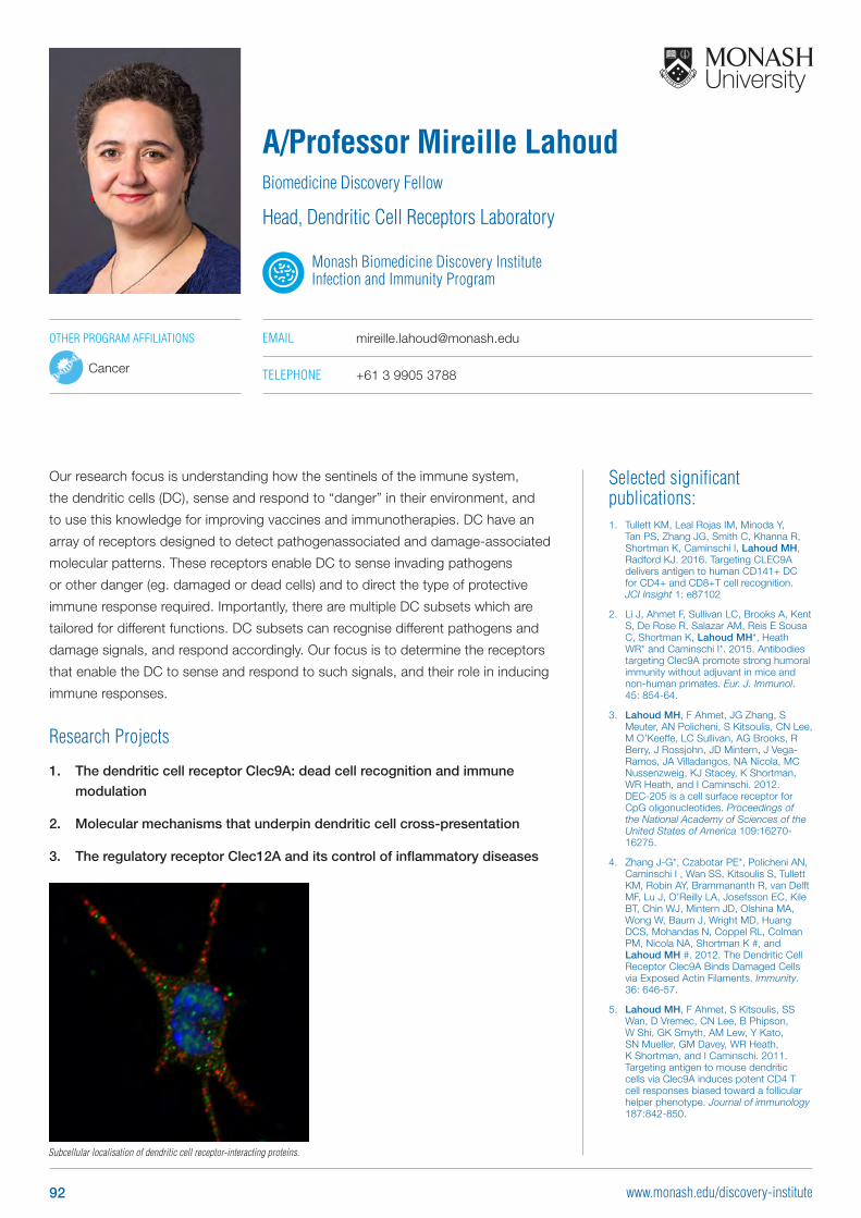
Selected significant publications: 1. Tullett KM, Leal Rojas IM, Minoda Y,
Tan PS, Zhang JG, Smith C, Khanna R, Shortman K, Caminschi I, Lahoud MH, Radford KJ. 2016. Targeting CLEC9A delivers antigen to human CD141+ DC for CD4+ and CD8+T cell recognition. JCI Insight 1: e87102
2. Li J, Ahmet F, Sullivan LC, Brooks A, Kent S, De Rose R, Salazar AM, Reis E Sousa C, Shortman K, Lahoud MH*, Heath WR* and Caminschi I*. 2015. Antibodies targeting Clec9A promote strong humoral immunity without adjuvant in mice and non-human primates. Eur. J. Immunol. 45: 854-64.
3. Lahoud MH, F Ahmet, JG Zhang, S Meuter, AN Policheni, S Kitsoulis, CN Lee, M O’Keeffe, LC Sullivan, AG Brooks, R Berry, J Rossjohn, JD Mintern, J Vega-Ramos, JA Villadangos, NA Nicola, MC Nussenzweig, KJ Stacey, K Shortman, WR Heath, and I Caminschi. 2012. DEC-205 is a cell surface receptor for CpG oligonucleotides. Proceedings of the National Academy of Sciences of the United States of America 109:16270-16275.
4. Zhang J-G*, Czabotar PE*, Policheni AN, Caminschi I , Wan SS, Kitsoulis S, Tullett KM, Robin AY, Brammananth R, van Delft MF, Lu J, O’Reilly LA, Josefsson EC, Kile BT, Chin WJ, Mintern JD, Olshina MA, Wong W, Baum J, Wright MD, Huang DCS, Mohandas N, Coppel RL, Colman PM, Nicola NA, Shortman K #, and Lahoud MH #. 2012. The Dendritic Cell Receptor Clec9A Binds Damaged Cells via Exposed Actin Filaments. Immunity. 36: 646-57.
5. Lahoud MH, F Ahmet, S Kitsoulis, SS Wan, D Vremec, CN Lee, B Phipson, W Shi, GK Smyth, AM Lew, Y Kato, SN Mueller, GM Davey, WR Heath, K Shortman, and I Caminschi. 2011. Targeting antigen to mouse dendritic cells via Clec9A induces potent CD4 T cell responses biased toward a follicular helper phenotype. Journal of immunology 187:842-850.
Our research focus is understanding how the sentinels of the immune system,
the dendritic cells (DC), sense and respond to “danger” in their environment, and
to use this knowledge for improving vaccines and immunotherapies. DC have an
array of receptors designed to detect pathogenassociated and damage-associated
molecular patterns. These receptors enable DC to sense invading pathogens
or other danger (eg. damaged or dead cells) and to direct the type of protective
immune response required. Importantly, there are multiple DC subsets which are
tailored for different functions. DC subsets can recognise different pathogens and
damage signals, and respond accordingly. Our focus is to determine the receptors
that enable the DC to sense and respond to such signals, and their role in inducing
immune responses.
Research Projects
1. The dendritic cell receptor Clec9A: dead cell recognition and immune
modulation
2. Molecular mechanisms that underpin dendritic cell cross-presentation
3. The regulatory receptor Clec12A and its control of inflammatory diseases
A/Professor Mireille LahoudBiomedicine Discovery Fellow
Head, Dendritic Cell Receptors Laboratory
Monash Biomedicine Discovery Institute Infection and Immunity Program
EMAIL [email protected]
TELEPHONE +61 3 9905 3788
Subcellular localisation of dendritic cell receptor-interacting proteins.
OTHER PROGRAM AFFILIATIONS
Cancer
92 www.monash.edu/discovery-institute
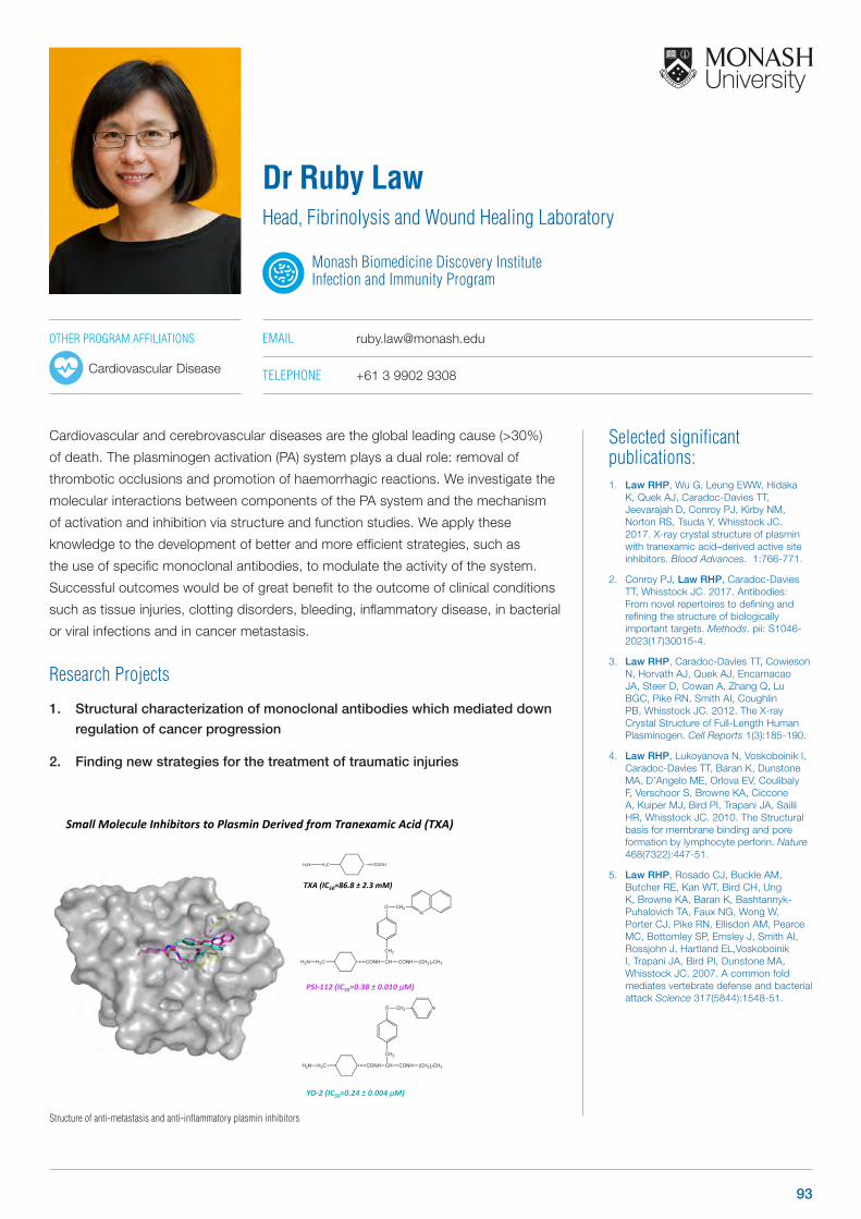
Cardiovascular and cerebrovascular diseases are the global leading cause (>30%)
of death. The plasminogen activation (PA) system plays a dual role: removal of
thrombotic occlusions and promotion of haemorrhagic reactions. We investigate the
molecular interactions between components of the PA system and the mechanism
of activation and inhibition via structure and function studies. We apply these
knowledge to the development of better and more efficient strategies, such as
the use of specific monoclonal antibodies, to modulate the activity of the system.
Successful outcomes would be of great benefit to the outcome of clinical conditions
such as tissue injuries, clotting disorders, bleeding, inflammatory disease, in bacterial
or viral infections and in cancer metastasis.
Research Projects
1. Structural characterization of monoclonal antibodies which mediated down
regulation of cancer progression
2. Finding new strategies for the treatment of traumatic injuries
OTHER PROGRAM AFFILIATIONS
Cardiovascular Disease
Selected significant publications: 1. Law RHP, Wu G, Leung EWW, Hidaka
K, Quek AJ, Caradoc-Davies TT, Jeevarajah D, Conroy PJ, Kirby NM, Norton RS, Tsuda Y, Whisstock JC. 2017. X-ray crystal structure of plasmin with tranexamic acid–derived active site inhibitors. Blood Advances. 1:766-771.
2. Conroy PJ, Law RHP, Caradoc-Davies TT, Whisstock JC. 2017. Antibodies: From novel repertoires to defining and refining the structure of biologically important targets. Methods. pii: S1046-2023(17)30015-4.
3. Law RHP, Caradoc-Davies TT, Cowieson N, Horvath AJ, Quek AJ, Encarnacao JA, Steer D, Cowan A, Zhang Q, Lu BGC, Pike RN, Smith AI, Coughlin PB, Whisstock JC. 2012. The X-ray Crystal Structure of Full-Length Human Plasminogen. Cell Reports 1(3):185-190.
4. Law RHP, Lukoyanova N, Voskoboinik I, Caradoc-Davies TT, Baran K, Dunstone MA, D’Angelo ME, Orlova EV, Coulibaly F, Verschoor S, Browne KA, Ciccone A, Kuiper MJ, Bird PI, Trapani JA, Sailil HR, Whisstock JC. 2010. The Structural basis for membrane binding and pore formation by lymphocyte perforin. Nature 468(7322):447-51.
5. Law RHP, Rosado CJ, Buckle AM, Butcher RE, Kan WT, Bird CH, Ung K, Browne KA, Baran K, Bashtannyk-Puhalovich TA, Faux NG, Wong W, Porter CJ, Pike RN, Ellisdon AM, Pearce MC, Bottomley SP, Emsley J, Smith AI, Rossjohn J, Hartland EL,Voskoboinik I, Trapani JA, Bird PI, Dunstone MA, Whisstock JC. 2007. A common fold mediates vertebrate defense and bacterial attack Science 317(5844):1548-51.
Dr Ruby LawHead, Fibrinolysis and Wound Healing Laboratory
Monash Biomedicine Discovery Institute Infection and Immunity Program
EMAIL [email protected]
TELEPHONE +61 3 9902 9308
Structure of anti-metastasis and anti-inflammatory plasmin inhibitors
H2CH2N COOH
SmallMoleculeInhibitorstoPlasminDerivedfrom TranexamicAcid(TXA)
TXA(IC50=86.8± 2.3mM)
YO-2(IC50=0.24± 0.004µM)
PSI-112(IC50=0.38± 0.010µM)
93
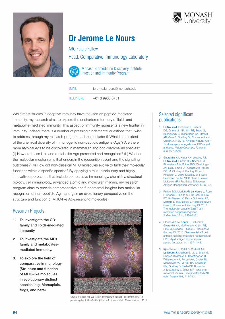
Selected significant publications: 1. Le Nours J, Praveena T, Pellicci
DG, Gherardin NA, Lim RT, Besra G, Keshipeddy S, Richardson SK, Howell AR, Gras S, Godfrey DI, Rossjohn J and Uldrich A. P. 2016. Atypical Natural Killer T-cell receptor recognition of CD1d-lipid antigens. Nature Commun. 7, article number 10570.
2. Gherardin NA, Keller AN, Woolley RE, Le Nours J, Ritchie DS, Neeson PJ, Birkinshaw RW, Eckle SBG, Waddington JN, Liu L, Fairlie DP, Uldrich AP, Pellicci DG, McCluskey J, Godfrey DI, and Rossjohn J. 2016. Diversity of T Cells Restricted by the MHC Class I-Related Molecule MR1 Facilitates Differential Antigen Recognition. Immunity 44, 32-45.
3. Pellicci DG, Uldrich AP, Le Nours J, Ross F, Chabrol E, Eckle SB, de Boer R, Lim RT, McPherson K, Besra G, Howell AR, Moretta L, McCluskey J, Heemskerk MH, Gras S, Rossjohn J, Godfrey DI. 2014. The molecular bases of δ/αβ T cell-mediated antigen recognition. J. Exp. Med. 211, 2599-615.
4. Uldrich AP, Le Nours J, Pellicci DG, Gherardin NA, McPherson K, Lim RT, Patel O, Beddoe T, Gras S, Rossjohn J, Godfrey DI. 2013. Gamma-delta T cell antigen receptor mediated recognition of CD1d-lipid antigen lipid complex. Nature Immunol. 14, 1137-1145.
5. Kjer-Nielsen L, Patel O, Corbett AJ, Le Nours J, Meehan B, Liu L, Bhati M, Chen Z, Kostenko L, Reantragoon R, Williamson NA, Purcell AW, Dudek NL, McConville MJ, O’Hair RA, Khairallah GN, Godfrey DI Fairlie DP, Rossjohn J, McCluskey J. 2012. MR1 presents microbial vitamin B metabolites to MAIT cells. Nature 491, 717-723.
While most studies in adaptive immunity have focused on peptide-mediated
immunity, my research aims to explore the unchartered territory of lipid- and
metabolite-mediated immunity. This aspect of immunity represents a new frontier in
immunity. Indeed, there is a number of pressing fundamental questions that I wish
to address through my research program and that include: (i) What is the extent
of the chemical diversity of immunogenic non-peptidic antigens (Ags)? Are there
more atypical Ags to be discovered in mammalian and non-mammalian species?
(ii) How are these lipid and metabolite Ags presented and recognized? (iii) What are
the molecular mechanisms that underpin the recognition event and the signalling
outcomes? (iv) How did non-classical MHC molecules evolve to fulfill their molecular
functions within a specific species? By applying a multi-disciplinary and highly
innovative approaches that include comparative immunology, chemistry, structural
biology, cell immunology, advanced atomic and molecular imaging, my research
program aims to provide comprehensive and fundamental insights into molecular
recognition of non-peptidic Ags, and gain an evolutionary perspective on the
structure and function of MHC-like Ag-presenting molecules.
Dr Jerome Le NoursARC Future Fellow
Head, Comparative Immunology Laboratory
Monash Biomedicine Discovery Institute Infection and Immunity Program
EMAIL [email protected]
TELEPHONE +61 3 9905 0751
Crystal structure of a γδ TCR in complex with the MHC-like molecule CD1d presenting the lipid α-GalCer (Uldrich & Le Nours et al., Nature Immunol., 2013)
Research Projects
1. To investigate the CD1
family and lipids-mediated
immunity.
2. To investigate the MR1
family and metabolites-
mediated immunity.
3. To explore the field of
comparative immunology
(Structure and function
of MHC-like molecules
in evolutionary distinct
species, e.g. Marsupials,
frogs, and bats).
94 www.monash.edu/discovery-institute

Selected significant publications: 1. Maifiah MH, Cheah SE, Johnson MD,
Han ML, Boyce JD, Thamlikitkul V, Forrest A, Kaye KS, Hertzog P, Purcell AW, Song J, Velkov T, Creek DJ, Li J. 2016. Global metabolic analyses identify key differences in metabolite levels between polymyxin-susceptible and polymyxin-resistant Acinetobacter baumannii. Sci Rep 6: 22287
2. Cheah SE, Johnson MD, Zhu Y, Tsuji BT, Forrest A, Bulitta JB, Boyce JD, Nation RL, Li J. 2016. Polymyxin Resistance in Acinetobacter baumannii: Genetic Mutations and Transcriptomic Changes in Response to Clinically Relevant Dosage Regimens. Sci Rep 6: 26233
3. Azad MA, Roberts KD, Yu HH, Liu B, Schofield AV, James SA, Howard DL, Nation RL, Rogers K, de Jonge MD, Thompson PE, Fu J, Velkov T, Li J. 2015. Significant accumulation of polymyxin in single renal tubular cells: a medicinal chemistry and triple correlative microscopy approach. Anal Chem 87: 1590-5
4. Zavascki AP, Goldani LZ, Cao G, Superti SV, Lutz L, Barth AL, Ramos F, Boniatti MM, Nation RL, Li J. 2008. Pharmacokinetics of intravenous polymyxin B in critically ill patients. Clin Infect Dis 47: 1298-304
5. Li J, Nation RL, Turnidge JD, Milne RW, Coulthard K, Rayner CR, Paterson DL. 2006. Colistin: the re-emerging antibiotic for multidrug-resistant Gram-negative bacterial infections. Lancet Infect Dis 6: 589-601
Professor Jian LiNHMRC Principal Research Fellow
Head, Antimicrobial Systems Pharmacology Laboratory
Monash Biomedicine Discovery Institute Infection and Immunity Program
EMAIL [email protected]
TELEPHONE +61 3 9903 9702
WEB monash.edu/pharm/research/areas/drug-delivery/labs/li-lab
Virtual cell modelling for bacteria.
Discovery of novel safer polymyxins.
Antibiotics are a cornerstone of modern medicine and over the last century have significantly decreased mortality worldwide. Unfortunately, resistance to these ‘magic bullets’ has become one of the greatest threats to human health that the world faces, now and in the coming decades. If proactive solutions are not found to prevent widespread antibiotic resistance, it is estimated that by 2050 ~10 million people per year will die of infections. The World Health Organization (WHO) has urged all government sectors and society to act on antimicrobial resistance (AMR). On 27 February 2017, multidrug-resistant K. pneumoniae, P. aeruginosa and A. baumannii were identified by WHO as the highest priority pathogens, which require urgent attention for the discovery of novel antibiotics. Over the last decade, ‘old’ polymyxins are increasingly used as the last defence against these Gram-negative ‘superbugs’. My research in the systems pharmacology of polymyxins and drug discovery has made a significant contribution to the global commitment to combat antibiotic resistance in Gram-negative pathogens.
Research ProjectsAs no new antibiotics will be available for Gram-negative ‘superbugs’ in the near future, it is crucial to optimise the clinical use of polymyxins and develop novel, safer polymyxins. My major research programs are:
1. Optimising clinical use of polymyxins and their synergistic combinations using pharmacokinetics/pharmacodynamics/toxicodynamics (PK/PD/TD) and systems pharmacology
2. Mechanisms of antibacterial activity, resistance, nephrotoxicity, pulmonary toxicity, and neurotoxicity of polymyxins using a multi-disciplinary approach
3. Development of virtual bacterial cells using systems pharmacology and computational biology
4. Discovery of new-generation polymyxins against multidrug-resistant P. aeruginosa, A. baumannii and K. pneumoniae
95
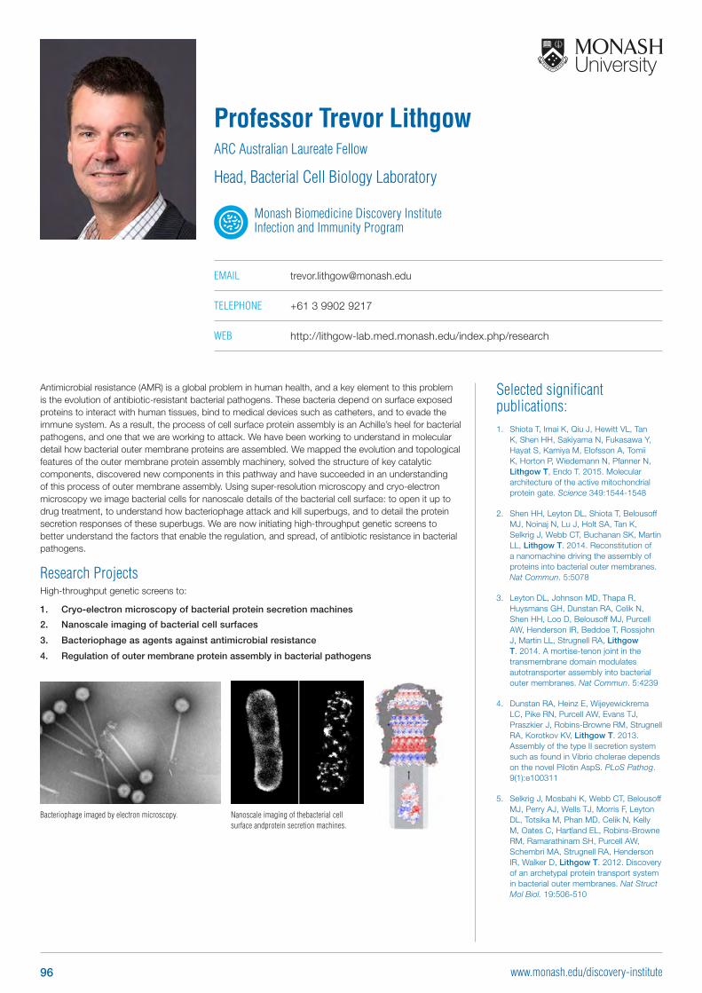
Bacteriophage imaged by electron microscopy.
Selected significant publications: 1. Shiota T, Imai K, Qiu J, Hewitt VL, Tan
K, Shen HH, Sakiyama N, Fukasawa Y, Hayat S, Kamiya M, Elofsson A, Tomii K, Horton P, Wiedemann N, Pfanner N, Lithgow T, Endo T. 2015. Molecular architecture of the active mitochondrial protein gate. Science 349:1544-1548
2. Shen HH, Leyton DL, Shiota T, Belousoff MJ, Noinaj N, Lu J, Holt SA, Tan K, Selkrig J, Webb CT, Buchanan SK, Martin LL, Lithgow T. 2014. Reconstitution of a nanomachine driving the assembly of proteins into bacterial outer membranes. Nat Commun. 5:5078
3. Leyton DL, Johnson MD, Thapa R, Huysmans GH, Dunstan RA, Celik N, Shen HH, Loo D, Belousoff MJ, Purcell AW, Henderson IR, Beddoe T, Rossjohn J, Martin LL, Strugnell RA, Lithgow T. 2014. A mortise-tenon joint in the transmembrane domain modulates autotransporter assembly into bacterial outer membranes. Nat Commun. 5:4239
4. Dunstan RA, Heinz E, Wijeyewickrema LC, Pike RN, Purcell AW, Evans TJ, Praszkier J, Robins-Browne RM, Strugnell RA, Korotkov KV, Lithgow T. 2013. Assembly of the type II secretion system such as found in Vibrio cholerae depends on the novel Pilotin AspS. PLoS Pathog. 9(1):e100311
5. Selkrig J, Mosbahi K, Webb CT, Belousoff MJ, Perry AJ, Wells TJ, Morris F, Leyton DL, Totsika M, Phan MD, Celik N, Kelly M, Oates C, Hartland EL, Robins-Browne RM, Ramarathinam SH, Purcell AW, Schembri MA, Strugnell RA, Henderson IR, Walker D, Lithgow T. 2012. Discovery of an archetypal protein transport system in bacterial outer membranes. Nat Struct Mol Biol. 19:506-510
Antimicrobial resistance (AMR) is a global problem in human health, and a key element to this problem is the evolution of antibiotic-resistant bacterial pathogens. These bacteria depend on surface exposed proteins to interact with human tissues, bind to medical devices such as catheters, and to evade the immune system. As a result, the process of cell surface protein assembly is an Achille’s heel for bacterial pathogens, and one that we are working to attack. We have been working to understand in molecular detail how bacterial outer membrane proteins are assembled. We mapped the evolution and topological features of the outer membrane protein assembly machinery, solved the structure of key catalytic components, discovered new components in this pathway and have succeeded in an understanding of this process of outer membrane assembly. Using super-resolution microscopy and cryo-electron microscopy we image bacterial cells for nanoscale details of the bacterial cell surface: to open it up to drug treatment, to understand how bacteriophage attack and kill superbugs, and to detail the protein secretion responses of these superbugs. We are now initiating high-throughput genetic screens to better understand the factors that enable the regulation, and spread, of antibiotic resistance in bacterial pathogens.
Research ProjectsHigh-throughput genetic screens to:
1. Cryo-electron microscopy of bacterial protein secretion machines
2. Nanoscale imaging of bacterial cell surfaces
3. Bacteriophage as agents against antimicrobial resistance
4. Regulation of outer membrane protein assembly in bacterial pathogens
Professor Trevor LithgowARC Australian Laureate Fellow
Head, Bacterial Cell Biology Laboratory
Monash Biomedicine Discovery Institute Infection and Immunity Program
EMAIL [email protected]
TELEPHONE +61 3 9902 9217
WEB http://lithgow-lab.med.monash.edu/index.php/research
Nanoscale imaging of thebacterial cell surface andprotein secretion machines.
96 www.monash.edu/discovery-institute
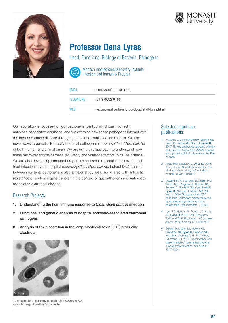
Selected significant publications: 1. Hutton ML, Cunningham BA, Mackin KE,
Lyon SA, James ML, Rood JI, Lyras D. 2017. Bovine antibodies targeting primary and recurrent Clostridium difficile disease are a potent antibiotic alternative. Sci Rep 7: 3665.
2. Awad MM, Singleton J, Lyras D. 2016. The Sialidase NanS Enhances Non-TcsL Mediated Cytotoxicity of Clostridium sordellii. Toxins (Basel) 8.
3. Cowardin CA, Buonomo EL, Saleh MM, Wilson MG, Burgess SL, Kuehne SA, Schwan C, Eichhoff AM, Koch-Nolte F, Lyras D, Aktories K, Minton NP, Petri WA, Jr. 2016 The binary toxin CDT enhances Clostridium difficile virulence by suppressing protective colonic eosinophilia. Nat Microbiol 1: 16108
4. Lyon SA, Hutton ML, Rood JI, Cheung JK, Lyras D. 2016. CdtR Regulates TcdA and TcdB Production in Clostridium difficile. PLoS Pathog 12: e1005758.
5. Stanley D, Mason LJ, Mackin KE, Srikhanta YN, Lyras D, Prakash MD, Nurgali K, Venegas A, Hill MD, Moore RJ, Wong CH. 2016. Translocation and dissemination of commensal bacteria in post-stroke infection. Nat Med 22: 1277-1284
Our laboratory is focussed on gut pathogens, particularly those involved in
antibiotic-associated diarrhoea, and we examine how these pathogens interact with
the host and cause disease through the use of animal infection models. We use
novel ways to genetically modify bacterial pathogens (including Clostridium difficile)
of both human and animal origin. We are using this approach to understand how
these micro-organisms harness regulatory and virulence factors to cause disease.
We are also developing immunotherapeutics and small molecules to prevent and
treat infections by the hospital superbug Clostridium difficile. Lateral DNA transfer
between bacterial pathogens is also a major study area, associated with antibiotic
resistance or virulence gene transfer in the context of gut pathogens and antibiotic-
associated diarrhoeal disease.
Research Projects
1. Understanding the host immune response to Clostridium difficile infection
2. Functional and genetic analysis of hospital antibiotic-associated diarrhoeal
pathogens
3. Analysis of toxin secretion in the large clostridial toxin (LCT) producing
clostridia
Professor Dena LyrasHead, Functional Biology of Bacterial Pathogens
Monash Biomedicine Discovery Institute Infection and Immunity Program
EMAIL [email protected]
TELEPHONE +61 3 9902 9155
WEB med.monash.edu/microbiology/staff/lyras.html
Transmission electron microscopy on a section of a Clostridium difficile spore within a vegetative cell (Dr Yogi Srikhanta).
97
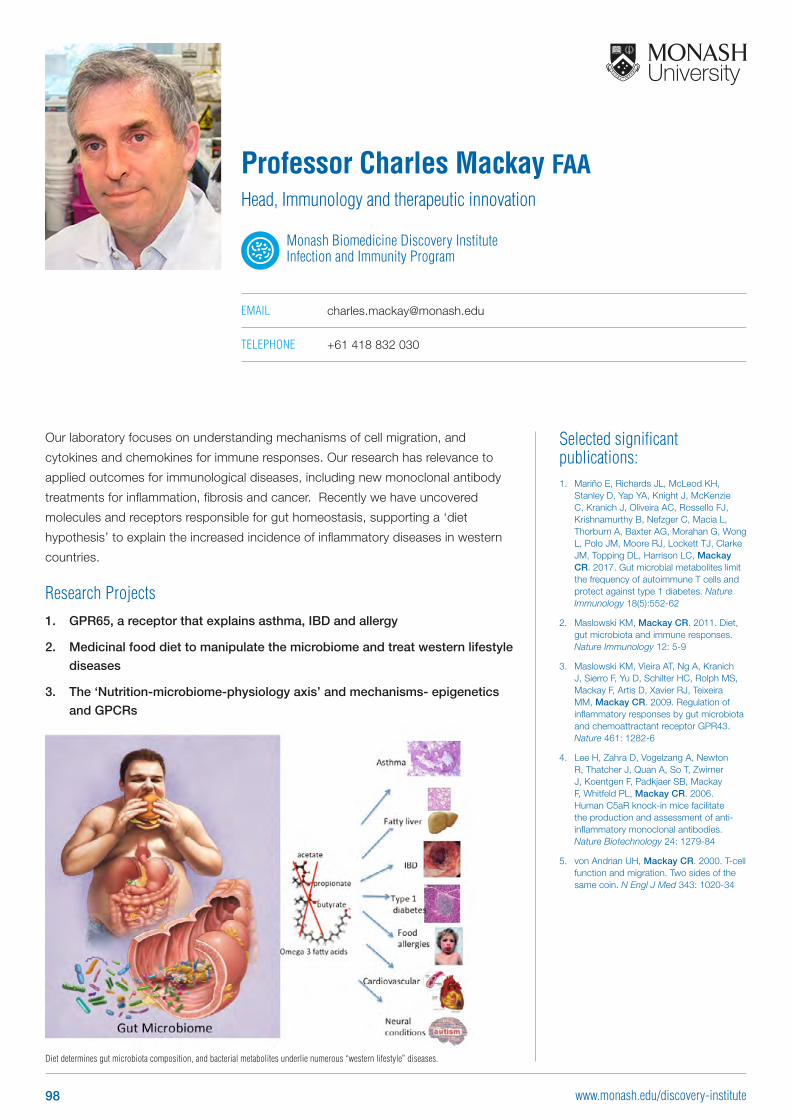
Diet determines gut microbiota composition, and bacterial metabolites underlie numerous “western lifestyle” diseases.
Selected significant publications: 1. Mariño E, Richards JL, McLeod KH,
Stanley D, Yap YA, Knight J, McKenzie C, Kranich J, Oliveira AC, Rossello FJ, Krishnamurthy B, Nefzger C, Macia L, Thorburn A, Baxter AG, Morahan G, Wong L, Polo JM, Moore RJ, Lockett TJ, Clarke JM, Topping DL, Harrison LC, Mackay CR. 2017. Gut microbial metabolites limit the frequency of autoimmune T cells and protect against type 1 diabetes. Nature Immunology 18(5):552-62
2. Maslowski KM, Mackay CR. 2011. Diet, gut microbiota and immune responses. Nature Immunology 12: 5-9
3. Maslowski KM, Vieira AT, Ng A, Kranich J, Sierro F, Yu D, Schilter HC, Rolph MS, Mackay F, Artis D, Xavier RJ, Teixeira MM, Mackay CR. 2009. Regulation of inflammatory responses by gut microbiota and chemoattractant receptor GPR43. Nature 461: 1282-6
4. Lee H, Zahra D, Vogelzang A, Newton R, Thatcher J, Quan A, So T, Zwirner J, Koentgen F, Padkjaer SB, Mackay F, Whitfeld PL, Mackay CR. 2006. Human C5aR knock-in mice facilitate the production and assessment of anti-inflammatory monoclonal antibodies. Nature Biotechnology 24: 1279-84
5. von Andrian UH, Mackay CR. 2000. T-cell function and migration. Two sides of the same coin. N Engl J Med 343: 1020-34
Our laboratory focuses on understanding mechanisms of cell migration, and
cytokines and chemokines for immune responses. Our research has relevance to
applied outcomes for immunological diseases, including new monoclonal antibody
treatments for inflammation, fibrosis and cancer. Recently we have uncovered
molecules and receptors responsible for gut homeostasis, supporting a ‘diet
hypothesis’ to explain the increased incidence of inflammatory diseases in western
countries.
Research Projects1. GPR65, a receptor that explains asthma, IBD and allergy
2. Medicinal food diet to manipulate the microbiome and treat western lifestyle
diseases
3. The ‘Nutrition-microbiome-physiology axis’ and mechanisms- epigenetics
and GPCRs
Professor Charles Mackay FAAHead, Immunology and therapeutic innovation
Monash Biomedicine Discovery Institute Infection and Immunity Program
EMAIL [email protected]
TELEPHONE +61 418 832 030
98 www.monash.edu/discovery-institute
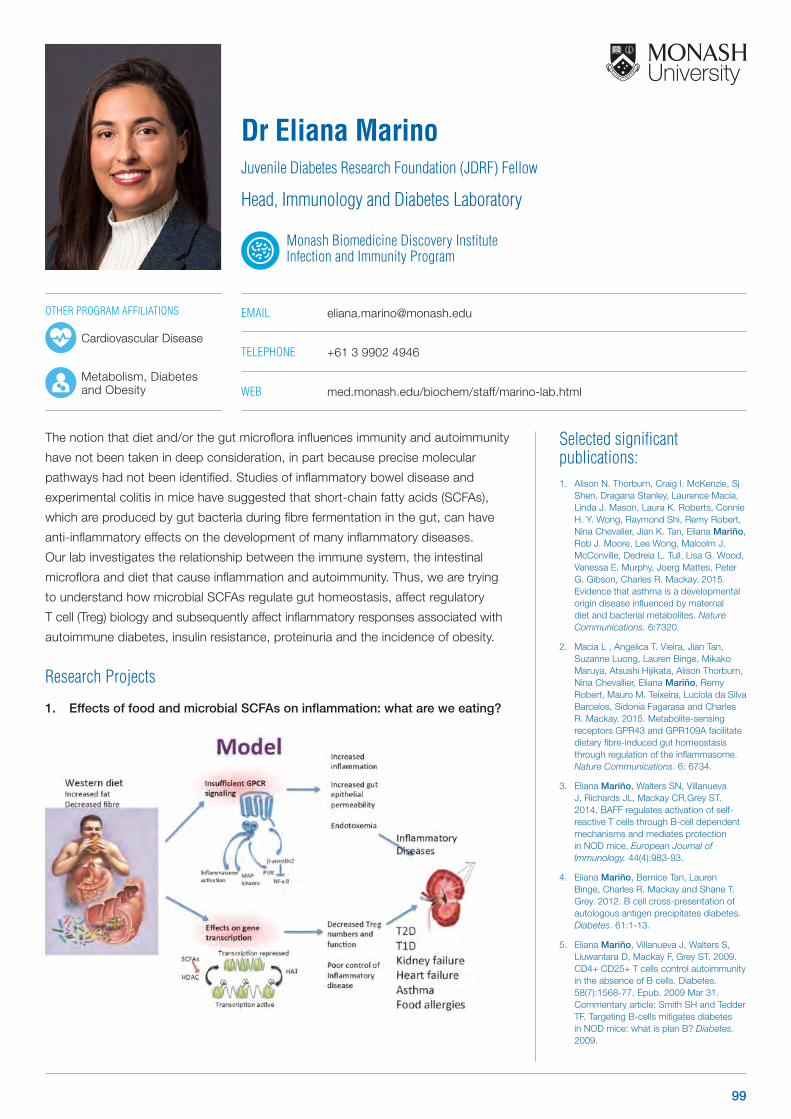
Selected significant publications: 1. Alison N. Thorburn, Craig I. McKenzie, Sj
Shen, Dragana Stanley, Laurence Macia, Linda J. Mason, Laura K. Roberts, Connie H. Y. Wong, Raymond Shi, Remy Robert, Nina Chevalier, Jian K. Tan, Eliana Mariño, Rob J. Moore, Lee Wong, Malcolm J. McConville, Dedreia L. Tull, Lisa G. Wood, Vanessa E. Murphy, Joerg Mattes, Peter G. Gibson, Charles R. Mackay. 2015. Evidence that asthma is a developmental origin disease influenced by maternal diet and bacterial metabolites. Nature Communications. 6:7320.
2. Macia L , Angelica T. Vieira, Jian Tan, Suzanne Luong, Lauren Binge, Mikako Maruya, Atsushi Hijikata, Alison Thorburn, Nina Chevallier, Eliana Mariño, Remy Robert, Mauro M. Teixeira, Lucíola da Silva Barcelos, Sidonia Fagarasa and Charles R. Mackay. 2015. Metabolite-sensing receptors GPR43 and GPR109A facilitate dietary fibre-induced gut homeostasis through regulation of the inflammasome. Nature Communications. 6: 6734.
3. Eliana Mariño, Walters SN, Villanueva J, Richards JL, Mackay CR,Grey ST. 2014. BAFF regulates activation of self-reactive T cells through B-cell dependent mechanisms and mediates protection in NOD mice. European Journal of Immunology. 44(4):983-93.
4. Eliana Mariño, Bernice Tan, Lauren Binge, Charles R. Mackay and Shane T. Grey. 2012. B cell cross-presentation of autologous antigen precipitates diabetes. Diabetes. 61:1-13.
5. Eliana Mariño, Villanueva J, Walters S, Liuwantara D, Mackay F, Grey ST. 2009. CD4+ CD25+ T cells control autoimmunity in the absence of B cells. Diabetes. 58(7):1568-77. Epub. 2009 Mar 31. Commentary article: Smith SH and Tedder TF. Targeting B-cells mitigates diabetes in NOD mice: what is plan B? Diabetes. 2009.
The notion that diet and/or the gut microflora influences immunity and autoimmunity
have not been taken in deep consideration, in part because precise molecular
pathways had not been identified. Studies of inflammatory bowel disease and
experimental colitis in mice have suggested that short-chain fatty acids (SCFAs),
which are produced by gut bacteria during fibre fermentation in the gut, can have
anti-inflammatory effects on the development of many inflammatory diseases.
Our lab investigates the relationship between the immune system, the intestinal
microflora and diet that cause inflammation and autoimmunity. Thus, we are trying
to understand how microbial SCFAs regulate gut homeostasis, affect regulatory
T cell (Treg) biology and subsequently affect inflammatory responses associated with
autoimmune diabetes, insulin resistance, proteinuria and the incidence of obesity.
Research Projects
1. Effects of food and microbial SCFAs on inflammation: what are we eating?
Dr Eliana MarinoJuvenile Diabetes Research Foundation (JDRF) Fellow
Head, Immunology and Diabetes Laboratory
Monash Biomedicine Discovery Institute Infection and Immunity Program
EMAIL [email protected]
TELEPHONE +61 3 9902 4946
WEB med.monash.edu/biochem/staff/marino-lab.html
OTHER PROGRAM AFFILIATIONS
Cardiovascular Disease
Metabolism, Diabetes and Obesity
99
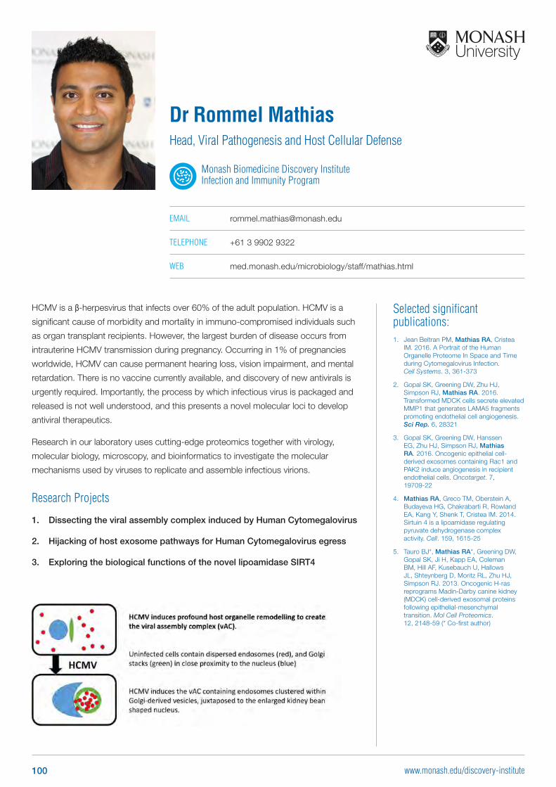
Selected significant publications: 1. Jean Beltran PM, Mathias RA, Cristea
IM. 2016. A Portrait of the Human Organelle Proteome In Space and Time during Cytomegalovirus Infection. Cell Systems. 3, 361-373
2. Gopal SK, Greening DW, Zhu HJ, Simpson RJ, Mathias RA. 2016. Transformed MDCK cells secrete elevated MMP1 that generates LAMA5 fragments promoting endothelial cell angiogenesis. Sci Rep. 6, 28321
3. Gopal SK, Greening DW, Hanssen EG, Zhu HJ, Simpson RJ, Mathias RA. 2016. Oncogenic epithelial cell-derived exosomes containing Rac1 and PAK2 induce angiogenesis in recipient endothelial cells. Oncotarget. 7, 19709-22
4. Mathias RA, Greco TM, Oberstein A, Budayeva HG, Chakrabarti R, Rowland EA, Kang Y, Shenk T, Cristea IM. 2014. Sirtuin 4 is a lipoamidase regulating pyruvate dehydrogenase complex activity. Cell. 159, 1615-25
5. Tauro BJ*, Mathias RA*, Greening DW, Gopal SK, Ji H, Kapp EA, Coleman BM, Hill AF, Kusebauch U, Hallows JL, Shteynberg D, Moritz RL, Zhu HJ, Simpson RJ. 2013. Oncogenic H-ras reprograms Madin-Darby canine kidney (MDCK) cell-derived exosomal proteins following epithelial-mesenchymal transition. Mol Cell Proteomics. 12, 2148-59 (* Co-first author)
Dr Rommel Mathias Head, Viral Pathogenesis and Host Cellular Defense
Monash Biomedicine Discovery Institute Infection and Immunity Program
EMAIL [email protected]
TELEPHONE +61 3 9902 9322
WEB med.monash.edu/microbiology/staff/mathias.html
HCMV is a β-herpesvirus that infects over 60% of the adult population. HCMV is a
significant cause of morbidity and mortality in immuno-compromised individuals such
as organ transplant recipients. However, the largest burden of disease occurs from
intrauterine HCMV transmission during pregnancy. Occurring in 1% of pregnancies
worldwide, HCMV can cause permanent hearing loss, vision impairment, and mental
retardation. There is no vaccine currently available, and discovery of new antivirals is
urgently required. Importantly, the process by which infectious virus is packaged and
released is not well understood, and this presents a novel molecular loci to develop
antiviral therapeutics.
Research in our laboratory uses cutting-edge proteomics together with virology,
molecular biology, microscopy, and bioinformatics to investigate the molecular
mechanisms used by viruses to replicate and assemble infectious virions.
Research Projects
1. Dissecting the viral assembly complex induced by Human Cytomegalovirus
2. Hijacking of host exosome pathways for Human Cytomegalovirus egress
3. Exploring the biological functions of the novel lipoamidase SIRT4
100 www.monash.edu/discovery-institute
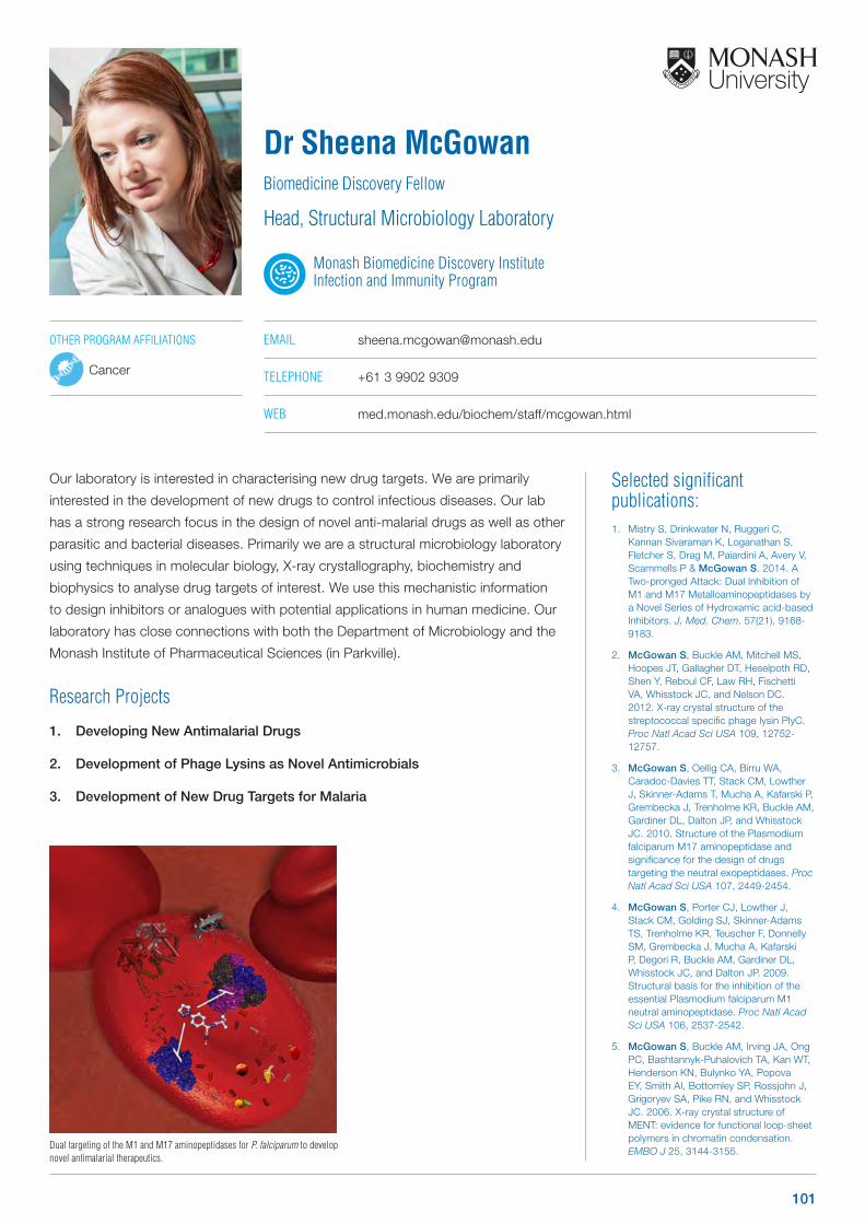
Selected significant publications: 1. Mistry S, Drinkwater N, Ruggeri C,
Kannan Sivaraman K, Loganathan S, Fletcher S, Drag M, Paiardini A, Avery V, Scammells P & McGowan S. 2014. A Two-pronged Attack: Dual Inhibition of M1 and M17 Metalloaminopeptidases by a Novel Series of Hydroxamic acid-based Inhibitors. J. Med. Chem. 57(21), 9168-9183.
2. McGowan S, Buckle AM, Mitchell MS, Hoopes JT, Gallagher DT, Heselpoth RD, Shen Y, Reboul CF, Law RH, Fischetti VA, Whisstock JC, and Nelson DC. 2012. X-ray crystal structure of the streptococcal specific phage lysin PlyC. Proc Natl Acad Sci USA 109, 12752-12757.
3. McGowan S, Oellig CA, Birru WA, Caradoc-Davies TT, Stack CM, Lowther J, Skinner-Adams T, Mucha A, Kafarski P, Grembecka J, Trenholme KR, Buckle AM, Gardiner DL, Dalton JP, and Whisstock JC. 2010. Structure of the Plasmodium falciparum M17 aminopeptidase and significance for the design of drugs targeting the neutral exopeptidases. Proc Natl Acad Sci USA 107, 2449-2454.
4. McGowan S, Porter CJ, Lowther J, Stack CM, Golding SJ, Skinner-Adams TS, Trenholme KR, Teuscher F, Donnelly SM, Grembecka J, Mucha A, Kafarski P, Degori R, Buckle AM, Gardiner DL, Whisstock JC, and Dalton JP. 2009. Structural basis for the inhibition of the essential Plasmodium falciparum M1 neutral aminopeptidase. Proc Natl Acad Sci USA 106, 2537-2542.
5. McGowan S, Buckle AM, Irving JA, Ong PC, Bashtannyk-Puhalovich TA, Kan WT, Henderson KN, Bulynko YA, Popova EY, Smith AI, Bottomley SP, Rossjohn J, Grigoryev SA, Pike RN, and Whisstock JC. 2006. X-ray crystal structure of MENT: evidence for functional loop-sheet polymers in chromatin condensation. EMBO J 25, 3144-3155.
Our laboratory is interested in characterising new drug targets. We are primarily
interested in the development of new drugs to control infectious diseases. Our lab
has a strong research focus in the design of novel anti-malarial drugs as well as other
parasitic and bacterial diseases. Primarily we are a structural microbiology laboratory
using techniques in molecular biology, X-ray crystallography, biochemistry and
biophysics to analyse drug targets of interest. We use this mechanistic information
to design inhibitors or analogues with potential applications in human medicine. Our
laboratory has close connections with both the Department of Microbiology and the
Monash Institute of Pharmaceutical Sciences (in Parkville).
Research Projects
1. Developing New Antimalarial Drugs
2. Development of Phage Lysins as Novel Antimicrobials
3. Development of New Drug Targets for Malaria
Dr Sheena McGowanBiomedicine Discovery Fellow
Head, Structural Microbiology Laboratory
Monash Biomedicine Discovery Institute Infection and Immunity Program
EMAIL [email protected]
TELEPHONE +61 3 9902 9309
WEB med.monash.edu/biochem/staff/mcgowan.html
Dual targeting of the M1 and M17 aminopeptidases for P. falciparum to develop novel antimalarial therapeutics.
OTHER PROGRAM AFFILIATIONS
Cancer
101
Selected significant publications: 1. Dando SJ, Naranjo-Golborne C, Chinnery,
HR, Ruitenberg, MJ, McMenamin PG. 2016. A case of mistaken identity: CD11c-eYFP+ cells in the normal mouse brain parenchyma and neural retina display the phenotype of microglia, not dendritic cells. Glia doi: 10.1002/ glia.23005 [Epub ahead of print].
2. Lioufas PL, Quayle MR, Leong JC, McMenamin PG. 2016. 3D printed models of cleft palate pathology for surgical education. J Plastic Reconstructive Surgery (in Press).
3. Chinnery HR, Leong J, Forrester JV, McMenamin PG. 2015. TLR9 and TLR7/8 activation induces formation of keratic precipitates and giant macrophages in the mouse cornea. J Leuk Biol 97(1):103-10.
4. McMenamin PG, Quayle MR, McHenry CR, Adams JW. 2014. The production of anatomical teaching resources using three-dimensional (3D) printing technology. Anat Sci Educ. 2014 7: 479-86
5. Chinnery HR, Pearlman E, McMenamin PG. 2008. Membrane nanotubes in vivo: A unique morphological feature of MHC class II+ macrophages and putative DCs in the mouse corneal stroma. ‘Cutting Edge’ J Immunol. 180:5779-5783.
Our group is investigating a number of issues that relate to eye diseases which may
have an immune or inflammatory basis to their pathogenesis. In addition, the group
also studies eye development and aging. The World Health Organization estimates
that over 45 M people are blind and 314 M people are visually impaired. The Ocular
Immunology group has a number of active projects that are aimed at understanding
common blinding diseases such as corneal infection, retinopathy of prematurity, and
age-related macular degeneration. The research group sees itself as part of a larger
global effort to understand these conditions - if we can help place one small piece in
the larger jigsaw puzzle of medical research that will help future treatment of these
conditions then we will have succeeded.
Research Projects
1. Are there antigen presenting cells in
the normal brain parenchyma and
retina?
2. Novel treatment protocols for
human retinopathy of prematurity
using a clinically relevant mouse
model
3. Investigating perivascular
macrophages in the ocular
microenvironment
Professor Paul McMenaminHead, Ocular Immunology Group
Monash Biomedicine Discovery Institute Infection and Immunity Program
EMAIL [email protected]
TELEPHONE +61 3 9905 6215
WEB med.monash.edu/anatomy/research/ocular-immunology.html
Prof. Paul McMenamin with 3D Anatomy Prints.
OTHER PROGRAM AFFILIATIONS
Development and Stem Cells
Neuroscience
102 www.monash.edu/discovery-institute
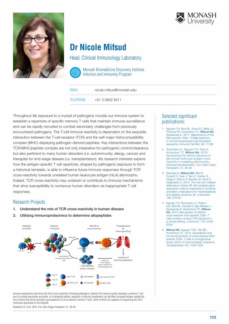
Selected significant publications: 1. Nguyen TH, Bird NL, Grant EJ, Miles JJ,
Thomas PG, Kotsimbos TC, Mifsud NA, Kedzierska K. 2017. Maintenance of the EBV-specific CD8+ TCRαβ repertoire in immunosuppressed lung transplant recipients. Immunol Cell Biol. 95, 77-86
2. Rowntree LC, Nguyen TH, Gras S, Kotsimbos TC, Mifsud NA. 2016. Deciphering the clinical relevance of allo-human leukocyte antigen cross-reactivity in mediating alloimmunity following transplantation. Curr Opin Organ Transplant. 21, 29-39
3. Banerjee A, Mifsud NA, Bird R, Forsyth C, Szer J, Tam C, Kellner S, Grigg A, Motum P, Bentley M, Opat S, Grigoriadis G. 2015. The oral iron chelator deferasirox inhibits NF-κB mediated gene expression without impacting on proximal activation: implications for myelodysplasia and aplastic anaemia. Br J Haematol. 168, 576-82
4. Nguyen TH, Rowntree LC, Pellicci DG, Bird NL, Handel A, Kjer-Nielsen L, Kedzierska K, Kotsimbos TC, Mifsud NA. 2014. Recognition of distinct cross-reactive virus-specific CD8+ T cells reveals a unique TCR signature in a clinical setting. J Immunol. 192, 5039-5049
5. Mifsud NA, Nguyen THO, Tait BD, Kotsimbos TC. 2010. Quantitative and functional diversity of cross-reactive EBV-specific CD8+ T cells in a longitudinal study cohort of lung transplant recipients. Transplantation 90, 1439-1449
Throughout life exposure to a myriad of pathogens moulds our immune system to establish a repertoire of specific memory T cells that maintain immune-surveillance and can be rapidly recruited to combat secondary challenges from previously encountered pathogens. The T-cell immune reactivity is dependent on the exquisite interaction between the T-cell receptor (TCR) and the self-major histocompatibility complex (MHC) displaying pathogen-derived peptides. Key interactions between the TCR/MHC/peptide complex are not only imperative for pathogenic control/clearance but also pertinent to many human disorders (i.e. autoimmunity, allergy, cancer) and therapies for end-stage disease (i.e. transplantation). My research interests explore how the antigen-specific T cell repertoire, shaped by pathogenic exposure to form a historical template, is able to influence future immune responses through TCR cross-reactivity towards unrelated human leukocyte antigen (HLA) allomorphs. Indeed, TCR cross-reactivity may underpin or contribute to immune mechanisms that drive susceptibility to numerous human disorders via inappropriate T cell
responses.
Research Projects1. Understand the role of TCR cross-reactivity in human disease
2. Utilising immunoproteomics to determine allopeptides
Dr Nicole MifsudHead, Clinical Immunology Laboratory
Monash Biomedicine Discovery Institute Infection and Immunity Program
EMAIL [email protected]
TELEPHONE +61 3 9902 9311
Immune mechanisms that drive allo-HLA cross-reactivity. Following pathogenic infection the immune system develops a memory T-cell pool to combat secondary encounter. In a transplant setting, cessation of antiviral prophylaxis can facilitate increased antigen availability (via viremia) that drives activation and expansion of virus-specific memory T cells, some of which are capable of recognising allo-HLA molecules expressed on the allograft.
Rowntree LC, et al. 2016. Curr Opin Organ Transplant. 21, 29-39.
Pathogenencounter
Memory T cell pool
Transplantation&
tissue specificity
Allo-HLAcross-reactivity
viral infection(active viremia)
reactivation ofvirus-specific memory T
cells cessation of
antiviral prophylaxis
expansion
viral T cell
allo-HLA cell
virus A
virus B
virus Callo-peptide
viral-peptide cross-reactive TCR
ineffectiveT cell response
under conditions of immunosuppression
allo-HLA TCR
103
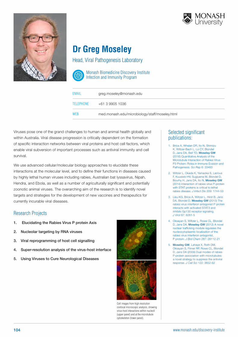
Cell images from high resolution confocal microscopic analysis, showing virus-host interactions within nucleoli (upper panel) and at the microtubule cytoskeleton (lower panel).
Selected significant publications: 1. Brice A, Whelan DR, Ito N, Shimizu
K, Wiltzer-Bach L, Lo CY, Blondel D, Jans DA, Bell TD, Moseley GW (2016) Quantitative Analysis of the Microtubule Interaction of Rabies Virus P3 Protein: Roles in Immune Evasion and Pathogenesis. Sci Rep 6: 33493
2. Wiltzer L, Okada K, Yamaoka S, Larrous F, Kuusisto HV, Sugiyama M, Blondel D, Bourhy H, Jans DA, Ito N, Moseley GW (2014) Interaction of rabies virus P-protein with STAT proteins is critical to lethal rabies disease. J Infect Dis 209: 1744-53
3. Lieu KG, Brice A, Wiltzer L, Hirst B, Jans DA, Blondel D, Moseley GW (2013) The rabies virus interferon antagonist P protein interacts with activated STAT3 and inhibits Gp130 receptor signaling. J Virol 87: 8261-5
4. Oksayan S, Wiltzer L, Rowe CL, Blondel D, Jans DA, Moseley GW (2012) A novel nuclear trafficking module regulates the nucleocytoplasmic localization of the rabies virus interferon antagonist, P protein. J Biol Chem 287: 28112-21
5. Moseley GW, Lahaye X, Roth DM, Oksayan S, Filmer RP, Rowe CL, Blondel D, Jans DA (2009) Dual modes of rabies P-protein association with microtubules: a novel strategy to suppress the antiviral response. J Cell Sci 122: 3652-62
Viruses pose one of the grand challenges to human and animal health globally and
within Australia. Viral disease progression is critically dependent on the formation
of specific interaction networks between viral proteins and host cell factors, which
enable viral subversion of important processes such as antiviral immunity and cell
survival.
We use advanced cellular/molecular biology approaches to elucidate these
interactions at the molecular level, and to define their functions in diseases caused
by highly lethal human viruses including rabies, Australian bat lyssavirus, Nipah,
Hendra, and Ebola, as well as a number of agriculturally significant and potentially
zoonotic animal viruses. The overarching aim of the research is to identify novel
targets and strategies for the development of new vaccines and therapeutics for
currently incurable viral diseases.
Research Projects
1. Elucidating the Rabies Virus P protein Axis
2. Nucleolar targeting by RNA viruses
3. Viral reprogramming of host cell signalling
4. Super-resolution analysis of the virus-host interface
5. Using Viruses to Cure Neurological Diseases
Dr Greg MoseleyHead, Viral Pathogenesis Laboratory
Monash Biomedicine Discovery Institute Infection and Immunity Program
EMAIL [email protected]
TELEPHONE +61 3 9905 1036
WEB med.monash.edu/microbiology/staff/moseley.html
104 www.monash.edu/discovery-institute
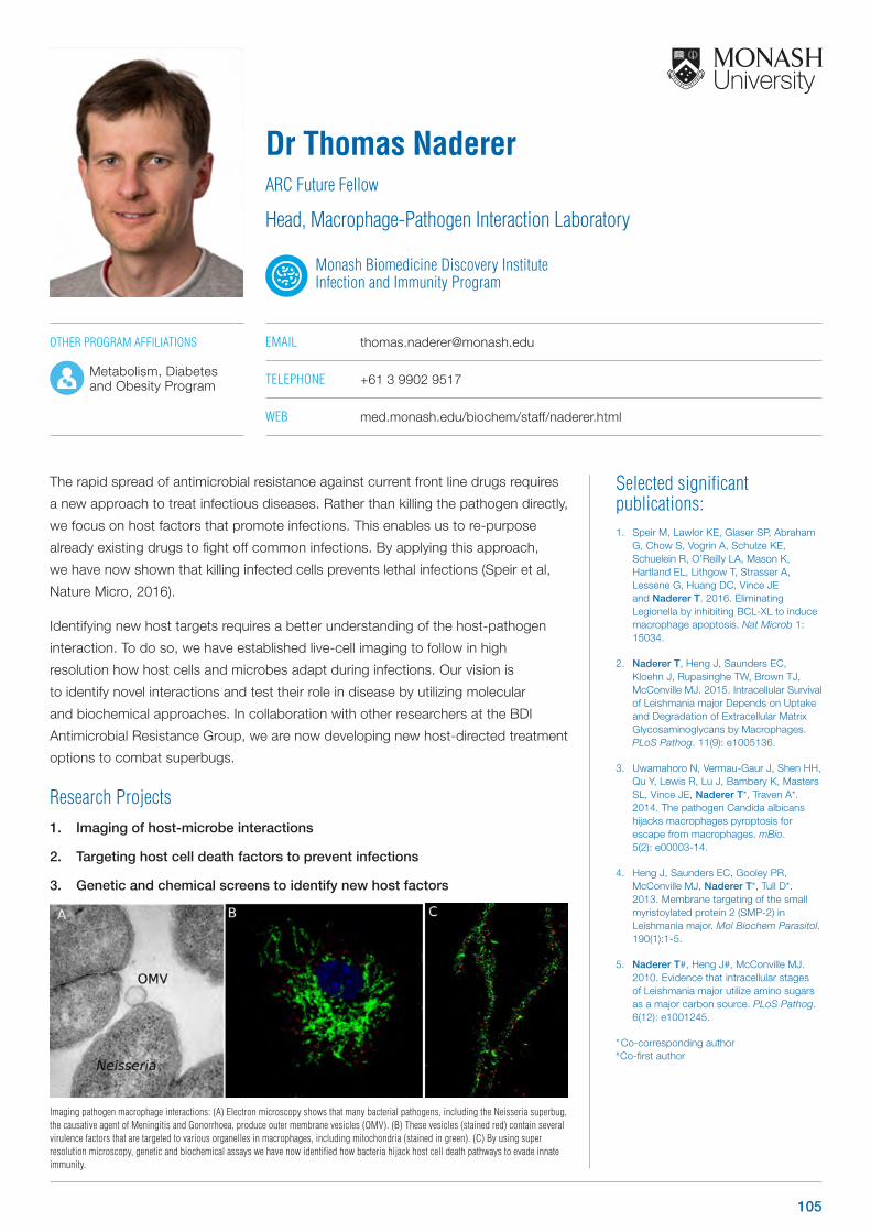
Selected significant publications: 1. Speir M, Lawlor KE, Glaser SP, Abraham
G, Chow S, Vogrin A, Schulze KE, Schuelein R, O’Reilly LA, Mason K, Hartland EL, Lithgow T, Strasser A, Lessene G, Huang DC, Vince JE and Naderer T. 2016. Eliminating Legionella by inhibiting BCL-XL to induce macrophage apoptosis. Nat Microb 1: 15034.
2. Naderer T, Heng J, Saunders EC, Kloehn J, Rupasinghe TW, Brown TJ, McConville MJ. 2015. Intracellular Survival of Leishmania major Depends on Uptake and Degradation of Extracellular Matrix Glycosaminoglycans by Macrophages. PLoS Pathog. 11(9): e1005136.
3. Uwamahoro N, Vermau-Gaur J, Shen HH, Qu Y, Lewis R, Lu J, Bambery K, Masters SL, Vince JE, Naderer T*, Traven A*. 2014. The pathogen Candida albicans hijacks macrophages pyroptosis for escape from macrophages. mBio. 5(2): e00003-14.
4. Heng J, Saunders EC, Gooley PR, McConville MJ, Naderer T*, Tull D*. 2013. Membrane targeting of the small myristoylated protein 2 (SMP-2) in Leishmania major. Mol Biochem Parasitol. 190(1):1-5.
5. Naderer T#, Heng J#, McConville MJ. 2010. Evidence that intracellular stages of Leishmania major utilize amino sugars as a major carbon source. PLoS Pathog. 6(12): e1001245.
* Co-corresponding author # Co-first author
The rapid spread of antimicrobial resistance against current front line drugs requires
a new approach to treat infectious diseases. Rather than killing the pathogen directly,
we focus on host factors that promote infections. This enables us to re-purpose
already existing drugs to fight off common infections. By applying this approach,
we have now shown that killing infected cells prevents lethal infections (Speir et al,
Nature Micro, 2016).
Identifying new host targets requires a better understanding of the host-pathogen
interaction. To do so, we have established live-cell imaging to follow in high
resolution how host cells and microbes adapt during infections. Our vision is
to identify novel interactions and test their role in disease by utilizing molecular
and biochemical approaches. In collaboration with other researchers at the BDI
Antimicrobial Resistance Group, we are now developing new host-directed treatment
options to combat superbugs.
Research Projects1. Imaging of host-microbe interactions
2. Targeting host cell death factors to prevent infections
3. Genetic and chemical screens to identify new host factors
Dr Thomas NadererARC Future Fellow
Head, Macrophage-Pathogen Interaction Laboratory
Monash Biomedicine Discovery Institute Infection and Immunity Program
EMAIL [email protected]
TELEPHONE +61 3 9902 9517
WEB med.monash.edu/biochem/staff/naderer.html
Imaging pathogen macrophage interactions: (A) Electron microscopy shows that many bacterial pathogens, including the Neisseria superbug, the causative agent of Meningitis and Gonorrhoea, produce outer membrane vesicles (OMV). (B) These vesicles (stained red) contain several virulence factors that are targeted to various organelles in macrophages, including mitochondria (stained in green). (C) By using super resolution microscopy, genetic and biochemical assays we have now identified how bacteria hijack host cell death pathways to evade innate immunity.
OTHER PROGRAM AFFILIATIONS
Metabolism, Diabetes and Obesity Program
105
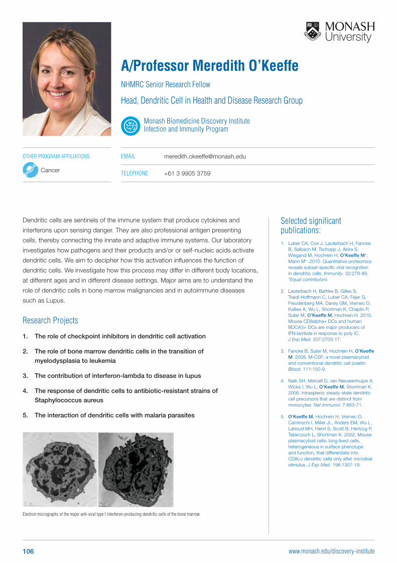
Electron micrographs of the major anti-viral type I interferon-producing dendritic cells of the bone marrow.
Selected significant publications: 1. Luber CA, Cox J, Lauterbach H, Fancke
B, Selbach M, Tschopp J, Akira S, Wiegand M, Hochrein H, O’Keeffe M*, Mann M* .2010. Quantitative proteomics reveals subset-specific viral recognition in dendritic cells. Immunity. 32:279-89. *Equal contributors
2. Lauterbach H, Bathke B, Gilles S, Traidl-Hoffmann C, Luber CA, Fejer G, Freudenberg MA, Davey GM, Vremec D, Kallies A, Wu L, Shortman K, Chaplin P, Suter M, O’Keeffe M, Hochrein H. 2010. Mouse CD8alpha+ DCs and human BDCA3+ DCs are major producers of IFN-lambda in response to poly IC. J Exp Med. 207:2703-17.
3. Fancke B, Suter M, Hochrein H, O’Keeffe M. 2008. M-CSF: a novel plasmacytoid and conventional dendritic cell poietin. Blood. 111:150-9.
4. Naik SH, Metcalf D, van Nieuwenhuijze A, Wicks I, Wu L, O’Keeffe M, Shortman K. 2006. Intrasplenic steady-state dendritic cell precursors that are distinct from monocytes. Nat Immunol. 7:663-71.
5. O’Keeffe M, Hochrein H, Vremec D, Caminschi I, Miller JL, Anders EM, Wu L, Lahoud MH, Henri S, Scott B, Hertzog P, Tatarczuch L, Shortman K. 2002. Mouse plasmacytoid cells: long-lived cells, heterogeneous in surface phenotype and function, that differentiate into CD8(+) dendritic cells only after microbial stimulus. J Exp Med. 196:1307-19.
Dendritic cells are sentinels of the immune system that produce cytokines and
interferons upon sensing danger. They are also professional antigen presenting
cells, thereby connecting the innate and adaptive immune systems. Our laboratory
investigates how pathogens and their products and/or or self-nucleic acids activate
dendritic cells. We aim to decipher how this activation influences the function of
dendritic cells. We investigate how this process may differ in different body locations,
at different ages and in different disease settings. Major aims are to understand the
role of dendritic cells in bone marrow malignancies and in autoimmune diseases
such as Lupus.
Research Projects
1. The role of checkpoint inhibitors in dendritic cell activation
2. The role of bone marrow dendritic cells in the transition of
myelodysplasia to leukemia
3. The contribution of interferon-lambda to disease in lupus
4. The response of dendritic cells to antibiotic-resistant strains of
Staphylococcus aureus
5. The interaction of dendritic cells with malaria parasites
A/Professor Meredith O’KeeffeNHMRC Senior Research Fellow
Head, Dendritic Cell in Health and Disease Research Group
Monash Biomedicine Discovery Institute Infection and Immunity Program
EMAIL [email protected]
TELEPHONE +61 3 9905 3759
OTHER PROGRAM AFFILIATIONS
Cancer
106 www.monash.edu/discovery-institute
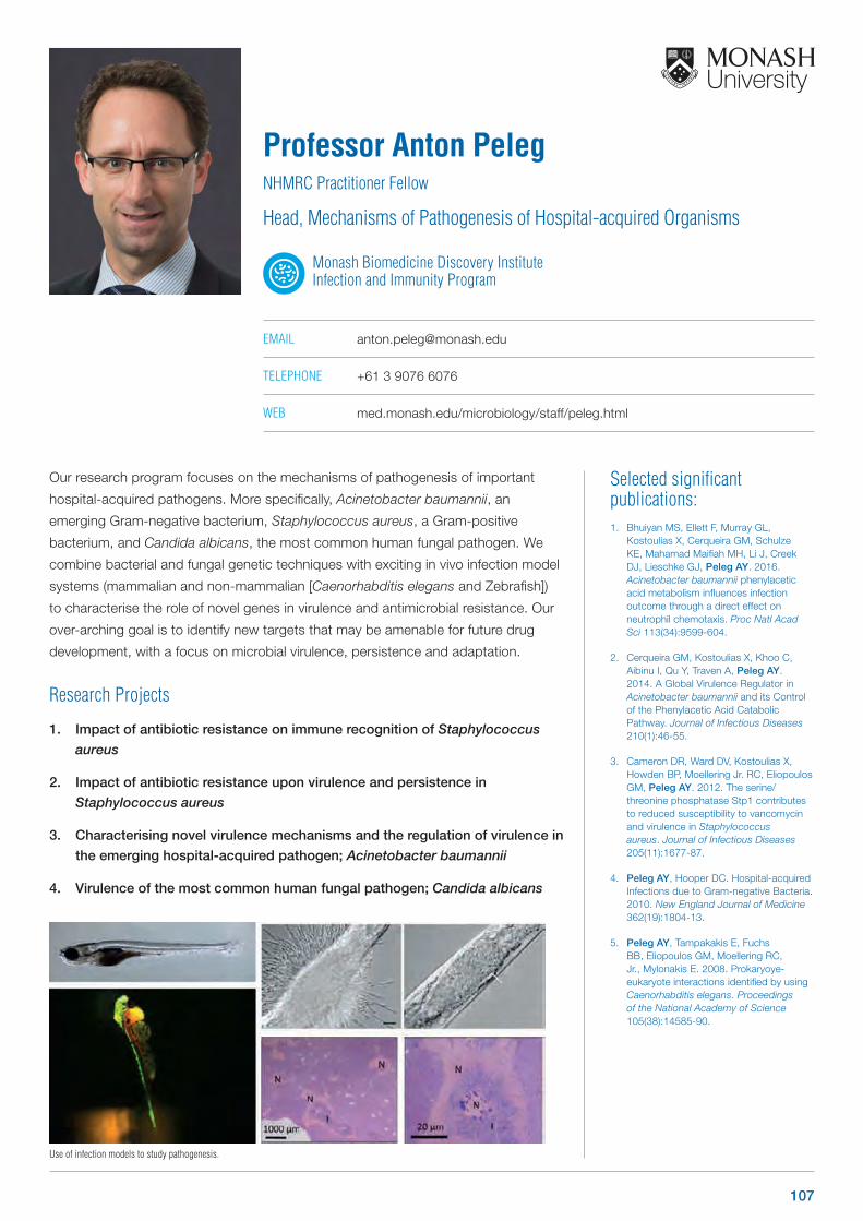
Selected significant publications: 1. Bhuiyan MS, Ellett F, Murray GL,
Kostoulias X, Cerqueira GM, Schulze KE, Mahamad Maifiah MH, Li J, Creek DJ, Lieschke GJ, Peleg AY. 2016.Acinetobacter baumannii phenylacetic acid metabolism influences infection outcome through a direct effect on neutrophil chemotaxis. Proc Natl Acad Sci 113(34):9599-604.
2. Cerqueira GM, Kostoulias X, Khoo C, Aibinu I, Qu Y, Traven A, Peleg AY. 2014. A Global Virulence Regulator in Acinetobacter baumannii and its Control of the Phenylacetic Acid Catabolic Pathway. Journal of Infectious Diseases 210(1):46-55.
3. Cameron DR, Ward DV, Kostoulias X, Howden BP, Moellering Jr. RC, Eliopoulos GM, Peleg AY. 2012. The serine/threonine phosphatase Stp1 contributes to reduced susceptibility to vancomycin and virulence in Staphylococcus aureus. Journal of Infectious Diseases 205(11):1677-87.
4. Peleg AY, Hooper DC. Hospital-acquired Infections due to Gram-negative Bacteria. 2010. New England Journal of Medicine 362(19):1804-13.
5. Peleg AY, Tampakakis E, Fuchs BB, Eliopoulos GM, Moellering RC, Jr., Mylonakis E. 2008. Prokaryoye-eukaryote interactions identified by using Caenorhabditis elegans. Proceedings of the National Academy of Science 105(38):14585-90.
Our research program focuses on the mechanisms of pathogenesis of important
hospital-acquired pathogens. More specifically, Acinetobacter baumannii, an
emerging Gram-negative bacterium, Staphylococcus aureus, a Gram-positive
bacterium, and Candida albicans, the most common human fungal pathogen. We
combine bacterial and fungal genetic techniques with exciting in vivo infection model
systems (mammalian and non-mammalian [Caenorhabditis elegans and Zebrafish])
to characterise the role of novel genes in virulence and antimicrobial resistance. Our
over-arching goal is to identify new targets that may be amenable for future drug
development, with a focus on microbial virulence, persistence and adaptation.
Research Projects
1. Impact of antibiotic resistance on immune recognition of Staphylococcus
aureus
2. Impact of antibiotic resistance upon virulence and persistence in
Staphylococcus aureus
3. Characterising novel virulence mechanisms and the regulation of virulence in
the emerging hospital-acquired pathogen; Acinetobacter baumannii
4. Virulence of the most common human fungal pathogen; Candida albicans
Professor Anton PelegNHMRC Practitioner Fellow
Head, Mechanisms of Pathogenesis of Hospital-acquired Organisms
Monash Biomedicine Discovery Institute Infection and Immunity Program
EMAIL [email protected]
TELEPHONE +61 3 9076 6076
WEB med.monash.edu/microbiology/staff/peleg.html
Use of infection models to study pathogenesis.
107
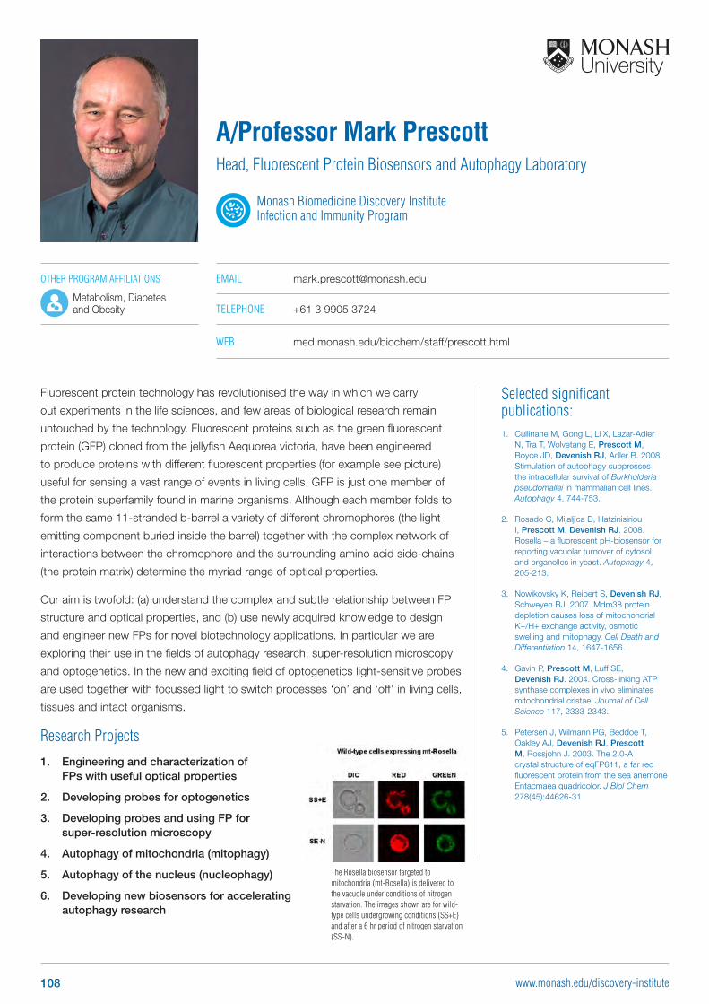
Selected significant publications: 1. Cullinane M, Gong L, Li X, Lazar-Adler
N, Tra T, Wolvetang E, Prescott M, Boyce JD, Devenish RJ, Adler B. 2008. Stimulation of autophagy suppresses the intracellular survival of Burkholderia pseudomallei in mammalian cell lines. Autophagy 4, 744-753.
2. Rosado C, Mijaljica D, Hatzinisiriou I, Prescott M, Devenish RJ. 2008. Rosella – a fluorescent pH-biosensor for reporting vacuolar turnover of cytosol and organelles in yeast. Autophagy 4, 205-213.
3. Nowikovsky K, Reipert S, Devenish RJ, Schweyen RJ. 2007. Mdm38 protein depletion causes loss of mitochondrial K+/H+ exchange activity, osmotic swelling and mitophagy. Cell Death and Differentiation 14, 1647-1656.
4. Gavin P, Prescott M, Luff SE, Devenish RJ. 2004. Cross-linking ATP synthase complexes in vivo eliminates mitochondrial cristae. Journal of Cell Science 117, 2333-2343.
5. Petersen J, Wilmann PG, Beddoe T, Oakley AJ, Devenish RJ, Prescott M, Rossjohn J. 2003. The 2.0-A crystal structure of eqFP611, a far red fluorescent protein from the sea anemone Entacmaea quadricolor. J Biol Chem 278(45):44626-31
Fluorescent protein technology has revolutionised the way in which we carry
out experiments in the life sciences, and few areas of biological research remain
untouched by the technology. Fluorescent proteins such as the green fluorescent
protein (GFP) cloned from the jellyfish Aequorea victoria, have been engineered
to produce proteins with different fluorescent properties (for example see picture)
useful for sensing a vast range of events in living cells. GFP is just one member of
the protein superfamily found in marine organisms. Although each member folds to
form the same 11-stranded b-barrel a variety of different chromophores (the light
emitting component buried inside the barrel) together with the complex network of
interactions between the chromophore and the surrounding amino acid side-chains
(the protein matrix) determine the myriad range of optical properties.
Our aim is twofold: (a) understand the complex and subtle relationship between FP
structure and optical properties, and (b) use newly acquired knowledge to design
and engineer new FPs for novel biotechnology applications. In particular we are
exploring their use in the fields of autophagy research, super-resolution microscopy
and optogenetics. In the new and exciting field of optogenetics light-sensitive probes
are used together with focussed light to switch processes ‘on’ and ‘off’ in living cells,
tissues and intact organisms.
Research Projects
1. Engineering and characterization of FPs with useful optical properties
2. Developing probes for optogenetics
3. Developing probes and using FP for super-resolution microscopy
4. Autophagy of mitochondria (mitophagy)
5. Autophagy of the nucleus (nucleophagy)
6. Developing new biosensors for accelerating autophagy research
A/Professor Mark PrescottHead, Fluorescent Protein Biosensors and Autophagy Laboratory
Monash Biomedicine Discovery Institute Infection and Immunity Program
EMAIL [email protected]
TELEPHONE +61 3 9905 3724
WEB med.monash.edu/biochem/staff/prescott.html
The Rosella biosensor targeted to mitochondria (mt-Rosella) is delivered to the vacuole under conditions of nitrogen starvation. The images shown are for wild-type cells undergrowing conditions (SS+E) and after a 6 hr period of nitrogen starvation (SS-N).
OTHER PROGRAM AFFILIATIONS
Metabolism, Diabetes and Obesity
108 www.monash.edu/discovery-institute
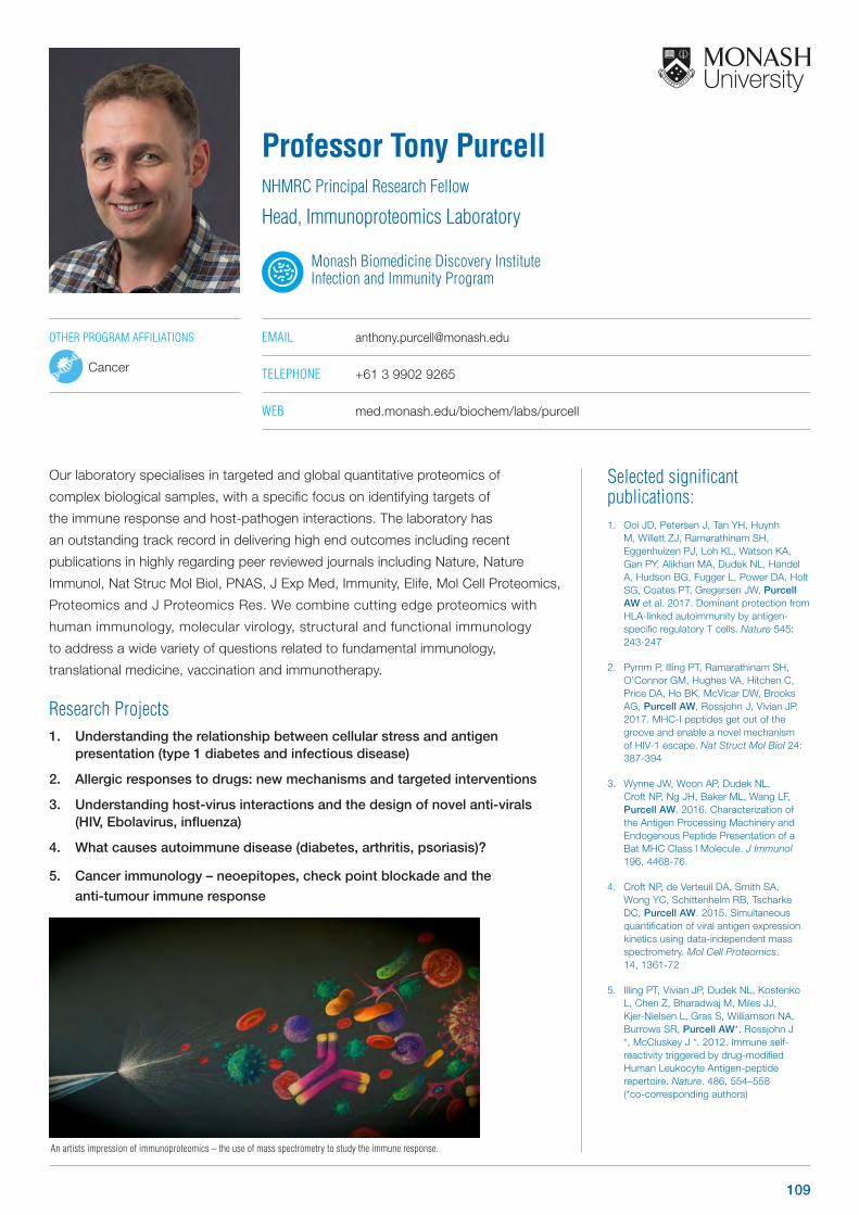
Selected significant publications: 1. Ooi JD, Petersen J, Tan YH, Huynh
M, Willett ZJ, Ramarathinam SH, Eggenhuizen PJ, Loh KL, Watson KA, Gan PY, Alikhan MA, Dudek NL, Handel A, Hudson BG, Fugger L, Power DA, Holt SG, Coates PT, Gregersen JW, Purcell AW et al. 2017. Dominant protection from HLA-linked autoimmunity by antigen-specific regulatory T cells. Nature 545: 243-247
2. Pymm P, Illing PT, Ramarathinam SH, O’Connor GM, Hughes VA, Hitchen C, Price DA, Ho BK, McVicar DW, Brooks AG, Purcell AW, Rossjohn J, Vivian JP. 2017. MHC-I peptides get out of the groove and enable a novel mechanism of HIV-1 escape. Nat Struct Mol Biol 24: 387-394
3. Wynne JW, Woon AP, Dudek NL, Croft NP, Ng JH, Baker ML, Wang LF, Purcell AW. 2016. Characterization of the Antigen Processing Machinery and Endogenous Peptide Presentation of a Bat MHC Class I Molecule. J Immunol 196, 4468-76.
4. Croft NP, de Verteuil DA, Smith SA, Wong YC, Schittenhelm RB, Tscharke DC, Purcell AW. 2015. Simultaneous quantification of viral antigen expression kinetics using data-independent mass spectrometry. Mol Cell Proteomics. 14, 1361-72
5. Illing PT, Vivian JP, Dudek NL, Kostenko L, Chen Z, Bharadwaj M, Miles JJ, Kjer-Nielsen L, Gras S, Williamson NA, Burrows SR, Purcell AW*, Rossjohn J *, McCluskey J *. 2012. Immune self-reactivity triggered by drug-modified Human Leukocyte Antigen-peptide repertoire. Nature. 486, 554–558 (*co-corresponding authors)
Our laboratory specialises in targeted and global quantitative proteomics of
complex biological samples, with a specific focus on identifying targets of
the immune response and host-pathogen interactions. The laboratory has
an outstanding track record in delivering high end outcomes including recent
publications in highly regarding peer reviewed journals including Nature, Nature
Immunol, Nat Struc Mol Biol, PNAS, J Exp Med, Immunity, Elife, Mol Cell Proteomics,
Proteomics and J Proteomics Res. We combine cutting edge proteomics with
human immunology, molecular virology, structural and functional immunology
to address a wide variety of questions related to fundamental immunology,
translational medicine, vaccination and immunotherapy.
Research Projects1. Understanding the relationship between cellular stress and antigen
presentation (type 1 diabetes and infectious disease)
2. Allergic responses to drugs: new mechanisms and targeted interventions
3. Understanding host-virus interactions and the design of novel anti-virals (HIV, Ebolavirus, influenza)
4. What causes autoimmune disease (diabetes, arthritis, psoriasis)?
5. Cancer immunology – neoepitopes, check point blockade and the
anti-tumour immune response
Professor Tony PurcellNHMRC Principal Research Fellow
Head, Immunoproteomics Laboratory
Monash Biomedicine Discovery Institute Infection and Immunity Program
EMAIL [email protected]
TELEPHONE +61 3 9902 9265
WEB med.monash.edu/biochem/labs/purcell
An artists impression of immunoproteomics – the use of mass spectrometry to study the immune response.
OTHER PROGRAM AFFILIATIONS
Cancer
109
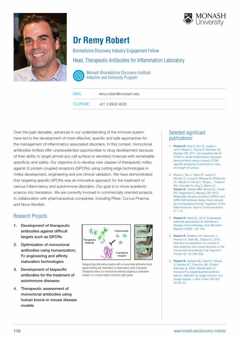
Antagonising chemokine receptor with a monoclonal antibodies block ligand binding and chemotaxis of inflammatory cells (Left panel). Therapeutic effect of a monoclonal antibody targeting a chemokine receptor in a mouse model of arthritis (right panel).
Selected significant publications: 1. Robert R, Ang C, Sun G, Juglair L,
Lim F, Mason L, Payne N, Bernard CA, Mackay CR. 2017. An essential role for CCR6 in certain inflammatory diseases demonstrated using a human CCR6-specific antagonist and knock-in mice. JCI insight (In press).
2. Macia L, Tan J, Vieira AT, Leach K, Stanley D, Luong S, Maruya M, McKenzie CI, Hijikata A, Wong C, Binge L, Thorburn AN, Chevalier N, Ang C, Marino E, Robert R, Teixeira MM, Moore RJ, Flavell RA, Fagarasan S, Mackay CR. 2015. Metabolite-sensing receptors GPR43 and GPR109A facilitate dietary fiber-induced gut homeostasis through regulation of the inflammasome. Nature Communications 6: 1-15.
3. Robert R, Wark KL. 2012. Engineered antibody approaches for Alzheimer’s disease immunotherapy. Arch Biochem Biophys 526(2): 132-138.
4. Robert R, Streltsov VA, Newman J, Pearce LA, Wark KL, Dolezal O. 2010. Germline humanization of a murine A beta antibody and crystal structure of the humanized recombinant Fab fragment. Protein Sci 19: 299-308.
5. Robert R, Jacobin MJ, Daret D, Miraux S, Nurden.A.T, Franconi JM, Clofent- Sanchez G. 2006. Identification of humanscFvs targeting atherosclerotic lesions: selection by single round in vivo phage-display. J. Biol. Chem 281(52): 40135-43.
Over the past decades, advances in our understanding of the immune system
have led to the development of more effective, specific and safe approaches for
the management of inflammatory associated disorders. In this context, monoclonal
antibodies (mAbs) offer unprecedented opportunities to drug development because
of their ability to target almost any cell surface or secreted molecule with remarkable
specificity and safety. Our objective is to develop new classes of therapeutic mAbs
against G protein-coupled receptors (GPCRs) using cutting edge technologies in
mAbs development, engineering and pre-clinical validation. We have demonstrated
that targeting specific GPCRs was an innovative approach for the treatment of
various inflammatory and autoimmune disorders. Our goal is to move academic
science into translation. We are currently involved in commercially oriented projects
in collaboration with pharmaceutical companies, including Pfizer, Corvus Pharma,
and Novo-Nordisk.
Dr Remy RobertBiomedicine Discovery Industry Engagement Fellow
Head, Therapeutic Antibodies for Inflammation Laboratory
Monash Biomedicine Discovery Institute Infection and Immunity Program
EMAIL [email protected]
TELEPHONE +61 3 9902 4635
Research Projects
1. Development of therapeutic
antibodies against difficult
targets such as GPCRs
2. Optimization of monoclonal
antibodies using humanization;
Fc engineering and affinity
maturation technologies
3. Development of bispecific
antibodies for the treatment of
autoimmune diseases
4. Therapeutic assessment of
monoclonal antibodies using
human knock-in mouse disease
models
110 www.monash.edu/discovery-institute
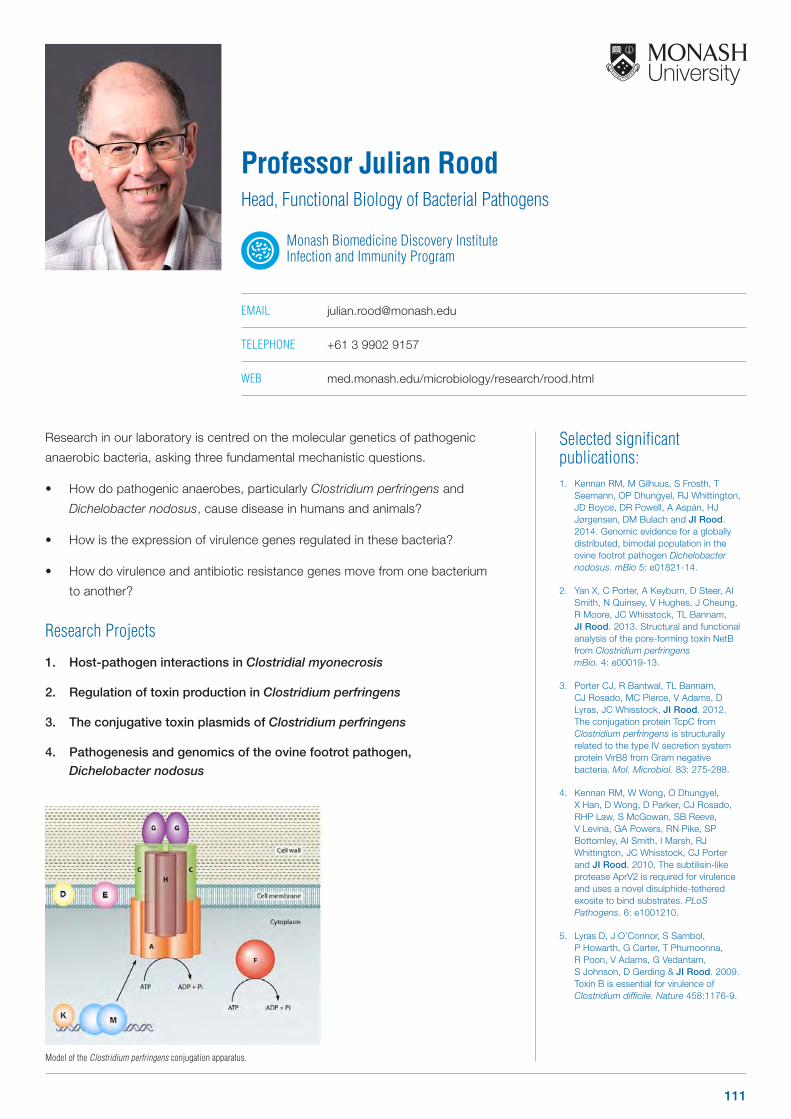
Selected significant publications: 1. Kennan RM, M Gilhuus, S Frosth, T
Seemann, OP Dhungyel, RJ Whittington, JD Boyce, DR Powell, A Aspán, HJ Jørgensen, DM Bulach and JI Rood. 2014. Genomic evidence for a globally distributed, bimodal population in the ovine footrot pathogen Dichelobacter nodosus. mBio 5: e01821-14.
2. Yan X, C Porter, A Keyburn, D Steer, AI Smith, N Quinsey, V Hughes, J Cheung, R Moore, JC Whisstock, TL Bannam, JI Rood. 2013. Structural and functional analysis of the pore-forming toxin NetB from Clostridium perfringens mBio. 4: e00019-13.
3. Porter CJ, R Bantwal, TL Bannam, CJ Rosado, MC Pierce, V Adams, D Lyras, JC Whisstock, JI Rood. 2012. The conjugation protein TcpC from Clostridium perfringens is structurally related to the type IV secretion system protein VirB8 from Gram negative bacteria. Mol. Microbiol. 83: 275-288.
4. Kennan RM, W Wong, O Dhungyel, X Han, D Wong, D Parker, CJ Rosado, RHP Law, S McGowan, SB Reeve, V Levina, GA Powers, RN Pike, SP Bottomley, AI Smith, I Marsh, RJ Whittington, JC Whisstock, CJ Porter and JI Rood. 2010. The subtilisin-like protease AprV2 is required for virulence and uses a novel disulphide-tethered exosite to bind substrates. PLoS Pathogens. 6: e1001210.
5. Lyras D, J O’Connor, S Sambol, P Howarth, G Carter, T Phumoonna, R Poon, V Adams, G Vedantam, S Johnson, D Gerding & JI Rood. 2009. Toxin B is essential for virulence of Clostridium difficile. Nature 458:1176-9.
Research in our laboratory is centred on the molecular genetics of pathogenic
anaerobic bacteria, asking three fundamental mechanistic questions.
• How do pathogenic anaerobes, particularly Clostridium perfringens and
Dichelobacter nodosus, cause disease in humans and animals?
• How is the expression of virulence genes regulated in these bacteria?
• How do virulence and antibiotic resistance genes move from one bacterium
to another?
Research Projects
1. Host-pathogen interactions in Clostridial myonecrosis
2. Regulation of toxin production in Clostridium perfringens
3. The conjugative toxin plasmids of Clostridium perfringens
4. Pathogenesis and genomics of the ovine footrot pathogen,
Dichelobacter nodosus
Professor Julian RoodHead, Functional Biology of Bacterial Pathogens
Monash Biomedicine Discovery Institute Infection and Immunity Program
EMAIL [email protected]
TELEPHONE +61 3 9902 9157
WEB med.monash.edu/microbiology/research/rood.html
Model of the Clostridium perfringens conjugation apparatus.
111

Selected significant publications: 1. Ooi JD, Petersen J, Tan YH, Huynh
M, Willett ZJ, Ramarathinam SH, Eggenhuizen PJ, Loh KL, Watson KA, Gan PY, Alikhan MA, Dudek NL, Handel A, Hudson BG, Fugger L, Power DA, Holt SG, Coates PT, Gregersen JW, Purcell AW, Holdsworth SR, La Gruta NL, Reid HH#, Rossjohn J# and Kitching AR#. 2017. Dominant protection from HLA-linked autoimmunity by antigen-specific regulatory T cells. Nature 545: 243-247
2. Birkinshaw RW, Pellicci DG, Cheng TY, Keller AN, Sandoval-Romero M, Gras S, de Jong A, Uldrich AP, Moody DB, Godfrey DI, Rossjohn J. 2015. alphabeta T cell antigen receptor recognition of CD1a presenting self lipid ligands. Nat Immunol 16: 258-66
3. Kjer-Nielsen L, Patel O, Corbett AJ, Le Nours J, Meehan B, Liu L, Bhati M, Chen Z, Kostenko L, Reantragoon R, Williamson NA, Purcell AW, Dudek NL, McConville MJ, O’Hair RA, Khairallah GN, Godfrey DI, Fairlie DP, Rossjohn# J & McCluskey# J. 2012. MR1 presents microbial vitamin B metabolites to MAIT cells. Nature. 491, 717-723.
4. Illing PT, Vivian JP, Dudek NL, Kostenko L, Chen Z, Bharadwaj M, Miles JJ, Kjer-Nielsen L, Gras S, Williamson NA, Burrows SR, Purcell AW#, Rossjohn# J & McCluskey# J. 2012. Immune self-reactivity triggered by drug-modified Human Leukocyte Antigen peptide repertoire. Nature. 486, 554-558.
5. Vivian JP, Duncan RC, Berry R, O’Connor GM, Reid HH, Beddoe T, Gras S, Saunders PM, Olshina MA, Widjaja JML, Harpur CM, Lin J, Maloveste SM, Price DA, Lafont BAP, McVicar DW, Clements CS, Brooks# AG & Rossjohn# J. 2011. Killer cell immunoglobulin-like receptor 3DL1-mediated recognition of human leukocyte antigen B. Nature. 479, 401-405.
# denotes joint senior author
The academic research program within this laboratory is concerned with defining the key molecular interactions underlying receptor recognition events that are the primary determinants of innate and adaptive immunity.
The laboratory’s research has provided an understanding of the basis of peptide, metabolite and lipid presentation, T-cell triggering, aberrant T-cell reactivity, monomorphic and polymorphic Natural Killer (NK) receptor recognition. The team’s research on anti-viral immunity has provided an understanding of the factors that shape MHC-restriction (e.g. Immunity, 2003, 2016; Nature Immunol, 2005, 2007, 2015). Moreover, we have demonstrated how the pre-TCR, a receptor crucial for T-cell development, functions by autonomous dimerization (Nature, 2010). In relation to aberrant T-cell reactivity, our team has provided insight into alloreactivity (Immunity, 2009), Celiac Disease (Immunity, 2012; NSMB, 2014) and HLA-linked drug hypersensitivities (Nature, 2012, NSMB 2014). Regarding innate and innate-like recognition, the team has shed light into how Natural Killer cell receptors interact with their cognate ligands (Nature 2011; J. Exp. Med. 2008 & 2016; Nature Immunol 2013; NSMB 2017; Cell 2017). Further, we have provided fundamental insight into how T cells recognise lipid-based antigens in the context of protective and aberrant immunity (Nature, 2007; Nature Immunol 2010, 2011, 2012, 2015, 2016; Nature Comms. 2016). Most recently, our team identified the long sought after ligand for MAIT cells, namely showing that MAIT cells are activated by metabolites of vitamin B (Nature 2012, 2014; Nat Commun 2012; Nat Immunol 2016; Nat Immunol, 2017). The industrial research program of the laboratory includes a close collaboration with Janssen (one of the Pharmaceutical companies of Johnson & Johnson), for the development of new therapies to treat
rheumatoid arthritis.
Research Projects1. MHC-restricted protective immunity
2. T-cell autoimmunity and alloreactivity
3. HLA-linked drug hypersensitivities
4. Lipid-mediated immunity
5. Metabolite-mediated immunity
6. NK cell recognition
7. T-cell signaling machinery
Professor Jamie Rossjohn FAA FAHMS FLSW FMedSci
ARC Australian Laureate Fellow
Head, Infection and Immunity Program Head, Immune Recognition Laboratory
Monash Biomedicine Discovery Institute Infection and Immunity Program
EMAIL [email protected]
TELEPHONE +61 3 9902 9236
WEB research.med.monash.edu/rossjohn
OTHER PROGRAM AFFILIATIONS
Cancer
T-cells becoming distracted when presented with drugs. Nature Immunology 18, 402–411 (2017). [Illustrator: Vanette Tran]
In Goodpasture’s disease when the molecule DR15 is present it can select and instruct T cells to attack the body. But when people also have the protective DR1 molecule present these T cells are held at bay and can be overturned. Nature. 545, 243-247 (2017). [Illustrator: Vanette Tran]
112 www.monash.edu/discovery-institute
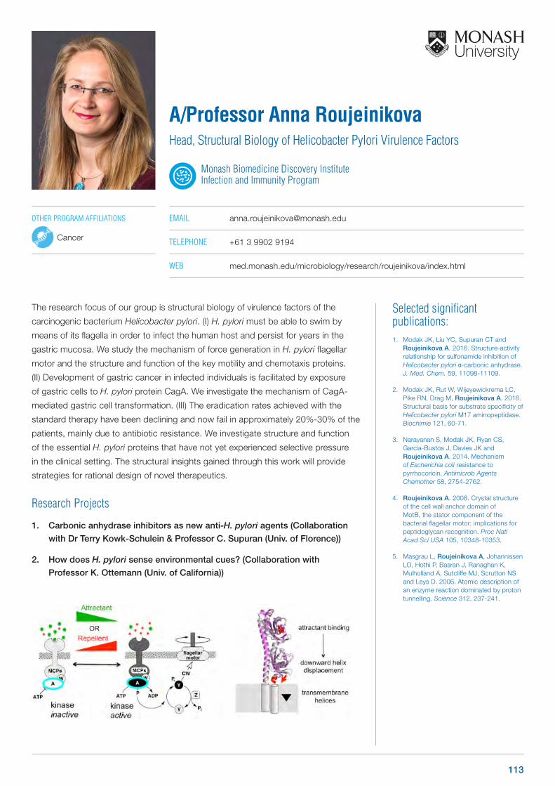
Selected significant publications: 1. Modak JK, Liu YC, Supuran CT and
Roujeinikova A. 2016. Structure-activity relationship for sulfonamide inhibition of Helicobacter pylori α-carbonic anhydrase. J. Med. Chem. 59, 11098-11109.
2. Modak JK, Rut W, Wijeyewickrema LC, Pike RN, Drag M, Roujeinikova A. 2016. Structural basis for substrate specificity of Helicobacter pylori M17 aminopeptidase. Biochimie 121, 60-71.
3. Narayanan S, Modak JK, Ryan CS, Garcia-Bustos J, Davies JK and Roujeinikova A. 2014. Mechanism of Escherichia coli resistance to pyrrhocoricin. Antimicrob Agents Chemother 58, 2754-2762.
4. Roujeinikova A. 2008. Crystal structure of the cell wall anchor domain of MotB, the stator component of the bacterial flagellar motor: implications for peptidoglycan recognition. Proc Natl Acad Sci USA 105, 10348-10353.
5. Masgrau L, Roujeinikova A, Johannissen LO, Hothi P, Basran J, Ranaghan K, Mulholland A, Sutcliffe MJ, Scrutton NS and Leys D. 2006. Atomic description of an enzyme reaction dominated by proton tunnelling. Science 312, 237-241.
The research focus of our group is structural biology of virulence factors of the
carcinogenic bacterium Helicobacter pylori. (I) H. pylori must be able to swim by
means of its flagella in order to infect the human host and persist for years in the
gastric mucosa. We study the mechanism of force generation in H. pylori flagellar
motor and the structure and function of the key motility and chemotaxis proteins.
(II) Development of gastric cancer in infected individuals is facilitated by exposure
of gastric cells to H. pylori protein CagA. We investigate the mechanism of CagA-
mediated gastric cell transformation. (III) The eradication rates achieved with the
standard therapy have been declining and now fail in approximately 20%-30% of the
patients, mainly due to antibiotic resistance. We investigate structure and function
of the essential H. pylori proteins that have not yet experienced selective pressure
in the clinical setting. The structural insights gained through this work will provide
strategies for rational design of novel therapeutics.
Research Projects
1. Carbonic anhydrase inhibitors as new anti-H. pylori agents (Collaboration
with Dr Terry Kowk-Schulein & Professor C. Supuran (Univ. of Florence))
2. How does H. pylori sense environmental cues? (Collaboration with
Professor K. Ottemann (Univ. of California))
A/Professor Anna RoujeinikovaHead, Structural Biology of Helicobacter Pylori Virulence Factors
Monash Biomedicine Discovery Institute Infection and Immunity Program
EMAIL [email protected]
TELEPHONE +61 3 9902 9194
WEB med.monash.edu/microbiology/research/roujeinikova/index.html
OTHER PROGRAM AFFILIATIONS
Cancer
113
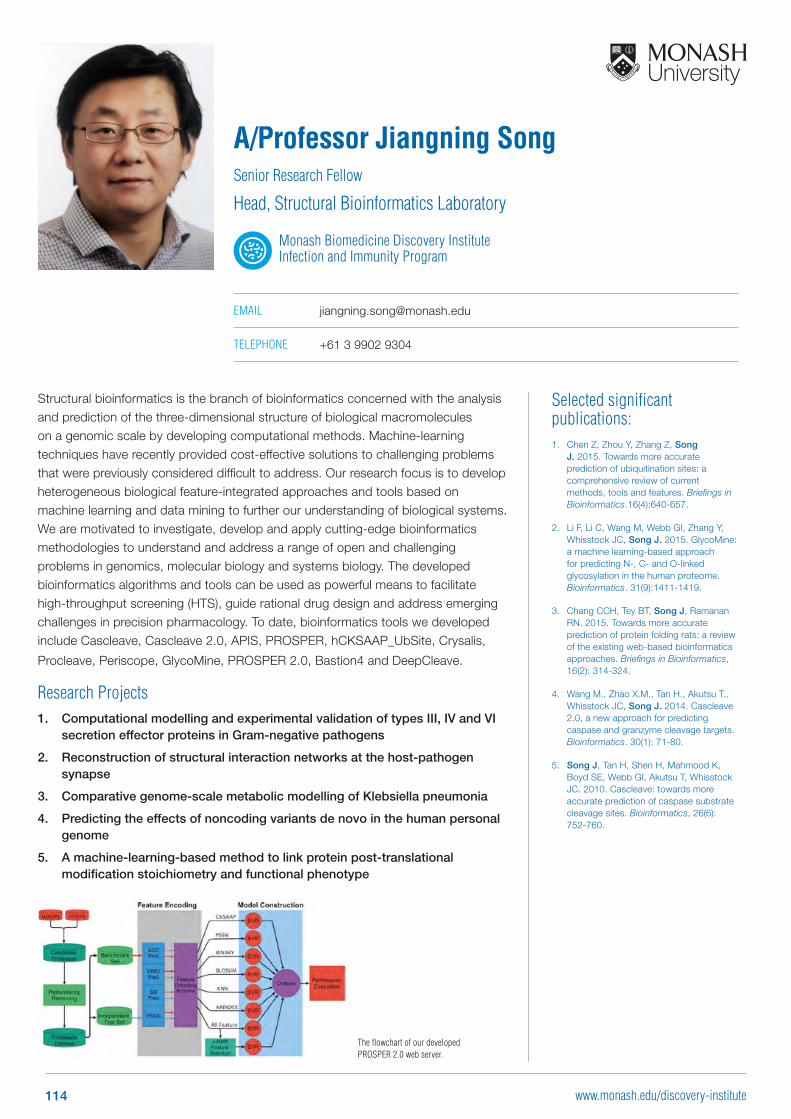
Selected significant publications: 1. Chen Z, Zhou Y, Zhang Z, Song
J. 2015. Towards more accurate prediction of ubiquitination sites: a comprehensive review of current methods, tools and features. Briefings in Bioinformatics.16(4):640-657.
2. Li F, Li C, Wang M, Webb GI, Zhang Y, Whisstock JC, Song J. 2015. GlycoMine: a machine learning-based approach for predicting N-, C- and O-linked glycosylation in the human proteome. Bioinformatics. 31(9):1411-1419.
3. Chang CCH, Tey BT, Song J, Ramanan RN. 2015. Towards more accurate prediction of protein folding rats: a review of the existing web-based bioinformatics approaches. Briefings in Bioinformatics, 16(2): 314-324.
4. Wang M., Zhao X.M., Tan H., Akutsu T., Whisstock JC, Song J. 2014. Cascleave 2.0, a new approach for predicting caspase and granzyme cleavage targets. Bioinformatics. 30(1): 71-80.
5. Song J, Tan H, Shen H, Mahmood K, Boyd SE, Webb GI, Akutsu T, Whisstock JC. 2010. Cascleave: towards more accurate prediction of caspase substrate cleavage sites. Bioinformatics, 26(6): 752-760.
Structural bioinformatics is the branch of bioinformatics concerned with the analysis and prediction of the three-dimensional structure of biological macromolecules on a genomic scale by developing computational methods. Machine-learning techniques have recently provided cost-effective solutions to challenging problems that were previously considered difficult to address. Our research focus is to develop heterogeneous biological feature-integrated approaches and tools based on machine learning and data mining to further our understanding of biological systems. We are motivated to investigate, develop and apply cutting-edge bioinformatics methodologies to understand and address a range of open and challenging problems in genomics, molecular biology and systems biology. The developed bioinformatics algorithms and tools can be used as powerful means to facilitate high-throughput screening (HTS), guide rational drug design and address emerging challenges in precision pharmacology. To date, bioinformatics tools we developed include Cascleave, Cascleave 2.0, APIS, PROSPER, hCKSAAP_UbSite, Crysalis,
Procleave, Periscope, GlycoMine, PROSPER 2.0, Bastion4 and DeepCleave.
Research Projects1. Computational modelling and experimental validation of types III, IV and VI
secretion effector proteins in Gram-negative pathogens
2. Reconstruction of structural interaction networks at the host-pathogen synapse
3. Comparative genome-scale metabolic modelling of Klebsiella pneumonia
4. Predicting the effects of noncoding variants de novo in the human personal genome
5. A machine-learning-based method to link protein post-translational modification stoichiometry and functional phenotype
A/Professor Jiangning SongSenior Research Fellow
Head, Structural Bioinformatics Laboratory
Monash Biomedicine Discovery Institute Infection and Immunity Program
EMAIL [email protected]
TELEPHONE +61 3 9902 9304
The flowchart of our developed PROSPER 2.0 web server.
114 www.monash.edu/discovery-institute
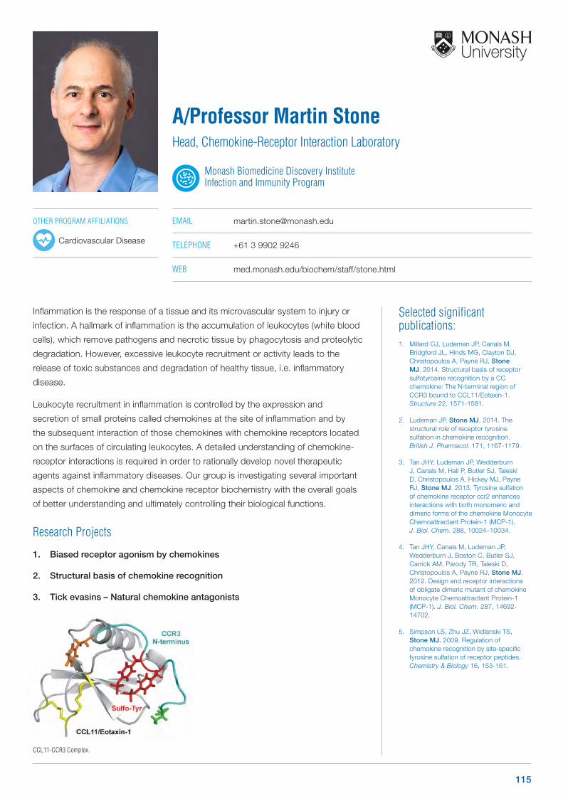
Selected significant publications: 1. Millard CJ, Ludeman JP, Canals M,
Bridgford JL, Hinds MG, Clayton DJ, Christopoulos A, Payne RJ, Stone MJ. 2014. Structural basis of receptor sulfotyrosine recognition by a CC chemokine: The N-terminal region of CCR3 bound to CCL11/Eotaxin-1. Structure 22, 1571-1581.
2. Ludeman JP, Stone MJ. 2014. The structural role of receptor tyrosine sulfation in chemokine recognition. British J. Pharmacol. 171, 1167-1179.
3. Tan JHY, Ludeman JP, Wedderburn J, Canals M, Hall P, Butler SJ, Taleski D, Christopoulos A, Hickey MJ, Payne RJ, Stone MJ. 2013. Tyrosine sulfation of chemokine receptor ccr2 enhances interactions with both monomeric and dimeric forms of the chemokine Monocyte Chemoattractant Protein-1 (MCP-1). J. Biol. Chem. 288, 10024–10034.
4. Tan JHY, Canals M, Ludeman JP, Wedderburn J, Boston C, Butler SJ, Carrick AM, Parody TR, Taleski D, Christopoulos A, Payne RJ, Stone MJ. 2012. Design and receptor interactions of obligate dimeric mutant of chemokine Monocyte Chemoattractant Protein-1 (MCP-1). J. Biol. Chem. 287, 14692-14702.
5. Simpson LS, Zhu JZ, Widlanski TS, Stone MJ. 2009. Regulation of chemokine recognition by site-specific tyrosine sulfation of receptor peptides. Chemistry & Biology 16, 153-161.
Inflammation is the response of a tissue and its microvascular system to injury or
infection. A hallmark of inflammation is the accumulation of leukocytes (white blood
cells), which remove pathogens and necrotic tissue by phagocytosis and proteolytic
degradation. However, excessive leukocyte recruitment or activity leads to the
release of toxic substances and degradation of healthy tissue, i.e. inflammatory
disease.
Leukocyte recruitment in inflammation is controlled by the expression and
secretion of small proteins called chemokines at the site of inflammation and by
the subsequent interaction of those chemokines with chemokine receptors located
on the surfaces of circulating leukocytes. A detailed understanding of chemokine-
receptor interactions is required in order to rationally develop novel therapeutic
agents against inflammatory diseases. Our group is investigating several important
aspects of chemokine and chemokine receptor biochemistry with the overall goals
of better understanding and ultimately controlling their biological functions.
Research Projects
1. Biased receptor agonism by chemokines
2. Structural basis of chemokine recognition
3. Tick evasins – Natural chemokine antagonists
A/Professor Martin StoneHead, Chemokine-Receptor Interaction Laboratory
Monash Biomedicine Discovery Institute Infection and Immunity Program
EMAIL [email protected]
TELEPHONE +61 3 9902 9246
WEB med.monash.edu/biochem/staff/stone.html
CCL11-CCR3 Complex.
OTHER PROGRAM AFFILIATIONS
Cardiovascular Disease
115
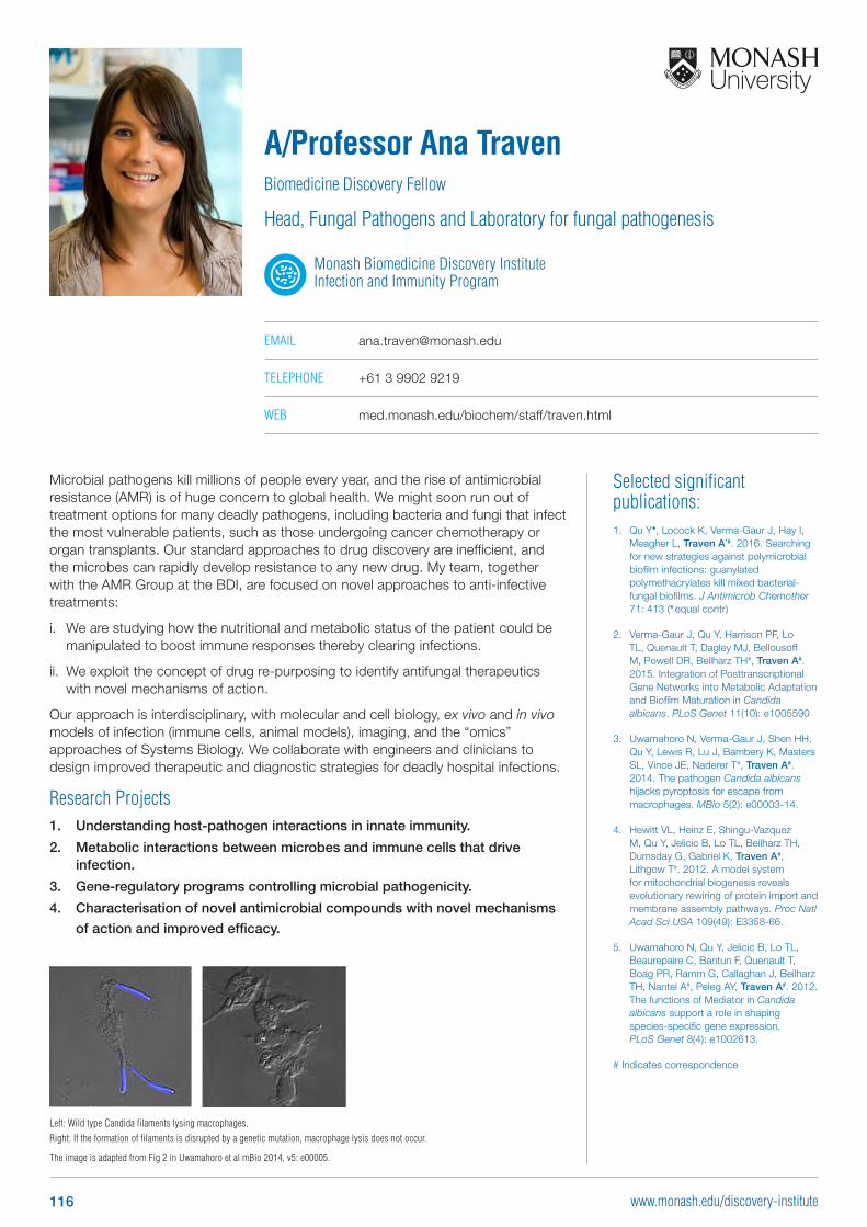
Selected significant publications: 1. Qu Y*, Locock K, Verma-Gaur J, Hay I,
Meagher L, Traven A*#. 2016. Searching for new strategies against polymicrobial biofilm infections: guanylated polymethacrylates kill mixed bacterial-fungal biofilms. J Antimicrob Chemother 71: 413 (* equal contr)
2. Verma-Gaur J, Qu Y, Harrison PF, Lo TL, Quenault T, Dagley MJ, Bellousoff M, Powell DR, Beilharz TH#, Traven A#. 2015. Integration of Posttranscriptional Gene Networks into Metabolic Adaptation and Biofilm Maturation in Candida albicans. PLoS Genet 11(10): e1005590
3. Uwamahoro N, Verma-Gaur J, Shen HH, Qu Y, Lewis R, Lu J, Bambery K, Masters SL, Vince JE, Naderer T#, Traven A#. 2014. The pathogen Candida albicans hijacks pyroptosis for escape from macrophages. MBio 5(2): e00003-14.
4. Hewitt VL, Heinz E, Shingu-Vazquez M, Qu Y, Jelicic B, Lo TL, Beilharz TH, Dumsday G, Gabriel K, Traven A#, Lithgow T#. 2012. A model system for mitochondrial biogenesis reveals evolutionary rewiring of protein import and membrane assembly pathways. Proc Natl Acad Sci USA 109(49): E3358-66.
5. Uwamahoro N, Qu Y, Jelicic B, Lo TL, Beaurepaire C, Bantun F, Quenault T, Boag PR, Ramm G, Callaghan J, Beilharz TH, Nantel A#, Peleg AY, Traven A#. 2012. The functions of Mediator in Candida albicans support a role in shaping species-specific gene expression. PLoS Genet 8(4): e1002613.
# Indicates correspondence
Microbial pathogens kill millions of people every year, and the rise of antimicrobial resistance (AMR) is of huge concern to global health. We might soon run out of treatment options for many deadly pathogens, including bacteria and fungi that infect the most vulnerable patients, such as those undergoing cancer chemotherapy or organ transplants. Our standard approaches to drug discovery are inefficient, and the microbes can rapidly develop resistance to any new drug. My team, together with the AMR Group at the BDI, are focused on novel approaches to anti-infective treatments:
i. We are studying how the nutritional and metabolic status of the patient could be manipulated to boost immune responses thereby clearing infections.
ii. We exploit the concept of drug re-purposing to identify antifungal therapeutics with novel mechanisms of action.
Our approach is interdisciplinary, with molecular and cell biology, ex vivo and in vivo models of infection (immune cells, animal models), imaging, and the “omics” approaches of Systems Biology. We collaborate with engineers and clinicians to design improved therapeutic and diagnostic strategies for deadly hospital infections.
Research Projects1. Understanding host-pathogen interactions in innate immunity.
2. Metabolic interactions between microbes and immune cells that drive infection.
3. Gene-regulatory programs controlling microbial pathogenicity.
4. Characterisation of novel antimicrobial compounds with novel mechanisms
of action and improved efficacy.
A/Professor Ana TravenBiomedicine Discovery Fellow
Head, Fungal Pathogens and Laboratory for fungal pathogenesis
Monash Biomedicine Discovery Institute Infection and Immunity Program
EMAIL [email protected]
TELEPHONE +61 3 9902 9219
WEB med.monash.edu/biochem/staff/traven.html
Left: Wild type Candida filaments lysing macrophages. Right: If the formation of filaments is disrupted by a genetic mutation, macrophage lysis does not occur.
The image is adapted from Fig 2 in Uwamahoro et al mBio 2014, v5: e00005.
116 www.monash.edu/discovery-institute
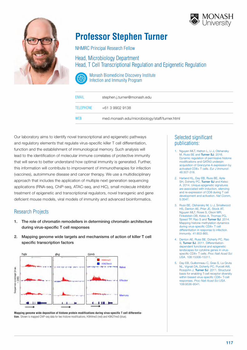
Mapping genome wide deposition of histone protein modifications during virus-specific T cell differentia-tion. Shown is mapped ChIP-seq data for two histone modifications, H3K4me3 (red) and H3K27me3 (blue).
Selected significant publications: 1. Nguyen MLT, Hatton L, Li J, Olshansky
M, Russ BE and Turner SJ. 2016. Dynamic regulation of permissive histone modifications and GATA3 underpin acquisition of Granzyme A expression by activated CD8+ T cells. Eur J Immunol. 46:307-318.
2. Harland KL, Day EB, Russ BE, Apte SH, Doherty PC, Turner SJ and Kelso A. 2014. Unique epigenetic signatures are associated with induction, silencing and re-expression of CD8 during T cell development and activation. Nat Comm, 5:3547.
3. Russ BE, Olshansky M, Li J, Smallwood HS, Denton AE, Prier JE, Stock AT, Nguyen MLT, Rowe S, Olson MR, Finkelstein DB, Kelso A, Thomas PG, Speed TP, Rao S and Turner SJ. 2014. Mapping histone methylation dynamics during virus-specific CD8+ T cell differentiation in response to infection. Immunity. 41:853-865.
4. Denton AE, Russ BE, Doherty PC, Rao S, Turner SJ. 2011. Differentiation-dependent functional and epigenetic landscapes for cytokine genes in virus-specific CD8+ T cells. Proc Natl Acad Sci USA. 108:15306-15311.
5. Day EB, Guillonneau C, Gras S, La Gruta NL, Vignali DA, Doherty PC, Purcell AW, Rossjohn J, Turner SJ. 2011. Structural basis for enabling T-cell receptor diversity within biased virus-specific CD8+ T-cell responses. Proc Natl Acad Sci USA. 108:9536-9541.
Our laboratory aims to identify novel transcriptional and epigenetic pathways
and regulatory elements that regulate virus-specific killer T cell differentiation,
function and the establishment of immunological memory. Such analysis will
lead to the identification of molecular immune correlates of protective immunity
that will serve to better understand how optimal immunity is generated. Further,
this information will contribute to improvement of immunotherapies for infection
(vaccines), autoimmune disease and cancer therapy. We use a multidisciplinary
approach that includes the application of multiple next generation sequencing
applications (RNA-seq, ChiP-seq, ATAC-seq, and HiC), small molecule inhibitor
treatment of epigenetic and transcriptional regulators, novel transgenic and gene
deficient mouse models, viral models of immunity and advanced bioinformatics.
Research Projects
1. The role of chromatin remodellers in determining chromatin architecture
during virus-specific T cell responses
2. Mapping genome wide targets and mechanisms of action of killer T cell
specific transcription factors
Professor Stephen TurnerNHMRC Principal Research Fellow
Head, Microbiology DepartmentHead, T Cell Transcriptional Regulation and Epigenetic Regulation
Monash Biomedicine Discovery Institute Infection and Immunity Program
EMAIL [email protected]
TELEPHONE +61 3 9902 9138
WEB med.monash.edu/microbiology/staff/turner.html
117
Selected significant publications: 1. Moradi S, Berry R, Pymm P, Hitchen C,
Beckham SA, Wilce MC, Walpole NG, Clements CS, Reid HH, Perugini MA, Brooks AG, Rossjohn J, Vivian JP. 2015. The structure of the atypical killer cell immunoglobulin-like receptor, KIR2DL4. J Biol Chem. 290(16):10460-71.
2. Vivian JP, Saunders PM, Baschuk N, Beddoe T, Widjaja J, O’Connor GM, Hitchen C, Pymm P, Andrews DM, Gras S, McVicar DW, Rossjohn J, Brooks AG. 2015. The interaction of KIR3DL1*001 with HLA class I molecules is dependent upon molecular microarchitecture within the Bw4 epitope. J Immunol. 194(2):781-9.
3. de Weerd NA, Vivian JP, Nguyen TK, Mangan NE, Gould JA, Braniff SJ, Zaker-Tabrizi L, Fung KY, Forster SC, Beddoe T, Reid HH, Rossjohn J, Hertzog PJ. 2013. Structural basis of a unique interferon-‐ signaling axis mediated via the receptor IFNAR1. Nat Immunol 14(9):901-7.
4. Illing PT, Vivian JP, Dudek NL, Kostenko L, Chen Z, Bharadwaj M, Miles JJ, Kjer-Nielsen L, Gras S, Williamson NA, Burrows SR, Purcell AW, Rossjohn J, McCluskey J. 2012. Immune self-reactivity triggered by drug-modified HLA-peptide repertoire. Nature. 486(7404):554-8.
5. Vivian JP, Duncan RC, Berry R, O’Connor GM, Reid HH, Beddoe T, Gras S, Saunders PM, Olshina MA, Widjaja JM, Harpur CM, Lin J, Maloveste SM, Price DA, Lafont BA, McVicar DW, Clements CS, Brooks AG, Rossjohn J. 2011. Killer cell immunoglobulin-like receptor 3DL1-mediated recognition of human leukocyte antigen B. Nature. 479(7373):401-5.
Our work is principally focused on events central to infection and immunity.
Specifically we work on deducing the structural arrangement of Killer-Cell
Immunoglobulin-like Receptors (KIR) and their ligands and detail the molecular mode
of interaction generating their complexes. This has important implications in disease
and transplant outcomes. We also investigate immune reactions to specific drugs.
This work is intended to lead to the better design and screening of new therapeutics.
Research Projects
1. Structural and functional investigation of KIR receptors
2. What causes drug hypersensitivity?
Dr Julian VivianResearch Fellow
Head, Vivian Research Group
Monash Biomedicine Discovery Institute Infection and Immunity Program
EMAIL [email protected]
118 www.monash.edu/discovery-institute
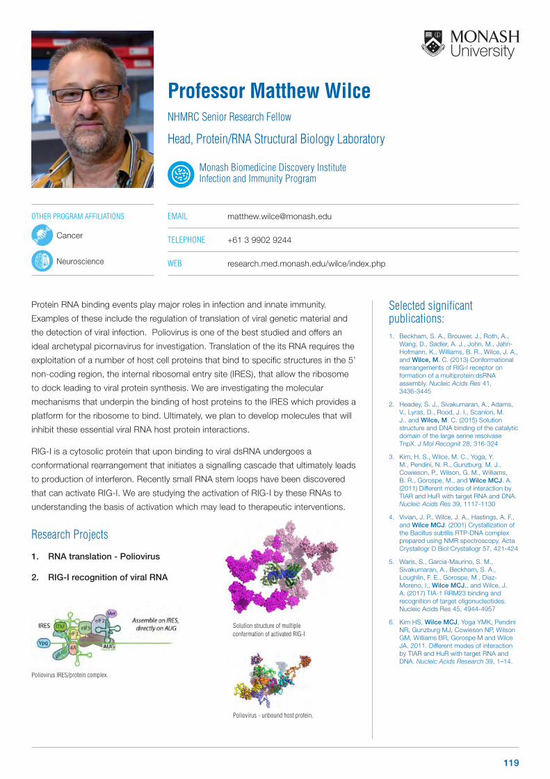
Selected significant publications: 1. Beckham, S. A., Brouwer, J., Roth, A.,
Wang, D., Sadler, A. J., John, M., Jahn-Hofmann, K., Williams, B. R., Wilce, J. A., and Wilce, M. C. (2013) Conformational rearrangements of RIG-I receptor on formation of a multiprotein:dsRNA assembly. Nucleic Acids Res 41, 3436-3445
2. Headey, S. J., Sivakumaran, A., Adams, V., Lyras, D., Rood, J. I., Scanlon, M. J., and Wilce, M. C. (2015) Solution structure and DNA binding of the catalytic domain of the large serine resolvase TnpX. J Mol Recognit 28, 316-324
3. Kim, H. S., Wilce, M. C., Yoga, Y. M., Pendini, N. R., Gunzburg, M. J., Cowieson, P., Wilson, G. M., Williams, B. R., Gorospe, M., and Wilce MCJ. A. (2011) Different modes of interaction by TIAR and HuR with target RNA and DNA. Nucleic Acids Res 39, 1117-1130
4. Vivian, J. P., Wilce, J. A., Hastings, A. F., and Wilce MCJ. (2001) Crystallization of the Bacillus subtilis RTP-DNA complex prepared using NMR spectroscopy. Acta Crystallogr D Biol Crystallogr 57, 421-424
5. Waris, S., Garcia-Maurino, S. M., Sivakumaran, A., Beckham, S. A., Loughlin, F. E., Gorospe, M., Diaz-Moreno, I., Wilce MCJ., and Wilce, J. A. (2017) TIA-1 RRM23 binding and recognition of target oligonucleotides. Nucleic Acids Res 45, 4944-4957
6. Kim HS, Wilce MCJ, Yoga YMK, Pendini NR, Gunzburg MJ, Cowieson NP, Wilson GM, Williams BR, Gorospe M and Wilce JA. 2011. Different modes of interaction by TIAR and HuR with target RNA and DNA. Nucleic Acids Research 39, 1–14.
Protein RNA binding events play major roles in infection and innate immunity.
Examples of these include the regulation of translation of viral genetic material and
the detection of viral infection. Poliovirus is one of the best studied and offers an
ideal archetypal picornavirus for investigation. Translation of the its RNA requires the
exploitation of a number of host cell proteins that bind to specific structures in the 5’
non-coding region, the internal ribosomal entry site (IRES), that allow the ribosome
to dock leading to viral protein synthesis. We are investigating the molecular
mechanisms that underpin the binding of host proteins to the IRES which provides a
platform for the ribosome to bind. Ultimately, we plan to develop molecules that will
inhibit these essential viral RNA host protein interactions.
RIG-I is a cytosolic protein that upon binding to viral dsRNA undergoes a
conformational rearrangement that initiates a signalling cascade that ultimately leads
to production of interferon. Recently small RNA stem loops have been discovered
that can activate RIG-I. We are studying the activation of RIG-I by these RNAs to
understanding the basis of activation which may lead to therapeutic interventions.
Research Projects
1. RNA translation - Poliovirus
2. RIG-I recognition of viral RNA
Professor Matthew WilceNHMRC Senior Research Fellow
Head, Protein/RNA Structural Biology Laboratory
Monash Biomedicine Discovery Institute Infection and Immunity Program
EMAIL [email protected]
TELEPHONE +61 3 9902 9244
WEB research.med.monash.edu/wilce/index.php
Solution structure of multiple conformation of activated RIG-I
Poliovirus - unbound host protein.
Poliovirus IRES/protein complex.
OTHER PROGRAM AFFILIATIONS
Cancer
Neuroscience
119

Selected significant publications: 1. Johnson TK, Henstridge MA, Warr CG,
Whisstock JC. 2015. Torso-like mediates extracellular accumulation of Furin-clevaed Trunk to pattern the Drosophila embryo termini. Nat Commun. 6: 8759.
2. Henstridge MA, Johnson TK, Warr CG, Whisstock JC. 2013. Trunk cleavage is essential for Drosophila terminal patterning and can occur independently of Torso-like. Nat Commun 5:3419.
3. Law RH, Caradoc-Davies T, Cowieson N, Horvath AJ, Quek AJ, Encarnacao JA, Steer D, Cowan A, Zhang Q, Lu BG, Pike RN, Smith AI, Coughlin PB, Whisstock JC. 2012. The X-ray crystal structure of full-length human plasminogen. Cell Rep.1(3):185-90.
4. Law RH, Lukoyanova N, Voskoboinik I, Caradoc-Davies TT, Baran K, Dunstone MA, D’Angelo ME, Orlova EV, Coulibaly F, Verschoor S, Browne KA, Ciccone A, Kuiper MJ, Bird PI, Trapani JA, Saibil HR, Whisstock JC. 2010. The structural basis for membrane binding and pore formation by lymphocyte perforin. Nature 468(7322):447-51.
5. Rosado CJ, Buckle AM, Law RH, Butcher RE, Kan WT, Bird CH, Ung K, Browne KA, Baran K, Bashtannyk-Puhalovich TA, Faux NG, Wong W, Porter CJ, Pike RN, Ellisdon AM, Pearce MC, Bottomley SP, Emsley J, Smith AI, Rossjohn J, Hartland EL, Voskoboinik I, Trapani JA, Bird PI, Dunstone MA, Whisstock JC. 2007. A common fold mediates vertebrate defense and bacterial attack. Science 317(5844):1548-51.
We use structural and molecular biology to investigate protein function and
dysfunction in immunity, haemostasis, cell signaling and developmental biology.
Support for the laboratory includes an ARC Federation Fellowship, NHMRC and ARC
grants, an ARC Centre of Excellence in Advanced Molecular Imaging together with
funding from the Wellcome Trust. Generally projects involve any or all of the following
techniques: X-ray crystallography, small angle x-ray scattering, electron microscopy,
bioinformatics, molecular and cell biology, enzymology and protein chemistry.
Research Projects:
1. Perforin in immunity & cancer (Collaboration with Professor Joe Trapani,
Peter MacCallum Cancer Centre; Professor Ray Norton, MIPS)
2. Fibrinolysis in diabetes (Collaboration with Professor J Shaw, Bakers IDI)
3. GABA functions in disease (Collaboration with Dr K Tuck, Chemistry
Department, Monash University)
4. Understanding the structural
basis for bacterial conjugation
(Collaboration with Professor
J Rood, Microbiology
Department, Monash
University)
Professor James WhisstockNHMRC Senior Principal Research Fellow ARC Australian Laurate Fellow
Head, Whisstock Structural Biology Laboratory
Monash Biomedicine Discovery Institute Infection and Immunity Program
EMAIL [email protected]
TELEPHONE +61 3 9902 9312
WEB whisstock-lab.med.monash.edu.au
“Polar Aurora” by Paul Martin (Research Media); A stylised depiction of signalling induced by the localized secretion of Trunk at the Drosophila embryo poles.
OTHER PROGRAM AFFILIATIONS
Cancer
Neuroscience
120 www.monash.edu/discovery-institute
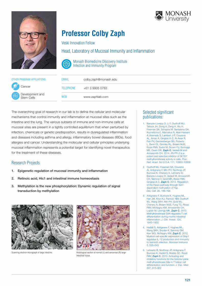
Selected significant publications: 1. Barsyte-Lovejoy D, Li F, Oudhoff MJ,
Tatlock JH, Dong A, Zeng H, Wu H, Freeman SA, Schapira M, Senisterra GA, Kuznetsova E, Marcellus R, Allali-Hassani A, Kennedy S, Lambert J-P, Couzens AL, Aman A, Gingras A-C, Al-Awar R, Fish PV, Gerstenberger BS, Roberts L, Benn CL, Grimley RL, Braam MJS, Rossi FMV, Sudol M, Brown PJ, Bunnage ME, Owen DR, Zaph C, Vedadi M and Arrowsmith CH. 2014. (R)-PFI-2 is a potent and selective inhibitor of SETD7 methyltransferase activity in cells. Proc. Natl. Acad. Sci U.S.A. 111, 12853–12858
2. Oudhoff MJ, Freeman SA, Couzens AL Antignano F, Min PH, Northrop JP, Burrows K, Chenery A, Lehnertz B, Barsyte-Lovejoy D, Vedadi M, Arrowsmith CH, Nishina H, Gold MR, Rossi FMV, Gingras A-C, Zaph C. 2013. Regulation of the Hippo pathway through Set7-dependent methylation of Yap. Dev. Cell. 26, 188–194
3. Antignano F, Burrows K, Hughes ML, Han JM, Kron KJ, Penrod, NM, Oudhoff MJ, Wang SKH, Min PH, Gold MJ, Chenery A, Braam MJS, Fung TC, Rossi FMV, McNagny KM, Arrowsmith CH, Lupien M, Levings MK, Zaph C. 2013. Methyltransferase G9A regulates T cell differentiation during murine intestinal inflammation. J. Clin. Invest. 124, 1945–1955
4. Hadidi S, Antignano F, Hughes ML, Wang SKH, Snyder K, Sammis GM, Kerr WG, McNagny KM, Zaph C. 2012. Myeloid cell-specific expression of Ship1 regulates IL-12 production and immunity to helminth infection. Mucosal Immunol. 5, 535–543
5. Lehnertz B, Northrop JP, Antignano F, Burrows K, Hadidi S, Mullaly SC, Rossi FMV, Zaph C. 2010. Activating and inhibitory functions for the histone lysine methyltransferase G9a in T helper cell differentiation and function. J. Exp. Med. 207, 915–922
The overarching goal of research in our lab is to define the cellular and molecular
mechanisms that control immunity and inflammation at mucosal sites such as the
intestine and the lung. The various subsets of immune and non-immune cells at
mucosal sites are present in a tightly controlled equilibrium that when perturbed by
infection, chemicals or genetic predisposition, results in dysregulated inflammation
and diseases including asthma and allergy, inflammatory bowel diseases (IBDs), food
allergies and cancer. Understanding the molecular and cellular principles underlying
mucosal inflammation represents a potential target for identifying novel therapeutics
for the treatment of these diseases.
Research Projects
1. Epigenetic regulation of mucosal immunity and inflammation
2. Retinoic acid, Hic1 and intestinal immune homeostasis
3. Methylation is the new phosphorylation: Dynamic regulation of signal
transduction by methylation
Professor Colby ZaphVeski Innovation Fellow
Head, Laboratory of Mucosal Immunity and Inflammation
Monash Biomedicine Discovery Institute Infection and Immunity Program
EMAIL [email protected]
TELEPHONE +61 3 9905 0783
WEB www.zaphlab.com
Scanning electron micrograph of large intestine. Histological section of normal (L) and cancerous (R) large intestinal tissue.
OTHER PROGRAM AFFILIATIONS
Cancer
Development and Stem Cells
121

Metabolism, Diabetes and Obesity Program Group Leaders
122122 www.monash.edu/discovery-institute

Selected significant publications: 1. Bayliss JA, Stark R, Lemus M, Santos VV,
Thompson A, Rees D, Galic S, Elsworth J, Kemp BE, Davies JS, Andrews ZB. 2016. Ghrelin-AMPK signalling mediates the neuroprotective effects of Calorie Restriction in Parkinson’s Disease. Journal of Neuroscience 36 (10): 3049-3063
2. Lockie SH, Dinan T, Lawrence AJ, Spencer SJ, Andrews ZB. 2015. Diet-induced obesity causes ghrelin resistance in reward processing tasks. Psychoneuroendocrinology 62: 114-120.
3. Lemus MB, Bayliss JA, Lockie SH, Santos VV, Reichenbach A, Stark R, Andrews ZB. 2015. A stereological analysis of NPY, POMC, orexin, GFAP astrocyte and Iba1 microglial cell number and volume in diet-induced obese male mice. Endocrinology 156(5)1701-13
4. Briggs DI, Enriori PJ, Lemus MB, Cowley MA, Andrews ZB. 2010. Diet-induced obesity causes ghrelin resistance in NPY/AgRP neurons. Endocrinology 151(10): 4745-4755
5. Andrews ZB, Liu ZW, Walllingford N, Erion DM, Borok E, Friedman JM, Tschöp MH, Shanabrough M, Cline G, Shulman GI, Coppola A, Gao XB, Horvath TL Diano S. 2008. Uncoupling protein-2 mediates ghrelin’s action on NPY/AgRP neurons. Nature 454(7206): 846-51.
We examine how the brain senses hormone and nutrient information in different
metabolic states and how the brain integrates this information to encode physiological
and behavioural changes that maintain energy homeostasis. The work is critical to help
identify the causes of obesity, anorexia and type-2 diabetes.
We are examining how metabolic states such as fasting or starvation influence
other brain systems, such as anxiety and stress, motivation and memory. Clearly
maintaining energy homeostasis is not only good for body weight, but also for mental
health. Similarly changes in mental health can affect food intake and body weight.
We are working to identify interconnected neural pathways linking metabolism to
mood, motivation and neuroprotection.
Research Projects
1. How does emotional, cognitive and motivational information from the
cerebral cortex influence hypothalamic control of energy homeostasis
2. How does endocrine feedback during negative energy balance coordinate
and integrate metabolism with stress, anxiety, motivation and memory; the
role of ghrelin and ghrelin receptors
3. How does the brain sense changes in metabolic state in order to control
energy homeostasis; implications for obesity and type 2 diabetes
A/Professor Zane AndrewsNHMRC Senior Research Fellow
Head, Neural Control of Energy Homeostasis Laboratory
Monash Biomedicine Discovery Institute Metabolism, Diabetes and Obesity Program
EMAIL [email protected]
TELEPHONE +61 3 9905 8165
WEB med.monash.edu/physiology/staff/andrews.html
Designer Receptors Exclusively Activated by Designer Drugs (DREADDs) provide a unique way to remotely control specific neuronal populations. Localised DREADD expression only in arcuate nucleus NPY neurons.
DREADD activation with an exogenous ligand increases cfos, a marker of neuronal activation (red nuclei) expression only in NPY (green) neurons.
OTHER PROGRAM AFFILIATIONS
Neuroscience
123
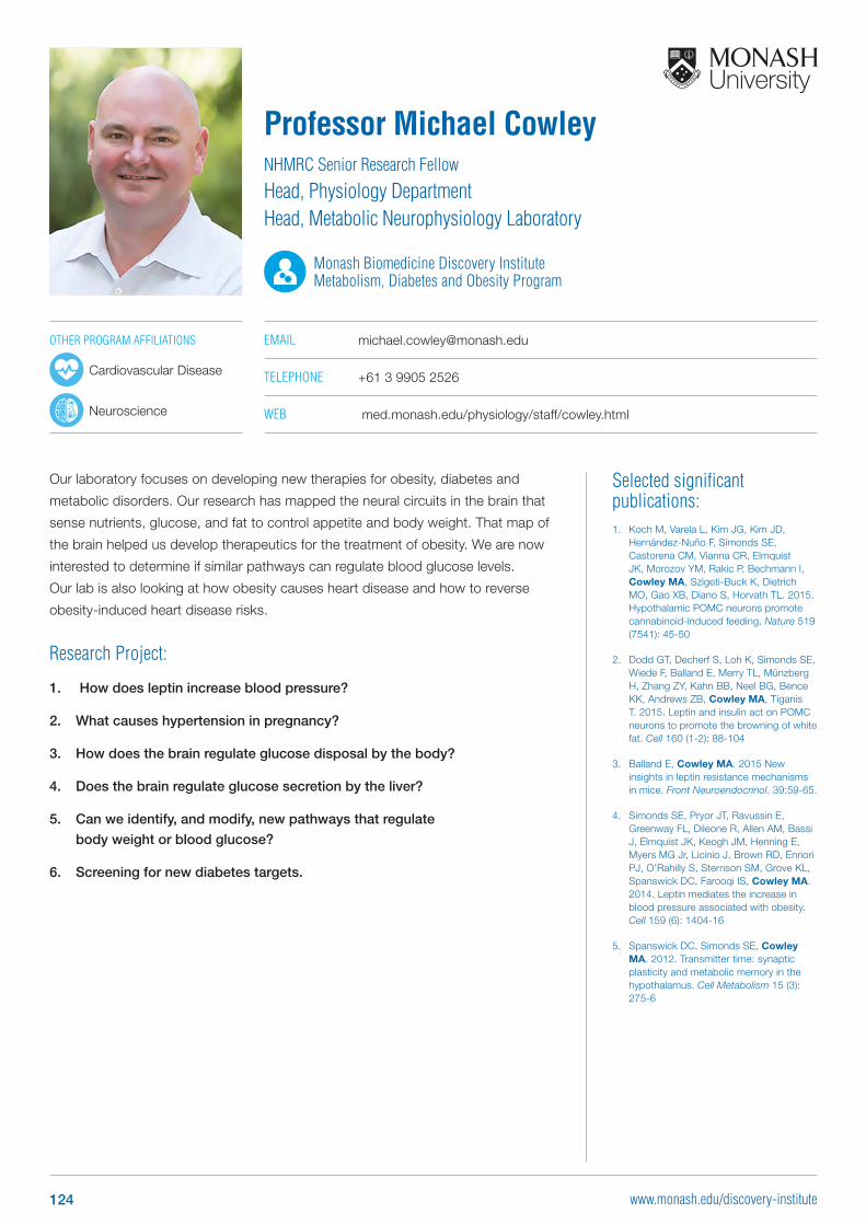
Selected significant publications: 1. Koch M, Varela L, Kim JG, Kim JD,
Hernández-Nuño F, Simonds SE, Castorena CM, Vianna CR, Elmquist JK, Morozov YM, Rakic P, Bechmann I, Cowley MA, Szigeti-Buck K, Dietrich MO, Gao XB, Diano S, Horvath TL. 2015. Hypothalamic POMC neurons promote cannabinoid-induced feeding. Nature 519 (7541): 45-50
2. Dodd GT, Decherf S, Loh K, Simonds SE, Wiede F, Balland E, Merry TL, Münzberg H, Zhang ZY, Kahn BB, Neel BG, Bence KK, Andrews ZB, Cowley MA, Tiganis T. 2015. Leptin and insulin act on POMC neurons to promote the browning of white fat. Cell 160 (1-2): 88-104
3. Balland E, Cowley MA. 2015 New insights in leptin resistance mechanisms in mice. Front Neuroendocrinol. 39:59-65.
4. Simonds SE, Pryor JT, Ravussin E, Greenway FL, Dileone R, Allen AM, Bassi J, Elmquist JK, Keogh JM, Henning E, Myers MG Jr, Licinio J, Brown RD, Enriori PJ, O’Rahilly S, Sternson SM, Grove KL, Spanswick DC, Farooqi IS, Cowley MA. 2014. Leptin mediates the increase in blood pressure associated with obesity. Cell 159 (6): 1404-16
5. Spanswick DC, Simonds SE, Cowley MA. 2012. Transmitter time: synaptic plasticity and metabolic memory in the hypothalamus. Cell Metabolism 15 (3): 275-6
Our laboratory focuses on developing new therapies for obesity, diabetes and
metabolic disorders. Our research has mapped the neural circuits in the brain that
sense nutrients, glucose, and fat to control appetite and body weight. That map of
the brain helped us develop therapeutics for the treatment of obesity. We are now
interested to determine if similar pathways can regulate blood glucose levels.
Our lab is also looking at how obesity causes heart disease and how to reverse
obesity-induced heart disease risks.
Research Project:
1. How does leptin increase blood pressure?
2. What causes hypertension in pregnancy?
3. How does the brain regulate glucose disposal by the body?
4. Does the brain regulate glucose secretion by the liver?
5. Can we identify, and modify, new pathways that regulate
body weight or blood glucose?
6. Screening for new diabetes targets.
Professor Michael CowleyNHMRC Senior Research Fellow
Head, Physiology Department Head, Metabolic Neurophysiology Laboratory
Monash Biomedicine Discovery Institute Metabolism, Diabetes and Obesity Program
EMAIL [email protected]
TELEPHONE +61 3 9905 2526
WEB med.monash.edu/physiology/staff/cowley.html
OTHER PROGRAM AFFILIATIONS
Cardiovascular Disease
Neuroscience
124 www.monash.edu/discovery-institute
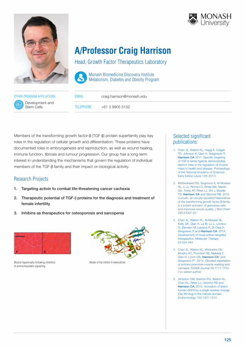
Selected significant publications: 1. Chen JL, Walton KL, Hagg A, Colgan
TD, Johnson K, Qian H, Gregorevic P, Harrison CA 2017. Specific targeting of TGF-b family ligands demonstrates distinct roles in the regulation of muscle mass in health and disease. Proceedings of the National Academy of Sciences - Early Edition (June 12th 2017).
2. Mottershead DG, Sugimura S, Al-Musawi SL, Li JJ, Richani D, White MA, Martin GA, Trotta AP, Ritter LJ, Shi J, Mueller TD, Harrison CA and Gilchrist RB. 2015. Cumulin, an oocyte-secreted heterodimer of the transforming growth factor-β family, is a potent activator of granulosa cells and improves oocyte quality. J Biol Chem 290:24007-20
3. Chen JL, Walton KL, Al-Musawi SL, Kelly, EK, Qian H, La M, Lu L, Lovrecz G, Ziemann M, Lazarus R, El-Osta A, Gregorevic P and Harrison CA. 2014. Development of novel activin-targeted therapeutics. Molecular Therapy 23:434-444
4. Chen JL, Walton KL, Winbanks CE, Murphy KT, Thomson RE, Makanji Y, Qian H, Lynch GS, Harrison CA* and Gregorevic P*. 2014. Elevated expression of activins promotes muscle wasting and cachexia. FASEB Journal 28:1711-1723. (*co-senior author)
5. Simpson CM, Stanton PG, Walton KL, Chan KL, Ritter LJ, Gilchrist RB and Harrison CA. 2012. Activation of latent human GDF9 by a single residue change (Gly 391Arg) in the mature domain. Endocrinology 153:1301-1310
Members of the transforming growth factor-β (TGF-β) protein superfamily play key
roles in the regulation of cellular growth and differentiation. These proteins have
documented roles in embryogenesis and reproduction, as well as wound healing,
immune function, fibrosis and tumour progression. Our group has a long-term
interest in understanding the mechanisms that govern the regulation of individual
members of the TGF-β family and their impact on biological activity.
Research Projects
1. Targeting activin to combat life-threatening cancer cachexia
2. Therapeutic potential of TGF-β proteins for the diagnosis and treatment of
female infertility
3. Inhibins as therapeutics for osteoporosis and sarcopenia
A/Professor Craig HarrisonHead, Growth Factor Therapeutics Laboratory
Monash Biomedicine Discovery Institute Metabolism, Diabetes and Obesity Program
EMAIL [email protected]
TELEPHONE +61 3 9905 5132
Muscle hypertrophy following inhibition of activin/myostatin signalling.
OTHER PROGRAM AFFILIATIONS
Development and Stem Cells
Model of the inhibin A heterodimer.
125
Selected significant publications: 1. Hewagalamulage SD, Clarke IJ, Young
IR, Rao A, Henry BA. 2015. High cortisol response to adrenocorticotrophic hormone identifies ewes with reduced melanocortin signalling and increased propensity to obesity. J Neuroendocrinol 27(1):44-56.
2. Lee TK, Lee C, Bischof R, Lambert GW, Clarke IJ and Henry BA. 2014. Stress-induced behavioural and metabolic adaptations lead to an obesity prone phenotype in ewes with high cortisol responses. Psychoneuroendocrinology 47: 166-77.
3. Lee TK, Clarke IJ, StJohn J, Young IR, Leury BJ, Rao A, Andrews ZB and Henry BA. 2014. High cortisol responses identify propensity to obesity that is linked to thermogenesis in skeletal muscle. Faseb J 28: 35-44
4. Henry BA, Andrews ZB, Rao A and Clarke IJ. 2011. Central leptin activates mitochondrial function and increases heat production in skeletal muscle. Endocrinology 152: 2609-18.
5. Henry BA, Dunshea FR, Gould M and Clarke IJ. 2008. Profiling post-prandial thermogenesis in muscle and fat of sheep and the central effect of leptin administration. Endocrinology 149(4):2019-26.
Our laboratory focuses on understanding how body weight is regulated. We
have a particular interest in understanding how energy expenditure occurs within
mammals. Our work primarily focuses on thermogenesis, which is a specialised
process where the body expends energy in the form of heat. Our work aims to
understand how the brain regulates thermogenesis. We have a number of unique
and novel models that allow us to characterise the control of body weight, food
intake and energy expenditure. The metabolic neuroendocrine group has a particular
interest in understanding how the following impact on energy homeostasis and
weight control:
1. Control of thermogenesis in humans.
2. Stress, stress responsiveness and obesity.
3. Exercise.
Research Projects
1. Sex differences in the control of thermogenesis
2. Stress, weight loss and predisposition to obesity
3. Role of thermogenesis in weight regulation in humans
Dr Belinda HenryHead, Metabolic Neuroendocrinology Laboratory
Monash Biomedicine Discovery Institute Metabolism, Diabetes and Obesity Program
EMAIL [email protected]
TELEPHONE +61 3 9905 2500
WEB www.modi.monash.edu.au/people/dr-belinda-henry
126 www.monash.edu/discovery-institute
Selected significant publications: 1. Adler ES, Hollis JH, Clarke IJ, Grattan
DR, Oldfield BJ. 2012. Neurochemical characterization and sexual dimorphism of projections from the brain to abdominal and subcutaneous white adipose tissue in the rat. J Neurosc 32 (45): 15913-21
2. Verty A, Lockie SH, Stefanidis A, Oldfield BJ. 2012. Anti-obesity effects of the combined administration of CB1 receptor antagonist rimonabant and melanocortin concentrating hormone antagonists in diet induced obese mice. International Journal of Obesity 32:279-287
3. Kampe J, Stefanidis A, Lockie SH, Brown WA, Dixon JB, Odoi A, Spencer SJ, Raven J, Oldfield BJ. 2012. Neural and humoral changes associated with the adjustable gastric band: insights from a rodent model. International Journal of Obesity 36: 1403–1411
4. Verty AN, Allen AM, Oldfield BJ. 2010. The endogenous actions of hypothalamic peptides on brown adipose tissue thermogenesis in the rat. Endocrinology 151(9):4236-46
5. Oldfield BJ, Giles ME, Watson A, Anderson C, Colvill LM, McKinley MJ. 2002. The neurochemical characterisation of hypothalamic pathways projecting polysynaptically to brown adipose tissue in the rat. Neuroscience 110(3):515-26
The primary focus of the laboratory is brown fat biology, particularly its innervation.
Given the consensus that activation of the ß-adrenoceptor on brown or “brown –
like” beige fat cells by noradrenaline is a critical element in the effective functional
recruitment of energy expenditure in these tissues, we have focussed on the details
of the central neural circuits involved. This theme is being pursued in the first four of
the funded projects listed below.
In addition, there are two other major projects in the lab which have unexpected
overlap with our focus on brown fat biology. One is the use of our rodent model of
the adjustable gastric band which is being used to define mechanisms underpinning
the efficacy of the procedure, particularly in relation to the use of adjunctive
pharmacotherapies with the band. The secod is a functional dissection of midbrain
reward pathways, using Cav-Cre viruses and DREADD technologies, to define the
contribution of these pathways to the mediation of voluntary starvation in a rodent
model of anorexia nervosa.
Research Projects
1. “Smart Food” – The fulcrum in the energy balance equation
2. Determining the impact of the nutrient milieu in the CNS on thermogenic
tone using obese prone and obese resistant rodents
3. Central neural regulation of brown fat function – glucose sensing
4. Central neural regulation of brown or beige fat function
5. The use of a rodent model of the AGB to define the mechanisms underlying
increased efficacy of the AGB when combined with pharmacotherapies
6. Reward pathways and their involvement in the etiology of anorexia nervosa
Professor Brian OldfieldNHMRC Principal Research Fellow
Head, Metabolic Neuroscience Laboratory
Monash Biomedicine Discovery Institute Metabolism, Diabetes and Obesity Program
EMAIL [email protected]
TELEPHONE +61 3 9905 2507
WEB med.monash.edu/physiology/staff/oldfield.html
OTHER PROGRAM AFFILIATIONS
Neuroscience
127
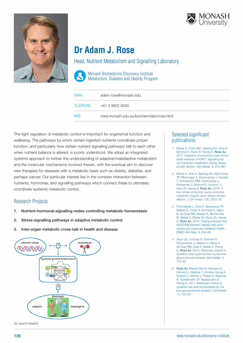
Selected significant publications: 1. Maida A, Chan JSK, Sjøberg KA, Zota A,
Schmoll D, Kiens B, Herzig S, Rose AJ. 2017. Repletion of branched chain amino acids reverses mTORC1 signaling but not improved metabolism during dietary protein dilution. Mol Metab. 6, 873-881
2. Maida A, Zota A, Sjøberg KA, Sijmonsma TP, Pfenninger A, Schumacher J, Gantert T, Christensen MM, Fuhrmeister J, Rothermel U, Schmoll D, Iovanna, J, Kiens B, Herzig S, Rose AJ. 2016. A liver stress-endocrine nexus promotes metabolic integrity upon dietary protein dilution. J Clin Invest. 126, 3263-78.
3. Fuhrmeister J, Zota A, Sijmonsma TP, Seibert O, Cingir S, Schmidt K, Vallon N, de Guia RM, Niopek K, Berriel Diaz M, Maida A, Blüher M, Okun JG, Herzig S, Rose AJ. 2016. Fasting-induced liver GADD45β restrains hepatic fatty acid uptake and improves metabolic health. EMBO Mol Med. 8, 654-69.
4. Okun JG, Conway S, Schmidt K, Schumacher J, Seibert O, Wang X, de Guia RM, Zota A, Maida A, Herzig S, Rose AJ. 2015. Molecular control of systemic urea cycle function by the liver glucocorticoid receptor. Mol Metab. 4, 732-40.
5. Rose AJ, Berriel Díaz M, Reimann A, Klement J, Walcher T, Krones-Herzig A, Strobel O, Werner J, Peters A, Kleyman A, Tuckermann JP, Vegiopoulos A, Herzig S. 2011. Molecular control of systemic bile acid homeostasis by the liver glucocorticoid receptor. Cell Metab. 14, 123-30
The tight regulation of metabolic control is important for organismal function and
wellbeing. The pathways by which certain ingested nutrients coordinate proper
function, and particularly how certain nutrient signalling pathways talk to each other
when nutrient balance is altered, is poorly understood. We adopt an integrated
systems approach to further the understanding of adaptive/maladaptive metabolism
and the molecular mechanisms involved therein, with the eventual aim to discover
new therapies for diseases with a metabolic basis such as obesity, diabetes, and
perhaps cancer. Our particular interest lies in the complex interaction between
nutrients, hormones, and signalling pathways which connect these to ultimately
coordinate systemic metabolic control.
Research Projects
1. Nutrient-hormonal-signalling nodes controlling metabolic homeostasis
2. Stress-signalling pathways in adaptive metabolic control
3. Inter-organ metabolic cross-talk in health and disease
Dr Adam J. RoseHead, Nutrient Metabolism and Signalling Laboratory
Monash Biomedicine Discovery Institute Metabolism, Diabetes and Obesity Program
EMAIL [email protected]
TELEPHONE +61 3 9902 9340
WEB med.monash.edu.au/biochem/labs/rose.html
Our research framework.
128 www.monash.edu/discovery-institute
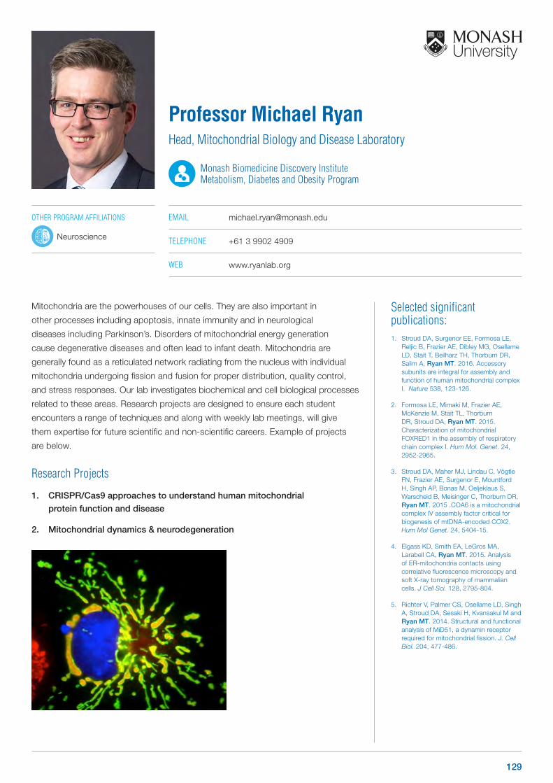
Selected significant publications: 1. Stroud DA, Surgenor EE, Formosa LE,
Reljic B, Frazier AE, Dibley MG, Osellame LD, Stait T, Beilharz TH, Thorburn DR, Salim A, Ryan MT. 2016. Accessory subunits are integral for assembly and function of human mitochondrial complex I. Nature 538, 123-126.
2. Formosa LE, Mimaki M, Frazier AE, McKenzie M, Stait TL, Thorburn DR, Stroud DA, Ryan MT. 2015. Characterization of mitochondrial FOXRED1 in the assembly of respiratory chain complex I. Hum Mol. Genet. 24, 2952-2965.
3. Stroud DA, Maher MJ, Lindau C, Vögtle FN, Frazier AE, Surgenor E, Mountford H, Singh AP, Bonas M, Oeljeklaus S, Warscheid B, Meisinger C, Thorburn DR, Ryan MT. 2015 .COA6 is a mitochondrial complex IV assembly factor critical for biogenesis of mtDNA-encoded COX2. Hum Mol Genet. 24, 5404-15.
4. Elgass KD, Smith EA, LeGros MA, Larabell CA, Ryan MT. 2015. Analysis of ER-mitochondria contacts using correlative fluorescence microscopy and soft X-ray tomography of mammalian cells. J Cell Sci. 128, 2795-804.
5. Richter V, Palmer CS, Osellame LD, Singh A, Stroud DA, Sesaki H, Kvansakul M and Ryan MT. 2014. Structural and functional analysis of MiD51, a dynamin receptor required for mitochondrial fission. J. Cell Biol. 204, 477-486.
Mitochondria are the powerhouses of our cells. They are also important in
other processes including apoptosis, innate immunity and in neurological
diseases including Parkinson’s. Disorders of mitochondrial energy generation
cause degenerative diseases and often lead to infant death. Mitochondria are
generally found as a reticulated network radiating from the nucleus with individual
mitochondria undergoing fission and fusion for proper distribution, quality control,
and stress responses. Our lab investigates biochemical and cell biological processes
related to these areas. Research projects are designed to ensure each student
encounters a range of techniques and along with weekly lab meetings, will give
them expertise for future scientific and non-scientific careers. Example of projects
are below.
Research Projects
1. CRISPR/Cas9 approaches to understand human mitochondrial
protein function and disease
2. Mitochondrial dynamics & neurodegeneration
Professor Michael RyanHead, Mitochondrial Biology and Disease Laboratory
Monash Biomedicine Discovery Institute Metabolism, Diabetes and Obesity Program
EMAIL [email protected]
TELEPHONE +61 3 9902 4909
WEB www.ryanlab.org
OTHER PROGRAM AFFILIATIONS
Neuroscience
129

Selected significant publications: 1. Simonds SE, Cowley MA. 2013.
Hypertension in obesity: is leptin the culprit? Trends Neurosci 36: 121-32
2. Simonds SE, Pryor JT, Ravussin E, Greenway FL, Dileone R, Allen AM, Bassi J, Elmquist JK, Keogh JM, Henning E, Myers MG, Jr., Licinio J, Brown RD, Enriori PJ, O’Rahilly S, Sternson SM, Grove KL, Spanswick DC, Farooqi IS, Cowley MA. 2014. Leptin mediates the increase in blood pressure associated with obesity. Cell 159: 1404-16
3. Simonds SE, Pryor JT, Cowley MA. 2017. Does leptin cause an increase in blood pressure in animals and humans? Curr Opin Nephrol Hypertens 26: 20-25
4. Simonds SE, Pryor JT, Cowley MA. 2018. Repeated weight cycling in obese mice causes increased appetite and glucose intolerance. Physiol Behav 194: 184-190
5. Simonds SE, Pryor JT, Koegler FH, Buch-Rasmussen AS, Kelly LE, Grove KL, Cowley MA. 2019. Determining the Effects of Combined Liraglutide and Phentermine on Metabolic Parameters, Blood Pressure, and Heart Rate in Lean and Obese Male Mice. Diabetes 68: 683-695
The primary research focus of the Integrated Physiology Lab is on the development
of obesity-associated diseases such as diabetes and cardiovascular diseases. We
use mouse models of disease states in order to probe the mechanisms underlying
obesity-associated disease development. We use world leading research techniques
in order to answer fundamental questions such as “what is a healthy diet? can I
be overweight and healthy?” “as a woman am I at increased risk of developing an
obesity-associated disease”. 2019 represents a particularly exciting year in the
Integrated Physiology Lab as we are embarking upon a novel research stream;
investigating the effects of air pollution on the cardiovascular, respiratory and
metabolic health.
Research Projects
1. Delineating the role of the melanocortin system in metabolic vs cardiac
control
2. The effect of particulate matter on metabolic and glycaemic control
3. The effects of air pollution, particulate matter on the cardiovascular system.
Dr Stephanie SimondsNHMRC Peter Doherty Biomedical Early Career Fellow National Heart Foundation of Australia Postdoctoral Fellow
Head, Integrated Physiology Laboratory
Monash Biomedicine Discovery Institute Metabolism, Diabetes and Obesity Program
EMAIL [email protected]
TELEPHONE +61 3 9905 2137
OTHER PROGRAM AFFILIATIONS
Cardiovascular Disease
130 www.monash.edu/discovery-institute
Selected significant publications: 1. Brown RM, Kupchik YM, Spencer S,
Garcia-Keller C, Spanswick D, Lawrence AJ, Simonds SE, Schwartz DJ, Jordan KA, Jhou TC, Kalivas PW. 2015. Addiction-like Synaptic Impairments in Diet-Induced Obesity. Biol Psychiatry pii: S0006-3223(15)00996-8..
2. Hebeisen S, Pires, N, Loureiro AI, Bonifácio MJ, Palma N, Whyment A, Spanswick D, Soares-da-Silva P. 2015. Eslicarbazepine and the enhancement of slow inactivation of voltage-gated sodium channels: A comparison with carbamazepine, oxcarbazepine and lacosamide. Neuropharmacology 89, 122-135.
3. Simonds SE, Pryor JT, Ravussin E, Greenway FL, Dileone R, Allen AM, Bassi J, Elmquist JK, Keogh JM, Henning E, Myers MG Jr, Licinio J, Brown RD, Enriori PJ, O’Rahilly S, Sternson SM, Grove KL, Spanswick DC, Farooqi IS, Cowley MA. 2014. Leptin mediates the increase in blood pressure associated with obesity. Cell, 159, 1404-1416.
4. Van den Top M, Lee K, Whyment A, Blanks A, Spanswick D. 2004. Orexigen-sensitive NPY/AgRP pacemaker neurons in the hypothalamic arcuate nucleus. Nature Neuroscience 7, 493-494
5. Spanswick D, Smith MA, Mirshamsi S, Routh VH, Ashford MLJ. 2000. Insulin activates ATP-sensitive potassium channels in hypothalamic neurones of lean, but not obese rats. Nature Neuroscience 3, 757-758.
Brain areas dedicated to controlling food intake and body weight include aspects of
the hypothalamus, key centres for sensing, integrating and formulating appropriate
behavioural responses to changes in energy status and the hedonic, reward-based
neural circuits. One nutrient that is controlled and maintained within narrow limits is
glucose. Glucose levels are maintained by a network of interacting peripheral and
central glucose-sensing systems. Consequently understanding the fundamental
mechanisms by which function-specific glucose-sensing neurons and networks
detect, respond and formulate appropriate output and if and how they are subject
to dysfunction in obesity and diabetes is critical to developing future intervention
strategies. We employ an electrophysiological approach to identify mechanisms
by which function-specific neurones and circuits detect changes in energy status to
co-ordinate appropriate behavioural responses and how they change depending on
the energy status of the organism.
Research Projects
1. Glucose-sensing neurons in the brain: how do they do it?
2. Motivation and reward: glucose, ghrelin and the mechanisms regulating
the dopaminergic neural circuits of the ventral tegmental area
Professor David SpanswickBiomedicine Discovery Fellow
Head, Obesity and Metabolic Neurophysiology Laboratory
Monash Biomedicine Discovery Institute Metabolism, Diabetes and Obesity Program
EMAIL [email protected]
TELEPHONE +61 3 9902 4307
WEB med.monash.edu/physiology/staff/spanswick.html
OTHER PROGRAM AFFILIATIONS
Neuroscience
131
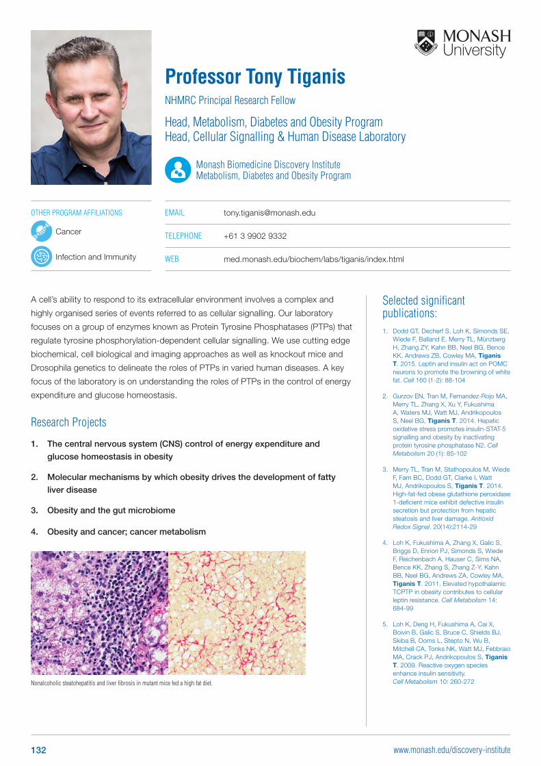
Selected significant publications: 1. Dodd GT, Decherf S, Loh K, Simonds SE,
Wiede F, Balland E, Merry TL, Münzberg H, Zhang ZY, Kahn BB, Neel BG, Bence KK, Andrews ZB, Cowley MA, Tiganis T. 2015. Leptin and insulin act on POMC neurons to promote the browning of white fat. Cell 160 (1-2): 88-104
2. Gurzov EN, Tran M, Fernandez-Rojo MA, Merry TL, Zhang X, Xu Y, Fukushima A, Waters MJ, Watt MJ, Andrikopoulos S, Neel BG, Tiganis T. 2014. Hepatic oxidative stress promotes insulin-STAT-5 signalling and obesity by inactivating protein tyrosine phosphatase N2. Cell Metabolism 20 (1): 85-102
3. Merry TL, Tran M, Stathopoulos M, Wiede F, Fam BC, Dodd GT, Clarke I, Watt MJ, Andrikopoulos S, Tiganis T. 2014. High-fat-fed obese glutathione peroxidase 1-deficient mice exhibit defective insulin secretion but protection from hepatic steatosis and liver damage. Antioxid Redox Signal. 20(14):2114-29
4. Loh K, Fukushima A, Zhang X, Galic S, Briggs D, Enriori PJ, Simonds S, Wiede F, Reichenbach A, Hauser C, Sims NA, Bence KK, Zhang S, Zhang Z-Y, Kahn BB, Neel BG, Andrews ZA, Cowley MA, Tiganis T. 2011. Elevated hypothalamic TCPTP in obesity contributes to cellular leptin resistance. Cell Metabolism 14: 684-99
5. Loh K, Deng H, Fukushima A, Cai X, Boivin B, Galic S, Bruce C, Shields BJ, Skiba B, Ooms L, Stepto N, Wu B, Mitchell CA, Tonks NK, Watt MJ, Febbraio MA, Crack PJ, Andrikopoulos S, Tiganis T. 2009. Reactive oxygen species enhance insulin sensitivity. Cell Metabolism 10: 260-272
A cell’s ability to respond to its extracellular environment involves a complex and
highly organised series of events referred to as cellular signalling. Our laboratory
focuses on a group of enzymes known as Protein Tyrosine Phosphatases (PTPs) that
regulate tyrosine phosphorylation-dependent cellular signalling. We use cutting edge
biochemical, cell biological and imaging approaches as well as knockout mice and
Drosophila genetics to delineate the roles of PTPs in varied human diseases. A key
focus of the laboratory is on understanding the roles of PTPs in the control of energy
expenditure and glucose homeostasis.
Research Projects
1. The central nervous system (CNS) control of energy expenditure and
glucose homeostasis in obesity
2. Molecular mechanisms by which obesity drives the development of fatty
liver disease
3. Obesity and the gut microbiome
4. Obesity and cancer; cancer metabolism
Professor Tony TiganisNHMRC Principal Research Fellow
Head, Metabolism, Diabetes and Obesity Program Head, Cellular Signalling & Human Disease Laboratory
Monash Biomedicine Discovery Institute Metabolism, Diabetes and Obesity Program
EMAIL [email protected]
TELEPHONE +61 3 9902 9332
WEB med.monash.edu/biochem/labs/tiganis/index.html
Nonalcoholic steatohepatitis and liver fibrosis in mutant mice fed a high fat diet.
OTHER PROGRAM AFFILIATIONS
Cancer
Infection and Immunity
132 www.monash.edu/discovery-institute
Neuroscience Program Group Leaders
133www.monash.edu/discovery-institute
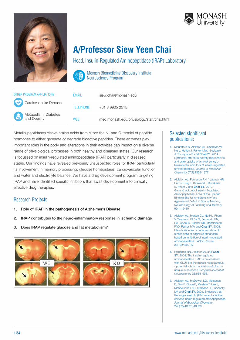
Selected significant publications: 1. Mountford S, Albiston AL, Charman W,
Ng L, Holien J, Parker MW, Nicolazzo J, Thompson P and Chai SY. 2014. Synthesis, structure-activity relationships and brain uptake of a novel series of benzopyran inhibitors of insulin-regulated aminopeptidase. Journal of Medicinal Chemistry 57(4):1368-1377.
2. Albiston AL, Fernando RN, Yeatman HR, Burns P, Ng L, Daswani D, Diwakarla S, Pham V and Chai SY. 2010. Gene Knockout of Insulin-Regulated Aminopeptidase: Loss of the Specific Binding Site for Angiotensin IV and Age-related Deficit in Spatial Memory. Neurobiology of Learning and Memory 93(1):19-30.
3. Albiston AL, Morton CJ, Ng HL, Pham V, Yeatman HR, Ye S, Fernando RN, De Bundel D, Ascher DB, Mendelsohn FAO, Parker MW and Chai SY. 2008. Identification and characterization of a new class of cognitive enhancers based on inhibition of insulin-regulated aminopeptidase. FASEB Journal 22(12):4209-17.
4. Fernando RN, Albiston AL and Chai SY. 2008. The insulin-regulated aminopeptidase IRAP is co-localised with GLUT4 in the mouse hippocampus - potential role in modulation of glucose uptake in neurons? European Journal of Neuroscience 28:588–598.
5. Albiston AL, McDowall SG, Matsacos D, Sim P, Clune E, Mustafa T, Lee J, Mendelsohn FAO, Simpson RJ, Connolly LM and Chai SY. 2001. Evidence that the angiotensin IV (AT4) receptor is the enzyme insulin regulated aminopeptidase. Journal of Biological Chemistry 276(52):48623-48626.
Metallo-peptidases cleave amino acids from either the N- and C-termini of peptide
hormones to either generate or degrade bioactive peptides. These enzymes play
important roles in the body and alterations in their activities can impact on a diverse
range of physiological processes in both healthy and diseased states. Our research
is focussed on insulin-regulated aminopeptidase (IRAP) particularly in diseased
states. Our findings have revealed previously unsuspected roles for IRAP particularly
its involvement in memory processing, glucose homeostasis, cardiovascular function
and water and electrolyte balance. We have a drug development program targeting
IRAP and have identified specific inhibitors that await development into clinically
effective drug therapies.
Research Projects
1. Role of IRAP in the pathogenesis of Alzheimer’s Disease
2. IRAP contributes to the neuro-inflammatory response in ischemic damage
3. Does IRAP regulate glucose and fat metabolism?
A/Professor Siew Yeen ChaiHead, Insulin-Regulated Aminopeptidase (IRAP) Laboratory
Monash Biomedicine Discovery Institute Neuroscience Program
EMAIL [email protected]
TELEPHONE +61 3 9905 2515
WEB med.monash.edu/physiology/staff/chai.html
OTHER PROGRAM AFFILIATIONS
Cardiovascular Disease
Metabolism, Diabetes and Obesity
134 www.monash.edu/discovery-institute
Selected significant publications: 1. George JT, Hendrikse M, Veldhuis JD,
Clarke IJ, Anderson RA, Millar RP. 2017. Inhibitory effect of gonadotropin inhibitory hormone (GnIH) on luteinizing hormone secretion in man. Clinical Endocrinology, 86:731-738.
2. Allen-Worthington K, Xie J, Brown JL, Edmunson AM, Dowling A, Navratil AM, Scavelli K, Yoon H, Kim D-G, Bynoe MS, Clarke IJ and Roberson MS. 2016. The F0F1 ATP synthase complex co-localizes with the GnRH receptor in membrane rafts in gonadotrope cells. Molecular Endocrinology 30:996-1011.
3. Ezzat A, Pereira A, Clarke IJ. 2015. Kisspeptin is a component of the pulse generator for GnRH secretion in female sheep but not the pulse generator. Endocrinology 156:1828-37.
4. Hewagalamulage S, Clarke IJ, Young IR, Rao A and Henry BA. 2015. High cortisol response to adrenocorticotrophic hormone (ACTH) identifies ewes with reduced melanocortin signalling and increased propensity to obesity. Journal of Neuroendocrinology 27:44-56.
5. Henry BA, Andrews Z, Rao A, Clarke IJ. 2011. Central leptin activates mitochondrial function and increases heat production in skeletal muscle. Endocrinology 152: 2609-2618
Work in our laboratory has contributed extensively to the field of neuroendocrinology
and currently have 3 main divisions:
• Reproduction: effects of gut peptides on the brain; role of kisspeptin and
gonadotropin inhibitory hormone on reproductive function; vaccination in early
life for life-long sterility.
• Metabolic neuroendocrinology: predisposition to obesity; relationship between
stress and metabolic function.
• Heat stress: effects on body tissues, gut and brain function.
We utilise sheep models, which allow a range of studies not easily undertaken in
small laboratory species. We have developed a number of novel neuroendocrine
methodologies that allow analysis ranging from the whole animal down to the single
cell and subcellular function. These techniques facilitate national and international
collaborations, with grant funding from Australian and offshore sources. In addition,
our laboratory undertakes a range of contract research projects.
Research Projects
Research in the Neuroendocrinology Lab currently focuses on the following areas:
1. Central regulation of reproduction by kisspeptin and gonadotropin
inhibitory hormone
2. Estrogen signalling in neuroendocrine systems
3. Control of food intake and energy expenditure by leptin and novel
regulatory factors
4. Optogenetic control of kisspeptin function
5. Neonatal sterility vaccination, using a novel approach
6. Heat stress in various genetic models
Professor Iain Clarke AMHead, Neuroendocrinology Laboratory
Monash Biomedicine Discovery Institute Neuroscience Program
EMAIL [email protected]
TELEPHONE +61 3 9905 2554
WEB med.monash.edu/physiology/staff/clarke.html
OTHER PROGRAM AFFILIATIONS
Development and Stem Cells
Metabolism, Diabetes and Obesity
135
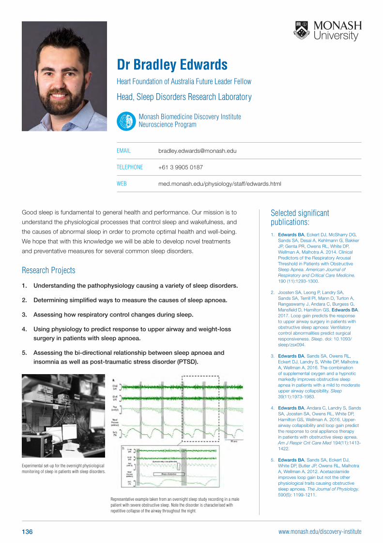
Selected significant publications: 1. Edwards BA, Eckert DJ, McSharry DG,
Sands SA, Desai A, Kehlmann G, Bakker JP, Genta PR, Owens RL, White DP, Wellman A, Malhotra A. 2014. Clinical Predictors of the Respiratory Arousal Threshold in Patients with Obstructive Sleep Apnea. American Journal of Respiratory and Critical Care Medicine. 190 (11):1293-1300.
2. Joosten SA, Leong P, Landry SA, Sands SA, Terrill PI, Mann D, Turton A, Rangaswamy J, Andara C, Burgess G, Mansfield D, Hamilton GS, Edwards BA. 2017. Loop gain predicts the response to upper airway surgery in patients with obstructive sleep apnoea: Ventilatory control abnormalities predict surgical responsiveness. Sleep. doi: 10.1093/sleep/zsx094.
3. Edwards BA, Sands SA, Owens RL, Eckert DJ, Landry S, White DP, Malhotra A, Wellman A. 2016. The combination of supplemental oxygen and a hypnotic markedly improves obstructive sleep apnea in patients with a mild to moderate upper airway collapsibility. Sleep 39(11):1973-1983.
4. Edwards BA, Andara C, Landry S, Sands SA, Joosten SA, Owens RL, White DP, Hamilton GS, Wellman A. 2016. Upper-airway collapsibility and loop gain predict the response to oral appliance therapy in patients with obstructive sleep apnea. Am J Respir Crit Care Med 194(11):1413-1422.
5. Edwards BA, Sands SA, Eckert DJ, White DP, Butler JP, Owens RL, Malhotra A, Wellman A. 2012. Acetazolamide improves loop gain but not the other physiological traits causing obstructive sleep apnoea. The Journal of Physiology. 590(5): 1199-1211.
Good sleep is fundamental to general health and performance. Our mission is to
understand the physiological processes that control sleep and wakefulness, and
the causes of abnormal sleep in order to promote optimal health and well-being.
We hope that with this knowledge we will be able to develop novel treatments
and preventative measures for several common sleep disorders.
Research Projects
1. Understanding the pathophysiology causing a variety of sleep disorders.
2. Determining simplified ways to measure the causes of sleep apnoea.
3. Assessing how respiratory control changes during sleep.
4. Using physiology to predict response to upper airway and weight-loss
surgery in patients with sleep apnoea.
5. Assessing the bi-directional relationship between sleep apnoea and
insomnia as well as post-traumatic stress disorder (PTSD).
Dr Bradley EdwardsHeart Foundation of Australia Future Leader Fellow
Head, Sleep Disorders Research Laboratory
Monash Biomedicine Discovery Institute Neuroscience Program
EMAIL [email protected]
TELEPHONE +61 3 9905 0187
WEB med.monash.edu/physiology/staff/edwards.html
Experimental set-up for the overnight physiological monitoring of sleep in patients with sleep disorders.
Representative example taken from an overnight sleep study recording in a male patient with severe obstructive sleep. Note the disorder is characterised with repetitive collapse of the airway throughout the night.
136 www.monash.edu/discovery-institute
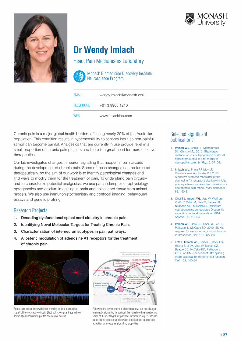
Selected significant publications: 1. Imlach WL, Bhola RF, Mohammadi
SA, Christie MJ. 2016. Glycinergic dysfunction in a subpopulation of dorsal horn interneurons in a rat model of neuropathic pain. Sci Rep. 6, 37104.
2. Imlach WL, Bhola RF, May LT, Christopoulos A, Christie MJ. 2015. A positive allosteric modulator of the adenosine A1 receptor selectively inhibits primary afferent synaptic transmission in a neuropathic pain model. Mol Pharmacol. 88, 460-8.
3. Choi BJ, Imlach WL, Jiao W, Wolfram V, Wu Y, Grbic M, Cela C, Baines RA, Nitabach MN, McCabe BD. Miniature neurotransmission regulates Drosophila synaptic structural maturation. 2014. Neuron. 82, 618-34.
4. Imlach WL, Beck ES, Choi BJ, Lotti F, Pellizzoni L, McCabe BD. 2012. SMN is required for sensory-motor circuit function in Drosophila. Cell. 151, 427-39.
5. Lotti F, Imlach WL, Saieva L, Beck ES, Hao le T, Li DK, Jiao W, Mentis GZ, Beattie CE, McCabe BD, Pellizzoni L. 2012. An SMN-dependent U12 splicing event essential for motor circuit function. Cell. 151, 440-54.
Chronic pain is a major global health burden, affecting nearly 20% of the Australian population. This condition results in hypersensitivity to sensory input so non-painful stimuli can become painful. Analgesics that are currently in use provide relief in a small proportion of chronic pain patients and there is a great need for more effective therapeutics.
Our lab investigates changes in neuron signalling that happen in pain circuits during the development of chronic pain. Some of these changes can be targeted therapeutically, so the aim of our work is to identify pathological changes and find ways to modify them for the treatment of pain. To understand pain circuitry and to characterize potential analgesics, we use patch-clamp electrophysiology, optogenetics and calcium imagining in brain and spinal cord tissue from animal models. We also use immunohistochemistry and confocal imaging, behavioural assays and genetic profiling.
Research Projects1. Decoding dysfunctional spinal cord circuitry in chronic pain.
2. Identifying Novel Molecular Targets for Treating Chronic Pain.
3. Characterization of interneuron subtypes in pain pathways.
4. Allosteric modulation of adenosine A1 receptors for the treatment
of chronic pain.
Dr Wendy ImlachHead, Pain Mechanisms Laboratory
Monash Biomedicine Discovery Institute Neuroscience Program
EMAIL [email protected]
TELEPHONE +61 3 9905 1210
WEB www.imlachlab.com
Spinal cord dorsal horn with inset showing an interneuron that is part of the nociceptive circuit. Electrophysiological trace in blue shows spontaneous firing of the nociceptive neuron.
Following the development of chronic pain we can see changes in synaptic signalling throughout the spinal cord pain pathways. Some of these changes are potential therapeutic targets. We use patch-clamp electrophysiology and electrical and optogenetic activation to investigate signalling properties.
137
Selected significant publications: 1. Kuruppu S, Rajapakse N, Parkington
H, Smith I. In Press. Pharmacological hypothesis: Nitric oxide induced inhibition of ADAM-17 activity as well as vesicle release can in turn prevent the production of soluble Endothelin Converting Enzyme. Pharmacology Research and Perspectives.
2. Han H, Baumann K, Casewell NR, Ali SA, Dobson J, Koludarov I, Debono J, Cutmore SC, Rajapakse NW, Jackson TN, Jones R, Hodgson WC, Fry BG, Kuruppu S. 2017. The cardiovascular and neurotoxic effects of the venoms of six bony and cartilaginous fish species. Toxins (Basel) 9(2).
3. Kuruppu S, Rajapakse NW, Spicer AJ, Parkington HC, Smith AI. 2016. Stimulating the activity of amyloid-beta degrading enzymes: a novel approach for the therapeutic manipulation of amyloid-beta levels. J Alzheimers Dis 54: 891-895.
4. Smith AI, Rajapakse NW, Kleifeld O, Lomonte B, Hodgson WC, Conroy PC, Spicer AJ, Sikanyika NL, Small DH, Kaye DM, Parkington HC, Whisstock JC, Kuruppu S. 2016. N-terminal domain of Bothrops asper Myotoxin II Enhances the Activity of Endothelin Converting Enzyme and Neprilysin. Sci Rep 6, 22413.
5. Kuruppu S, Chou SH, Feske SK, Suh S, Hanchapola I, Lo EH, Ning M, Smith AI. 2014. Soluble and catalytically active endothelin converting enzyme-1 is present in cerebrospinal fluid of subarachnoid hemorrhage patients. Mol Cell Proteomics 13, 1091-4
Development of novel drugs from animal venoms is a major focus of our current
research. We have developed an ideal drug candidate that targets one of the main
pathological features of Alzheimer’s disease. Preclinical trials on this molecule are
currently underway funded by the National Foundation for Medical Research and
Innovation. Other major focus of our group is the role of the endothelin system in
cardiovascular and renal disease. Together with researchers in engineering, our
laboratory is aiming to develop a device similar to a glucometer with capacity to
measure endothelin levels in circulation to detect renal disease early.
Research Projects
1. Examining the effect of venom derived molecule(s) as novel drug leads for
Alzheimer’s disease
2. Examining the role of the endothelin system in cardiovascular and renal
disease with a particular emphasis on characterising endothelins as
biomarkers
3. Characterisation of toxins in animal venoms as potential lead compounds
targeting cardiovascular disease
Dr Sanjaya KuruppuHead, Kuruppu Research Group
Monash Biomedicine Discovery Institute Neuroscience Program
EMAIL [email protected]
TELEPHONE +61 3 9902 9372
OTHER PROGRAM AFFILIATIONS
Cardiovascular Disease
138 www.monash.edu/discovery-institute
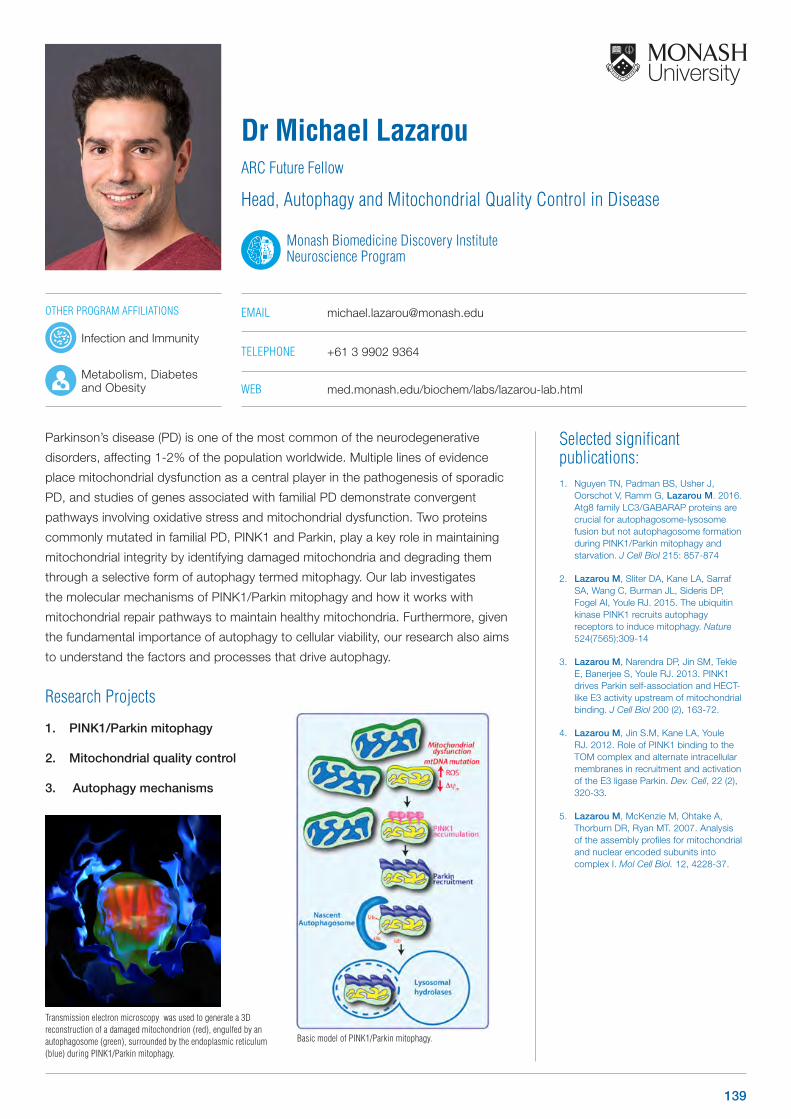
Selected significant publications: 1. Nguyen TN, Padman BS, Usher J,
Oorschot V, Ramm G, Lazarou M. 2016. Atg8 family LC3/GABARAP proteins are crucial for autophagosome-lysosome fusion but not autophagosome formation during PINK1/Parkin mitophagy and starvation. J Cell Biol 215: 857-874
2. Lazarou M, Sliter DA, Kane LA, Sarraf SA, Wang C, Burman JL, Sideris DP, Fogel AI, Youle RJ. 2015. The ubiquitin kinase PINK1 recruits autophagy receptors to induce mitophagy. Nature 524(7565):309-14
3. Lazarou M, Narendra DP, Jin SM, Tekle E, Banerjee S, Youle RJ. 2013. PINK1 drives Parkin self-association and HECT-like E3 activity upstream of mitochondrial binding. J Cell Biol 200 (2), 163-72.
4. Lazarou M, Jin S.M, Kane LA, Youle RJ. 2012. Role of PINK1 binding to the TOM complex and alternate intracellular membranes in recruitment and activation of the E3 ligase Parkin. Dev. Cell, 22 (2), 320-33.
5. Lazarou M, McKenzie M, Ohtake A, Thorburn DR, Ryan MT. 2007. Analysis of the assembly profiles for mitochondrial and nuclear encoded subunits into complex I. Mol Cell Biol. 12, 4228-37.
Parkinson’s disease (PD) is one of the most common of the neurodegenerative
disorders, affecting 1-2% of the population worldwide. Multiple lines of evidence
place mitochondrial dysfunction as a central player in the pathogenesis of sporadic
PD, and studies of genes associated with familial PD demonstrate convergent
pathways involving oxidative stress and mitochondrial dysfunction. Two proteins
commonly mutated in familial PD, PINK1 and Parkin, play a key role in maintaining
mitochondrial integrity by identifying damaged mitochondria and degrading them
through a selective form of autophagy termed mitophagy. Our lab investigates
the molecular mechanisms of PINK1/Parkin mitophagy and how it works with
mitochondrial repair pathways to maintain healthy mitochondria. Furthermore, given
the fundamental importance of autophagy to cellular viability, our research also aims
to understand the factors and processes that drive autophagy.
Research Projects
1. PINK1/Parkin mitophagy
2. Mitochondrial quality control
3. Autophagy mechanisms
Dr Michael LazarouARC Future Fellow
Head, Autophagy and Mitochondrial Quality Control in Disease
Monash Biomedicine Discovery Institute Neuroscience Program
EMAIL [email protected]
TELEPHONE +61 3 9902 9364
WEB med.monash.edu/biochem/labs/lazarou-lab.html
Transmission electron microscopy was used to generate a 3D reconstruction of a damaged mitochondrion (red), engulfed by an autophagosome (green), surrounded by the endoplasmic reticulum (blue) during PINK1/Parkin mitophagy.
Basic model of PINK1/Parkin mitophagy.
OTHER PROGRAM AFFILIATIONS
Infection and Immunity
Metabolism, Diabetes and Obesity
139
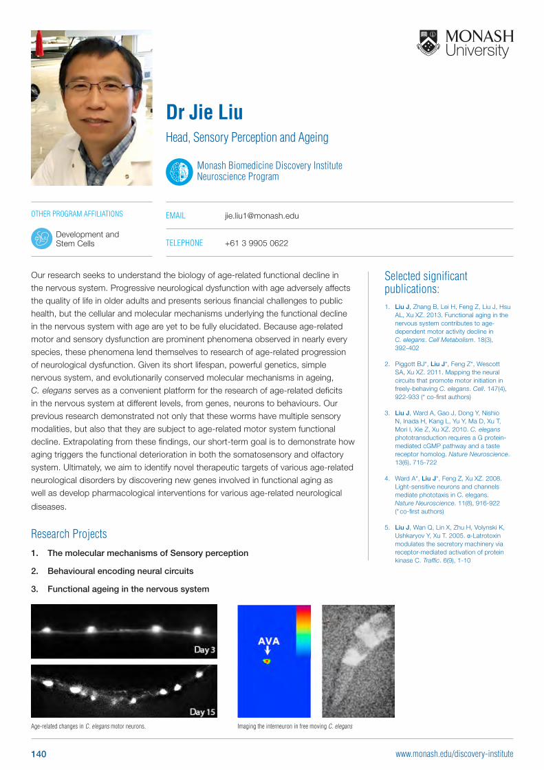
Selected significant publications: 1. Liu J, Zhang B, Lei H, Feng Z, Liu J, Hsu
AL, Xu XZ. 2013. Functional aging in the nervous system contributes to age-dependent motor activity decline in C. elegans. Cell Metabolism. 18(3), 392-402
2. Piggott BJ*, Liu J*, Feng Z*, Wescott SA, Xu XZ. 2011. Mapping the neural circuits that promote motor initiation in freely-behaving C. elegans. Cell. 147(4), 922-933 (* co-first authors)
3. Liu J, Ward A, Gao J, Dong Y, Nishio N, Inada H, Kang L, Yu Y, Ma D, Xu T, Mori I, Xie Z, Xu XZ. 2010. C. elegans phototransduction requires a G protein-mediated cGMP pathway and a taste receptor homolog. Nature Neuroscience. 13(6), 715-722
4. Ward A*, Liu J*, Feng Z, Xu XZ. 2008. Light-sensitive neurons and channels mediate phototaxis in C. elegans. Nature Neuroscience. 11(8), 916-922 (* co-first authors)
5. Liu J, Wan Q, Lin X, Zhu H, Volynski K, Ushkaryov Y, Xu T. 2005. α-Latrotoxin modulates the secretory machinery via receptor-mediated activation of protein kinase C. Traffic. 6(9), 1-10
Our research seeks to understand the biology of age-related functional decline in the nervous system. Progressive neurological dysfunction with age adversely affects the quality of life in older adults and presents serious financial challenges to public health, but the cellular and molecular mechanisms underlying the functional decline in the nervous system with age are yet to be fully elucidated. Because age-related motor and sensory dysfunction are prominent phenomena observed in nearly every species, these phenomena lend themselves to research of age-related progression of neurological dysfunction. Given its short lifespan, powerful genetics, simple nervous system, and evolutionarily conserved molecular mechanisms in ageing, C. elegans serves as a convenient platform for the research of age-related deficits in the nervous system at different levels, from genes, neurons to behaviours. Our previous research demonstrated not only that these worms have multiple sensory modalities, but also that they are subject to age-related motor system functional decline. Extrapolating from these findings, our short-term goal is to demonstrate how aging triggers the functional deterioration in both the somatosensory and olfactory system. Ultimately, we aim to identify novel therapeutic targets of various age-related neurological disorders by discovering new genes involved in functional aging as well as develop pharmacological interventions for various age-related neurological
diseases.
Research Projects1. The molecular mechanisms of Sensory perception
2. Behavioural encoding neural circuits
3. Functional ageing in the nervous system
Dr Jie LiuHead, Sensory Perception and Ageing
Monash Biomedicine Discovery Institute Neuroscience Program
EMAIL [email protected]
TELEPHONE +61 3 9905 0622
OTHER PROGRAM AFFILIATIONS
Development and Stem Cells
Age-related changes in C. elegans motor neurons. Imaging the interneuron in free moving C. elegans
140 www.monash.edu/discovery-institute
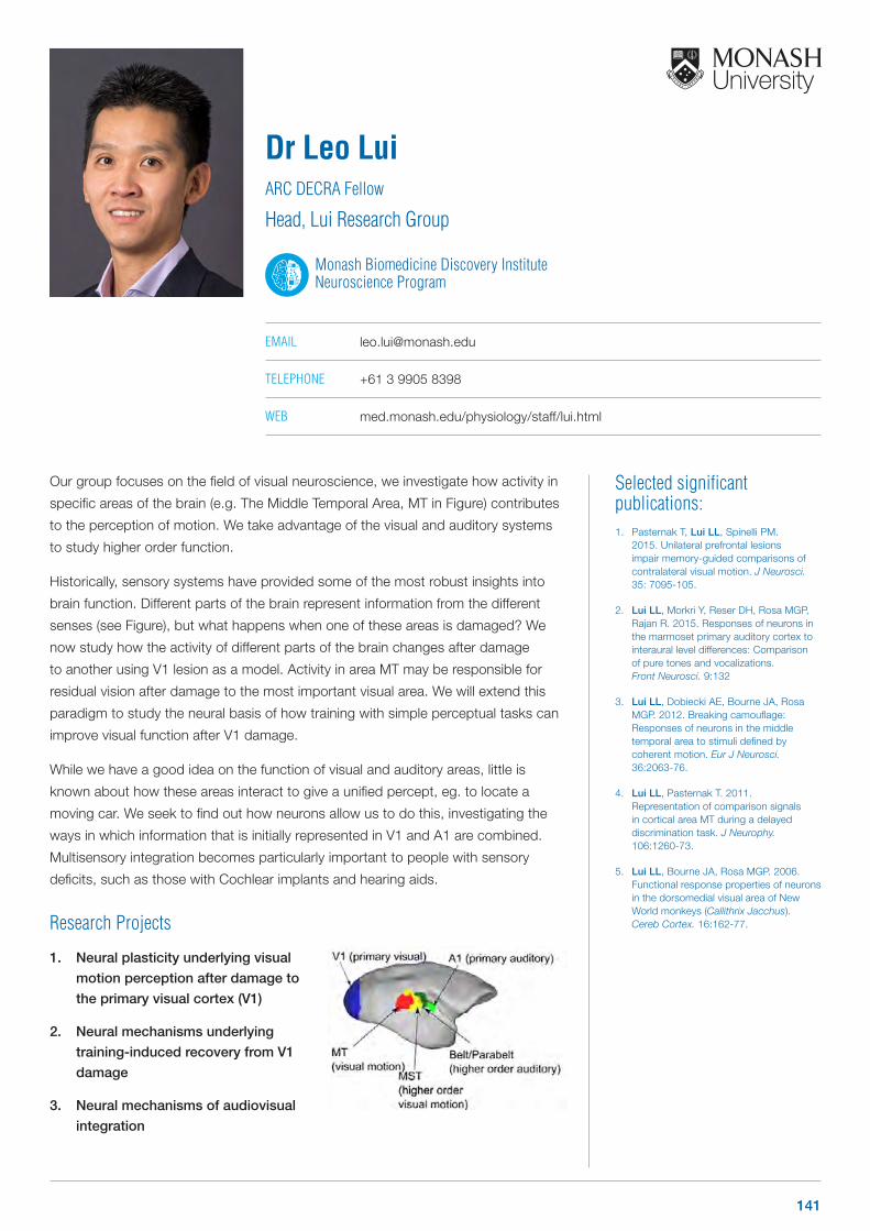
Selected significant publications: 1. Pasternak T, Lui LL, Spinelli PM.
2015. Unilateral prefrontal lesions impair memory-guided comparisons of contralateral visual motion. J Neurosci. 35: 7095-105.
2. Lui LL, Morkri Y, Reser DH, Rosa MGP, Rajan R. 2015. Responses of neurons in the marmoset primary auditory cortex to interaural level differences: Comparison of pure tones and vocalizations. Front Neurosci. 9:132
3. Lui LL, Dobiecki AE, Bourne JA, Rosa MGP. 2012. Breaking camouflage: Responses of neurons in the middle temporal area to stimuli defined by coherent motion. Eur J Neurosci. 36:2063-76.
4. Lui LL, Pasternak T. 2011. Representation of comparison signals in cortical area MT during a delayed discrimination task. J Neurophy. 106:1260-73.
5. Lui LL, Bourne JA, Rosa MGP. 2006. Functional response properties of neurons in the dorsomedial visual area of New World monkeys (Callithrix Jacchus). Cereb Cortex. 16:162-77.
Our group focuses on the field of visual neuroscience, we investigate how activity in
specific areas of the brain (e.g. The Middle Temporal Area, MT in Figure) contributes
to the perception of motion. We take advantage of the visual and auditory systems
to study higher order function.
Historically, sensory systems have provided some of the most robust insights into
brain function. Different parts of the brain represent information from the different
senses (see Figure), but what happens when one of these areas is damaged? We
now study how the activity of different parts of the brain changes after damage
to another using V1 lesion as a model. Activity in area MT may be responsible for
residual vision after damage to the most important visual area. We will extend this
paradigm to study the neural basis of how training with simple perceptual tasks can
improve visual function after V1 damage.
While we have a good idea on the function of visual and auditory areas, little is
known about how these areas interact to give a unified percept, eg. to locate a
moving car. We seek to find out how neurons allow us to do this, investigating the
ways in which information that is initially represented in V1 and A1 are combined.
Multisensory integration becomes particularly important to people with sensory
deficits, such as those with Cochlear implants and hearing aids.
Research Projects
1. Neural plasticity underlying visual
motion perception after damage to
the primary visual cortex (V1)
2. Neural mechanisms underlying
training-induced recovery from V1
damage
3. Neural mechanisms of audiovisual
integration
Dr Leo LuiARC DECRA Fellow
Head, Lui Research Group
Monash Biomedicine Discovery Institute Neuroscience Program
EMAIL [email protected]
TELEPHONE +61 3 9905 8398
WEB med.monash.edu/physiology/staff/lui.html
141
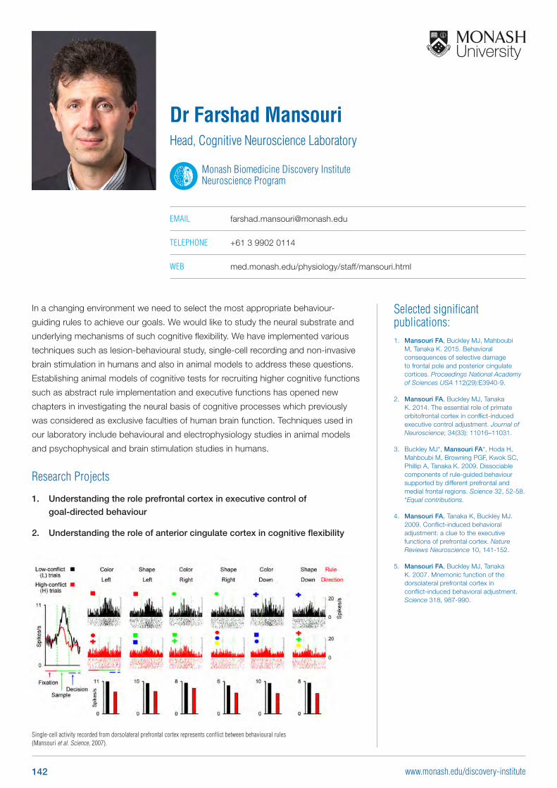
Selected significant publications: 1. Mansouri FA, Buckley MJ, Mahboubi
M, Tanaka K. 2015. Behavioral consequences of selective damage to frontal pole and posterior cingulate cortices. Proceedings National Academy of Sciences USA 112(29):E3940-9.
2. Mansouri FA, Buckley MJ, Tanaka K. 2014. The essential role of primate orbitofrontal cortex in conflict-induced executive control adjustment. Journal of Neuroscience; 34(33): 11016–11031.
3. Buckley MJ*, Mansouri FA*, Hoda H, Mahboubi M, Browning PGF, Kwok SC, Phillip A, Tanaka K. 2009. Dissociable components of rule-guided behaviour supported by different prefrontal and medial frontal regions. Science 32, 52-58. *Equal contributions.
4. Mansouri FA, Tanaka K, Buckley MJ. 2009. Conflict-induced behavioral adjustment: a clue to the executive functions of prefrontal cortex. Nature Reviews Neuroscience 10, 141-152.
5. Mansouri FA, Buckley MJ, Tanaka K. 2007. Mnemonic function of the dorsolateral prefrontal cortex in conflict-induced behavioral adjustment. Science 318, 987-990.
In a changing environment we need to select the most appropriate behaviour-
guiding rules to achieve our goals. We would like to study the neural substrate and
underlying mechanisms of such cognitive flexibility. We have implemented various
techniques such as lesion-behavioural study, single-cell recording and non-invasive
brain stimulation in humans and also in animal models to address these questions.
Establishing animal models of cognitive tests for recruiting higher cognitive functions
such as abstract rule implementation and executive functions has opened new
chapters in investigating the neural basis of cognitive processes which previously
was considered as exclusive faculties of human brain function. Techniques used in
our laboratory include behavioural and electrophysiology studies in animal models
and psychophysical and brain stimulation studies in humans.
Research Projects
1. Understanding the role prefrontal cortex in executive control of
goal-directed behaviour
2. Understanding the role of anterior cingulate cortex in cognitive flexibility
Dr Farshad MansouriHead, Cognitive Neuroscience Laboratory
Monash Biomedicine Discovery Institute Neuroscience Program
EMAIL [email protected]
TELEPHONE +61 3 9902 0114
WEB med.monash.edu/physiology/staff/mansouri.html
Single-cell activity recorded from dorsolateral prefrontal cortex represents conflict between behavioural rules (Mansouri et al. Science, 2007).
142 www.monash.edu/discovery-institute
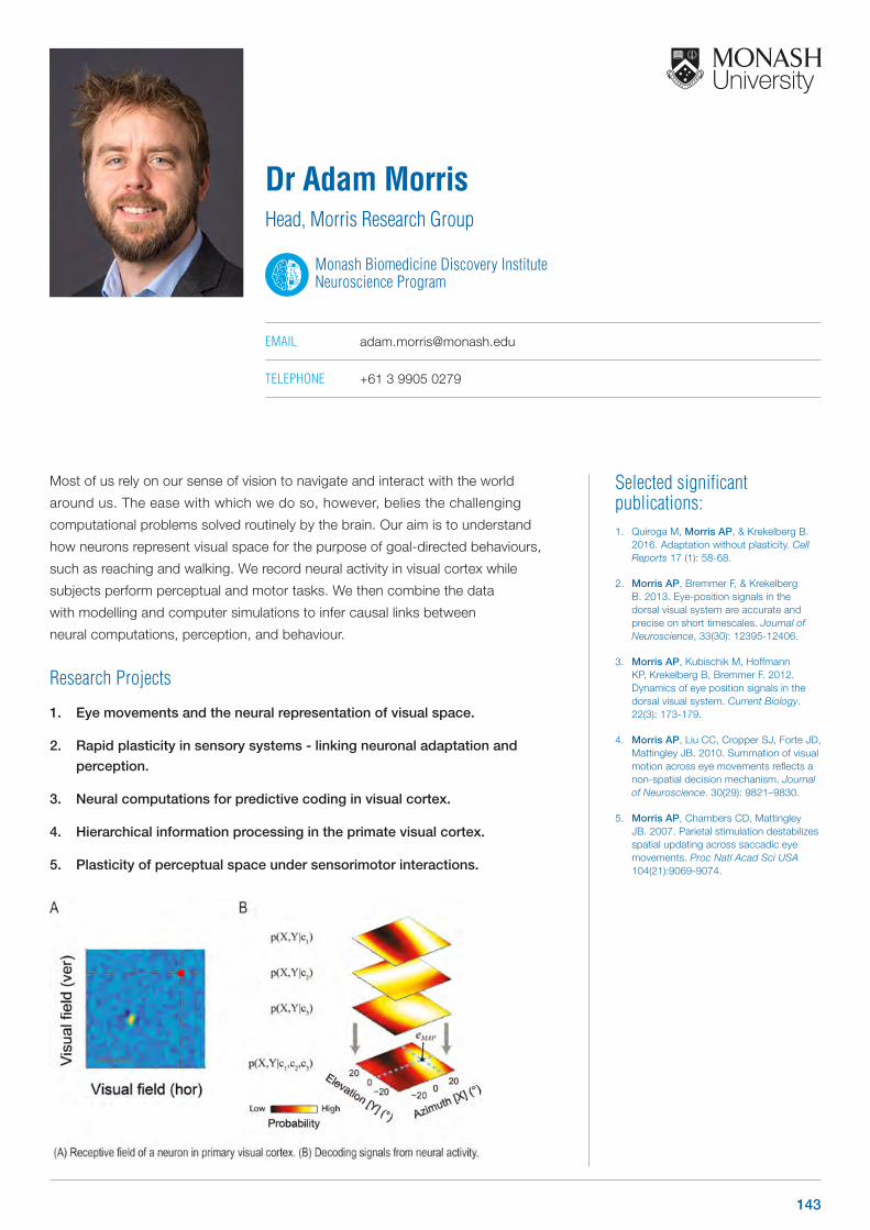
Selected significant publications: 1. Quiroga M, Morris AP, & Krekelberg B.
2016. Adaptation without plasticity. Cell Reports 17 (1): 58-68.
2. Morris AP, Bremmer F, & Krekelberg B. 2013. Eye-position signals in the dorsal visual system are accurate and precise on short timescales. Journal of Neuroscience, 33(30): 12395-12406.
3. Morris AP, Kubischik M, Hoffmann KP, Krekelberg B, Bremmer F. 2012. Dynamics of eye position signals in the dorsal visual system. Current Biology. 22(3): 173-179.
4. Morris AP, Liu CC, Cropper SJ, Forte JD, Mattingley JB. 2010. Summation of visual motion across eye movements reflects a non-spatial decision mechanism. Journal of Neuroscience. 30(29): 9821–9830.
5. Morris AP, Chambers CD, Mattingley JB. 2007. Parietal stimulation destabilizes spatial updating across saccadic eye movements. Proc Natl Acad Sci USA 104(21):9069-9074.
Most of us rely on our sense of vision to navigate and interact with the world
around us. The ease with which we do so, however, belies the challenging
computational problems solved routinely by the brain. Our aim is to understand
how neurons represent visual space for the purpose of goal-directed behaviours,
such as reaching and walking. We record neural activity in visual cortex while
subjects perform perceptual and motor tasks. We then combine the data
with modelling and computer simulations to infer causal links between
neural computations, perception, and behaviour.
Research Projects
1. Eye movements and the neural representation of visual space.
2. Rapid plasticity in sensory systems - linking neuronal adaptation and
perception.
3. Neural computations for predictive coding in visual cortex.
4. Hierarchical information processing in the primate visual cortex.
5. Plasticity of perceptual space under sensorimotor interactions.
Dr Adam MorrisHead, Morris Research Group
Monash Biomedicine Discovery Institute Neuroscience Program
EMAIL [email protected]
TELEPHONE +61 3 9905 0279
143
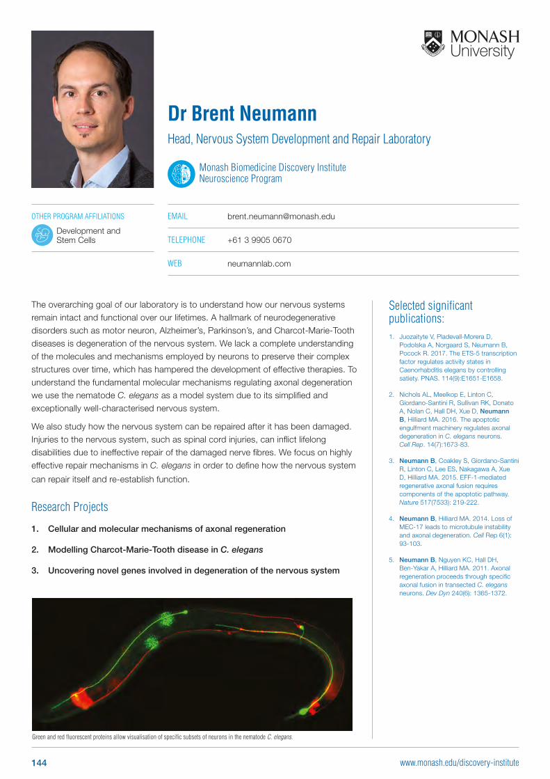
Selected significant publications: 1. Juozaityte V, Pladevall-Morera D,
Podolska A, Norgaard S, Neumann B, Pocock R. 2017. The ETS-5 transcription factor regulates activity states in Caenorhabditis elegans by controlling satiety. PNAS. 114(9):E1651-E1658.
2. Nichols AL, Meelkop E, Linton C, Giordano-Santini R, Sullivan RK, Donato A, Nolan C, Hall DH, Xue D, Neumann B, Hilliard MA. 2016. The apoptotic engulfment machinery regulates axonal degeneration in C. elegans neurons. Cell Rep. 14(7):1673-83.
3. Neumann B, Coakley S, Giordano-Santini R, Linton C, Lee ES, Nakagawa A, Xue D, Hilliard MA. 2015. EFF-1-mediated regenerative axonal fusion requires components of the apoptotic pathway. Nature 517(7533): 219-222.
4. Neumann B, Hilliard MA. 2014. Loss of MEC-17 leads to microtubule instability and axonal degeneration. Cell Rep 6(1): 93-103.
5. Neumann B, Nguyen KC, Hall DH, Ben-Yakar A, Hilliard MA. 2011. Axonal regeneration proceeds through specific axonal fusion in transected C. elegans neurons. Dev Dyn 240(6): 1365-1372.
The overarching goal of our laboratory is to understand how our nervous systems remain intact and functional over our lifetimes. A hallmark of neurodegenerative disorders such as motor neuron, Alzheimer’s, Parkinson’s, and Charcot-Marie-Tooth diseases is degeneration of the nervous system. We lack a complete understanding of the molecules and mechanisms employed by neurons to preserve their complex structures over time, which has hampered the development of effective therapies. To understand the fundamental molecular mechanisms regulating axonal degeneration we use the nematode C. elegans as a model system due to its simplified and exceptionally well-characterised nervous system.
We also study how the nervous system can be repaired after it has been damaged. Injuries to the nervous system, such as spinal cord injuries, can inflict lifelong disabilities due to ineffective repair of the damaged nerve fibres. We focus on highly effective repair mechanisms in C. elegans in order to define how the nervous system
can repair itself and re-establish function.
Research Projects
1. Cellular and molecular mechanisms of axonal regeneration
2. Modelling Charcot-Marie-Tooth disease in C. elegans
3. Uncovering novel genes involved in degeneration of the nervous system
Dr Brent NeumannHead, Nervous System Development and Repair Laboratory
Monash Biomedicine Discovery Institute Neuroscience Program
EMAIL [email protected]
TELEPHONE +61 3 9905 0670
WEB neumannlab.com
Green and red fluorescent proteins allow visualisation of specific subsets of neurons in the nematode C. elegans.
OTHER PROGRAM AFFILIATIONS
Development and Stem Cells
144 www.monash.edu/discovery-institute
Selected significant publications: 1. Zavitz E, Yu HH, Rowe EG, Rosa MG,
Price NSC. 2016. Rapid adaptation induces persistent biases in population codes for visual motion. J Neurosci 36(16):4579-90.
2. Blum J and Price NSC. 2014. Reflexive tracking eye movements and motion perception: one or two neural populations? Journal of Vision 13(4):23.
3. Price NSC and Born RT. 2013. Adaptation to speed in macaque middle temporal and medial superior temporal areas. J Neurosci. 33(10): 4359-68.
4. Price NSC and Born RT. 2010. Sensory- and decision- related activity in macaque MT and MST. J Neurosci. 30(42):14036-45.
5. Price NSC, Ono S, Mustari MJ, Ibbotson MR. 2005. Comparing acceleration and speed tuning in macaque MT: physiology and modeling. J Neurophys. 94(5):3451-64.
We study how the activity of small populations of sensory neurons underlies
conscious visual perception and the control of eye movements. We employ
a diverse range of methods including studies of human and animal behaviour,
extracellular neuronal recordings using multi-electrode arrays and computational
modelling. We are particularly interested in two main questions:
1. How does sustained exposure to a visual stimulus affect neuronal encoding
and perception of subsequently seen stimuli?
2. How does the activity of small population of sensory neurons encode visual
motion, and how can we decode this population activity to predict perception
and behaviour?
Research Projects
1. The neural correlates of perceptual backward masking
2. Is visual cortex self-aware?
3. How do sensory neurons incorporate dynamic stimulus statistics?
Dr Nicholas PriceHead, Price Research Group
Monash Biomedicine Discovery Institute Neuroscience Program
EMAIL [email protected]
TELEPHONE +61 3 9905 5131
WEB med.monash.edu/physiology/staff/price.html
145
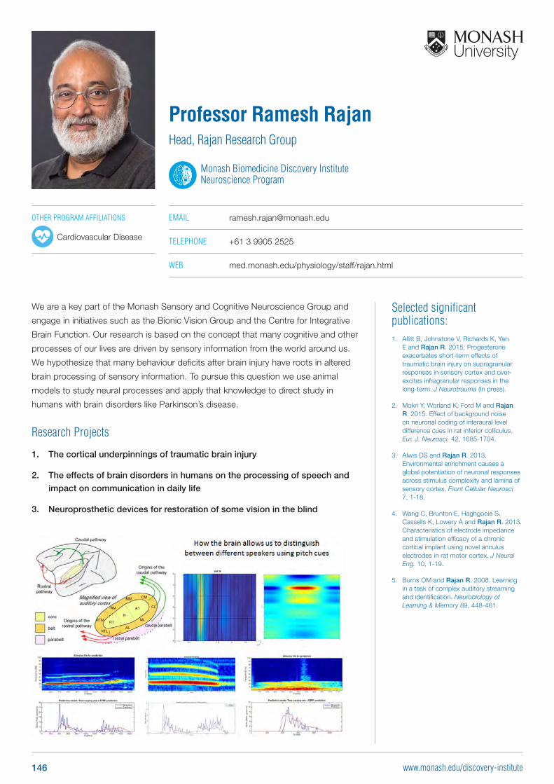
Selected significant publications: 1. Allitt B, Johnstone V, Richards K, Yan
E and Rajan R. 2015. Progesterone exacerbates short-term effects of traumatic brain injury on supragranular responses in sensory cortex and over-excites infragranular responses in the long-term. J Neurotrauma (In press).
2. Mokri Y, Worland K, Ford M and Rajan R. 2015. Effect of background noise on neuronal coding of interaural level difference cues in rat inferior colliculus. Eur. J. Neurosci. 42, 1685-1704.
3. Alwis DS and Rajan R. 2013. Environmental enrichment causes a global potentiation of neuronal responses across stimulus complexity and lamina of sensory cortex. Front Cellular Neurosci 7, 1-18.
4. Wang C, Brunton E, Haghgooie S, Cassells K, Lowery A and Rajan R. 2013. Characteristics of electrode impedance and stimulation efficacy of a chronic cortical implant using novel annulus electrodes in rat motor cortex. J Neural Eng. 10, 1-19.
5. Burns OM and Rajan R. 2008. Learning in a task of complex auditory streaming and identification. Neurobiology of Learning & Memory 89, 448-461.
We are a key part of the Monash Sensory and Cognitive Neuroscience Group and
engage in initiatives such as the Bionic Vision Group and the Centre for Integrative
Brain Function. Our research is based on the concept that many cognitive and other
processes of our lives are driven by sensory information from the world around us.
We hypothesize that many behaviour deficits after brain injury have roots in altered
brain processing of sensory information. To pursue this question we use animal
models to study neural processes and apply that knowledge to direct study in
humans with brain disorders like Parkinson’s disease.
Research Projects
1. The cortical underpinnings of traumatic brain injury
2. The effects of brain disorders in humans on the processing of speech and
impact on communication in daily life
3. Neuroprosthetic devices for restoration of some vision in the blind
Professor Ramesh RajanHead, Rajan Research Group
Monash Biomedicine Discovery Institute Neuroscience Program
EMAIL [email protected]
TELEPHONE +61 3 9905 2525
WEB med.monash.edu/physiology/staff/rajan.html
OTHER PROGRAM AFFILIATIONS
Cardiovascular Disease
146 www.monash.edu/discovery-institute
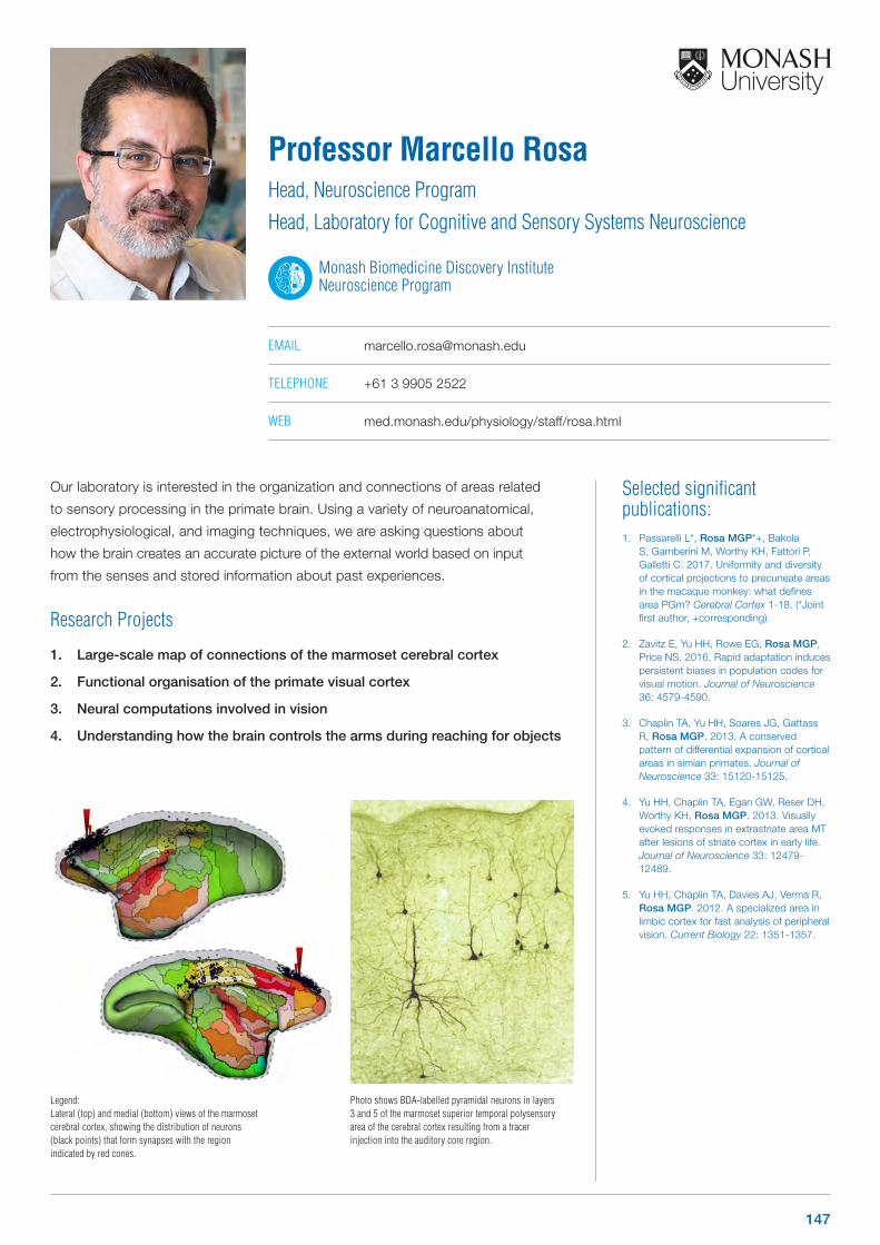
Selected significant publications: 1. Passarelli L*, Rosa MGP*+, Bakola
S, Gamberini M, Worthy KH, Fattori P, Galletti C. 2017. Uniformity and diversity of cortical projections to precuneate areas in the macaque monkey: what defines area PGm? Cerebral Cortex 1-18. (*Joint first author, +corresponding)
2. Zavitz E, Yu HH, Rowe EG, Rosa MGP, Price NS. 2016. Rapid adaptation induces persistent biases in population codes for visual motion. Journal of Neuroscience 36: 4579-4590.
3. Chaplin TA, Yu HH, Soares JG, Gattass R, Rosa MGP. 2013. A conserved pattern of differential expansion of cortical areas in simian primates. Journal of Neuroscience 33: 15120-15125.
4. Yu HH, Chaplin TA, Egan GW, Reser DH, Worthy KH, Rosa MGP. 2013. Visually evoked responses in extrastriate area MT after lesions of striate cortex in early life. Journal of Neuroscience 33: 12479-12489.
5. Yu HH, Chaplin TA, Davies AJ, Verma R, Rosa MGP. 2012. A specialized area in limbic cortex for fast analysis of peripheral vision. Current Biology 22: 1351-1357.
Our laboratory is interested in the organization and connections of areas related
to sensory processing in the primate brain. Using a variety of neuroanatomical,
electrophysiological, and imaging techniques, we are asking questions about
how the brain creates an accurate picture of the external world based on input
from the senses and stored information about past experiences.
Research Projects
1. Large-scale map of connections of the marmoset cerebral cortex
2. Functional organisation of the primate visual cortex
3. Neural computations involved in vision
4. Understanding how the brain controls the arms during reaching for objects
Professor Marcello RosaHead, Neuroscience Program
Head, Laboratory for Cognitive and Sensory Systems Neuroscience
Monash Biomedicine Discovery Institute Neuroscience Program
EMAIL [email protected]
TELEPHONE +61 3 9905 2522
WEB med.monash.edu/physiology/staff/rosa.html
Legend:Lateral (top) and medial (bottom) views of the marmoset cerebral cortex, showing the distribution of neurons (black points) that form synapses with the region indicated by red cones.
Photo shows BDA-labelled pyramidal neurons in layers 3 and 5 of the marmoset superior temporal polysensory area of the cerebral cortex resulting from a tracer injection into the auditory core region.
147
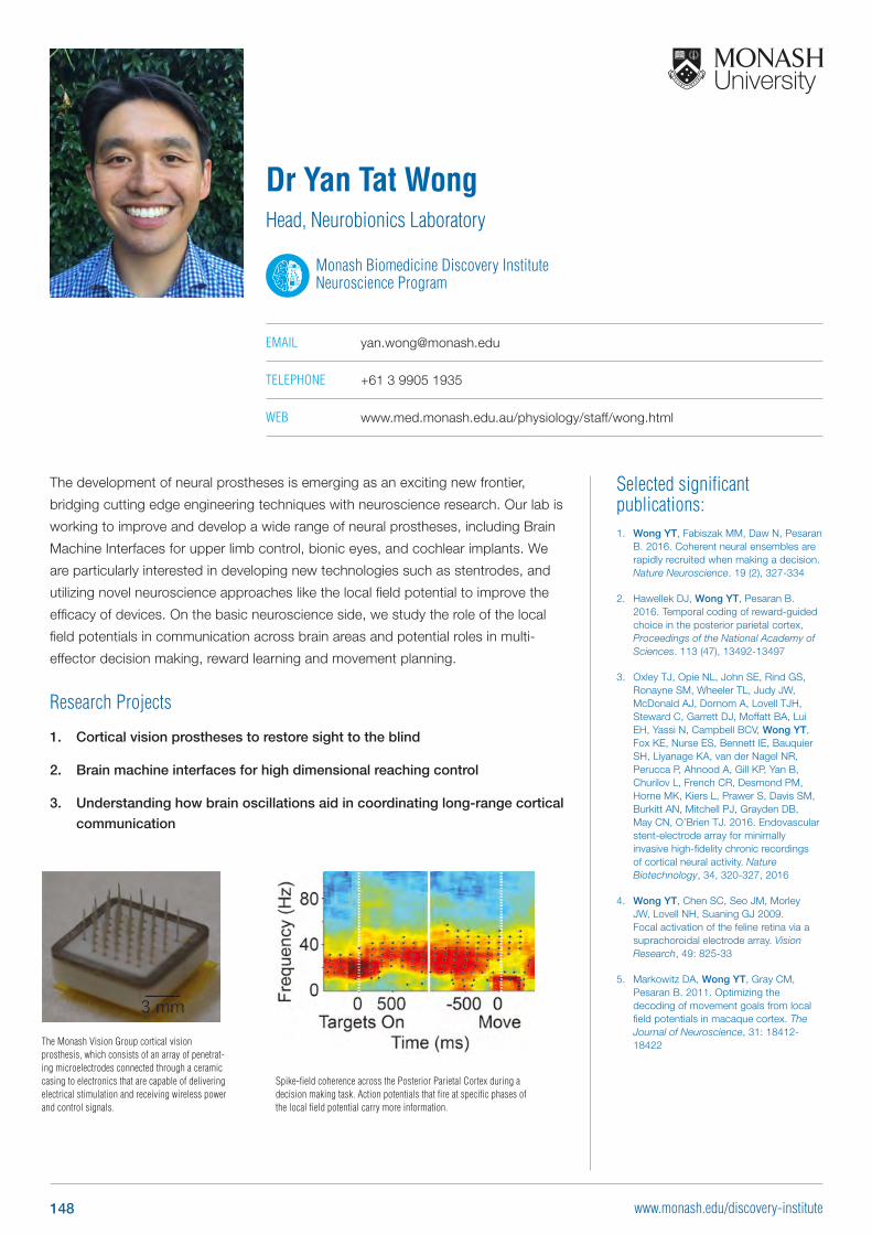
Selected significant publications: 1. Wong YT, Fabiszak MM, Daw N, Pesaran
B. 2016. Coherent neural ensembles arerapidly recruited when making a decision.Nature Neuroscience. 19 (2), 327-334
2. Hawellek DJ, Wong YT, Pesaran B.2016. Temporal coding of reward-guidedchoice in the posterior parietal cortex,Proceedings of the National Academy ofSciences. 113 (47), 13492-13497
3. Oxley TJ, Opie NL, John SE, Rind GS,Ronayne SM, Wheeler TL, Judy JW,McDonald AJ, Dornom A, Lovell TJH,Steward C, Garrett DJ, Moffatt BA, LuiEH, Yassi N, Campbell BCV, Wong YT, Fox KE, Nurse ES, Bennett IE, BauquierSH, Liyanage KA, van der Nagel NR,Perucca P, Ahnood A, Gill KP, Yan B,Churilov L, French CR, Desmond PM,Horne MK, Kiers L, Prawer S, Davis SM,Burkitt AN, Mitchell PJ, Grayden DB,May CN, O’Brien TJ. 2016. Endovascularstent-electrode array for minimallyinvasive high-fidelity chronic recordingsof cortical neural activity. Nature Biotechnology, 34, 320-327, 2016
4. Wong YT, Chen SC, Seo JM, MorleyJW, Lovell NH, Suaning GJ 2009.Focal activation of the feline retina via asuprachoroidal electrode array. Vision Research, 49: 825-33
5. Markowitz DA, Wong YT, Gray CM,Pesaran B. 2011. Optimizing thedecoding of movement goals from localfield potentials in macaque cortex. The Journal of Neuroscience, 31: 18412-18422
The development of neural prostheses is emerging as an exciting new frontier,
bridging cutting edge engineering techniques with neuroscience research. Our lab is
working to improve and develop a wide range of neural prostheses, including Brain
Machine Interfaces for upper limb control, bionic eyes, and cochlear implants. We
are particularly interested in developing new technologies such as stentrodes, and
utilizing novel neuroscience approaches like the local field potential to improve the
efficacy of devices. On the basic neuroscience side, we study the role of the local
field potentials in communication across brain areas and potential roles in multi-
effector decision making, reward learning and movement planning.
Research Projects
1. Cortical vision prostheses to restore sight to the blind
2. Brain machine interfaces for high dimensional reaching control
3. Understanding how brain oscillations aid in coordinating long-range cortical
communication
Dr Yan Tat WongHead, Neurobionics Laboratory
Monash Biomedicine Discovery Institute Neuroscience Program
EMAIL [email protected]
TELEPHONE +61 3 9905 1935
WEB www.med.monash.edu.au/physiology/staff/wong.html
Spike-field coherence across the Posterior Parietal Cortex during a decision making task. Action potentials that fire at specific phases of the local field potential carry more information.
The Monash Vision Group cortical vision prosthesis, which consists of an array of penetrat-ing microelectrodes connected through a ceramic casing to electronics that are capable of delivering electrical stimulation and receiving wireless power and control signals.
148 www.monash.edu/discovery-institute
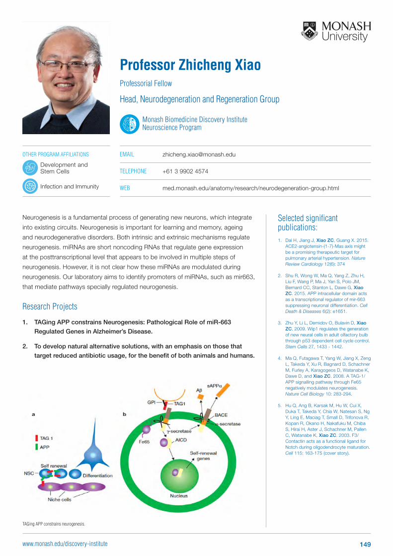
Selected significant publications: 1. Dai H, Jiang J, Xiao ZC, Guang X. 2015.
ACE2-angiotensin-(1-7)-Mas axis mightbe a promising therapeutic target forpulmonary arterial hypertension. Nature Review Cardiology 12(6): 374
2. Shu R, Wong W, Ma Q, Yang Z, Zhu H,Liu F, Wang P, Ma J, Yan S, Polo JM,Bernard CC, Stanton L, Dawe G, Xiao ZC. 2015. APP intracellular domain actsas a transcriptional regulator of mir-663suppressing neuronal differentiation. Cell Death & Diseases 6(2): e1651.
3. Zhu Y, Li L, Demidov O, Bulavin D, Xiao ZC. 2009. Wip1 regulates the generationof new neural cells in adult olfactory bulbthrough p53 dependent cell cycle control.Stem Cells 27, 1433 - 1442.
4. Ma Q, Futagawa T, Yang W, Jiang X, ZengL, Takeda Y, Xu R, Bagnard D, SchachnerM, Furley A, Karagogeos D, Watanabe K,Dawe D, and Xiao ZC. 2008. A TAG-1/APP signalling pathway through Fe65negatively modulates neurogenesis.Nature Cell Biology 10: 283-294.
5. Hu Q, Ang B, Karsak M, Hu W, Cui X,Duka T, Takeda Y, Chia W, Natesan S, NgY, Ling E, Maciag T, Small D, Trifonova R,Kopan R, Okano H, Nakafuku M, ChibaS, Hirai H, Aster J, Schachner M, PallenC, Watanabe K, Xiao ZC. 2003. F3/Contactin acts as a functional ligand forNotch during oligodendrocyte maturation.Cell 115: 163-175 (cover story).
Neurogenesis is a fundamental process of generating new neurons, which integrate
into existing circuits. Neurogenesis is important for learning and memory, ageing
and neurodegenerative disorders. Both intrinsic and extrinsic mechanisms regulate
neurogenesis. miRNAs are short noncoding RNAs that regulate gene expression
at the posttranscriptional level that appears to be involved in multiple steps of
neurogenesis. However, it is not clear how these miRNAs are modulated during
neurogenesis. Our laboratory aims to identify promoters of miRNAs, such as mir663,
that mediate pathways specially regulated neurogenesis.
Research Projects
1. TAGing APP constrains Neurogenesis: Pathological Role of miR-663
Regulated Genes in Alzheimer’s Disease.
2. To develop natural alternative solutions, with an emphasis on those that
target reduced antibiotic usage, for the benefit of both animals and humans.
Professor Zhicheng XiaoProfessorial Fellow
Head, Neurodegeneration and Regeneration Group
Monash Biomedicine Discovery Institute Neuroscience Program
EMAIL [email protected]
TELEPHONE +61 3 9902 4574
WEB med.monash.edu/anatomy/research/neurodegeneration-group.html
TAGing APP constrains neurogenesis.
OTHER PROGRAM AFFILIATIONS
Development and Stem Cells
Infection and Immunity
149www.monash.edu/discovery-institute


Monash University has been recognised for its enduring work in gender equity and for fostering an inclusive workplace culture with this citation from the Workplace Gender Equality Agency.
CONTACT USWhether you want to research, invest, study or partner with us, we’d be delighted to hear from you.
Biomedicine Graduate [email protected]
Monash Biomedicine Discovery Institute 23 Innovation WalkMonash UniversityClayton, Victoria, 3800
monash.edu/discovery-institutefacebook.com/MonashBDItwitter.com/MonashBDI
MONASH BDI PROGRAMS
Cancer [email protected]
Cardiovascular [email protected]
Development and Stem [email protected]
Infection and [email protected]
Metabolism, Diabetes and [email protected]
All information contained in this document is current at the time of publication. Monash University reserves the right to alter this information at any time (should the need arise). Please check the Monash University website for updates: www.monash.edu CRICOS provider: Monash University 00008C. Published April 2019. TRSUAPRIL2019