discoid lupus erythematosus in dogs - dog, cat & horse ...lupus... · the most common immune...
TRANSCRIPT

Discoid Lupus Erythematosus in Dogs
Dr Robert HiltonBVSc(Hons) MACVSc (Canine Medicine) Cert.VD MRCVS
Mobile 0433853560
Website www.skinvet.org
Email [email protected]

The most common immune mediated skin disease in Australia
There are gross differences in how the canine and human “discoid” lupus appears; hence, cutaneous lupus erythematosus (CLE) is a more appropriate name for the canine condition.
Collies and Shelties predisposed but may occur in a variety of breeds.
Severely exacerbated by UV radiation
Target = basement membrane area. Target antigens unclear.

Nasal planum is common target but may involve other mucocutaneous junctions, pinnae, scrotum and other sites
Inflammatory depigmentation, erosions and ulceration are typical lesions.
Limited to skin only. ANA –ve
Severe cases are complicated by infections and occasionally severe bleeding

Cutaneous (Discoid) Lupus

Cutaneous (Discoid) Lupus

Cutaneous (Discoid) Lupus

Cutaneous “discoid”lupus.
Alternative sites

Differential diagnosis
• Differential diagnosis Mucocutaneous pyoderma (major
differential)Other immune-mediated diseases (pemphigus,
SLE, dermatomyositis, vasculitis and the uveo-dermatologic syndrome).
Nasal dermatophytosisSolar or physical dermatitisNeoplasia

CAUTION
Diagnosis is by histopathologyDo not submit samples less than 4mm
in diameter
Histopathology is NOT RELIABLE in
differentiating DLE from muco-cutaneous pyoderma. Prior to biopsy,
assess response to 3 weeks of antibiotics. If complete cure, Dx = MCP

Mucocutaneous pyoderma pre
and post antibiotics

Cutaneous (Discoid) Lupus
Areas showing depigmentationand erythema should be selected for biopsy, rather than areas of ulceration and/or crusts.

Balance the severity of cutaneous lupus against the risks of therapy.
Use the least toxic drugs and control sunlight exposure. 90% resolution is target!

Treatment OverviewSun Avoidance essential. Control without this almost impossible.
Short term immunosuppressive doses of prednisolone to induce remission. Long term immunosuppession, as per pemphigus foliaceus, is the last resort.
Topical Corticosteroids : skin thinning, calcinosis cutis and infections. Short term use OK. Long term 1-2x week but watch for skin thinning.
Tetracyclines and niacinamideTopical TacrolimusHydroxycloroquinine (Plaquinil)Cyclosporine (severe refractory cases)
Ancillary therapy - Vitamin E- Omega 3/6 oils- Antibiotics for secondary infection

Topical Tacrolimus
Similar action but different binding site to cyclosporine
Pimecrolimus : No studies and dubious absorption
10 cases, 0.1% tacrolimus once a day , 80% responded. 75% of responding cases could be maintained on topical tacrolimus alone.
Compounded tacrolimus is the only form available in Australia and the author has NOT enjoyed success using it as monotherapy.
Unregistered and wear gloves

Tetracycline and Niacinamide (nicotinamide, nicotinic acid)
Multiple anti-inflammatory properties. Neither effective alone in dogs. Widely used.
70% success rate in maintaining. Assess after 10 weeks of use.
Dogs >10kg BW 500mg of each TID. Smaller dogs 250mg of each TID
Doxycycline (7.5-10mg/kg SID) may be substituted for tetracycline. Not cheap.
With three times a day medication, compliance is an issue. The author has had good results with doxycycline 1x day and 350-750 mg of niacinamide 2x day

Other immunosuppressants
Hydroxychloroquinine
(Plaquinil)Antimalarial with immuno-
modulating properties
Good safety profile in dogs
5mg/kg once a day dose and cheap. Assess after 10 weeks.
This is the “go to” drug for human DLE. The author has had success in a limited number of cases.
Oral Cyclosporine
Many anecdotal reports that effective at atopic dermatitis protocol
For refractory cases
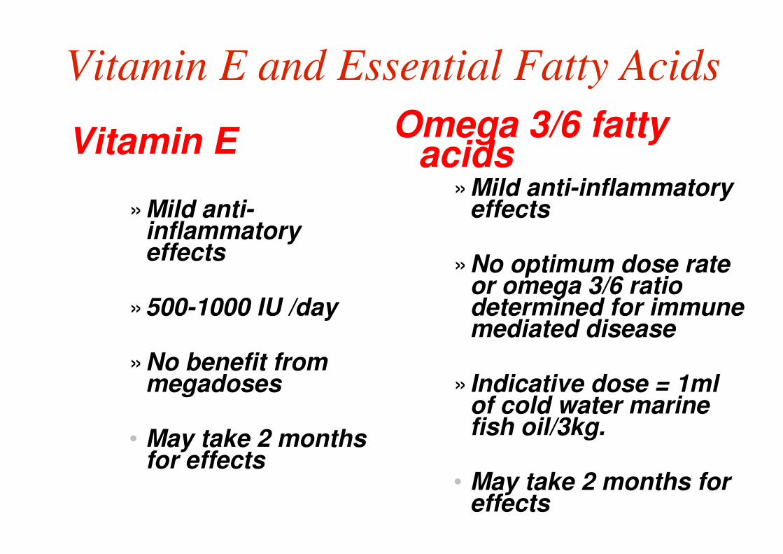
Vitamin E and Essential Fatty Acids
Vitamin E
»Mild anti-inflammatory effects
»500-1000 IU /day
»No benefit from megadoses
• May take 2 months for effects
Omega 3/6 fatty acids
»Mild anti-inflammatory effects
»No optimum dose rate or omega 3/6 ratio determined for immune mediated disease
» Indicative dose = 1ml of cold water marine fish oil/3kg.
• May take 2 months for effects

A sample treatment plan
• Confirm diagnosis
• Aim for 90% symptom control. Pigment may never return.
• Keep out of sun. Non zinc sunscreens high SPF not a substitute.
• Treat secondary infection
• 4-6 weeks reducing course of corticosteroids (topical and parental to induce remission. Topical steroids no more than 1-2x week long term. Watch skin thinning.
• Same time, start Doxycycline/niacinamide or hydroxychloroquinine. Vit E and omega-3 oil may help.
• Re-assess in 10 weeks. If not good response, change systemic meds over and/or add in topical tacrolimus
• If refractory, cyclosporine