directed differentiation of human embryonic stem cells: a
TRANSCRIPT
Thesis for doctoral degree (Ph.D.)2008
Directed differentiation ofhuman embryonic stem cells:
A model for early bone development
Elerin Kärner
Thesis for doctoral degree (Ph.D.) 2008
Elerin Kärner
Directed differen
tiation of H
ESC
s: A m
odel for early bone developm
ent

From the CENTER FOR ORAL BIOLOGY,
INSTITUTE OF ODONTOLOGY, Karolinska Institutet, Stockholm, Sweden
DIRECTED DIFFERENTIATION OF
HUMAN EMBRYONIC STEM CELLS: A MODEL FOR
EARLY BONE DEVELOPMENT
Elerin Kärner
Stockholm 2008

2008
Gårdsvägen 4, 169 70 Solna
Printed by
All previously published papers were reproduced with permission from the publisher. Published by Karolinska Institutet. © Elerin Kärner, 2008 ISBN 978-91-7409-020-8

To my family

ABSTRACT Research in stem cell biology is an important and necessary requirement for the
better understanding of cell differentiation and formation of tissues, while also
contributing to the field of regenerative medicine. The establishment of human
embryonic stem cell (HESC) lines offers the potential to study the earliest
developmental processes and provides an unlimited source of cells which can be used
for the differentiation into functional osteoblasts. Bone matrix production and
mineralization are guided by complicated mechanisms that differ from other tissues in
many ways. There is the initial formation of an organic extracellular matrix (ECM) into
which inorganic hydroxyapatite crystals are later deposited. Our first study investigated
the molecular processes that occur pre- and post-mineralization within the primary
ossification centre during early bone formation using global gene expression analysis.
We then continued investigating the osteogenic differentiation potential of several
HESC lines. Novel to our studies was the use of commercially available human
foreskin fibroblasts to support the undifferentiated growth of the HESC colonies and
their propagation in serum-replacement containing culture medium. Two different
approaches to differentiate HESCs into the osteogenic lineage were evaluated. Firstly,
undifferentiated cells were cultured in suspension, facilitating the formation of
embryoid bodies (EB), and secondly in monolayer; both methods were in the presence
of osteogenic supplements. Characterization of the osteogenic phenotype revealed that
all HESC lines differentiated towards the osteoblastic lineage, demonstrating also that
EB formation is not necessary for the initiation of osteogenic differentiation.
Mineralization of the ECM occurred through a cell-mediated calcification process.
Study of the expression profile of bone-associated genes revealed that the HESC model
differs from the standard osteogenesis model, which has been characterized by
osteoprogenitor cells. In the redefined model there is first the general cellular
proliferation and secretion of pre-maturational matrix stage that is needed for cell
migration, and second, the appearance of osteoprogenitors with characteristic ECM
synthesis. A gene modification approach to enhance potential osteoblastic
differentiation was employed in the fourth and final study. We found that for enhanced
osteogenesis originating from in vitro cultured HESCs, the correct levels of ectopic
transcription factors need to be established. Our data adds additional confirmation of a
close relationship between early blood and bone development.

LIST OF PUBLICATIONS I. Sugars RV., Kärner E., Petersson U.,Ganss B., Wendel M.
Transcriptome analysis of fetal metatarsal long bones by microarray, as a
model for endochondral bone formation. (2006) Biochim Biophys Acta.
Oct;1763(10):1031-9.
II. Kärner E., Unger C., Sloan AJ., Ährlund-Richter L., Sugars RV., Wendel
M. Bone Matrix Formation in Osteogenic Cultures Derived from Human
Embryonic Stem Cells In Vitro. (2007) Stem Cells Dev., Feb;16(1):39-52.
III. Kärner E., Bäckesjö C-M., Cedervall J., Sugars RV., Ährlund-Richter L.,
Wendel M. Dynamics of gene expression during bone matrix formation in
osteogenic cultures derived from human embryonic stem cells in vitro.
Submitted.
IV. Kärner E.*, Unger C.*, Cerny R., Ährlund-Richter L., Ganss B., Dilber S.,
Wendel M. * Authors have contributed equally to this study.
Differentiation of human embryonic stem cells into osteogenic or
hematopoietic lineages: a dose-dependent effect of Osterix over-expression.
Submitted.
CONTENTS 1 INTRODUCTION ......................................................................................1
1.1 STEM CELLS...................................................................................1 1.2 Embryonic stem cells ........................................................................2
1.2.1 Derivation of human ESCs....................................................3 1.2.2 Maintaining undifferentiated HESCs ....................................5 1.2.3 Markers of HESCs ................................................................9 1.2.4 Transcriptional networks in HESCs....................................10 1.2.5 Differentiation of HESCs ....................................................11
1.3 BONE TISSUE ...............................................................................14 1.3.1 Bone formation....................................................................14 1.3.2 Bone-producing cells...........................................................15 1.3.3 Osteoblast differentiation process .......................................16 1.3.4 Transcriptional control of osteoblast differentiation ...........17 1.3.5 Regulation of osteoblast differentiation ..............................19 1.3.6 Extracellular matrix of bone................................................22 1.3.7 Mechanisms of mineralization ............................................25 1.3.8 In vitro models for osteogenesis..........................................25 1.3.9 Differentiating ESCs to osteoblasts.....................................26
2 AIMS OF THE PRESENT INVESTIGATION .......................................33 3 MATERIALS AND METHODS .............................................................34
3.1 In vivo model for osteogenesis (paper I) .........................................34 3.2 Osteogenic differentiation of HESCs in vitro (papers II, III, IV)....34
3.2.1 HESC culture maintenance .................................................34 3.2.2 Control cell lines and culture conditions .............................35 3.2.3 Osteogenic differentiation in vitro.......................................35 3.2.4 Cellular proliferation and metabolic activity (paper III) .....36 3.2.5 Assessment of osteogenic phenotype..................................36 3.2.6 Lentiviral transgene expression (paper IV) .........................40
4 RESULTS AND DISCUSSION...............................................................42 4.1 Paper I..............................................................................................42 4.2 Paper II ............................................................................................43 4.3 Paper III ...........................................................................................44 4.4 Paper IV...........................................................................................46
5 CONCLUSIONS.......................................................................................48 6 FUTURE PERSPECTIVES......................................................................49 7 ACKNOWLEDGMENTS ........................................................................50 8 REFERENCES .........................................................................................52

LIST OF ABBREVIATIONS
AA Ascorbic acid ALP Alkaline phosphatase AR Alizarin Red S staining
-GP -glycerophosphate bFGF Basic fibroblast growth factor BMP Bone morphogenetic protein BSP Bone sialoprotein Cbf Core-binding factor Dex Dexamethasone EB Embryoid body EGFP Enhanced green fluorescent
protein ECM Extracellular matrix EGF Epidermal growth factor ESC Embryonic stem cell FACS Fluorescence activated cell
sorting FBS Fetal bovine serum Flt Fms-like tyrosine kinase FTIR Fourier-transform infrared GAG Glycosaminoglycan GSK Glycogen synthase kinase HESC Human embryonic stem cells HGF Hepatocyte growth factor HLA Human leukocyte antigen HoxB4 Homeobox B4 HSC Hematopoietic stem cell ICM Inner cell mass IGF Insuline-like growth factor IL Interleukin IVF In vitro fertilization JAK Janus kinase KO-SR Knockout serum replacement LIF Leukemia inhibitory factor LRP Low density lipoprotein
receptor-related protein M-CSF Macrophage colony-
stimulating factor MEF Mouse embryonic fibroblasts MSC Mesenchymal stem cell
NC Neural crest NCP Non-collagenous
glycoproteins and proteoglycans NF- B Nuclear factor kappa B OCN Osteocalcin Oct-4 Octamer binding protein-4 ON Osteonectin OPN Osteopontin OSAD Osteoadherin OSX Osterix PI3K Phosphoinositide kinase-3 PK Protein kinase RA Retinoic acid RANK Receptor activation of nuclear
factor kappa B RANKL RANK ligand RC Fetal rat calvaria-derived cells ROCK P160-Rho-associated coiled-coil
kinase RT-PCR Reverse-transcriptase PCR Runx Runt-related factor SCID Severe combined immunodeficient SIBLINGS Small Integrin Binding Ligand N-
linked Glycoproteins Sox SRY (sex determining region Y)-
box Sp Specificity protein SPARC Secreted protein, acidic, rich in
cysteine SSEA Stage-specific embryonic antigen STAT Signal-transduced and activator of
transcription TC Tetracycline TGF Transforming growth factor TNF Tumour necrosis factor TRA Tumour recognition antigen VEGF Vascular endothelial growth factor vitD3 1,25-dihydroxy vitamin D3 Wnt Wingless


Osteogenic differentiation of HESCs
1
1 INTRODUCTION Stem cells serve as a fundamental source for tissues throughout the life of every
organism. They provide the body with cells for replacement during growth, and are
responsible for regeneration following disease or injury. Such cells are found not only
during early development, but also in the adult body. Research into stem cell biology is
likely to provide useful information to applications such as tissue replacement and drug
screening.
1.1 STEM CELLS Stem cells are able to differentiate into other types of cells of the organism, and
in addition stem cells possess the ability to self-renew. All developing tissues retain
cells with stem cell properties; however whether this is the case throughout the entire
adult body remains to be clearly demonstrated. Certain tissues, such as skin, muscle,
and the hematopoietic system are capable of renewal, although recent medical research
demonstrates that tissues previously believed to be non-regenerative, such as brain and
heart may possess similar properties [1, 2].
Developmental potency is a functional characterization of stem cells, and does
not necessarily describe the range of genes expressed by the cells, their origin and
whether they represent an endogenous cell type in the organism. The potency may be
revealed experimentally in vitro, by i) forming aggregates in suspension culture, ii) in
vivo within a teratoma following injection into immunocompromised mice, and iii)
within an embryo that has had pluripotent cells injected into the blastocyst and results
in the birth of chimeras [3]. Different types of stem cells exist, depending on the ability
to maintain stem cell–like properties and the variability of derivatives that they give rise
to. Unipotent stem cells undergo self-renewal and are able to generate only one mature
cell type. Multipotent stem cells give rise to two or more differentiated cell types. A
large number of multipotential cells exist; an example is seen in the adult organism, and
during early development, where tissues in the nervous system contain neural crest
(NC) stem cells and neural stem cells. Hematopoietic stem cells (HSCs) give rise to
lineage restricted stem cells, which can further differentiate into numerous blood cell
types [4]. HSCs, together with mesenchymal stem cells (MSCs) reside in the bone
marrow. Bone-producing cells, osteoblasts, originate from MSCs along with
adipocytes, muscle cells and chondrocytes [5]. The hallmark of pluripotent stem cells
is the potential to give rise to the representatives of the three germ layers; endoderm,
Elerin Kärner
2
mesoderm, and ectoderm. This is determined using cell type specific molecular
markers, morphological criteria, and functionality. Three types of pluripotent stem cells
have been described so far; i) embryonic germ (EG) cells of the gonads of a post-
implantation embryo, ii) embryonal carcinoma (EC) cells, originating from tumorigenic
germinal tissue, and iii) embryonic stem cells (ESCs) [6]. Recently, new human
pluripotent cell lines were induced through a process of reprogramming somatic cells
(iPS) [7-9].
Moreover, despite on the above mentioned definitions of pluripotency and
multipotency, it is clear that cells with intermediate potencies could exist, for example,
the existence of mesoangioblasts has been suggested [10].
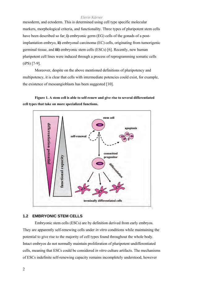
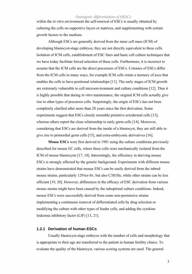
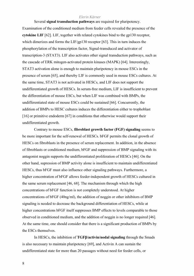
Figure 1. A stem cell is able to self-renew and give rise to several differentiated
cell types that take on more specialized functions.
1.2 EMBRYONIC STEM CELLS Embryonic stem cells (ESCs) are by definition derived from early embryos.
They are apparently self-renewing cells under in vitro conditions while maintaining the
potential to give rise to the majority of cell types found throughout the whole body.
Intact embryos do not normally maintain proliferation of pluripotent undifferentiated
cells, meaning that ESCs could be considered in vitro culture artifacts. The mechanisms
of ESCs indefinite self-renewing capacity remains incompletely understood, however
Osteogenic differentiation of HESCs
3
within the in vitro environment the self-renewal of ESCs is usually obtained by
culturing the cells on supportive layers or matrices, and supplementing with certain
growth factors to the medium.
Although ESCs are generally derived from the inner cell mass (ICM) of
developing blastocyst-stage embryos, they are not directly equivalent to these cells.
Isolation of ICM cells, establishment of ESC lines and basic cell culture techniques that
we have today facilitate forced selection of these cells. Furthermore, it is incorrect to
assume that the ICM cells are the direct precursors of ESCs. Colonies of ESCs differ
from the ICM cells in many ways, for example ICM cells retain a memory of axes that
enables the cells to have positional relationships [11]. The early stages of ICM growth
are extremely vulnerable to cell microenvironment and culture conditions [12]. Thus it
is highly possible that during in vitro maintenance, the original ICM cells actually give
rise to other types of precursor cells. Surprisingly, the origin of ESCs has not been
completely clarified after more than 20 years since the first derivation. Some
experiments suggest that ESCs closely resemble primitive ectodermal cells [13],
whereas others report the close relationship to early germ cells [14]. Moreover,
considering that ESCs are derived from the inside of a blastocyst, they are still able to
give rise to primordial germ cells [15], and extra-embryonic derivatives [16].
Mouse ESCs were first derived in 1981 using the culture conditions previously
described for mouse EC cells, where these cells were mechanically isolated from the
ICM of mouse blastocysts [17, 18]. Interestingly, the efficiency in deriving mouse
ESCs is strongly affected by the genetic background. Experiments with different mouse
strains have demonstrated that mouse ESCs can be easily derived from the inbred
mouse strains, particularly 129/ter-Sv, but also C3H/He, while other strains can be less
efficient [19, 20]. However, differences in the efficacy of ESC derivation from various
mouse strains might have been caused by the suboptimal culture conditions. Indeed,
mouse ESCs were successfully derived from some non-permissive strains
implementing a continuous removal of differentiated cells by drug selection or
modifying the culture with other types of feeder cells, and adding the cytokine
leukemia inhibitory factor (LIF) [13, 21].
1.2.1 Derivation of human ESCs Usually blastocyst-stage embryos with the number of cells and morphology that
is appropriate to their age are transferred to the patient in human fertility clinics. To
evaluate the quality of the blastocyst, various scoring systems are used. The general
Elerin Kärner
4
strategy is based on the morphological grading criteria of blastocyst, ICM and
trophectoderm used in IVF (in vitro fertilization) treatments and described by Gardner
and co-workers [22, 23]. Typically, it is the donated low quality blastocyst-stage
embryos that are available for the derivation of pluripotent human ESCs (HESCs).
The in vitro culture of isolated ICMs from human blastocysts was first reported
in 1994 [24], however these cells were kept only for a couple of passages. It was not
until 1998 that the first derivation of a HESC line from the ICM of a blastocyst was
published [25]. The developmental stages and morphological characteristics of the
embryos used to generate the first HESC lines were not well documented [25-27].
Generally, embryos lagging behind in normal development, with poor morphology, or
blastocysts without a distinct ICM were discarded by IVF clinics because they lacked
full developmental potential. However, such embryos have been used for the
establishment of new HESC lines [28, 29]. Recently, it was demonstrated that embryos,
which arrested in early development or were highly fragmented seldom yielded cell
lines, whereas those that had achieved the blastocyst stage were a good source of
normal HESCs [29]. It must be noted that derivation of HESC lines has not followed a
common uniform procedure among different laboratories. Moreover, the culture and
manipulation of HESCs differs considerably between laboratories and pose several
unique challenges. Although similarities in marker expression were observed, different
cell lines have a distinct human leukocyte antigen (HLA) profile and blood antigen
types O, A and B [30]. Other variabilities among different HESC lines have been
reported by several groups, including differences in growth characteristics,
differentiation potential, karyotype and gene expression pattern. In fact, such
differences might reflect the genetic heterogeneity of the derived HESCs lines, as they
are from a genetically diverse, outbread population [31, 32]. Large international
networks, such as ESTOOLS (www.estools.org) in Europe, are now formed to compare
and share experiences in the HESC research field, and recently 59 HESC lines from 17
laboratories were compared by The International Stem Cell Initiative [33].
To date HESCs have been derived from variety of sources, including earlier
morula-stage embryos [34, 35], single human blastomeres [36], and later blastocyst-
stage embryos [37]. It is also possible to obtain disease-specific HESCs from embryos
with diagnosed mutations by preimplantation genetic diagnosis [38], and such cells
could be extremely valuable to study small molecular changes that are characteristic to
disease phenotypes.
Osteogenic differentiation of HESCs
5
Interestingly, derivation of two HESC lines in defined conditions were reported
[39]. Unfortunately, one developed trisomy 12 and the other had a XXY karyotype. For
these lines, a feeder-independent HESC culture system was employed and protein
components solely derived from recombinant sources or human material were used.
This study described for the first time the now widely used TeSR1 medium (containing
basic fibroblast growth factor (bFGF), lithium chloride (LiCl), -aminobutyric acid
(GABA), pipecolic acid and transforming growth factor beta (TGF ), and established
that the optimal in vitro conditions for HESCs are 10%CO2/5%O2, and pH of 7.2.
1.2.2 Maintaining undifferentiated HESCs Undifferentiated HESCs possess a distinct morphology when viewed under
the light microscope. Individual cells contain a large nucleus, prominent nucleoli and a
cytoplasm of relatively small ratio. The undifferentiated cells appear as a tightly packed
monolayer [40], forming a colony with a defined border at the periphery. HESC
cultures are often heterogenous as they they contain both undifferentiated stem cells
and spontaneously arising differentiated derivatives. The single colonies are often
surrounded by differentiated cells that appear stroma-like [41] or fibroblast-like [30]. In
addition, if HESCs are grown in feeder-free conditions, the HESCs can differentiate
into fibroblast-like cells, which surround the undifferentiated cells [12].
Once established, HESCs display an almost unlimited proliferative capacity
while maintaining their developmental potential. The long-term stability of HESCs is
an important issue and a specialized growth environment is required to retain an
undifferentiated phenotype. However, a number of alternative methods exist for the in
vitro culture of HESCs, and several reviews and protocols have been published
regarding the propagation and maintenance of undifferentiated HESCs [42]. HESCs
require a growth medium with specific properties to maintain the undifferentiated state.
A chemically-defined medium was shown to maintain the characteristic expression of
HESC-specific markers, where the cells retained their characteristic morphology, and
possessed a normal karyotype in vitro, as well as developed teratomas [43]. The
propagation medium usually contains Knockout Dulbecco´s Modified Eagle Medium
(KO-DMEM), approximately 20% commercially available Knockout Serum
Replacement (KO-SR), 2mM L-glutamine or its stabilized form GlutaMAXTM
(www.invitrogen.com), 0.1µM non-essential amino acids and 0.1µM -
mercaptoethanol. Various concentrations of bFGF have been used successfully to
sustain undifferentiated HESCs [44-46]. Even though fetal bovine serum (FBS) is still
Elerin Kärner
6
used, the use of a defined serum substitute in HESC medium is preferred. KO-SR
(patent WO 98/30679) is better defined than FBS, but it must be recognized that it is a
proprietary product that cannot be regarded as fully defined [47] and includes proteins
like transferrin, which are likely to be from animal sources. This is an important issue,
not only for establishing consistent research standards, but also for the eventual
development of cell therapies.
In vitro, the first pluripotent EC cells or ESCs were cultured on feeder cells or
in media conditioned by the cells [18]. The exact biochemical identity of feeder cells
remains unclear, however they contribute various factors essential for the maintenance
of HESC pluripotency. Interactions, by means of growth factors, cell-surface
molecules, the extracellular matrix (ECM), or neutralizers of toxic metabolites
produced by the stem cells themselves, exist between HESCs and feeders. As a rule,
feeder cells are mitotically inactivated using irradiation or mitomycin C prior to culture
with the HESCs. Dissimilarities between HESCs grown on irradiated or mitomycin C-
treated feeders have not been reported.
Mitotically-inactivated mouse embryonic fibroblasts (MEFs) have been used
successfully to support the growth and maintenance of HESCs. Even medium, which is
conditioned by co-culture with fibroblasts is known to sustain HESCs. Several groups
have reported that certain human cell lines are capable of supporting the growth and
maintenance of undifferentiated HESCs, and changing the type of feeders does not
affect the state of HESCs. HESCs can be adapted to cell types other than MEFs
including human muscle cells, adult fallopian tubal epithelial cells, adult marrow cells,
foreskin fibroblasts, human uterine endometrium cells, breast parenchyma cells and
fetal fibroblasts [26, 48-51]. Feeder cells derived from HESCs, as an autogenic system
efficiently support the growth and maintenance of pluripotency of HESCs [52, 53].
However, the morphology of HESC colonies grown on human fibroblasts layers was
described as slightly different from the ones cultured on MEFs. The cells tended to
organize according to the direction of the human feeder layers and the colonies were
not so round [54].
In addition to conventional feeder-based cultures, feeder-free systems have
been established. The very first report of successful culture of HESCs in feeder-free
conditions used MEF-conditioned medium and the cells were cultured on Matrigel and
laminin coated plates [41]. Matrigel is a basement membrane preparation extracted
from a murine Englebreth-Holm-Swarm sarcoma, and conditioning with FBS or KO-

Osteogenic differentiation of HESCs
7
SR containing medium on fibroblasts reduces its bone morphogenetic protein (BMP)
signaling activity. However, this method still requires expansion of MEFs for the
production of the conditioned medium. In addition, as often is described, the use of
MEF-conditioned medium may still expose the HESCs to pathogen transmission and
viral infection, such as mouse retroviruses. Thus, methods describing totally cell-free
and even serum-free systems for HESC lines have been established [12]. HESCs
cultured with animal cells or serum products express Neu5Gc, a non-human sialic acid
that would be immunogenic if used for human transplantation [55]. Recently, a study
demonstrated that HESCs cultured in serum-free conditions acquired the bovine
apolipoprotein B-100 from feeder cell layers and KO-SR [56].
Several alternative methods exist for the culture of HESCs. For maintenance of
self-renewal, the HESC colonies are routinely passaged by dissociating them and
replating onto new tissue culture plates. Enzymatic dissociation with trypsin solution
(0.05% trypsin/ ethylene diamine tetraacetic acid (EDTA)) is often used. Advantages of
using enzymatic dissociation with collagenase or dispase over trypsin/EDTA include
reduced cell death and greater karyotypic stability, but in contrast, disadvantages are
the inability to accurately assess cell number and the failure to generate single cell
clones. Although subcloning is possible, HESC colonies are usually passaged by
dissociating into clumps before plating. When plated at low densities, only 1% of
individual HESCs survive and form colonies [57]. Undifferentiated HESCs possess gap
junctions that express high levels of connexins 43 and 45 [30, 58]. Dissociation of
HESCs to single cells causes considerable cell death, and it is highly possible that gap
junctional communication is important to the survival of these cells [58]. However,
recently it was shown that treatment with p160-Rho-associated coiled-coil kinase
(ROCK) inhibitor Y-27632 increased the survival of dissociated HESCs, and the
cloning efficiency was about 26% [59].
The long-term stability of HESCs is also an important issue, and despite normal
karyotypes being maintained for extended culture times in vitro, others have reported
the instability of chromosomes 12 and 17 [60, 61]. Thus, it is important to reassess the
karyotypes regularly for HESC specific cell lines, particularly in those which are
passaged into single-cell suspension as they may continue to express pluripotent
markers even when they have become aneuploid.
Elerin Kärner
8
Several signal transduction pathways are required for pluripotency.
Examination of the conditioned medium from feeder cells revealed the presence of the
cytokine LIF [62]. LIF, together with related cytokines bind to the gp130 receptor,
which dimerizes and forms the LIF/gp130 receptor [63]. This in turn induces the
phosphorylation of the transcription factor, Signal-transduced and activator of
transcription-3 (STAT3). LIF also activates other signal transduction pathways, such as
the cascade of ERK mitogen-activated protein kinases (MAPK) [64]. Interestingly,
STAT3 activation alone is enough to maintain pluripotency in mouse ESCs in the
presence of serum [65], and thereby LIF is commonly used in mouse ESCs cultures. At
the same time, STAT3 is not activated in HESCs, and LIF does not support the
undifferentiated growth of HESCs. In serum-free medium, LIF is insufficient to prevent
the differentiation of mouse ESCs, but when LIF was combined with BMPs, the
undifferentiated state of mouse ESCs could be sustained [66]. Concurrently, the
addition of BMPs to HESC cultures induces the differentiation either to trophoblast
[16] or primitive endoderm [67] in conditions that otherwise would support their
undifferentiated growth.
Contrary to mouse ESCs, fibroblast growth factor (FGF) signaling seems to
be more important for the self-renewal of HESCs. bFGF permits the clonal growth of
HESCs on fibroblasts in the presence of serum replacement. In addition, in the absence
of fibroblasts or conditioned medium, bFGF and suppression of BMP signaling with its
antagonist noggin supports the undifferentiated proliferation of HESCs [46]. On the
other hand, supression of BMP activity alone is insufficient to maintain undifferentiated
HESCs, thus bFGF must also influence other signaling pathways. Furthermore, a
higher concentration of bFGF allows feeder-independent growth of HESCs cultured in
the same serum replacement [46, 68]. The mechanism through which the high
concentrations of bFGF function is not completely understood. At higher
concentrations of bFGF (40ng/ml), the addition of noggin or other inhibitors of BMP
signaling is needed to decrease the background differentiation of HESCs, while at
higher concentrations bFGF itself suppresses BMP effects to levels comparable to those
observed in conditioned medium, and the addition of noggin is no longer required [46].
At the same time, one should consider that there is a significant production of BMPs by
the ESCs themselves.
In HESCs, the inhibition of TGF /activin/nodal signaling through the Smads
is also necessary to maintain pluripotency [69], and Activin A can sustain the
undifferentiated state for more than 20 passages without need for feeder cells, or
Osteogenic differentiation of HESCs
9
conditioned medium [70]. Several other factors have been identified supporting the
pluripotent growth of HESCs, for example, pleiotropin, which is secreted by mouse
fibroblasts, and enhances clonal growth of HESCs. HESCs express the receptor for
pleiotropin, which is down-regulated upon differentiation [71].
Furthermore, the Wnt pathway is represented in HESCs. Signaling
downstream of the Wnt/ Frizzled receptor leads to the inactivation of glycogen synthase
kinase-3beta (GSK-3), resulting in the nuclear accumulation of -catenin, which in turn
activates the transcription of target genes. Wnt signaling can also be activated by direct
intracellular inhibition of GSK-3 function. In short-term cultures, activation of Wnt
signaling by a pharmacological GSK-3-specific inhibitor (6-bromoindirubin-3´-oxime
(BIO)) has been reported to have a positive effect on HESC self-renewal, as detected
by the expression of undifferentiation markers Octamer binding protein-4 (Oct-4),
Rex1 (Zfp-42), and Nanog. However, another Wnt inhibitor, LiCl, did not possess
similar effect [72].
1.2.3 Markers of HESCs A large panel of markers are now recognized as important to define HESC
pluripotentiality, and include Oct-4, Nanog, SRY (sex determining region Y)-box 2
(Sox2), Forkhead box protein D3 (FoxD3), Rex1, Telomeric repeat binding factor
(NIMA-interacting)-1 (TERF1), Growth and differentiation factor-1 (GDF1) receptor,
and Stella (reviewed in [11]). In addition, for HESC characterization it is common to
report also alkaline phosphatase (ALP) and telomerase activities, the presence of stage-
specific embryonic antigens 3 and 4 (SSEA3, 4), Thy1 (also known as CD90), and
several keratin sulfate proteoglycans; tumour-recognition antigen (TRA)-1-60, TRA1-
81, GCTM2 amongst others [47]. Other stem cells antigens, such as CD117 (c-kit) and
CD135 (fms-like tyrosine kinase (Flt)-3 receptor) are also sometimes reported. A
comparison study between the common HESC lines cultured in conditioned medium
supplemented with 8ng/ml bFGF revealed that undifferentiation markers were
expressed similarly between these lines [40]. However, the expression of TRA1-81 and
SSEA4 differed between HESC colonies, with some HESC populations expressing
higher levels than others [40]. Nevertheless, because early embryonic cells are not
maintained as tissue-sustaining stem cells throughout the life of the organism, it is
perhaps reasonable to expect that the mechanisms are distinct from those that control
adult stem cells [73].
Elerin Kärner
10
1.2.4 Transcriptional networks in HESCs The nuclear factors that regulate pluripotency and convert extrinsic signals into
intrinsic cellular responses have been the subject of intense research. Recently three
transcription factors have been identified that coordinately regulate the pluripotency
program: Oct-4, Sox2 and Nanog. Oct-4 (POU5F1) is a POU domain-containing
transcription factor, and interacts with Sox2 to regulate down-stream genes [74]. Target
genes for Oct-4 include Rex1, Lefty1 as well as others, and genes that co-operate with
Oct-4, such as Sox2. During early mouse development, Oct-4 is activated at the four
cell-stage, and is later restricted only to pluripotent ICM and germ cells. Interestingly,
exact levels of Oct-4 seem to be important, in that overexpression causes differentiation
into endoderm and mesoderm, while lower levels induce the differentiation towards
trophoblast [75]. Oct-4 is the most widely used HESC marker for undifferentiated cells,
but examination of Oct-4 expression alone may be misleading. This transcription factor
does not immediately shut down RNA transcription in differentiating HESCs, taking
some time and is also found in other pluripotent cells, as well as in some adult and fetal
multipotent stem cells [11]. It has been reported that under certain circumstances
differentiating ESCs show a transient burst of Oct-4 expression prior to its down-
regulation [73].
As mentioned, Oct-4 binds with Sox2, and in turn Sox2 contributes to
pluripotency by regulating Oct-4 levels [76]. Common to Oct-4, Sox2 and Nanog is the
ability to i) bind to their own promoter and function together to maintain their own
expression, ii) co-occupy their target genes, and iii) target such genes that are actively
expressed, or those that are silent in ESCs but are poised for subsequent expression
during differentiation [76].
Nanog is needed to maintain pluripotency, but it is not necessary for induced
pluripotency following the somatic cell reprogramming [76]. Although, the exact
mechanisms of how Nanog regulates stem cell pluripotency remain unclear, it has been
proposed that it represses the down-stream genes that are important for differentiation,
but at the same time Nanog can activate other genes that are important for self-renewal,
such as Oct-4 and Rex1 [74].
However, there are still plenty of other factors and interactions that regulate
pluripotency and need to be either identified or studied.
Osteogenic differentiation of HESCs
11
1.2.5 Differentiation of HESCs Spontaneous differentiation of HESC colonies occurs in vitro in prolonged
suboptimal cultures and in the absence of active feeder cells. Early differentiation
events may be observed in many HESC colonies within a week after the last passage,
and heterogeneous expression of pluripotent markers, such as Oct-4, can be observed in
early differentiating HESC colonies [11]. When monolayer cultures of HESCs are
permitted to overgrow in a two-dimensional system, cells within the multiplying colony
begin to pile up and start to differentiate at the central and border areas. A wide range
of differentiating cell types can be observed in these flat cultures, including ectodermal
neuronal cells, mesodermal muscle, and endodermal organ tissue types [27]. HESCs
can also form extraembryonic tissues that differentiate from the embryo before
gastrulation [67]. BMP4, for example, induces the differentiation of HESCs to
trophoblasts, which even secrete placental hormones, such as chorionic gonadotrophin
[16].
Differentiation of HESCs occurs through symmetric cell division suggesting
that ESCs more closely resemble transit amplifying cells rather than adult stem cells
[73].
1.2.5.1 Basic methods to promote differentiation of ESCs
The physical microenvironment within which cells reside plays an important
role [77]. Studies utilizing the culture of ESCs as monolayers on ECM proteins
demonstrated the role of complex ECMs in tissue-specific differentiation of ESCs,
whereas single compartments of ECM such as laminin-1 and collagen type I did not
support the growth or morphology of ESCs [78]. A more widely used method is the
culture of ESCs directly on supportive stromal layers, such as mouse stromal cells
that have been used to drive the ESCs towards neuronal fates [79]. Bone marrow
stromal cells have been used efficiently to support hematopoietic differentiation [80].
However, such culture systems with stromal cells of animal origin contain still
unknown components, and differentiation can be dependent on the culture conditions of
the stromal cell line. The formation of three-dimensional aggregates known as
embryoid bodies (EBs) has been a widely used tool eliminating the need for other
cells to support differentiation. It is obvious that the nature of the three-dimensional
environment provides a different organization of ECM, thus facilitating the formation
of structures that are not otherwise possible on flat surfaces. EBs are spherical
structures composed of aggregated ESCs. Aggregation induces ESC differentiation and

Elerin Kärner
12
the formation of derivatives of the three germ layers [81], for example visceral
endoderm was consistently identified in the outer layer of HESC-derived EBs.
Moreover, cellular aggregation in mouse ESCs has been shown to induce the repression
of Nanog at the outer layer, which occurs independently from LIF/STAT3 or BMP
pathways [82]. Most of the early differentiation protocols were based on EBs. EBs can
be induced to form by culturing the ESCs in “hanging drops” or in plastic culture
dishes that do not favour cell attachment, albeit, cultivation of clumps of HESCs in
hanging-drop cultures resulted in considerable cell death [27]. However, HESC-derived
EBs possess a consistent appearance and structure with variety of cell types that
appeared to develop in a less organized pattern than mouse EBs [83]. Recently, a new
reproducible method for production of uniform and synchronously differentiating EBs
from HESCs using spinning in low attachment plates was reported [84].
1.2.5.2 Modulation of differentiation in vitro
HESCs provide a potentially unlimited source of specialized cell types for
regenerative medicine. One of the key requirements to fulfill this potential is the
competence to direct the in vitro differentiation of HESCs to selective fates. However,
it is the same plasticity that permits ESCs to generate differentiated cell types which
makes it difficult to control the very same process. In similarity to all cells, the fate of
stem cells is influenced by chemical and physical signals within the surrounding
microenvironment. Within in vitro conditions, such signals can be manipulated to affect
stem cell fate, and it is possible to induce the HESC differentiation towards any specific
lineage. On the other hand, the detailed molecular control of this differentiation is
poorly understood.
Activation of endogenous transcription factors or transfection of HESCs
with ubiquitously expressed transcription factors have often been used to
manipulate the natural genetic program within HESCs. Traditional techniques are based
around homologous recombination, but HESCs have proven more difficult to
manipulate compared to mouse ESCs. One reason could be attributable to that the
HESCs clonal propagation efficiency is poor, thus making it difficult to screen for
induced changes. In addition, the cell size differs, as HESCs are larger (14µm) than
mouse ESCs (8µm), and therefore the transfection methods are different. The first
report that studied several chemical-based methods and isolated genetically engineered
HESCs lines demonstrated that transfection with ExGen500 (Fermentas) delivered
DNA into HESCs more efficiently than other reagents ((Lipofectamine Plus (from
Osteogenic differentiation of HESCs
13
Invitrogen), Fugene (from Boehringer Mannheim)). The best chemical reagents yielded
stable drug selectable transfectants at rates about 10-5 cells [85]. Generally, it is
acknowledged that HESCs do not survive electroporation well, however a successful
electroporation study used HESCs in clumps and a modified protocol in a protein-rich
solution [57]. Therefore, neither chemical transfection nor electroporation are
considered as efficient methods to induce stable transgene expression in HESCs, and as
a result studies have turned to viral-based gene delivery, in order to achieve long-term
transgene expression. Adenovirus-derived vectors have been successfully used in
mouse ESC studies [86], however their application in HESCs is still under
investigation. Retroviral vectors, including lentiviral vectors which are also derived
from retroviruses, are a common and efficient means to transduce HESCs [87-91].
Exposure of HESCs to selected growth factors or their antagonists has
become a widely used strategy for directing the differentiation of HESCs. Evaluation of
the effects of several growth factors on pre-differentiated HECSs demonstrated that
TGF and Activin A induced mainly mesodermal differentiation; epidermal growth
factor (EGF), FGF, retinoic acid (RA) and BMP4 stimulated ectodermal differentiation;
and nerve growth factor (NGF) and hepatocyte growth factor (HGF) gave rise to all
three germ layers [92]. Co-culture of HESCs with cell types capable of lineage
induction are an interesting field. Mummery et al showed that if HESCs were grown
with mouse visceral endoderm cells (END2), they formed beating heart muscle
colonies [93].
Despite the progressive interest in developing various differentiation protocols,
the selection of differentiating cells for specific lineages has been difficult due to the
lack of markers for the earliest progenitor cells.
Environmental and epigenetic factors also play an important role in
regulating the differentiation of pluripotent HESCs. For example, DNA methylation is
required for differentiation, and together with the chromatin regulators, such as the
polycomb group proteins, they are important for epigenetic modifications. Among the
environmental factors that influence the state of potency, is oxygen concentration. At
low oxygen levels, hypoxia has been shown to promote more pluripotent and
multipotent cell types at the expense of their differentiated progeny [94].
Elerin Kärner
14
1.3 BONE TISSUE The skeleton, composed of cartilage and bone, is essential for providing a
scaffold for soft tissues but serves also as a reservoir for calcium, magnesium and
phosphate ions that are of critical importance in physiology. Bone is an unique tissue
since i) it possesses the ability to become calcified by a physiologic mechanism called
mineralization, ii) it is composed of various cell types within this mineralized matrix,
and iii) it constantly undergoes a remodeling process. The composition of bone
includes 70-90% mineral, and 10-30% is represented by the organic component.
Proteins are usually classified as collagenous proteins comprising 90% of the organic
matrix, and non-collagenous proteins the remaining 10% [95].
Two types of bone are recognized; woven bone, which is highly cellular and
formed in response to growth or injury, and lamellar bone. Woven bone, eventually is
converted into lamellar bone, a mature bone with collagen fibres arranged in lamellae
and the principal load-bearing bone of the adult skeleton. Interestingly, the biochemical
composition of woven and lamellar bone differs with woven bone being rich in acidic
phosphoproteins such as bone sialoprotein (BSP), which are not expressed in lamellar
bone. Whereas, on the other hand, lamellar bone contains large quantities of osteocalcin
(OCN). Also, mineralization of woven bone occurs faster than in lamellar bone by
means of a matrix-vesicle-assisted mechanism [96].
1.3.1 Bone formation Throughout development, the vertebrate skeleton is formed by mesenchymal
cells condensing in areas of future bones (patterning phase). The craniofacial skeleton
is formed by cranial NC cells, the axial skeleton from paraxial mesoderm (somites),
and the limb skeleton is the product of lateral plate mesodermal cells [97].
Throughout embryogenesis, bone tissue forms by two distinct processes.
During intramembranous ossification clusters of cells adhere through the expression
of adhesion molecules, and differentiate into osteoblasts [98]. In regions of
endochondral ossification, the process first involves cell migration to locations in the
embryo where skeletal elements will develop, where they form characteristic
mesenchymal condensations of high cell density. This is followed by the differentiation
to cartilage producing cells, chondrocytes, and subsequent growth generates cartilage
scaffolds for future bones. The cells lay down an ECM particularly rich in collagen
type II and aggrecan, and express characteristic chondrogenic transcription factors,
Sox5/6/9 [99], stop proliferating, become hypertrophic, and synthesize a distinctive

Osteogenic differentiation of HESCs
15
ECM containing collagen type X. Hypertrophic chondrocytes attract blood vessels
through the production of angiogenic factors, they direct adjacent perichondral cells to
become osteoblasts, and thereafter undergo apoptotic cell death, creating bone marrow
cavity.
Figure 2. Bone is formed either by direct ossification of embryonic connective
tissue (intramembranous ossification) or by replacement of hyaline cartilage
(endochondral ossification). Intramembranous ossification takes place in the bones of
skull, while endochondral ossification is characteristic to the bones of the trunk and
extremities.
1.3.2 Bone-producing cells Active osteoblasts are cuboidal, polarized bone matrix producing cells. In in
vitro cell culture, osteoblasts are nearly indistinguishable from fibroblasts, and all the
genes expressed in fibroblasts are also expressed in osteoblasts [100]. The only
morphological feature specific to osteoblasts is the formation of the mineralized ECM.
Similar to fibroblasts, myoblasts, chondrocytes and adipocytes, osteoblasts originate
from MSCs located in the bone marrow, endosteum and periosteum. During
differentiation of multipotent mesenchymal cells into several lineages, the progenitors
of these lineages acquire specific phenotypes under the control of regulatory factors of
the restricted lineages [99, 101, 102]. Osteoblasts deposit osteoid, the unmineralized
Elerin Kärner
16
ECM, which subsequently becomes calcified. During this process, a proportion of cells
becomes trapped within the lacunae of the matrix and are termed osteocytes.
Osteocytes are connected by a system of canaliculi, and their proposed function is to
regulate the response of bone to mechanical stimuli [103]. The other proportion of
osteoblasts becomes bone-lining cells, which are flat cells lining the surface of bone.
Osteoblasts also influence the differentiation of osteoclasts, bone resorbing
cells, which belong to the family of monocyte/macrophage lineage. Osteoblasts express
in vivo the receptor for activation of nuclear factor kappa B (NF- B) (RANK) ligand
(RANKL) and macrophage colony-stimulating factor (M-CSF) [104], which in turn
activate a number of signaling pathways in osteoclasts, such as NF- B and MAPK
pathways.
1.3.3 Osteoblast differentiation process The population of cells that is committed to the osteoblastic phenotype are
called osteoprogenitors. Such cells divide and differentiate into osteoblasts forming
bone. Analysis of fetal rat calvaria-derived osteoblast cultures (RC cells) has indicated
that less than 1% of cells are actually destined to form bone [105, 106].
Continuous recruitment, proliferation and differentiation of cells within bone
tissue is regulated by the expression of genes providing the characteristics to the bone
phenotype. Studies using RC cells have determined a pattern for the expression of
marker genes encoding the osteoblast phenotype, which can be subdivided in three
chronologically related distinct stages, defined as:
A growth or proliferation phase,
A matrix development phase,
A mineralization phase.
Each stage is characterized by expression of distinctive set of genes and
between each growth period there appears to be restriction points to which cells
progress but cannot pass without further signals (reviewed in [107], [108]).
The growth or proliferation phase is reflected by a high mitotic activity that
is accompanied by the expression of cell-cycle genes, such as those encoding for
histones, and cell growth genes, such as C-myc, C-fos, and C-jun. During this period,
genes associated with the formation of ECM, such as collagen type I, osteopontin
(OPN), and fibronectin are actively expressed, but are then gradually down-regulated.
Collagen type I mRNA remains, however, it is expressed at lower levels during the
Osteogenic differentiation of HESCs
17
following stages of osteoblast differentiation. Following the down-regulation of the
proliferation genes, an increase in ALP activity is evident. In the matrix development
phase, the composition and organization of the ECM is greatly modified, providing an
environment favourable for mineralization. As the culture matures towards
mineralization, all cells possess high ALP activity. The mechanism of mineralization
is coordinated by the osteoblasts and involves the deposition of a calcium phosphate
apatite within an organic framework. Several ECM proteins play role in the
mineralization process, and it is generally accepted that the formation of mineral does
not occur without a three-dimensional matrix, which consists of collagen together with
a number of acidic macromolecules, including proteoglycans, glycoproteins and
phosphoproteins. These macromolecules regulate the transport and concentration of
mineral ions at the site of mineralization.
1.3.4 Transcriptional control of osteoblast differentiation Commitment of MSCs to tissue-specific cells is orchestrated by transcriptional
regulators (review [109]). A central regulator of bone formation is Runx2, also known
as Core-binding factor 1 (Cbf 1), a member of the Runx (Runt-related factors) family
of transcription factors. The family members, Runx1 (Acute myeloid leukemia gene
(AML) -1), Runx2 (AML3), and Runx3 (AML2), are encoded by distinct genes but
share a common DNA recognition motif. Runx2 activates the OCN and collagen type I-
1 genes [110], and serves as an initial marker of the osteogenic cell lineage (review in
[111]). Runx2 is abundantly expressed in calcified cartilage and bone tissues and is
transcribed from two separate promoters. The upstream promoter drives the expression
of osteoblast-specific isoforms, whereas the second promoter drives the expression of
isoforms that are mainly expressed in T-cells, but they can be found also in osteoblasts
and other mesenchymal cells [112-114]. Targeted disruption of Runx2 results in the
complete lack of bone formation by osteoblasts, revealing that Runx2 is essential for
both endochondral and intramembranous bone formation [115]. Forced expression of
Runx2 in skin fibroblasts leads to osteoblast-specific gene expression [116], and in vivo
ectopic expression of Runx2 leads to endochondral ossification in regions of the
skeleton that would not normally ossify [117]. Interestingly, co-cultures with human
prostate cancer cells and mouse osteoblasts demonstrated that osteoblast differentiation
was induced by tumour cells, which was associated with the up-regulation of Runx2
[118]. Runx2 has been designated as the most pleiotropic regulator of skeletogenesis
Elerin Kärner
18
[99], it functions as an inhibitor of proliferation of progenitors [119], and is also
required for osteoblast function beyond differentiation [120, 121].
A few transcription factors that act up-stream of Runx2 to control its expression
have been identified, such as Msx2 and Bapx1, two homeobox-containing transcription
factors. Their inactivation in mice causes a marked delay in ossification and an overall
decrease in bone volume accompanied by a down-regulation of Runx2 expression,
thereby indicating that they directly or indirectly regulate Runx2 expression [122].
Twist-1, a mediator of dorsal-ventral patterning and mesoderm formation, is down-
regulated for Runx2-induced osteoblast gene expression [122]. p53 tumor suppressor
plays a pivotal role in preventing cancer, and suppresses osteoblast differentiation by
repressing the expression of either Runx2 or Osterix (OSX) [122-124]. Schnurri-3, a
large zinc-finger protein, was found to control protein levels of Runx2 by promoting its
degradation and repressing the Runx2-mediated ECM mineralization [122].
Functioning as a transcription factor, Runx2 protein interacts with a number of
co-activators and co-repressors. The most important co-activating protein, essential for
enhancement of Runx DNA binding is Cbfß, the non-DNA-binding partner of all three
Runx proteins. Inactivation of Cbfß causes embryo hemorrhagia and lethality in mice
because Cbfß normally dimerizes with Runx1 and Runx3, which are essential for
haematopoiesis. Interestingly, transgenic rescue and 'knock-in' experiments
demonstrated a delayed ossification phenotype. Other well-characterized co-activators
of Runx2 are p300, Creb-binding protein (CBP), Monocytic leukemia zinc finger
protein (MOZ), and Mortality factor (MORF). Among co-repressor molecules, histone
deacetylases have been shown to inhibit Runx2, as well as OPN, BSP, and OCN
expression (review in [122]). Another pathway, the proteosome degradation pathway
decreases Runx2 protein levels and slows down osteoblastic differentiation. Within this
pathway, Smurf1, the ubiquitin-protein isopeptide ligase, induces Runx2 degradation,
while Smad6 enhances it. Tumour necrosis factor (TNF)- up-regulates the expression
of Smurf1 and consequently promotes Runx2 proteasomal degradation resulting in the
inhibition of osteoblast differentiation [122].
Even though Runx2 is essential for osteoblast differentiation, this differentiation
program also requires other genes, such as OSX (Sp7), which encodes a transcription
factor genetically down-stream of Runx2. OSX, a zinc finger-containing transcription
factor and BMP2-inducible gene, was identified as a regulator for the final stages of
bone tissue formation [125]. OSX contains a DNA-binding domain, and its C-terminus

Osteogenic differentiation of HESCs
19
shares a high degree of sequence identity with similar motifs in Specificity protein
(Sp)-l, Sp-3, and Sp-4. OSX activates OCN and collagen type I- 1 genes, and in mutant
OSX-null mice, no endochondral or intramembranous bone formation occurs [125].
The mesenchymal cells in such mutant mice cannot differentiate into osteoblasts,
although the cells express normal levels of Runx2. Interestingly, OSX-null osteoblast
precursors in the periosteum express chondrocytic markers, such as Sox9 and collagen
type II, suggesting that Runx2-expressing progenitors are still bipotential cells, and that
OSX acts down-stream of Runx2 [125]. To date there is no evidence as to whether
Runx2 and OSX interact [122]. However, it has been demonstrated that OSX gene
includes an OSE2 element in the regulatory region, so the OSX promoter might be a
direct target for Runx2 [126].
Several studies have implicated additional signaling pathways which may act in
parallel to, or independent of, Runx2 during osteoblast differentiation. MAPK and
protein kinase D (PKD) signaling pathways mediate the OSX expression upon
induction with BMP2 and Insulin-like growth factor (IGF)-I in MSCs [127].
Additionally, Dlx5, a homeobox transcription factor, is a BMP2-regulated gene, and
has been shown to regulate OSX independently from Runx2 [128]. Koga and co-
workers showed that nuclear factor of activated T-cells (NFAT) co-operates with OSX
to accelerate osteoblast differentiation and bone formation [129]. In another study,
Activating transcription factor (ATF4) was identified as being a critically important
molecule for the onset of osteoblast differentiation, for osteoblast terminal
differentiation, for BSP and OCN synthesis and for post-transcriptional regulation of
collagen type I [130].
1.3.5 Regulation of osteoblast differentiation Factors that are produced by osteoblasts or a range of circulating growth factors
are all bound to the proteins of the bone ECM, where they locally influence the
osteoblast differentiation process.
Endocrine control of osteoblast differentiation is regulated by two principal
hormonal factors, parathyroid hormone (PTH) which is synthesized by parathyroid
gland, and leptin, produced by adipocytes. Calcium release into the bloodstream
requires bone destruction by osteoclasts, and the principal mediators of this process are
PTH hormone and its downstream effector vitamin D3. The osteoblasts together with
their precursors have a central role in directing the bone resorbing effect of PTH.
Continous PTH administration stimulates the expression of RANKL and M-CSF,

Elerin Kärner
20
molecules that support osteoclastogenesis, in osteoblasts. At the same time, PTH
inhibits the expression of osteoprotegerin by osteoblasts, a RANKL-binding receptor
which then prevents RANKL binding to RANK. On the other hand, intermittent
application of PTH has an anabolic effect on bone, by increasing the osteoblast number
and activity. However, the mechanism is complicated and less well understood
(reviewed [131]).
Leptin, an adipocyte produced hormone, acts as a physiological inhibitor of
bone formation. This inhibition is achieved by leptin action on a subpopulation of
hypothalamic neurons, which then act through the sympathetic nervous system, and 2
adrenergic receptors present on osteoblasts. Mice lacking leptin or the leptin-receptor
gene have increased bone formation (reviewed [132]).
Figure 3. Signaling pathways in osteoblasts.
Figure 3 shows a simplified scheme covering the main signaling pathways that
are involved during osteoblastogenesis. PTH and many local effectors, such as
prostaglandins initiate autocrine and paracrine events through G-protein-coupled
receptors, thereafter activating adenylyl cyclase (cAMP) and the protein kinase A
(PKA) pathway. ECM signaling through focal adhesion kinase (FAK) activates
MAPKs, and a number of growth factors have been identified to be important for the
local control of osteoblast differentiation. Transforming growth factor -1 (TGF- 1)
is a 25kD polypeptide synthesized in an inactive form bound to latent TGF- binding

Osteogenic differentiation of HESCs
21
protein (latent TGF- ) [133]. It regulates osteoblast proliferation and matrix synthesis
including mineralization (reviewed [134]). Osteoclasts activate latent TGF- during
bone resorption to release active TGF- 1 to stimulate new bone formation [135].
However, TGF- 1 cannot initiate the bone formation cascade in extraskeletal sites like
the bone morphogenetic proteins (BMPs) [136]. BMPs were originally identified as
proteins present in demineralized bone matrix that could induce ectopic osteogenesis
[137, 138]. At present, over 30 members have been identified, all structurally related to
the TGF- superfamily of secreted signaling molecules. The BMPs are found in the
bone matrix and are synthesized by skeletal and extraskeletal tissues as larger precursor
molecules, which are processed to around 30kD dimers before their secretion to the cell
(reviewed [139]). Both TGF- 1 and BMPs exert their signaling effects via BMP
receptors type I and II, and Smad1/5/8 molecules. Smads become phosphorylated by
the BMP binding to the receptor, and are translocated to the nucleus in a complex with
Smad4, where they regulate the target genes. Another set of inhibitory Smads (Smad
6/7) compete for binding with Smad4, and present a negative regulation of this pathway
[139]. Several BMPs (BMP2, BMP4, BMP7) have been shown to induce ectopic bone
formation, and are thus called osteogenic BMPs [139]. The regulation of osteoblast
gene expression involves the interaction between the Smad1/5/8-Smad4 complex and
enhancer-sequences of target genes, the most important being Runx2 and OSX [125,
140, 141]. An additional Smad-independent pathway has been described. BMP2, for
example can activate ERK, JNK and p38 in osteoblastic cells, thus providing the
evidence of MAPKs are involved as well.
The mineralized bone matrix also contains heparin-binding fibroblast growth
factors (FGFs), which are powerful mitogenic stimulants for osteoblasts [142] and
important during chondrogenesis and osteoblast differentiation [143]. The FGF ligands
are usually between 20-35 kD and bind to the FGF receptor’s extracellular ligand
binding domain to induce FGF signaling. Upon ligand binding, the FGF receptors
(FGFRs 1-4) dimerize and subsequently cause autophosphorylation of the intrinsic
kinase residues, setting off the FGF signaling cascade through the MAPK pathways
[144]. The FGF signaling has been shown to activate ERK, p38, JNK, PKC, and PI3K
pathways to transduce cell signaling in osteoblasts [145]. The role of FGF signaling
during osteogenic differentiation from mouse ESCs has been demonstrated [146], and
bFGF was shown to induce ALP activity in rat bone marrow precursor cells, and to
induce the expression of Runx2 [146]. Furthermore, other members of the FGF-family
can induce OCN expression and are important in matrix mineralization [145].

Elerin Kärner
22
However, conflicting evidence exists regarding the effect of FGF on osteoblast
proliferation and the expression of the osteoblast markers, and this discrepancy appears
to be a result of the stage-specific effect of the FGF signaling [145].
Wnts, a family of secreted glycoproteins bind to the Frizzled receptors (FZD).
Wnts signal through several pathways, however it is the Wnt/ -catenin (canonical)
pathway that appears to be important for bone biology [147]. Wnt signaling functions
downstream of Indian hedgehog (Ihh) in development of the osteoblast lineage [148],
and is activated by the formation of a complex with FZD/low density lipoprotein
receptor-related proteins 5/6 (LRP5/6) at the cell surface. The following signals are
generated through the protein Dishevelled, which inhibits a protein complex of Axin/
adenomatous polyposis coli (APC) /GSK3. In the absence of a suitable ligand, -
catenin becomes phosphorylated and is degraded. Free cytosolic -catenin is
translocated to nucleus where it activates target gene transcription, such as Runx2 in the
developing osteoblasts [149]. Although it is known that pathologically high levels of
Wnt signaling result in higher bone density, the exact function of Wnt in bone biology
remains unclear [150]. LRP5 deficient mice were viable but postnatally developed a
low bone mass phenotype because of reduced osteoblast proliferation and function
[151], and an activating mutation has been linked in two cases to individuals with high
bone density [152, 153]. Several studies have shown that Wnt proteins inhibit the
ability of human MSCs to differentiate to osteoblasts [154, 155], while others show the
opposite [156, 157]. In the absence of -catenin, osteoprogenitors fail to express OSX
and instead differentiate into chondrocytes [158], thus -catenin seems to be required
for osteoblast differentiation at a very early stage.
1.3.6 Extracellular matrix of bone Collagen fibers play critical roles in maintaining the structure and function of
bone tissue. Collagens, in general, cover a large family of proteins with up to 38 genes
giving rise to more than 20 different collagens [159]. They can be subdivided into:
fibrillar collagens (types I, II, III, V, XI),
non-fibrillar or basement membrane collagens (types IV, VI, VII, XII),
fibril associated collagens with interrupted triple helices (FACIT) (types VIII,
IX, X, XIII).
It is the fibrillar collagens that have been suggested to be of primary importance
in the process of mineralization, providing the framework for crystal nucleation and

Osteogenic differentiation of HESCs
23
environment for cellular migration and differentiation [160]. The FACIT collagens are
found for example in the developing cartilage, where they may serve as molecular
bridges that are important for the organization and stabilization of the ECM [161].
An important part of the ECM is composed of the non-collagenous
glycoproteins and proteoglycans (NCP), secreted by osteoblasts into the surrounding
millieu. The highly anionic complexes have a high ion-binding capacity and are
thought to play an important part in the calcification process and the fixation of
hydroxyapatite crystals to collagen fibers. The most important NCPs are:
Small Integrin Binding Ligand N-linked Glycoproteins (SIBLINGS)
(phosphoproteins osteopontin, bone sialoprotein),
Cell-matrix mediating proteins: osteonectin,
Proteoglycans (PGs):
small leucine-rich PGs (SLRPs) (biglycan, decorin,
osteoadherin),
large aggregating PGs (aggrecan, versican),
cell-surface PGs,
CD44, glypican,
basement membrane PGs,
intracellular PGs,
GLA-carboxylated (osteocalcin, matrix Gla),
Other specialized proteins: fibronectin, laminin, tenascin, vitronectin,
integrins, serum proteins.
One of the most abundant NCPs in bone is a phosphorylated glycoprotein
osteonectin (ON), an important molecule for cell-matrix interactions and encoded by
the SPARC (secreted protein, acidic, rich in cysteine) gene. ON is expressed in a wide
variety of adult and embryonic tissues, such as developing bone, odontoblasts, kidneys
and lining epithelium. As an acidic protein it has a high affinity for binding collagen
and calcium ions [162]. Several other functions have been proposed; ON inhibits
cellular proliferation, modulates cell-matrix interactions, and binds and regulates
negatively apatite crystal growth in hard tissues as well as at sites of ectopic
calcification [163, 164].
The proteoglycan (PG) family contain more than 30 proteins that are post-
translationally modified by glycosylation or the addition of negatively charged
Elerin Kärner
24
glycosaminoglycans (GAGs) [165]. Small leucine-rich PGs (SLRPs) constitute of a
core protein and contain either dermatan/chondroitin, heparan or keratan sulphates.
Several of SLRPs have been shown to bind to collagen and regulate mineral crystal
formation [166, 167]. Osteoadherin (OSAD) is currently believed to be an osteoblast-
specific SLRP, and has a role in inhibiting actively proliferating cells. It was found to
possess a similar distribution to BSP in rat long bone and calvaria [168].
At an early stage of osteogenic development, osteoblasts secrete OPN.
Expressed by cells in numerous tissues throughout the body [169], OPN is also found
in body fluids like plasma, urine, bile and milk. During bone developmnt OPN
mediates cellular interactions and is expressed by proliferating osteoprogenitors prior to
other matrix proteins including BSP and OCN [170, 171]. The early expression of OPN
has been linked to its role in cell attachment and the control of relationships between
cells and the ECM [172, 173]. Owing to its overall acidity, OPN binds to calcified
matrices and has been proposed to link organic and inorganic phases [174]. Indeed,
OPN is abundant in mineralized tissue and has therefore been implicated both in bone
formation and remodeling [175].
OPN and BSP are both phosphorylated sialoproteins containing tyrosine
sulphates, regions enriched in acidic amino acids and possessing an Arg-Gly-Asp
(RGD) cell attachment sequence. BSP is involved in the nucleation of hydroxyapatite at
the mineralization front of bone [176], whilst OCN delays and OPN inhibits nucleation
[177]. OCN, a member of the Gla-protein family, is a small, highly conserved molecule
only associated with the mineralized matrix of bone. As OCN is only expressed at the
later stages of the osteoblast development sequence, it provides an ideal marker for the
mature osteoblast.
The current belief is also that mineralization of the matrix is initiated by the
expression of the membrane-bound glycoprotein ALP. ALP is expressed in large
amounts by osteoblasts in vivo [178], and has also been found in differentiation studies
with osteoblast-like cell lines in vitro [179]. Although ALP is assumed to play a role in
the early stages of osteogenesis, the role of this enzyme in bone development is still
uncertain.
Bone also contains large amounts of some serum proteins, including albumin
and 2-HS-glycoprotein that accumulate in bone because of their affinity for
hydroxyapatite [180].
Osteogenic differentiation of HESCs
25
1.3.7 Mechanisms of mineralization Biomineralization is the process by which mineral crystals are deposited in an
organized fashion in the matrix.
Matrix-mediated mineralization is the generally accepted mechanism for the
formation of hard tissues. Two functions must be synchronized in order to provide a
structurally hard tissue, first the matrix must provide a highly ordered scaffold
possessing a suitable reactive surface for nucleation and crystal deposition, and second
the matrix must provide means for the transport of calcium and phosphate ions to the
appropriate site.
It is proposed that the ECM controls the formation of initial mineral deposits
(nucleation) and orientation of the resulting crystals (crystal growth). The ECM
proteins stabilize the smallest mineral crystals (nuclei) and/or bind to the crystal
surfaces and regulate their morphology. The inorganic fraction of the matrix consists of
plate- or spindle-shaped mineral crystals of hydroxyapatite that are deposited on the
collagenous framework. It appears less likely that the framework itself is directly
responsible for mineralization, but plays role in the orientation of crystal nucleators and
in the regulation of crystals size. Before nucleation, the clusters of mineral continuously
form and dissolve. However, needed is the formation of stable clusters, known as the
critical nucleus.
Another mechanism for mineralization is termed as matrix-vesicle mediated
mineralization, in which enzyme-rich membrane-bound organelles, matrix-vesicles,
regulate the process. Such vesicles contain phosphatases that are involved in the
nucleation process. Moreover, they are also likely to provide a protective
microenvironment for crystal growth. It has been proposed that matrix-vesicle mediated
mineralization occurs in dystrophic mineralization [181], calcifying cartilage [182], and
in intramembranous bone [183]. However, the precise mechanism of this type of
calcification remains unclear.
1.3.8 In vitro models for osteogenesis Bone generating, osteogenic culture conditions were first established using bone
tumour cell lines or tissue-derived cells of non-human origin [184-191]. The traditional
bone nodule assay, a standard culture model originating from early studies using RC
cells has contributed significantly to the increased understanding of osteoblast
differentiation [192]. However, models based on animal cells express inappropriate
Elerin Kärner
26
signaling systems regarding humans, and tumour cell lines often have an impaired cell
cycle, and thus do not exhibit the true phenotype of the bone tissue [191].
Many growth factors and cytokines have been shown to promote the
differentiation of osteoblasts in vitro, including IGF-1 [193], melatonin [194], and
interleukin (IL)-6 related cytokines [195]. Furthermore, BMP family members have
been shown to exert particularly strong osteoinductive effects [196, 197]. Osteoblasts
display also receptors for LIF [198-200]. Cadherin-11 was identified as a cell adhesion
molecule constitutively expressed by osteoblasts, particularly during the early stages of
their differentiation [201]. The authors demonstrated that a cadherin-11-positive cell
population, following subculture in the presence of osteogenic stimuli including
dexamethasone (Dex) and BMP2, differentiated into osteoblastic cells. The cadherin-
11-negative cell fraction, which was subcultured under identical conditions, showed no
evidence of bone nodule formation or expression of other osteoblastic markers
suggesting that this population had few or no cells with osteoblast lineage potential.
This suggests that cadherin-11 is a potential marker for osteoblastic precursors.
When osteogenic cells are maintained for extended periods in the presence of
serum, the cells develop into well defined three-dimensional structures termed “ bone-
like nodules” (reviewed [186]). In the presence of -glycerophosphate ( GP) such
nodules become mineralized, and stain with von Kossa reagent and Alizarin Red S
(AR) dye. A nodule consists of a central collagenous unmineralized ECM region,
which is covered by a continuous layer of cuboidal osteoblast-like cells and resembles
woven bone.
1.3.9 Differentiating ESCs to osteoblasts Several studies have investigated the osteogenic differentiation potential of
ESCs, and the most important of these are presented in Table 1.
Table 1. List of published studies on the in vitro osteogenic differentiation
potential of ESCs, their experimental set-up and results
ECS line and
type of feeders
Induction of differentiation Results References
MESC: CEE
F: MEF
EB: 10%FBS 5days,
protein: OCN, Col I
AR
Buttery et
al, 2001
[202]

Osteogenic differentiation of HESCs
27
MESC: CGR8 EB: HD 2 days, then cultured
w RA 3 days. OG: AA, GP,
compactin, BMP2
mRNA: OCN, OPN, ALP
vonKossa, AR
Phillips et
al., 2001
[203]
MESC: D3
F: MEF
EB: 5 days,
OG: AA, GP, vitD3;
mRNA: OCN; ON, BSP,
OPN, Col I, ALP, Runx2;
protein: ALP, OCN;
vonKossa, AR, TC.
zur
Nieden et
al., 2003
[204]
MESC: CEE
F: SNL
(LIF+)
EB: 10%FBS 5days, SD:
5x10⁴/6w plates, after 14 days
added AA, GP, Dex
mRNA: Oct-4, Runx2, OCN,
OPN, IGF-II, STRA13,
cadherin11. Microarray.
Bourne et
al, 2004
[205]
MESC: CGR8,
E14/Tg2a, EFC1
EB: HD 2 days, OG: 3 days
w/wo RA, then 2days wo RA,
then AA, Dex, GP, BMP4
mRNA (Q-PCR): several,
ALP, AR
Kawaguchi
et al., 2005
[206]
MESC: D3
F: MEF
EB: 5 days, OG: BMP2, AA,
vitD3, TGF 1, insulin.
mRNA: chondrocytic
markers, OCN, BSP, Runx2,
ALP, OPN, ON, Col I. prot:
Col II, AB
zur Nieden
et al., 2005
[207]
MESC: E14/
TG2a
EB: 1,3,5 days
SD: 3x10⁴c/cm2,
OG: AA, GP, after 14 days
added 1µM Dex, CC: HepG2
protein: cadherin11, tested
also cardiogenic markers.
CPA, AR.
Hwang et
al, 2006
[208]
MESC: R1 EB: HD method, then i)
cultured 35 days,
ii) single cells 10 days.
OG: AA; Dex, vitD3
mRNA: ALP, OCN, Runx2.
AR
Duplomb
et al, 2007
[209]
Monkey ESC:
CMSA2
F: MEF
EB: HD 15%FBS 3 days,
then RA 2 days, OG:
100nMDex, AA, GP, BMP2
mRNA: Col I, OPN; Runx2,
OCN; protein: OCN;
calcium (quantified)
Yamashita
et al., 2005
[210]
HESC: H1, H9
F: Matrigel +
MEF CM
EB: 48h in CM + 4 days
10%FBS,
OG: AA, GP, 100nM Dex
mRNA: Oct-4, PTHR, OPN,
Runx2, Col I, OCN, BSP,
ALP. protein: OCN,
AR, calcium (quantified), x-
ray diffraction
Sottile et
al., 2003
[211]
HESC: H1
F: MEF
EB: 15%FBS 5days,
dissociated; OG: AA
50µg/ml, GP 10 mM, Dex
1µM
mRNA: Oct-4, Runx2;
protein: Oct-4, SSEA4,
OCN; in vivo 35 days, HE,
AR
Bielby et
al, 2004
[212]
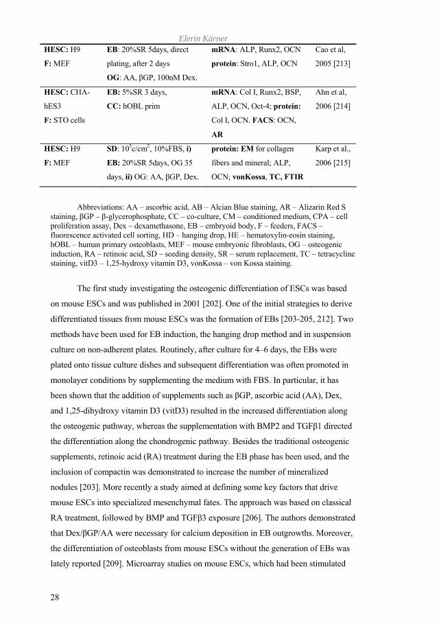
Elerin Kärner
28
HESC: H9
F: MEF
EB: 20%SR 5days, direct
plating, after 2 days
OG: AA, GP, 100nM Dex.
mRNA: ALP, Runx2, OCN
protein: Stro1, ALP, OCN
Cao et al,
2005 [213]
HESC: CHA-
hES3
F: STO cells
EB: 5%SR 3 days,
CC: hOBL prim
mRNA: Col I, Runx2, BSP,
ALP, OCN, Oct-4; protein:
Col I, OCN. FACS: OCN,
AR
Ahn et al,
2006 [214]
HESC: H9
F: MEF
SD: 105c/cm2, 10%FBS, i)
EB: 20%SR 5days, OG 35
days, ii) OG: AA, GP, Dex.
protein: EM for collagen
fibers and mineral; ALP,
OCN; vonKossa, TC, FTIR
Karp et al.,
2006 [215]
Abbreviations: AA – ascorbic acid, AB – Alcian Blue staining, AR – Alizarin Red S
staining, GP – -glycerophosphate, CC – co-culture, CM – conditioned medium, CPA – cell proliferation assay, Dex – dexamethasone, EB – embryoid body, F – feeders, FACS – fluorescence activated cell sorting, HD – hanging drop, HE – hematoxylin-eosin staining, hOBL – human primary osteoblasts, MEF – mouse embryonic fibroblasts, OG – osteogenic induction, RA – retinoic acid, SD – seeding density, SR – serum replacement, TC – tetracycline staining, vitD3 – 1,25-hydroxy vitamin D3, vonKossa – von Kossa staining.
The first study investigating the osteogenic differentiation of ESCs was based
on mouse ESCs and was published in 2001 [202]. One of the initial strategies to derive
differentiated tissues from mouse ESCs was the formation of EBs [203-205, 212]. Two
methods have been used for EB induction, the hanging drop method and in suspension
culture on non-adherent plates. Routinely, after culture for 4–6 days, the EBs were
plated onto tissue culture dishes and subsequent differentiation was often promoted in
monolayer conditions by supplementing the medium with FBS. In particular, it has
been shown that the addition of supplements such as GP, ascorbic acid (AA), Dex,
and 1,25-dihydroxy vitamin D3 (vitD3) resulted in the increased differentiation along
the osteogenic pathway, whereas the supplementation with BMP2 and TGF 1 directed
the differentiation along the chondrogenic pathway. Besides the traditional osteogenic
supplements, retinoic acid (RA) treatment during the EB phase has been used, and the
inclusion of compactin was demonstrated to increase the number of mineralized
nodules [203]. More recently a study aimed at defining some key factors that drive
mouse ESCs into specialized mesenchymal fates. The approach was based on classical
RA treatment, followed by BMP and TGF 3 exposure [206]. The authors demonstrated
that Dex/ GP/AA were necessary for calcium deposition in EB outgrowths. Moreover,
the differentiation of osteoblasts from mouse ESCs without the generation of EBs was
lately reported [209]. Microarray studies on mouse ESCs, which had been stimulated

Osteogenic differentiation of HESCs
29
with serum-containing culture medium supplemented with GP, AA, and Dex revealed
a combination of up-regulated genes involved in osteoblast differentiation (OPN, IGF-
II), and a down-regulation of those genes that were involved in the differentiation of
other phenotypes, such as neuroectoderm [205]. In addition, using an antibody against
cadherin-11, the authors purified a subpopulation of cells with osteoblastic
characteristics.
Another approach to differentiate ESCs has come from using co-culture
systems. In order to promote potential osteoblast formation, the mouse ESCs were
cultured together with fetal murine osteoblasts [202]. Recently, a study with HESCs by
co-culture with primary human osteoblasts was reported [214].
In most stem cell osteogenic in vitro models, bone-like nodules tend to develop
after a certain time in culture, yet, there is no agreement if all HESC lines are able to
form bone-like nodules. Conflicting reports exist as to whether, for example human
MSCs form nodules or if instead the matrix represents a diffusely mineralized network
[5, 216].
In general, only a few studies have been published with HESCs. In these
experiments the differentiation of HESCs was induced by EB formation in 10% FBS,
however differentiation by omitting the EB generation step was also recently reported
[215]. Osteogenic induction was often performed with AA, GP and Dex, three
supplements that have been extensively used in studies investigating the formation of
bone-like mineralized matrices in vitro (for review [186]). It is generally accepted that
AA promotes the proliferation and differentiation of cells, and induces the synthesis of
collagen, whereas GP is a precursor to inorganic phosphate and has been shown to
induce the nuclear export of Runx2 and lower the expression of OCN in mouse
osteoblastic cells [217]. Glucocorticoids, such as Dex, affect both the nodule formation
[186] and induce the osteoblastic gene expression [218]. However, the stimulatory
effect of these three factors is not limited just to osteoblasts, and when the process of
osteogenesis is induced using HESCs, cells from other lineages can be generated, too
[219-221].
Following cell culture, in order to detect successful osteogenesis, the most
common method seems to be the histochemical staining for the detection of calcium
deposition using AR and von Kossa staining. Several studies have used tetracycline
(TC) incorporation into the developing bone-like nodules. However, it must be noted
that neither ALP activity nor calcium deposition is an exclusive feature of osteogenic
cells and can be misleading in studies with ESCs.

Elerin Kärner
30
Taken together, all these models still lack defined conditions for the
differentiation and isolation of pure osteogenic precursor populations.
Figure 4. Schematic overview of currently used differentiation techniques to drive
osteogenic differentiation of HESCs. (HES-MPCs – human embryonic stem cell derived
mesenchymal progenitors).
1.3.9.1 Differentiating ESCs to osteoblasts through mesenchymal precursors
Two distinct strategies have been followed in the field of osteogenic
differentiation from ESCs; first and the most often followed approach is the induction
of differentiation directly from ESCs, and the second approach involves a pre-
differentiation step into mesenchymal progenitors prior to the induction of the desired
cell lineages. Some authors state that the length of time that the differentiating ESCs are
maintained in culture (~ 21 days) is in accordance with the time scale for the osteogenic
differentiation pathway of murine and human primary osteoblasts, and MSC cultures
[202]. However, these are more committed progenitor cells than the ESCs. Thus the
strategy based on the initial isolation of multipotent mesenchymal precursor cell
populations rather than specific mesenchymal derivatives has gained attention.
Induction of mesodermal fates from mouse ESCs follows a highly reproducible
and stereoptypic time-frame. In that the undifferentiated ESCs reproduce the

Osteogenic differentiation of HESCs
31
developmental stages during in vitro differentiation, an example was shown by the
appearance of neuronal, hematopoietic and cardiac mesoderm (reviewed [222]). A pure
population of mesenchymal precursors can be isolated using fluorescence activated cell
sorting (FACS)-based isolation of CD73+ progeny, [223], or other conditions [224],
and the cells could then be further expanded as mesenchymal precursors or
differentiated into various mesenchymal derivatives. Interestingly, cells isolated under
these conditions support the undifferentiated growth of HESCs [224]. In order to obtain
pure mesenchymal precursors from HESCs, the cells require pre-differentiation in co-
culture with the stromal cell line (OP9) for 40 days (Figure 4). Thereafter, the
mesenchymal precursor cells could be isolated by FACS based on the expression of
CD73, a marker routinely used for the isolation of adult bone marrow-derived MSCs.
The resulting CD73+ population could be proliferated or differentiated further. Such
cultures do not express detectable levels of ESC-specific markers, such as Nanog and
Oct-4, and work with severe combined immunodeficient (SCID)/Beige mice suggested
that they do not form teratomas in vivo [222].

Elerin Kärner
32
Osteogenic differentiation of HESCs
33
2 AIMS OF THE PRESENT INVESTIGATION The overall aim of the project was to study the process of osteoblastic
differentiation in HESCs.
Specific aims:
To investigate the molecular processes that occur during bone formation in the
primary ossification center in vivo, using global gene expression analysis;
To characterize bone matrix formation within the HESC model of osteogenesis
by:
comparing various differentiation methods,
comparing more than two HESC lines,
examining the expression of both early and late osteoblast-related genes
that are generally acknowledged as marker genes,
characterizing the synthesis of bone matrix proteins into bone-like
nodules,
investigating whether the mineralization mechanism is occurring through
a cell-mediated processes,
To further elucidate the dynamics of bone marker gene expression, in order to
establish a time-frame for supplementation with growth factors, so that a more
defined/directed differentiation model can be established.
To investigate the gain-of-function effects of an early osteoblast-related gene in
order to enhance the osteogenic differentiation potential of HESCs.
To establish a protocol for stable transgene expression for mineralized tissue
formation within the HESC model.

Elerin Kärner
34
3 MATERIALS AND METHODS A more detailed description of Materials and Methods can be found in the
individual papers. Here, only a summary is given and specific issues are discussed.
3.1 IN VIVO MODEL FOR OSTEOGENESIS (PAPER I) The key to successful characterization of any cell type is the information
obtained from in vivo conditions. In order to establish the global gene expression
profile during early ossification processes, we studied two specific time-points during
endochondral long bone development, representing the conditions before and after the
onset of mineralization. For tissue preparation, mice from NMR1 strain (Scanbur BK
AB, Stockholm, Sweden) were used. Central mouse metatarsals were dissected free
under sterile conditions on embryonic days 15 (E15) and 19 (E19), and total RNA was
extracted. Microarray gene expression analysis was performed using the GeneChip®
Mouse Genome 430 2.0 Array (Affymetrix, Inc., Santa Clara, CA, USA). The analysis,
including RNA processing, labeling and hybridization to the GeneChips®, was
performed at the Bioinformatics and Expression Analysis core facility at NOVUM,
Karolinska Institutet, Sweden.
In addition, entire mouse paws from E15 and E19, were stained with Masson's
Trichrome in order to identify the synthesized collagenous matrix, and to show the
presence of preosteoclasts and osteoclasts, sections were stained for tartrate-resistant
acid phosphatase (TRAP) with fast red violet.
3.2 OSTEOGENIC DIFFERENTIATION OF HESCS IN VITRO (PAPERS II, III, IV)
3.2.1 HESC culture maintenance HESC lines HS181, HS237, HS306 (from Karolinska Institutet Stem Cell
Network, Stockholm, Sweden), and commercially available H9 cell line (from WiCell
Research Institute, Madison, WI) with a normal karyotype were used. The HESCs were
maintained in medium that consisted of KO-DMEM containing 17% KO-SR, 1 mM L-
glutamine, 1% non-essential amino acids, 0.1 mM - mercaptoethanol and 4-6 ng/ml of
bFGF. Cells were kept in an undifferentiated state at 37°C in a humid 6.8% CO2
atmosphere. The cultured HESCs were checked visually and the culture medium
changed daily. HESCs were passaged by incubation in collagenase (1 mg/ml) for 7–10
min at 37°C every 5–6 days or when colonies revealed signs of differentiation. HS181,

Osteogenic differentiation of HESCs
35
HS237, and HS306 cell lines were derived and maintained on human foreskin
fibroblasts. In comparison, the H9 line was propagated on a layer of MEFs (paper II)
and switched onto human fibroblasts (paper IV). Human foreskin fibroblasts were
obtained from ATCC (CRL- 2429; ATCC, Manassas, VA), and MEFs were kindly
provided by S. Teglund, (Institute for Biosciences, Karolinska Institutet, Sweden). The
human fibroblasts were kept up to 20 passages, mouse cells up to 9 passages, and both
were mitotically inactivated by irradiation at 35-40 Gy before use. The seeding density
for feeder cells was ~200 000 cells/well in a six-well plate (Corning, NY), and the
culture medium consisted of Iscove’s modified Dulbecco’s medium (IMDM) and 10%
FBS. The cells were allowed to attach to the plate 3–5 days prior to use as feeder layers.
3.2.2 Control cell lines and culture conditions Human mesenchymal stem cells (hMSCs) (paper II) and human osteoblasts
(hOBL) (papers II, III and IV) were purchased from Cambrex Bioscience
(Walkersville, Inc., MD). The epithelial line HEK293-EBNA and the human
osteosarcoma cell line Saos-2 (HTB-85, ATCC, Manassas, VA) were used in papers III
and IV. The 293FT (Invitrogen) lentivirus packaging cell line and Hela cells were
used in paper IV. All cell lines were kept in medium consisting of DMEM (high
glucose) containing 1 mM L-glutamine and 10% FBS. The K562 cell line (LGC
Promochem/ ATCC) used in paper IV was cultured in RPMI-1640 (Invitrogen)
supplemented with 10% FBS.
The cells were incubated at 37°C in a humid 5% CO2 atmosphere, and the
medium was changed twice a week.
For differentiation purposes the cells were dissociated to single cells and ~5000
cells/cm2 were seeded onto tissue culture plates. Basal medium was supplemented with
GP, AA, and Dex. The control cultures were maintained for 15 days, because by day
25 true osteogenic cells had become fully mineralized, thus making it impossible to
extract RNA.
3.2.3 Osteogenic differentiation in vitro To initiate cell differentiation in monolayer, the HESCs were removed from
the culture dish by incubation with collagenase solution and mechanical scraping from
the culture plate. Approximately 5 colonies of 400 cells each (1000 cells/cm2) were
seeded onto gelatin-coated 24-well-plates. EB formation was induced in the HESC
colonies by again enzymatically detaching the cells with collagenase and transferring
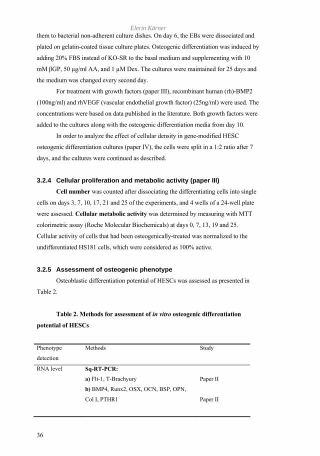
Elerin Kärner
36
them to bacterial non-adherent culture dishes. On day 6, the EBs were dissociated and
plated on gelatin-coated tissue culture plates. Osteogenic differentiation was induced by
adding 20% FBS instead of KO-SR to the basal medium and supplementing with 10
mM GP, 50 µg/ml AA, and 1 µM Dex. The cultures were maintained for 25 days and
the medium was changed every second day.
For treatment with growth factors (paper III), recombinant human (rh)-BMP2
(100ng/ml) and rhVEGF (vascular endothelial growth factor) (25ng/ml) were used. The
concentrations were based on data published in the literature. Both growth factors were
added to the cultures along with the osteogenic differentiation media from day 10.
In order to analyze the effect of cellular density in gene-modified HESC
osteogenic differentiation cultures (paper IV), the cells were split in a 1:2 ratio after 7
days, and the cultures were continued as described.
3.2.4 Cellular proliferation and metabolic activity (paper III) Cell number was counted after dissociating the differentiating cells into single
cells on days 3, 7, 10, 17, 21 and 25 of the experiments, and 4 wells of a 24-well plate
were assessed. Cellular metabolic activity was determined by measuring with MTT
colorimetric assay (Roche Molecular Biochemicals) at days 0, 7, 13, 19 and 25.
Cellular activity of cells that had been osteogenically-treated was normalized to the
undifferentiated HS181 cells, which were considered as 100% active.
3.2.5 Assessment of osteogenic phenotype Osteoblastic differentiation potential of HESCs was assessed as presented in
Table 2.
Table 2. Methods for assessment of in vitro osteogenic differentiation
potential of HESCs
Phenotype
detection
Methods Study
RNA level Sq-RT-PCR:
a) Flt-1, T-Brachyury
b) BMP4, Runx2, OSX, OCN, BSP, OPN,
Col I, PTHR1
Paper II
Paper II
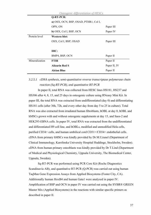
Osteogenic differentiation of HESCs
37
Q-RT-PCR:
a) OSX, OCN, BSP, OSAD, PTHR1, Col I,
OPN, ON
b) OSX, Col I, BSP, OCN
Paper III
Paper IV
Protein level Western blot:
OSX, Col I, BSP, OSAD
IHC:
BMP4, BSP, OCN
Paper III
Paper II
Mineralization FTIR
Alizarin Red S
Alcian Blue
Paper II
Paper II, IV
Paper II
3.2.5.1 cDNA synthesis, semi-quantitative reverse transcriptase polymerase chain
reaction (Sq-RT-PCR), and quantitative RT-PCR
In paper II, total RNA was collected from HESC lines HS181, HS237 and
HS306 after 4, 8, 15, and 25 days in osteogenic culture using RNeasy Mini Kit. In
paper III, the total RNA was extracted from undifferentiated (day 0) and differentiating
HS181 cells (after 36h, 72h, and every other day from day 5 to 25 in culture). Total
RNA was also extracted from irradiated human fibroblasts, hOBL at day 0, hOBL and
hMSCs grown with and without osteogenic supplements at day 15, and Saos-2 and
HEK293-EBNA cells. In paper IV, total RNA was extracted from the undifferentiated
and differentiated H9 cell line, and hOBLs, modified and unmodified Hela cells,
purified CD34+ cells, and human umbilical cord CD31+/CD34+ endothelial cells.
cDNA from primary hMSCs was kindly provided by Dr M.Uzunel (Department of
Clinical Immunology, Karolinska University Hospital Huddinge, Stockholm, Sweden).
cDNA from human primary osteoblasts was kindly provided by Dr T.Lind (Department
of Medical and Physiological Chemistry, Uppsala University, The Biomedical Center,
Uppsala, Sweden).
Sq-RT-PCR was performed using PCR Core Kit (Roche Diagnostics
Scandinavia AB), and quantitative RT-PCR (Q-PCR) was carried out using human
TaqMan Gene Expression Assays from Applied Biosystems (Foster City, CA).
Additionally human HoxB4 and human Gata1 were analysed in paper IV.
Amplification of BSP and OCN in paper IV was carried out using the SYBR® GREEN
Master Mix (Applied Biosystems) in the reactions with similar specific primers as
described in paper II.
Elerin Kärner
38
The comparative cycle threshold (Ct) method was used to analyze data,
hydroxymethylbilane synthase (HMBS) was used to standardize the Ct values, and
undifferentiated HS181 (paper III) or H9 (paper IV) were used to calibrate the values of
differentiating HESCs.
3.2.5.2 SDS-PAGE and Western blot (paper III and IV)
Cells were lysed using TRIzol reagent and protein extracts were quantified.
Each sample was electrophoresed on a SDS-PAGE and the proteins were electroblotted
onto nitrocellulose membranes. After blocking, the membranes were probed with
primary antibodies followed by a corresponding horseradish peroxidase (HRP)-
conjugated secondary antibody (DAKO, Glostrup, Denmark). Proteins were detected
with ECL Plus Western Blotting Detection System (GE Healthcare).
For the detection of HoxB4 modified and unmodified Hela cells in paper IV,
the total protein was extracted from unmodified K562 and Hela cells, and
electrophoresed on a 10% pre-cast gel (BioRad Laboratories). Gels were blotted onto
PVDF membranes (BioRad Laboratories), which were probed with rat anti-HoxB4
hybridoma supernatant (Developmental Studies Hybridoma Bank, Iowa, USA)
overnight, followed by secondary anti-rat IgG antibody conjugated to HRP. Bound
antibodies were detected using SuperSignal West Pico Chemoluminescent Substrate
(Pierce, Rockford, IL, USA).
3.2.5.3 Histological and immunocytochemical studies
As an indicator of mineralization within the HESC cultures, calcium deposition
was analyzed by AR staining in papers II and IV. The calcium salt crystals within the
bone-like nodules stain dark red, while the collagenous ECM turns yellow. It is
important to distinguish between mineralizing nodules, and fibrous nodules, which also
are three-dimensional structures, but do not mineralize. AR is often preferred to another
staining method, von Kossa, which can detect calcium sediments within the cell
culture.
The synthesis of glycosaminoglycans (GAGs) was analyzed by Alcian Blue
staining in paper II, which is a widely used method based on copper. The stained parts
are blue; however the specificity can be manipulated by the pH to selectively identify
neutral, sulphated, and phosphated mucopolysaccharides. pH 2.5 is commonly used to
detect GAGs within the cartilaginous matrix.
Osteogenic differentiation of HESCs
39
Lipid droplets in developing adipocytes were stained with Oil Red O in paper
II. However, no positive signal was detected in osteogenically differentiated HESC
cultures under these experimental circumstances.
For immunocytochemical analysis, the cell cultures described in paper II were
fixed, rinsed and treated with 0.2 M HCl and 3% H2O2 to clean the epitopes. The
antibodies were non-specifically blocked with 4% normal goat serum (DAKO) and the
cells were incubated with primary antibodies directed against human tissue diluted in
4% normal goat serum at 4°C overnight followed by incubation for 1 h at room
temperature with the corresponding secondary antibody prepared in blocking solution.
After washing with Tris buffered saline (TBS), the samples were mounted with
Vectashield containing DAPI (Vector labs Inc., Burlingame, CA). For the detection of
BSP and OCN antibodies, the cells were incubated with the HRP-conjugated secondary
antibody, and the signal was detected with freshly prepared DAB (DAKO) solution
activated with 0.1% H2O2. The sections were mounted with glycerol. Controls for
primary and secondary antibodies revealed neither non-specific staining nor antibody
cross-reactivity.
To investigate if pluripotent cells remained within the differentiating HESC
cultures in paper III, the cultures were fixed, rinsed, treated with 0.2M HCl and 3%
H2O2. The antibodies were non-specifically blocked with phosphate buffered saline
(PBS), 3% BSA (Fraction V, Sigma-Aldrich), 0.1% Tween-20, 0.1% bovine serum
albumin (BSA)-c (Aurion) for 40min. After blocking, the cells were incubated with
primary antibody diluted in PBS, 0.1% Tween-20, 0.1% BSA-c for 1h. The cells were
washed with PBS, 0.01% Tween-20, 0.01% BSA-c and incubated for 1h at room
temperature with fluorescent labeled secondary antibody. After washing again with
PBS, the samples were mounted with Vectashield containing DAPI.
3.2.5.4 Fourier-transform infrared spectroscopic analyzes (paper II)
The crystalline structure of the calcium phosphate deposits were analyzed by
Fourier-transform infrared (FTIR) spectroscopy. This method was important in order to
establish whether the deposited mineral resembled that of hydroxyapatite. We
compared two HESC lines, HS181 and H9 after osteogenic induction. The cells were
fixed, washed with TBS, treated with the buffer containing 10 mM Tris-HCl, pH 7.5,
0.5 mM MgCl2, and 0.1% Triton X-100. After centrifugation, the supernatant was
removed and the cells were incubated 48h in 0.1 M Tris-HCl, pH 7.5 and 10 mM CaCl2
containing 10 mg/ml non-specific protease at 55°C. The pellet was centrifuged again

Elerin Kärner
40
and washed with TBS. Thereafter, the mineral crystals were lyophilized and combined
with dried spectroscopic grade potassium bromide in the ratio 1:200. The samples were
resuspended in acetone and thoroughly dried. Spectra were obtained using a Thermo
Nicolet Avatar 360 FTIR.
3.2.6 Lentiviral transgene expression (paper IV) 3.2.6.1 Construction and production of lentiviral vectors
OSX cDNA, originally cloned into the TOPO vector was PCR-amplified, and
inserted into pEGFP-N1 vector (Clontech, Palo Alto, CA) in order to create the fusion
with EGFP (enhanced green fluorescent protein). Thereafter, the OSX-EGFP fusion
sequence was cleaved from the pEGFP-N1-OSX construct and ligated into the same
sites of the Gateway plasmid pENTR4 (Invitrogen). In order to create the final
lentiviral vector, the construct was transferred into the lentiviral backbone
pLenti6/UbC/V5-DEST (Invitrogen). The EGFP-control plasmid was constructed by
subcloning the EGFP gene from pEGFP-N1 into the Gateway plasmid pENTR4, and
was recombined with the lentiviral backbone pLenti6/UbC/V5-DEST.
Human genomic HoxB4 was cloned from extracted donor buffycoat DNA,
subcloned into pEGFP-C3 (Clontech), E2A sequence was inserted between EGFP and
HoxB4, and subcloned into the Gateway pENTR4 plasmid. To obtain the final
lentiviral transfer vector, the EGFP-E2a-HoxB4 fusion gene in pENTR4 was
recombined with pLenti6/UbC/V5-DEST using the Gateway LR reaction. The DNA
structure in the individual steps was tested by restriction enzyme analysis and the final
constructs were verified by sequencing.
Virus was produced by transient co-transfection of three plasmids into 293FT
cells; an envelope plasmid pMDG harbouring the gene encoding vesicular stomatitis
virus glycoprotein (VSV-G), pCMV_R8.91 expressing Gag and Pol, and our transfer
vector constructs.
Viral titers (TU/ml) were determined by transduction of Hela cells with serial
dilutions of the viral supernatant and flow cytometric analysis of the percentage of
EGFP-expressing cells. For transduction with lentiviral vectors, passages between 35-
50 H9 HESCs were used. Blasticidin resistant feeder cells were specifically prepared
for this study by transducing the human fibroblasts with pLenti6/UbC/V5-GW-lacZ
control vector (Invitrogen) followed by chemical selection with blasticidin.
Osteogenic differentiation of HESCs
41
3.2.6.2 Flow cytometric analysis
The cells were washed with PBS and dissociated with TrypLE Express
(Invitrogen). Fluorescent-labeled monoclonal antibodies: CD34-Allophycocyanine
(APC), CD31- Phycoerythrin (PE) and their corresponding isotype controls (BD
Biosciences) were used to detect the hemato-endothelial phenotype. For detection of
pluripotency SSEA3, and TRA1-60 (kindly provided by Mark Jones from the lab of
Peter Andrews, The University of Sheffield, Sheffield, UK) were used. All antibodies
were previously optimized in terms of their concentration. Flow cytometric analysis
was performed on a FACSCalibur (BD Biosciences, San Jose, CA). Acquisition and
analysis was performed using BD CellQuest™ Pro (BD Biosciences) and FlowJo (Tree
Star, Ashland, OR, USA) software.
Elerin Kärner
42
4 RESULTS AND DISCUSSION 4.1 PAPER I
An individual gene expression pattern is demarcating to each phase during
endochondral bone formation: condensation of mesenchymal progenitors, chondrocyte
differentiation with the eventual vascular invasion followed by osteoblast
differentiation. In order to obtain the best overview of such dynamic changes in gene
expression pattern we used microarray analysis. Two time points and mouse metatarsal
bones were chosen to model the formation of the primary ossification center in vivo. At
embryonic stage, E15 an avascular cartilage anlagen and pre-hypertrophic
chondrocytes in the diaphysis were detected, whereas by E19 the formation of the
primary ossification center and a primary marrow cavity could be seen.
Gene expression analysis of the total RNA isolated from mouse metatarsals
embryonic stages E15 and E19 identified 1285 genes, of which 543 were up-regulated
on E19 compared to E15, and 742 were down-regulated (selection criteria: 2-fold
change, P value of <0.005). Analysis of the data followed the gene ontology categories
Biological Process, Cellular Component and Molecular Function, however this study
focused on the two first categories. In summary, the gene expression data followed the
expected scheme for developmental progression of osteogenesis. We found that Hoxd
genes 10–12, Gli2 and Noggin were down-regulated post-mineralization (E19). No
change in gene expression was identified for BMP2,-4,-5 and -7. TGF- 1 and BSP
were highly up-regulated from E15 to E19, as well as OPN and DMP1. There was a
7.8-fold increase in OCN levels, a marker for terminally differentiated osteoblasts.
However, within the Cellular Component classifications, a large number of
genes related to bone remodeling predominantly featured. They included a number of
proteases, such as matrix metalloproteinases, TIMP 1 (tissue inhibitor of matrix
metalloproteinase), and cathepsin K. The presence of these enzymes demonstrates the
full differentiation and activation of osteoclasts, which was also observed in the TRAP
positive cells at E19. Structural molecules like the SLRP family; fibromodulin,
biglycan, asporin, and decorin were up-regulated. Using the metatarsal long bone
model we were able to identify and examine the genes associated with the formation of
the primary ossification center in an in vivo system.
Osteogenic differentiation of HESCs
43
4.2 PAPER II The derivation and establishment of culture systems for HESC lines provided us
with a novel model system by which investigate the process of osteogenesis within a
distinct environment. The focus of the study II was to examine the capacity of HESCs
to differentiate towards the osteoblastic lineage and their subsequent ability to form a
mineralized ECM. A selection of marker genes defining osteogenesis, which were
identified from study I were used, reaching from the earliest progenitor cells to the
differentiated osteoblasts. Four pluripotent HESC cell lines were studied and two
methods were used to initiate differentiation, first by plating the HESCs in monolayer
onto gelatin-coated plates, and, second, initiating the differentiation within EBs. The
cells were allowed to differentiate further in the presence of Dex, AA, and GP. Novel
to our study was the use of HESC cell lines (HS181, HS237, and HS306) derived and
maintained on commercially available human foreskin fibroblasts to support the
undifferentiated growth of the HESC cell colonies.
We ensured that the HESCs followed a typical differentiation pathway from
early mesodermal progenitors to the fully differentiated osteoblastic phenotype.
Monolayer cultures exhibited similar levels of T-Brachyury expression examined in the
two cell lines (H9 and HS181). However, following growth within EBs, the levels of T-
Brachyury declined in the H9 line earlier than the HS181 cells. Immunohistochemical
staining against human BMP4 in the osteogenic-induced monolayer cultures
demonstrated that the signal was specifically localized to the cells aggregating to form
eventual bone-like nodules. In the HS181 monolayer culture, the levels of BMP4 gene
expression increased earlier compared to H9, correlating also with the formation of
larger bone-like nodules. Screening for the osteoblast-specific gene mRNAs
demonstrated that the markers were detected in all HESC cell lines, and within both
monolayer and EB-derived cultures. It was observed in our study that the highest levels
of OSX expression were accompanied by raised levels of BSP and OCN. The SqRT-
PCR analysis also showed that BSP and OCN were expressed to a higher degree in
monolayer cultures, whereas the EB-derived cultures revealed more variable expression
levels.
It is known that in in vitro cultures, it is often hard to distinguish between cell-
mediated calcification and dystrophic calcium depositions. In the current study, mineral
deposition in the ECM was assessed by AR staining, and positive staining was detected
in all the cell lines examined. In order to further examine whether the deposited
calcium phosphate is similar to the biological apatite crystalline form, as found in de
Elerin Kärner
44
novo bone, the samples were also analyzed by FTIR spectroscopy. This method
provided confirmatory information at the biochemical level that indeed the mineral
phase within the osteogenic cultures resembled a crystalline apatite, which had been
formed by a cell-mediated calcification process.
Taken together, we were able to show that the cultures differentiated towards
the osteogenic lineage, however some differences were apparent between the gene
expression patterns for the bone matrix markers, which were dependent on the method
used to induce differentiation and between the cell lines. Overall, cells cultured in
monolayer conditions revealed higher levels of osteoblastic markers, whereas the EB-
derived cultures displayed generally lower levels of expression. We concluded that
lineage potential is not dependent on the mode of differentiation induction but on a cell
line itself.
4.3 PAPER III In the third paper, we tried to analyze further the standard model system for
osteogenesis of HESCs in order to establish the expression profile of bone-related
genes during differentiation triggered by supplementing the medium with AA, -GP
and Dex, three factors which are widely used to trigger osteogenesis from HESCs.
Based on our pilot studies and previously published work (paper II), we established that
the initial cell density plays an important role in differentiation. The optimal seeding
density for osteogenic HESC cultures (HS181 cell line) was about ~1000 cells/cm2.
Such cultures reached confluency 7-8 days after seeding, followed by the up-regulation
of the bone specific transcription factor, OSX. We believe that such density provides
the cells with enough space to proliferate until reaching cell-cell contact at confluency,
followed by the interaction with the produced ECM to switch on the optimal signaling
pathways. We show that the experimental period needed to induce the expression of the
latest osteoblast marker, OCN, was 25 days. In addition, we show that “osteogenically”
treated cultures retain a potentially undifferentiated population of cells.
Osteoblastic development is usually subdivided into certain developmental
stages: proliferation and differentiation of cells, and ECM synthesis, maturation and
mineralization. In this study, we used an alternative approach to the HESC osteogenic
model, and considered separately the cellular compartment activity on one side, and
matrix formation and mineralization on the other. We believe the first regulatory
transition, triggering the initiation of osteoblastic gene expression, takes place after the
active proliferation step even though several ECM-associated gene mRNAs were
Osteogenic differentiation of HESCs
45
expressed in actively proliferating immature cells. We show that at the end of active
proliferation, the osteoblast-specific transcription factor OSX was up-regulated
suggesting that its expression was regulated by the onset of contact-inhibition and its
function preceedes matrix maturation. ON, a major non-collagenous component of
bone was up-regulated straight after the end of the proliferative phase. Another
currently believed mineralizing tissue-specific NCP, OSAD was expressed at the
beginning of the culture period, supporting the possibility that it has a role in inhibiting
the actively proliferating cells. However, OSAD is also associated with the terminally
differentiated osteoblastic phenotype and to our knowledge it is so far considered as
osteoblast-specific. The second regulatory transition mediates the initiation of gene
expression for ECM formation, maturation and mineralization. OPN gene expression
was progressively down-regulated towards the end of the culture, which is in agreement
with the reports that low OPN levels are required for apatite crystal growth.
Q-PCR analysis revealed that OCN was expressed at the end of matrix
maturation, being rapidly down-regulated before mineralization, but thereafter
increased again. PTHR1, receptor for PTH and parathyroid hormone-like hormone, was
up-regulated during matrix maturation. PTHR1 has been described as a “globally”
expressed marker for osteoblastic cells, whereas OPN, BSP, and OCN can be
differentially expressed at mRNA and protein levels in only a subset of osteoblasts,
depending on the maturational state of the cells.
The direction of differentiation towards osteogenic lineage with growth factors
are essential to either increase the outcome of osteoblastic cells or decrease the
presence of other cell types. Due to the specificity of HESCs as an undifferentiated and
pluripotent system, the timing is of utmost importance. Here, our results showed that
HESCs seeded at 1000cells/cm2, reached confluency around day 7-8, followed by the
up-regulation of OSX.
VEGF-treated cells demonstrated down-regulated levels of known osteoblast
associated mRNAs. However, we also show that inclusion of BMP2 rescued
expression, which could be due to the fact that during osteogenic lineage progression,
in addition to the BMP pathway, several other signal transduction pathways mediate
osteoblastic gene expression. The combined addition of both growth factors
demonstrated that BMP2 decreased the inhibitory effect of VEGF on most of the bone-
related gene mRNAs. OSX, OCN and OSAD all showed increased expression levels
compared to levels in the VEGF-treated cells. Addition of BMP2 induced an earlier
significant up-regulation of BSP compared to “osteogenically”-treated cells. The
Elerin Kärner
46
finding that OCN was not significantly increased by BMP2, could be because OCN is
expressed at low levels in the young bone, where BSP along with other acidic
phosphoproteins are expressed at high levels. The overall higher expression of OSX
and BSP, indicative of immature mineralized tissue formation confirms that
assumption. Perhaps, continuation of the culture period would have exposed an
increased expression level for OCN. Interestingly, the combination of growth factors
had an inhibitory effect on BSP expression throughout the culture time. A similar
observation was reported in another study where a cross-communication between the
two pathways was suggested.
4.4 PAPER IV The aim of the paper IV was to study whether ectopic expression of an early
bone-specific gene could enhance HESC differentiation towards the osteoblastic
lineage. We used a lentiviral vector-based system, which has previously been reported
to be less affected by gene silencing during HESC differentiation, and evaluated the
effects of gain of function of OSX, currently recognized as the earliest bone-specific
transcription factor. The transcription factor OSX has been identified as a crucial
regulator of osteogenesis and is predominantly expressed by early osteoblastic cells.
OSX-deficient mice show a complete lack of osteoblast differentiation, and no
endochondral or intramembranous bone formation can be detected. To evaluate the
effects of the forced expression of OSX, we established a HESC line stably expressing
the transgene under the control of the Ubiquitin promoter to enhance the directed
differentiation into osteoblasts. However, it was not the main aim of the study to focus
on the analysis of osteogenesis. Within the study, we also included the analysis of
another transcription factor, HoxB4, which is an early hematopoietic transcription
factor. This factor was ectopically expressed in a similar lentiviral system. The
transduction of HESCs resulted in two HESC populations exhibiting different levels of
expression, which were compared to naturally occurring levels. We show that the
expression of OSX at low levels induced the transcription of endogenous HoxB4.
Furthermore, the up-regulated levels of mineralization-associated gene mRNAs, such
as collagen I, BSP and OCN, by high HoxB4 could also indicate a role for HoxB4
during pathological mineralization, perhaps similar to that found in blood vessels. Our
findings support the notion of cell-cell-interactions between early preosteoblasts and
HSCs on the bone marrow endosteal surface, required for hematopoiesis. We
concluded that for an enhanced osteogenesis originating from in vitro cultured HESC,

Osteogenic differentiation of HESCs
47
the correct levels of ectopic transcription factors need to be established. Our data also
highlights the notion of a close relationship between early blood and bone
development.
Elerin Kärner
48
5 CONCLUSIONS In this thesis my goal was to study the osteoblastic differentiation potential in HESCs
and to establish the model of ostegenesis in HESCs. The specific conclusions are the
following:
Paper I
We concluded that the metatarsal long bone model is a valuable and reliable
tool for examining the genes associated with the formation of the primary
ossification center.
Paper II
All HESC lines are able to express osteoblast-related gene mRNAs, but the
differentiation capacity towards the osteoblastic lineage is dependent on the cell
line.
Initiation of the differentiation process through EB formation is not necessary in
HESCs.
The mineralization of the ECM is a cell-mediated calcification process.
Paper III
We characterized the step-by-step expression profile of bone-associated genes.
We identified the time-frame for further supplementation with growth factors.
We established that two distinguishable phases occur during osteogenesis
within the HESC model that differ from the standard osteogenesis model
characterized by progenitor cells. Firstly, there is the cellular proliferation and
secretion of pre-maturational matrix stage that is needed for cell migration, and
second, the appearance of osteoprogenitors with characteristic ECM synthesis.
Paper IV
Lentiviral expression system is an efficient method to study osteoblast-
associated transgene expression in HESCs.
We found that for enhanced osteogenesis originating from in vitro cultured
HESCs, the correct levels of ectopic transcription factors need to be established.
Our data adds additional confirmation of a close relationship between early
blood and bone development.

Osteogenic differentiation of HESCs
49
6 FUTURE PERSPECTIVES In summary, the methods described in this thesis clearly demonstrate that
HESCs can be differentiated towards the osteoblastic lineage. However, it is important
to compare the derivation and differentiation potential towards osteogenic cells among
a larger numbers of HESC lines. The effect of growth factors, either alone or in
combination with the three classical osteogenic supplements is important to investigate.
Furthermore, the effect of a total ECM extract or its single substances on osteoblastic
gene expression needs to be further examined.
One interesting future perspective is the use of HESC lines in functional studies
in vivo, exploiting various animal models of musculoskeletal diseases.
We have shown that cells with a potential pluripotent phenotype remain present
within the osteoblastically differentiated HESC cultures. An important concern for
clinical applications of HESC-derived progeny in regenerative medicine is the risk of
teratoma formation due to the presence of residual undifferentiated ESCs among the
differentiated progeny. Thus, more studies are needed in order to sort the
osteoprogenitor cells and/or eliminate the potential multipotential cells.

Elerin Kärner
50
7 ACKNOWLEDGMENTS I am extremely lucky to have met a number of great people during the past
years. My special gratitude goes to my main supervisor, Associate Professor Mikael
Wendel, for your enormous patience and warm support. You gave me the opportunity
to carry out this work, provided finance and facilities, and surprisingly - optimism
never abandoned you.
Professor Lars Ährlund-Richter, my essential mentor through all these years,
for spreading your unlimited enthusiasm and knowledge in the field of … everything. I
am sure we will continue our “coffee and kanelbulle” meetings in future.
Dr. Rachael Sugars, my co-supervisor and in fact my main supervisor when it
comes to the scientific writing and thinking, for being always in good humor and
introducing us to British chick-movies, Mom´s lemon curd and the importance of
rugby.
I am also extremely lucky to have met the people in the group of Professor M
Sirac Dilber. The previous group member Dr. Alar Aints who showed me the elegant
ways of PCR, for all good times when he was still in Sweden. Christian Unger, my lab
partner. We shared the weekends, coffee and last cake pieces. You have been fun to
work with, being the last person who still believes in real science. We have laughed a
lot about “miserable” life-stories, we have fought a lot being both stubborn, but in the
end we established the HESC culture techniques in both of our labs. Evren Alici, for
being such a triggering personality, you are full of ideas and it has been great to know
you.
Anna Berglöf, for our funny coffee breaks which should always happen
“immediately” or “NOW”, for just being The Anna. Magnus Bäckesjö, what would I
have done without your Red Party? Thank you so much for always being helpful in the
lab, you know everybody and have access to every little machine room. I am happy I
managed to finish my lab-work before you left.
Ben and Eszter, the famous family Ganss. You two have brightened up the
life during my stay in COB. We all had fun when you two met, we enjoyed your
wedding and recently I had a pleasure to visit you and meet your two lovely off-springs
in Toronto.
Marie-Louise Olsson, for keeping a relaxed athmosphere in the lab, for all
your support with western blotting that always came in time, even when it was 30 min
before submitting the manuscript. Cecilia Christersson, for introducing me to the field
Osteogenic differentiation of HESCs
51
of molecular biology during my early years in COB. Professor Radim Cerny, for
spending so many summers with you. No, not on the beach! In the lab, pipetting, and
eating ice-cream. You are like a sign of summer, when you come then the rest of the
people go on vacation. Kaj Fried, my Favourite-Professor-Next-Door. It has been a
great fun to know you. You make the best coffee in Sweden! Ulrika Petersson,
Yingwei Hu, Anders Rehn, Erik Karlström, Ion Tcacencu, Thomas Lind, Jörgen
Jönsson, Jonas Thun, Nina Kaukua and Gregory Pilipjuk, and all other past and
present members in COB. Anna-Karin Persson, for being the best room-mate.
Karin Gertow and Jessica Cedervall, for always being kind and helpful, for
our discussions about stem cells and difficulties of being a PhD student. Marta Imreh,
for teaching me how to culture the ESCs, for being the best coach in the cell culture lab,
but also sharing your knowledge about different other techniques. Hernan Concha,
you have been full of unexpected surprises, and very helpful with FACS. Yes, Hernan,
I learned that controls are important!!!
Kerstin Smedberg, for always kindly reminding me of some deadlines which I
repeatedly missed. For taking care of all this boring document work and keeping your
eye on that things go smoothly.
Associate Professor Aavo Lang for taking care of my records in the Medical
Faculty, University of Tartu in Estonia, for being the most tolerant PhD coordinator one
could ever wish!
People of the previous Dental Clinic Dentista in Haapsalu and Dr. Katrin
Aasav, a tough Estonian dentist, who took over my patients. I know you don’t regret it
and I know the patients are happy. Anu Kõve and Pille Runnel, you represent the
successful young Estonian ladies, being creative and hard working. I am happy that you
find time to visit me and never forget to send e-cards for my birthday, Christmas or
Easter. I in turn always forget that….. Friendship really starts from kindergarten (please
see the PhD Thesis of Eszter Somogyi-Ganss). Helen Lasn, we met through the most
original mother-in-law, however you became indispensable to help me to get through
all the KI bureaucratic labyrinths.
C. Johannes Lindvall, for packing my lunch boxes, for driving me to work at
weekends to save the starving cells. You deserve the co-authorship on all my
publications, without you I would have not managed with things in time. I have
destroyed your Christmas vacations and summer vacations, all because I always had to
go and feed the cells. Thank you for being so patient. I promise, I will never work on
weekends again.
Elerin Kärner
52
8 REFERENCES 1. Steele, A. and P. Steele, Stem cells for repair of the heart. Curr Opin Pediatr,
2006. 18(5): p. 518-23. 2. Stocum, D.L., Stem cells in regenerative biology and medicine. Wound Repair
Regen, 2001. 9(6): p. 429-42. 3. Verfaillie, C.M., M.F. Pera, and P.M. Lansdorp, Stem cells: hype and reality.
Hematology Am Soc Hematol Educ Program, 2002: p. 369-91. 4. Kondo, M., et al., Biology of hematopoietic stem cells and progenitors:
implications for clinical application. Annu Rev Immunol, 2003. 21: p. 759-806. 5. Pittenger, M.F., et al., Multilineage potential of adult human mesenchymal stem
cells. Science, 1999. 284(5411): p. 143-7. 6. Denham, M., et al., Stem cells: an overview. Curr Protoc Cell Biol, 2005.
Chapter 23: p. Unit 23 1. 7. Takahashi, K., et al., Induction of pluripotent stem cells from adult human
fibroblasts by defined factors. Cell, 2007. 131(5): p. 861-72. 8. Park, I.H., et al., Reprogramming of human somatic cells to pluripotency with
defined factors. Nature, 2008. 451(7175): p. 141-6. 9. Yu, J., et al., Induced pluripotent stem cell lines derived from human somatic
cells. Science, 2007. 318(5858): p. 1917-20. 10. Minasi, M.G., et al., The meso-angioblast: a multipotent, self-renewing cell that
originates from the dorsal aorta and differentiates into most mesodermal tissues. Development, 2002. 129(11): p. 2773-83.
11. Trounson, A., The production and directed differentiation of human embryonic stem cells. Endocr Rev, 2006. 27(2): p. 208-19.
12. Klimanskaya, I., et al., Human embryonic stem cells derived without feeder cells. Lancet, 2005. 365(9471): p. 1636-41.
13. Brook, F.A. and R.L. Gardner, The origin and efficient derivation of embryonic stem cells in the mouse. Proc Natl Acad Sci U S A, 1997. 94(11): p. 5709-12.
14. Zwaka, T.P. and J.A. Thomson, A germ cell origin of embryonic stem cells? Development, 2005. 132(2): p. 227-33.
15. Clark, A.T., et al., Spontaneous differentiation of germ cells from human embryonic stem cells in vitro. Hum Mol Genet, 2004. 13(7): p. 727-39.
16. Xu, R.H., et al., BMP4 initiates human embryonic stem cell differentiation to trophoblast. Nat Biotechnol, 2002. 20(12): p. 1261-4.
17. Evans, M.J. and M.H. Kaufman, Establishment in culture of pluripotential cells from mouse embryos. Nature, 1981. 292(5819): p. 154-6.
18. Martin, G.R., Isolation of a pluripotent cell line from early mouse embryos cultured in medium conditioned by teratocarcinoma stem cells. Proc Natl Acad Sci U S A, 1981. 78(12): p. 7634-8.
19. Ledermann, B. and K. Burki, Establishment of a germ-line competent C57BL/6 embryonic stem cell line. Exp Cell Res, 1991. 197(2): p. 254-8.
20. Kitani, H., et al., Isolation of a germline-transmissible embryonic stem (ES) cell line from C3H/He mice. Zoolog Sci, 1996. 13(6): p. 865-71.
21. McWhir, J., et al., Selective ablation of differentiated cells permits isolation of embryonic stem cell lines from murine embryos with a non-permissive genetic background. Nat Genet, 1996. 14(2): p. 223-6.
22. Gardner, D.K., et al., Blastocyst score affects implantation and pregnancy outcome: towards a single blastocyst transfer. Fertil Steril, 2000. 73(6): p. 1155-8.
23. Gardner, D.K. and D. Sakkas, Assessment of embryo viability: the ability to select a single embryo for transfer--a review. Placenta, 2003. 24 Suppl B: p. S5-12.
24. Bongso, A., et al., Isolation and culture of inner cell mass cells from human blastocysts. Hum Reprod, 1994. 9(11): p. 2110-7.
25. Thomson, J.A., et al., Embryonic stem cell lines derived from human blastocysts. Science, 1998. 282(5391): p. 1145-7.
Osteogenic differentiation of HESCs
53
26. Richards, M., et al., Human feeders support prolonged undifferentiated growth of human inner cell masses and embryonic stem cells. Nat Biotechnol, 2002. 20(9): p. 933-6.
27. Reubinoff, B.E., et al., Embryonic stem cell lines from human blastocysts: somatic differentiation in vitro. Nat Biotechnol, 2000. 18(4): p. 399-404.
28. Mitalipova, M., et al., Human embryonic stem cell lines derived from discarded embryos. Stem Cells, 2003. 21(5): p. 521-6.
29. Lerou, P.H., et al., Human embryonic stem cell derivation from poor-quality embryos. Nat Biotechnol, 2008. 26(2): p. 212-4.
30. Carpenter, M.K., et al., Properties of four human embryonic stem cell lines maintained in a feeder-free culture system. Dev Dyn, 2004. 229(2): p. 243-58.
31. Abeyta, M.J., et al., Unique gene expression signatures of independently-derived human embryonic stem cell lines. Hum Mol Genet, 2004. 13(6): p. 601-8.
32. Bhattacharya, B., et al., Gene expression in human embryonic stem cell lines: unique molecular signature. Blood, 2004. 103(8): p. 2956-64.
33. Adewumi, O., et al., Characterization of human embryonic stem cell lines by the International Stem Cell Initiative. Nat Biotechnol, 2007. 25(7): p. 803-16.
34. Zhang, X., et al., Derivation of human embryonic stem cells from developing and arrested embryos. Stem Cells, 2006. 24(12): p. 2669-76.
35. Strelchenko, N., et al., Morula-derived human embryonic stem cells. Reprod Biomed Online, 2004. 9(6): p. 623-9.
36. Klimanskaya, I., et al., Human embryonic stem cell lines derived from single blastomeres. Nature, 2006. 444(7118): p. 481-5.
37. Stojkovic, M., et al., Derivation of human embryonic stem cells from day-8 blastocysts recovered after three-step in vitro culture. Stem Cells, 2004. 22(5): p. 790-7.
38. Verlinsky, Y., et al., Human embryonic stem cell lines with genetic disorders. Reprod Biomed Online, 2005. 10(1): p. 105-10.
39. Ludwig, T.E., et al., Derivation of human embryonic stem cells in defined conditions. Nat Biotechnol, 2006. 24(2): p. 185-7.
40. Rosler, E.S., et al., Long-term culture of human embryonic stem cells in feeder-free conditions. Dev Dyn, 2004. 229(2): p. 259-74.
41. Xu, C., et al., Feeder-free growth of undifferentiated human embryonic stem cells. Nat Biotechnol, 2001. 19(10): p. 971-4.
42. Bodnar, M.S., et al., Propagation and maintenance of undifferentiated human embryonic stem cells. Stem Cells Dev, 2004. 13(3): p. 243-53.
43. Yao, S., et al., Long-term self-renewal and directed differentiation of human embryonic stem cells in chemically defined conditions. Proc Natl Acad Sci U S A, 2006. 103(18): p. 6907-12.
44. Amit, M., et al., Clonally derived human embryonic stem cell lines maintain pluripotency and proliferative potential for prolonged periods of culture. Dev Biol, 2000. 227(2): p. 271-8.
45. Wang, L., et al., Human embryonic stem cells maintained in the absence of mouse embryonic fibroblasts or conditioned media are capable of hematopoietic development. Blood, 2005. 105(12): p. 4598-603.
46. Xu, R.H., et al., Basic FGF and suppression of BMP signaling sustain undifferentiated proliferation of human ES cells. Nat Methods, 2005. 2(3): p. 185-90.
47. Draper, J.S., et al., Culture and characterization of human embryonic stem cells. Stem Cells Dev, 2004. 13(4): p. 325-36.
48. Lee, J.B., et al., Available human feeder cells for the maintenance of human embryonic stem cells. Reproduction, 2004. 128(6): p. 727-35.
49. Miyamoto, K., et al., Human placenta feeder layers support undifferentiated growth of primate embryonic stem cells. Stem Cells, 2004. 22(4): p. 433-40.
50. Cheng, L., et al., Human adult marrow cells support prolonged expansion of human embryonic stem cells in culture. Stem Cells, 2003. 21(2): p. 131-42.
51. Richards, M., et al., Comparative evaluation of various human feeders for prolonged undifferentiated growth of human embryonic stem cells. Stem Cells, 2003. 21(5): p. 546-56.

Elerin Kärner
54
52. Xu, C., et al., Immortalized fibroblast-like cells derived from human embryonic stem cells support undifferentiated cell growth. Stem Cells, 2004. 22(6): p. 972-80.
53. Stojkovic, P., et al., An autogeneic feeder cell system that efficiently supports growth of undifferentiated human embryonic stem cells. Stem Cells, 2005. 23(3): p. 306-14.
54. Amit, M., et al., Human feeder layers for human embryonic stem cells. Biol Reprod, 2003. 68(6): p. 2150-6.
55. Martin, M.J., et al., Human embryonic stem cells express an immunogenic nonhuman sialic acid. Nat Med, 2005. 11(2): p. 228-32.
56. Hisamatsu-Sakamoto, M., N. Sakamoto, and A.S. Rosenberg, Embryonic stem cells cultured in serum-free medium acquire bovine apolipoprotein B-100 from feeder cell layers and serum replacement medium. Stem Cells, 2008. 26(1): p. 72-8.
57. Zwaka, T.P. and J.A. Thomson, Homologous recombination in human embryonic stem cells. Nat Biotechnol, 2003. 21(3): p. 319-21.
58. Wong, R.C., et al., Presence of functional gap junctions in human embryonic stem cells. Stem Cells, 2004. 22(6): p. 883-9.
59. Watanabe, K., et al., A ROCK inhibitor permits survival of dissociated human embryonic stem cells. Nat Biotechnol, 2007. 25(6): p. 681-6.
60. Draper, J.S., et al., Recurrent gain of chromosomes 17q and 12 in cultured human embryonic stem cells. Nat Biotechnol, 2004. 22(1): p. 53-4.
61. Imreh, M.P., et al., In vitro culture conditions favoring selection of chromosomal abnormalities in human ES cells. J Cell Biochem, 2006. 99(2): p. 508-16.
62. Williams, R.L., et al., Myeloid leukaemia inhibitory factor maintains the developmental potential of embryonic stem cells. Nature, 1988. 336(6200): p. 684-7.
63. Yoshida, K., et al., Maintenance of the pluripotential phenotype of embryonic stem cells through direct activation of gp130 signalling pathways. Mech Dev, 1994. 45(2): p. 163-71.
64. Takahashi-Tezuka, M., et al., Gab1 acts as an adapter molecule linking the cytokine receptor gp130 to ERK mitogen-activated protein kinase. Mol Cell Biol, 1998. 18(7): p. 4109-17.
65. Matsuda, T., et al., STAT3 activation is sufficient to maintain an undifferentiated state of mouse embryonic stem cells. Embo J, 1999. 18(15): p. 4261-9.
66. Ying, Q.L., et al., BMP induction of Id proteins suppresses differentiation and sustains embryonic stem cell self-renewal in collaboration with STAT3. Cell, 2003. 115(3): p. 281-92.
67. Pera, M.F., et al., Regulation of human embryonic stem cell differentiation by BMP-2 and its antagonist noggin. J Cell Sci, 2004. 117(Pt 7): p. 1269-80.
68. Levenstein, M.E., et al., Basic fibroblast growth factor support of human embryonic stem cell self-renewal. Stem Cells, 2006. 24(3): p. 568-74.
69. James, D., et al., TGFbeta/activin/nodal signaling is necessary for the maintenance of pluripotency in human embryonic stem cells. Development, 2005. 132(6): p. 1273-82.
70. Beattie, G.M., et al., Activin A maintains pluripotency of human embryonic stem cells in the absence of feeder layers. Stem Cells, 2005. 23(4): p. 489-95.
71. Soh, B.S., et al., Pleiotrophin enhances clonal growth and long-term expansion of human embryonic stem cells. Stem Cells, 2007. 25(12): p. 3029-37.
72. Sato, N., et al., Maintenance of pluripotency in human and mouse embryonic stem cells through activation of Wnt signaling by a pharmacological GSK-3-specific inhibitor. Nat Med, 2004. 10(1): p. 55-63.
73. Zwaka, T.P. and J.A. Thomson, Differentiation of human embryonic stem cells occurs through symmetric cell division. Stem Cells, 2005. 23(2): p. 146-9.
74. Liu, N., et al., Molecular mechanisms involved in self-renewal and pluripotency of embryonic stem cells. J Cell Physiol, 2007. 211(2): p. 279-86.
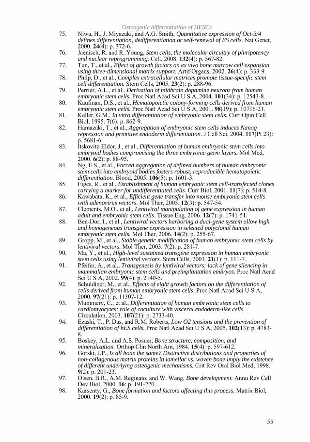
Osteogenic differentiation of HESCs
55
75. Niwa, H., J. Miyazaki, and A.G. Smith, Quantitative expression of Oct-3/4 defines differentiation, dedifferentiation or self-renewal of ES cells. Nat Genet, 2000. 24(4): p. 372-6.
76. Jaenisch, R. and R. Young, Stem cells, the molecular circuitry of pluripotency and nuclear reprogramming. Cell, 2008. 132(4): p. 567-82.
77. Tun, T., et al., Effect of growth factors on ex vivo bone marrow cell expansion using three-dimensional matrix support. Artif Organs, 2002. 26(4): p. 333-9.
78. Philp, D., et al., Complex extracellular matrices promote tissue-specific stem cell differentiation. Stem Cells, 2005. 23(2): p. 288-96.
79. Perrier, A.L., et al., Derivation of midbrain dopamine neurons from human embryonic stem cells. Proc Natl Acad Sci U S A, 2004. 101(34): p. 12543-8.
80. Kaufman, D.S., et al., Hematopoietic colony-forming cells derived from human embryonic stem cells. Proc Natl Acad Sci U S A, 2001. 98(19): p. 10716-21.
81. Keller, G.M., In vitro differentiation of embryonic stem cells. Curr Opin Cell Biol, 1995. 7(6): p. 862-9.
82. Hamazaki, T., et al., Aggregation of embryonic stem cells induces Nanog repression and primitive endoderm differentiation. J Cell Sci, 2004. 117(Pt 23): p. 5681-6.
83. Itskovitz-Eldor, J., et al., Differentiation of human embryonic stem cells into embryoid bodies compromising the three embryonic germ layers. Mol Med, 2000. 6(2): p. 88-95.
84. Ng, E.S., et al., Forced aggregation of defined numbers of human embryonic stem cells into embryoid bodies fosters robust, reproducible hematopoietic differentiation. Blood, 2005. 106(5): p. 1601-3.
85. Eiges, R., et al., Establishment of human embryonic stem cell-transfected clones carrying a marker for undifferentiated cells. Curr Biol, 2001. 11(7): p. 514-8.
86. Kawabata, K., et al., Efficient gene transfer into mouse embryonic stem cells with adenovirus vectors. Mol Ther, 2005. 12(3): p. 547-54.
87. Clements, M.O., et al., Lentiviral manipulation of gene expression in human adult and embryonic stem cells. Tissue Eng, 2006. 12(7): p. 1741-51.
88. Ben-Dor, I., et al., Lentiviral vectors harboring a dual-gene system allow high and homogeneous transgene expression in selected polyclonal human embryonic stem cells. Mol Ther, 2006. 14(2): p. 255-67.
89. Gropp, M., et al., Stable genetic modification of human embryonic stem cells by lentiviral vectors. Mol Ther, 2003. 7(2): p. 281-7.
90. Ma, Y., et al., High-level sustained transgene expression in human embryonic stem cells using lentiviral vectors. Stem Cells, 2003. 21(1): p. 111-7.
91. Pfeifer, A., et al., Transgenesis by lentiviral vectors: lack of gene silencing in mammalian embryonic stem cells and preimplantation embryos. Proc Natl Acad Sci U S A, 2002. 99(4): p. 2140-5.
92. Schuldiner, M., et al., Effects of eight growth factors on the differentiation of cells derived from human embryonic stem cells. Proc Natl Acad Sci U S A, 2000. 97(21): p. 11307-12.
93. Mummery, C., et al., Differentiation of human embryonic stem cells to cardiomyocytes: role of coculture with visceral endoderm-like cells. Circulation, 2003. 107(21): p. 2733-40.
94. Ezashi, T., P. Das, and R.M. Roberts, Low O2 tensions and the prevention of differentiation of hES cells. Proc Natl Acad Sci U S A, 2005. 102(13): p. 4783-8.
95. Boskey, A.L. and A.S. Posner, Bone structure, composition, and mineralization. Orthop Clin North Am, 1984. 15(4): p. 597-612.
96. Gorski, J.P., Is all bone the same? Distinctive distributions and properties of non-collagenous matrix proteins in lamellar vs. woven bone imply the existence of different underlying osteogenic mechanisms. Crit Rev Oral Biol Med, 1998. 9(2): p. 201-23.
97. Olsen, B.R., A.M. Reginato, and W. Wang, Bone development. Annu Rev Cell Dev Biol, 2000. 16: p. 191-220.
98. Karsenty, G., Bone formation and factors affecting this process. Matrix Biol, 2000. 19(2): p. 85-9.

Elerin Kärner
56
99. Karsenty, G. and E.F. Wagner, Reaching a genetic and molecular understanding of skeletal development. Dev Cell, 2002. 2(4): p. 389-406.
100. Ducy, P., T. Schinke, and G. Karsenty, The osteoblast: a sophisticated fibroblast under central surveillance. Science, 2000. 289(5484): p. 1501-4.
101. Rosen, E.D. and B.M. Spiegelman, Molecular regulation of adipogenesis. Annu Rev Cell Dev Biol, 2000. 16: p. 145-71.
102. Arnold, H.H. and B. Winter, Muscle differentiation: more complexity to the network of myogenic regulators. Curr Opin Genet Dev, 1998. 8(5): p. 539-44.
103. Ehrlich, P.J. and L.E. Lanyon, Mechanical strain and bone cell function: a review. Osteoporos Int, 2002. 13(9): p. 688-700.
104. Teitelbaum, S.L. and F.P. Ross, Genetic regulation of osteoclast development and function. Nat Rev Genet, 2003. 4(8): p. 638-49.
105. Aubin, J.E., et al., Osteoblast and chondroblast differentiation. Bone, 1995. 17(2 Suppl): p. 77S-83S.
106. Malaval, L., et al., Kinetics of osteoprogenitor proliferation and osteoblast differentiation in vitro. J Cell Biochem, 1999. 74(4): p. 616-27.
107. Aubin, J.E., Advances in the osteoblast lineage. Biochem Cell Biol, 1998. 76(6): p. 899-910.
108. Aubin, J.E., Regulation of osteoblast formation and function. Rev Endocr Metab Disord, 2001. 2(1): p. 81-94.
109. Marie, P.J., Transcription factors controlling osteoblastogenesis. Arch Biochem Biophys, 2008.
110. Komori, T., Regulation of bone development and maintenance by Runx2. Front Biosci, 2008. 13: p. 898-903.
111. Franceschi, R.T., et al., Transcriptional regulation of osteoblasts. Ann N Y Acad Sci, 2007. 1116: p. 196-207.
112. Xiao, Z.S., et al., Genomic structure and isoform expression of the mouse, rat and human Cbfa1/Osf2 transcription factor. Gene, 1998. 214(1-2): p. 187-97.
113. Ogawa, S., et al., Cbfa1, an essential transcription factor for bone formation, is expressed in testis from the same promoter used in bone. DNA Res, 2000. 7(3): p. 181-5.
114. Harada, H., et al., Cbfa1 isoforms exert functional differences in osteoblast differentiation. J Biol Chem, 1999. 274(11): p. 6972-8.
115. Komori, T., et al., Targeted disruption of Cbfa1 results in a complete lack of bone formation owing to maturational arrest of osteoblasts. Cell, 1997. 89(5): p. 755-64.
116. Ducy, P., et al., Osf2/Cbfa1: a transcriptional activator of osteoblast differentiation. Cell, 1997. 89(5): p. 747-54.
117. Takeda, S., et al., Continuous expression of Cbfa1 in nonhypertrophic chondrocytes uncovers its ability to induce hypertrophic chondrocyte differentiation and partially rescues Cbfa1-deficient mice. Genes Dev, 2001. 15(4): p. 467-81.
118. Yang, J., et al., Prostate cancer cells induce osteoblast differentiation through a Cbfa1-dependent pathway. Cancer Res, 2001. 61(14): p. 5652-9.
119. Pratap, J., et al., Cell growth regulatory role of Runx2 during proliferative expansion of preosteoblasts. Cancer Res, 2003. 63(17): p. 5357-62.
120. Ducy, P., et al., A Cbfa1-dependent genetic pathway controls bone formation beyond embryonic development. Genes Dev, 1999. 13(8): p. 1025-36.
121. Liu, W., et al., Overexpression of Cbfa1 in osteoblasts inhibits osteoblast maturation and causes osteopenia with multiple fractures. J Cell Biol, 2001. 155(1): p. 157-66.
122. Huang, W., et al., Signaling and transcriptional regulation in osteoblast commitment and differentiation. Front Biosci, 2007. 12: p. 3068-92.
123. Lengner, C.J., et al., Osteoblast differentiation and skeletal development are regulated by Mdm2-p53 signaling. J Cell Biol, 2006. 172(6): p. 909-21.
124. Wang, X., et al., p53 functions as a negative regulator of osteoblastogenesis, osteoblast-dependent osteoclastogenesis, and bone remodeling. J Cell Biol, 2006. 172(1): p. 115-25.
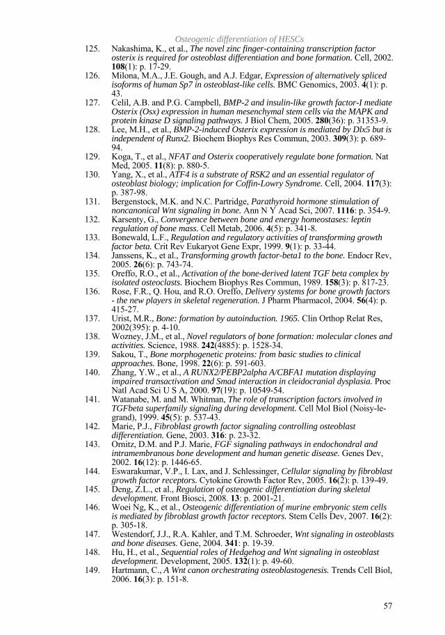
Osteogenic differentiation of HESCs
57
125. Nakashima, K., et al., The novel zinc finger-containing transcription factor osterix is required for osteoblast differentiation and bone formation. Cell, 2002. 108(1): p. 17-29.
126. Milona, M.A., J.E. Gough, and A.J. Edgar, Expression of alternatively spliced isoforms of human Sp7 in osteoblast-like cells. BMC Genomics, 2003. 4(1): p. 43.
127. Celil, A.B. and P.G. Campbell, BMP-2 and insulin-like growth factor-I mediate Osterix (Osx) expression in human mesenchymal stem cells via the MAPK and protein kinase D signaling pathways. J Biol Chem, 2005. 280(36): p. 31353-9.
128. Lee, M.H., et al., BMP-2-induced Osterix expression is mediated by Dlx5 but is independent of Runx2. Biochem Biophys Res Commun, 2003. 309(3): p. 689-94.
129. Koga, T., et al., NFAT and Osterix cooperatively regulate bone formation. Nat Med, 2005. 11(8): p. 880-5.
130. Yang, X., et al., ATF4 is a substrate of RSK2 and an essential regulator of osteoblast biology; implication for Coffin-Lowry Syndrome. Cell, 2004. 117(3): p. 387-98.
131. Bergenstock, M.K. and N.C. Partridge, Parathyroid hormone stimulation of noncanonical Wnt signaling in bone. Ann N Y Acad Sci, 2007. 1116: p. 354-9.
132. Karsenty, G., Convergence between bone and energy homeostases: leptin regulation of bone mass. Cell Metab, 2006. 4(5): p. 341-8.
133. Bonewald, L.F., Regulation and regulatory activities of transforming growth factor beta. Crit Rev Eukaryot Gene Expr, 1999. 9(1): p. 33-44.
134. Janssens, K., et al., Transforming growth factor-beta1 to the bone. Endocr Rev, 2005. 26(6): p. 743-74.
135. Oreffo, R.O., et al., Activation of the bone-derived latent TGF beta complex by isolated osteoclasts. Biochem Biophys Res Commun, 1989. 158(3): p. 817-23.
136. Rose, F.R., Q. Hou, and R.O. Oreffo, Delivery systems for bone growth factors - the new players in skeletal regeneration. J Pharm Pharmacol, 2004. 56(4): p. 415-27.
137. Urist, M.R., Bone: formation by autoinduction. 1965. Clin Orthop Relat Res, 2002(395): p. 4-10.
138. Wozney, J.M., et al., Novel regulators of bone formation: molecular clones and activities. Science, 1988. 242(4885): p. 1528-34.
139. Sakou, T., Bone morphogenetic proteins: from basic studies to clinical approaches. Bone, 1998. 22(6): p. 591-603.
140. Zhang, Y.W., et al., A RUNX2/PEBP2alpha A/CBFA1 mutation displaying impaired transactivation and Smad interaction in cleidocranial dysplasia. Proc Natl Acad Sci U S A, 2000. 97(19): p. 10549-54.
141. Watanabe, M. and M. Whitman, The role of transcription factors involved in TGFbeta superfamily signaling during development. Cell Mol Biol (Noisy-le-grand), 1999. 45(5): p. 537-43.
142. Marie, P.J., Fibroblast growth factor signaling controlling osteoblast differentiation. Gene, 2003. 316: p. 23-32.
143. Ornitz, D.M. and P.J. Marie, FGF signaling pathways in endochondral and intramembranous bone development and human genetic disease. Genes Dev, 2002. 16(12): p. 1446-65.
144. Eswarakumar, V.P., I. Lax, and J. Schlessinger, Cellular signaling by fibroblast growth factor receptors. Cytokine Growth Factor Rev, 2005. 16(2): p. 139-49.
145. Deng, Z.L., et al., Regulation of osteogenic differentiation during skeletal development. Front Biosci, 2008. 13: p. 2001-21.
146. Woei Ng, K., et al., Osteogenic differentiation of murine embryonic stem cells is mediated by fibroblast growth factor receptors. Stem Cells Dev, 2007. 16(2): p. 305-18.
147. Westendorf, J.J., R.A. Kahler, and T.M. Schroeder, Wnt signaling in osteoblasts and bone diseases. Gene, 2004. 341: p. 19-39.
148. Hu, H., et al., Sequential roles of Hedgehog and Wnt signaling in osteoblast development. Development, 2005. 132(1): p. 49-60.
149. Hartmann, C., A Wnt canon orchestrating osteoblastogenesis. Trends Cell Biol, 2006. 16(3): p. 151-8.

Elerin Kärner
58
150. Glass, D.A., 2nd and G. Karsenty, Molecular bases of the regulation of bone remodeling by the canonical Wnt signaling pathway. Curr Top Dev Biol, 2006. 73: p. 43-84.
151. Kato, M., et al., Cbfa1-independent decrease in osteoblast proliferation, osteopenia, and persistent embryonic eye vascularization in mice deficient in Lrp5, a Wnt coreceptor. J Cell Biol, 2002. 157(2): p. 303-14.
152. Boyden, L.M., et al., High bone density due to a mutation in LDL-receptor-related protein 5. N Engl J Med, 2002. 346(20): p. 1513-21.
153. Little, R.D., et al., A mutation in the LDL receptor-related protein 5 gene results in the autosomal dominant high-bone-mass trait. Am J Hum Genet, 2002. 70(1): p. 11-9.
154. Boland, G.M., et al., Wnt 3a promotes proliferation and suppresses osteogenic differentiation of adult human mesenchymal stem cells. J Cell Biochem, 2004. 93(6): p. 1210-30.
155. de Boer, J., et al., Wnt signaling inhibits osteogenic differentiation of human mesenchymal stem cells. Bone, 2004. 34(5): p. 818-26.
156. Luo, Q., et al., Connective tissue growth factor (CTGF) is regulated by Wnt and bone morphogenetic proteins signaling in osteoblast differentiation of mesenchymal stem cells. J Biol Chem, 2004. 279(53): p. 55958-68.
157. Bain, G., et al., Activated beta-catenin induces osteoblast differentiation of C3H10T1/2 cells and participates in BMP2 mediated signal transduction. Biochem Biophys Res Commun, 2003. 301(1): p. 84-91.
158. Hill, T.P., et al., Canonical Wnt/beta-catenin signaling prevents osteoblasts from differentiating into chondrocytes. Dev Cell, 2005. 8(5): p. 727-38.
159. Myllyharju, J. and K.I. Kivirikko, Collagens and collagen-related diseases. Ann Med, 2001. 33(1): p. 7-21.
160. Sutmuller, M., J.A. Bruijn, and E. de Heer, Collagen types VIII and X, two non-fibrillar, short-chain collagens. Structure homologies, functions and involvement in pathology. Histol Histopathol, 1997. 12(2): p. 557-66.
161. Shaw, L.M. and B.R. Olsen, FACIT collagens: diverse molecular bridges in extracellular matrices. Trends Biochem Sci, 1991. 16(5): p. 191-4.
162. Termine, J.D., et al., Osteonectin, a bone-specific protein linking mineral to collagen. Cell, 1981. 26(1 Pt 1): p. 99-105.
163. Bradshaw, A.D., et al., SPARC-null mice exhibit increased adiposity without significant differences in overall body weight. Proc Natl Acad Sci U S A, 2003. 100(10): p. 6045-50.
164. Brekken, R.A. and E.H. Sage, SPARC, a matricellular protein: at the crossroads of cell-matrix communication. Matrix Biol, 2001. 19(8): p. 816-27.
165. Iozzo, R.V., Matrix proteoglycans: from molecular design to cellular function. Annu Rev Biochem, 1998. 67: p. 609-52.
166. Sugars, R.V., et al., Molecular interaction of recombinant decorin and biglycan with type I collagen influences crystal growth. Connect Tissue Res, 2003. 44 Suppl 1: p. 189-95.
167. Vogel, K.G., M. Paulsson, and D. Heinegard, Specific inhibition of type I and type II collagen fibrillogenesis by the small proteoglycan of tendon. Biochem J, 1984. 223(3): p. 587-97.
168. Ramstad, V.E., et al., Ultrastructural distribution of osteoadherin in rat bone shows a pattern similar to that of bone sialoprotein. Calcif Tissue Int, 2003. 72(1): p. 57-64.
169. Brown, L.F., et al., Expression and distribution of osteopontin in human tissues: widespread association with luminal epithelial surfaces. Mol Biol Cell, 1992. 3(10): p. 1169-80.
170. Liu, F., et al., Simultaneous detection of multiple bone-related mRNAs and protein expression during osteoblast differentiation: polymerase chain reaction and immunocytochemical studies at the single cell level. Dev Biol, 1994. 166(1): p. 220-34.
171. Sodek, J., B. Ganss, and M.D. McKee, Osteopontin. Crit Rev Oral Biol Med, 2000. 11(3): p. 279-303.
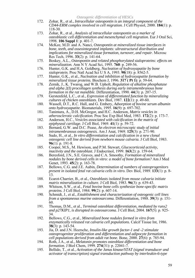
Osteogenic differentiation of HESCs
59
172. Zohar, R., et al., Intracellular osteopontin is an integral component of the CD44-ERM complex involved in cell migration. J Cell Physiol, 2000. 184(1): p. 118-30.
173. Zohar, R., et al., Analysis of intracellular osteopontin as a marker of osteoblastic cell differentiation and mesenchymal cell migration. Eur J Oral Sci, 1998. 106 Suppl 1: p. 401-7.
174. McKee, M.D. and A. Nanci, Osteopontin at mineralized tissue interfaces in bone, teeth, and osseointegrated implants: ultrastructural distribution and implications for mineralized tissue formation, turnover, and repair. Microsc Res Tech, 1996. 33(2): p. 141-64.
175. Boskey, A.L., Osteopontin and related phosphorylated sialoproteins: effects on mineralization. Ann N Y Acad Sci, 1995. 760: p. 249-56.
176. Hunter, G.K. and H.A. Goldberg, Nucleation of hydroxyapatite by bone sialoprotein. Proc Natl Acad Sci U S A, 1993. 90(18): p. 8562-5.
177. Hunter, G.K., et al., Nucleation and inhibition of hydroxyapatite formation by mineralized tissue proteins. Biochem J, 1996. 317 ( Pt 1): p. 59-64.
178. Zernik, J., K. Twarog, and W.B. Upholt, Regulation of alkaline phosphatase and alpha 2(I) procollagen synthesis during early intramembranous bone formation in the rat mandible. Differentiation, 1990. 44(3): p. 207-15.
179. Gerstenfeld, L.C., et al., Expression of differentiated function by mineralizing cultures of chicken osteoblasts. Dev Biol, 1987. 122(1): p. 49-60.
180. Wassell, D.T., R.C. Hall, and G. Embery, Adsorption of bovine serum albumin onto hydroxyapatite. Biomaterials, 1995. 16(9): p. 697-702.
181. Tanimura, A., D.H. McGregor, and H.C. Anderson, Matrix vesicles in atherosclerotic calcification. Proc Soc Exp Biol Med, 1983. 172(2): p. 173-7.
182. Anderson, H.C., Vesicles associated with calcification in the matrix of epiphyseal cartilage. J Cell Biol, 1969. 41(1): p. 59-72.
183. Bernard, G.W. and D.C. Pease, An electron microscopic study of initial intramembranous osteogenesis. Am J Anat, 1969. 125(3): p. 271-90.
184. Sudo, H., et al., In vitro differentiation and calcification in a new clonal osteogenic cell line derived from newborn mouse calvaria. J Cell Biol, 1983. 96(1): p. 191-8.
185. Cooper, M.S., M. Hewison, and P.M. Stewart, Glucocorticoid activity, inactivity and the osteoblast. J Endocrinol, 1999. 163(2): p. 159-64.
186. Beresford, J.N., S.E. Graves, and C.A. Smoothy, Formation of mineralized nodules by bone derived cells in vitro: a model of bone formation? Am J Med Genet, 1993. 45(2): p. 163-78.
187. Bellows, C.G. and J.E. Aubin, Determination of numbers of osteoprogenitors present in isolated fetal rat calvaria cells in vitro. Dev Biol, 1989. 133(1): p. 8-13.
188. Ecarot-Charrier, B., et al., Osteoblasts isolated from mouse calvaria initiate matrix mineralization in culture. J Cell Biol, 1983. 96(3): p. 639-43.
189. Whitson, S.W., et al., Fetal bovine bone cells synthesize bone-specific matrix proteins. J Cell Biol, 1984. 99(2): p. 607-14.
190. Schmidt, J., et al., Establishment and characterization of osteogenic cell lines from a spontaneous murine osteosarcoma. Differentiation, 1988. 39(3): p. 151-60.
191. Thomas, D.M., et al., Terminal osteoblast differentiation, mediated by runx2 and p27KIP1, is disrupted in osteosarcoma. J Cell Biol, 2004. 167(5): p. 925-34.
192. Bellows, C.G., et al., Mineralized bone nodules formed in vitro from enzymatically released rat calvaria cell populations. Calcif Tissue Int, 1986. 38(3): p. 143-54.
193. Jia, D. and J.N. Heersche, Insulin-like growth factor-1 and -2 stimulate osteoprogenitor proliferation and differentiation and adipocyte formation in cell populations derived from adult rat bone. Bone, 2000. 27(6): p. 785-94.
194. Roth, J.A., et al., Melatonin promotes osteoblast differentiation and bone formation. J Biol Chem, 1999. 274(31): p. 22041-7.
195. Bellido, T., et al., Activation of the Janus kinase/STAT (signal transducer and activator of transcription) signal transduction pathway by interleukin-6-type
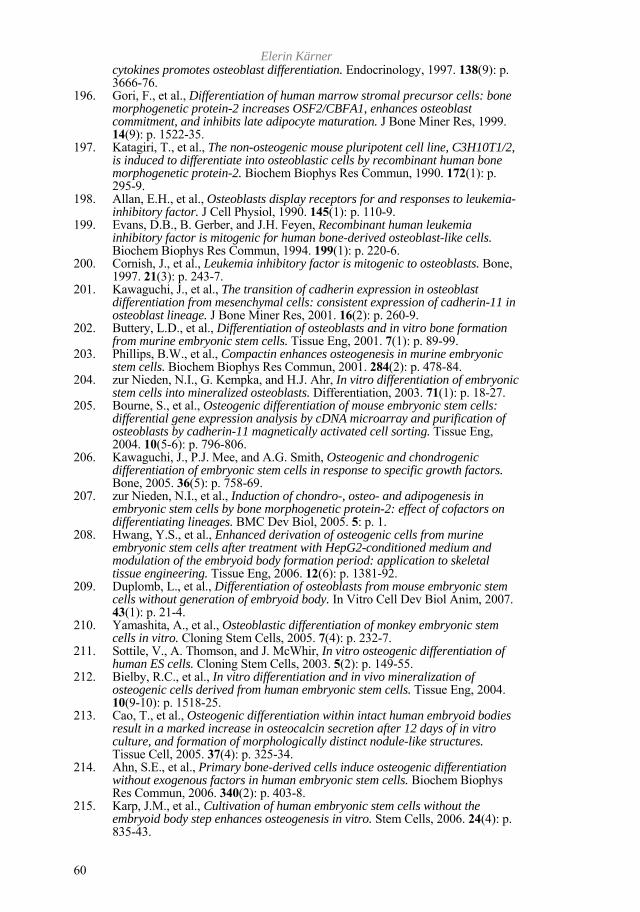
Elerin Kärner
60
cytokines promotes osteoblast differentiation. Endocrinology, 1997. 138(9): p. 3666-76.
196. Gori, F., et al., Differentiation of human marrow stromal precursor cells: bone morphogenetic protein-2 increases OSF2/CBFA1, enhances osteoblast commitment, and inhibits late adipocyte maturation. J Bone Miner Res, 1999. 14(9): p. 1522-35.
197. Katagiri, T., et al., The non-osteogenic mouse pluripotent cell line, C3H10T1/2, is induced to differentiate into osteoblastic cells by recombinant human bone morphogenetic protein-2. Biochem Biophys Res Commun, 1990. 172(1): p. 295-9.
198. Allan, E.H., et al., Osteoblasts display receptors for and responses to leukemia-inhibitory factor. J Cell Physiol, 1990. 145(1): p. 110-9.
199. Evans, D.B., B. Gerber, and J.H. Feyen, Recombinant human leukemia inhibitory factor is mitogenic for human bone-derived osteoblast-like cells. Biochem Biophys Res Commun, 1994. 199(1): p. 220-6.
200. Cornish, J., et al., Leukemia inhibitory factor is mitogenic to osteoblasts. Bone, 1997. 21(3): p. 243-7.
201. Kawaguchi, J., et al., The transition of cadherin expression in osteoblast differentiation from mesenchymal cells: consistent expression of cadherin-11 in osteoblast lineage. J Bone Miner Res, 2001. 16(2): p. 260-9.
202. Buttery, L.D., et al., Differentiation of osteoblasts and in vitro bone formation from murine embryonic stem cells. Tissue Eng, 2001. 7(1): p. 89-99.
203. Phillips, B.W., et al., Compactin enhances osteogenesis in murine embryonic stem cells. Biochem Biophys Res Commun, 2001. 284(2): p. 478-84.
204. zur Nieden, N.I., G. Kempka, and H.J. Ahr, In vitro differentiation of embryonic stem cells into mineralized osteoblasts. Differentiation, 2003. 71(1): p. 18-27.
205. Bourne, S., et al., Osteogenic differentiation of mouse embryonic stem cells: differential gene expression analysis by cDNA microarray and purification of osteoblasts by cadherin-11 magnetically activated cell sorting. Tissue Eng, 2004. 10(5-6): p. 796-806.
206. Kawaguchi, J., P.J. Mee, and A.G. Smith, Osteogenic and chondrogenic differentiation of embryonic stem cells in response to specific growth factors. Bone, 2005. 36(5): p. 758-69.
207. zur Nieden, N.I., et al., Induction of chondro-, osteo- and adipogenesis in embryonic stem cells by bone morphogenetic protein-2: effect of cofactors on differentiating lineages. BMC Dev Biol, 2005. 5: p. 1.
208. Hwang, Y.S., et al., Enhanced derivation of osteogenic cells from murine embryonic stem cells after treatment with HepG2-conditioned medium and modulation of the embryoid body formation period: application to skeletal tissue engineering. Tissue Eng, 2006. 12(6): p. 1381-92.
209. Duplomb, L., et al., Differentiation of osteoblasts from mouse embryonic stem cells without generation of embryoid body. In Vitro Cell Dev Biol Anim, 2007. 43(1): p. 21-4.
210. Yamashita, A., et al., Osteoblastic differentiation of monkey embryonic stem cells in vitro. Cloning Stem Cells, 2005. 7(4): p. 232-7.
211. Sottile, V., A. Thomson, and J. McWhir, In vitro osteogenic differentiation of human ES cells. Cloning Stem Cells, 2003. 5(2): p. 149-55.
212. Bielby, R.C., et al., In vitro differentiation and in vivo mineralization of osteogenic cells derived from human embryonic stem cells. Tissue Eng, 2004. 10(9-10): p. 1518-25.
213. Cao, T., et al., Osteogenic differentiation within intact human embryoid bodies result in a marked increase in osteocalcin secretion after 12 days of in vitro culture, and formation of morphologically distinct nodule-like structures. Tissue Cell, 2005. 37(4): p. 325-34.
214. Ahn, S.E., et al., Primary bone-derived cells induce osteogenic differentiation without exogenous factors in human embryonic stem cells. Biochem Biophys Res Commun, 2006. 340(2): p. 403-8.
215. Karp, J.M., et al., Cultivation of human embryonic stem cells without the embryoid body step enhances osteogenesis in vitro. Stem Cells, 2006. 24(4): p. 835-43.
Osteogenic differentiation of HESCs
61
216. Jaiswal, N., et al., Osteogenic differentiation of purified, culture-expanded human mesenchymal stem cells in vitro. J Cell Biochem, 1997. 64(2): p. 295-312.
217. Fujita, T., et al., Phosphate provides an extracellular signal that drives nuclear export of Runx2/Cbfa1 in bone cells. Biochem Biophys Res Commun, 2001. 280(1): p. 348-52.
218. Igarashi, M., et al., Inductive effects of dexamethasone on the gene expression of Cbfa1, Osterix and bone matrix proteins during differentiation of cultured primary rat osteoblasts. J Mol Histol, 2004. 35(1): p. 3-10.
219. Sato, H., et al., Collagen synthesis is required for ascorbic acid-enhanced differentiation of mouse embryonic stem cells into cardiomyocytes. Biochem Biophys Res Commun, 2006. 342(1): p. 107-12.
220. Shin, D.M., et al., Ascorbic acid responsive genes during neuronal differentiation of embryonic stem cells. Neuroreport, 2004. 15(12): p. 1959-63.
221. Srivastava, A.S., et al., Dexamethasone facilitates erythropoiesis in murine embryonic stem cells differentiating into hematopoietic cells in vitro. Biochem Biophys Res Commun, 2006. 346(2): p. 508-16.
222. Barberi, T. and L. Studer, Mesenchymal cells. Methods Enzymol, 2006. 418: p. 194-208.
223. Barberi, T., et al., Derivation of multipotent mesenchymal precursors from human embryonic stem cells. PLoS Med, 2005. 2(6): p. e161.
224. Olivier, E.N., A.C. Rybicki, and E.E. Bouhassira, Differentiation of human embryonic stem cells into bipotent mesenchymal stem cells. Stem Cells, 2006. 24(8): p. 1914-22.
