direct [email protected]
TRANSCRIPT

Clinical principle ofClinical principle ofDirect OphthalmoscopyDirect Ophthalmoscopy
&&methods of recording methods of recording
observation observation
Farhana AdninFarhana Adnin
B.Optom,4B.Optom,4thth BatchBatch
IICO,CU.CO,CU.

What is What is ophthalmoscopyophthalmoscopy…???…???
&&
ophthalmoscopeophthalmoscope…???…???
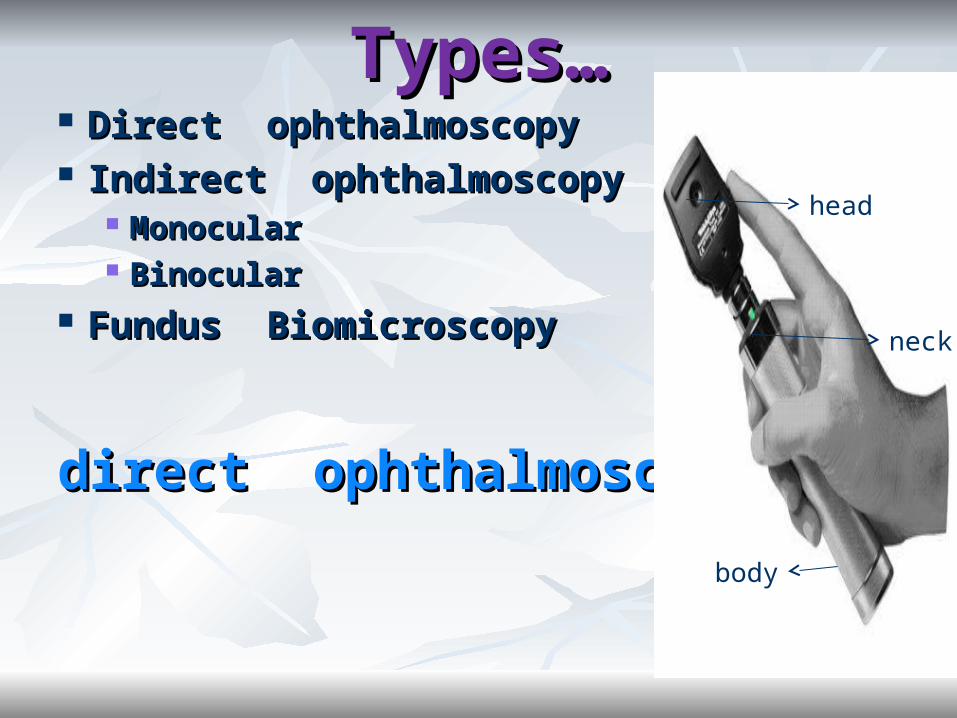
TTypes…ypes… Direct Direct ophthalmoscopyophthalmoscopy Indirect Indirect ophthalmoscopyophthalmoscopy
MonocularMonocular BinocularBinocular
Fundus BiomicroscopyFundus Biomicroscopy
direct ophthalmoscopy??direct ophthalmoscopy??
head
neck
body

IInstrumentation…nstrumentation…
Direct Ophthalmoscope consists of ~Direct Ophthalmoscope consists of ~ Illumination system & observation Illumination system & observation
systemsystem
Illumination system Illumination system –light source–light source -condensing lenses-condensing lenses -reflecting prism-reflecting prism -a series of aperture-a series of aperture Observation system Observation system –a peephole–a peephole -a bank of spherical -a bank of spherical
lenseslenses

Ophthalmoscope Ophthalmoscope head,neck, bodyhead,neck, body
Lens strength selector wheel
Selects light size,filter & grid
Concave mirror with a hole in centre
Bulb in here
Connects to rheostate and handle containing batteries
peephole
On/off rheostate
Contains battery here

PrinciplePrinciple

Optics of Optics of direct ophthalmoscope…direct ophthalmoscope…

Cont..Cont..

Characteristics of the Characteristics of the imageimage
VirtualVirtual Erect and Erect and Magnified image.Magnified image. Magnification;Magnification;
MME E = distance from viewers eye to patient’s = distance from viewers eye to patient’s fundusfundus
focal lengthfocal length
= 250mm/16.67mm = 250mm/16.67mm =15X=15X
For emmetrope, MFor emmetrope, MAA =15X =15X
For myope ,moreFor myope ,more
For hyperope, less.For hyperope, less.

04/17/2304/17/23
Methods of Methods of OphthalmoscopyOphthalmoscopy
Distance direct ophthalmoscopyDistance direct ophthalmoscopy Illuminating eye from 25-40 inchesIlluminating eye from 25-40 inches
Direct ophthalmoscopyDirect ophthalmoscopy Approach closer to patient.Approach closer to patient.

Set the patient in a semi dark room & instruct Set the patient in a semi dark room & instruct to look at a distant target.to look at a distant target.
Hold ophthalmoscope in right hand and look Hold ophthalmoscope in right hand and look through examiners’r right eye at patient’s right through examiners’r right eye at patient’s right eyeeye
Examine for red reflex at arm’s length (20-Examine for red reflex at arm’s length (20-40cm)40cm) Normal - red glow from choroidNormal - red glow from choroid Look for opacities or loss of reflexLook for opacities or loss of reflex
The examiner moves as close as to the patient The examiner moves as close as to the patient to examine anterior segment with high + powerto examine anterior segment with high + power
ProcedureProcedureof direct of direct
ophthalmoscopy…ophthalmoscopy…

Cont…Cont… Then by reducing + power,crystaline Then by reducing + power,crystaline
lens,vitreous & finally the optic nerve head lens,vitreous & finally the optic nerve head can be observed.can be observed.
Optic disc is visualized first & blood vessels Optic disc is visualized first & blood vessels can be followed then.can be followed then.
After quadrant by quadrant scan of After quadrant by quadrant scan of fundus,the macula is examined. fundus,the macula is examined.
……left eye…left eye…


Clinical UsesClinical Uses1)Light to check pupillary reaction1)Light to check pupillary reaction
2)Media opacities can be detected2)Media opacities can be detected
3)3)Estimation of patients refractive errorEstimation of patients refractive error4)Visuoscopy4)Visuoscopy
5)Small aperture and half moon:5)Small aperture and half moon: when pupil is miosed and in case of media opacitieswhen pupil is miosed and in case of media opacities6)Slit beam6)Slit beam
To view the contours in iris and retinaTo view the contours in iris and retina
7)7)Red Free filterRed Free filter8)Blue filter- 8)Blue filter- stainingstaining


04/17/2304/17/23
Clinical Clinical IImportancemportanceTo observe the optical clarity of To observe the optical clarity of
Human EyeHuman Eye
Dark or semi-dark spot over red reflex Dark or semi-dark spot over red reflex OpacityOpacity
If no reflex (whole field dark/Gray)If no reflex (whole field dark/Gray) Totally opaque lensTotally opaque lens Hemorrhage within the eyeHemorrhage within the eye Total RDTotal RD
Crescentic ring in pupillary Crescentic ring in pupillary areaareaSubluxated LensSubluxated Lens
Grayish patchesGrayish patchesRD, Choroidal RD, Choroidal detachment or intra-ocular tumordetachment or intra-ocular tumor

Clinical Clinical IImportancemportance
To evaluate A/C angleTo evaluate A/C angle Shadow technique of evaluating angles
Anterior segment:Anterior segment: # Grossly examine the cornea, lids, sclera, lashes and iris. # Grossly examine the cornea, lids, sclera, lashes and iris.
#Use +13 or 13D lenses in your ophthalmoscope. The #Use +13 or 13D lenses in your ophthalmoscope. The ophthalmoscope and your face should be about 2 to 3 inches ophthalmoscope and your face should be about 2 to 3 inches away from the patient to examine the rest.away from the patient to examine the rest.
Vitreous: Vitreous: Vitreous floaters are best seen with a + 6 or 7 Vitreous floaters are best seen with a + 6 or 7 D lens in place.D lens in place.
DiscDisc FundusFundus MaculaMacula

Basics & Fundamentals of Basics & Fundamentals of Recording Ophthalmoscopic Recording Ophthalmoscopic
Findings:Findings:1]Fundal glow1]Fundal glow
2] Disc 2] Disc ~shape ~ margin~color~vessels emerged~shape ~ margin~color~vessels emerged
~cup/disc ratio ~neural rim~cup/disc ratio ~neural rim3]Retina3]Retina~periphery{4quadrants}~periphery{4quadrants}haemorrhage,exudate,pighaemorrhage,exudate,pig
mentarymentary
changes,vascularizationchanges,vascularization
~macula (~macula (foveal reflex,any abnormality like foveal reflex,any abnormality like
hole,scar,oedema,haemorrhage)hole,scar,oedema,haemorrhage)
~vessels ~vessels (vascular reflexes,A/V ratio,crossing,new vessels(vascular reflexes,A/V ratio,crossing,new vessels))
~choroidal vessels~choroidal vessels


optic discoptic disc Move as close to the patient as possible.Move as close to the patient as possible. Find the optic discFind the optic disc
Examine the optic discExamine the optic disc
shapeshape--round to oval; diameter (1.5-1.7)round to oval; diameter (1.5-1.7)
marginmargin--sharp or dull;occasionally a pigment ring or sharp or dull;occasionally a pigment ring or conus.conus.
colour: colour: red-yellow ; temporal part may appear pale.red-yellow ; temporal part may appear pale.
vessels emerged from disc: vessels emerged from disc: Noticed to see Noticed to see neovascularization.neovascularization.

Cont…Cont…
C/D ratio (cup/disc)C/D ratio (cup/disc)

ISNT ruleISNT rule

RetinaRetina
PeripheryPeriphery- lighter than central area- lighter than central area AV ratio AV ratio (normal 2:3)(normal 2:3) Fundus colour :Fundus colour :Darker with pigmented Darker with pigmented
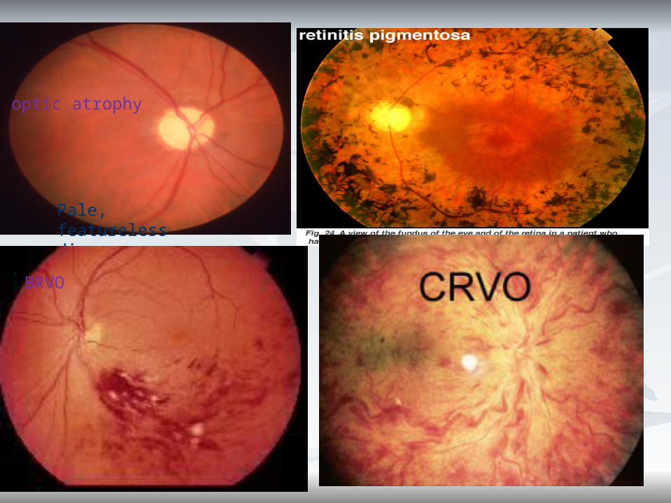
skin or retinitisskin or retinitis pigmentosapigmentosa Pale with arterial occlusionPale with arterial occlusion
MaculaMacula- - appears darker than central areaappears darker than central area
-3mm temporal to disc.-3mm temporal to disc.
- Central foveal reflex- Healthy/ dull- Central foveal reflex- Healthy/ dull

Clinical recording of Clinical recording of Ophthalmoscopic Ophthalmoscopic
findings…findings…In normal case…In normal case… Shape- roundShape- round Marjin-sharpMarjin-sharp Colour-pinkColour-pink C/D ratio- 0.3:1 (supposing)C/D ratio- 0.3:1 (supposing) A/V ratio- 2:3A/V ratio- 2:3 Macula – healthy & bright foveal Macula – healthy & bright foveal
reflex. reflex.

Blurred disc margin
pink disc
Disc swollen / raised
glaucoma
normal
papilloedema
04/17/2304/17/23
Pale, featureless disc
optic atrophy
BRVO

04/17/2304/17/23
Hard exudates
Haemorrhage
Microaneurysm
non-proliferative diabetic
retinopathy
Proliferative diabetic
retinopathy

Age-related Macular Age-related Macular DegenetationDegenetation
Wet form: abnormal blood Wet form: abnormal blood vessel growth w/ hemorrhage vessel growth w/ hemorrhage and protein leakageand protein leakage
Dry form: Drusen Dry form: Drusen
(cellular debris) build-(cellular debris) build-upup

04/17/2304/17/23
Move on to the front of Move on to the front of the eyethe eye
Dendritic ulcer (herpes virus) on the cornea inspected with cobalt blue light
Foreign body
cataract
Corneal ulcer
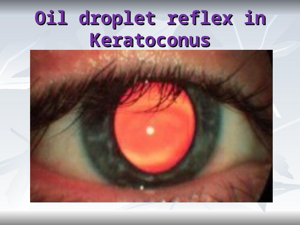
Oil droplet reflex in Oil droplet reflex in KeratoconusKeratoconus
References1.Theory & practice of Optics & Refraction…A.K.Khurana
2.Clinical Procedure in Optometry
3.Primary Care Optometry… Theodore Grosvenor
4.Essentials of Ophthalmology…S.K.Basak
5.Internet