direct measurement of the optical modulation transfer
TRANSCRIPT

Rochester Institute of TechnologyRIT Scholar Works
Theses Thesis/Dissertation Collections
6-1-1977
Direct Measurement of the Optical ModulationTransfer Function of Non-Developed EmulsionsGlen Elie
Follow this and additional works at: http://scholarworks.rit.edu/theses
This Thesis is brought to you for free and open access by the Thesis/Dissertation Collections at RIT Scholar Works. It has been accepted for inclusionin Theses by an authorized administrator of RIT Scholar Works. For more information, please contact [email protected].
Recommended CitationElie, Glen, "Direct Measurement of the Optical Modulation Transfer Function of Non-Developed Emulsions" (1977). Thesis.Rochester Institute of Technology. Accessed from

School of Photographic Arts and Sciences
Rochester Institute of Technology
Rochester, New York
CERTIFICATE OF APPROVAL
MASTER'S THESIS
This is to certify that the Master's Thesis of
Glen C. Elie
with a major in Photographic Science and Instrumentation
has been approved by the Thesis Committee as
satisfactory for the thesis requirement for the
Master of Science degree at the convocation of
Thesis Committee: M. Abouelata Thesis adviser
Name Illegible Graduate adviser
Burt H. Carroll Director or designate

DIRECT MEASUREMENT OF THE OPTICAL MODULATION TRANSFER
FUNCTION OF NON-DEVELOPED EMULSIONS
by
Glen C. Elie
A thesis submitted in partial fulfillment of the
requirements for the degree of Master of Science in the
Photographic Science and Instrumentation Division of the
School of Photographic Arts and Sciences
June, 1977
Thesis adviser: Professor Mohamed F. Abouelata

f
ACKNOWLEDGEMENTS
I would like to express my sincere thanks to my
adviser, Professor M. Abouelata, for his constant support
and guidance throughout this thesis, to Professor
J. Carson for his valuable assistance in the preliminary
testing of the equipment. I am also very grateful to
Dr. B. Carroll for his suggestions concerning the analysis
of the experimental results.
11

TABLE OF CONTENTS
Page
LIST OF TABLES iv
LIST OF FIGURES v
INTRODUCTION 1
THEORY 17
EXPERIMENTAL 31
Equipment 32
ANALYSIS 38
CONCLUSION 50
LIST OF REFERENCES 53
APPENDIX A 54
APPENDIX B 57
lll

LIST OF TABLES
TABLE 1 MTF Data for test films. Run Number 1 . .
Page
. 41
TABLE 2 MTF Data for test films. Run Number 2 . . . 41
TABLE 3 MTF Data for test films. Run Number 3 . . . 42
TABLE 4 MTF Data for test films. Run Number 4 . . . 42
TABLE 5 MTF Data for test films. Run Number 5 . . . 43
TABLE 6 MTF Data for test films. Run Number 6 . . . 43
TABLE 7 MTF Data for test films . Run Number 7 . . . 44
TABLE 8 MTF Data for test films. Run Number 8 . . . 44
IV
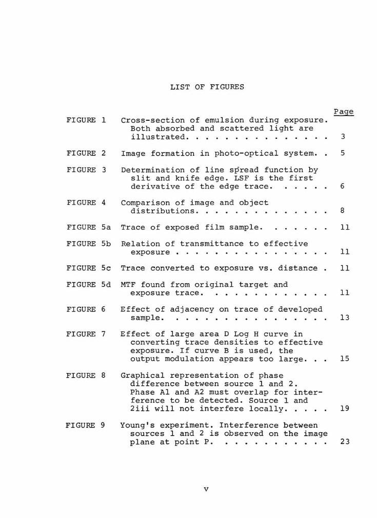
LIST OF FIGURES
Page
FIGURE 1 Cross-section of emulsion during exposure.
Both absorbed and scattered light are
illustrated 3
FIGURE 2 Image formation in photo-optical system. . 5
FIGURE 3 Determination of line sp'read function byslit and knife edge. LSF is the first
derivative of the edge trace 6
FIGURE 4 Comparison of image and object
distributions 8
FIGURE 5a Trace of exposed film sample 11
FIGURE 5b Relation of transmittance to effective
exposure 11
FIGURE 5c Trace converted to exposure vs. distance . 11
FIGURE 5d MTF found from original target and
exposure trace 11
FIGURE 6 Effect of adjacency on trace of developed
sample 13
FIGURE 7 Effect of large area D Log H curve in
converting trace densities to effective
exposure. If curve B is used, the
output modulation appears too large. . . 15
FIGURE 8 Graphical representation of phase
difference between source 1 and 2.
Phase Al and A2 must overlap for inter
ference to be detected. Source 1 and
2iii will not interfere locally 19
FIGURE 9 Young's experiment. Interference between
sources 1 and 2 is observed on the image
plane at point P 23
v

FIGURE 10
FIGURE 11
FIGURE 12
FIGURE 13
FIGURE 14
FIGURE 15
FIGURE 16
FIGURE 17
FIGURE 18
FIGURE 19
FIGURE 20
FIGURE 21
FIGURE 22
Page
Shape of fringes resulting from inter
ference of monochromatic sources 24
Interference due to primary source of two
emitted wavelengths is the sum of each
pattern separately 25
Interference pattern of source of band
width AX. Coherence length decreases as
AX increases 25
Relation of parameters S and C 26
Spectral distribution and visibility
curves for Helium-Neon laser 2 8
Lloyd's mirror. Interference occurs between
primary source and its virtual image. . . 29
Layout of experimental apparatus. Photo
multiplier feeds information to radio
meter (not shown) 32
Fourier transform of interference fringes
with frequency of 10 c/mm. Six cycles are
visible along the image plane 35
Transmission vs. time tests 37
MTF for Kodak Panatomic-X. The solid curve
shows the frequency response derived from
traditional methods. The dashed curve
represents the approximate average values
of the data 45
MTF for Kodalith Type 3. The dashed curve
represents the approximate average values
of the data 46
MTF for Kodak High Contrast Copy. The solid
curve shows the frequency response
derived from traditional methods. No
average value curve is drawn here due to
the large variability in the data. ... 47
Effect of k-value of film on oblique inci
dence of light. The larger the k-value of
the emulsion, the more asymmetric the
spread function 4 8
vi

Page
FIGURE 23 MTF of Kodak High Contrast Copy plotted
against angle between sample and mirror.
The average value closely agrees with
the estimated average from Figure 21. . . 49
FIGURE 24 Calculation of region of interference, y,
in Lloyd's mirror arrangement 58
FIGURE 25 A true cosine wave limited by a window of
width y 60
FIGURE 26 Fourier transform of limited cosine
function is two sine functions about
co=a)0 and w=-w0. . 60
vii

ABSTRACT
A new procedure for measuring the MTF of non-developed
emulsions is discussed, which uses a coherent energy source
and interference system to produce sinusoidal fringes on a
film sample. The transmitted light is projected onto the
scanning slit of a photomultiplier, and its variations
registered on a chart recorder. Measurements are taken
directly from the traces of the irradiance distribution to
calculate the optical part of the MTF of the sample. This
system does not require development of the film, and is
therefore free of the non-linearities inherent in traditional
test methods.
Results are obtained for Kodak Panatomic-X, Kodak High
Contrast Copy, and Kodalith Ortho Type 3 films, and compared
where possible to the MTF derived from standard techniques.
The MTF found from this study is that of the light distribu
tion in the undeveloped emulsion layer, while the conven
tional method gives the MTF of the developed film. Adjacency
effects in development cause the two methods to produce
different results. It is concluded that this system is
capable of producing excellent results for some types of
films. Recommendations are made for improvement of the
apparatus to render the procedure more universally suitable.

INTRODUCTION
Image quality is a concept often referred to in general
terms, but determining an overall basis for any judgement is
a difficult if not impossible task. Because of the number of
different film characteristics involved, image quality
depends on which of these is being assessed. A perfect repro
duction in all respects is not possible, so the photographer
must compromise by choosing a film which will best represent
the subject, knowing that this may mean only a second best
reproduction of another area. A photograph containing sharp
architectural lines may appear as clear as the original
scene, while an inscription on the side of the same building
is almost illegible. It is important to be able to rate the
performance of films with respect to their characteristics,
and to do so, standard tests are required. This thesis is
concerned with one of these tests, the optical part of the
Modulation Transfer Function.
The evaluation of an image occurs at different levels,
and has as its culmination the subjective opinion, itself
dependent on image content. In this section, the basic
levels of image evaluation will be reviewed and inter
related, showing their progression towards the final subjective
appraisal. Graphically, the levels may be represented as

follows :
Line Spread Function (LSF) Basis of all other tests, yet
difficult to relate to final
image .
Modulation Transfer Function Spatial frequency response
curve, used to measure contrast
reduction of targets.
Resolution, Acutance Each expressed as single number
related to specific areas of
MTF curve. Resolution depends on
shape of the neck of the LSF,
and Acutance depends on the
shape of the base of the LSF1.
Subjective Opinion Visual appearance evaluated
by observer without special
equipment .
Each level leads to the one below, with the basic
requirement being that the system is struck by light. Just
how a photographic emulsion reacts to a point of light is the
first concern, since all more complicated sources are made
up of a series of points, and are therefore based on this2.
When an infinitesimal surface area of film is exposed to
light, one of several things can happen to the photons
entering the emulsion. They may be absorbed by the first
grain they encounter. They may be reflected to a second or
higher number grain before being absorbed. Or they may pass

out of the emulsion without being absorbed at all. As the
photons travel through the film, they are reflected sideways
to a certain extent. This sideways scatter will be approx
imately symmetrical in photographic emulsions. If we took a
cross-section of the film through the location where it was
light-struck, we would see something as in Figure 1. Moving
SOURCE'"*
(_J
BACK SCATTER
*i EMULSION
Figure 1. Cross section of emulsion during exposure. Both
absorbed and scattered light are illustrated.
along the film and summing all the exposed grains above the
interval Ax, a curve could be obtained showing the emulsion's
reproduction of the point of light. This operation, in effect,
is performed by a microdensitometer equipped with a small
circular aperture. By scanning the image of a point, a trace
is obtained of the illuminance distribution of the image, and
is called the point spread function of the film. A second

scan would be necessary in a two dimensional distribution.
Because we are dealing with symmetric distributions, a
rectangular aperture is used, and only a single scan needed.
The result is called the line spread function, and is
defined as the illuminance distribution of the image of a
line. If the PSF is denoted by p(x,y), then the line spread
function, s (x) = /p (x,y) 3y . The point or line spread function
-co-J
represents the most basic form of illuminance distribution;
and all other such distribution's can be found by summing
the spread functions from each point on the object curve.
This is the reason the spread function is expressed in terms
of illuminance or exposure. These quantities are additive,
as opposed to transmittance, an exponential function, which
would be much more difficult to apply.
It is possible to determine the image distribution, by
knowing the illuminance function of the object, and the
line spread function of the imageing system. The one dimen
sional image function, g(x) , is found byconvoluting* the
object function, f (x) , with the system line spread function,
s (x) . To see how this is produced, imagine an object
function, f (x). , divided into small elements of f(n)Ar), so
that f(x)= Sf(n)An. By making An very small,f(x)= /f(n)3n.
This object is then imaged by a photo-optical system of
line spread function, s (x) , and the resulting contribution
*see Appendix

System line spread function
Image distribution
Figure 2. Image formation in photo-optical system.

of each element to the image function is 9g(x)= f(x~n)s(n)9n
The total image function is g(x)= If (x-n) s (n) 9n (1)
and is shown in Figure 2. Formula (1) is called the
convolution integral, and is denoted symbolically f(x)*s(x).
If a system contains more than one spread function, the
total LSF can be found by convolution of the individual LSF's,
s (x) =s i (x) *s2 (x) .
Since an illuminance distribution can be found from the
system LSF and the object function, then one way of evaluat
ing a photographic system is to find the LSF representing
that system. Practically, the determination of the LSF is
carried out using a knife edge as object, as there is not
enough energy passing through an infinitely narrow slit to
expose the film sample. The effect of the edge is shown in
Figure 3, beside an equivalent illustration of the
theoretical function.
u.Source-
Edge
g(x)
Figure 3. Determination of line spread function by slitand knife edge. LSF is the first derivative of the edge
trace.

In the case of the knife edge, the object distribution is
f (x)= j 1 for x>0
0 for x<0 .
Let the spread function be s(x). Then the illuminance
function of the image is/OO A- OO
f (n)s(x-n) 3n= js(x-n)9n
and settingx-n=
g(x)= |sU)3C
showing that the line spread function is the first derivative
of the edge trace. The edge trace method incorporates an
averageing effect due to the graphical analysis, so is not
overly susceptible to image noise. In contrast, by the
theoretical method
f(x)= jl for x=0 (unit impulse function)
0 elsewhere
so g(x)= f(x)*s(x), and the convolution of any function with
the delta function gives that function itself. So for this
instance, g(x)= s (x) .
We can apply the same technique to any object dist
ribution3. If the object is a sinusoidal function, then
f(x)= afi-a cos wx whereaQ is the D.C. level,
and a is the amplitude at frequency id. This object, repro
duced by an emulsion, yieldsg(x)= f(x)*s(x). Representing
cos wx by the real part of the complex e ,
f(x)= aQ-a Rl[eiwX]
and the image becomes

Now
-00
g(x)= f(x)*sCx)= J(a0-a Rl [eXuJx~n,]l s(n)9n
=
an/7(n)9n-
aRl[eia)X fe"lu)T,s (n) 9n] .
ls(n)9n is a constant= c, and
fe-iwT1s(n)9n-00-'
is the Fourier transform*, F(w), of s(n)/ so
g(x)= aQc-a Rl
[elwxF(w)]
F(w)| Rl[ei{wX-^]=aQc-
a
and normalizing, g(x)= g(x)v c
gn(x)= aQ-a-fe^fl- cos (wx-cf>)
In dealing with film, s (x) is generally symmetrical, and <J>=0
F(w)|so g (x)
^na
a0
cos wx. (2)
Comparing this to the object, the D.C. level has not
changed, but the variation in amplitude is now influenced
by a factor dependent on the frequency of oscillation. The
effect is illustrated in Figure 4.
f(x)
x
Figure 4. Comparison of image and object distributions,
* see Appendix

9
This modulation factor forms the basis for all further
discussion. In curves such as the one above, it shows the
relationship between maximum or minimum amplitude, and the
D.C. level. In f (x) , the object distribution, modulation=a/an,
I Jand in g(x) , the image distribution, M.= ^"^
. To see how1
a0well the output distribution represents the input, the two
modulations are compared, and the result called the
modulation transfer, M./M =
'
- ^. This is in the case
' i'
o c
where the spread function is symmetrical, and normalization
has occurred. The modulation transfer function is the
Fourier transform of the line spread function of the photo-
optical system. This transform has bridged level 1 and 2 in
our four level image quality scheme.
If the curves f (x) and g(x) are known, as from micro
densitometer traces, MTF can be determined directly from
the maxima and minima of these curves. For any frequencyr
MTF =?max~9min
/max-
fmjn^
9max 9min ^max tmin*
Continued over the desired frequency range, a curve is
produced, representing the frequency response of the system.
This is the method used in this experiment to determine MTF.
The formulas above were derived from sinusoidal input
functions. If the object is other than sinusoidal, Fourier
analysis must be used to first split the object and image
into their sinusoidal components. Input and output modulations
are found by summing the modulations of the corresponding
harmonics. Then MTF= M. . . as before.image oo^ect

10
When a system containing several elements is to be
tested, this method can be used if only the overall MTF is
desired. However, the MTF of a single element can be found
by proceeding as follows. For a system of three parts,
s(x)=s- (x) *s2 (x)*s3(x)
^[s(x)] = 7[Sl(x)*s2(x)*s3(x)]so
therefore
and
|f(w)|= ^(w)!Fx(w) F(w) /
F2(w)
F2(w)
F3(w)
F3(id)
(3)
MTF1=c MTFtQt /cic2c3MTF2MTF3r
where c= /s(x) 3x .
-oaJ
Note that the Fourier method applies exactly only if the
system is linear, while the convolution method always holds.
A summary of the standard techniques in determining
MTF from a sinusoidal target follows . First the target
itself is scanned at each frequency to give a plot of the
object function. Then a photographic emulsion is exposed,
and developed, to show the image of the target, and is in
turn placed in the microdensitometer to trace illuminance
vs. distance. An example is shown in Figure 5 . Becausecl
development is a non-linear operation, D and D . frommax min
each trace must be converted to H and H ., the effective
max min
exposure values. This is done by exposing another strip of
film to a photographic step tablet, and scanning the
developed image in the same microdensitometer. This produces
a plot of density vs. exposure, Figure 5,. By reflecting
each desired density value on this graph, equivalent values

11
T(x)
Figure 5a. Trace of exposed film sample.
D(x)
Average value curve
Log H
Figure 5b. Relation of transmittance to effective exposure,
H(x)
x
Figure 5c. Trace converted to exposure vs. distance,
1.0
M(v)
.1
Figure 5d. MTF found from original target and exposure
trace .

12
of exposure are obtained. If the entire trace is reflected,
the curve will appear as in Figure 5 . Usually, only the
maximum and minimum values are needed, and the output
modulation found from (H -H . )/(H +H . ) for eachmax rain
'max mm
frequency. A sample MTF curve is drawn in Figure 5,.
MTF gives the overall frequency response of the
emulsion, but there are two areas of the function which are
of particular interest. These are the shape of the curve
near v=0 , and the frequency at which MTF=.10. The curve
shape at low frequency will indicate how well the film will
reproduce edges. High MTF values in low frequencies indicate
high acutance. Similarly, the higher the frequency value at
the point where MTF=.1, the better the film is at repro
ducing fine detail. This is the resolving power of the
emulsion. Both these parameters are part of the third level
of image analysis.
The final stage, subjective opinion, is dependent on
all the previous parameters, plus others such as graininess,
and even the pictorial content of the image.
The methods described so far are useful in determining
the capabilities of the photographic system as a whole, as
they deal with the combined film-developer effect. Problems
arise when either the input or developer is changed
drastically. These problems are caused by the non-linear
adjacency of developers, and will be outlined here as they
affect the MTF of the film.

13
The strict definition of MTF deals with light scatter
in the film alone, but because of the mechanism of develop
ment, the MTF will be modified to some extent. This
modification will depend on the degree of adjacency effect,
which itself will depend on the type of developer, amount
of agitation, temperature, etc. In Figure 6 a sharp
exposure gradient exists between areas A and B of a film
sample. Upon processing, the developer over B exhausts
faster than over A. There is some lateral developer
migration between the two areas, causing an adjacency
effect. Fresh developer diffuses part way into B, and
exhausted developer part way into A, with the result that
D(x)
Development
D(x)
Without adjacency
x
_&1
Exposed sample / _,,
Adjacency present
x
Figure 6. Effect of adjacency on trace of developed sample.
the processed film has a density distribution as inA'
and B'. The apparent sharpness has increased. As a general
rule, the less active the developer, the more evident the
adjacency effects. More problems occur because of different

14
types and gradients of exposure boundaries. Figure 6 shows a
knife edge exposure, but when the input is a sine wave, the
extent of adjacency will not necessarily be the same. This
shows that to some degree, MTF as measured here depends on
the inputtarget*4
, a situation that makes predicting image
distributions difficult. Different average densities will
also produce different amounts of adjacency in the same type
target. This has been related to the characteristic curve
in a number ofarticles5 6 7
. The problem is that the large
area D log H curve used to relate trace density to effective
exposure does not in general represent the true character
istic curve for the type of input used. When adjacency is
present, the density difference between D and D . is* ' 2
max min
greater than the difference due to exposure alone. If this
difference is reflected on the large area D log H curve,
the apparent log H interval will in turn be greater than the
actual amount. In Figure 7, the apparent density difference,
when reflected on the large area curve, gives log H interval
B. If this is used, calculations of the output modulation
will be too large, yielding an inflated MTF. It frequently
happens that MTF> 1 at low frequencies because of this. Note
that in Figure 7, the less the average density of the scene,
the closer the two curves are to each other, illustrating
that lower average density produces less adjacency.
There are simply too many variables for the MTF of the
film alone, or for MTF of inputs other than those used in

15
the original test, to be found from the standard method. For
a generally valid prediction of a film's frequency response,
Small area curve
(adjacency present)
Large area curve
Log H
Figure 7. Effect of large area D Log H curve in convertingtrace densities to effective exposure. If curve B is used,
the output modulation appears too large.
the Optical MTF must be used. A method devised byC.N.Nelson5
employs mathematical means to remove the effect of the
adjacency from the measured MTF. But Nelson's models are
empirical, including approximations and assumptions which
depend on the film-developer combination. There is no
general formula at this time for mathematically predicting
the effect of development. An experimental method for
determining optical MTF was first described byH.Thiry8
. The
technique uses undeveloped samples in the tests, so is not
susceptible to adjacency effects, and gives a result
independent of the type of input. It also provides a method
for direct comparison of emulsions. Finally, it can be used
to monitor the effect of development, as any difference
between Optical MTF and apparent MTF can be directly

16
related to the type of development used. In this way,
Nelson's chemical spread function concept can be evaluated.
Although Thiry briefly mentions a test of a negative
material, his paper deals mainly with holographic emulsions
at high frequencies. The experiment is redone here to expand
the results, using the same basic design to study negative
films at relatively low frequencies ( <200 c/mm ) .
Suggestions for improvements to the apparatus are made in
the conclusion of the thesis.

17
THEORY
In the study of modulation transfer functions, some
sort of sinusoidal object is normally used, but here instead
of the target being permanent, it is set up optically by
interference. The fringe pattern is imaged directly through
a microscope onto a radiometer. The trace on a chart recorder
shows the object or image pattern, depending on whether a
film is in place in the sample plane. A brief discussion of
interference theory will be included before describing the
exact experimental set-up.
By the principle of superposition, when two waves
cross , the resultant displacement at any point is the sum
of the displacements due to each wave separately. This can
be seen for water waves simply by tossing two stones into
a pool, such that the disturbance set up by one will cross
the other. Both sets of waves have their own amplitude,
frequency, and phase, dependent on the parameters of the
source. Phase can be interpreted as that fraction of a
complete vibration attained at a particular time. It will
vary from point to point depending on x, the distance along
the wave. When x changes by a wavelength, the phase will be
the same as its starting value. The constant k, called the
wave number, represents the proportion between the phase

18
and the displacement along the wave. The phase difference
between two pointsx2
and x, on the wave, at the same time,
is -6, where <5=k(x2-x,).
So far we have spoken of unrelated waves, but now if
it is required that both waves have the same frequency, and
that the sources vary in phase by a constant, an interference
effect is produced. If the origins differ by a phase <f>, the
phase difference at point P will be S=k*(x -x. )-c|> . Water
waves from two oscillating sources of the same frequency can
be studied easily, and 6 calculated at any point, since <j>
is determinable, and constant.
The same general terms apply in theory to light. The
light paths from two different sources can be made to
superpose. But light is emitted as a burst of waves lasting
about10~9
seconds, each packet with random phase. So <(>
changes each10~9
seconds. Interference occurs, but its
pattern and position changes each time <J> changes. The
recorded result is just the average of all these inter
ferences, or simply a steady state irradiance. For measure
ment of light interference, there must be some conditions
established for thesources.9
1. Coherent sources are needed. All apparent sources must
emit light of the same, or proportional phase*. Inthis way,
even though the phase shifts randomly everyIO-9
seconds,
* In the set-up used here, apparent sources are 180 degrees
out of phase, but this relationship continually exists.

19
it does so the same for each source, so the effects cancel.
Such sources may be devised by using a source and its
optical image, or by using two images of the same source.
2. The amplitude of the two apparent sources must be roughly
the same, otherwise the difference between maxima and minima
in the interference pattern will be small, if not measureless.
3. The optical path difference should not be too great. Due
to the finite speed of light, the beginning of one burst of
light leads the other by approximately 30 cm. The illustration
in Figure 8 represents the parts of two waves which will
interfere in a stationary location. Between source 2 and 2.,
SOURCE 1 {PHASE A,} {PHASE B^ {PHASE C^
SOURCE 2i {PHASE A2. } {PHASE B^} {PHASE C^}
2ii {PHASE A_..} {PHASE B_..} {PHASE C, . . )Zll ^-11 fell
2iii {PHASE A2iii} {PHASE B2iii)
Figure 8. Graphical representation of phase difference
between source 1 and 2 . Phase Al and A2 must overlap for
interference to be detected. Source 1 and 2iii will not
interfere locally.
the path difference is zero, and maximum interference will
occur. Source 2.. will partially interfere, and source 2...
will not interfere locally with source 1, since the phase
difference B-A will differ from C-B, and the situation will
be similar to that of incoherent sources.
The discussion to this point has dealt with sources of
infinitesimal spectral width. For real sources, a second

20
type of coherence restriction, temporal coherence, is placed
on the system, and is related to the spectral width of the
source. As the spectral width, AX, increases, the visibility
of interference approaches zero in only a few fringe widths.
In terms of coherence length Al, the distance from the
maximum interference to the point where the visibility
reaches zero, Al= X2/AX*. As an example, for white light
_ o o o.
X=55 A, AX=30 A, so Al =100 A. Since the average wavelength
o
is approximately 50 A, this means there will be two fringes
visible. This concept of visibility will be further
discussed later.
To summarize: A single source is devided by wavefront
or amplitude to produce two waves of the same frequency
which then travel along different optical paths, and are
then made to intersect. Interference is the study of the
superposition of these two waves.
* From the wave uncertainty relation for the propagation
of wave packets J AtAv>-^
-1 ,where At, the coherence
time, is the average duration of wave trains of a light
disturbance, and Av is the effective frequency range of
the Fourier spectrum of the disturbance. Now v= c/X
so Av= cAX/X2. Also Al= cAt, so Al= X2/AX, where Al is
the coherence length.

21
In studying interference, a wave of the form =Ae
will be used, and is shown here to satisfy the general wave
equation 92y (x,t) /9x2= (1/v2) d2V (x,t)/9t2
. This equation
determines the properties of motion of a wave travelling along
the x-axis, and has a general solution = f (xvt) . Taking the
complex solution of a transverse wave, the real part of
_ i(kx-wt) ,,,, a.j-
Ae will be shown to satisfy the wave equation.
<x,t)- Ae1^"^
a2|_= Ak ei(kx-wt)= k2Y(Xft)9x
Similarly32^/9t2= w2(x,t)
Now w2y(x,t)/k2(x,t)= w2/k2=v2
So 92Y(x,t)/8x2= (l/v2)92^(x,t)/9t2
showingY(x,t)=
Ae1 sis a valid solution. Furthermore,
if the wave travels through a series of mediums, then
(x,t)= Ae*- ixi/
f Where k^ changes in proportion to
the velocity of the wave in the medium. The quantity Ik^x^
represents the phase <J>, so the equation may be rewritten
(x,t)=Ae1 '
. When two waves are made to superpose,
the total effect is
(x,t)- A.e^-^^^+A.e1^^*^
orAe^= A1ei*M-A2e1*7S
It is useful to determine time average power of the wave
Ae , since this indicates the irradiance, which can be
measured by a radiometer.
Since
(P(t) = (Aei((>)(Ae~i(,,)= (A1ei*'+A2ei**)(A1e"i*'+A2e"i**)

22
2setting E=A
E=E +E 2+2 v^TeT cos 5
where 6 is the phase difference. The total irradiance is the
sum of the two waves plus a modifying term dependent on the
phase difference between the waves. This is responsible for
the interference, containing a harmonic term cos 6 , and a
modulation factor. When dealing with light, it is optical
path difference which is measured. Instead of 4>, we use kXi
and for the case where each path is through a single medium,
<}>=kx, 6=k(x2-x1) =kAs. The optical path difference, As, is a
variable which increases as the point under observation
moves away from the central position, producing the fringes
in the interference pattern. The general equation now
becomes E=(E1+E2) Fl+(2/E1E2) /(E1+E2) cos 2TrAs~] (4)
where k=2 7T.
Thomas Young was the first to produce interference effects
using division of wavefront. This is a valuable experiment
to study, as many other designs are quite similar to it, and
only slight mathematical changes are required to derive
equivalent parameters. In Young's set up, monochromatic
light from a primary point source is intercepted by a plate
with two holes whose size and separation is small (the
diameter of each aperature is of the order of the wavelength
of light) . An image screen is placed a distance R from the
secondary sources, parallel to the object plane, such that
R>>D. Waves leave 1 and 2 in phase (both apertures are

23
equidistant from the primary source), and intersect at P.
The difference in their optical paths is s2-sa=As. We wish
to determine this quantity to predict the appearance of the
interference pattern on the image screen.
Secondarysources
source
Object plane
Figure 9. Young's experiment. Interference between sources
1 and 2 is observed on the image plane at point P.
In Figure 9, at any point P,
and
2, B, 2 2
s2 = (Y+2> +R
2 2s2 -Sj =2yD
2,
D,2 LlSi
= (y-n) +R
s2-s i=As2yD
s2+ Si
A-Jg
SO
giving
The irradiance at point P is now
E=(Ea+E2)
and since R>>D, 2R=s2 + s5
. 2/EiE2 ~ D
^"e^cos
2^XR.XR
(5)
where the period of the disturbance is =-. The maximum value
of this function occurs when cos-y^
=1, and the minimum
when cos-- \-
=-1. That is
when *-~ m m= 0,1,2,3,... there is a maximum
or Y=mR
D'

24
and when ^= m+|m- 0 , 1 , 2 , 3 , . . .
or
y=(m+l)2|
there is a minimum. The integer m is the order of inter
ference, and the fringes called the zero, first, second, and
third orders respectively.
The irradiance distribution in the image plane of a
monochromatic source is shown in Figure 10. With the
secondary sources in phase, the pattern. has a maximum at
x=0, the perpendicular bisector of the line connecting the
sources. Secondary sources out of phase cause the curve to
shift laterally by an amount proportional to the phase
difference. If this difference is ir radians, the pattern at
x=0 will have minimum irradiance.
Figure 10. Shape of fringes resulting from interference
of monochromatic sources.
Spatial frequency depends on the distance between the
secondary sources, the distance between the source and image
planes, and the wavelength of the illumination. If no change
is made in the structure of the apparatus, it can be seen

25
pfrom the formula, fringe separation=--r, that the period of
the interference pattern is directly proportional to the
wavelength of the source. If the light consists of two emitted
wavelengths, the contributions are mutually incoherent, and
the total distribution is just the sum of each function
individually, as in Figure 11. The coherence length is very
short in this case, with the visibility reaching zero after
only one period. For a real source of spectral width AX,
there is a distinctive envelope on the pattern as in
Figure 12, which can be predicted if the spectral distri
bution of the source isknown.10
Interference from A+B
Wfyoo^oWavelength A Wavelength B
Figure 11. Interference due to primary source of two
emitted wavelengths is the sum of each pattern separately,
Figure 12. Interference pattern of source of bandwidth AX.
Coherence length decreases as AX increases.

26
To determine the envelope, or visibility curve, the
light source is regarded as a summation of monochromatic
sources over the spectral width AX. The irradiance of each
element over the wave number range 3kQ may be calculated by
equation 5 as
9k0
(6)e0(k0,As)=2e1(k()) [l+cos kQAs
where k0=2TT/X0 , optical path difference As=6/kQ, and e,(kQ)
is the spectral irradiance distribution .of the section of the
beam at k..(Both beams will have the same irradiance for the
purpose of this discussion) . All spectral components add
incoherently, and the total irradiance is
E(As)=2/e1(k(J) l+cos kQAs 3k0
*
If kQ is the central position of the source, let x be such
that kft=kn+x ;then the irradiance in the interval 3kn isL0
Now
j(x)=e1(kQ+x).
E=2|j(x) [l+cos ((k0+x)As]J 3x
=P+cos kQAs C (As) -sin kQAs S(As)
L /c^+s2
where
=P+/C2+S2
P=2 I j(x) 9x
C=2/j(x) cos xAs 3x
S=2/ j(x) sin xAs 3x
From Figure 13 , with tan =S/C
C cos kpAs S sin kpAs
y^+s-
Figure 13. Relation of parameters S and C.
E=P+/C2+S2
(cose cos kQAs-sin6 sin kQAs)

27
which may also be written
E=PJr 2+c 2
1+p
scos kQAs+e (7)
For the normalized distribution, E/P, the visibility
(E -E . )/(E +E . )=/C2+S2/P=Vmax mm max min
'
This procedure is equivalent to taking the Fourier
transform of the spectral distribution, going to As space
instead of w space. C and S are the Fourier cosine and sine
transforms of the function j (x) .
/C2+S2is F(AS) and in
complex form
F(AS) = fj(x)e"iAsx3x. (8)
P is the normalization factor. An equivalent expression was
used in the introduction to show that MTF is the Fourier
transform of the spread function. Here MTF is replaced by
the visibility envelope V(As) , and the spread function by
the spectral distribution.
For a spectral distribution of gaussian shape,
j(x)=j0e"a xa=/2TH2/Ak .
Visibility V(As)=J j (x)e~lAsx3x
-As2/(4a2)
("
Je-(ax+i=j0e
' Jeza'
9x .
Setting y=ax+^||, 3y=a9x ,
v(As)4e-^2/(4a!/.-**
=D0v/rfya e"As2/4a2=l at As=0 in the
normalized function, so
V (as)- e"(As/2a)2.n
This is the type of visibility function resulting from the
spectral distribution of a Helium-Neon laser, Figure 14. In

28
this case, the optical path difference As, has a value
of 4.5 cm when V(As)=.l. To that point, 71000 wavelengths
can be observed.
Spectral distribution
For helium-neon laser
1=630 nm
AX=1/15 A
AX
Visibility curve
1 2 3 4 As
centimeters
Figure 14. Spectral distribution and visibility curves for
Helium-Neon laser.
The Lloyd's mirror arrangement in Figure 15 consists of
a primary source placed a distance from an optical flat, such
that light from it hits the mirror at near grazing incidence.
The image plane is located perpendicular to the mirror. A
point P located on this plane receives light directly from
source 1, and from its virtual image at 2. Interference will
occur at P just as in Young's experiment, with only a few
differences. First, the virtual source is a reflected image

29
of the real source, and this results in a phase difference
of tt between the sources, giving an irradiance minimum at the
zero order fringe. Second, interference can only occur when
both sources are visible to the point on the image plane.
Image plane
R ^
, Primary source
^6?c*i><><v<>^xS2A'
0
t-'
Figure 15. Lloyd's mirror. Interference occurs between
primary source and its virtual imacre.
When the light reaching P comes from the front edge of the
mirror, the maximum extent of interference has been reached;
that is, 6 can get no larger unless changes are made to the
apparatus. Third, the irradiance of the two sources can
never be identical, since reflectance is at least slightly
less than 100%. E . on the interference pattern thereforemm
never reaches zero. This is certainly not a drawback since
radiometer error increases as irradiance approaches zero.
One of the great advantages of the Lloyd's mirror
set-up is the ease with which spatial frequency of the
pattern is changed. From Young's experiment, fringe separa-
tion=XR/D. Here, R/D is approximately l/2sin0 , so for

30
Lloyd's mirror, fringe separation^ /2sin 6=X/26 for small e
As e increases, fringe separation decreases, so simply by
rotating the mirror and image plane together about their
intersection, the spatial frequency of the irradiance distri
bution is changed.

31
EXPERIMENTAL
The design of this experiment is formed of two parts;
an image -forming or interference section, and animage-
receiving or analysis area. The layout of the apparatus is
shown in Figure 16.
The image -forming section includes a Helium-Neon
laser, made to act as a point source by directing the beam
through a negative lens. A mirror mounted on a rotating
table then forms sinusoidal fringes at the sample plane.
If no film is in place, the fringes represent the input.
With a film at the sample plane, an output function will
result. In either case the receiving apparatus is the same.
A microscope focused on the sample plane enlarges the
fringes and projects the image through a slit onto a
photomultiplier tube. This entire analysis section is mounted
on an x-y moveable platform to allow scanning of the fringe
pattern.
Recalling the theory of Lloyd's mirror, to change the
frequency of the disturbance, all that is required is that
the angle of the optical flat change, thus changing the
angle of intersection of the two paths of light at a given
point on the sample plane. At each angle, an Input and Output
modulation is determined for a given set of films. Values

32
are obtained at specified spatial frequencies between 13 c/mm
and 225 c/mm. Eight such sets are made to give sufficient
statistical validity to the evaluation. By mounting a ruler
over the radiometer slit, but still within the projected
image of the fringes, the spatial frequency can be approx
imately noted, so that for subsequent sets, the same
frequencies can be used.
Optical
Flat
Light source Lens
Side view
Sample plane
Top view Scanning platform
Photomultiplier
Figure 16. Lay out of experimental apparatus. Photo
multiplier feeds information to radiometer (not shown) .
Equipment
Light Source. A Helium -Neon laser from University
Labratories (model L261) is used. This produces red light
at 630 nm, and gives an output at that wavelength of approx
imately 3 mW. It provides the monochromatic light needed
to obtain interference of high coherence length, insuring
that the pattern closely resembles a sinusoidal distribution
over several periods. The light is also sufficiently intense

33
to be recorded by the radiometer without excessive amplifi
cation, thereby minimizing instrument noise.
There would be a problem in using the laser alone,
since the gaussian intensity distribution of its narrow beam
would create a pronounced envelope on the interference
fringes. By placing a negative lens in the incident beam, the
gaussian distribution is widened, making the fringe envelope
much flatter. The lens used has a focal* length of -155 mm,
and a diameter of 35 mm.
Optical Flat. The mirror used in this study must be
optically flat, otherwise the fringes formed on the sample
plane will not be straight, and vary from the calculated
spatial frequency. The mirror must also allow a sufficient
number of fringes to be produced for the pattern to approx
imate a sine wave. This sets a lower limit on obtainable
frequencies, and is calculated by the following considera
tions*.
If the interference pattern is to represent a cosine
wave, then its Fourier transform must approximate two delta
functions, one located on each side of the origin at a
distance 1/t, where x=wavelength of the cosine wave. Now the
mirror acts as a window. Its Fourier transform is a sine
function, and represents the energy spread of the fringes.
The wider the window, the narrower the sine function, and
the closer its approximation to a delta function. Too narrow
* A complete analysis is given in the appendix

34
a window will cause the interference pattern to be distorted
in the lower frequencies. The minimum acceptable spatial
frequency, vQ , is determined by comparing the graph of the
combined sine functions for different small values of v
with the Fourier transform of a cosine function. As v decreases,
the width of the function increases. From Figure 17, 5 or 6
cycles will produce an adequate cosine wave. The mirror used
here has a length of 200 mm, which will. produce 6 or more
cycles down to about 10 c/mm. In this experiment, a minimum
of 13 c/mm is used.
To change the spatial frequency of the fringes the
mirror is simply rotated and positioned in the proper
lateral location using a micrometer with an x-y mount. The
lateral location is changed each time the mirror is rotated
to insure that the same portion of the incident light beam
is used in each case. Again, this is necessary because of
the gaussian intensity distribution of the light.
Although the reflectance of the mirror should be high
so that both sources have about the same apparent irrad
iance, it is important that it not be too close to 100%, as
then the minimum value on the traces would approach zero for
input modulation, causing excessive instrument noise. This
mirror exhibits an 85% reflentance, found to be quite
acceptable.
Sample Plane. The film holder consists of a thin black
ened metal plate in which a small hole is cut to allow the

35
passage of light. Film samples are attached over this window,
This is a simple method, but quite convenient, as changing
samples can be performed quickly and easily.
F(w) for cos w-.x
F(w) for
fringes
w
Figure 17. Fourier transform of interference fringes with
frequency of 10 c/mm. Six cycles are visible along the
image plane.
Image Analysis Section. The microscope is a Rolyn
Arcadia, with a 215 mm, .40 NA, 20 X Bausch & Lomb objective,
and a 5X eyepiece. It is focused at that location where,
with the film sample in place, irradiance variation is at
its maximum. This setting is then maintained throughout the
tests.
In this study, the microscope acts as a projector,
directing the image onto the slit of the photomultiplier at
a magnification of 65.6 times. The entire microscope-
photomultiplier system is set on a moveable stage, whose

36
lateral adjustment enables the slit to scan the fringes at
the film plane.
A photomultiplier tube, General Electric Model 931a
integrates the irradiance on the slit and displays the result
on a Microphotometer (Aminco Model 10-213).. The entrance
slit is adjusted to measure 10 rami . 1 mm by .05 mm + .0005 mm
using an eyepiece calibrated by a Bausch & Lomb calibration
slide. Because of the 65.6 microscope magnification, the
effective slit dimensions are 152 ym x .76 ym, giving an
approximate 1/200 width to lengthratio.11
Attached to the photomultiplier is a Varian chart
recorder Model G-11A. Its motor is used simultaneously to
advance the graph paper onto which the pattern is traced,
and to move the stage micrometer. By matching maximum
irradiances with values taken from the micrometer, an
accurate calculation is made of the pattern frequency.
Transmission vs. Time Test. One of the possible
problems with direct scanning of film is the buildup of
photolytic silver on the emulsion due to the length of
exposure from the high intensity source. If this were to
happen to a significant extent, the transmittance and degree
of diffusion of the light would change, giving an inaccurate
MTF of the sample. Different types of film were tested by
aligning the laser directly on the slit of the radiometer,
and placing samples in this beam. Plots of transmittance vs.
time were made for Kodak Plus-X Pan 4147, Kodak High Contrast

37
Copy Film, Kodak Panatomic-X, GAF Versapan 2 831, and
Kodalith Ortho Type 3. Some of these results are shown in
Figure 18. Kodak Plus-X decreased in transmittance by 16% in
one minute, indicating a relatively large build-up ofphoto-
lytic silver. GAF Versapan decreased 6%, also too large
an amount here. But Kodak Panatomic-X, Kodak High Contrast
Copy, and Kodalith Ortho Type 3 films show only a 2%, Js%,
and %% decrease after two minutes, so they were chosen for
this study.
o
Sl00%-p
H
EW
c(C
u
En
90%
>-H
P
n
cu
Kodak Panatomic-X
1 min
Figure 18. Transmission vs. time tests
2 min
Since we are determining MTF due to scattered light
alone, any change in the scattering properties will alter
output modulation. To determine the effect of this change,
I would suggest that in a separate study, the MTF of Kodak
Panatomic-X be found as described here, then another sample
be pre-exposed long enough to show a 7 or 8% drop in
transmittance. Redetermination of MTF and comparison of the
curves would illustrate the effect of print-out silver.

38
ANALYSIS
Using the methods described in the previous section,
three films, Kodak Panatomic-X, Kodalith Ortho Type 3, and
Kodak High Contrast Copy film were tested, and their
calculated MTF's appear in Tables 1 to 8.
In Figure 19, the experimental results for Panatomic-X
may be compared with a reference curve drawn to show MTF by
the conventionalmethod12
The proximity of these curves
indicates that the Lloyd's mirror arrangement is feasible
for at least this type of sample. Absolute values of the two
curves cannot be related below 13 c/mm, since data was not
obtained in that region. However, since adjacency effects
tend to enhance detail in the lower frequencies, it is
probable that the conventional curve has slightly higher
values in that area.
Figure 20 shows the MTF for Kodalith Type 3, a litho
graphic film. The data is quite repeatable, and the average
value curve behaves as would be expected from practical
useage. No comparison curve is shown since conventional
methods cannot produce a unique MTF function due to both the
extremely high densities envolved (>4.0), and the strong
dependence on developer activity.
The data for Kodak High Contrast Copy film, in Figure 21,

39
is scattered and apparently inconclusive. This variation may
be explained by looking at the relation between film spread
function and the type of imageing apparatus used here. With
light at normal incidence, emulsion spread functions are
symmetrical; but this is not strictly the case in a pattern
formed by Lloyd's mirror interference. Each point on the
film plane receives light from two different directions,
shown in Figure 22. Furthermore, as the distance along the
film increases, these angles of incidence change. This may
have a drastic effect on MTF, depending on the type of
spread function involved. The large k-value, corresponding
to theyq
width1 /of spread functions similar to Kodak High
Contrast Copy, greatly increase the lateral diffusion of
light entering from an oblique angle. This produces a
significant light loss, and a deflated value of output
modulation. Spread functions with smaller base widths exhibit
less sideways scatter, so are less dependent on light angle.
Kodak Panatomic-X, and Kodalith Ortho Type 3 films fall into
this category ; so though the same angular variations of light
are present as for High Contrast Copy film, the effect is not
as pronounced, and the values of output modulation, and
therefore MTF, are much more consistent.
There is a second factor which contributes to the
angular variation of light incidence. In this arrangement,
the film plane remains stationary, and does not rotate with
the mirror. Ideally, the apparatus should maintain an angle

40
of 90 between the mirror and the film plane. This dictates
that each time the fringe frequency is changed, the film
rotates accordingly. And the scan direction, which must be
parallel to the film plane to allow precise focusing, must
move through the same angle. This would involve placing the
entire analysis system on a rotational micrometer of a type
not available in this study.
A test has been conducted to evaluate the effects of
film angle on the MTF of Kodak High Contrast Copy film
between the frequencies 130 and 175 cycles/mm. This is the
area showing the largest MTF variation. Fortunately, in this
high frequency range, the scan direction is short enough so
that the microscope need not be re focused along the scan. At
each of the frequencies 130 c/mm, 140 c/mm, and 175 c/mm,
the film was mounted on a rotational micrometer, and turned
through a total of 10. The graphs in Figure 23 show the
resulting variation. A shift of only 1 from normal incidence
can produce the large differences in the MTF values. These
differences agree in each case with the observed fluctuations
in the experimental data of Figure 21.
A curious result of the test is the fluctuation of MTF
rather than a steady deterioration as the angle of film tilt
was increased. This is perhaps caused by internal reflections,
or by the film samples not maintaining their flatness
throughout the tests. More extensive study of this fluctua
tion is left to future experiments.

41
frequency(cycles/mm)
(E -E . )/(E +E )1
max mm'
max min
Panatomic-X Kodalith 3 High Con.
14
20
33
45
59
81
106
129
146
170
189
213
.89
.78
.66
.54
.41
.33
.20
.16
.17
.83
.83
.71
.59
.41
.34
.24
.13
.13
.97
.95
.90
.77
.60
.57
.70
.54
.49
.49
.57
.47
TABLE 1 MTF Data for test films. Run Number 1
frequency(cycles/mm)
(E -E . )/(E +E . )1max mm
" xmax
mm'
Panatomic-X Kodalith 3 High Con.
13
20
33
46
60
84
104
136
155
171
192
226
.88
.81
.65
.54
.42
.35
.20
.16
.18
.88
.84
.70
.61
.38
.38
.29
.12
.13
.96
.91
.80
.80
.82
.54
.53
.52
.52
.53
.51
.48
TABLE 2 MTF Data for test films. Run Number 2

42
(Em -E . )/(E +E . )
frequency(cycles/mm)
max mm max mm
Panatomic-X Kodalith 3 High Con.
13 .88 .89 .9.9
20 .73 .79 .94
34 .58 .67 .88
46 .50 .59 .91
60 .44 .49 .83
80 .30 .79
10 3 .26 .25 .48
131 .18 .15 .71
152 .17 .10 .73
170.66
208 .45
228.48
TABLE 3 MTF Data for test films. Run Number 3
frequency(cycles/mm)
(E -E . )/(E +E . )max mm max mm
Panatomic-X Kodalith 3 High Con.
13
20
34
45
61
82
106
138
162
179
202
226
.88
.81
.62
.47
.41
.32
.18
.09
.09
.93
.89
.68
.58
.49
.38
.25
.16
.18
.99
.95
.95
.86
.82
.80
.56
.49
.46
.46
.44
.47
TABLE 4 MTF Data for test films. Run Number 4

43
frequency(cycles/mm)
(E -E . )/(E +E . )vmax
mm'/v
maxmm'
Panatomic-X Kodalith 3 High Con.
14
21
34
47
58
83
102
134
150
164
19 3
222
.&2
.75
.62
.50
.35
.34
.22
.13
.05
.92
.76
.72
.50
.47
.38
.28
.19
*
.16
.99
.89
.88
.90
.82
.65
.69
.46
.72
.52
.48
.40
TABLE 5 MTF Data for test films. Run Number 5
frequency(cycles/mm)
(E -E . )/(E +E . )vmax mm
'max mm
Panatomic-X Kodalith 3 High Con.
14
20
34
43
62
78
102
131
149
172
189
2 36
.90
.79
.60
.55
.43
.31
.26
.18
.12
.91
.83
.69
.59
.52
.40
.28
.21
.21
.96
.93
.94
.93
.87
.75
.51
.50
.51
.52
.51
.49
TABLE 6 MTF Data for test films. Run Number 6

44
frequency(cycles/mm)
(E -E . )/(E +E . )xmax
rain'M
maxmin'
Panatomic-X Kodalith 3 High Con.
13
19
32
47
60
76
104
130
144
175
187
233
.88
.84
.64
.50
.40
.30
.18
.14
.14
.94
.85
.72
.58
.49
.38
.31
.18
.97
.94
.91
.85
.75
.80
.52
.53
.57
.48
.50
.56 |TABLE 7 MTF Data for test films. Run Number 7
frequency
(cycles/mm)
(E -E )/(E +E . )max mm max mm
Panatomic-X Kodalith 3 High Con.
14
20
29
48
56
81
10 3
131
149
171
207
228
.86
.79
.62
.52
.42
.32
.24
.08
.15
.98
.79
.70
.56
.48
.39
.29
.14
.13
1.0
.95
.93
.90
.85
.84
.82
.67
.70
.51
.49
.47
TABLE 8 MTF Data for test films. Run Number 8

45
1.0
.8
0.6
.4
.2
^vExperimental MTF for Kodak Panatomic-X
'X'Denotes MTF values from
standard* methods
Nx;Nx
^>\\
20 40 60 80 100 120
Spatial frequency c/mm
140 160
Figure 19. MTF for Kodak Panatomic-X. The solid curve shows
the frequency response derived from traditional methods .
The dashed curve represents the approximate average values
of the data.

46
1.0
.8
v6
.4
\.
Experimental MTF for Kodalith Type 3
\
\
X.
\
.N
20 40 60 80 100 120
Spatial frequency c/mm
140 160
Figure 20. MTF for Kodalith Type 3. The dashed curve
represents the approximate average values of the data.

47
Experimental MTF for Kodak High Contrast Copy
*X'Denotes MTF values from
standard methods
50 100 150
Spatial frequency c/mm
200
Figure 21. MTF for Kodak High Contrast Copy. The solid
curve shows the frequency response derived from
traditional methods. No average value curve is drawn
here due to the large variability in the data.

48
Normal incidence
Oblique
incidence
(Lloyd's mirror)
e-k^
Spread function with small k-value
S
Normal incidence
Oblique
incidence
(Lloyd's mirror)
Spread function with large k-value
Figure 22. Effect of k-value of film on oblique incidence
of light. The larger the k-value of the emulsion, the more
asymmetric the spread function.

49
.8
.6
fo
S.4
.2
.8
fo
S.4
.2
.8
fo-6
Eh
2.4
.2
Kodak High Contrast Copy 130c/mm
.69
Average MTF=.58
.48
Estimated MTF from data is .60
90' +4'
Angle of light incidence
Kodak High Contrast Copy 140c/mm
_ .69
Average MTF=.59
.49
Estimated MTF from data is .60
90
+4'
Angle of light incidence
Kodak High Contrast Copy 175c/mm
Average MTF=.55
.49
Estimated MTF from data is .56
90( +4'
Angle of light incidence
Figure 23. MTF of Kodak High Contrast Copy plotted against
angle between sample and mirror. The average value closelyagrees with the estimated average from Figure 21.

50
CONCLUSION
By employing an imageing system that simultaneously
projects a sinusoidal pattern onto a film and scans the
transmitted fringes, a sample's output modulation may be
recorded directly. The resulting MTF shows the light
distribution in the emulsion, and is independent of develop
ment effects. It represents the optical part of the frequency
response of the film. This has been accomplished with a
Lloyd's mirror interference system, and is used here in
testing three films.
Kodak Panatomic-X produces results similar to those
obtained by conventional methods. Kodalith Ortho Type 3 is
evaluated without difficulty because of the elimination of
the development process; and this represents an important
advantage over standard procedures, since MTF determination
of lithographic films is not possible by traditional methods.
Kodak High Contrast Copy film gives a wide variation in
results over part of its frequency range. One explanation is
that such fluctuations are a function of the angle of
incidence of light.
There are a number of ways this apparatus may be
improved, and the major recommendations follow. When Lloyd's
mirror is used, a transparent material placed in the path of

51
the unreflected beam will increase that beam's optical path
length, and shift the position of the zero order fringe
above the edge of the mirror 1<f. This allows the region on
both sides of the interference maximum to be used, and
reduces the effect of the fringe envelope.
Because of the type of recording method, MTF is deter
mined from the scattered light. Any light absorbed by the
silver halide grains will not be detected by the radiometer.
The color of the source light will have a definite effect on
the extent of scattering, dependent on the spectral sensi
tivity of the film being examined. Tests with other source
colors would produce interesting comparisons, both in the
transmission vs. time study of Figure 18, and in the MTFs
obtained. Indeed with Kodalith Ortho Type 3 film, red light
is not used in practical applications, so a blue or green
source would definitely be more appropriate (both Panatomic-X
and High Contrast Copy films are panchromatic) .
A problem evident in analyzing the data is that frequen
cies differ slightly on each run, so no statistical analysis
was applied. The spatial frequency was adjusted manually,
and not known exactly until the trace of the pattern was
evaluated. Future studies should employ a rotational micro
meter for the mirror, allowing frequencies to be accurately
preset.
Lloyd's mirror produces good quality fringes in the
higher frequencies, but below about 15 c/mm, blemishes in

52
the mirror contribute significantly to interference distor
tion. Also, in this low frequency range, there may be
insufficient cycles of the pattern to represent a sine wave.
If data is required for low frequencies, an alternate set-up
should be used, such as a Michelson interferometer. This
would be a major improvement, since problems with conven
tional methods are most evident in this region.
The variation in MTF of High Contrast Copy film may be
the result of the microscope collecting light of too specular
a nature. More diffuse measurements would record much of the
light which reflects sideways enough to miss the objective
used in the present scheme. More tests showing MTF vs. film
angle should be run, using microscope objectives of a variety
of numerical apertures.
Finally, when the radiometer scans the output pattern,
it reads the light transmitted by the entire film sample,
not simply the emulsion. Actinic light is strongly absorbed
by the anti-halation backing, and transmission of other
frequencies is influenced by the color of the pelloid. These
layers should be removed in future studies. Alternatively,
if the emulsion is removed in a preliminary test, the MTF of
the film support alone may be determined. Then the emulsion
MTF is found from MTF^^-
MTFtot/MTFsupport.

53
LIST OF REFERENCES
1. G. C. Higgins and F. H. Perrin,
Photogr. Sci. Eng., 2:73 (1958)
2. J. J. DePalmer and J. Gasper ,, Photogr. Sci. Eng.
(19 72)
3. M. Abouelata, Analysis and Evaluatiqn Of Imaging Systems
PPHS 74 1", Rochester Institute of Technology, 1972
4. J. L. Simonds, Photogr. Sci. Eng., 9:294 (1965)
5. C. N. Nelson, Photogr. Sci. Eng., 15:82-97 (1971)
6. G. Langner and R. Muller, J. Photogr. Sci., 15_:9-10 (1967)
7. M. DeBelder, J. Jespers, and R. Verbrugghe , Photogr . Sci.
Eng., 9:314-318 (1965)
8. H. Thiry, Photogr. Sci. Eng., 16:430-432 (1972)
9. C. B. Dach, "Light", English Universities Press,
London, 1954
10. M. Born and E. Wolf, "Principles of Optics", Pergamon Press,
London, 1970, Chp. 7
11. R. Welch. Photogram. Eng. '254-255 (1971)
12. Kodak Pamphlet P-49 /'Modulation Transfer for Kodak Films",
Eastman Kodak Co., Rochester, N.Y., 1967
13. H. Frieser, Photogr. Sci. Eng., 4:326-327 (1960)
14. R. A. Houstoun, "A Treatise on Light", Longmans, London,
1915, pg. 138

54
APPENDIX A

55
APPENDIX A
For two functions f^t) and f2 (t) , the convolution f(t)
is defined by the integral
f (t)=|f1(n)f2(t-n) 3n (Al)
and is expressed f (t) =f1(t) *f2 (t)
. Equation Al states that
the convolution of two functions f, (t) and f2 (t) is found by
shifting f2 (n) to the right by a quantity t, reflecting
f2 (n-t) about n=t, and multiplying the reflected function by
f,(n). Integrating over the extent of the two functions gives
f(t). The process is communatative , so either function may
be folded.
One of the important properties of the convolution
integral is its relation to the Fourier transform. This
property states that the Fourier transform of the convolution
of two functions is the product of the Fourier transforms
of the individual functions. This is known as the Time
Convolution Theorem, and is shown by the following.
If the Fourier transform of f, (t) is F1(w), then
r00.
F1(w) = /f1(t)e"lwt3t (A2)
-ooJ
where w is the spatial frequency in radians/mm. It follows
that the transform of f , (t) *f2 (t)is
/f1(n)f2Ct-Ti)] 3ne"iut3t
-O0/-O0
F(w) =
CO
= /?]_ (n) 9n ff2 (t-n)e"ia)t
at

56
/
3tf2(t-n)e-lut=F2(w)e"lwn
r
-iwn
Therefore F(w)= lF2(w)fa(n)e n9n-to-'
F(w)=F1 (w)F2 (w) .
(A3)

57
APPENDIX B

58
APPENDIX B
As the spatial frequency of the interference pattern
decreases, fewer and fewer cycles are visible. The angle
that the light from the source makes with the mirror becomes
so small that the region of interference^,produces only a
few cycles. In FIGURE 24, the mirror has a fixed length
L=200mm. For small angles, d=6L. We wish to determine if the
fringe quality at frequency v=10 Cycles/mm is acceptable.
Now v=2 0/X=10 where X is the wavelength of the source,
-4 -3
6.3x10 mm. So 6=3x10 , giving y=.6mm. Therefore, at this
frequency, 6 cycles are visible.
.Source
Figure 24. Calculation of region of interference, y, in
Lloyd's mirror arrangement.
To test the quality of the fringes, the situation may
be treated as a cosine function restricted to a window of
width y. The function in Figure 25 may be expressed as

59
f(t)=u(t) coswQt (Bl)
where u(t)= 1 \t\4hy
0 |t|>Jsy.
To see how closely it resembles cos wQt, we find the Fourier
transform of f(t), and compare it to the transform of cos w t.
y[f^ =7[u(t) cos wQt]
^ju(t)e-i(w-u0)t-t + ^(tje-i^O)^Now if ^[u(tj] =F(w)
then ^[-^(t)] =3sF(w-w0) +%F(w+wQ) .
Finding F(w)
7[u(t,]=/u (t)e-iut8t = e-iMt3t
=d sin(wy/2) .
wy/2
Therefore 3[f(t)] =^F(w-w0)+ JsF(w+w )
7[f(t)]= %d sin Jsy(w-w0) + 3jd sin Jjyfw+Wg) . (B2)
%y(w-wQ) Jsy(w+w0)
Equation B2 describes two sine functions, about "=<->.., and
w=-wQ.i7 |f(t)j is plotted in Figure 26.
The transform of cos wQt is two delta functions centered
at wn and -wn, and the approximations in Figure 26 compare
favorably with this. So down to 10 cycles/mm, it can be
concluded that the interference fringes produced are of
acceptable quality.

60
^ AA/
u(t) cos w0t
(- 1 / \ / \ / / y
W 1/ V w \J {J*
Figure 25. A true cosine wave limited by a window of width y.
'-'' t]
Figure 26. Fourier transform of limited cosine function
is two sine functions about w=w0 and w=-w0.