dioscin inhibits osteoclast differentiation and bone resorption though down-regulating the akt...
TRANSCRIPT

Biochemical and Biophysical Research Communications 443 (2014) 658–665
Contents lists available at ScienceDirect
Biochemical and Biophysical Research Communications
journal homepage: www.elsevier .com/locate /ybbrc
Dioscin inhibits osteoclast differentiation and bone resorptionthough down-regulating the Akt signaling cascades
0006-291X/$ - see front matter � 2013 Elsevier Inc. All rights reserved.http://dx.doi.org/10.1016/j.bbrc.2013.12.029
⇑ Corresponding authors. Fax: 86 21 63139920.E-mail addresses: [email protected] (A. Qin), [email protected] (K. Dai).
1 These authors contributed equally to this work.
Xinhua Qu a,1, Zanjing Zhai a,1, Xuqiang Liu a,1, Haowei Li a, Zhengxiao Ouyang a,b, Chuanlong Wu a,Guangwang Liu c, Qiming Fan a, Tingting Tang a, An Qin a,⇑, Kerong Dai a,⇑a Shanghai Key Laboratory of Orthopaedic Implants, Department of Orthopaedics, Ninth People’s Hospital, Shanghai Jiao Tong University School of Medicine, Shanghai, Chinab Department of Orthopaedics, Hunan Provincial Tumor Hospital and Tumor Hospital of Xiangya School of Medicine, Central South University, Changsha, Chinac Department of Orthopaedic Surgery, The Central Hospital of Xuzhou, Affiliated Hospital of Medical Collage of Southeast University, Xuzhou, China
a r t i c l e i n f o
Article history:Received 28 November 2013Available online 11 December 2013
Keywords:DioscinOsteoclastOsteolysisAKT cascades
a b s t r a c t
Bone resorption is the unique function of osteoclasts (OCs) and is critical for both bone homeostasis andpathologic bone diseases including osteoporosis, rheumatoid arthritis and tumor bone metastasis. Thus,searching for natural compounds that may suppress osteoclast formation and/or function is promising forthe treatment of osteoclast-related diseases. In this study, we for the first time demonstrated that dioscinsuppressed RANKL-mediated osteoclast differentiation and bone resorption in vitro in a dose-dependentmanner. The suppressive effect of dioscin is supported by the reduced expression of osteoclast-specificmarkers. Further molecular analysis revealed that dioscin abrogated AKT phosphorylation, which subse-quently impaired RANKL-induced nuclear factor-kappaB (NF-jB) signaling pathway and inhibitedNFATc1 transcriptional activity. Moreover, in vivo studies further verified the bone protection activityof dioscin in osteolytic animal model. Together our data demonstrate that dioscin suppressedRANKL-induced osteoclast formation and function through Akt signaling cascades. Therefore, dioscin isa potential natural agent for the treatment of osteoclast-related diseases.
� 2013 Elsevier Inc. All rights reserved.
1. Introduction
Bone is a rigid yet dynamic organ that is continuously shapedand repaired [1]. The delicate balance between osteoblastic boneformation and osteoclastic bone resorption is necessary for bonemetabolic homeostasis. Excessive osteoclast formation and boneresorption can cause adult skeletal diseases including osteoporosis,rheumatoid arthritis, multiple myeloma and tumor bone metasta-sis [2].
The formation of functional osteoclasts requires two key mole-cules, monocyte/macrophage-colony stimulating factor (M-CSF)and the receptor activator of nuclear factor jB (NF-jB) ligand(RANKL) [3]. RANKL is crucial for osteoclast function by bindingto its receptor RANK and thereby activating downstream signalingcascades including the NF-jB pathway, Src/phosphatidylinositide(PI) 3-kinase/Akt axis, mitogen activated protein kinases (MAPK)signaling pathway and so on[4]. Previous literatures demonstratedblockage of key RANKL signalings are potential for the treatment ofosteoclast-related diseases.
Dioscin is a natural product derived from medicinal plants suchas Dioscorea nipponica Makino and Dioscorea zingiberensis Wright[5,6]. Pharmacological researches have demonstrated that dioscinhas anti-inflammatory, lipid-lowering, anti-tumor and hepatopro-tective properties [7–9]. It has also been widely used as an impor-tant raw material for the synthesis of steroid hormone drugs suchas cortisone [10]. However, to the best of our knowledge, theeffects of dioscin on bone biology is yet unknown. Therefore, thisstudy aims to investigate the pharmacological effects of dioscinon osteoclast in vitro and in vivo.
2. Materials and methods
2.1. Media and reagents
Dioscin was purchased from Sigma–Aldrich (St. Louis, MO,USA). Alpha-MEM, foetal bovine serum (FBS), and penicillin werepurchased from Gibco BRL (Gaithersburg, MD, USA). Soluble mouserecombinant M-CSF and RANKL were purchased from R&D Systems(USA). Tartrate-resistant acid phosphatase (TRAP) staining solutionwas from Sigma–Aldrich. The Cell Counting Kit-8 (CCK-8) wasobtained from Dojindo Molecular Technology (Japan). Primaryantibodies targeting b-actin, phospho-IjBa, IjBa, phospho-AKT,
X. Qu et al. / Biochemical and Biophysical Research Communications 443 (2014) 658–665 659
AKT, phospho-ERK, ERK, phospho-JNK, JNK, phospho-p38, p38 andNFATc1 were purchased from Cell Signaling Technology (CST,Danvers, MA, USA).
2.2. Cell viability assay
The anti-proliferative effect of dioscin on BMMs cells was as-sessed with a cell counting kit-8 (CCK-8, Dojindo Laboratories,Kumamoto, Japan). Briefly, after treatment, 10 ll CCK-8 solutionwas added to each well; after 4 h incubation, absorbance was mea-sured at 450 nm using a microplate reader. The effect of dioscin oncell viability was expressed as percent cell viability with vehicle-treated control cells set at 100%.
2.3. In vitro osteoclastogenesis assay
In vitro osteoclastogenesis assays were preformed to examinethe effects of dioscin on osteoclast differentiation. Bone marrowmacrophages (BMM) cells were prepared as previously described[11–14]. Briefly, cells extracted from the femur and tibiae of a6-week-old C57/BL6 mouse were incubated in complete cell cul-ture media and 30 ng/mL M-CSF in a T-75 cm2 flask for prolifera-tion. When changing the medium, the cells were washed in orderto deplete residual stromal cells. After reaching 90% confluence,cells were washed with phosphate-buffered saline (PBS) threetimes and trypsinised for 30 min to harvest BMMs. Adherent cellson dish bottoms were classified as BMMs; these BMMs were platedin the 96-well plates at a density of 8 � 103 cells/well in triplicateand incubated in a humidified incubator containing 5% CO2 at 37 �Cfor 24 h. The cells were then treated with various concentrations ofdioscin (0, 1, or 4 lM) plus M-CSF (30 ng/mL) and RANKL (50 ng/mL). After five days, cells were fixed and stained for TRAP activity.TRAP+ multinucleated cells with more than five nuclei werecounted as osteoclasts.
2.4. Resorption pit assay
For the bone resorption assay were carried out as previously de-scribed [15,16], BMMs were seeded on bone slices in 96-well platesat a density of 8 � 103 cells/well with three replicates and stimu-lated with M-CSF (30 ng/mL) plus RANKL (50 ng/mL). Three dayslater, cells were treated with the indicated concentrations ofdioscin for 48 h post-culture. Cells were then fixed with 2.5%glutaraldehyde. Bone slices were imaged using a scanning electronmicroscope (SEM; FEI Quanta 250) with 200�magnification and at10 kV. Three view fields were randomly selected for each boneslice for further analysis. Pit areas were quantified using Image Jsoftware (National Institutes of Health). Similar independentexperiments were repeated for at least three times.
2.5. Western blot analysis
BMMS cells were seeded at 5 � 105 cells/well into 6-well platesand pretreated with or without dioscin (4 lM) for 4 h prior toRANKL stimulation (50 ng/mL) for the indicated times (0, 5, 15 or30 min). BMMs were seeded at 5 � 105 cells/well into 6-well platesand treated with or without dioscin (4 lM) and RANKL (50 ng/mL)for the indicated times. Cells were lysed in RIPA lysis buffercontaining 50 mM Tris–HCl, 150 mM NaCl, 5 mM EDTA, 1% TritonX-100, 1 mM sodium fluoride, 1 mM sodium vanadate, 1% deoxy-cholate, and protease inhibitor cocktail. The lysate was centrifugedat 12,000 rcf for 10 min, and the protein in the supernatant wascollected. Protein concentrations were measured though BCAassay. Thirty micrograms of each protein lysate was resolved bysodium dodecyl sulfate–polyacrylamide gel electrophoresis (SDS–PAGE) using 8–10% gels, and proteins were then transferred to
polyvinylidene difluoride membranes (Millipore, Bedford, MA,USA). Nonspecific interactions were blocked with 5% skim milkfor 1 h, and membranes were then probed with the indicated pri-mary antibodies overnight at 4 �C as indicated. Membranes wereincubated with the appropriate secondary antibodies conjugatedwith IRDye 800CW (molecular weight, 1166 Da), and the antibodyreactivity was detected by exposure in an Odyssey infrared imag-ing system (Li-COR).
2.6. Luciferase reporter gene activity assay
The effects of dioscin on RANKL-induced NF-jB activation weremeasured using RAW264.7 cells that had been stably transfectedwith an NF-jB luciferase reporter construct, as previously de-scribed [14,17]. Briefly, cells were seeded into 48-well plates andmaintained in cell culture media for 24 h. Cells were then pre-treated with or without the indicated concentrations of dioscinfor 1 h, followed by addition of RANKL (50 ng/mL) for 8 h. Lucifer-ase activity was measured using the Promega Luciferase AssaySystem (Promega, Madison, WI, USA) and normalised to that ofthe vehicle control. Similarly, the effect of dioscin on RANKL-induced AP-1- or NFATc1-dependent luciferase reporter assayswas determined as described previously [18,19].
2.7. Quantitative PCR analysis
For real-time PCR, 10 � 104 BMMs were seeded in each well of a24-well plate and cultured in complete medium containing a-MEM, 10% FBS, 100 U/mL penicillin, M-CSF (30 ng/mL), and RANKL(50 ng/mL). Cells were then treated with or without dioscin (4 lM)for the indicated times. Total RNA was prepared using an RNeasyMini kit (Qiagen, Valencia, CA, USA) according to the manufac-turer’s instructions, and cDNA was synthesised from 1 lg of totalRNA using reverse transcriptase (TaKaRa Biotechnology, Otsu,Japan). Real-time PCR was performed using the SYBR Premix ExTag kit (TaKaRa Biotechnology) and an ABI 7500 Sequencing Detec-tion System (Applied Biosystems, Foster City, CA, USA). The detec-tor was programmed with the following PCR conditions: 40 cyclesfor 5 s denaturation at 95 �C and 34 s amplification at 60 �C. Allreactions were run in triplicate and were normalised to the house-keeping gene b-actin. The following primer sets were used aspreviously described [20,21]: mouse b-actin: forward, 50-TCTGCTGGAAGGTGGACAGT-30 and reverse, 50-CCTCTATGCCAACACAGTGC-30; mouse NFATc1: forward, 50-CCGTTGCTTCCAGAAAATAACA-30 and reverse, 50-TGTGGGATGTGAACTCGGAA-30; mouseTRAP: forward, 50-CTGGAGTGCACGATGCCAGCGACA-30 and re-verse, 50-TCCGTGCTCGGCGATGGACCAGA-30; mouse cathepsin K:forward, 50-CTTCCAATACGTGCAGCAGA-30 and reverse, 50-TCTTCAGGGCTTTCTCGTTC-30; mouse CTR: forward, 50-TGCAGACAACTCTTGGTTGG-30 and reverse, 50-TCGGTTTCTTCTCCTCTGGA-30.
2.8. In vivo experiments
The Animal Care and Experiment Committee of Shanghai JiaoTong University School of Medicine approved all experimental pro-cedures, and the study was carried out according to the guidelinesfor Ethical Conduct in the Care and Use of Nonhuman Animals inResearch by the American Psychological Association. Mice were in-jected intraperitoneally dioscin (5 mg/kg body weight) or PBS ascontrol 1 day before injection of LPS (5 lg/g body weight). dioscinor PBS was injected intraperitoneally every other day for 8 days.LPS was injected intraperitoneally on days 1 and 4. All mice weresacrificed 8 days after the initial LPS injection and the femurs weredissected and fixed in 4% paraformaldehyde (Sigma–Aldrich, St.Louis, MO) for 1 day at 4 �C and were then decalcified in 12% EDTA.Decalcified bones were paraffin-embedded and sectioned. For
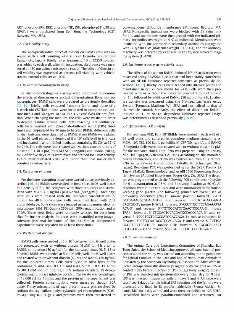
Fig. 1. Dioscin inhibited RANKL-induced osteoclast formation and bone resorption in a concentration-dependent manner without any cytotoxicity. (A) Viability of dioscin-treated BMMs cells. (B) The half-maximal inhibitory concentration (IC50) of dioscin was 9.004 lM. (C) Bone marrow-derived monocytes/macrophages (BMMs) were treatedwith various concentrations of dioscin followed by M-CSF (30 ng/mL) and RANKL (50 ng/mL) stimulation for five days. Cells were then fixed with 4% PFA and subjected toTRAP staining. (D) BMM-derived pre-osteoclasts were stimulated with M-CSF (30 ng/mL) and RANKL (50 ng/mL for three days. Later, cells were cultured in the presence of theindicated concentrations of dioscin with M-CSF (30 ng/mL) and RANKL (50 ng/mL) for another 48 h. SEM images of bone resorption pits are shown. (E) TRAP-positivemultinuclear cells were counted. (F) Resorption pit areas were measured using Image J and are presented graphically. All experiments were carried out at least three times,and the significance was determined by Student–Newman–Keul’s tests (⁄P < 0.05; ⁄⁄P < 0.01).
660 X. Qu et al. / Biochemical and Biophysical Research Communications 443 (2014) 658–665

Fig. 2. Dioscin suppressed RANKL-induced expression of osteoclast-specific genes. (A) BMMs were cultured with M-CSF (30 ng/mL) and RANKL (50 ng/mL), with or without4 lM dioscin, for 0, 1, 3, or 5 days. Osteoclast-specific gene expression (TRAP, CTR, Cts k, and NFATc1) was analysed by real-time PCR, and results were normalised to theexpression of b-actin. (B) BMMs were cultured with M-CSF (30 ng/mL) and RANKL (50 ng/mL), with indicated concentrations of dioscin for 5 days. Osteoclast-specific geneexpression (TRAP, CTR, Cts k, and NFATc1) was analysed by real-time PCR, and results were normalised to the expression of b-actin. All experiments were performed at leastthree times (⁄P < 0.05; ⁄⁄P < 0.01).
X. Qu et al. / Biochemical and Biophysical Research Communications 443 (2014) 658–665 661
histologic examination, sections were stained with hematoxylinand eosin (H&E), and another section was stained with TRAP toidentify osteoclasts on the bone surface. Parameters for the per-centage osteoclast surface per bone surface (OcS/BS, %), numberof osteoclasts per field of tissue, as well as the microstructural indi-ces of trabecular bone density (BV/TV), trabecular thickness(Tb.Th), trabecular number (Tb.N), and trabecular space (Tb.Sp)were measured to assess the trabecular bone microstructure ofthe femurs were quantified by using the Image Pro-Plus program,version 4.0 (Media Cybernetics, Silver Spring, MD).
2.9. Statistical analysis
All values are presented as the mean ± standard deviation (S.D.)of the values obtained from three or more experiments. Statisticalsignificances were determined by Student’s t-test. A value ofP < 0.05 was considered significant.
3. Results
3.1. Dioscin does not inhibit BMMs proliferation at low concentrations
After 48-h culture, a CCK-8 proliferation assay showed that dios-cin did not affect BMMs cell proliferation at concentrations 4 lM(Fig. 1A). Dioscin partially suppressed cell proliferation at concentra-tions P8 lM. The calculated IC50 for dioscin was 9.004 lM (Fig. 1B).In order to exclude dioscin-mediated cytotoxicity, non-lethal con-centrations (64 lM) were used in subsequent experiments.
3.2. Dioscin inhibits RANKL-induced osteoclast formation and boneresorption in vitro
To investigate the effects of dioscin on osteoclastogenesis,BMMs were treated with various concentrations of dioscin duringthe course of osteoclast formation. As shown in Fig. 1C, the control

Fig. 3. Dioscin inhibited RANKL-induced activation of NF-jB/NFATc1 and AKT signaling pathways. (A) RAW264.7 cells were seeded at 5 � 105 cells/well in 6-well plates andpretreated with or without dioscin (4 lM) for 4 h prior to RANKL stimulation (50 ng/mL) for the indicated times. Cells were lysed for Western blotting with specific antibodiesagainst phospho-IjBa, IjBa, phospho-AKT, AKT and actin. (B) Stably transfected RAW264.7 cells with an NF-jB luciferase reporter construct were seeded in 48-well plates andmaintained in the cell culture media for 24 h. The cells were then pretreated with or without the indicated concentrations of dioscin for 1 h, followed by addition of RANKL (50 ng/mL) for 8 h. NF-jB luciferase activity was measured. (C) RAW264.7 cells were seeded at 5 � 105 cells/well in 6-well plates and treated with or without dioscin (4 lM) for theindicated days. Cells were lysed for Western blotting with specific antibodies against NFATc1 and actin. (D) Stably transfected RAW264.7 cells with an NFATc1 luciferase reporterconstruct were seeded in 48-well plates and maintained in the cell culture media for 24 h. The cells were then pretreated with or without the indicated concentrations of dioscinfor 1 h, followed by addition of RANKL (50 ng/mL) for 24 h. NFATc1 luciferase activity was measured. (E) RAW264.7 cells were seeded at 5 � 105 cells/well in 6-well plates andpretreated with or without dioscin (4 lM) for 4 h prior to RANKL stimulation (50 ng/mL) for the indicated times. Cells were lysed for Western blotting with specific antibodiesagainst phospho-ERK, ERK, phospho-JNK, JNK, phospho-p38 and p38. (F) Stably transfected RAW264.7 cells with an AP-1 luciferase reporter construct were seeded in 48-wellplates and maintained in the cell culture media for 24 h. The cells were then pretreated with or without the indicated concentrations of dioscin for 1 h, followed by addition ofRANKL (50 ng/mL) for 24 h. AP-1 luciferase activity was measured. All experiments were performed at least three times (⁄P < 0.05; ⁄⁄P < 0.01).
662 X. Qu et al. / Biochemical and Biophysical Research Communications 443 (2014) 658–665
group formed numerous TRAP-positive multinucleated osteoclasts.In contrast, the formation of osteoclasts was inhibited after dioscintreatment, especially at the 4 lM concentration (Fig. 1C). This isfurther supported by statistical analysis of the number of osteo-clasts formed, where the control group formed 172 ± 26.8 osteo-clasts per well, but only 61 ± 11.3 osteoclasts per well in the4 lM concentration group (Fig. 1E).
Since dioscin inhibited osteoclast formation, we next investi-gated whether dioscin could impair osteoclastic bone resorptionin vitro. As shown in Fig. 1D, dioscin treatment substantially re-duced osteoclastic bone resorption area. Osteoclastic bone resorp-tion was almost completely abrogated after being treated with4 lM dioscin (Fig. 1F). Collectively, these findings suggested thatdioscin impaired osteoclast formation and bone resorption in vitro.
3.3. Dioscin suppressed osteoclastic gene expression in vitro
To further confirm the inhibitory effect of dioscin on osteo-clast differentiation, osteoclastic gene expression profile was
investigated by realtime PCR. As shown in Fig. 2A, the expressionof osteoclastic specific genes was gradually induced during osteo-clastogenesis, including TRAP, CtsK and CTR. What’s more,NFATc1 reached its peak at the mid-phrase of osteoclast differen-tiation. However, the induction of these genes was suppresseddramatically by the presence of dioscin. In addition, dioscindose-dependently suppressed these osteoclast specific genes(TRAP, Cts K, CTR, and NFATc1) at 1 and 4 lM respectively(Fig. 2B). Collectively, these data supported the inhibition ofosteoclast formation by dioscin.
3.4. Dioscin inhibited the RANKL-induced AKT/NF-jB and AKT /NFATc1activation
To further elucidate the mechanisms through which dioscinsuppressed osteoclast formation, RANKL-induced signaling path-ways were investigated. Here, we found the phosphorylation ofAkt was significantly inhibited by dioscin (Fig. 3A). Since Akt isimportant for both Akt/NF-jB and Akt/NFATc1 cascades, we thus

Fig. 4. Dioscin prevented LPS-induced bone loss by inhibiting osteoclast activity. (A) Femurs were fixed, decalcified, dehydrated, embedded, and sectioned. Sections werestained with H&E (100� and 200�) and TRAP (400�). (B) Percentage osteoclast surface per bone surface (OcS/BS, %), and (C) number of osteoclasts per field of tissue (No. ofOCs/field) in 400� magnification were analysed. Asterisks indicate statistically significant differences (⁄P < 0.05; ⁄⁄P < 0.01) between groups.
X. Qu et al. / Biochemical and Biophysical Research Communications 443 (2014) 658–665 663
further investigated these two key signaling pathways duringosteoclast differentiation. As shown in Fig. 3A, RANKL-inducedIjBa phosphorylation and degradation was markedly suppressedby dioscin. More specifically, IjBa was phosphorylated anddegraded upon RANKL stimulation for 5 min in the controlgroup. In contrast, dioscin treatment suppressed the phosphory-lation and therefore degradation of IjBa (Fig. 3A). This wasfurther evidenced by luciferase assay that dioscin blockedof NF-jB activation in a concentration-dependent manner(Fig. 3B).
Akt is also known to regulate osteoclast differentiation by regu-lating Akt/NFATc1 signaling pathway [22]. Therefore, we furtherchecked the expression of NFATc1 after dioscin treatment. Here,our Western blot analysis demonstrated that RANKL inducedNFATc1 expression during osteoclast formation. However, the
expression of NFATc1 was significantly suppressed after dioscintreatment (Fig. 3C). This was in consistent with our real-time PCR re-sults, which demonstrated the suppression of NFATc1 mRNA afterdioscin treatment (Fig. 2). Luciferase analysis results of declinedtranscriptional activity further demonstrated dioscin’s inhibitory ef-fect on NFATc1 (Fig. 3D).
In addition, we also investigated other key signaling pathwaysinvolved in osteoclast differentiation [23–25]. We examined thephosphorylation of p38, JNK and ERK and showed that dioscinhad no obvious effect on these signaling pathways (Fig. 3E). Fur-thermore, we also demonstrated that dioscin does not affect AP-1activity by AP-1 luciferase reporter gene assays.
Together, these results revealed that dioscin specifically inhib-ited the Akt/NF-jB and Akt/NFATc1 activation during osteoclastdifferentiation without affecting MAPK/AP-1 pathways.

664 X. Qu et al. / Biochemical and Biophysical Research Communications 443 (2014) 658–665
3.5. Dioscin prevented bone destruction induced by LPS in vivo
To address the effect of dioscin in vivo, osteolytic mouse modelwas chosen by LPS injection as previously described in our group[14]. Mice were intraperitoneally injected with LPS with or withoutdioscin. No fatalities were recorded after LPS and dioscin adminis-tration, and the animals retained normal activity throughout theduration of the experiment. Histological examination confirmedthe protective effects of dioscin on LPS-induced bone loss. Asshown in Fig. 4A, RANKL injection led to bone erosion and in-creased numbers of TRAP-positive osteoclasts. However, bone ero-sion was rescued in dioscin-treated mouse femur tissue sections,which was consistent with decreased TRAP-positive osteoclasts(Fig. 4A). Furthermore, histomorphometric analysis of OcS/BS andNo. of OCs demonstrated that dioscin reduced LPS-activated osteo-clast numbers, and microstructural indices of BV/TV, Tb.Th, andTb.Sp further identified dioscin protective effect in inflammatorybone loss, even though no significantly increased Tb.N was wit-nessed (Fig. 4B). Taken together, our data indicated that dioscinprevented LPS-induced bone loss in vivo.
4. Discussion
In this study, we have verified for the first time that naturalcompound dioscin inhibited osteoclast differentiation and boneresorption, suggesting an additional protective effect of dioscinon osteoclast-related diseases. In addition, we revealed the molec-ular mechanisms of dioscin on osteoclasts are through suppressingAkt/NF-jB and Akt/NFATc1 signaling pathways.
In osteoclasts, the Akt signaling cascades is a critical down-stream of three osteoclast surface receptors including c-fms,avb3 integrin and RANK [22,26]. Previous studies demonstratedAkt phosphorylation is activated upon both M-CSF and RANKLstimulation and play critical roles in osteoclastogenesis by affect-ing both NF-jB and NFATc1 activation. Moon et al. demonstratedthat overexpression of Akt in BMMs strongly induced NFATc1expression and lead to enhanced osteoclastogenesis [22]. Besides,activation of NF-jB can be initiated by several different kinasessuch as Akt and NF-jB inducing kinase. Gingery et al. demon-strated that AKT/NF-jB axis is critical in osteoclastogenesis andmaintaining mature osteoclast survival [27,28]. In consistent withthese studies, we demonstrated that dioscin inhibited Akt phos-phorylation and thus suppressed the RANKL-induced NF-jB activ-ity and NFATc1 activity, both of which are critical for osteoclastdifferentiation. Interestingly, in the process of detecting the func-tion of dioscin on MAPKs pathway, no significantly inhibitory im-pact was witnessed.
In summary, dioscin is capable of inhibiting osteoclast forma-tion and function, indicating additional therapeutic benefits ofdioscin for osteoclast-related diseases. In addition, this study alsoclearly revealed the molecular mechanisms of dioscin on osteo-clasts are via impairing Akt/NF-jB and Akt/NFATc1 signaling path-ways in vitro. In addition, our in vitro results further verified thebone protective role of dioscin on LPS-induced osteolysis model.However, further investigation of dioscin on other cells withinbone is still required.
Conflict of interest
No authors have any conflicts of interest to declare.
Acknowledgments
This work was supported by the Program for InnovativeResearch Team of Shanghai Municipal Education Commission
(Phase I), a Grant awarded for Innovative Research from ShanghaiMunicipal Education Commission (13YZ031), a Grant awarded bythe Key Disciplines of Shanghai Municipal Education Commissionof China (J50206), a Grant for scientific research from the NationalNatural Science Foundation for the Youth of China (No. 81201364),and a Grant awarded by the Scientific Research Foundation forReturned Overseas Chinese Scholars from the State HumanResource Ministry.
References
[1] S.L. Teitelbaum, Bone resorption by osteoclasts, Science 289 (2000) 1504–1508.
[2] Y. Tanaka, S. Nakayamada, Y. Okada, Osteoblasts and osteoclasts in boneremodeling and inflammation, Curr. Drug Targets: Inflammation Allergy 4(2005) 325–328.
[3] T. Suda, N. Takahashi, N. Udagawa, E. Jimi, M.T. Gillespie, T.J. Martin,Modulation of osteoclast differentiation and function by the new membersof the tumor necrosis factor receptor and ligand families, Endocr. Rev. 20(1999) 345–357.
[4] Z.H. Lee, H.-H. Kim, Signal transduction by receptor activator of nuclear factorkappa B in osteoclasts, Biochem. Biophys. Res. Commun. 305 (2003) 211–214.
[5] T. Nakamura, C. Komori, Y.-Y. Lee, F. Hashimoto, S. Yahara, T. Nohara, A. Ejima,Cytotoxic activities of Solanum steroidal glycosides, Biol. Pharm. Bull. 19(1996) 564–566.
[6] X. Wang, The expanding role of mitochondria in apoptosis, Genes Dev. 15(2001) 2922–2933.
[7] Y. Wang, C.-M. Che, J.-F. Chiu, Q.-Y. He, Dioscin (saponin)-induced generationof reactive oxygen species through mitochondria dysfunction: a proteomic-based study, J. Proteome Res. 6 (2007) 4703–4710.
[8] M. Sautour, A.-C. Mitaine-Offer, T. Miyamoto, A. Dongmo, M.-A. Lacaille-Dubois, Antifungal steroid saponins from Dioscorea cayenensis, Planta Med. 70(2004) 90–92.
[9] M.J. Kaskiw, M.L. Tassotto, M. Mok, S.L. Tokar, R. Pycko, J. Th’ng, Z.-H. Jiang,Structural analogues of diosgenyl saponins: synthesis and anticancer activity,Bioorg. Med. Chem. 17 (2009) 7670–7679.
[10] N. Brautbar, J. Williams II, Industrial solvents and liver toxicity: riskassessment, risk factors and mechanisms, Int. J. Hyg. Environ. Health 205(2002) 479–491.
[11] A. Qin, T.S. Cheng, Z. Lin, L. Cao, S.M. Chim, N.J. Pavlos, J. Xu, M.H. Zheng, K.R.Dai, Prevention of wear particle-induced osteolysis by a novel V-ATPaseinhibitor saliphenylhalamide through inhibition of osteoclast bone resorption,PLoS One 7 (2012) e34132.
[12] M. Kogawa, K. Hisatake, G.J. Atkins, D.M. Findlay, Y. Enoki, T. Sato, P.C. Gray, Y.Kanesaki-Yatsuka, P.H. Anerson, S. Wada, The paired-box domain transcriptionfactor Pax6 binds to the Upstream Region of the TRAP gene promoter andsuppresses RANKL-induced osteoclast differentiation, J. Biol. Chem. 288 (2013)31299–31312.
[13] W. Zou, T. Izawa, T. Zhu, J. Chappel, K. Otero, S.J. Monkley, D.R. Critchley, B.G.Petrich, A. Morozov, M.H. Ginsberg, Talin1 and Rap1 are critical for osteoclastfunction, Mol. Cell Biol. 33 (2013) 830–844.
[14] Z. Zhai, H. Li, G. Liu, X. Qu, B. Tian, W. Yan, Z. Lin, T. Tang, A. Qin, K. Dai,Andrographolide suppresses RANKL-induced osteoclastogenesis in vitro andprevents inflammatory bone loss in vivo, Br. J. Pharmacol. (2013), http://dx.doi.org/10.1111/bph.12463 [Epub ahead of print].
[15] H. Li, Z. Zhai, G. Liu, T. Tang, Z. Lin, M. Zheng, A. Qin, K. Dai, SanguinarineInhibits Osteoclast formation and Bone Resorption via suppressing RANKL-Induced Activation of NF-kappaB and ERK signaling pathways, Biochem.Biophys. Res. Commun. 430 (2013) 951–956.
[16] B. Tian, A. Qin, Z.Y. Shao, T. Jiang, Z.J. Zhai, H.W. Li, T.T. Tang, Q. Jiang, K.R. Dai,M.H. Zheng, Y.P. Yu, Z.A. Zhu, OA-4 inhibits osteoclast formation and boneresorption via suppressing RANKL induced P38 signaling pathway, Curr. Med.Chem. (2013) [Epub ahead of print].
[17] C. Wang, J.H. Steer, D.A. Joyce, K.H.M. Yip, M.H. Zheng, J. Xu, 12-O-tetradecanoylphorbol-13-acetate (TPA) inhibits osteoclastogenesis bysuppressing RANKL-induced NF-jB activation, J. Bone Miner. Res. 18 (2003)2159–2168.
[18] F. Ikeda, R. Nishimura, T. Matsubara, S. Tanaka, J.-I. Inoue, S.V. Reddy, K. Hata,K. Yamashita, T. Hiraga, T. Watanabe, Critical roles of c-Jun signaling inregulation of NFAT family and RANKL-regulated osteoclast differentiation, J.Clin. Invest. 114 (2004) 475–484.
[19] T.J. Weber, L.M. Markillie, Regulation of activator protein-1 by 8-iso-prostaglandin E2 in a thromboxane A2 receptor-dependent and-independentmanner, Mol. Pharmacol. 63 (2003) 1075–1081.
[20] A. Qin, T.S. Cheng, Z. Lin, N.J. Pavlos, Q. Jiang, J. Xu, K.R. Dai, M.H. Zheng,Versatile roles of V-ATPases accessory subunit Ac45 in osteoclast formationand function, PLoS One 6 (2011) e27155.
[21] A. Qin, T. Cheng, N. Pavlos, Z. Lin, K. Dai, M. Zheng, V-ATPases in osteoclasts:Structure, function and potential inhibitors of bone resorption, Int. J. Biochem.Cell Biol. 44 (2012) 1422–1435.
[22] J.B. Moon, J.H. Kim, K. Kim, B.U. Youn, A. Ko, S.Y. Lee, N. Kim, Akt inducesosteoclast differentiation through regulating the GSK3b/NFATc1 signalingcascade, J. Immunol. 188 (2012) 163–169.

X. Qu et al. / Biochemical and Biophysical Research Communications 443 (2014) 658–665 665
[23] D.A. Stevenson, E. Schwarz, J.C. Carey, D.H. Viskochil, H. Hanson, S. Bauer, H.Y.Cindy Weng, T. Greene, K. Reinker, J. Swensen, Bone resorption in syndromesof the Ras/MAPK pathway, Clin. Genet. 80 (2011) 566–573.
[24] F. Ikeda, R. Nishimura, T. Matsubara, S. Tanaka, J. Inoue, S.V. Reddy, K. Hata, K.Yamashita, T. Hiraga, T. Watanabe, Critical roles of c-Jun signaling inregulation of NFAT family and RANKL-regulated osteoclast differentiation, J.Clin. Invest. 114 (2004) 475–484.
[25] A.E. Grigoriadis, Z.-Q. Wang, M.G. Cecchini, W. Hofstetter, R. Felix, H.A. Fleisch,E.F. Wagner, C-Fos: a key regulator of osteoclast-macrophage lineagedetermination and bone remodeling, Science 266 (1994) 443–448.
[26] H. Cao, K. Zhu, L. Qiu, S. Li, H. Niu, M. Hao, S. Yang, Z. Zhao, Y. Lai, J.L. Anderson,Critical of AKT in myeloma-induced osteoclast formation and osteolysis, J. Biol.Chem. 288 (2013) 30399–30410.
[27] A. Gingery, E.W. Bradley, L. Pederson, M. Ruan, N.J. Horwood, M.J. Oursler, TGF-b coordinately activates TAK1/MEK/AKT/NFkB and SMAD pathways topromote osteoclast survival, Exp. Cell Res. 314 (2008) 2725–2738.
[28] A. Gingery, E. Bradley, A. Shaw, M.J. Oursler, Phosphatidylinositol 3-kinasecoordinately activates the MEK/ERK and AKT/NFkappaB pathways to maintainosteoclast survival, J. Cell Biochem. 89 (2003) 165–179.