digital pathology and tissue-based diagnosis. how … · digital pathology and tissue-based...
TRANSCRIPT

10.12.2014 1 Institute of Pathology – Charité Berlin
P. Hufnagl
Institute of Pathology (Rudolf-Virchow-Haus). Humboldt University, Berlin
Digital Pathology and Tissue-based Diagnosis.
How do they differ?
?

10.12.2014 2 Institute of Pathology – Charité Berlin
Structure of the talk
• Possible workflow
• routine diagnostic
• diagnostic support
• Which scanner?
• Quantification / image analysis
• Conclusions

10.12.2014 3 Institute of Pathology – Charité Berlin
Structure of the talk
• Possible workflow
• routine diagnostic
• diagnostic support
• Which scanner can we trust?
• Quantification / image analysis
• Conclusions

10.12.2014 4 Institute of Pathology – Charité Berlin
The Conventional Workflow
Lab: Cutting,
Staining, Coverslip
PLIMS
Physician: Assignment, diagnosing, consultation
HIS
Tissue
Report Physician: Dictation, Images,
Annotation
Clinical request

10.12.2014 5 Institute of Pathology – Charité Berlin
The Digital Workflow
Lab: Cutting,
Staining, Coverslip
Slide Scanner: Registration, Digitalisation
PACS: Storage,
Image streaming
PLIMS
Physician: Assignment, diagnosing, consultation
HIS
Tissue
Report Physician: Dictation, Images,
Annotation
Image analysis: marker quantification
Clinical request

10.12.2014 6 Institute of Pathology – Charité Berlin
The Digital Workflow
Lab: Cutting,
Staining, Coverslip
Slide Scanner: Registration, Digitalisation
PACS: Storage,
Image streaming
PLIMS
Physician: Assignment, diagnosing, consultation
HIS
Tissue
Report Physician: Dictation, Images,
Annotation
Image analysis: marker quantification
Clinical request
Missing link between cover
slipper and scanner Missing link between
scanner and PLIS

10.12.2014 7 Institute of Pathology – Charité Berlin
Advantages of Virtual Microscopy
Slide Server Viewer User • No glass transportation, no glass archive (?)
• Previous slides are always available
• Microscopic diagnostic - anytime – anywhere -
• Parallel viewing of different stainings, positions
• Viewing and handling parallel at different locations
• Facilitated second opinion - online
• Quantification and image analysis just in time
• Annotations are simple to be handled
• much more …..
Slide Scanner
10.12.2014 8 Institute of Pathology – Charité Berlin
Problems of the digital workflow
Disadvantages
• Additional process step: Digitalisation
• Huge and continuous hardware and
software investments
• Training of personnel
• Diagnosis on the monitor is unfamiliar for
pathologists
• Legal Problems

10.12.2014 9 Institute of Pathology – Charité Berlin
Strategy
• Start with a less critical application
• Biobanking
• Introduce VM to applications with most effects
• Tumour board
• Second opinion in-house
• Solve all problems along the workflow
• LIMS integration
• Sure barcode identification
• Establishing of continuous testing
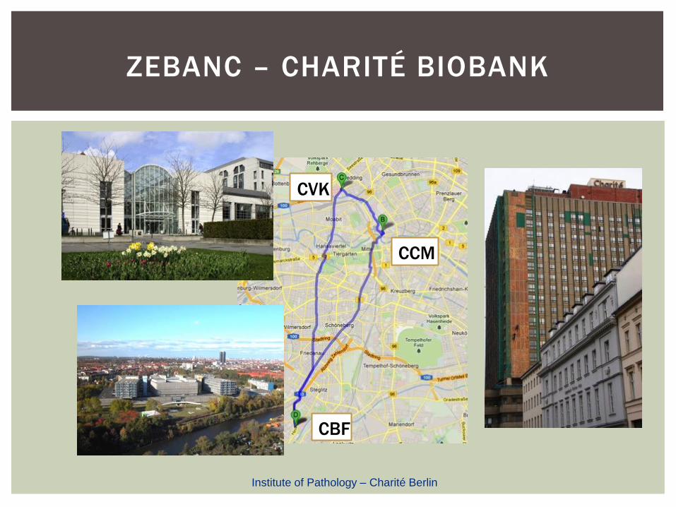
ZEBANC – CHARITÉ BIOBANK
CCM
CVK
CBF
Institute of Pathology – Charité Berlin
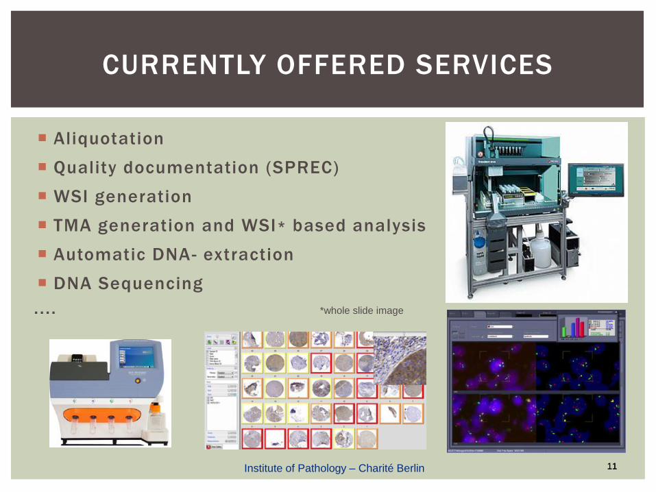
CURRENTLY OFFERED SERVICES
Aliquotation
Quality documentation (SPREC)
WSI generation
TMA generation and WSI* based analysis
Automatic DNA- extraction
DNA Sequencing
. . . . *whole slide image
11 11 Institute of Pathology – Charité Berlin

VALUE OF A SAMPLE
Sample
Whole Slide Images
Clinical Data
Quantification of Morphology
Next Generation Sequencing Data
Data generated during experiments ......
V
A L
U E
Institute of Pathology – Charité Berlin

Digitalisation established for samples from frozen section labs
Technical quality control implemented
Infrastructure for block-centric navigation established
Medical quality control in use (prototype tumor area detection)
TMA as sample array in CentraXX integrated
Several quantification procedures implemented to generate additional sample features
Virtual studies on virtual slides instead of real samples
Virtual microscopy has a huge potential for services in the context of a biomaterial bank
ZEBANC AND VM
Institute of Pathology – Charité Berlin

10.12.2014 14 Institute of Pathology – Charité Berlin
Clinical Pathology
Medical Workstation - All Information in One View
Second Opinion, Studies, Marker Quantification

10.12.2014 15 Institute of Pathology – Charité Berlin
Workflow of Tumor Board Meetings
Before meeting On meeting
Set bookmarks and make annotations Browse slides and move to annotations

10.12.2014 16 Institute of Pathology – Charité Berlin
Quantification

10.12.2014 17 Institute of Pathology – Charité Berlin
Structure of the talk
• Possible workflow
• routine diagnostic
• diagnostic support
• Which scanner?
• Quantification / image analysis
• Conclusions

10.12.2014 18 Institute of Pathology – Charité Berlin
Requirements on Slide Scanning
• Correct slide information is present
• Completeness of tissue
• Image sharpness
• Color fidelity

VIRTUAL MICROSCOPY –
SCANNER CONTEST
P. Hufnagl1,2, N.Zerbe2
1University of Applied Science Berlin, Berlin, German 2Institute for Pathology, Charité Berlin, Germany
technology meets pathology
2nd International Scanner Contest

Institute of Pathology – Charité Berlin
technology meets pathology
2nd International Scanner Contest
Determination of the state of the art in slide scanning
Support of pathologists and scientists to find the appropriate
scanner for their applications
Support of vendors to understand the needs of pathologists
Determination of „quality of WSI“ within the context of
pathology
Development of a set of standard features for the
characterization and comparison of scanning devices
MISSION

Institute of Pathology – Charité Berlin
technology meets pathology
2nd International Scanner Contest
High Throughput
Quality
Fluorescence
Image Analysis
Technical
DISCIPLINES

Institute of Pathology – Charité Berlin
technology meets pathology
2nd International Scanner Contest
QUALITY | EVALUATION TERMINALS

Institute of Pathology – Charité Berlin
technology meets pathology
2nd International Scanner Contest
VENDORS….

Institute of Pathology – Charité Berlin
technology meets pathology
2nd International Scanner Contest
Aims: Measurement of colour fidelity and colour resolution
of devices
Determination of true effective pixel size
Detection of image distortions
Material: IT8.7/1 Colour Target mounted on glas slide
264 colour & 24 grey value fields
known absorption spectra and colourimetric coordinates
Grid pattern glas slide overall size: 1” x 3” (25mm x 75mm )
image area: 20mm x 50mm
clear aperture: 8.5 µm²
opaque lines: 1.5 µm²
pitch: 10 µm
TECHNICAL – COLOUR & GEOMETRY | AIMS &
MATERIAL

Institute of Pathology – Charité Berlin
technology meets pathology
2nd International Scanner Contest
General Conditions:
All participants had to scan the same slide
Any manual interaction was allowed
Rescan of slide was allowed
1h time limit
Evaluation:
Colour difference calculation to CIEDE2000
average inside middle 50% of each field
low-resolution scan
Measurements inside whole slide images
Inside sensor field (no stitching)
9 sensor fields – 18 measurements each
TECHNICAL – COLOUR & GEOMETRY | TASK

Institute of Pathology – Charité Berlin
technology meets pathology
2nd International Scanner Contest
TECHNICAL – COLOUR | TEST METHOD
Fidelity test: average dE over 144 fields
dEavg= Σ dE(c*mes, c ref) /144
Colour difference calculation to CIEDE2000
colour step wedges
mix-colours
matrix for
fidelity test

Institute of Pathology – Charité Berlin
technology meets pathology
2nd International Scanner Contest
TECHNICAL – COLOUR | SOFTWARE

Institute of Pathology – Charité Berlin
technology meets pathology
2nd International Scanner Contest
COMPLETENESS OF SCAN

Institute of Pathology – Charité Berlin
technology meets pathology
2nd International Scanner Contest
HIGH THROUGHPUT | AIMS & MATERIAL
29

Institute of Pathology – Charité Berlin
technology meets pathology
2nd International Scanner Contest
HIGH THROUGHPUT | AIMS & MATERIAL
30

Institute of Pathology – Charité Berlin
technology meets pathology
2nd International Scanner Contest
Sample identifikation (barcode / OCR)
Sharpness assessment + Completeness of particles
DIGITALISATION - SCANMASTER

Institute of Pathology – Charité Berlin
technology meets pathology
2nd International Scanner Contest
FOCUS QUALITY ASSESSMENT
Green: quality sufficiant
Red: not sharp, possible artefacts

Institute of Pathology – Charité Berlin

Institute of Pathology – Charité Berlin
technology meets pathology
2nd International Scanner Contest

VIEWING ON A
VIRTUAL
MICROSCOPE
technology meets pathology
2nd International Scanner Contest

Institute of Pathology – Charité Berlin
technology meets pathology
2nd International Scanner Contest
RESOLUTION
MAGNIFICATION IS NOT RESOLUTION
AND OPTICAL RESOLUTION IS NOT
DIGITAL RESOLUTION !

10.12.2014 37 Institute of Pathology – Charité Berlin
Several Positions in Test
Leap Motion
under acryl glass pane
Leap Motion
over the table

10.12.2014 38 Institute of Pathology – Charité Berlin
Leap Motion
Stereo imaging based on infrared cameras (https://www.leapmotion.com/)

10.12.2014 39 Institute of Pathology – Charité Berlin
Handling Possible workflow

10.12.2014 40 Institute of Pathology – Charité Berlin
Gesture Control
Next/ previous slide Zoom in/ out

10.12.2014 41 Institute of Pathology – Charité Berlin
Histological Image Registration
• Goal
– Inter-Modal Registration (Stain-To-Stain)
• Applications
– WSI Navigation Support
– Virtual Staining
– 3D Reconstruction
• Approach
– Intensity Based
– Multi-Resolution
• Similarity Measure
– Mutual Information
• Transformation Models
– Rigid: Rotation + Translation
– Affine: Linear Transformation + Translation
– Free Form Deformation: B-Splines
• Optimization
– Gradient Descent

10.12.2014 42 Institute of Pathology – Charité Berlin
Registration Of Renal Images:
Reference Image (H&E)
Template Image (SFOG)

10.12.2014 43 Institute of Pathology – Charité Berlin Reference Image (H&E)

10.12.2014 44 Institute of Pathology – Charité Berlin Template Image (SFOG)

10.12.2014 45 Institute of Pathology – Charité Berlin
Coarse to Fine Image Registration:
Rigid, Affine, Free Form Model

10.12.2014 46 Institute of Pathology – Charité Berlin Rigid Registration

10.12.2014 47 Institute of Pathology – Charité Berlin Affine Registration

10.12.2014 48 Institute of Pathology – Charité Berlin Free Form Registration

10.12.2014 49 Institute of Pathology – Charité Berlin Reference Image

10.12.2014 50 Institute of Pathology – Charité Berlin
Structure of the talk
• Possible workflow
• routine diagnostic
• diagnostic support
• Which scanner?
• Quantification / image analysis
• Conclusion
10.12.2014 51 Institute of Pathology – Charité Berlin
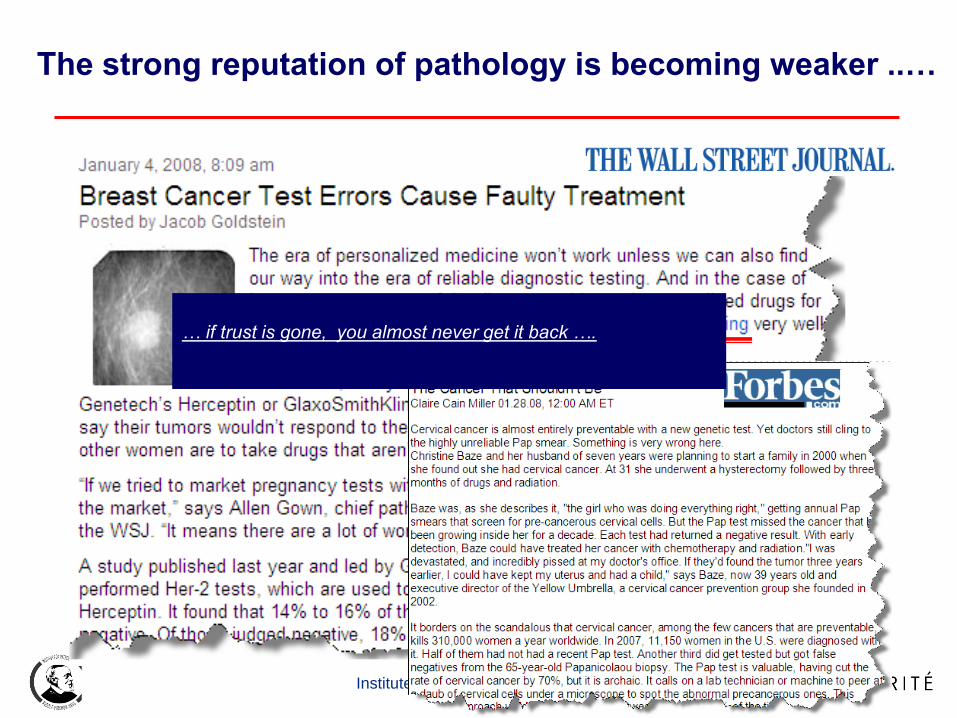
The strong reputation of pathology is becoming weaker ..…
… if trust is gone, you almost never get it back ….

10.12.2014 52 Institute of Pathology – Charité Berlin
Nat J Inst., preprint November 2013
Standardized Quantification in Tumor Pathology

10.12.2014 53 Institute of Pathology – Charité Berlin
…. At the St. Gallen cutoff of
13.5 % there are 32.3 % high
Ki67 by Lab A while Lab B
would call the same cases low
Ki67. ..

10.12.2014 54 Institute of Pathology – Charité Berlin
Optical Illusions
Not always funny, sometimes really critical…

10.12.2014 55 Institute of Pathology – Charité Berlin
Human brain:
Square a is
lighter than
square b !
Reality:
Both are identical

10.12.2014 56 Institute of Pathology – Charité Berlin
Simulated Ki67 15%
100
625
225 400
900

10.12.2014 57 Institute of Pathology – Charité Berlin
Estimation of variation of Ki67 scoring

FEATURE BASED MULTIRESOLUTION
CORRESPONDENCE
tumor
non-tumor
Combined
tumor
annotations
Individual
tumor
annotations
by 10 pathologists
By 4 pathologists
by no pathologist
By 2 pathologists
Annotated as tumor
Institute of Pathology – Charité Berlin

CLASSIFICATION RESULTS
59
Gastric Cancer (HER2) Transmission to HE Learning Sample
Institute of Pathology – Charité Berlin

10.12.2014 60 Institute of Pathology – Charité Berlin
Structure of the talk
• Possible workflow
• routine diagnostic
• diagnostic support
• Which scanner?
• Quantification / image analysis
• Conclusions

10.12.2014 61 Institute of Pathology – Charité Berlin
Summary on Relevance of VM
• Workflow
• routine diagnostic
• diagnostic support
• Which scanner?
• Quantification / image analysis
• Clinical-pathological tumor-
conferences (tumor board)
• Biobanking
In routine path not yet
active on a broad level
Excellent instruments exist,
but they have to be integrated properly
Will become very important in
personalized medicine
Is already very important
Is important in research institutions

10.12.2014 62 Institute of Pathology – Charité Berlin
Most important requirements on VM
• Clinical data (LIMS) are correctly connected to WSI
• Completeness of tissue
• Image sharpness
• Color fidelity
• Compression is adequate
• Resolution and quality of the monitor is sufficiant
• Test continously!
• Correct slide information is present
• Completeness of tissue
• Image sharpness
• Color fidelity

10.12.2014 63 Institute of Pathology – Charité Berlin
Let‘s go virtual
13th European Conference on
Digital Pathology
Berlin May 25. / 26. 2016
www.digitalpathology2016.org

Acknowledgement
Team
Norman Zerbe, Karsten Schlüns, Sebastian Lohmann, Mario Domhardt, Björn Lindequist,
Daniel Heim, Stephan Wienert, Kai Saeger, Thorsten Knape, Arend Müller,
Wolfram Schädel, Uwe Brunner, Thomas Schrader, Manfred Dietel
technology meets pathology
2nd International Scanner Contest
Institute of Pathology – Charité Berlin