differing strategies despite shared lineages of motor ... · jonathan enriquez,1,2,* laura quintana...
TRANSCRIPT

Article
Differing Strategies Despite Shared Lineages ofMotor Neurons and Glia to Achieve RobustDevelopment of an Adult Neuropil in Drosophila
Highlightsd The development of the neuropil glia and legmotor neurons is
highly coordinated
d All adult thoracic neuropil glia are born from two lineages per
hemisegment
d These lineages also give rise only to leg motor neurons
d Unlike neurons, glia generation is plastic and exhibits
interlineage compensation
Authors
Jonathan Enriquez,
Laura Quintana Rio, Richard Blazeski,
Stephanie Bellemin, Pierre Godement,
Carol Mason, Richard S. Mann
[email protected] (J.E.),[email protected] (R.S.M.)
In BriefEnriquez et al. show that the glia
surrounding each thoracic neuropil come
from stem cells that also give rise to leg
motor neurons. Unlike the hard-wired
birth order andmorphology of eachmotor
neuron, the number and morphologies of
the glia born from single lineages are
variable, yet the structure they contribute
to is highly stereotyped.
Enriquez et al., 2018, Neuron 97, 1–17February 7, 2018 ª 2018 Elsevier Inc.https://doi.org/10.1016/j.neuron.2018.01.007

Neuron
Article
Differing Strategies Despite Shared Lineagesof Motor Neurons and Glia to Achieve RobustDevelopment of an Adult Neuropil in DrosophilaJonathan Enriquez,1,2,* Laura Quintana Rio,2 Richard Blazeski,3 Stephanie Bellemin,1 Pierre Godement,1 Carol Mason,3
and Richard S. Mann2,4,*1Institut de Genomique Fonctionnelle de Lyon, ENS de Lyon, CNRS, Univ Lyon 1, 46 Allee d’Italie, 69364 Lyon Cedex 07, France2Departments of Biochemistry and Molecular Biophysics, and Neuroscience, Mortimer B. Zuckerman Mind Brain Behavior Institute,Columbia University, New York, NY 10027, USA3Departments of Pathology and Cell Biology, Neuroscience and Ophthalmology, Mortimer B. Zuckerman Mind Brain Behavior Institute,Columbia University, New York, NY 10027, USA4Lead Contact*Correspondence: [email protected] (J.E.), [email protected] (R.S.M.)https://doi.org/10.1016/j.neuron.2018.01.007
SUMMARY
In vertebrates and invertebrates, neurons and gliaare generated in a stereotyped manner from neuralstem cells, but the purpose of invariant lineagesis not understood. We show that two stem cellsthat produce leg motor neurons in Drosophila alsogenerate neuropil glia, which wrap and sendprocesses into the neuropil wheremotor neuron den-drites arborize. The development of the neuropil gliaand leg motor neurons is highly coordinated. How-ever, although motor neurons have a stereotypedbirth order and transcription factor code, the numberand individual morphologies of the glia born fromthese lineages are highly plastic, yet the final struc-ture they contribute to is highly stereotyped.We sug-gest that the shared lineages of these two cell typesfacilitate the assembly of complex neural circuits andthat the two birth order strategies—hardwired formotor neurons and flexible for glia—are importantfor robust nervous system development, homeo-stasis, and evolution.
INTRODUCTION
Cell lineages play a critical role in generating cellular diversityduring animal development yet range widely in the number andtypes of cells they produce. Embryonic stem cells, for example,have the potential to give rise to all cell types, whereas hemato-poietic stem cells have much more limited potential, giving riseonly to myeloid and lymphoid cells (Seita and Weissman, 2010;Wu et al., 2016). The wide range of developmental potentials isalso reflected in the degree to which cell lineages are hardwiredor plastic in the types and numbers of cells they can generate. Atone extreme, each of the 959 somatic cells of the adultC. elegans hermaphrodite is derived from an invariant lineagethat can be traced back to the single-celled zygote (Sulston,
1976). In contrast, cell lineages of the mammalian immunesystem are plastic and can be modified depending on the envi-ronmental challenges that an animal confronts (Boettcher andManz, 2017). Similarly, intestinal stem cell lineages exhibitplasticity in response to injury and cell death (Apidianakis andRahme, 2011; Jiang and Edgar, 2011).Although the capacity of some cell lineages to respond to
varying environmental conditions makes sense in light of thecell types these lineages generate, the underlying logic ofwhy some lineages are invariant is usually not understood.For example, although the entire lineage is hardwired inC. elegans, there is almost no correlation between where a cellis born in the lineage and cell-type identity; most sub-lineagescontribute broadly to endodermal, mesodermal, and nervoussystem tissues. Consequently, a single cell type such as a motorneuron can be born from many different sub-lineages inC. elegans (Hobert, 2016). In D. melanogaster, although thereare neuroblasts (NBs), stem cells that are dedicated to the gen-eration of neurons, during embryogenesis each NB can generatea mixture of motor neurons, interneurons, and glia that do notshare an obvious function (Bossing et al., 1996; Schmidt et al.,1997). These observations raise the question of whether thereis a biological purpose for the structure of invariant lineagesthat may be difficult to discern by only examining the final prog-eny. Consistent with this notion, in the adult, the neuronal prog-eny of individual Drosophila NB hemilineages share anatomicalfeatures such as stereotyped axon paths and, when activated,the progeny of individual hemilineages can evoke specificbehaviors in decapitated flies (Harris et al., 2015; Truman et al.,2004). Thus, by both anatomical and behavioral criteria, thereis emerging evidence that individual Drosophila NBs generatefunctionally related neurons in the adult fly.Another reason for the existence of hardwired lineages might
be to facilitate the assembly of complex structures during devel-opment, which is an especially challenging problem for nervoussystems. For example, the !50 motor neurons (MNs) thatinnervate each leg in adult Drosophila are born from invariantNB lineages and have highly stereotyped birthdates andmorphologies (Baek and Mann, 2009; Brierley et al., 2012).
Neuron 97, 1–17, February 7, 2018 ª 2018 Elsevier Inc. 1
NEURON 14084
Please cite this article in press as: Enriquez et al., Differing Strategies Despite Shared Lineages of Motor Neurons and Glia to Achieve Robust Devel-opment of an Adult Neuropil in Drosophila, Neuron (2018), https://doi.org/10.1016/j.neuron.2018.01.007

A1
D1
G
I
HP
Q
J
K
M N O
L1 L2
D2 E F
A2 B C
(legend on next page)
NEURON 14084
2 Neuron 97, 1–17, February 7, 2018
Please cite this article in press as: Enriquez et al., Differing Strategies Despite Shared Lineages of Motor Neurons and Glia to Achieve Robust Devel-opment of an Adult Neuropil in Drosophila, Neuron (2018), https://doi.org/10.1016/j.neuron.2018.01.007

These stereotyped morphologies can be seen both in the mus-cles these motor neurons target and in their elaborate dendriticarbors that establish a very large number of synapses in denseneuropils present in each thoracic neuromere of the ventralnerve cord (VNC) (Court et al., 2017). Although each motorneuron morphology is thought to be determined by unique com-binations of morphology transcription factors (mTFs; Enriquezet al., 2015), how these stereotyped morphologies form thecorrect synaptic connections with interneurons and sensoryneurons is not understood. Interestingly, at least one of themajor NB lineages that give rise to adult motor neurons in thefly also generates another important cell type in the nervoussystem, glia (Baek et al., 2013; Lacin and Truman, 2016), whichare critical for the establishment of synapses and the mainte-nance of neuronal activity (Freeman, 2015; Freeman andRowitch, 2013). The observation that glia and legmotor neuronsmay be derived from common progenitors (neuroglioblast[NGB]) raised the possibility that their development may becoordinated, and that being born from the same lineages mightalso play a role in neural circuit assembly. However, in contrastto motor neurons, very little is known about the glial types pro-duced by these NGBs, how their morphologies are geneticallyspecified, and how neurons and glia communicate during devel-opment to build a functional CNS.In other contexts, Drosophila glia have been subdivided
into three classes based on their functions and morphologies:surface glia, which isolate the CNS from the hemolymph, cortexglia, which send processes around neuronal cell bodies, andneuropil glia (NG), which surround neuropil and glomeruli (Omotoet al., 2016). NG are further subdivided into astrocytes andensheathing glia (EG) (Muthukumar et al., 2014; Peco et al.,2016). EG extend flat processes that surround neuropil andglomeruli as well as a small portion of axon bundles as they enter
or leave the neuropil (Omoto et al., 2015; Peco et al., 2016). Morerecently, EG have been shown to extend small protrusions insidethe neuropil of the adult brain (Kremer et al., 2017). In themedulla(region of the brain that processes visual information), EG areassociated with axons (Kremer et al., 2017) and trachea (Kremeret al., 2017; Pereanu et al., 2007). In contrast, astrocytes sendhighly ramified processes deep into neuropil regions in closecontact with synapses (Awasaki et al., 2008; Muthukumaret al., 2014; Stork et al., 2014). Although NG exhibit complexand diverse morphologies and are generally derived from stemcells that also generate neurons (Awasaki et al., 2008; Omotoet al., 2016), the developmental mechanisms leading to glialdiversity remains unknown.Here, we show that two of the !11 lineages that generate leg
motor neurons in Drosophila also generate all of the NG thatpopulate the adult thoracic neuropils. As a consequence ofbeing born from the same lineages, the development of theNG is physically and temporally coordinated with motor neurondevelopment. However, unlike the hardwired birth order andunique mTF codes of the motor neurons that are born fromthese NGBs, individual NG do not have a stereotypedmorphology, birth order, or unique transcription factor code.Moreover, using a unique mode of division, the gliogenesisphase of these lineages is plastic and highly adaptable: whengliogenesis in one lineage is compromised, other lineagescompensate to maintain the correct number of NG. Thus,even though NG and motor neurons come from the samestem cells, and generate adult neuromeres with highly stereo-typed structures, there are fundamental differences in howthese two cell types are specified. We suggest that the combi-nation of hardwired motor neuron generation and flexible gliaproduction facilitates the robust construction, homeostasis,and evolution of complex neural circuits.
Figure 1. Cellular Organization of the Adult Thoracic NG(A–C) Adult VNCs immunostained with anti-BRP (neuropil marker, blue) (A1–C) and anti-Dll (NG marker, green) (B) with astrocytes expressing mCD8::GFP
(membranemarker, green) (A1 and A2), H2B::RFP (nuclear marker, red) (B) or themulticolor system FB1.1 (single-cell marker, red, yellow, and green) (C) under the
control of alrm-Gal4.
(B) Astrocyte (H2B::RFP+, Dll+): arrow, EG (Dll+): arrowhead.
(C) Different astrocytes with processes occupying different neuropil territories: arrows. See also Figure S1 and Movie S1.
(D–F) Adult VNCs immunostained with anti-BRP (neuropil marker, blue) (D1–F) and anti-Dll (NG marker, green) (E) with EG-expressing mCD4::GFP (membrane
marker, green) (D1 and D2), H2B::RFP (nuclear marker, red) (E) or the multicolor system FB1.1 (single-cell marker, red, yellow, and green; Hadjieconomou et al.,
2011) (F) under the control of R56F03-Gal4. (E) Astrocyte: arrow (Dll+), EG (H2B::RFP+, Dll+): arrowhead.
(D) arrow: in more than half of the samples analyzed (n = 10) a cell body of an EG (GFP +, Dll+) was observed inside the neuropil, next to axon bundles.
(F) Different EG with processes occupying different neuropil territories: arrows. See also Figure S1 and Movie S1.
(G and H) Prothoracic neuromeres with EG-expressing mCD4::GFP and immunostained with anti-BRP (blue) and anti-Elav (neuron marker, red) (G) or anti-NCad
(neuropil marker, red) and Nrg (axon marker, blue) (H).
(G) Asterisk: Elav+ neuron wrapped by an EG. Enlargements of the boxed regions are to the right of each panel. See also Movie S2.
(I and J) Prothoracic neuromeres with EG-expressing mCD4::GFP (I) and GFP::mCD8::HRP (J) immunostained with anti-BRP (blue) (I) or labeled with
DAB (brown) (J).
(K) Low-magnification electron microscope image of the boxed region in (J).
(L–O) Enlargement of the boxed regions in (K). (L2) Enlargement of the boxed region in (L1). Note: (L1) and (L2) show the organization of the EG processes around
the neuropil while (M)–(O) show the EG processes inside the neuropil. Pink lines outline axons. PG, perineurial glia; SPG, subperineurial glia; NG, neuropil glia;
C, cortex; Np, neuropil; NL, neural lamella.
(P) Bottom, schematic of adult CNS (blue: neuropils, gray: cortex); top, single astrocyte and EG labeled with mCD8::GFP under the control of alrm-Gal4 and
R56F03-Gal4 using MARCM. Axes: green (posterior), blue (dorsal), red (medial).
(Q) Average number of NG in the adult VNC, in which EG or astrocytes were expressing H2B::RFP under the control of R56F03-Gal4 or alrm-Gal4, respectively,
and immunostained with anti-Dll. Number of samples = 4/genotype. Error bars indicate SD.
ProNm, Prothoracic neuromere; AMesoNm, Accessory mesothoracic neuromere; MesoNm, Mesothoracic neuromere; MetaNm, Metathoracic neuromere;
ANm, Abdominal neuromeres; FS, frontal cross-section; TS, transverse cross-section; 3D, 3-dimensional reconstruction of confocal image stack; pMP, partial
maximum projection.
NEURON 14084
Neuron 97, 1–17, February 7, 2018 3
Please cite this article in press as: Enriquez et al., Differing Strategies Despite Shared Lineages of Motor Neurons and Glia to Achieve Robust Devel-opment of an Adult Neuropil in Drosophila, Neuron (2018), https://doi.org/10.1016/j.neuron.2018.01.007

A1
G
B1
B2
B3 C3 D3
C2 D2
C1 D1 E1 F1
F2E2
E3
E4
E5
F4
F5
F3
A2
(legend on next page)
NEURON 14084
4 Neuron 97, 1–17, February 7, 2018
Please cite this article in press as: Enriquez et al., Differing Strategies Despite Shared Lineages of Motor Neurons and Glia to Achieve Robust Devel-opment of an Adult Neuropil in Drosophila, Neuron (2018), https://doi.org/10.1016/j.neuron.2018.01.007

RESULTS
Cellular Organization of the Adult Thoracic Neuropil GliaIn order to characterize the cellular organization of the NG in theadult VNC, we expressed fluorescent reporters under the controlof two Gal4 drivers, alrm-Gal4 (astrocyte specific; Doherty et al.,2009) and R56FO3-Gal4 (EG specific; Kremer et al., 2017; Pecoet al., 2016; Pfeiffer et al., 2008), which we confirmed areexpressed in all adult astrocytes and EG, respectively (Figures1 and S1). We also used as a marker Distalless (Dll), which wefound to be expressed in all NG in the adult VNC (Figure S1).As with the NG in the larval CNS (Beckervordersandforth et al.,2008; Omoto et al., 2015), the number of NG surrounding eachthoracic neuropil is very consistent between animals (with avery low SD; Figures 1P and 1Q).As described for other regions of the CNS (Awasaki et al.,
2008; Muthukumar et al., 2014), adult thoracic astrocytes sendprocesses deep into the neuropil that respect each other’s terri-tory, thus exhibiting a tiling-like phenomenon (Figures 1A and1C;Movie S1). EG extend processes that surround each neuropiland also respect each other’s territory with very infrequent over-laps (Figures 1D and 1F;Movie S1). In addition, the adult thoracicEG feature a more complex morphology than previouslydescribed for larval EG. First, adult EG wrap the cell bodies ofneurons (Elav+) that are localized next to the neuropil (Figure 1G;Movie S2) as well as astrocyte cell bodies (Figure S1). Second,they send processes inside the neuropil where they wrap axonbundles (Neuroglian+) where no synapses are formed (BRP-negative, a marker of the active presynaptic zone) (Figures 1Gand 1H; Movie S2). Interestingly, EG only wrap these axonsinside the neuropil; outside of the neuropil these axons aresurrounded by a distinct class of wrapping glia (Figure 1H;data not shown). Thus, EG isolate nerves inside neuropil beforethey make synapses with other neurons.We next performed electron microscopy of transverse
sections of prothoracic neuromeres (ProNs) (Figures 1K–1O).EG wrap the thoracic neuropils very densely with several layers(Figure 1N). These results, in combination with the tiling resultsdescribed above, suggest that one EG can send processesorganized in layers around the neuropil. Inside the neuropil,axon bundles and single axons can be wrapped by layers ofEG processes (Figures 1M–1O). Interestingly, each nerve alsocontains trachea bundles that are surrounded by EG, forming astructure with all three cell types: EG, axons, and trachea (Fig-ure 1O). Notably, axons close to the midline are not wrappedby EG (data not shown).
Developmental Origins of the Adult Thoracic NGAs described above, the adult thoracic neuropils are complexstructures containing glia processes and axons/dendrites in closejuxtaposition to each other, raising the question of how thesestructures develop. A previous study suggested that pockets ofglia,marked by the general gliamarker Repo, surround the imma-ture leg neuropils in late third-instar larvae (just prior to the onsetof metamorphosis) and produce adult astrocytes (Li et al., 2014),while another study suggests that these glia express Dll and maymigrate to the leg to form wrapping glia, which are distinct fromEG and astrocytes (Plavicki et al., 2016). In order to definitivelydetermine what type of adult cells these Repo+ glia give rise to,we used aDll-Gal4 enhancer trap and aDll antibody to label thesecells during development (Figure 2).In third-instar larvae (L3), there were !40 Repo+ Dll+ cells sur-
rounding each immature leg neuropil (Figures 2B, 2C, and 2G),where nascent motor neuron axons enter and then exit (Fig-ure 4A). If these cells are the progenitors of the adult NG, theyshould be mitotically active. To test this idea, we first confirmedthat they express phosphorylated histone H3 (pH3), a marker formitotically active cells (Figure 2D).Although Dll protein is expressed in adult NG, Dll-Gal4 is only
active in immature NG (data not shown), allowing us to mark theprogeny of Dll-Gal4-expressing larval cells in lineage tracingexperiments (see STAR Methods). In the first experiment, welabeled the adult progeny of Dll-Gal4-expressing L3 glia with nu-clear GFP (UAS-nGFP) and co-stained with an antibody againstRepo and Dll (Figure 2E). Only the adult Repo+ Dll+ NG werelabeled with nGFP, demonstrating that the Dll-Gal4-expressingcells in the larval CNS only generate adult NG. In a secondexperiment, we used Dll-Gal4 to activate the Flybow system(UAS-FB1.1), which permanently labels cells with different fluo-rescent markers (Hadjieconomou et al., 2011). We co-stainedthe resulting adult VNCs with Prospero, which is expressed inastrocytes but not in EG (Griffiths and Hidalgo, 2004; Pecoet al., 2016) (Figures 2F and S1). Based on Prospero expressionand morphology, we found that both astrocytes and EG werelabeled. Significantly, neither experiment labeled other classesof glia, such as surface glia or cortex glia, arguing that theseglia subtypes are not derived from Dll-Gal4-expressing cells.We confirmed this result with another driver (R31F10-Gal4) thatis also expressed in immature NG (Figure S2).Taken together, we conclude that the!40Repo+Dll+Dll-Gal4-
expressing cells surrounding each immature leg neuropil in L3are the precursors of the >280 NG that surround each adultthoracic neuropil.
Figure 2. Developmental Origin of the Adult NG(A) 3D rendering of the thoracic segments of a L3 VNC (A1) labeled with anti-BRP (mature neuropil marker, blue) and anti-NCad (mature and immature neuropil
marker, red). The glia surrounding the six immature leg neuropils expressmCD8::GFP (green, left image only) under the control ofDll-Gal4. (T1, T2, and T3 indicate
the three thoracic segments.) (A2) Following metamorphosis, 3D rendering of adult VNCs with astrocytes and EG-expressing GFP under the control of alrm-Gal4
(right) and R56F03-Gal4 (left) and costained with anti-BRP (red).
(B–D) L3 CNSs in which glia surrounding the six immature leg neuropils express mCD8::GFP under the control of Dll-Gal4 and co-stained with anti-BRP (blue),
anti-NCad (red) (B1–B3), anti-Repo (blue), anti-Dll (red) (C1–C3), or anti-Repo (blue), anti-PH3 (red) (D1–D3). (D2 and D3) Arrows point to glia expressing PH3.
(E and F) Adult VNCs with NG expressing nuclear GFP (green) (E1–E5) or the multicolor Flybow system FB1.1 (yellow, red, and green) (see STAR Methods for
details) and immunostained with anti-Repo (blue), anti-Dll (red) (E1–E5), or anti-Pros (blue) (F1–F5). (E1–E5) Arrow points to adult NG-expressing GFP. (F1–F5)
Arrow: astrocyte (mCherry+, Pros+), arrowhead: EG (mCherry+, Pros"). See also Figure S2.
(G) Left: schematic of an L3 CNS. The immature leg neuropils and proliferating NG are indicated. Right: schematic of an adult VNC with the adult leg NG.
N, average number of proliferating NG in each L3 thoracic hemisegment.
NEURON 14084
Neuron 97, 1–17, February 7, 2018 5
Please cite this article in press as: Enriquez et al., Differing Strategies Despite Shared Lineages of Motor Neurons and Glia to Achieve Robust Devel-opment of an Adult Neuropil in Drosophila, Neuron (2018), https://doi.org/10.1016/j.neuron.2018.01.007

Coordinated Development of the Adult NG and NeuropilTo characterize the development of the adult neuropil, we con-ducted several experiments to trace the origins of this complexstructure using BRP to label the larval neuropils, N-cadherin tolabel the immature adult neuropils, and Dll-Gal4; UAS-GFP(Dll>GFP) to label the proliferating NG precursors (Figure 3;Movie S3).
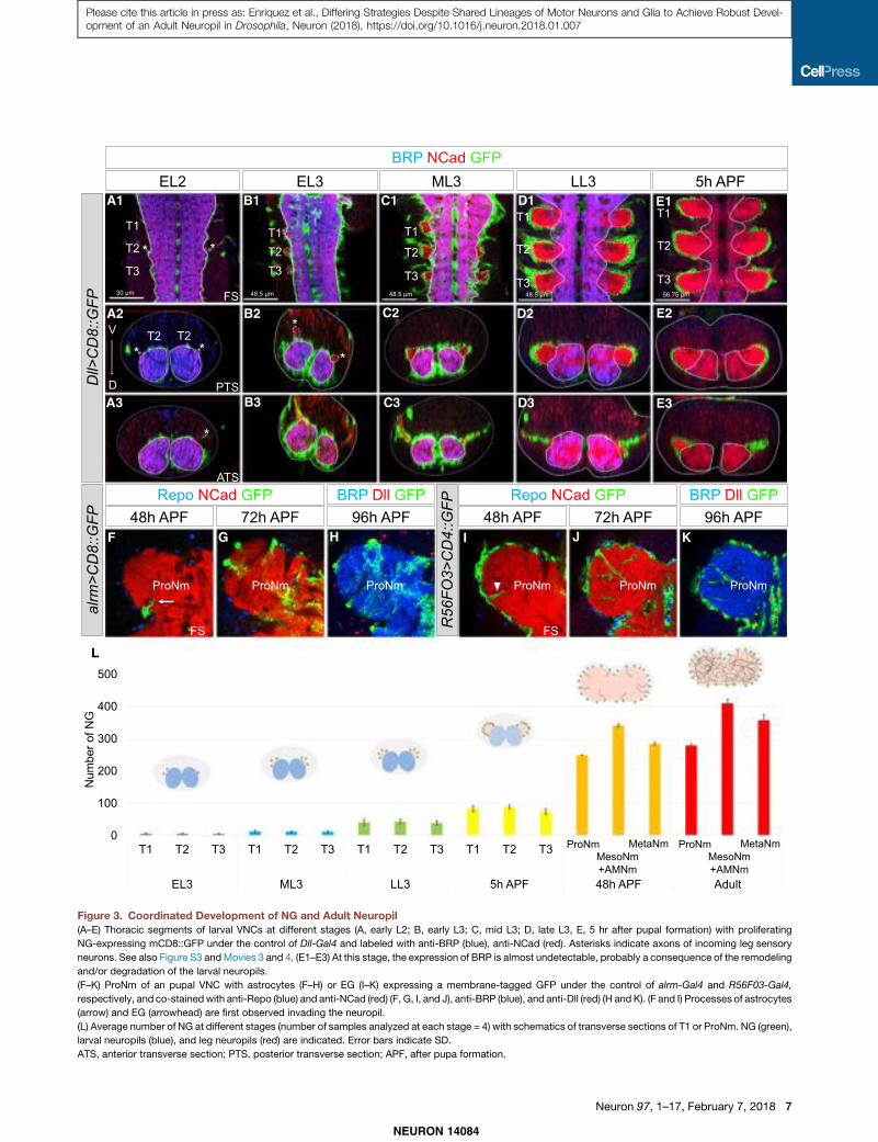
In early second-instar larvae (EL2), most of the adult legmotor neurons are not yet born (Baek and Mann, 2009) and noDll > GFP+-proliferating NG are visible. However, at this stageincoming leg sensory axons are visible and mark the locationwhere the adult neuropil will form (Figure 3A). In early L3 (EL3;72 hr after egg laying, AEL), 0–10 proliferating NG are observedper thoracic hemisegment (Figures 3B, 3L, and S3), and theirnumbers gradually expand through subsequent larval and pupalstages (Figures 3C–3E and S3). As proliferating NG are pro-duced, they send processes that surround the growing neuropiland wrap the leg nerves (Figures 3A–3E and S3). By the mid pu-pal stage (48 hr APF), most of EG and astrocytes have beengenerated and start to differentiate by sending processes insidethe neuropil (Figures 3F–3I). By the late pupal stage (96 hr APF),the adult neuropils are fully invaded by astrocyte processes andwrapped by EG (Figures 3H and 3K).
These results suggest that the adult thoracic NG are producedfrom early L3 to mid pupa. These glia create a structure that en-compasses the immature adult neuropil (Movies S3 and S4); byL3, the motor neuron neurites are completely surrounded by theprocesses of the immature NG. During metamorphosis, the den-drites and axons of the neurons establish their final morphol-ogies within the glia-defined neuropil.
The Adult Thoracic NG Are Produced by NGBs that AlsoGenerate Leg Motor NeuronsPrevious work established that the larval NG are born from asingle glioblast in each hemisegment of the larval CNS (Becker-vordersandforth et al., 2008; Ito et al., 1995; Jacobs et al., 1989)and from a single glioblast or NGB in each hemisphere of thebrain (Hartenstein et al., 1998; Omoto et al., 2015), while inthe adult brain NG are derived from a group of uncharacterizedNGBs (Omoto et al., 2015). To characterize the lineages thatgive rise to the adult thoracic NG, we carried out several clonalanalysis experiments. Previous observations demonstratedthat NB lineages such as Lin A (also called Lin 15), produceboth legmotor neurons and glial cells (Baek et al., 2013; Trumanet al., 2004). This observation raised the intriguing possibilitythat NG and leg motor neurons may share common develop-mental origins.
First, we used MARCM to express the membranereporter UASmCD8::GFP with two different drivers, repo-Gal4(expressed in all glia, Sepp and Auld, 2003) and VGlut-Gal4(expressed in all motor neurons and some interneurons, Mahrand Aberle, 2006). We induced these clones in L1 larvae anddissected late L3 CNS and co-stained for Dll, to mark immatureadult NG, and BRP to mark the mature larval neuropil. If a stemcell produces both motor neurons and NG, individual MARCMclones should contain both cell types. We identified glial cellsby their position and expression of Dll, and motor neurons bytheir morphology, particularly motor neuron axons, which exit
the CNS and target the leg imaginal discs (Baek and Mann,2009; Truman et al., 2004). 62/62 clones that labeled NG alsolabeled motor neurons (Figure 4). We found 3 types of clonesbased on the number of motor neurons they contained.Type 1 clones (n = 36) contained !28 leg motor neurons and!20 immature adult NG. Based on motor neuron morphology,this lineage is Lin A (Figure 4A). Type 2 clones (n = 23) contained2 motor neurons and !10 immature NG (Figure 4B). In type 3(n = 3), the clones contained 1 motor neuron and !7 glia(Figure 4A). The total number of glia (!37) produced by thesethree NGBs is very close to the total number of NG progenitorsthat surround each neuropil at L3 (!40), suggesting that thesethree lineages produce most, and perhaps all, of the adultthoracic NG.In a second approach, we generated clones using a modified
QMARCM/MARCM method (cis2-MARCM) in which the twotranscriptional repressors, GAL80 and QS, are recombined onthe same chromosome arm (see STAR Methods for details).We used two drivers, VGlut-QF and repo-Gal4, allowing us tovisualize VGlut+ motor neurons and Repo+ glia derived fromthe same stem cell with two different fluorescent markers. Usingthis method, and analyzing the adult legs and VNCs, weconfirmed that there are three NGBs that produce leg motorneurons and adult NG. Lin A clones produce NG and!28 motorneurons targeting the trochanter, femur, and tibia (n = 14,in ProN) (Figures 4C and 4F). Type 2 clones labeled NGand 2 motor neurons that targeted the coxa; based on themorphology of these motor neurons, we recognized this lineageas Lin D (Baek andMann, 2009) (n = 2) (Figure 4E). Type 3 cloneslabeled NG and a single motor neuron that targeted a bodywall muscle at the base of the coxa (n = 16) (Figure 4F). Notably,this motor neuron was previously described to be derived fromLin A (Brierley et al., 2012), a conclusion that we confirm andrevise below.
The Lin A NGB Initially Produces Glioblasts andPostmitotic Motor NeuronsBecause Lin A generates both glia and a stereotyped set ofmotor neurons, we used this lineage to investigate the birth orderof these two types of progeny. In the first set of experiments weused a lineage tracing system that is restricted to Dpn+ NBs(Awasaki et al., 2014). When combined with 10C12-Gal4, whichis expressed early in the Lin A lineage, this system specificallylabels all Lin A progeny with GFP (Lacin and Truman, 2016).We immunostained the resulting larval CNSs with Repo, Elav,and Dpn to visualize glia, neurons, and NBs, respectively, anddissected larva at different time points during L3 (Figures 5A–5J). As a consequence of NB division patterns, progeny furtherfrom the NB tend to be older compared to progeny closer tothe NB, thus potentially providing birth order information.Using this approach, we found that Repo+ glia and Elav+
neurons are first observed at about the same time, as early as75–78 hr AEL (Figures 5F and 5K). In some samples at this earlytime point, only a single Dpn+ NGB or clones with a NGBsurrounded by cells not marked with Repo, Elav, or Dpn areobserved (Figures 5D, 5E, and 5K). These unlabeled cells arealways close to the NB and therefore may be ganglionmother cells (GMCs) or another type of intermediate cell type.
NEURON 14084
6 Neuron 97, 1–17, February 7, 2018
Please cite this article in press as: Enriquez et al., Differing Strategies Despite Shared Lineages of Motor Neurons and Glia to Achieve Robust Devel-opment of an Adult Neuropil in Drosophila, Neuron (2018), https://doi.org/10.1016/j.neuron.2018.01.007
A1
A2
A3
F G H I J K
B2
B3
C2
C3 D3 E3
D2 E2
B1 C1 D1 E1
L
Figure 3. Coordinated Development of NG and Adult Neuropil(A–E) Thoracic segments of larval VNCs at different stages (A, early L2; B, early L3; C, mid L3; D, late L3, E, 5 hr after pupal formation) with proliferating
NG-expressing mCD8::GFP under the control of Dll-Gal4 and labeled with anti-BRP (blue), anti-NCad (red). Asterisks indicate axons of incoming leg sensory
neurons. See also Figure S3 andMovies 3 and 4. (E1–E3) At this stage, the expression of BRP is almost undetectable, probably a consequence of the remodeling
and/or degradation of the larval neuropils.
(F–K) ProNm of an pupal VNC with astrocytes (F–H) or EG (I–K) expressing a membrane-tagged GFP under the control of alrm-Gal4 and R56F03-Gal4,
respectively, and co-stainedwith anti-Repo (blue) and anti-NCad (red) (F, G, I, and J), anti-BRP (blue), and anti-Dll (red) (H and K). (F and I) Processes of astrocytes
(arrow) and EG (arrowhead) are first observed invading the neuropil.
(L) Average number of NG at different stages (number of samples analyzed at each stage = 4) with schematics of transverse sections of T1 or ProNm. NG (green),
larval neuropils (blue), and leg neuropils (red) are indicated. Error bars indicate SD.
ATS, anterior transverse section; PTS, posterior transverse section; APF, after pupa formation.
NEURON 14084
Neuron 97, 1–17, February 7, 2018 7
Please cite this article in press as: Enriquez et al., Differing Strategies Despite Shared Lineages of Motor Neurons and Glia to Achieve Robust Devel-opment of an Adult Neuropil in Drosophila, Neuron (2018), https://doi.org/10.1016/j.neuron.2018.01.007

In addition, due to the limitations of labeling recently born cells,they may be immature glia or neurons. As development pro-ceeds, the cells close to the NGB are either unlabeled or Elav+
neurons, suggesting that starting at mid L3 (!96 hr AEL) theLin A stem cell produces only neurons (Figures 5G–5K). Although
the number of Repo+ glia increases over time, they are far fromthe NGB, consistent with the idea (and supported by the pH3staining shown in Figure 2D) that early born glia continue todivide. We also note that as development proceeds, the Repo+
glia appear to migrate, such that by late L3 they surround the
A1 A2 A3 A4 A5
B1 B2 B3 B4 B5
C1 C2 E1 E2D1 D2
C3 E3D3
Figure 4. NGBs Produce Leg Motor Neurons and All Thoracic NG(A and B) Thoracic segments of an L3 VNC labeled with anti-BRP (blue) and anti-Dll (red) containing MARCM clones expressing mCD8::GFP under the control of
both VGlut-Gal4 and repo-Gal4. Lin A, Lin ?, and Lin D clones produce motor neurons (arrowhead) and glia (arrow). (A1–A5) Sample with Lin A, Lin B and Lin ?
clones; note that Lin B does not produce glia. (B1–B5) Sample with a Lin D clone. Asterisks mark motor neuron axons exiting the VNC.
(C–E) Lin A (C1–C3), Lin ? (D1–D3) and Lin D (E1–E3) cis2 MARCM clones in a ProN labeled with anti-BRP (blue) (C1–C2, D1–D2, E1–E2) and in a T1 Leg (C3, D3,
E3) expressing mCD8::RFP in NG (red) and mCD8::GFP (green) under the control of repo-Gal4 and VGlut-QF, respectively.
NEURON 14084
8 Neuron 97, 1–17, February 7, 2018
Please cite this article in press as: Enriquez et al., Differing Strategies Despite Shared Lineages of Motor Neurons and Glia to Achieve Robust Devel-opment of an Adult Neuropil in Drosophila, Neuron (2018), https://doi.org/10.1016/j.neuron.2018.01.007

A1 A2 B1 C1 C3
C2B2
D1 E1 F1 G1 H1 I1
D2 E2 F2 G2 H2 I2 J2
J1
D3 E3 F3 G3 H3 I3 J3
L1 L2
K
L3 M N
(legend on next page)
NEURON 14084
Neuron 97, 1–17, February 7, 2018 9
Please cite this article in press as: Enriquez et al., Differing Strategies Despite Shared Lineages of Motor Neurons and Glia to Achieve Robust Devel-opment of an Adult Neuropil in Drosophila, Neuron (2018), https://doi.org/10.1016/j.neuron.2018.01.007

immature adult neuropil. These results suggest that the Lin Astem cell initially produces mitoticially active glioblasts and asmall number ofmotor neurons, while at later stages it transitionsto a NB that only produces neurons.
To complement these lineage tracing experiments, we usedthe QMARCM/MARCM twin spot system (Potter et al., 2010;see STAR Methods for details) in which both daughter lineagesfrom a single cell division are permanently labeled with RFPand GFP, respectively. In these experiments, we induced clonesduring L1 to capture the first post-embryonic division andlabeled motor neurons with GFP, allowing us to recognize LinA due to the stereotyped morphology of Lin A dendrites. Sisterprogeny were marked with tub>RFP to label all cell types.Remarkably, in twin clones where GFP labels Lin A motor neu-rons, the RFP+ progeny are composed of Repo+ NG and a singlemotor neuron that targets a body wall muscle at the base of thecoxa (Figures 5L–5M), the samemotor neuron that was observedin our cis2 MARCM experiments (Figure 4D). This motor neuronhas also been described previously to be part of Lin A (Brierleyet al., 2012). Similar results were obtained when Lin AQMARCM/MARCM clones were generated in L2 or early L3,consistent with the timing inferred from our lineage tracingexperiments (data not shown).
Taken together, these results suggest that the first post-em-bryonic division of the Lin A NGB results in two sub-lineages (Fig-ure 5N): one, which we refer to as Lin A.1, generates a glioblastand a single post-mitotic motor neuron, and a second (Lin A.2)generates at least one additional glioblast and the remaining28 Lin Amotor neurons. To our knowledge, this mode of division,in which a NGB generates an intermediate mother cell (IMC) thatdivides to produce a post-mitotic motor neuron and a glioblast,has not been observed previously (Figure 5N).
Lin A.1 and A.2 Generate Both Astrocytes and EGTo determinewhich types of NG are produced by Lin A and Lin D,we repeated the cis2-MARCM experiments with alrm-Gal4 orR56FO3-Gal4 (instead of repo-Gal4), to label astrocytes andEG, respectively (Table S2). We found that Lin A.1 (n = 33) pro-
duces both types of NG, while Lin D (n = 4) only generates astro-cytes (Figures 6A–6I; Movie S5; Table S2). Lin A.2 also producesboth types of NG (Figures 6A–6I).To determine whether Lin A and Lin D generate only motor
neurons and NG, we repeated the MARCM experiments usinga tubulin driver (tub-Gal4), to label all cell types. We stained theresulting L3 larvae with Elav, Dpn, and Repo (Figures 6J–6O).The results confirmed that Lin A.2 generates motor neuronsand NG. Lin A.2 clones also usually included two cells that didnot stain for either Elav or Repo. Because of their proximity tothe NB, they are likely to be GMCs (Figures 6J and 6M; datanot shown). All non-Lin A.2 clones (n = 4) that generated Repo+
NG labeled only 1 (Lin A.1) or 2 (Lin D) Elav+ neurons (Figure 6K).Together with our earlier experiments, these results suggest that,post-embryogenesis, Lin A and Lin D give rise to only leg motorneurons and NG. Notably, at late L3, a Dpn+ NB remains associ-ated with Lin A, but not with Lin D.We conclude that in each thoracic hemisegment, all of the
adult NG are generated from only 2 NGBs, which also give riseonly to leg motor neurons.
Unlike Motor Neurons, Individual NG Born from theSame Lineage Are Not StereotypedPrevious studies have revealed that each of the 47 leg motorneurons have a specific birth date and morphology, character-ized by their axonal targeting, dendritic arbor, and cell bodyposition (Baek andMann, 2009; Brierley et al., 2012).We recentlycharacterized Lin B, a lineage that produces only 7 motor neu-rons, and described a post-mitotic code of mTFs that controlsindividual motor neuron morphologies (Enriquez et al., 2015).We identified these TFs by an expression screen using !250antibodies, yet, surprisingly, none of these TFs are differentiallyexpressed in subsets of thoracic NG (data not shown). Instead,we found TFs, such as Dll, expressed in all NG. The one excep-tion is Prospero (Pros), which is expressed in all astrocytes butnot EG (Griffiths andHidalgo, 2004; Peco et al., 2016) (Figure S1).If no single-cell TF code exists for NG, it follows that the numberand morphology of the NG progeny from the same lineage may
Figure 5. Lin A Initially Generates Postmitotic Motor Neurons and Proliferating Glia and Then Only Motor Neurons(A–C) 3D reconstruction of all six (A1), three (B1), or a single (C) Lin A clones labeled with myr::GFP (green) generated by a Lin A tracing system in the thoracic
segments of a late L3 VNC, and immunostained with anti-Dpn (red), anti-Elav (cyan), and anti-Repo (blue); A1 and C1 are ventral views, B1 and C2 are lateral
views. C3 shows a series of frontal cross-sections (FS) of Lin A from ventral (1) to dorsal (4). NB: Lin A NB/NGB, red arrowhead, GMC: Lin A GMC, black
arrowhead, Ns: Lin A neurons, cyan arrowhead. Axes in A2, B2: green (posterior, Post), blue (dorsal, D), red (medial, M). A2 and B2 show plots of Lin A progeny as
seen in A1 and B1, respectively, color coded according to cell type. Blue, Repo+; red, Dpn+; cyan, Elav+, orange: (GMC/IMC, ganglion mother cell/intermediate
mother cell, Dpn", Elav", Repo"); note: some of these GMC/IMC could be immature neurons or glia.
(D–J) Ventral view of a 3D reconstruction of single Lin A clones in a T2 segment at different time points during L3 (D1–J1) and graphs of each Lin A cell from two
perspectives (D2–J2 and D3–J3). Axes: green (posterior, Post), blue (dorsal, D), red (medial, M). Shown are: (D) 75–78 hr AEL, (E) 96–99 hr AEL, (F) 99–102 hr AEL,
(G) 102–105 hr AEL, (H) 120–123 hr AEL, (I) 123–126 hr AEL, and (J) 126–129 hr AEL.
(K) Average number of Lin A cell types at different L3 stages. Number of samples analyzed at each stage: (75- to 78-hr AEL): n = 16; (78- to 81-hr AEL): n = 16;
(96- to 99-hr AEL): n = 8; (99- to 102-hr AEL): n = 10; (102- to 105-hr AEL): n = 7; (120- to 123-hr AEL): n = 9; (123- to 126-hr AEL): n = 8; (126- to 129-hr AEL: n = 7.
Error bars indicate SD. AEL, after egg laying. ‘‘Lin A’’ refers to all cells labeled by GFP.
(L andM) Lin AQMARCM/MARCMclone in a ProNmgenerated before the first post-embryonic division and labeled with Repo (blue). Motor neurons produced by
one daughter cell are labeled withmCD8::GFP under the control of VGlut-QF, and all the cells produced by the sister daughter cell are labeled with RFP under the
control of tubulin-Gal4 (L1, L2). RFP labels a single motor neuron targeting a body wall muscle (M) and NG when the clones are induced at early L3 or earlier.
Number of samples analyzed: n > 30.
(N) Inferred pattern of Lin A divisions. Lin A.1 produces at least one glioblast (GB) and onemotor neuron targeting the body wall; Lin A.2 also produces at least one
glioblast and the remaining Lin A motor neurons. We refer to the mother of a glioblast and motor neuron as an intermediate mother cell (IMC) and the progeny of
the Glioblast as proliferating glia (pG) Later cell divisions of Lin A.2 generate a GMC, which produce one motor neuron and a sister cell that undergoes apoptosis
(blue star; Truman et al., 2010).
NEURON 14084
10 Neuron 97, 1–17, February 7, 2018
Please cite this article in press as: Enriquez et al., Differing Strategies Despite Shared Lineages of Motor Neurons and Glia to Achieve Robust Devel-opment of an Adult Neuropil in Drosophila, Neuron (2018), https://doi.org/10.1016/j.neuron.2018.01.007

D2 E2D1 E1 F1 F2
G1 G2 H1 H2
O1M2 N2 O2M1
BA C
I
KJ L
N1
(legend on next page)
NEURON 14084
Neuron 97, 1–17, February 7, 2018 11
Please cite this article in press as: Enriquez et al., Differing Strategies Despite Shared Lineages of Motor Neurons and Glia to Achieve Robust Devel-opment of an Adult Neuropil in Drosophila, Neuron (2018), https://doi.org/10.1016/j.neuron.2018.01.007

differ from animal to animal. Below we used the cis2-MARCMtechnique to test this prediction (Figures 7A–7R; Table S3).
Lin A.2, D and A.1 give rise on average to 79 (n = 10), 57 (n = 4),and 33 (n = 13) astrocytes, respectively, but with a very high SD,suggesting that each lineage has the potential to generate a var-iable number of NG (Figure S5).We also find that the final positionof NG progeny can vary from animal to animal: for each of thethree lineages born in a T1 hemisegment, astrocyte processesmostly invade the ProNp (Prothoracic Neuropil) but, dependingon the sample, can also send processes to the Accessory Meso-thoracic Neuropil (AMesoNp, wherewingmotor neuron dendritesinnervate), the mesothoracic neuropil (MesoNp), and occasion-ally cross the mid-line to populate the contralateral neuropil(Figures 7A–7L). Similarly, although most T1-born astrocyte cellbodies remain in the ProNm (Prothoracic Neuromere), theycan also end up in the Accessory Mesothoracic Neuromere(AMesoNm) and occasionally in the Mesothoracic Neuromere(MesoNm) (Figures 7A–7L, data not shown for Lin D, Table S3).Lin A astrocytes born in a T2 and T3 hemisegment have thesame ability to populate neighboring neuromeres (Figure S5).Thus, unlike the highly stereotypedmorphology of motor neuronsborn from the same lineage (Figures 7M–7R), the number, finalcell body position, and neuropil regions invaded by astrocyteprocesses born from individual NGBs are variable from animalto animal (Figures 7 and S5; Table S3).
Although we were unable to count the number of EG producedby Lin A.1 (see STARMethods), Lin A.2 generates!114EG (n = 4)with a lowSD (±6), suggesting that the number of EGproducedbyLin A is similar in different animals. However, as with astrocytes,theneuropil region surroundedand invadedbyLinAEG is variablefrom animal to animal, and these glia can populate neighboringneuromeres and cross the midline (Figures 7 and S5; Table S3).
In summary, an individual NGB produces a variable numberof NG with different morphologies and final locations, butthe same number of motor neurons, each with a stereotypedmorphology.
Final Astrocyte Number Depends on Inter-lineageCompetitionAlthough the number of NGproduced by a single NGB (e.g., Lin A)can be highly variable, the total number of NG produced by all lin-eages is very constant from animal to animal (Figures 1 and S5). Inaddition, the non-stereotyped morphology and final positions ofNG derived from single NGBs led us to hypothesize that NGmay compete during development to ensure that each neuropilis fully wrapped and innervated. We tested this hypothesis for as-trocytes, by generating Dllmutant Lin A.2 MARCM clones, which
results in a severely compromised number of astrocytes bornfrom this lineage without affecting motor neuron number ormorphology (Figures 7W and S6). Dll" Lin A.2 clones were gener-ated in a background where all astrocytes were labeled, thusallowing us to assess whether other lineages could compensatefor the fewer number of Lin A.2-derived astrocytes. We consid-ered three possible outcomes (Figures 7S and 7T): (1) no compen-sation, in whichWT (non-Lin A.2) astrocytes do not respond to thereduced number of astrocytes derived from Lin A; (2) compensa-tion where WT astrocytes respond to the lower number of Lin A.2astrocytes by increasing the number or extent of processesinvading the neuropil; and (3) compensation where WT lineagesgenerate more astrocytes to maintain the total final number.To distinguish between these possibilities, we generatedWT or
DllmutantLinAMARCMclonesexpressingmCD8::GFPunder thecontrol of repo-Gal4 and VGlut-Gal4. In this genetic background,we labeled all astrocytes with a cytoplasmic QUAS-mCherry un-der the control of alrm-QF. As a result, astrocytes produced byLin A are labeled by mCherry and mCD8::GFP while astrocytesproduced by other lineages express only mCherry. Astrocyteswere counted in a ProNm containing a single Lin AMARCM cloneand in the contralateral neuromere that had no clones, whichserved as an internalWT control. AlthoughDllmutant Lin A clones(n = 4) producedon average!18 astrocytes (compared to!80 forWT Lin A clones), the total number of astrocytes in these neuro-meres was normal (!140) (Figures 7U–7X). Altogether, these re-sults suggest that Lin D and Lin A.1 compensate for the reducednumber of astrocytes produced by Lin A.2.To determine when during development NG compensation is
likely to occur, we generated Dll" Lin A.2 MARCM clones(n = 4) and counted the number of Dll" Repo+ cells in late L3larva. We found that the number was similar to the number inWT clones (!20) (Figure S6), arguing that the absence of Dlldoes not affect the generation of astrocytes until the pupal stage,when NG number continue to increase (Figure 3L). It follows thatcompensation from WT lineages also occurs during this pupalamplification phase, long after the glioblasts are born.These results suggest that the production of NG is extremely
plastic during development, and that their progenitors continueto divide until the entire neuropil is occupied.
DISCUSSION
The Logic of Lineages Producing Multiple, FunctionallyRelated Cell TypesHere, we show that two NGBs per thoracic larval hemisegmentgive rise to leg motor neurons, astrocytes, and EG. All three of
Figure 6. Lin A.1 and Lin A.2 Produce Astrocytes and EG, While Lin D Produces Only Astrocytes(A–C) Schematic representation of the cis2 MARCM experiments labeling Lin A.2 (A), Lin A.1 (B), and Lin D (C).
(D–H) Lin A.2 (D and G), Lin A.1 (E and H), and Lin D (F) cis2MARCM clones in a ProNm stained with anti-BRP (blue), with motor neurons expressing mCD8::GFP,
astrocytes expressing mCherry, and EG-expressing mCD8::RFP, under the control of VGlut-QF, and alrm-Gal4 or R56F03-Gal4, respectively. Number of VNCs
analyzed: Lin A astrocyte: n = 10, Lin A EG: n = 4, Lin D astrocyte: n = 4, Lin Z astrocytes: n = 13. See also Figure S4 and Movie S5.
(I) Summary of progeny produced by Lin A and Lin D.
(J–L) Schematic representation of tub-Gal4 MARCM experiments in (M)–(O) labeling Lin A.2 (J), Lin A.1 (K), and Lin D (L).
(M–O) Lin A.2 (M), Lin A.1 (N), Lin D (O)MARCM clones labeled with mCD8::GFP and nGFP under the control of tub-Gal4 in a thoracic segment of an L3 VNC and
immunostainedwith anti-Dpn (red), anti-Elav (cyan), and anti-Repo (blue). (M2, N2, O2) are successive frontal cross-sections (FS) from ventral (V) to dorsal (D). pG,
proliferating glia (Repo+).
NEURON 14084
12 Neuron 97, 1–17, February 7, 2018
Please cite this article in press as: Enriquez et al., Differing Strategies Despite Shared Lineages of Motor Neurons and Glia to Achieve Robust Devel-opment of an Adult Neuropil in Drosophila, Neuron (2018), https://doi.org/10.1016/j.neuron.2018.01.007

(legend on next page)
NEURON 14084
Neuron 97, 1–17, February 7, 2018 13
Please cite this article in press as: Enriquez et al., Differing Strategies Despite Shared Lineages of Motor Neurons and Glia to Achieve Robust Devel-opment of an Adult Neuropil in Drosophila, Neuron (2018), https://doi.org/10.1016/j.neuron.2018.01.007

these cell types are key components of each thoracic neuro-mere: motor neurons extend axons to innervate leg musclesand elaborate complex dendritic arbors inside the neuropil,EG wrap the developing neuropil and incoming sensory axons,and astrocytes send processes inside the neuropil wherethey associate with synapses between motor neuron dendrites,interneurons, and sensory neurons. Together, these cellsdevelop in a coordinated manner to form a complex functionalunit that comprises the neural circuitry used for many adultbehaviors such as walking.
Glial cells are a major component of nervous systems, criticalnot only for the activity of neurons, but also for their develop-ment (Freeman, 2015; Freeman and Rowitch, 2013). Neuronsand glia are always associated with each other and exist inall bilateria, even in primitive phyla such as flatworms (Hartline,2011). As nervous system complexity increases, there is anincrease in both neuronal diversity and in the diversity of gliatypes and morphologies (Paredes et al., 2016). Given this closerelationship, it is striking that in the thorax of the adult fly all NGare derived from lineages that also—and only—give rise to legmotor neurons. In the absence of extensive cell migration,being born from the same stem cell results in an anatomicalproximity that may be important for subsequent steps indevelopment. In particular, we propose that the shared line-ages of the leg motor neurons, astrocytes, and EG facilitatesthe assembly of anatomically complex neuropils and neuralcircuits. This idea helps explain why lineage relationshipsappear to play less of a role in simpler nervous systems suchas in C. elegans. Consistent with the idea that anatomical prox-imity of functionally related cell types is important for buildingcomplex nervous systems, in mammals, astrocytes and oligo-dendrocytes are born from the same progenitor domains asmotor neurons and interneurons (Ravanelli and Appel, 2015;Rowitch and Kriegstein, 2010). Although it is currently unclearwhether these cell types are derived from the same lineagesin vertebrates, our results provide a striking precedent andcompelling reasons that this may be the case. Alternatively,vertebrate nervous systems may have solved the neuropilassembly problem differently, by having distinct progenitorsborn in the same domain but at different times that give riseto either neurons or glia.
Divergent Mechanisms for Generating Motor Neuronand Glia StereotypyOne of the most striking conclusions stemming from our findingsis how cell lineages can use very different mechanisms toproduce stereotyped outcomes. On the one hand, each motorneuron is morphologically distinct and born from a specificlineage and with a specific birth order, properties that are aconsequence of the unique TF code that they express. At theother extreme, based on the hundreds of TFs we have surveyed,individual astrocytes or EG appear to share the same TF code asall other astrocytes or EG, respectively, and can end up withdifferent morphologies in multiple positions within or even inneighboring neuropils. Yet, despite this plasticity, the endresult—!280 NG evenly distributed throughout each thoracicleg neuropil—is highly stereotyped. Nevertheless, these twovery different modes of achieving stereotypy—hardwired (formotor neurons) and plastic (for NG)—arise from the same stemcells. We also found that, although the total number of NG ineach thoracic neuropil is stereotyped (!140 astrocytes and!140 EG), the individual contributions from the two NGBlineages can vary from animal to animal. Moreover, when onelineage is compromised the others can compensate to maintainthe correct number of total NG. Although the mechanism regu-lating final NG number is currently unknown, we suggest that itis analogous to what has been referred to as neutral competitionin mammalian and Drosophila gut homeostasis (de Navascueset al., 2012; Snippert et al., 2010). It is possible that this compe-tition occurs via direct communication between NG. This idea issupported by the observation that individual astrocytes and EGrespect each other’s territory and thus exhibit tiling-like behavior.Alternatively, communication between motor neuron dendrites(or other components of the thoracic neuropil) and NG may beresponsible for NG stereotypy. For example, astrocytes andEG may be able to sense and adapt to the scaffold generatedby motor neuron dendrites in order to fully invade and wrapeach thoracic neuropil. Accordingly, NG stereotypy, both cellnumber and morphology, would be an indirect consequence ofthe mTF codes that generate the unique morphologies of motorneurons and likely other neurons.What might be the reason for the existence of such different
strategies to achieve stereotypy? One answer may be
Figure 7. Gliogenesis Is Not Stereotyped and Depends on Inter-lineage Competition(A–R) Three examples each of Lin A.2 (A–C, G–I, M–O) and Lin A.1 (D–F, J–L, P–R) cis2 MARCM clones expressing alrm >mCherry (A–F) or R56F03 >mCD8:RFP
(G–L) and VGlut >mCD8::GFP (M–R). Astrocyte and EG clone morphology is variable from animal to animal, while motor neuron clones have a uniform, lineage-
specificmorphology. (A–G) Arrows point to NGprocesses and arrowheads point to NG cell bodies that extend into or are located in a different neuromere than the
ProNm in which the clone originated.
(S) Schematic summarizing the Lin A and Lin D NGBs and their progeny. Green indicates a Lin A.2 MARCM clone induced in the NGB; red indicates all
proliferating glia. Bottom right: schematic of a thoracic neuromere, showing all (red) or just Lin A.2 (green outline) astrocytes with their processes entering the
neuropil.
(T) Same as (S) showing three possible outcomes for a Dll" Lin A.2 MARCM clone: no compensation, an increase in astrocyte processes, or an increase in
astrocyte number.
(U and V) WT (U) and Dll" (V) Lin A.2 MARCM clones in the right ProNm, labeled with mCD8::GFP (green) under the control of VGlut-Gal4 and repo-Gal4, and
co-stainedwith anti-BRP (blue). All astrocytes are labeledwithmCherry under the control alrm-QF. Frontal (FS) (V3) and transverse (TS) (V4) sections showing that
both thoracic neuropils appear to be equally invaded by astrocyte processes even if the neuropil contains a Dll" Lin A.2 clone.
(W) Average number of repo > H2B:RFP+ NG, alrm > H2B:RFP+ astrocytes and R56F03 > H2B:RFP+ EG in WT and Dll" cis2 MARCM Lin A.2 clones. Error bars
indicate SD. See also Figure S6. Number of samples analyzed = 4
(X) Average number of astrocytes in WT and Dll" MARCM Lin A.2 clones (GFP+, mCherry+), compared to the total number of astrocytes (GFP", mCherry+).
Error bars represent SD. Number of samples analyzed for each type of neuromere = 4.
NEURON 14084
14 Neuron 97, 1–17, February 7, 2018
Please cite this article in press as: Enriquez et al., Differing Strategies Despite Shared Lineages of Motor Neurons and Glia to Achieve Robust Devel-opment of an Adult Neuropil in Drosophila, Neuron (2018), https://doi.org/10.1016/j.neuron.2018.01.007

developmental robustness, which is especially challenging forestablishing neural circuits. While motor neurons need tomake precise connections (both in the CNS and in the legs),and therefore may require precise TF codes to achieve this pre-cision, our results suggest that astrocytes and EG may have amore generic role in neuropil development. Nevertheless, NGare likely to be just as critical for neuropil development andfunction. However, instead of specifying each glia on asingle-cell basis, the system has evolved a different strategyto achieve robustness: a generic TF code, but the ability tocommunicate with each other to ensure that the entire neuropilis appropriately and evenly populated by the correct number ofNG. There are several potential advantages to such a strategy.One is that as the number of motor neurons or size andcomplexity of nervous systems vary during evolution, the num-ber of NG can readily adapt in response. Second, NG plasticitymay be crucial during normal development to adjust to naturalvariations in neuronal morphology. For example, in the antennallobe of Drosophila the morphology and connectivity of localinterneurons can vary between animals and may require NGto respond accordingly (Chou et al., 2010). Finally, NG mayalso play an important role in maintaining axons and in moni-toring and pruning synapses to ensure the proper numberand type of synaptic connections, as they do in invertebrates(O’Connor et al., 2017) and vertebrates (Risher et al., 2014),respectively, during normal development and after injury (Burdaet al., 2016; He and Jin, 2016; Stephan et al., 2012). Glial devel-opmental plasticity may be essential to achieve robustness andmaintain homeostasis by readily adapting to these and perhapsother variations in nervous system size and morphology.
STAR+METHODS
Detailed methods are provided in the online version of this paperand include the following:
d KEY RESOURCES TABLEd CONTACT FOR REAGENT AND RESOURCE SHARINGd EXPERIMENTAL MODEL AND SUBJECT DETAILSd METHOD DETAILS
B Immunostaining of L3 LarvaB Immunostaining of adult VNCB Primary and secondary antibodiesB Time course of NG developmentB EM protocolB Fly geneticsB CloningB Leg imagingB Microscopy and 2D ImagingB 3D Leg analysisB 3D images of adult VNC and larval CNS
d QUANTIFICATION AND STATISTICAL ANALYSIS
SUPPLEMENTAL INFORMATION
Supplemental Information includes six figures, three tables, and five movies
and can be found with this article online at https://doi.org/10.1016/j.neuron.
2018.01.007.
ACKNOWLEDGMENTS
We thank members of the Mann lab for comments and suggestions and
Jeremy Dasen, Oliver Hobert, Laura Johnston, Emilie Peco, and Alain Vincent
for comments on themanuscript. This work was supported by the EllisonMed-
ical Foundation grant AG-SS-2945-12 and NIH grant NS070644 to R.S.M. and
funding from the ALS Association (#256) and FRM (#AJE20170537445) to J.E.
AUTHOR CONTRIBUTIONS
Conceptualization, J.E. and R.S.M.; Methodology, J.E. and R.S.M.; Investiga-
tion, J.E., L.Q.R., R.B., S.B., and P.G.; Writing – Original Draft, J.E. and R.S.M.;
Writing – Review & Editing, J.E. and R.S.M.; Funding Acquisition, J.E. and
R.S.M.; Resources, J.E., C.M., and R.S.M.; Supervision, J.E. and R.S.M.
DECLARATION OF INTERESTS
The authors declare no competing interests.
Received: June 27, 2017
Revised: December 4, 2017
Accepted: January 3, 2018
Published: January 25, 2018
REFERENCES
Alexandre, C., Baena-Lopez, A., and Vincent, J.P. (2014). Patterning and
growth control by membrane-tethered Wingless. Nature 505, 180–185.
Apidianakis, Y., and Rahme, L.G. (2011). Drosophila melanogaster as a model
for human intestinal infection and pathology. Dis. Model. Mech. 4, 21–30.
Awasaki, T., Lai, S.L., Ito, K., and Lee, T. (2008). Organization and postembry-
onic development of glial cells in the adult central brain of Drosophila.
J. Neurosci. 28, 13742–13753.
Awasaki, T., Kao, C.F., Lee, Y.J., Yang, C.P., Huang, Y., Pfeiffer, B.D., Luan,
H., Jing, X., Huang, Y.F., He, Y., et al. (2014). Making Drosophila lineage-
restricted drivers via patterned recombination in neuroblasts. Nat. Neurosci.
17, 631–637.
Baek, M., and Mann, R.S. (2009). Lineage and birth date specify motor neuron
targeting and dendritic architecture in adult Drosophila. J. Neurosci. 29,
6904–6916.
Baek, M., Enriquez, J., andMann, R.S. (2013). Dual role for Hox genes andHox
co-factors in conferring leg motoneuron survival and identity in Drosophila.
Development 140, 2027–2038.
Beckervordersandforth, R.M., Rickert, C., Altenhein, B., and Technau, G.M.
(2008). Subtypes of glial cells in the Drosophila embryonic ventral nerve cord
as related to lineage and gene expression. Mech. Dev. 125, 542–557.
Boettcher, S., and Manz, M.G. (2017). Regulation of inflammation- and
infection-driven hematopoiesis. Trends Immunol. 38, 345–357.
Bossing, T., Udolph, G., Doe, C.Q., and Technau, G.M. (1996). The embryonic
central nervous system lineages of Drosophila melanogaster. I. Neuroblast
lineages derived from the ventral half of the neuroectoderm. Dev. Biol.
179, 41–64.
Brierley, D.J., Rathore, K., VijayRaghavan, K., and Williams, D.W. (2012).
Developmental origins and architecture of Drosophila leg motoneurons.
J. Comp. Neurol. 520, 1629–1649.
Burda, J.E., Bernstein, A.M., and Sofroniew, M.V. (2016). Astrocyte roles in
traumatic brain injury. Exp. Neurol. 275, 305–315.
Calleja, M., Moreno, E., Pelaz, S., and Morata, G. (1996). Visualization of gene
expression in living adult Drosophila. Science 274, 252–255.
Chou, Y.H., Spletter, M.L., Yaksi, E., Leong, J.C., Wilson, R.I., and Luo, L.
(2010). Diversity and wiring variability of olfactory local interneurons in the
Drosophila antennal lobe. Nat. Neurosci. 13, 439–449.
Cohen, S.M., and J€urgens, G. (1989). Proximal-distal pattern formation in
Drosophila: Cell autonomous requirement for Distal-less gene activity in limb
development. EMBO J. 8, 2045–2055.
NEURON 14084
Neuron 97, 1–17, February 7, 2018 15
Please cite this article in press as: Enriquez et al., Differing Strategies Despite Shared Lineages of Motor Neurons and Glia to Achieve Robust Devel-opment of an Adult Neuropil in Drosophila, Neuron (2018), https://doi.org/10.1016/j.neuron.2018.01.007

Court, R.C., Armstrong, J.D., Borner, J., Card, G., Costa, M., Dickinson, M.,
Duch, C., Korff, W., Mann, R., Merritt, D., et al. (2017). A systematic nomencla-
ture for the Drosophila ventral nervous system. bioRxiv. https://doi.org/10.
1101/122952.
de Navascues, J., Perdigoto, C.N., Bian, Y., Schneider, M.H., Bardin, A.J.,
Martınez-Arias, A., and Simons, B.D. (2012). Drosophila midgut homeostasis
involves neutral competition between symmetrically dividing intestinal stem
cells. EMBO J. 31, 2473–2485.
Doherty, J., Logan, M.A., Tasdemir, O.E., and Freeman, M.R. (2009).
Ensheathing glia function as phagocytes in the adult Drosophila brain.
J. Neurosci. 29, 4768–4781.
Enriquez, J., Venkatasubramanian, L., Baek, M., Peterson, M., Aghayeva, U.,
and Mann, R.S. (2015). Specification of individual adult motor neuron
morphologies by combinatorial transcription factor codes. Neuron 86,
955–970.
Estella, C., andMann, R.S. (2008). Logic of Wg and Dpp induction of distal and
medial fates in the Drosophila leg. Development 135, 627–636.
Freeman, M.R. (2015). Drosophila central nervous system glia. Cold Spring
Harb. Perspect. Biol. Published online February 26, 2015. https://doi.org/10.
1101/cshperspect.a020552.
Freeman, M.R., and Rowitch, D.H. (2013). Evolving concepts of gliogenesis:
A look way back and ahead to the next 25 years. Neuron 80, 613–623.
Griffiths, R.L., and Hidalgo, A. (2004). Prospero maintains the mitotic potential
of glial precursors enabling them to respond to neurons. EMBO J. 23,
2440–2450.
Hadjieconomou, D., Rotkopf, S., Alexandre, C., Bell, D.M., Dickson, B.J., and
Salecker, I. (2011). Flybow: Genetic multicolor cell labeling for neural circuit
analysis in Drosophila melanogaster. Nat. Methods 8, 260–266.
Harris, R.M., Pfeiffer, B.D., Rubin, G.M., and Truman, J.W. (2015). Neuron
hemilineages provide the functional ground plan for the Drosophila ventral
nervous system. eLife 4. Published online July 20, 2015. https://doi.org/10.
7554/eLife.04493.
Hartenstein, V., Nassif, C., and Lekven, A. (1998). Embryonic development of
the Drosophila brain. II. Pattern of glial cells. J. Comp. Neurol. 402, 32–47.
Hartline, D.K. (2011). The evolutionary origins of glia. Glia 59, 1215–1236.
He, Z., and Jin, Y. (2016). Intrinsic control of axon regeneration. Neuron 90,
437–451.
Hobert, O. (2016). A map of terminal regulators of neuronal identity in
Caenorhabditis elegans. Wiley Interdiscip. Rev. Dev. Biol. 5, 474–498.
Ito, K., Urban, J., and Technau, G.M. (1995). Distribution, classification, and
development of Drosophila glial cells in the late embryonic and early larval
ventral nerve cord. Rouxs Arch. Dev. Biol. 204, 284–307.
Jacobs, J.R., Hiromi, Y., Patel, N.H., and Goodman, C.S. (1989). Lineage,
migration, and morphogenesis of longitudinal glia in the Drosophila CNS as
revealed by a molecular lineage marker. Neuron 2, 1625–1631.
Jiang, H., and Edgar, B.A. (2011). Intestinal stem cells in the adult Drosophila
midgut. Exp. Cell Res. 317, 2780–2788.
Kitamoto, T. (2002). Conditional disruption of synaptic transmission induces
male-male courtship behavior in Drosophila. Proc. Natl. Acad. Sci. USA 99,
13232–13237.
Kremer, M.C., Jung, C., Batelli, S., Rubin, G.M., and Gaul, U. (2017). The glia of
the adult Drosophila nervous system. Glia 65, 606–638.
Kurusu, M., Katsuki, T., Zinn, K., and Suzuki, E. (2012). Developmental
changes in expression, subcellular distribution, and function of Drosophila
N-cadherin, guided by a cell-intrinsic program during neuronal differentiation.
Dev. Biol. 366, 204–217.
Lacin, H., and Truman, J.W. (2016). Lineage mapping identifies molecular and
architectural similarities between the larval and adult Drosophila central
nervous system. eLife 5, e13399.
Li, H.H., Kroll, J.R., Lennox, S.M., Ogundeyi, O., Jeter, J., Depasquale, G., and
Truman, J.W. (2014). AGAL4 driver resource for developmental and behavioral
studies on the larval CNS of Drosophila. Cell Rep. 8, 897–908.
Mahr, A., and Aberle, H. (2006). The expression pattern of the Drosophila
vesicular glutamate transporter: A marker protein for motoneurons and
glutamatergic centers in the brain. Gene Expr. Patterns 6, 299–309.
Mayer, B., Emery, G., Berdnik, D., Wirtz-Peitz, F., and Knoblich, J.A. (2005).
Quantitative analysis of protein dynamics during asymmetric cell division.
Curr. Biol. 15, 1847–1854.
Muthukumar, A.K., Stork, T., and Freeman, M.R. (2014). Activity-dependent
regulation of astrocyte GAT levels during synaptogenesis. Nat. Neurosci. 17,
1340–1350.
O’Connor, R.M., Stone, E.F., Wayne, C.R., Marcinkevicius, E.V., Ulgherait, M.,
Delventhal, R., Pantalia, M.M., Hill, V.M., Zhou, C.G., McAllister, S., et al.
(2017). A Drosophila model of Fragile X syndrome exhibits defects in phagocy-
tosis by innate immune cells. J. Cell Biol. 216, 595–605.
Omoto, J.J., Yogi, P., and Hartenstein, V. (2015). Origin and development of
neuropil glia of the Drosophila larval and adult brain: Two distinct glial popula-
tions derived from separate progenitors. Dev. Biol. 404, 2–20.
Omoto, J.J., Lovick, J.K., and Hartenstein, V. (2016). Origins of glial
cell populations in the insect nervous system. Curr. Opin. Insect Sci.
18, 96–104.
Paredes, M.F., Sorrells, S.F., Garcia-Verdugo, J.M., and Alvarez-Buylla, A.
(2016). Brain size and limits to adult neurogenesis. J. Comp. Neurol. 524,
646–664.
Peco, E., Davla, S., Camp, D., Stacey, S.M., Landgraf, M., and vanMeyel, D.J.
(2016). Drosophila astrocytes cover specific territories of the CNS neuropil and
are instructed to differentiate by Prospero, a key effector of Notch.
Development 143, 1170–1181.
Pereanu, W., Spindler, S., Cruz, L., and Hartenstein, V. (2007). Tracheal
development in the Drosophila brain is constrained by glial cells. Dev. Biol.
302, 169–180.
Pfeiffer, B.D., Jenett, A., Hammonds, A.S., Ngo, T.T., Misra, S., Murphy, C.,
Scully, A., Carlson, J.W.,Wan, K.H., Laverty, T.R., et al. (2008). Tools for neuro-
anatomy and neurogenetics in Drosophila. Proc. Natl. Acad. Sci. USA 105,
9715–9720.
Plavicki, J.S., Squirrell, J.M., Eliceiri, K.W., and Boekhoff-Falk, G. (2016).
Expression of the Drosophila homeobox gene, Distal-less, supports an ances-
tral role in neural development. Dev. Dyn. 245, 87–95.
Potter, C.J., Tasic, B., Russler, E.V., Liang, L., and Luo, L. (2010). The Q sys-
tem: A repressible binary system for transgene expression, lineage tracing,
and mosaic analysis. Cell 141, 536–548.
Ravanelli, A.M., and Appel, B. (2015). Motor neurons and oligodendrocytes
arise from distinct cell lineages by progenitor recruitment. Genes Dev. 29,
2504–2515.
Rideout, E.J., Dornan, A.J., Neville, M.C., Eadie, S., and Goodwin, S.F. (2010).
Control of sexual differentiation and behavior by the doublesex gene in
Drosophila melanogaster. Nat. Neurosci. 13, 458–466.
Risher, W.C., Patel, S., Kim, I.H., Uezu, A., Bhagat, S., Wilton, D.K., Pilaz, L.J.,
Singh Alvarado, J., Calhan, O.Y., Silver, D.L., et al. (2014). Astrocytes refine
cortical connectivity at dendritic spines. eLife. Published online December
17, 2014. https://doi.org/10.7554/eLife.04047.
Rowitch, D.H., and Kriegstein, A.R. (2010). Developmental genetics of
vertebrate glial-cell specification. Nature 468, 214–222.
Schmidt, H., Rickert, C., Bossing, T., Vef, O., Urban, J., and Technau, G.M.
(1997). The embryonic central nervous system lineages of Drosophila
melanogaster. II. Neuroblast lineages derived from the dorsal part of the
neuroectoderm. Dev. Biol. 189, 186–204.
Seita, J., and Weissman, I.L. (2010). Hematopoietic stem cell: Self-
renewal versus differentiation. Wiley Interdiscip. Rev. Syst. Biol. Med. 2,
640–653.
Sepp, K.J., and Auld, V.J. (2003). Reciprocal interactions between neurons
and glia are required for Drosophila peripheral nervous system development.
J. Neurosci. 23, 8221–8230.
Snippert, H.J., van der Flier, L.G., Sato, T., van Es, J.H., van den Born, M.,
Kroon-Veenboer, C., Barker, N., Klein, A.M., van Rheenen, J., Simons, B.D.,
NEURON 14084
16 Neuron 97, 1–17, February 7, 2018
Please cite this article in press as: Enriquez et al., Differing Strategies Despite Shared Lineages of Motor Neurons and Glia to Achieve Robust Devel-opment of an Adult Neuropil in Drosophila, Neuron (2018), https://doi.org/10.1016/j.neuron.2018.01.007

and Clevers, H. (2010). Intestinal crypt homeostasis results from neutral
competition between symmetrically dividing Lgr5 stem cells. Cell 143,
134–144.
Stephan, A.H., Barres, B.A., and Stevens, B. (2012). The complement system:
An unexpected role in synaptic pruning during development and disease.
Annu. Rev. Neurosci. 35, 369–389.
Stork, T., Sheehan, A., Tasdemir-Yilmaz, O.E., and Freeman, M.R. (2014).
Neuron-glia interactions through the Heartless FGF receptor signaling
pathway mediate morphogenesis of Drosophila astrocytes. Neuron 83,
388–403.
Sulston, J.E. (1976). Post-embryonic development in the ventral cord of
Caenorhabditis elegans. Philos. Trans. R. Soc. Lond. B Biol. Sci. 275,
287–297.
Truman, J.W., Schuppe, H., Shepherd, D., and Williams, D.W. (2004).
Developmental architecture of adult-specific lineages in the ventral CNS of
Drosophila. Development 131, 5167–5184.
Truman, J.W., Moats, W., Altman, J., Marin, E.C., and Williams, D.W. (2010).
Role of Notch signaling in establishing the hemilineages of secondary neurons
in Drosophila melanogaster. Development 137, 53–61.
Wu, J., Yamauchi, T., and Izpisua Belmonte, J.C. (2016). An overview of
mammalian pluripotency. Development 143, 1644–1648.
NEURON 14084
Neuron 97, 1–17, February 7, 2018 17
Please cite this article in press as: Enriquez et al., Differing Strategies Despite Shared Lineages of Motor Neurons and Glia to Achieve Robust Devel-opment of an Adult Neuropil in Drosophila, Neuron (2018), https://doi.org/10.1016/j.neuron.2018.01.007

STAR+METHODS
KEY RESOURCES TABLE
REAGENT or RESOURCE SOURCE IDENTIFIER
Antibodies
mouse anti BRP DSHB RRID:AB_528108
mouse anti-repo DSHB RRID:AB_528448
rat anti-Elav DSHB RRID:AB_528218
mouse anti-Nrg DSHB RRID:AB_10804674
mouse anti-Pros DSHB RRID:AB_528440
rabbit anti-PH3 Abcam RRID:AB_2164915
rat anti-NCad DSHB RRID:AB_528121
guinea-pig anti-Dll Estella and Mann, 2008 N/A
guinea-pig anti-Dpn (gift from Jim skeath) Gift from Jim Skeath N/A
Goat anti-mouse Alexa 647 Invitrogen cat# A-32728
Goat anti-guinea pig Alexa 488 Invitrogen cat# A-11073
Goat anti-rat Alexa 555 Abcam cat# ab150199
Goat anti-rabbit Alexa 555 Invitrogen cat# A-32732
Goat anti-rat Alexa 405 Abcam cat# ab175673
Goat anti-guinea pig Alexa 555 Invitrogen cat# A-21435
Goat anti-mouse Alexa 488 Invitrogen cat# A-32723
Chemicals, Peptides, and Recombinant Proteins
formaldehyde Thermo Scientific cat# 28908
PBS Dutscher cat# X0515-500
triton Sigma cat# T8787-100mL
BSA Sigma cat# A7906-500 g
Vectashield mounting medium Vector Laboratories cat# H1000
DAB substrate kit ThermoFisher cat# 34002
pCR8/GW/TOPO/VGlut vector Enriquez et al., 2015 N/A
pattb-QF-hsp70 vector Addgene cat# 24368
pattb-QF-VGlut-hsp70 vector This work N/A
Experimental Models: Organisms/Strains
Alrm-GAl4 (3rd chromosome) BDSC RRID:BDSC_67031
UAS-mCD8::GFP (2nd chromosome) BDSC RRID:BDSC_60707
UAS-mCD8::GFP (3rd chromosome) BDSC RRID:BDSC_60726
UAS-nGFP BDSC RRID:BDSC_4776
tub-QS BDSC RRID:BDSC_30132
UAS-FLP BDSC BDSC_4539
y, w, hs-Flp1.22, tub-Gal4, UAS-nGFP Gary Struhl N/A
act > CD2 > Gal4 BDSC BDSC_4779
FRT42D BDSC RRID:BDSC_1802
UAS-H2B::GFP (3rd chromosome) Mayer et al., 2005 N/A
UAS-FB1.1 (2nd chromosome) BDSC RRID:BDSC_35537
R56F03-Gal4 (attP2 insertion) BDSC RRID:BDSC_39157
UAS-GFP::CD8::HRP (3rd chromosome) Alexandre et al., 2014 N/A
R31F10-GAL4 (attP2 insertion) BDSC RRID:BDSC_49685
UAS-CD4-tdGFP (3rd chromosome) BDSC RRID:BDSC_35839
Dll-Gal4 Calleja et al., 1996 RRID:BDSC_3038
(Continued on next page)
NEURON 14084
e1 Neuron 97, 1–17.e1–e5, February 7, 2018
Please cite this article in press as: Enriquez et al., Differing Strategies Despite Shared Lineages of Motor Neurons and Glia to Achieve Robust Devel-opment of an Adult Neuropil in Drosophila, Neuron (2018), https://doi.org/10.1016/j.neuron.2018.01.007

CONTACT FOR REAGENT AND RESOURCE SHARING
Further information and requests for resources and reagents should be directed to and will be fulfilled by the Lead Contact, RichardMann ([email protected]).
EXPERIMENTAL MODEL AND SUBJECT DETAILS
All in vivo experiments were carried out using standard laboratory strains of D. melanogaster.With the execpetion of R56F03-Gal4, UAS-CD4-tdGFP recombinant (Peco et al., 2016), all genotypes used to make recombinants
and fly stocks are listed on the REAGENT or RESOURCE sheet.Unless otherwise noted, fly stocks were obtained from the Bloomington Stock Center: alrm-Gal4-III, UAS-mCD8::GFP-III, UAS-
mCD8::GFP-II UAS-H2B::RFP-III (Mayer et al., 2005), UAS-FB1.1-II, R56F03-Gal4; UAS-GFP::CD8::HRP (Alexandre et al., 2014);R56F03-Gal4, UAS-CD4-tdGFP recombinant (Peco et al., 2016), UAS-mFlp5 (Enriquez et al., 2015); dll-Gal4, R31F10-Gal4,w hs-Flp1.22 FRT82B tub-Gal80 and FRT42D tub-Gal80 and y, w, hs-Flp1.22, tub-Gal80, FRT19A (Gary Struhl), tub-QS#0,FRT19A, Mhc-RFP, VGlut-Gal4 (also called OK371-Gal4), tub-QS, repo-Gal4, 10C12-Gal4, UAS-mCD8::RFP, VGlut-QF (thiswork), Dll-(Dll[SA1]) (Cohen and J€urgens, 1989), tub-Gal4, QUAS-mCD8::GFP/TM6b, QUAS-mCD8::GFP-II, QUAS-mcherry-III,
Continued
REAGENT or RESOURCE SOURCE IDENTIFIER
UAS-mFlp5 (X chromosome) Enriquez et al., 2015 N/A
UAS-mFlp5 (2nd chromosome) Enriquez et al., 2015 N/A
w, hs-Flp1.22, FRT82B tub-Gal80 Gary Struhl N/A
y, w, hs-Flp1.22, tub-Gal80, FRT19A Gary Struhl N/A
y, w, hs-Flp1.22, FRT42D tub-Gal80 Gary Struhl N/A
tub-QS#0, FRT19A BDSC RRID:BDSC_30129
Mhc-RFP (2nd chromosome) BDSC RRID:BDSC_38464
VGlut-Gal4 (also called OK371) (3rd chromosome) BDSC RRID:BDSC_26160
repo-gal4 (3rd chromosome) BDSC RRID:BDSC_7415
R22C10-GAL4 (attP2 insertion) BDSC RRID:BDSC_47841
UAS-mCD8::RFP (2nd chromosome) BDSC RRID:BDSC_27398
UAS-mCD8::RFP (3rd chromosome) BDSC RRID:BDSC_27399
VGlut-QF (attp 86f) This work N/A
Dll-(Dll[SA1]) Cohen and J€urgens, 1989 N/A
tub-Gal4, QUAS-mCD8::GFP/TM6b BDSC not available anymore, SN:BDSC_30031
QUAS-mCD8::GFP (2nd chromosome) BDSC RRID:BDSC_30002
QUAS-mcherry(attP2 insertion) BDSC RRID:BDSC_52270
UAS-mcherry(attP2 insertion) BDSC RRID:BDSC_52268
alrm-QF (3rd chromosome) BDSC RRID:BDSC_66464
elav-Gal80 Rideout et al., 2010 N/A
cha-Gal80 Kitamoto, 2002 N/A
dpn > KDRT > Cre//; act > LoxP > LexA,
LexA-myr::GFP//; UAS-Kd//
Awasaki et al., 2014 N/A
Recombinant DNA
pCR8/GW/TOPO/VGlut vector Enriquez et al., 2015 N/A
pattb-QF-hsp70 vector Addgene cat# 24368
pattb-QF-VGlut-hsp70 vector This work N/A
Software and Algorithms
Amira 3D software version 6.2 https://www.fei.com/
ImageJ version 1.48 https://imagej.nih.gov/ij/
Microsoft Excel version 2016 https://products.office.com/en-US/
Microsoft Power Point version 2016 https://products.office.com/en-US/
NEURON 14084
Neuron 97, 1–17.e1–e5, February 7, 2018 e2
Please cite this article in press as: Enriquez et al., Differing Strategies Despite Shared Lineages of Motor Neurons and Glia to Achieve Robust Devel-opment of an Adult Neuropil in Drosophila, Neuron (2018), https://doi.org/10.1016/j.neuron.2018.01.007

UAS-mcherry-III, alrm-QF-III, elav-Gal80-I, cha-Gal80 (Kitamoto, 2002), dpn > KDRT > Cre//; act > LoxP > LexA, LexA-myr::GFP//;UAS-Kd// (Awasaki et al., 2014).
METHOD DETAILS
Immunostaining of L3 LarvaInverted L3 larvae were fixed in 4% formaldehyde with PBS for 20 minutes. L3 larval CNSwere dissected in PBS triton and incubatedwith primary antibodies for two days and secondary antibodies for one day at 4#C. Fresh PBT (PBSwith 0.1% Triton X-100 0,3%, 1%BSA) was used for the blocking, incubation, and washing steps: five times for 20 minutes at room temperature after fixation and afterprimary/secondary antibodies. L3 CNS mounted onto glass slides using Vectashield mounting medium (Vector Labs).
Immunostaining of adult VNCAfter removing the abdominal and head segments, the thoraces of the flies were opened and fixed in 4% formaldehyde with PBS for20 minutes at room temperature and blocked in the blocking buffer for one hour. After dissection, adult VNC were incubated withprimary antibodies for two days and secondary antibodies for one day at 4#C. Fresh PBT (PBS with 0.1% Triton X-100, 1% BSA)was used for the blocking, incubation, and washing steps: five times for 20 minutes at room temperature after fixation and after pri-mary/secondary antibodies. VNCswere dissected andmounted onto glass slides using Vectashieldmountingmedium (Vector Labs).
Primary and secondary antibodiesUnless otherwise noted primary antibodies were obtained from Hybridoma Bank: mouse anti-BRP, mouse anti-repo, rat anti-Elav,mouse anti-Nrg (neuronal specific form), mouse anti-Pros, rat anti-NCad, guinea-pig anti-Dll (Estella and Mann, 2008), guinea-piganti-Dpn (gift from Jim Skeath). Secondary antibodies: Alexa 555, 647 and Alexa 405 (Invitrogen and Abcam).
Time course of NG developmentWe expressed a membrane reporter (UASmCD8::GFP) with Dll-Gal4 and dissected larva and pupae at different development timepoints (Figure 3; Movie S3). In order to specifically visualize the immature leg neuropil, larval CNS were co-stained with anti-BRP(which labels mature neuropils) and anti-N-Cadherin (NCad, which labels mature and immature neuropils at high and low levels,respectively [Kurusu et al., 2012) (Figures 3A–3E). Dll-Gal4 is also expressed in adult leg sensory neurons, which allowed us to visu-alize where sensory axons first enter the CNS andwas used as a reference point in these developing CNS.We also stained larval CNSfor Dll and BRP or Repo and Dll in order to quantify the number of glia at each development stage (Figures 3F–3J and data not shown).Finally, we used alrm-Gal4 and R56FO3-Gal4 to drive the expression of mCD8::GFP to determine when EG and astrocytes morpho-logically differentiate.
EM protocolAfter removal of abdominal and head segments, the thorax of the flies was opened and fixed in 4% glutaraldehyde, washed 5 timesquickly in PBS, incubated in PBSwith 0.03%Triton X-100 for 30mins andwashed 5 times over 20mins in PBS. To specifically recog-nize the EG in EMwe expressedUAS-GFP::mCD8::HRP (HRP: horseradish peroxidase) and DAB (3,30-Diaminobenzidine) was addedto catalyze the HRP to form an electron dense product. HRP expression was revealed overnight using the DAB substrate kit(Thermofisher).
In an EM microwave (Pelco model 3451 system with coldspot), specimens were post-fixed with 1% osmium tetroxide in 0.1Mphosphate buffer (PB) for 2 3 40 s (with each 40 s exposure in fresh osmium), and then washed 3 3 10 minutes in 0.1M PB. Spec-imens were dehydrated in ethanol grades of 50%, 70%, 95% (13 40 s for each grade), and 100% (23 40 s). Dehydrated tissue wasthen infiltrated with epoxy resin (Electron Microscopy Sciences Embed 812) and 100% ethanol (1:1 mixture) for 15 minutes, and thenin 100% epoxy resin (2 3 15 minutes with fresh resin each time).
Specimens were mounted between 2 plastic slides with 100% epoxy resin and polymerized overnight at 60#C. The next day thepolymerized resin wafers were separated from the plastic slides and placed flat on a glass slide to choose specimens for thinsectioning. An entire specimen sample was then cut out of the wafer using a razor blade and carefully placed on its side in a Dykstraflat embedding mold with the protoracic neuromere (PN) end (region of interest) closest to the beveled tip in one of the cavities of themold. Small slits were made at the tip and floor of the silicon mold to secure the perpendicularly oriented tissue. This orientation isused so that thin sections will be cut transversely through the specimen starting at the PN. The mold was then filled with 100% epoxyresin and polymerized for 18-24 hours at 60#C.
After polymerization, the block was removed from the cavity and trimmed down at the beveled tip in the shape of a 2mm squarewith the specimen near the center of the blockface. With an ultramicotome, 10 micron thick sections were cut and collected seriallyusing a diamond Histo-knife (Diatome) and then mounted and coverslipped on a glass slide with immersion oil. The 10 micronsections were then observed with a light microscope and sections with the best areas of interest are photographed. The coverslipwas carefully removed and the sections of interest are washed in 95% ethanol to remove any oil by passing them through the ethanolin 3 wells for 15 s in each well. Each 10 micron section was then carefully placed on a piece of lens tissue to dry and then remountedon a tiny drop of epoxy resin on a blank polymerized Beemcapsule resin block (blockface should be flat and smooth) and polymerized
NEURON 14084
e3 Neuron 97, 1–17.e1–e5, February 7, 2018
Please cite this article in press as: Enriquez et al., Differing Strategies Despite Shared Lineages of Motor Neurons and Glia to Achieve Robust Devel-opment of an Adult Neuropil in Drosophila, Neuron (2018), https://doi.org/10.1016/j.neuron.2018.01.007

overnight at 60#C; a spring tension apparatus was used to keep the 10 micron section flat against a plastic slide. The polymerizedblockface with the 10 micron section was trimmed down to size and 60 nanometer thin sections were cut with an ultramicrotomeusing a Diatome diamond knife. The ultrathin sections were collected onto formvar coated slot grids and then stained with uranylacetate and lead citrate, and examined on a JEOL 1200EX electron microscope.
Fly geneticsCrosses or stocks used to visualize NG (Figure 1, 2, 3, S1, S3):
UAS-mCD8::GFP-II, alrm-Gal4-IIIalrm-Gal4, UAS-FB1.1-III crossed with UAS-mFlp5-IIalrm-Gal4, UAS-H2B::RFP-IIIUAS-mCD8::GFP-II, R56F03-Gal4-IIIR56F03, UAS-FB1.1-III crossed with UAS-mFlp5-IIR56F03, UAS-H2B::RFP-IIIR56F03-Gal4-III crossed with UAS-GFP::CD8::HRPR56F03-Gal4, UAS-nGFP-IIIR56F03-Gal4, UAS-nGFP-III crossed with alrm-Gal4Dll-Gal4, UAS-mCD8::GFP-II
Lineage tracing systems (Figures 2 and S2):Elav-Gal80; Dll-Gal4, UAS-Flp1/cyo; cha-Gal80/MKRS crossed with act > cd2 > Gal4, UAS-nGFP-III. Note: since Dll-Gal4 is ex-pressed in sensory neurons we used cha-Gal80 and elav-Gal80 to inhibit Gal4 in these neurons.Elav-Gal80; Dll-Gal4, UAS-Flp1/Cyo; cha-gal80/MKRS crossed with UAS-mFlp5; UAS-FB1.1//; act > CD2 > Gal4R31F10-Gal4 crossed with UAS-mFlp5-X, UAS-Flybow-I.I
MARCM (Figures 4, 6, 7, and S6):Y, w, hs-Flp1.22; FRT42 tub-gal80//; repo-Gal4, UAS-mCD8::GFP/MKRS crossed withy, w, hs-Flp1.22; VGlut-Gal4, UAS-mCD8::GFP, Mhc-RFP FRT42D//; repo-Gal4, UAS-mCD8::GFP/Tm6b ory, w, hs-Flp1.22; VGlut-Gal4, UAS-mCD8::GFP, Mhc-RFP FRT42D Dll-//; repo-Gal4, UAS-mCD8::GFP/Tm6by, w, hs-Flp1.22, tub-Gal4; sp/cyo; FRT82B tub-Gal80 crossed withy, w, hs-Flp1.22, UAS-mCD8::GFP//; FRT82B//y, w, hs-Flp1.22; FRT42D tub-Gal80 //; alrm-QF, QUAS-mcherry/MKRScrossed withy, w, hs-Flp1.22; VGlut-Gal4, UAS-mCD8::GFP, Mhc-RFP FRT42D Dll-//; repo-Gal4, UAS-mCD8::GFP/Tm6b ory, w, hs-Flp1.22; VGlut-Gal4, UAS-mCD8::GFP, Mhc-RFP FRT42D //; repo-Gal4, UAS-mCD8::GFP/Tm6b
cis2 MARCM (Figures 4 and 6):y, w, hs-Flp1.22; FRT42 tub-Gal80, tub-QS/cyo; repo-Gal4, UAS-mCD8::RFP/TM6b ory, w, hs-Flp1.22; FRT42 tub-Gal80, tub-QS/cyo; R56F03-Gal4, UAS-mCD8::RFP/TM6b ory, w, hs-Flp1.22; FRT42 tub-Gal80, tub-QS/cyo; alrm-Gal4, UAS-mCherry/TM6b ory, w, hs-Flp1.22; FRT42 tub-Gal80, tub-QS/cyo; repo-Gal4, UAS-H2B::RFP, UAS-mCD8::GFP/TM6b ory, w, hs-Flp1.22; FRT42 tub-Gal80, tub-QS/cyo; R56F03-Gal4, UAS-H2B::RFP/tm6b ory, w, hs-Flp1.22; FRT42 tub-Gal80, tub-QS/cyo; alrm-Gal4, UAS-H2B::RFP/tm6b crossed withy, w, hs-Flp1.22; QUAS-mCD8:GFP FRT42D //; VGlut-QF/MKRS ory, w, hs-Flp1.22; QUAS-mCD8:GFP FRT42D Dll-//; VGlut-QF/MKRSNote: cis2 MARCM and MARCM clones where generated by heat shocks of first instar larva.
QMARCM/MARM (Figure 5):y,w, hs-Flp1.22, tub-Gal80, FRT19A; QUAS-mCD8::GFP, UAS-mCD8::RFP/Cyo; tub-Gal4, QUAS-mCD8::GFP/TM6bcrossed withtub-QS#0, FRT19A; QUAS-mCD8::GFP, UAS-mCD8::RFP/Cyo; DVGlut-QF::hsp70term (attp86F)/MKRSNote: MARCM/MARCM clones where generated by heat shocks from late embryogenesis to early third instar larva.
Lin A tracing system (Figure 5; Lacin and Truman, 2016)10C12-Gal4 crossed withdpn KDRT > Stop > KDRT Cre//; act LoxP > Stop > LoxP-LexA, LexA-myr::GFP//; UAS-Kd//
Cloningpattb-VGlut-QF-hsp70. A SpeI-VGlut-Spe1 fragment containing the 5.9 kb sequence upstream of the VGlut translationstart site was taken from a pCR8/GW/TOPO (Enriquez et al., 2015) vector and clone in a pattb-QF-hsp70 vector (addgene).
NEURON 14084
Neuron 97, 1–17.e1–e5, February 7, 2018 e4
Please cite this article in press as: Enriquez et al., Differing Strategies Despite Shared Lineages of Motor Neurons and Glia to Achieve Robust Devel-opment of an Adult Neuropil in Drosophila, Neuron (2018), https://doi.org/10.1016/j.neuron.2018.01.007

The resulting pattb-VGlut-QF-hsp70 construct was inserted in position 86f on chromosome III by injection into embryos carryingattP-86f landing site.
Leg imagingSee also Figure 4. Legs were dissected and fixed overnight at 4#C, washed five times for 20 minutes at room temperature inPBS withTriton X-100 (0.3%) and mounted onto glass slides using Vectashield mounting medium (Vector Labs). Due to their largesize, final leg images may be a composite of more than one image.
Microscopy and 2D ImagingMultiple 1-mm-thick sections in the z axis (dorsoventral for L3 CNS or adult VNC and mediolateral for adult legs) were imaged with aLeica TCS SP5 II or a Zeiss LSM 780 confocal microscope. Binary images for z stack images were generated using NIH ImageJ andPhotoshop (Adobe Systems).
3D Leg analysisSee also Figure 4. Z stacks were generated using NIH ImageJ. Specific cuticle background was generated using the Argon laser 488and by placing the confocal detector out of the range of the GFP emission wavelength. This cuticle background of the leg was used toremove the nonspecific signal from the GFP channel using ImageJ. Z stacks were used to generate 3D reconstructions of legs withAmira 3D software. The color codes used were Volrengreen to visualize mCD8::GFP, color used for cuticles was gray.
3D images of adult VNC and larval CNSVentral to dorsal Z stacks were generated using NIH ImageJ. and were used to generate 3D reconstructions using the Volren orvolume rendering in Amira 3D software. The color codes used were constant color in correlation with the fluorescent proteins orthe secondary Alexa used.
QUANTIFICATION AND STATISTICAL ANALYSIS
All bar plots were generated with Microsoft Excel. All bar errors represent standard deviation of a minimum of 4 samples. The totalnumber of samples used for each genotype is indicated in the text and/or the figures legends.
All Lin A cell plots were generated with Microsoft Excel. All bar errors represent standard deviation of a minimum of 7 samples.The total number of samples used for each genotype is indicated in the Figure 5 legend. The spatial coordinates were assignedto each cell using the cell counter plug-in of NIH ImageJ software. In Figure 5 (D2-J3) the coordinates of each cell were normalizedwith Microsoft Excel in order to have the positon of Lin A NB at the origin of the plot graph.
NEURON 14084
e5 Neuron 97, 1–17.e1–e5, February 7, 2018
Please cite this article in press as: Enriquez et al., Differing Strategies Despite Shared Lineages of Motor Neurons and Glia to Achieve Robust Devel-opment of an Adult Neuropil in Drosophila, Neuron (2018), https://doi.org/10.1016/j.neuron.2018.01.007