differential role of pi-3kinase p85 (α & β) regulatory
TRANSCRIPT

DIFFERENTIAL ROLE OF PI-3KINASE p85 (α & β) REGULATORY SUBUNITS
IN MAST CELL DEVELOPMENT
Subha Krishnan
Submitted to the faculty of the University Graduate School in partial fulfillment of the requirements
for the degree Doctor of Philosophy
in the Department of Biochemistry and Molecular Biology Indiana University
August 2011

ii
Accepted by the Faculty of Indiana University, in partial fulfillment of the requirements for the degree of Doctor of Philosophy.
_________________________ Reuben Kapur Ph.D, Chair
_________________________ Ronald C. Wek Ph.D
Doctoral Committee
_________________________ Lawrence A. Quilliam Ph.D
May 2, 2011
_________________________ Sean D. Mooney Ph.D

iii
DEDICATION
In loving memory of my dear father

iv
ACKNOWLEDGEMENTS
It is a pleasure to sincerely thank all the people who made this thesis possible. I would
like to express my deep and sincere gratitude to my supervisor Dr. Reuben Kapur for his
guidance throughout my graduate career. Thanks for giving me an opportunity to work
on this wonderful project. I would like to thank each member of my committee –
Dr. Ronald C. Wek, Dr. Lawrence A. Quilliam, Dr. Sean D. Mooney for their support,
guidance and advices during my graduate research. I would like to thank all dedicated
faculty of the Department of Biochemistry and molecular biology for the interesting
lectures which has helped me to invision this area of science in depth. I would like to
thank all members of Kapur lab for providing a stimulating environment of research. I
would like to thank all the staff of Department of Biochemistry and molecular biology for
providing administrative help any time.
I would like to extend a special word of thanks to Dr. Simon Rhodes - Dean of Indiana
School of Medicine and Dr. Mark Goebl - my graduate advisor, for helping me
successfully finishing this program. I would like to take this opportunity to thank my
Masters mentor Dr. Michael J. Econs for his continued support and inspiration.
I am indebted to many my family and I whole heartedly thank god for blessing me with
such a beautiful family, whose continued support made this thesis happen. I thank my
mother Vijayalakshmi Krishnan and my brother Veeraraghavan Krishnan for all their
love, mental support and encouragement which helped me get through tough times. I
thankfully remember all the support and help extended by my uncle
Dr. P.V.Ramachandran during early stages of my life in U.S. I am so thankful to my
Mother-in-law Alamelu and my Father-in-law Ramanathan for the immense support they
provided, and the trust they had in me. I would like to thank my dear husband

v
Narayanan Pallaseni for his guidance, encouragement and support. A special word of
thanks to my little daughter Lalita for her patience. and for her amazing co-operation
during my busy days of doctoral program. Above all, I thank god for blessing me with
strength, wisdom and patience to complete this task.

vi
ABSTRACT
Subha Krishnan
DIFFERENTIAL ROLE OF PI-3KINASE p85 (α & β) REGULATORY SUBUNITS IN
MAST CELL DEVELOPMENT
Stem cell factor (SCF) mediated c-Kit signaling, and downstream activation of
Phosphatidylinositol-3 Kinase (PI-3K) is critical for multiple biological effects mediated by
mast cells. Mast cells express multiple regulatory subunits of PI-3Kinase, including p85α,
p85β, p50α and p55α. In the present study, we have examined the relationship between
p85α and p85β subunit in mast cell development and show that loss of p85α in mast cell
progenitors impairs their growth, maturation and survival whereas loss of p85β enhances
this process. To further delineate the mechanism (s) by which p85α provides specificity
to mast cell biology, we compared the amino acid sequences between p85α and p85β
subunits. The two isoforms share significant structural homology in the two SH2
domains, but show significant differences in the N-terminal SH3 domain as well as the
BCR homology domain. To determine whether the c-Kit induced reduction in growth of
mast cells is contributed via the N-terminal SH3 or the BCR homology domain, we
cloned and expressed the shorter splice variant p50α, and various truncated mutant
versions of p85α in p85α deficient mast cells. We demonstrate both invitro and invivo
that while the SH3 and the BH domains of p85 are dispensable for mast cell maturation;
they are essential for normal growth and survival. In contrary to existing dogma on
redundant functional role of PI-3K regulatory subunits, this study proves that p85α and
p85β regulatory subunits of PI-3K have unique roles in mast cell development. We prove
that p85α deficiency impairs the expression of multiple growth, survival and maturation
related genes whereas p85β deficiency inhibits c-Kit receptor internalization and
degradation. This novel finding on negative role of p85β in mast cell development has

vii
significant clinical implication, as this knowledge could be used to develop treatments for
mast-cell-associated leukemia and mastocytosis.
Reuben Kapur, Ph.D., Chair

viii
TABLE OF CONTENTS
LIST OF TABLES ......................................................................................................... xii
LIST OF FIGURES ....................................................................................................... xiii
INTRODUCTION ............................................................................................................. 1
I. Origin of mast cells ....................................................................................................... 1
II. Proposed mast cell developmental pathways .............................................................. 3
III. Mast cell trafficking to peripheral tissues .................................................................... 5
IV. Mast cell homing ........................................................................................................ 6
A. Scf ....................................................................................................................... 6
B. Integrins ............................................................................................................... 7
C. Chemokines ......................................................................................................... 7
V. Mast cell subsets ......................................................................................................... 8
VI. c-Kit ............................................................................................................................ 9
A. Structure ............................................................................................................ 10
B. Function ............................................................................................................. 13
C. Negative regulation of c-Kit signaling ................................................................. 13
D. Abnormal c-Kit signaling .................................................................................... 14
VII. PI-3K (Phosphatidylinositol-3-kinase) and c-Kit signaling ........................................ 18
VIII. SCF and enhanced mast cell survival .................................................................... 20
IX. Role of IL-3 in mast cell maturation .......................................................................... 21
X. Role of transcription factors in mast cell maturation .................................................. 22
A. GATA ................................................................................................................. 23
B. PU.1................................................................................................................... 24
C. Mitf ..................................................................................................................... 24
XI. In vivo experiments to determine the function of p85 regulatory subunits and their
amino terminal domains in mast cell development ......................................................... 25

ix
XII. Focus of the dissertation ......................................................................................... 26
MATERIALS AND METHODS ...................................................................................... 27
I. Cytokines, Antibodies and Reagents .......................................................................... 27
II. Mice ........................................................................................................................... 28
III. Cell lines ................................................................................................................... 28
IV. Cloning ..................................................................................................................... 28
A. Construction of the HA tagged-full length p85 consturcts ................................... 28
i. p85α ................................................................................................................ 28
ii. p85β ............................................................................................................... 29
B. Construction of the HA-tagged p50α construct ................................................... 29
C. Construction of the HA-tagged p85 mutant constructs ....................................... 29
i. p85αΔSH3 ...................................................................................................... 30
ii. p85αΔBH ........................................................................................................ 30
D. Construction of the HA-tagged p85 chimeric constructs ..................................... 31
i. p85αβ .............................................................................................................. 32
ii. p85βα ............................................................................................................. 32
V. Preparation of retroviral supernatants for transduction .............................................. 32
VI. Generation of mast cells ........................................................................................... 33
VII. Expression of p85 constructs in 32D cells and Mast Cell Progenitors (MCps) ......... 34
VIII. Transplant studies .................................................................................................. 34
IX. Immunoprecipitation ................................................................................................. 36
X. Immunoblotting ......................................................................................................... 37
XI. Proliferation assay .................................................................................................... 37
XII. Apoptosis and Cell Cycle ........................................................................................ 38
XIII. Sample preparation for microarray analysis ........................................................... 38
XIV. Microarray processing and data analysis ............................................................... 39
x
XV. c-Kit internalization experiment ............................................................................... 40
RESULTS ..................................................................................................................... 41
I. Mast cells express multiple regulatory subunits of class IA PI-3 kinase (PI-3K) .......... 41
II. The p85α regulatory subunit of PI-3K is critical for biological functions
of mast cells .................................................................................................................. 43
A. Deficiency of p85α in MCps results in defective growth of MCps in
response to SCF stimulation .............................................................................. 43
B. Deficiency of p85α results in defective maturation of MCps ............................... 43
C. Deficiency of p85α in MCps results in defective survival of MCps in
response to SCF stimulation .............................................................................. 44
III. p85β does compensate for the loss of p85α regulatory subunit in mast cell .............. 48
IV. Differential roles of p85α and p85β regulatory subunits in mast cell biology ............. 52
A. p85α and p85β differentially regulate mast cell growth in response
to SCF stimulation .............................................................................................. 52
B. p85α and p85β differentially regulate mast cell survival in response to SCF
stimulation.......................................................................................................... 54
C. p85α and p85β differentially regulate maturation of MCps ................................. 57
D. The p85β regulatory subunit of PI-3K binds to c-Kit and becomes
activated upon SCF stimulation .......................................................................... 59
E. p85α and p85β differentially regulate c-Kit receptor-mediated signaling
events in MCps ................................................................................................. 61
V. p85β negatively regulates c-Kit receptor signaling by binding to phophorylated
E3 ubiquitin ligase, c-Cbl ............................................................................................... 63
A. p85β negatively regulates c-Kit receptor internalization and degradation ........... 63
B. Cells over-expressing p85β demonstrate enhanced activation of E3
ubiquitin ligase c-Cbl, compared to cells over-expressing p85α ......................... 66

xi
C. p85β regulatory subunit of PI-3K shows enhanced binding to E3 ubiquitin
ligase, c-Cbl, as compared to p85α regulatory subunit of PI-3K ......................... 66
D. 32D cells over-expressing p85β show enhanced c-Kit ubiquitination upon
SCF stimulation as compared to those over-expressing p85α ............................ 66
VI. Cooperation between the SH3 and BH domains of p85α is required for mast
cell growth, but not maturation ....................................................................................... 71
A. Over-expression of the p50α regulatory subunit in p85α-/- MCps corrects
maturation; and partially corrects growth in response to SCF stimulation ........... 71
B. Amino terminal mutants of p85α (p85αΔSH3 and p85αΔBH) rescue
maturation and Mitf expression in p85α-/- MCps ................................................. 73
C. The amino terminal domains of p85α (p85αΔSH3 and p85αΔBH) are
critical for growth of p85α-/- MCps ....................................................................... 76
D. The amino terminal domains of p85α (p85αΔSH3 and p85αΔBH) are
critical for survival of p85α-/- MCps ..................................................................... 77
E. The amino terminal SH3 and BH domains of p85α are important for
SCF-induced AKT, ERK, and JNK1 activation, but not JNK2 activation ............. 77
F. p85α mutant constructs bind to Gab1, Gab2, and Rac2 upon SCF
stimulation.......................................................................................................... 80
VII. Deficiency of p85α alters the gene expression profile of cultured mast cells ............ 82
VIII. PI-3K regulatory subunits p85α and p85β differentially regulate mast cell
development in vivo ....................................................................................................... 93
DISCUSSION ................................................................................................................ 97
REFERENCES ............................................................................................................ 108
CURRICULUM VITAE

xii
LIST OF TABLES
Table 1. List of of genes altered in p85α-/- in response to SCF stimulation ..................... 85
Table 2. Functional categories of genes upregulated in p85α-/- in response to
SCF stimulation ............................................................................................................. 88
Table 3. Functional categories of genes downregulated in p85α-/- in response to
SCF stimulation ............................................................................................................. 89

xiii
LIST OF FIGURES
Figure 1. Mast cell developmental pathway ..................................................................... 4
Figure 2. Schematic structure of c-Kit receptor and overview of the signaling
pathways activated upon SCF ligation to c-Kit ............................................................... 12
Figure 3. c-Kit structure and mutations found in human disorders ................................. 15
Figure 4. Schematic representation of different regulatory subunits of Class IA PI-3
Kinase ........................................................................................................................... 19
Figure 5. Expression of Class IA PI-3K regulatory subunits in WT, p85α-/- and
p85β-/- bone-marrow–derived mast cells (BMMCs) ....................................................... 42
Figure 6. The p85α regulatory subunit of PI-3K is critical for biological functions
of mast cells .................................................................................................................. 46
Figure 7. Over-expression of p85β in MCps results in reduced growth
and differentiation .......................................................................................................... 49
Figure 8. The p85β regulatory subunit of PI-3K binds to c-Kit and is activated
upon SCF stimulation .................................................................................................... 51
Figure 9. p85α and p85β regulatory subunits differentially regulate mast cell
growth in response to SCF stimulation .......................................................................... 53
Figure 10. p85α and p85β regulatory subunits differentially regulate
mast cell survival ........................................................................................................... 55
Figure 11. p85α and p85β regulatory subunits differentially regulate the
cell cycle of mast cells’ .................................................................................................. 56
Figure 12. p85α and p85β differentially regulate mast cell maturation ........................... 58
Figure 13. The p85β regulatory subunit of PI-3K binds to c-Kit and is activated
upon SCF stimulation .................................................................................................... 60
Figure 14. Reduced activation of AKT, ERK and JNK in p85α-/- MCps in
response to SCF stimulation .......................................................................................... 62

xiv
Figure 15. Reduced c-Kit receptor internalization in p85β-deficient MCps
compared to WT in response to SCF ............................................................................. 64
Figure 16. Enhanced. c-Kit receptor internalization in 32D cells over-expressing
p85β as compared to p85α subunit upon SCF stimulation 32D ..................................... 65
Figure 17. Enhanced c-Kit degradation and c-Cbl activation in 32D cells over-expressing
p85β regulatory subunit upon SCF stimulation .............................................................. 68
Figure 18. Sequence comparison between p85α and p85β regulatory subunits of
PI-3K ............................................................................................................................. 70
Figure 19.Over-expression of the PI-3kinase regulatory subunit of p50α restores
maturation and partially restores growth in response to SCF in p85α-/-MCps ................ 72
Figure 20. Amino terminal mutants of p85α (p85αΔSH3 and p85αΔBH) rescue
maturation and Mitf expression in p85α-/- MCps ............................................................ 75
Figure 21. The SH3 and BH domains of p85α are important for SCF-induced
growth and survival of mast cells ................................................................................... 79
Figure 22. p85α mutant constructs bind to Gab1, Gab2, and Rac2 upon SCF
stimulation ..................................................................................................................... 81
Figure 23. Methodology followed for microarray analysis............................................... 82
Figure 24. IPA results on functional network of significantly altered genes in
p85α-/- cells in response to SCF stimulation .................................................................. 93
Figure 25. The amino terminal SH3 and BH domains of p85α are critical for
mast cell development in vivo ........................................................................................ 95
Figure 26. PI-3K regulatory subunits p85α and p85β differentially regulates
mast cell development in vivo. ....................................................................................... 96
Figure 27. A model describing the possible mechanisms involved in Mitf
regulation by PI3K pathway ......................................................................................... 104

1
INTRODUCTION
Mast cells originate from multipotent stem cells in the bone marrow (BM). These cells,
which are critical mediators of inflammation, innate immunity and host defense are found
most abundantly in areas that interface with the external environment, such as the skin,
respiratory tract, lung tissue, gastrointestinal tract and the urinary system (Kirshenbaum,
Kessler et al. 1991). Upon activation, mast cells release various mediators including
histamine, leukotrienes, prostaglandins, serine proteases, and various cytokines,
chemokines, and growth factors (Metcalfe, Baram et al. 1997; Kinet 1999; Galli,
Kalesnikoff et al. 2005). Mast cell products such as proteases and interleukin-10 play
essential roles in inflammatory responses, as well as in tumor pathophysiology
(Kalesnikoff and Galli 2008). Roles for mast cells have also been described in multiple
sclerosis (Secor, Secor et al. 2000), rheumatoid arthritis (Lee, Friend et al. 2002) and
coronary artery disease (Lee, Friend et al. 2002). However, recent studies challenge the
dogma of a pathological role for mast cell activation, demonstrating its prominent role in
early phases of innate immunity to pathogenic bacteria (Feger, Varadaradjalou et al.
2002).
I. Origin of mast cells
In 1878, Paul Ehrlich first observed mast cells in connective tissue; he concluded that
these cells differentiated from fibroblasts (Ehrlich 1878). For the next century, mast cells
were believed to be a connective tissue component that was derived from
undifferentiated mesenchymal cells. Later, Kitamura established the hematopoietic origin
of mast cells in mice. The origin of mast cells from multipotent bone marrow cells was
demonstrated by transplant studies done in W/Wv mice, which are devoid of mast cells.
These mice developed mast cells when they received bone marrow cells from normal

2
littermates (Kitamura, Shimada et al. 1977; Kitamura, Go et al. 1978; Kitamura,
Yokoyama et al. 1981).
The origin of human mast cells from hematopoietic stem cells has been demonstrated in
vitro and in vivo (Kirshenbaum, Kessler et al. 1991; Fodinger, Fritsch et al. 1994). Jamur
et al. purified and characterized undifferentiated mast cell progenitors (MCps) from bone
marrow of adult Balb/c mice. These progenitor cells were isolated by using two
monoclonal antibodies, mAb-AA4 and mAb-BGD6, to obtain a homogeneous population
of undifferentiated mast cells (AA4-/BGD6+) from adult murine bone marrow (Jamur,
Grodzki et al. 2005). These cells which were characterized as CD34(+), CD13(+), c-
kit(+) and FcεRI- exclusively gave rise to mast cells in vitro in the presence of IL-3 and
SCF; and reconstituted spleen mast cells in lethally irradiated mice (Jamur, Grodzki et
al. 2005). MCps were also identified in the bone marrow of adult C57BL/6 mice and were
characterized by another group of researchers around the same time. These cells were
characterized as Lin−c-Kit+Sca1−Ly6c−FcεRIα−CD27−β7+T1/ST2+ and gave rise to mast
cells in vitro. These progenitor cells reconstituted mast cells when transplanted to mast-
cell deficient mice (Chen, Grimbaldeston et al. 2005). Collectively, these studies
establish the origin of mast cells from multipotent hematopoietic cells.

3
II. Proposed mast cell developmental pathways
The developmental pathway of mast cell progenitors from hematopoietic stem cells has
been a topic of controversy, and currently there are three proposed models for mast cell
hematopoiesis.
It has been proposed that common myeloid progenitor (CMP) in bone marrow gives rise
to all myeloid cells including mast cells (Janeway, 2001). Arinobu’s model illustrated in
Figure 1 is based on his identification of a population of basophil mast cell precursors
(BMCPs) in the spleen of mice (Arinobu, Iwasaki et al. 2005). BMCPs
(Lin−Kit+FcγRII/IIIhiβ7hi) are derived from granulocyte/macrophage progenitors (GMP) in
the bone marrow and are thought to be bipotent progenitors for basophil and mast cell
lineages. Chen’s model of MCps being derived directly from multipotent progenitor cells
(MPPs) (Chen, Grimbaldeston et al. 2005) agrees with an earlier report by Kempuraj et
al. that suggested that mast cell progenitors develop from multipotent hematopoietic
cells through a pathway distinct from other myeloid lineages (Kempuraj, Saito et al.
1999). MCps are derived from bone marrow, and then disseminated hematogenously to
peripheral tissues.
4
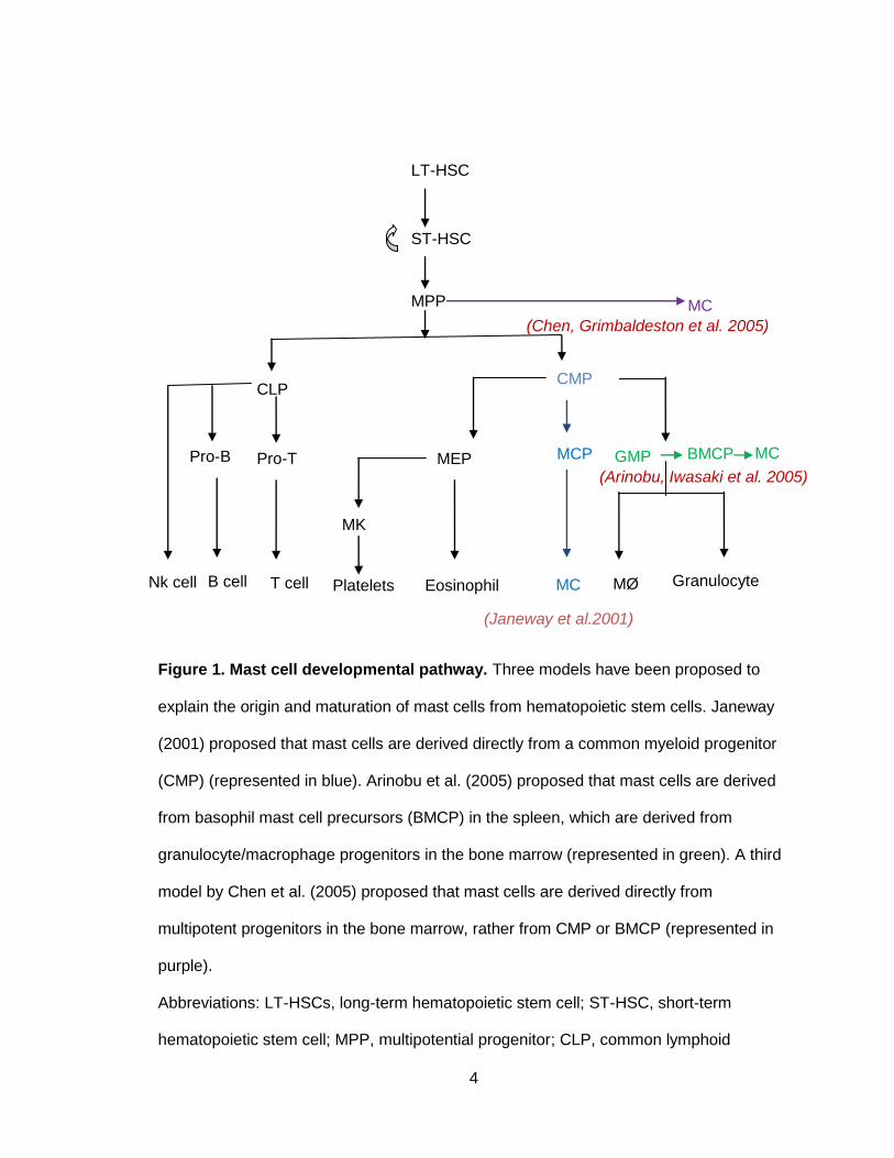
Figure 1. Mast cell developmental pathway. Three models have been proposed to
explain the origin and maturation of mast cells from hematopoietic stem cells. Janeway
(2001) proposed that mast cells are derived directly from a common myeloid progenitor
(CMP) (represented in blue). Arinobu et al. (2005) proposed that mast cells are derived
from basophil mast cell precursors (BMCP) in the spleen, which are derived from
granulocyte/macrophage progenitors in the bone marrow (represented in green). A third
model by Chen et al. (2005) proposed that mast cells are derived directly from
multipotent progenitors in the bone marrow, rather from CMP or BMCP (represented in
purple).
Abbreviations: LT-HSCs, long-term hematopoietic stem cell; ST-HSC, short-term
hematopoietic stem cell; MPP, multipotential progenitor; CLP, common lymphoid
(Chen, Grimbaldeston et al. 2005)
MC
MC BMCP MCP GMP MEP (Arinobu, Iwasaki et al. 2005)
MC Eosinophil Platelets T cell B cell
CLP CMP
Pro-T Pro-B
MK
Nk cell Granulocyte MØ
(Janeway et al.2001)
LT-HSC
ST-HSC
MPP

5
progenitor; CMP, common myeloid progenitor; MEP, megakaryocyte/erythrocyte
progenitor; MCP, mast cell progenitor; GMP, granulocyte/monocyte progenitor; BMCP,
basophil mast cell precursor; MC, Mast cell; MK, megakaryocyte; MØ, macrophages.
III. Mast cell trafficking to peripheral tissues
Although mature mast cells are never detected in circulation, the presence of MCps in
blood and various tissues has been demonstrated by in vitro cultures and limiting dilution
assays (Kasugai, Tei et al. 1995; Khalil, Luz et al. 1996; Gurish, Tao et al. 2001). A
committed precursor for the mast cell lineage was first identified in fetal murine blood by
Rodewald et al. (Rodewald, Dessing et al. 1996). These cells were characterized as
Thy-1loc-kithiFcεRI− and expressed mast cell specific proteases: mast cell
carboxypeptidase A, mMCP-2 and mMCP-4. These cells give rise to mast cell colonies
in vivo in the presence of IL-3 and SCF, and reconstitute a peritoneal mast cell
population in mast-cell–deficient W/Wv mice.
Additional support for the existence of circulating MCps was provided by Kitamura and
Fujita, who used methylcellulose colony-forming assays to demonstrate that mast cell
colonies can be formed from circulating mononuclear cells in the presence of growth
factors including SCF and IL-3. Committed mast cells progenitors (CD34+/ FcεRI−)
identified in human peripheral blood differentiated into mast cells in the presence of SCF
in vitro (Kitamura and Fujita 1989; Kirshenbaum, Kessler et al. 1991; Rottem, Okada et
al. 1994). These results suggested that, unlike other progeny of multipotent stem cells
(erythrocytes, neutrophils, eosinophils and basophils) that leave the bone marrow after
they differentiate, mast cells leave the bone marrow as immature but committed MCps
(Galli 1990; Kitamura, Kasugai et al. 1993).
6
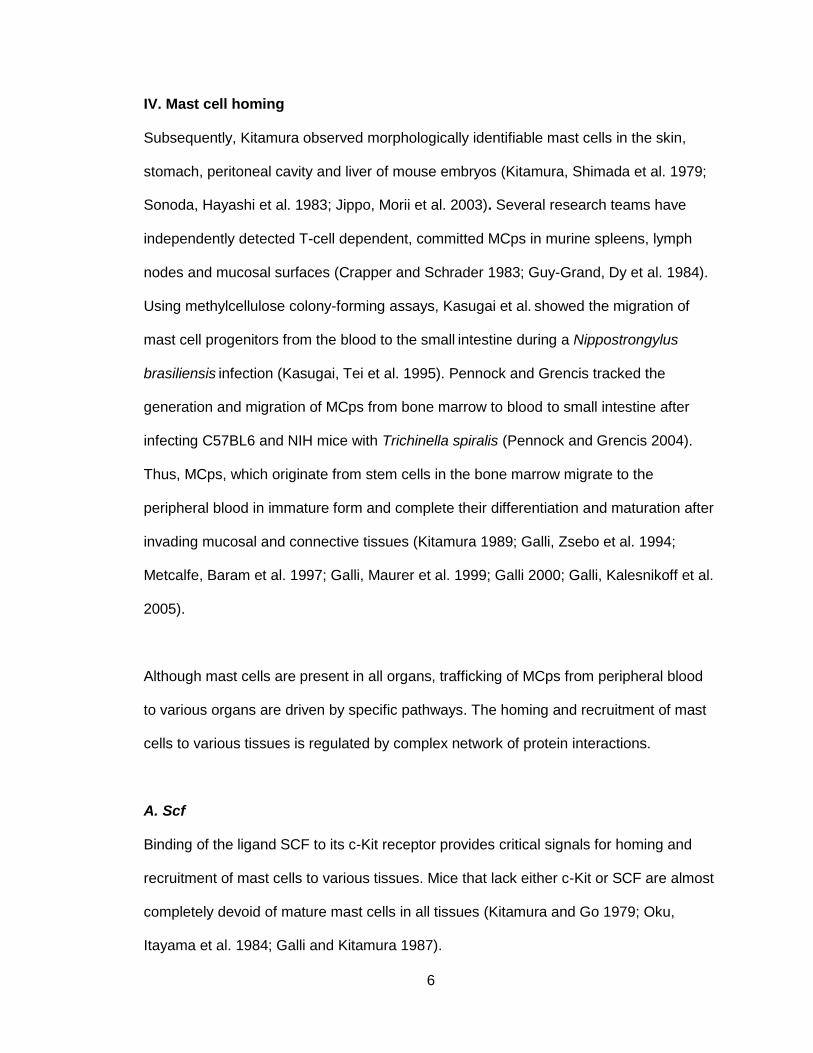
IV. Mast cell homing
Subsequently, Kitamura observed morphologically identifiable mast cells in the skin,
stomach, peritoneal cavity and liver of mouse embryos (Kitamura, Shimada et al. 1979;
Sonoda, Hayashi et al. 1983; Jippo, Morii et al. 2003). Several research teams have
independently detected T-cell dependent, committed MCps in murine spleens, lymph
nodes and mucosal surfaces (Crapper and Schrader 1983; Guy-Grand, Dy et al. 1984).
Using methylcellulose colony-forming assays, Kasugai et al. showed the migration of
mast cell progenitors from the blood to the small intestine during a Nippostrongylus
brasiliensis infection (Kasugai, Tei et al. 1995). Pennock and Grencis tracked the
generation and migration of MCps from bone marrow to blood to small intestine after
infecting C57BL6 and NIH mice with Trichinella spiralis (Pennock and Grencis 2004).
Thus, MCps, which originate from stem cells in the bone marrow migrate to the
peripheral blood in immature form and complete their differentiation and maturation after
invading mucosal and connective tissues (Kitamura 1989; Galli, Zsebo et al. 1994;
Metcalfe, Baram et al. 1997; Galli, Maurer et al. 1999; Galli 2000; Galli, Kalesnikoff et al.
2005).
Although mast cells are present in all organs, trafficking of MCps from peripheral blood
to various organs are driven by specific pathways. The homing and recruitment of mast
cells to various tissues is regulated by complex network of protein interactions.
A. Scf
Binding of the ligand SCF to its c-Kit receptor provides critical signals for homing and
recruitment of mast cells to various tissues. Mice that lack either c-Kit or SCF are almost
completely devoid of mature mast cells in all tissues (Kitamura and Go 1979; Oku,
Itayama et al. 1984; Galli and Kitamura 1987).

7
B. Integrins
Integrin heterodimer α4β7 and its corresponding ligands, vascular cell adhesion
molecule-1 (VCAM-1) and mucosal addressin cell adhesion molecule (MAdCAM1) are
critical for maintaining a pool of MCps in the small intestine (Gurish,2006). Mice deficient
in integrin α4β7, or those in which α4β7 integrin or their ligands are blocked by
antibodies, are devoid of MCps or matured mast cells in the intestine. VCAM-1 (which
interacts with both α4β1 and α4β7 integrins), but not MAdCAM1 is essential for the
recruitment of MCps into the lungs during pulmonary inflammation (Abonia, Hallgren et
al. 2006). Integrin αIIbβ3 mediates mast cell homing and retention in the peritoneal
cavity; mice that are deficient in the glycoprotein IIb subunit of the αIIbβ3 integrin have
significantly reduced peritoneal mast cell populations (Shattil and Newman 2004;
Berlanga, Emambokus et al. 2005).
C. Chemokines
In addition to integrins, chemokine receptors expressed by mast cell progenitors play a
critical role in directing the migration of MCps from circulation into the tissues (Humbles,
Lu et al. 2002; Abonia, Austen et al. 2005). Human mast cells express various
chemokine receptors CXCR2, CXCR4, CCR3 and CCR5 (Ochi, Hirani et al. 1999). The
interaction of chemokine receptor 2 (CXCR2) with its ligand is critical for directing MCps
to the intestine (Abonia, Austen et al. 2005). A profound decrease in the mast cell
progenitor population in the small intestine was reported in mice deficient in CXCR2 or
those in which anti-CXCR2 was administered (Abonia, Austen et al. 2005). The role of
CCR3 in mast cell homing has been identified in CCR3-deficient mice (Humbles, Lu et
al. 2002). A significant (2- to 4-fold) increase in intra-epithelial mast cells is found in the
tracheas of CCR3-deficient mice after an allergen challenge, which suggests that CCR3
is involved in the egress of mast cells from the mucosal intraepithelial compartment of
8
the lung (Humbles, Lu et al. 2002). These studies highlight the role of multiple factors—
the integrins, the growth factors, and the cytokine receptors—in influencing tissue
localization of MCps.
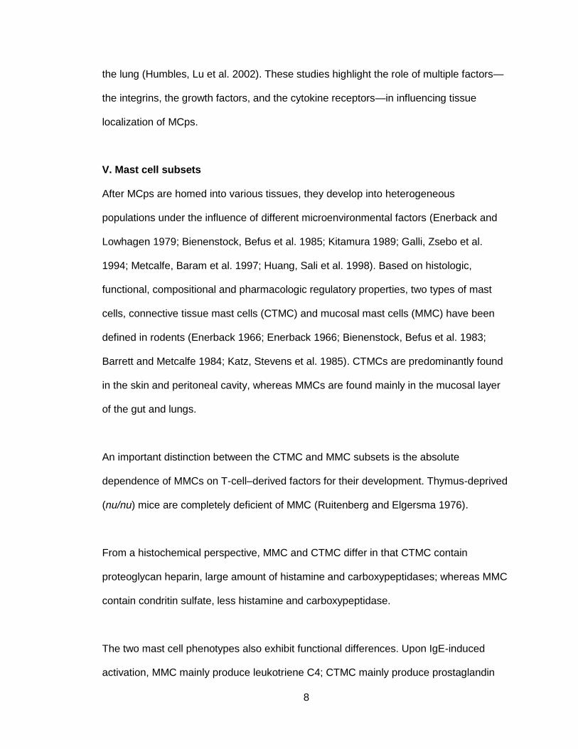
V. Mast cell subsets
After MCps are homed into various tissues, they develop into heterogeneous
populations under the influence of different microenvironmental factors (Enerback and
Lowhagen 1979; Bienenstock, Befus et al. 1985; Kitamura 1989; Galli, Zsebo et al.
1994; Metcalfe, Baram et al. 1997; Huang, Sali et al. 1998). Based on histologic,
functional, compositional and pharmacologic regulatory properties, two types of mast
cells, connective tissue mast cells (CTMC) and mucosal mast cells (MMC) have been
defined in rodents (Enerback 1966; Enerback 1966; Bienenstock, Befus et al. 1983;
Barrett and Metcalfe 1984; Katz, Stevens et al. 1985). CTMCs are predominantly found
in the skin and peritoneal cavity, whereas MMCs are found mainly in the mucosal layer
of the gut and lungs.
An important distinction between the CTMC and MMC subsets is the absolute
dependence of MMCs on T-cell–derived factors for their development. Thymus-deprived
(nu/nu) mice are completely deficient of MMC (Ruitenberg and Elgersma 1976).
From a histochemical perspective, MMC and CTMC differ in that CTMC contain
proteoglycan heparin, large amount of histamine and carboxypeptidases; whereas MMC
contain condritin sulfate, less histamine and carboxypeptidase.
The two mast cell phenotypes also exhibit functional differences. Upon IgE-induced
activation, MMC mainly produce leukotriene C4; CTMC mainly produce prostaglandin

9
D2 (Kitamura 1989). Similar to rodents, humans have two distinct types of mast cells:
MCT and MCTC. MCT s are predominantly found in the lungs and gastrointestinal mucosa;
MCTCs are most abundant in the skin and the gastrointestinal submucosa (Schwartz,
1987, Irani, 1986). Similar to MMC, the development of MCT critically depends on T-cell–
derived factors as seen by a significant reduction in their numbers in patients with
immunodeficiencies (Irani, Craig et al. 1987).The two subsets can be distinguished on
the basis of their protease content and secretory granules. MCTC contains tryptase,
chymase, cathepsin G, and carboxypeptidase, whereas MCT express only tryptase
(Irani, Schechter et al. 1986). In addition to exhibiting heterogeneity with respect to
neutral protease content of the secretory granules, human mast cells also vary with
respect to their cytokine content. MCT produce both IL-5 and IL-6, but MCTC produce
only IL-4 (Bradding, Okayama et al. 1995). Despite such histochemical and functional
differences, these cells reportedly are derived from a common lineage. It is also thought
that the above phenotypes are plastic and interchangeable (Sonoda, Sonoda et al. 1986;
Arinobu, Iwasaki et al. 2005).
VI. c-Kit
c-Kit, a member of the type III receptor tyrosine kinase subfamily, is a transmembrane
protein expressed in mast cells, hematopoietic progenitor cells, melanocytes, germ cells
and gastrointestinal pacemaker cells (Galli, Tsai et al. 1993). c-Kit is downregulated as
all hematopoietic lineages mature except mast cells. Mast cells retain high levels of c-Kit
expression throughout their maturity.
Normal mast cell development requires direct interaction between the c-Kit receptor
(expressed in MCps and mast cells) and the ligand SCF secreted by fibroblasts and
other cells in the microenvironment where mast cells develop. In mice, c-Kit is expressed

10
from two alternately spliced mRNAs, which, following protein glycosylation, give rise to
products with molecular weights around 145kDa (Yarden, Kuang et al. 1987). c-Kit is
encoded by the white-spotting (W) locus on chromosome 5, and the c-Kit ligand SCF is
encoded by the steel (SI) locus on chromosome 10 of mice (Chabot, Stephenson et al.
1988; Copeland, Gilbert et al. 1990). The mutations at the W locus abolish the c-Kit
tyrosine kinase receptor on the cell surface or produce receptors with markedly deficient
tyrosine kinase activity (Geissler, Ryan et al. 1988; Nocka, Tan et al. 1990). Mutations at
the Sl locus result in the absence of the c-Kit receptor ligand, stem cell factor (SCF), or
the production of abnormal forms of SCF (Flanagan and Leder 1990; Flanagan, Chan et
al. 1991). Mice with mutations at the W locus (W/Wv) or the Sl locus (Sl/Sld) are
profoundly deficient in mast cells (Kitamura, Go et al. 1978; Kitamura and Go 1979).
While several cytokines influence the growth, survival and maturation of mast cells, SCF
and its interaction with the c-Kit receptor are critical for normal mast cell development
and function.
A. Structure
c-Kit is characterized by the presence a signal sequence at its N-terminus, which is
followed by five immunoglobin (Ig)-like motifs, a transmembrane domain, and a cytosolic
tyrosine kinase domain that is split into proximal and distal regions by an insert
sequence. The second and third Ig-like motifs constitute a pocket for SCF binding
(Zhang, Zhang et al. 2000). When SCF binds to the c-Kit receptor, the latter undergoes
dimerization to initiate intrinsic tyrosine kinase activity. The fourth Ig motif of c-Kit
contains the dimerization site, the deletion of which completely abolishes receptor
dimerization and subsequent downstream signal transduction events.
11
SCF affinity for the c-Kit receptor depends on receptor dimerization, as indicted by
accelerated ligand dissociation in the monomeric form of c-Kit and in c-Kit with defective
dimerization sites (Blechman, Lev et al. 1995). SCF binding induces a conformational
change to dimerize the receptor, which stabilizes the ligand-receptor interaction
(Blechman, Lev et al. 1995). The juxtamembrane domain of c-Kit inhibits receptor
dimerization and enzyme activity, maintaining an inactive conformation (Roskoski 2005).
After initiation of kinase activity, various tyrosine residues in the cytoplasmic tail of the c-
Kit receptor become phosphorylated, and function as docking sites for the Src
homology2 (SH2) domain containing signal transduction molecules (Pawson 1995). The
kinase domain of c-Kit is responsible for catalyzing and transfering a phosphate group
from ATP to the substrate and activating them thus initiating downstream signaling
(Roskoski 2005).

12
Figure 2. Schematic structure of c-Kit receptor and overview of the signaling
pathways activated upon SCF ligation to c-Kit. The distinct molecular domains
comprise three functional structures: NH2-terminal extracellular, transmembrane and
COOH-terminal intracellular domains. The extracellular domain contains five
immunoglobulin-like repeats and the intracellular domain encodes two tandem repeats of
the enzyme catalytic domains. The juxtamembrane region binds to Src family members,
and changes in several residues within this region have been shown to result in
constitutive activation of c-Kit. The proximal kinase domain of c-Kit contains amino acid
residues involved in binding ATP. The kinase insert domain contains a tyrosine residue
that contributes to the binding of PI-3K. Asterisks indicate the position of aspartic acid-
814 in the second catalytic domain, and substitution of this residue to valine (D814V)
results in constitutive activation of c-Kit.
LynPP
PPP
P
P
Tec P
Dok1
P
GRB2
P
p85p110
GRB2
GRB7
P
Socs
CblGab1
Gab2
PLCγ1
P
P
P
P
SCF
*

13
B. Function
Signaling cascades initiated by c-Kit stimulate mast cell functions related to
differentiation, proliferation, survival and activation (Taylor and Metcalfe 2000).
c-Kit and its ligand, stem cell factor (SCF) induce proliferation of mouse mast cells in
vitro (Huang, Nocka et al. 1990; Martin, Suggs et al. 1990; Matsui, Zsebo et al. 1990;
Nocka, Buck et al. 1990; Williams, Eisenman et al. 1990; Zsebo, Williams et al. 1990;
Zsebo, Wypych et al. 1990) and in vivo (Tsai, Shih et al. 1991; Tsai, Takeishi et al.
1991). SCF-induced c-Kit kinase activity is essential for mast cell homeostasis, growth
and differentiation of CD34+ human mast cell progenitor cells in vitro (Kirshenbaum, Goff
et al. 1999). SCF is a potent chemotactic agent for MCps and mast cells, and also acts
as a major factor responsible for adhesion of mast cells to connective tissue matrices
(Dastych and Metcalfe 1994; Nilsson, Butterfield et al. 1994; Dastych, Taub et al. 1998).
Thus, SCF plays a crucial role in the migration and homing of MCps to mucosal and
connective tissues where they reside and terminally differentiate (Meininger, Yano et al.
1992; Okayama and Kawakami 2006). Survival of mature mast cells is also dependent
on SCF (Yee, Hsiau et al. 1994; Metcalfe, Mekori et al. 1995; Da Silva, Reber et al.
2006). In addition to their role in triggering differentiation, maturation and migration of
mast cells, SCF is also recognized as potent modifier of mast cell activation and
secretion of mediators including tumor necrosis factors, proteolytic enzymes,
glycosaminoglycans, and lipid mediators.
C. Negative regulation of c-Kit signaling
To maintain homeostasis, c-Kit signaling is attenuated after a period of time in mast
cells. Miyazawa et al. reported polyubiquitination and degradation of c-Kit in response to
SCF, which regulates c-Kit signaling in M07e cells (Miyazawa, Toyama et al. 1994).
Unrestricted activation of c-Kit results in abnormal growth and survival of mast cells,
14
causing acute myeloid leukemia, systemic mastocytosis and gastrointestinal stromal
tumors.
SCF stimulation, which initiates several signals for positive regulation of cell growth and
proliferation, also initiates the phosphorylation and activation of c-Cbl ubiquitin ligase. c-
Cbl is thought to associate with Src kinase in initiating the c-Kit degradation process,
thus attenuating various intracellular signals (Levkowitz, Waterman et al. 1998; Lee,
Wang et al. 1999; Miyake, Mullane-Robinson et al. 1999; Taher, Tjin et al. 2002;
Masson, Heiss et al. 2006). Activated c-Cbl mediates the degradation of c-Kit through
the proteasomal pathway (Zeng, Xu et al. 2005). Expression of a Cbl mutation
(CblR420Q) in mice inhibited SCF-induced ubiquitination and internalization of c-Kit and
led to mastocytosis and myeloid leukemia (Bandi, Brandts et al. 2009).
D. Abnormal c-Kit signaling
Although c-Kit is a critical molecule for mast cell development, certain activating
mutations of this receptor, result in ligand-independent autophosphorylation that leads to
constitutive activation of c-Kit, causing dysregulated cell growth and induction of
tumurogenesis (Kitayama, Kanakura et al. 1995; Tsujimura, Morimoto et al. 1996).
15
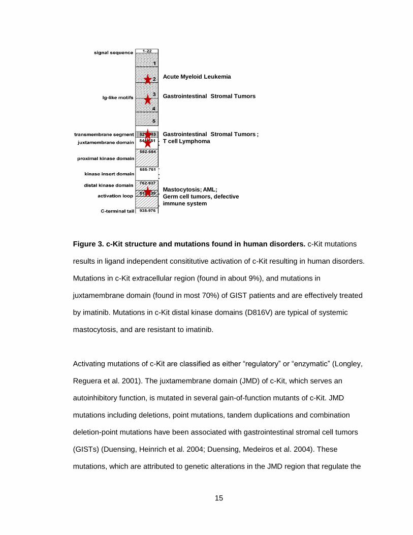
Figure 3. c-Kit structure and mutations found in human disorders. c-Kit mutations
results in ligand independent consititutive activation of c-Kit resulting in human disorders.
Mutations in c-Kit extracellular region (found in about 9%), and mutations in
juxtamembrane domain (found in most 70%) of GIST patients and are effectively treated
by imatinib. Mutations in c-Kit distal kinase domains (D816V) are typical of systemic
mastocytosis, and are resistant to imatinib.
Activating mutations of c-Kit are classified as either ―regulatory‖ or ―enzymatic‖ (Longley,
Reguera et al. 2001). The juxtamembrane domain (JMD) of c-Kit, which serves an
autoinhibitory function, is mutated in several gain-of-function mutants of c-Kit. JMD
mutations including deletions, point mutations, tandem duplications and combination
deletion-point mutations have been associated with gastrointestinal stromal cell tumors
(GISTs) (Duensing, Heinrich et al. 2004; Duensing, Medeiros et al. 2004). These
mutations, which are attributed to genetic alterations in the JMD region that regulate the
Gastrointestinal Stromal Tumors
Acute Myeloid Leukemia
Gastrointestinal Stromal Tumors ;
T cell Lymphoma
Mastocytosis; AML;
Germ cell tumors, defective
immune system

16
kinase activity of c-Kit, are the ―regulatory‖ type, and they might result in conformational
changes in c-Kit that cause it to dimerize and activate.
Another set of c-Kit–activating mutations are referred to as the ―enzymatic pocket type‖
mutation. These directly affect the sequence of c-Kit’s active enzyme site. This has been
studied in humans (D816V), mice (D814V) and rats (D817V). Constitutively activating
mutations of the c-Kit gene involve alterations of its phosphotransferase domain, which
was reported to confer factor-independent growth in human mast cell leukemia (HMC-1),
murine mastocytoma (P-815) and rat mast cell leukemia (RBL-2H3) (Furitsu, Tsujimura
et al. 1993; Tsujimura, Furitsu et al. 1994). D816V mutation of the human c-Kit
(homologous to D841V of mouse) has been associated with mast cell proliferative
disorder, mastocytosis, acute myeloid leukemia and germ cell tumors (Tian, Frierson et
al. 1999; Beghini, Peterlongo et al. 2000; Valent, Horny et al. 2001).
The aspartic acid (asp) residue encoded by the 816 codon of human c-Kit is located in
the tyrosine kinase domain and is involved in ATP binding and subsequent
phosphotransferase activity (Mol, Lim et al. 2003; Vendome, Letard et al. 2005). Amino
acid substitution of Asp-816 to valine in human c-Kit stabilizes the kinase in its active
conformation, thus resulting in ligand-independent activation of c-Kit (Mol, Lim et al.
2003; Vendome, Letard et al. 2005).
Interestingly, activation of c-Kit caused by this JMD mutation is due to constitutive
dimerization of c-Kit in absence of SCF, whereas a D814V mutation induces SCF-
independent growth without receptor dimerization (Kitayama, Kanakura et al. 1995;
Tsujimura, Morimoto et al. 1996). Unlike gastrointestinal stromal tumors, which are
treated effectively with Gleevac, systemic mastocytosis associated with a c-Kit D816V

17
mutation does not respond to Gleevac. This is thought to be due to the inability of the
drug to bind to the ATP binding site, whose conformation is altered by a c-Kit point
mutation at position 816 (Scheinfeld 2006). Currently there are no drugs on the market
can specifically target the kinase domain mutants of c-Kit.
Previous studies report that PI-3K, which binds to the 719 tyrosine residue of murine c-
Kit (721 a.a. in human c-Kit) through its regulatory subunit, substantially contributes to
factor-independent abnormal growth in c-Kit–mutated cells (Chian, Young et al. 2001).
PI-3K is constitutively activated in c-Kit (D814V)–induced myeloproliferative disorders,
and treatment of these cells with the PI-3K inhibitor wortmannin specifically inhibits
ligand-independent growth (Chian, Young et al. 2001). In hematopoietic neoplasms and
tumors, PI-3K is reported to be persistently active, which might contribute to the
abnormal growth of those cells (Vivanco and Sawyers 2002). Hadhimoto et al. reported
that PI3-K plays an important role in ligand-independent growth and tumorigenicity in c-
Kit (D814V) mutant cells (Hashimoto, Matsumura et al. 2003). The abnormal growth
induced by this c-Kit (D814V) mutation can be suppressed by genetic disruption of the
p85α regulatory subunit of PI-3K (Munugalavadla, Sims et al. 2007). Alterations of c-Kit
signaling was not observed when the p85β regulatory subunit was disrupted in these
cells, which suggests a unique role for the PI-3K regulatory subunits in the control of
constitutively active c-Kit signaling.
In this thesis, we evaluated specific role of various PI-3K regulatory subunits in
mediating c-Kit signals. Since there are no effective drugs available for the treatment of
c-Kit (D814V)– related diseases, this information is likely to be important for the design
of peptides that specifically target and correct the abnormal phenotype caused by this c-
Kit (D814V) mutation.
18
VII. PI-3K (Phosphatidylinositol-3-kinase) and c-Kit signaling
PI-3K comprises a family of lipid kinases that are essential for the growth, differentiation,
proliferation, survival and migration of mast cells. Structural characteristics and substrate
specificity divide PI-3K into four classes: Class IA, IB, II and III (Fruman, Meyers et al.
1998; Wymann and Pirola 1998; Walker, Perisic et al. 1999). Class IA PI-3Kinase is
heterodimeric kinases consisting of a regulatory subunit and a catalytic subunit (Sasaki,
Suzuki et al. 2002; Okkenhaug and Vanhaesebroeck 2003). Class IA has three types of
catalytic subunits: p110α, p110β and p110δ. Of those three, p110α and p110β are
expressed in many tissues, whereas p110δ is expressed mainly in leukocytes. Five
different proteins (p85α, p55α, p50α, p85β and p55γ) have been identified to date as the
Class IA regulatory subunits. The p85α, p55α and p50α proteins are derived from mRNA
splice variants encoded in the gene Pik3r1, while p85β is encoded by Pik3r2 and p55γ is
derived from Pik3r3 (Fruman, Cantley et al. 1996). The regulatory subunits have several
motifs implicated in protein-protein interactions. All the class IA regulatory subunits have
two Src homology 2 (SH2) domains that bind phosphorylated tyrosine residues of
various receptors and adaptor molecules (Sasaki, Suzuki et al. 2002; Okkenhaug and
Vanhaesebroeck 2003). The inter-SH2 domain constitutively interacts with a specific
domain of p110 catalytic subunit (Sasaki, Suzuki et al. 2002; Okkenhaug and
Vanhaesebroeck 2003). The dual SH2 domains are functionally important because they
recruit the p110 catalytic subunit to tyrosine-phosphorylated proteins at the the
cytoplasmic membrane (Sasaki, Suzuki et al. 2002; Okkenhaug and Vanhaesebroeck
2003). Furthermore, the interaction of the dual SH2 domains with the phosphotyrosine
residue of c-Kit releases the p110-kinase activity that is normally blocked by complex
formation between the regulatory and catalytic subunits (Sasaki, Suzuki et al. 2002;
Okkenhaug and Vanhaesebroeck 2003).
19
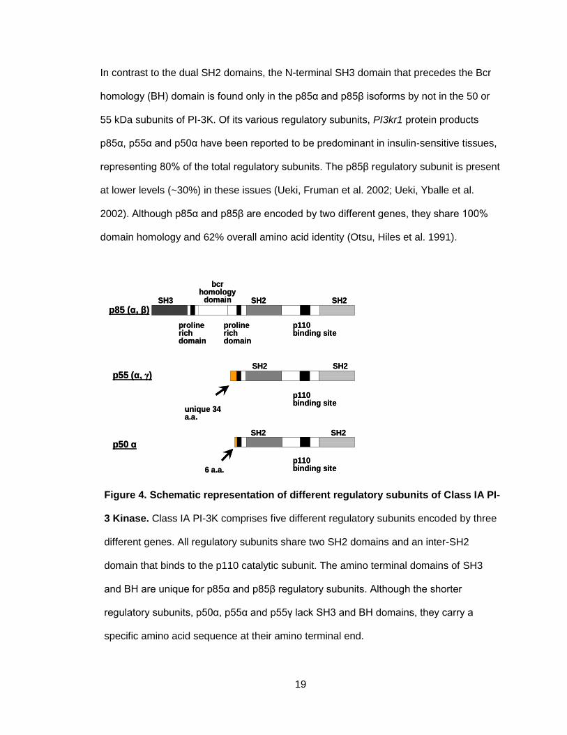
In contrast to the dual SH2 domains, the N-terminal SH3 domain that precedes the Bcr
homology (BH) domain is found only in the p85α and p85β isoforms by not in the 50 or
55 kDa subunits of PI-3K. Of its various regulatory subunits, PI3kr1 protein products
p85α, p55α and p50α have been reported to be predominant in insulin-sensitive tissues,
representing 80% of the total regulatory subunits. The p85β regulatory subunit is present
at lower levels (~30%) in these issues (Ueki, Fruman et al. 2002; Ueki, Yballe et al.
2002). Although p85α and p85β are encoded by two different genes, they share 100%
domain homology and 62% overall amino acid identity (Otsu, Hiles et al. 1991).
Figure 4. Schematic representation of different regulatory subunits of Class IA PI-
3 Kinase. Class IA PI-3K comprises five different regulatory subunits encoded by three
different genes. All regulatory subunits share two SH2 domains and an inter-SH2
domain that binds to the p110 catalytic subunit. The amino terminal domains of SH3
and BH are unique for p85α and p85β regulatory subunits. Although the shorter
regulatory subunits, p50α, p55α and p55γ lack SH3 and BH domains, they carry a
specific amino acid sequence at their amino terminal end.
p85 (α, β)
p55 (α, γ)
p50 α
SH3 SH2 SH2
bcrhomology
domain
prolinerichdomain
prolinerichdomain
p110binding site
SH2 SH2
p110binding site
unique 34 a.a.
SH2 SH2
p110binding site6 a.a.
p85 (α, β)
p55 (α, γ)
p50 α
SH3 SH2 SH2
bcrhomology
domain
prolinerichdomain
prolinerichdomain
p110binding site
SH2 SH2
p110binding site
unique 34 a.a.
SH2 SH2
p110binding site6 a.a.

20
Upon SCF stimulation, the p85 regulatory subunit of PI-3K binds to the phosphorylated
719 tyrosine residue of murine c-Kit receptor (Y721 of human c-Kit) in its kinase insert
region (Rottapel, Reedijk et al. 1991; Lev, Givol et al. 1992). Mutation of tyrosine 719 of
murine c-Kit to phenylalanine eliminates the capacity for p85 to associate with c-Kit but
enhances SCF-induced increased PI-3K activity (Serve, Hsu et al. 1994). Association of
the PI-3K complex with the activated c-Kit receptor via the p85 regulatory subunit allows
conformational change and activation of p110 catalytic subunit. The activated PI3K
complex then gets translocated to the membrane where its lipid substrate resides.
PI-3Ks activate phospholipid substrates by phosphorylating the D3 hydroxyl position of
the inositol ring, which generates the active products phosphatidylinositol-(3) phosphate
(PtdIns(3)P3), phosphatidylinositol-(4,5)-bisphosphate (PtdInsP2) and
phosphatidylinositol-(3,4,5)-triphosphate (PtdInsP3) (Hawkins, Anderson et al. 2006).
These products of class I PI-3K are crucial secondary messengers that recruit proteins
containing a pleckstrin homology (PH) domain. Recruitment to cellular membranes
activates them and initiates signaling cascades to enhance mast cell migration,
adhesion, activation, growth and survival (Serve, Yee et al. 1995; Vosseller, Stella et al.
1997).
VIII. SCF and enhanced mast cell survival
Stem cell factor is the ligand for c-Kit that is primarily produced by stromal cells. SCF is a
critical regulator of mast cell survival and mast cell numbers in various tissues in vivo.
SCF rescues mast cells from spontaneous apoptosis in vitro (Mekori, Oh et al. 1993;
Iemura, Tsai et al. 1994; Finotto, Mekori et al. 1997). Inhibition of SCF synthesis in vivo
leads to mast cell apoptosis (Mekori, Oh et al. 1993; Iemura, Tsai et al. 1994; Finotto,
Mekori et al. 1997). SCF stimulation of mast cells increases the level of pro-survival

21
proteins including Bcl-2, Bcl-XL (Mekori, Gilfillan et al. 2001; Baghestanian, Jordan et al.
2002), which are critical for mast cell survival. Another mechanism by which SCF
enhances mast cell survival is by actively preventing Bim expression via phosphorylation
of forkhead box (FOX) proteins FOXO1a and FOXO3a. Bim is involved in mast cell
apoptosis induced by growth factor deprivation (Alfredsson, Puthalakath et al. 2005).
PI-3K, an immediate downstream effector of c-Kit, is a well-known mediator of anti-
apoptotic signaling, and elevated levels of PI-3K products are reported in many tumor
cells (Downward 2004). Use of inhibitor LY294002, which selectively blocks PI-3K
activation in cells, induces apoptosis and suppresses growth of tumor cells (Krystal,
Sulanke et al. 2002). Abolition of PI-3K activation by mutating its docking site to c-Kit
(Y719F) in mast cells results in significantly reduced survival (Serve, Yee et al. 1995).
SCF-mediated c-Kit signaling in normal cells activates AKT, a survival-promoting serine-
threonine protein kinase that enhances cell survival by inducing phosphorylation and
inactivation of the pro-apoptotic molecule Bad. The phosphorylation and activation of
AKT is, in turn, mediated by PI-3K upon SCF stimulation (Blume-Jensen, Janknecht et
al. 1998). Activation of PI-3K also promotes phosphorylation of Bim and its proteosome-
dependent degradation, thus enhancing survival. Although PI-3K is known to be critical
for mast cell survival, the specific roles of PI-3K regulatory subunits in mediating the
process are poorly understood. Using genetic and molecular approaches, we examined
the role of p85α and p85β in regulating mast cell survival in response to SCF stimulation.
IX. Role of IL-3 in mast cell maturation
Differentiation and maturation of mast cells involve the synthesis and storage of an array
of inflammatory mediators including histamine, mast cell proteases (mainly tryptases and
chymases) and cytokines. IL-3 has been identified as one of the principle cytokines
22
regulating mast cell growth and terminal differentiation (Yong 1997; Galli, Nakae et al.
2005). In vitro cultures of murine bone marrow cells differentiate into a population of
homogeneous mast cells when cultured in a medium supplemented with IL-3 (Razin, Ihle
et al. 1984; Thompson, Metcalfe et al. 1990). IL-3 signaling activates Stat5, a
transcription factor that is critical for mast cell development, as shown by the loss of
mast cells in Stat5-deficient mice (Shelburne, McCoy et al. 2003). IL-3 also stimulates
TNF secretion induced via the PI-3K pathway, which is crucial for the maturation of mast
cells from their progenitors (Wright, Bailey et al. 2006).
In this thesis study, we characterized the specific role of p85α and p85β in regulating
mast cell maturation in vitro in response to IL-3 stimulation. We further performed
reconstitution studies to examine the role of the SH3 and BH domains of p85α and p85β
in mast cell maturation. Furthermore, we confirmed our in vitro observations in vivo, by
performing transplant studies.
X. Role of transcription factors in mast cell maturation
Transcription factors play a crucial role in the development of various lineages from
uncommitted precursor cells. According to the current models for hematopoietic
development, uncommitted stem cells express low levels of transcription factors (Orkin
2000; Cantor and Orkin 2001; Orkin 2003). Differentiation of mature blood cell types
from the multipotential hematopoietic cell is controlled in part through the expression of
lineage-specific transcription factors that regulate the expression of downstream genes
that determine the function of each blood cell type (Iwasaki, Mizuno et al. 2006). The
transcription factors GATA-1, GATA-2, PU.1, and the microphthalmia-associated
transcription factor (Mitf) play essential roles in mast cell development. GATA-1, GATA-
2, and PU.1 transcription factors are involved in the maturation of mast cells, while Mitf is

23
involved in the migration, phenotypic expression and survival of mast cells (Kitamura,
Oboki et al. 2006).
A. GATA
GATA-1 is a member of highly conserved family of zinc finger protein that regulates the
expression of several mast cell target genes, including FcεRI (Nishiyama, Hasegawa et
al. 2002; Maeda, Nishiyama et al. 2003), carboxypeptidases (Zon, Gurish et al. 1991),
IL-4 (Henkel and Brown 1994) and IL-13 (Masuda, Yoshikai et al. 2004). GATA-1low mice
generated by targeted deletion of upstream enhancer and promoter sequences of the
GATA-1 gene show impaired differentiation of mast cells (Migliaccio, Rana et al. 2003).
Mast cell defects associated with decreased GATA-1 expression was characterized by
hyperproliferation of early mast cell precursors, susceptibility to apoptosis and impaired
expression of FcεRI and mast-cell–specific carboxypeptidase-A in mature mast cells.
(Migliaccio, Rana et al. 2003). Abberant insertion of a neomycin cassette between
GATA-1 promoter regulatory elements and the GATA-1 IE exon also resulted in low
GATA-1 expression (Takahashi,1997). These mutant mice had lower GATA-1 levels and
low populations of mature mast cells in the skin (Harigae, Takahashi et al. 1998). The
increased apoptotic rate and defective differentiation of mast cells in GATA-1low mice
were reversed in vitro by forced GATA-1 expression (Migliaccio, Rana et al. 2003).
GATA-2 is another member of the GATA family that plays a crucial role in mast cell
development. A GATA-2 deficiency in embryonic stem cells impaired the response to the
c-Kit ligand SCF. Furthermore, those cells are incapable of differentiating into MCps
(Tsai, Keller et al. 1994). Expression of GATA-2 is highest in proliferating mast cell
lineage cells and is downregulated in differentiated mast cells (Jippo, Mizuno et al.
1996).

24
B. PU.1
Ets family member PU.1 is another transcription factor required for normal development
of mast cells. PU.1-deficient murine fetuses lack dermal mast cells; PU.1 in cooperation
with GATA regulates expression of crucial mast cell genes including FcεRI and IL-4
(Henkel and Brown 1994; Nishiyama, Hasegawa et al. 2002). In addition, evidence
exists regarding an interplay between PU.1 and GATA in the regulation of mast cell
development.
C. Mitf
Mitf (Microphtalmia transcription factor) is a basic helix-loop-helix leucine zipper
transcription factor that regulates transcription of several genes essential for the growth,
maturation, differentiation and normal histochemical composition of mast cells. Mitf
directly targets protease genes including, mast cell proteases 2,4,8,9, granzyme,
tryptophan hydozylase, protease inhibitor Serpin E1, and metabolic enzyme hPGDs, and
thus play a key role in mast cell biology (Morii, Tsujimura et al. 1996; Ito, Morii et al.
1998; Morii, Oboki et al. 2004)
Mitf is crucial of mast cell survival, and is expressed in mast cells as well as in the
tissues where mast cells develop. Cultured mast cells derived from Mitf-mutant mice
adhere poorly to fibroblasts. This defective adhesion has been attributed to deficient
transcription of adhesion factor SgIGSF (Morii, Oboki et al. 2004), the factor also
responsible for reduced peritoneal mast cell expression in Mitf mutants (Morii, Oboki et
al. 2004). Mitf regulates c-Kit expression in mast cells; and, in turn, c-Kit regulates the
transcriptional activity of Mitf, suggesting the possibility of homeostatic regulation
between these factors (Isozaki, Tsujimura et al. 1994). Interestingly, a phenotypic
overlap is seen among mice a carrying germ line mutation in the locus that encodes

25
SCF, receptor c-Kit, PI-3K and Mitf transcription factor (Dubreuil, Forrester et al. 1991;
Moore 1995; Hemesath, Price et al. 1998; Fukao, Yamada et al. 2002).
In our study, we observed defective differentiation of mast cells in the absence of the
p85α regulatory subunit of PI-3K, which could be corrected upon restoring expression of
either full-length p85α or mutant constructs of p85α that lack either amino terminal SH3
or BH domain. Although Mitf is critical in mast cell differentiation, the molecular
mechanism regulating the expression of Mitf in mast cells is not well understood. Here
we investigated the role of the p85α regulatory subunit and its amino terminal domains in
mediating Mitf expression in mast cells, which thereby regulates their maturation.
XI. In vivo experiments to determine the function of p85 regulatory subunits and
their amino terminal domains in mast cell development
c-Kit and its ligand SCF are critical for the biological functions of mast cells and mast cell
development is disrupted or severely affected in mice that either lack c-Kit expression or
carry c-Kit mutations that result in its loss of function. Such mast-cell–deficient rodents
are widely used as in vivo tools to investigate mast cell biology. One c-Kit mutation
resulting in loss of function is W-Sash (Wsh), an inversion mutation upstream of c-Kit’s
transcriptional site on murine chromosome 5 (Berrozpe, Timokhina et al. 1999). This
mutation specifically impairs the development of mast cells and melanocytes, and in vitro
cultures from those mice do not express c-Kit mRNA (Duttlinger, Manova et al. 1993;
Yamazaki, Tsujimura et al. 1994). Our study sought to investigate the role of p85
regulatory subunits in mediating mast cell development using an in vivo Wsh model. To
determine the specific roles of p85 regulatory subunits and their amino terminal domains
(SH3 and BH) in signal transduction of c-Kit/SCF mediated mast cell development, W
sh/sh mice were transplanted with p85α-deficient BMMC reconstituted with p85α, p85β,

26
p85αΔSH3 and p85αΔBH constructs, and the mast cell population reconstituted in
various tissues was investigated.
XII. Focus of the dissertation
Previous studies revealed the importance of PI-3K in mediating normal and abnormal c-
Kit signaling, thereby regulating growth, differentiation, and survival mast cell biology. PI-
3K directly interacts with c-Kit via its regulatory subunit; five different regulatory subunits
have been reported (Inukai, Funaki et al. 1997). We hypothesize that p85α and p85β
regulatory subunits might have unique mast cell functions that might be imparted by their
amino terminal SH3 and BH domains, regions where the p85 subunits differ the most.
We sought to investigate the specific roles of p85α and p85β, and the amino terminal
SH3 and BH domains of p85α in mediating mast cell growth, differentiation and survival.
PI-3K might also enhance c-Kit receptor internalization and degradation via binding to
the activated ubiquitin ligase, c-Cbl. We therefore investigated the interaction of p85
regulatory subunits with c-Cbl, and their role in regulating the c-Kit internalization and
degradation process upon SCF stimulation. Information on specific roles of PI-3K
regulatory subunits and their domains in regulating c-Kit signals in mast cells advances
our understanding of the mechanisms critical to the biology of mast cell growth and
differentiation for future translational applications for the treatment of mast cell disorders.

27
MATERIALS AND METHODS
I. Cytokines, Antibodies and Reagents
Recombinant murine interleukin-3 (IL-3), stem cell factor (SCF), interleukin-6 (IL-6), Flt3
and Thrombopoietin (TPO) were purchased from Pepro Tech, Rocky Hill, NJ.
Phycoerythrin (PE)-conjugated c-Kit antibody, fluorescence isothyocyanate (FITC)-
conjugated IgE receptor antibody, PE-conjugated Annexin V antibody, 7-Amino
actinomycin D (7-AAD), and allophycocyanin (APC)-conjugated c-Kit antibody were
purchased from BD Biosciences, San Jose, CA; and PE-conjugated anti-mouse FcεRIα
antibody was purchased from eBioscience Inc, San Diego, CA. Rabbit anti-c-Kit receptor
antibody (C-19) was purchased from Santa Cruz Biotechnology, Santa Cruz, CA. Mouse
anti-hemagglutinin (anti-HA) antibody, rabbit anti-p85 pan antibody (clone UB93-3),
mouse anti-p85α–specific antibody (clone AB6) and rabbit phosphotyrosine antibody
were purchased from Upstate Biotechnology Inc., Lake Placid, NY. Rabbit anti-phospho-
AKT (193H12), anti-phospho-ERK (197G2), Gab1, Gab2 (26B6), Grb2, mouse-anti ERK
(clone M12), rabbit anti AKT and anti-Ubiquitin (P4D1) antibodies were acquired from
Cell signaling Technology, Beverly, MA, phospho-c-Cbl antibody was purchased from
Epitomics Inc, Burlingame, CA, and anit-p110α antibody (RB1700) was obtained from
Abgent Inc, San Diego CA. Anti-p110δ antibody (Clone 29) and anti-SHIP antibody
(Clone 32) was purchased from BD Biosciences, San Jose, CA, mouse anti-Mitf
antibody was generated by Dr. Clifford Takemoto, Protein A- or Protein G-sepharose
beads were purchased from Amersham Biosciences, Piscataway, NJ. Retronectin was
purchased from Takara, Madison, WI. Iscove’s Modified Dulbecco’s Medium (IMDM)
was purchased from Invitrogen, Carlsbad, CA, and [3H] thymidine was purchased from
PerkinElmer, Boston, MA. Antibody dilutions were followed as per manufacurer’s
protocol.
28
II. Mice
C57BL/6 mice and C3H/HeJ mice were purchased from Jackson Laboratory (Bar
Harbor, ME). p85α-/- mice have been previously described (Terauchi, Tsuji et al. 1999).
Generation of mice lacking p85β (p85β-/-) with disruption of the first exon of the Pik3r2
gene by homologous recombination has been described previously (Ueki, Yballe et al.
2002). Mast cell deficient Wsh mice have been previously described (Duttlinger, Manova
et al. 1993). All mice were maintained under specific pathogen-free conditions at the
Indiana University Laboratory Animal Research Center (Indianapolis, IN).
III. Cell lines
The murine IL-3 dependent myeloid cell line 32D cells were used to express c-Kit and
p85 regulatory subunits of PI-3 kinase. These cells were cultured in IMDM supplemented
with 10% fetal bovine serum and IL-3 (10 ng/ml).
IV. Cloning
A. Construction of the HA tagged-full length p85 consturcts
Mouse full-length p85α and p85β cDNA were inserted into a MIEG3 vector, which
encoded an enhanced green fluorescence (EGFP) protein creating GFP-tagged p85
constructs. These p85α constructs also carry an HA tag at its amino terminus.
i. p85α
After synthesizing cDNA, the following primers were used for amplification of p85α by
PCR: forward, 5′-GAATTCATGTACCCATACGATGTTCCAGATTAC
GCTATGAGTGCAGAGGGCTACCAG; reverse, 5′-CTCGAGTCATCGCCTCTG
TTGTGCATATAC. Restriction sites used for cloning purposes are underlined.
29
ii. p85β
A pGEX-4T3 plasmid containing murine full-length p85β gene was purchased from
Addgene, Cambridge, MA, and the full-length p85β was amplified using primers:
forward, 5′-CAAGAATTCATGTACCCATACGATGTTCCAGATTACGCTGCAGGAGCC
GAG and reverse, 5′-CACCTCGAGTCAGCGTGCT GCAGACGG. (Restriction sites are
underlined.)
HA-tagged constructs were cloned to the EcoRI/XhoI site upstream of an internal entry
site of bicistronic retroviral vector MIEG3.
B. Construction of the HA-tagged p50α construct
The shorter isoform p50α was cloned from p85α using primers: forward, 5′-
AGAATTCATGTACCCATACGATGTTCCAGATTACGCTCATAACCTGCAAACACTGCC
CCCC and reverse 5′-CTCGAGTCATCGCCTCTGTTGTGC. The forward primer binds to
amino acid residues 307-311 of p85α, and the reverse primer binds to amino acid
residues 719-724 of p85α. The PCR product was subjected to restriction digestion with
EcoRI and XhoI enzymes, and was cloned to the MIEG3 vector to create GFP-tagged
p50α construct. The construct also carries an HA tag at its amino terminus.
C. Construction of the HA-tagged p85 mutant constructs
For cloning of p85α with a deletion of either SH3 domain (i.e., p85αΔSH3; amino acid
residues 81–724 of p85α), or BH domain (i.e., p85αΔBH; amino acid residues 1–101
and 289–724 of p85α), the full-length version of p85α was used as a template.

30
i. p85αΔSH3
For the amplification of p85αΔSH3 from p85α, we used the primers: forward, 5′-
CCAGAATTCATGTACCCATACGATGTTCCAGATTACGCTAGAATTTCACCCCCTACT
CCC and reverse: 5′-CCACTCGAGTCATCGCCTCTGTTGTGCAT ATACTGG.
(Restriction sites are underlined.) The forward primer binds to amino acids 81–83, and
reverse primers binds to 716–724 amino acids of the p85α gene.
ii. p85αΔBH
We used two sets of primer to clone the p85αΔBH construct from p85α. To amplify the
amino acid region 1–101 of p85α, we used the primers: forward, 5′-CAAGAATTCAT
GTACCCATACGATGTTCCAGATTACGCTAGTGCAGAGGGCTAC and reverse: 5′-
CTCTATCGCAGAACCCGGAGCAACAGGAAGC GGTCG. The forward primer binds to
2–5 amino acid residues of the p85α template, and the reverse primer binds to 95–101
amino acid residue of p85α.
A second set of primer: forward, 5′-CCGGGTTCTGCGATAGAGATTTTAATC and
reverse, 5′-CACCTCGAGTCATCGCCTCTGTTGTGC, was used to amplify the amino
acid region 289–724 of p85α. The forward primer binds to 289-291 amino acid residues
and the reverse primer binds to 719–724 amino acid residues of p85α.
The two amplified products were linked together by another PCR using primers: forward,
5′-CAAGAATTCATTACCCATACGATGTTCCAGATTACGCTAGTGCAGAGGGCTAC
and reverse, 5′-CACCTCGAGTCATCGCCTCTGTTGTGC (restriction sites are
underlined) to construct p85αΔBH.

31
Amino terminal HA-tagged mutant p85α constructs were then cloned to the EcoRI/XhoI
site of the bicistronic retroviral vector MIEG3.
D. Construction of the HA-tagged p85 chimeric constructs
Chimeric constructs, p85αβ (which has the amino terminal SH3 and BH domain of p85α,
and carboxy terminal containing two SH2 and inter SH2 domain of p85β) and p85βα
(which has the amino terminal SH3 and BH domains of p85β, and carboxy terminal
containing two SH2 and inter SH2 domain of p85α) were cloned using the full-length
version of p85α and the full-length version of p85β as templates
i. p85αβ
For construction of p85αβ, the portion of the p85α encoding the amino terminal half of
the protein was as a template and amplified by PCR using a full-length p85α cDNA as
template and primers: forward, 5′-CAAGAATTCATGTACCCATACCCATACGAT
GTTCCAGATTACGCTAGTGCAGAG and reverse, 5′-CCCCAGTACCATTCAGC. Here,
the forward primer binds to codons 2–4 of p85α and reverse binds to codons 331–334 of
p85α. The carboxy terminal half of p85β was amplified using a full-length p85β cDNA as
a template and amplified using primers: forward, 5′-TGGTACTGGGGGGACATC and
reverse, 5′-CAACTCGAGTCAGCGTGCTGCAG. The forward primer binds to codons
326–329 of p85β, and the reverse binds to codons 718–722 of p85β cDNA template.
The two products were joined together by performing another PCR using these amplified
products and primers: forward, 5′-CAAGAATTCATGTACCCATACCCATAC
GATGTTCCAGATTACGCTAGTGCAGAG and reverse, 5′-CAACTCGAGTCAGCGTG
CTGCAG. (Restriction sites are underlined.)

32
ii. p85βα
Similarly, for construction of p85βα clone, the p85β cDNA was used as a template to
amplify the portion encoding the N-terminal portion of the protein using the primers:
forward, 5′-CAAGAATTCATGTACCCATACGATGTTCCAGATTACGCTGCAGGAGCC
CGA and reverse: 5′-GTCTCCCCAGTACCACTCTGCATCCTGAAGC and full length
p85β as the template. The forward primer binds to codons 2–5, and reverse primer binds
to codons 319–325 of the p85β cDNA. The portion of the p85α encoding the carboxy
terminal half was amplified using the full-length p85α cDNA template and primers,
forward: 5′-GAGTGGTACTGGGGAGACATCTCAAGG and reverse: 5′-CACCTCGAGTC
ATCGCCTCTGTTGTGC. The forward primer binds to codons 335–340, and the reverse
binds to codons 719–724 of p85α. The two amplified products were linked together by
performing another PCR using primers: forward, 5′-CAAGAATTCATGTACCCATACGA
TGTTCCAGATTACGCTGCAGGAGCCCGA and reverse, 5′-CACCTCGAGTCATC
GCCTCTGTTGTGC.
Amino terminal HA-tagged chimeric p85 constructs were then cloned to the EcoRI/XhoI
site of the bicistronic retroviral vector MIEG3.PCR was performed using the following
conditions: an initial denaturation step at 94°C for 2 min, followed by 23 cycles at 94°C
for 30 s, 60°C for 1 min, and 72°C for 2 min, with a final step at 72°C for 7 min. All the
constructs having HA tag at the amino termini were cloned to the EcoRI/XhoI site
upstream of an internal entry site and the enhanced green fluorescence (EGFP) protein
containing the bicistronic retroviral vector MIEG3.
V. Preparation of retroviral supernatants for transduction
Retroviral supernatants for transduction of 32D cells and primary bone marrow cells
were generated using the Phoenix ecotropic packaging cell line transfected with

33
retroviral vector plasmids using a calcium phosphate transfection kit. Phoenix GP cells
were cultured in gelatin coated tissue culture plates in Dulbecco's Modified Eagle
Medium (DMEM high glucose media), supplemented with 10% FBS, 2%
penicillin/streptomycin and 1% glutamine. 70% confluent GP cells were transfected to
generate retroviral supernatants for various constructs. For transfection, 15 μg of MIEG3
plasmid was used, in which we cloned various constructs of p85; 10 μg of gag-pol
plasmid; 3 μg of envelope plasmid; 36 μl of 2M calcium chloride. The reaction volume
was adjusted to 300 μl by adding ddH2O. To this reaction mixture, 300 μl of 2xHBS
buffer was added, and the contents were mixed thoroughly by bubbling air for 30 s. The
reaction mixture was then incubated at room temperature for 30 min, after which, 9 ml of
growth media (DMEM containing 10% FBS, 2% Penstrep, 1% Hepes buffer solution and
1% glutamine) was added. The pH of the growth media was adjusted to 7.9 before the
reaction mixture was added to each GP plate. On the first day of infection, 9 μl of
chloroquine (20mM) was also added to the plates. Forty-eight hours post transfection,
viral supernatants were collected from these plates after filtering them through 0.45 μM
membranes.
VI. Generation of mast cells
Low-density bone marrow cells (LDBM) were harvested from wild-type (WT) and
knockout mice. Bone marrow cells were flushed with prewarmed IMDM using a 231/2 G
needle. Bone marrow cells were suspended in 5 ml IMDM and were subjected to ficol
gradient separation by spinning at 1500 RPM at 25°C in spin cycles without breaks. After
washing out leftover ficol, cells were cultured in IMDM supplemented with 10% fetal
bovine serum and IL-3 (10 ng/ml). These cells were used to generate mast cells or to do
retroviral transduction and transplantation studies.
34
VII. Expression of p85 constructs in 32D cells and Mast Cell Progenitors (MCps)
To express different PI-3K regulatory subunits in 32D cells, cells were infected with 2 ml
of high-titer retroviral supernatants in the presence of 8 μg/ml polybrene and 10 ng/ml
IL-3 for 48 h. To express different regulatory subunits in mast cell progenitors, low-
density bone marrow cells were collected from WT and p85α-/- mice, and pre-stimulated
in IMDM supplemented with 20% FBS, 1% penicillin/streptomycin, and cytokines (100
ng/ml SCF and 10 ng/ml IL-3) for 2 days, prior to retroviral infection on fibronectin
fragment (Retronectin) in non-tissue culture plates. Three days after harvesting, MCps
were transfected with high-titer retroviral supernatants for the indicated constructs.
Retronectin-coated non-tissue culture 6-well plates were preloaded with the retrovirus by
spinning them with 4ml viral supernatants at 1500 RPM at 25°C for 30 min. After
centrifugation, viral supernatants were aspirated, and the plates were loaded with 4 ml
fresh viral supernatants from the same constructs, along with 2 × 10⁶ cells from WT or
p85α-/- mice. These plates were subjected to another round of spinning at 1500 RPM for
30 min at 25°C, 800 μl of FBS, 10 ng/ml IL-3 and 100 ng/ml SCF were added to each
well and incubated at 37°C. The next day, cells were subjected to another round of
retroviral infection. On the following day, cells were harvested and resuspended in IMDM
supplemented with 10% FBS and 10 ng/ml of IL-3. The cells expressing EGFP were
sorted and further studied.
VIII. Transplant studies
For the mast cell reconstitution study in vivo, p85α-/- LDBM cells were transduced with
various constructs of p85α (p85α, p85β, p85αΔSH3, p85αΔBH) and empty vectors were
transplanted to Wsh/sh mice. This transplantation was also carried out using fluorouracil
(5-FU)-treated LDBM cells.

35
For LDBM transplants, 10–12 wk old p85α-/- mice were sacrificed; femur, tibia and iliac
bones were harvested and flushed with prewarmed IMDM using a 231/2 G needle. Bone
marrows were subjected to ficol gradient separation, and LDBM were prestimulated for
48 h at 37°C in non-tissue culture plates with a cytokine cocktail: 10 ng/ml interleukin-6,
100 ng/ml stem cell factor, 50 ng/ml Flt-3 ligand, and 100 ng/ml thrombopoietin in IMDM
supplemented with 20% FBS. On Day 3 after harvest, LDBM cells were transfected with
4 ml of high-titer retroviral supernatants for various constructs (p85α, p85β, p85αΔSH3,
p85αΔBH and empty MIEG3 vector). A second shot of viral infection was given 24 h
later. Forty-eight hours after the first infection, cells were collected and grown in IMDM
supplemented with 10% FBS and cytokines (10 ng/ml interleukin-6, 100 ng/ml stem cell
factor, 50 ng/ml Flt-3 ligand, and 100 ng/ml thrombopoietin). The following day,
enhanced green fluorescent protein (EGFP)-expressing cells were purified by
fluorescence-activated cell sorting (FACS).
For 5-FU transplant studies, a single dose of 5-FU (150 mg/kg body weight) was injected
intra peritoneally into p85α-/- mice. 3 days after injection, low-density bone marrow cells
were isolated as described above. LDBM cells were pre-stimulated in IMDM
supplemented with 20% fetal bovine serum and murine cytokines (10 ng/ml interleukin-6,
100 ng/ml stem cell factor, 50 ng/ml Flt-3 ligand, and 100 ng/ml thrombopoietin) for 2
days. Prestimulated cells were transduced with a retrovirus encoding an empty MIEG3
vector, full-length p85α, full-length p85β, p85αΔSH3 or p85αΔBH. After 2 rounds of
infection, EGFP-expressing cells were purified by FACS.
For mast cell reconstitution study with LDBM or (5-FU)-treated LDBM cells, 1 × 10⁶
transduced; and sorted cells were mixed with 1 × 10⁵ supporting fresh splenocytes from

36
Wsh/sh mice and administrated intravenously by tail-vein injection into lethally irradiated
Wsh/sh recipient mice (1100 cGy split dose with 4 h interval).
IX. Immunoprecipitation
Immunoprecipitation (IP) was performed using lysates prepared from 32D cells
expressing c-Kit and either full-length or mutant p85 constructs. 32D cells transduced
with various constructs were cultured in IMDM supplemented with 10% fetal bovine
serum and 10 ng/ml IL-3. Cells were starved for serum and then treated with SCF (100
ng/ml) to visualize the specific protein interaction in response to stimulation of c-Kit with
ligand SCF. For starvation, cells in culture were pelleted down, washed twice with IMDM
to remove serum and cytokine, then resuspended in starvation media (IMDM
supplemented with 0.2% BSA). 5 × 10⁶ cells were resuspended in 10 ml starvation
media and plated in petridish overnight. Starved and SCF stimulated cells were lysed in
a lysis solution (10 mM K2HPO4, 1 mM EDTA, 50 mM EGTA, 10 mM MgCl2, 1 mM
Na2VO4 and 50 mM β-glycerol phosphate) supplemented with protease inhibitors
(10 µg/ml aprotinin, 10 µg/ml leupeptin, 1µg/ml pepstatin A [pH 7.2]). Lysates were then
clarified by centrifugation at 10,000 × g for 30 min at 4°C. Protein content in the lysates
was determined by the BCA assay (Thermo Fisher Scientific Inc., Rockford, IL).
The immunoprecipitation assays were carried out as previously described (Kapur,
Majumdar et al. 1998). Briefly, 100 μg of cell lysate was incubated with 10 μl anti-c-Kit
receptor antibody or anti-hemagglutinin (anti-HA) antibody overnight at 4°C. Protein-A
and Protein-G Sepharose beads (GE Healthcare) were washed 3 times with ice-cold
PBS to remove any ethanol, and then resuspended with one bed volume of PBS. 25 μl
of beads suspended in PBS were added to each microfuge tube and incubated for 4 h at
4°C to insure that the protein bound to is complexed to beads. The immunoprecipitates
37
recovered with the beads were washed twice with 1 ml ice-cold PBS and one time with
1ml 1x cell lysis buffer to remove any nonspecific binding, and then boiled with 60 μl of
1x sample buffer. The bound proteins were then subjected to sodium dodecyl sulfate
polyacrylamide gel electrophoresis (SDS-PAGE) followed by and immunoblotting using
the indicated antibodies.
X. Immunoblotting
Equal amounts of the protein lysates were fractionated by electrophoresis using a 10%
SDS-polyacrylamide gel and then electrophoretically transferred onto a nitrocellulose
membrane. The nitrocellulose membrane was first incubated with a blocking solution
consisting of 5% nonfat dry milk in Tris-buffered saline containing 0.1% Tween-20 (TBS-
T). Membranes were then probed the indicated antibodies (1:2000 dilutions) at 4°C
overnight using rocker for gentle agitation of the solutions. The membranes were
washed three times with TBS-T for 10 min, followed by incubation with the appropriate
horseradish peroxidase (HRP)-conjugated secondary antibodies (1:3000 dilution) for 1 h
at room temperature. Then membranes were again washed three times with the TBS-T
solution. Finally, the targeted proteins on the membranes were detected with
SuperSignal West Dura luminol/enhancer solution and the membranes were exposed to
X-ray film.
XI. Proliferation assay
Cell proliferation was assessed by conducting a thymidine incorporation assay as
described (Vemula, Ramdas et al.). In vitro mast cells generated from different mice and
EGFP-sorted in vitro-generated MCps expressing either empty vector or various
constructs of p85 were measured for proliferation. Briefly, cells were washed twice with
warm IMDM and starved in IMDM supplemented with 0.2% BSA for 6 to 7 h. 5 × 10⁴

38
cells were plated in a 96-well plate in 200 μl of IMDM supplemented with 10% fetal
bovine serum plus 2% penicillin/streptomycin in the presence or absence of SCF. Cells
were cultured for 48 h and subsequently pulsed with 1.0 µCi of [3H ] thymidine for 6 to 8
h at 37°C. Cells were harvested using an automated 9-well cell harvester (Brandel;
Gaithersburg, MD) and thymidine incorporation was determined as counts per minute
(CPM).
XII. Apoptosis and Cell Cycle
Low-density bone marrow cells harvested from WT and knockout mice were cultured in
IMDM supplemented with 10% fetal bovine serum and 10 ng/ml IL-3. To determine the
cell cycle and measure apoptosis in these cells in response to SCF stimulation, cells
were collected by centrifugation, and then washed 2 times with IMDM to remove any
serum or cytokines. The cells were then starved for 7 h in IMDM supplemented with
0.2% BSA. After this, the number of cells were determined, and 0.2 × 10⁶ cells were
resuspended in IMDM supplemented with 10% FBS and plated into each well of a 24-
well tissue culture plate. For stimulated conditions, we supplemented the wells with100
ng/ml SCF, while unstimulated wells were deprived of any cytokines. After 48 h, the
levels of apoptosis were determined by first incubating the cells in 100 μl of 1x annexin
binding buffer for 20 min at 4 C, after which the cells were stained with 1 μg 7-AAD and
PE-Conjugated Annexin-V antibody and analyzed by flow cytometry. For cell-cycle
analysis, starved and stimulated or unstimulated (2 × 10⁶) cells were labeled by
propidium iodide (PI)/RNase and analyzed by flow cytometry.
XIII. Sample preparation for microarray analysis
Low-density bone marrow cells were harvested from 3 independent WT and p85α-/-
mice and cultured in IMDM supplemented with 10% fetal bovine serum, 2%

39
penicillin/streptomycin, and 10 ng/ml of IL-3 for 1 week. Cells were collected by
centrifugation, resuspended in 200 μl PBS containing 0.2% BSA, and then incubated
with 5 µg of anti-PE- c-Kit and anti-FITC-IgE antibody for 30 min at 4°C. Cells were
washed with a PBS solution containing 0.2% BSA to remove any unbound antibodies.
The mast cell population that demonstrated double expression of c-Kit and IgE receptor
was then sorted and collected. The sorted cells were then stimulated with SCF for 48 h,
after which, they were pelleted, frozen immediately and stored at -80oC until use for
microarray analysis.
XIV. Microarray processing and data analysis
Frozen cell pellets collected from 3 independent biological samples of WT and
p85α-/- mice were sent to Miltenyi Biotech to carry out the microarray analysis. Because
our sample size was significantly small (<10,000 cells), super-amplification technology
was required for the analysis. This amplification was based on a global PCR protocol
using mRNA-derived cDNA. mRNA was isolated via magnetic bead technology.
Amplified cDNA from the mRNA samples was quantified, and quality was evaluated by
capillary electrophoresis on an Agilent 2100 Bioanalyzer platform (Agilent Technologies;
Santa Clara, CA). 250 ng of WT and p85α-/- cDNAs was labeled with fluorescent dye cy3
or cy5 and simultaneously hybridized overnight (17 h, 65°C) to a Agilent whole-mouse
genome oligo microarray 4 × 44K. In order to avoid differential incorporation of the 2
dyes into target samples, dye incorporation rate was carefully monitored. To avoid
breakdown of Cy5 by ozone, an ozone-free microarray facility in Miltenyi Biotech was
maintained with specially installed ozone extractors. Subsequently, microarrays were
washed and signals were detected using Agilents DNA microarray scanner. Probe-set
intensities were obtained and a quantile normalization procedure was used to adjust for
40
the difference in probe intensity distribution across different chips. For our analysis, we
included genes that showed a fold-change >1.5 and p<0.05.
XV. c-Kit internalization experiment
32D cells co-transduced with c-Kit and p85 constructs (p85α or p85β) were first sorted
for the cells that were double-positive for the expression of c-Kit and p85 construct. The
surface expression of c-Kit in cells was detected by staining the cells with c-Kit
biotinylated antibody, and the p85 constructs that were cloned into the MIEG3 vector
were detected by green fluorescence. Sorted cells were grown in IMDM media
supplemented with 10% fetal bovine serum and IL-3 (10 ng/ml). For our internalization
experiment, sorted 32D cells were starved overnight in IMDM supplemented with 0.2%
BSA and then treated with cyclohexamide (50 ng/ml) for 30 min at 37°C. Cells were then
stimulated with SCF (100 ng/ml) at 37°C for 0, 5, 10 and 30 min, as indicated, and
subjected to c-Kit staining. For this analysis, we added 1μg anti-PE c-Kit antibody to the
cells in a100 μl of PBS and 0.2% BSA, and then incubated the mixture for 30 min at 4°C.
To remove any unbound antibody, cells were then washed with PBS containing 0.2%
BSA and analyzed by FACS.

41
RESULTS
I. Mast cells express multiple regulatory subunits of class IA PI-3 kinase (PI-3K)
To determine the expression of various Class IA PI-3K regulatory subunits in mast cells,
Western blot analysis was performed on in vitro bone-marrow–derived mast cells
(BMMC) from WT, p85α-/- and p85β-/- mice using a pan-p85 antibody, p85α-specific
antibody and p85β-specific antibody to differentiate the subunits of p85. As seen in
Figure 5A, mast cells express multiple Class IA PI3-K regulatory subunits including
p85α, p85β, p55α and p50α. Loss of p85α and p85β in p85α-/- and p85β-/- mast cells was
confirmed by Western blotting (Figure 5B and 5C).
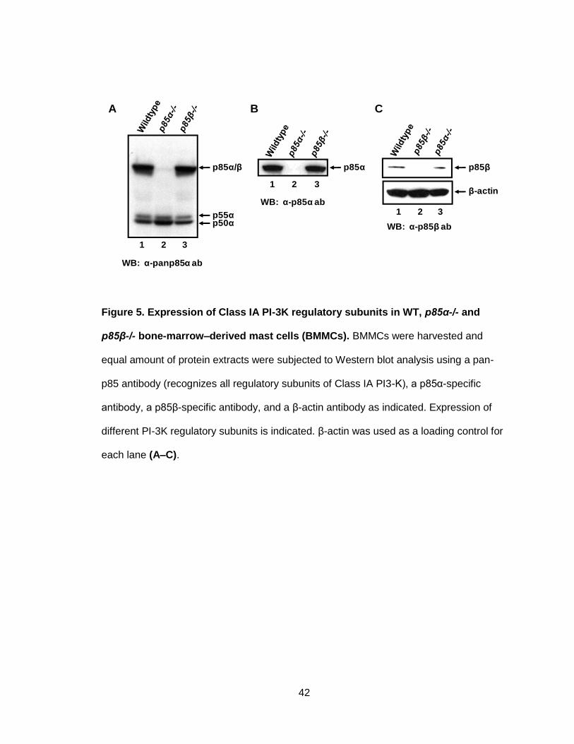
42
Figure 5. Expression of Class IA PI-3K regulatory subunits in WT, p85α-/- and
p85β-/- bone-marrow–derived mast cells (BMMCs). BMMCs were harvested and
equal amount of protein extracts were subjected to Western blot analysis using a pan-
p85 antibody (recognizes all regulatory subunits of Class IA PI3-K), a p85α-specific
antibody, a p85β-specific antibody, and a β-actin antibody as indicated. Expression of
different PI-3K regulatory subunits is indicated. β-actin was used as a loading control for
each lane (A–C).
1 2 3
1 2 3
1 2 3
β-actin
p85βp85αp85α/β
p55αp50α
WB: α-panp85α ab
WB: α-p85α ab
A B C
WB: α-p85β ab

43
II. The p85α regulatory subunit of PI-3K is critical for biological functions of mast
cells
A. Deficiency of p85α in MCps results in defective growth of MCps in response to
SCF stimulation
Various regulatory subunits of Class 1A PI-3K get activated upon binding to the 719
tyrosine residue of c-Kit upon SCF stimulation (Serve, Hsu et al. 1994). While mast cells
express various regulatory subunits of Class 1A PI-3K (Figure 5A), the physiological
roles of the various subunits in regulating c-Kit–induced mast cell growth and
differentiation are not known. Here we sought to evaluate the specific roles of p85α
regulatory subunits in mast cell biology in response to SCF stimulation. To assess the
contributions of p85α to mast cell growth, the differences in proliferation was determined
between mast cell progenitors (MCps) obtained from WT and p85α-/- mice. MCps were
starved of serum and cytokines for 6 h, after which they were cultured in the presence or
absence of SCF for 48 h. These cells were then pulsed with [3H] thymidine for 6 h.
Thymidine incorportation is a direct assessment of the proliferation rate of these cells in
response to SCF stimulation. MCps from WT and p85α-/- mice grown in the absence of
growth factors showed minimal thymidine incorporation. As shown in Figure 6A, WT
MCps demonstrated significant increase in thymidine incorporation in presence of SCF,
whereas deficiency of p85α resulted in complete loss of SCF mediated growth. This
result suggests that the p85α regulatory subunit is critical for SCF-induced mast cell
growth in response to SCF stimulation.
B. Deficiency of p85α results in defective maturation of MCps
We assessed how the loss of p85α alters the maturation of MCps in response to IL-3.
Earlier studies had demonstrated that media supplemented with the cytokine IL-3

44
facilitates the maturation of murine bone marrow cells to mast cells in vitro (Razin, Ihle et
al. 1984). Low density bone marrow cells from WT and p85α-/- mice cells were harvested
and cultured in media supplemented with IL-3 (10 ng/ml). Maturation of these cells to
mast cells was evaluated based on mast cell surface marker (c-Kit and IgE receptor)
expression. Mast cell maturation was analyzed on weekly basis by staining MCps with
antibodies to detect the expression of c-Kit and IgE receptors by flow cytometry. Figure
6B represents a dot blot profile of mast cell maturation at different points during
development. While 91% of WT MCps fully matured into c-Kit and IgE receptor double-
positive cells, p85α deficiency significantly reduced the maturation of MCps: only 20% of
those cells matured into a mast cell population at the end of 4 weeks. The results
suggest that p85α plays a crucial role in mast cell maturation as well.
C. Deficiency of p85α in MCps results in defective survival of MCps in response to
SCF stimulation
Significantly reduced proliferation in p85α-/- MCps might be due to an altered survival
potential of these cells in the specific absence of p85α. Here we evaluated the role of the
p85α regulatory subunit in mediating mast cell survival after SCF stimulation. In vitro
cultures of low-density bone marrow cells harvested from WT and p85α-/- mice were
starved for 6 h and grown in the presence or absence of SCF. After 48 h, apoptosis was
assessed by staining the cells with antibodies to Annexin V and 7-AAD, followed by flow
cytometry analysis. As seen in Figure 6C, less than 10% of MCps from WT and p85α-/-
mice survived for 48 h in the absence of any growth factors, as determined by the
presence of cells negative for Annexin V and 7-AAD. Although WT MCps showed
increased survival in the presence of SCF (50%), there was a significant reduction in the
survival of p85α-deficient MCps (19%). These results suggest that p85α acts as a
positive regulator of survival.
45
We then examined the cell cycle progression in WT and p85α-/- MCps in response to
SCF stimulation. Starved and SCF-stimulated (50 ng/ml) MCps were labeled with
propidium iodide, and cell cycling was analyzed by flow cytometry. As shown in Figure
6D, SCF stimulation enhanced cell cycle progress in WT MCps, whereas cell cycle
progression and S phase entry was significantly impaired in p85α-/- MCps. The result
suggests that p85α plays a critical role in the cell cycle of MCps in response to SCF in
mast cells.

46
Figure 6. The p85α regulatory subunit of PI-3K is critical for biological functions of
mast cells. (A) MCps from WT and p85α-/- cells were starved for 6 h in serum- and
cytokine-free media and cultured in the presence or absence of SCF (50 ng/ml). After 48
h, proliferation was evaluated by [3H] thymidine incorporation. The histograms represent
the mean [3H] thymidine incorporation in MCps (CPM ± SD), with the results from one
representative experiment performed in quadruplicate. Similar results were observed in
six independent experiments. *p<0.01, WT vs. p85α-/-. (B) LDBMCs from WT and p85α-
/- mice were cultured in the presence of IL-3 (10 ng/ml) for total of 4 weeks. At indicated
time points, maturation was analyzed by staining the cells with antibodies that recognize
WT
p85α-/-
0
4000
8000
12000
16000
20000
No GF SCF
Th
ym
idin
e in
co
rpo
rati
on
(CP
M)
*
A
Week 1
WT
p85α-/-
c-Kit
IgE
recep
tor
Week 2 Week 3 Week 4
1.5%
0.3%
27% 78% 91%
0.8% 5% 20%
B
C
p85α-/-WT
0
15
30
45
60
75
No GF SCF
% S
urv
ival
*
D

47
c-Kit and IgE receptor followed by flow cytometry. Shown is dot blot profile from one of
four independent experiments each resulting in the same conclusion (C) MCps from WT
and p85α-/- cells were starved for 6 h in serum- and cytokine-free media and cultured in
the presence or absence of SCF (50 ng/ml). After 48 h, cells were stained with
phycoerythrin-conjugated Annexin V and 7-AAD, followed by flow cytometry analysis. A
representative dot blot showing the percent survival of MCps as determined by the lack
of staining of cells by either Annexin V and/or 7-AAD (i.e., lower-left coordinate). (D)
MCps from WT and p85α-/- cells were starved for 6 h in serum- and cytokine-free media
and cultured in the presence or absence of SCF (50 ng/ml).Cells were stained with
propidium iodide and analyzed by flow cytometry. The percentage of cells in the S phase
of the cell cycle is shown. Similar results were observed in three independent
experiments; *p<0.05.
Although previously it was thought that all regulatory subunits of PI-3K functioned the
same way, MCps lacking p85α regulatory subunit show severe defects in mast cell
biological functions, irrespective of the presence of p85β, p50α and p55α (Figure 5).
p85α and p85β, regulatory subunits, although encoded by two different genes, they
share 100% protein domain homology and 60% amino acid identity (Figure 18).
Noticably as seen in Figure 5, p85α is the most abundant PI-3K regulatory subunit
expressed in MCps, and p85β is expressed in significantly lesser amounts.

48
III. p85β does compensate for the loss of p85α regulatory subunit in mast cell
To evaluated whether the defective mast cell phenotypes in p85α-/- MCps, as well as;
the inability of p85β to compensate for the loss of p85α is due to the quantitative
reduction in overall expression of PI-3K regulatory subunits, we performed restoration
experiments. For this, we cloned full length HA tagged p85α or p85β cDNA into
EcoRI/XhoI site of EGFP expressing MIEG3 retroviral vector. Expression of HA tagged
p85α or p85β regulatory subunit was restored in MCps by infecting these cells with
retroviral supernatants generated from various retroviral vector plasmid. Infected cells
were sorted based on EGFP expression, and were used for the experiments.
.
As seen in Figure 7A, restoring the expression of p85α in the p85α-deficient cells
completely restored the loss of growth induced by SCF. By comparison, over-expression
of p85β in p85α-deficient cells did not rescue SCF induced growth. Moreover, over-
expression of p85β in both WT and p85α-deficient MCps significantly reduced SCF-
induced proliferation. These results further demonstrate that p85α and p85β subunits
play unique roles in both the growth and differentiation of mast cells.
A significant reduction of surface c-Kit expression was also observed in WT and p85α-/-
MCps over expressing p85β regulatory subunit (Figure 7B). Restoring the expression of
p85α in p85α-deficient cells completely corrected the defective maturation of these cells.
In contrast, over-expression of p85β in p85α-deficient MCps did not correct the defective
maturation Furthermore, over-expression of p85β in WT and p85α-deficient MCps
significantly reduced maturation, as indicated by significantly reduced c-Kit and IgE
receptor double-positive cells (Figure 7C), as compared to controls (WT and p85α-/-
MCps transduced with empty vector).

49
Figure 7. Over-expression of p85β in MCps results in reduced growth and
differentiation. WT and p85α-/- MCps transduced with vector, p85α or p85β were
sorted to homogeneity and cultured in the presence of IL-3 (10 ng/ml). (A) Cells were
starved for 6 hours in serum- and cytokine-free media and cultured in the presence or
absence of SCF (50 ng/ml). After 48 hours, proliferation was evaluated by a [3H]
thymidine incorporation assay. Bars represent the mean [3H] thymidine incorporation in
BMMCs (CPM + SD) from one representative experiment performed in quadruplicate.
Similar results were observed in three to four additional independent experiments.
1%
22%
94%
2%
31%
84%
0.3%
3%
64%
0.3%
7%
3%
2%
44%
97%
0.4%
7%
14%
c-Kit
IgE
recep
tor
Week 1
Week 2
Week 3
vector p85α p85β vector p85α p85β
p85α-/-WT
A B
C
0
5000
10000
15000
20000
25000
30000
No GF SCF
Th
ym
idin
e in
co
rpo
rati
on
(CP
M)
WT-vector
p85α-/- -vectorp85α-/- -p85α
WT-p85β
p85α-/- -p85β
*
* *
0
5000
10000
15000
20000
25000
30000 wt/vectorp85α-/- /vector
p85α-/- /p85α
p85α-/- /p85β
wt/p85β
*
*
No GF SCF
Pro
life
rati
on
(c
pm
)

50
*p<0.01, WT-vector vs. WT-p85β, WT-vector vs. p85α-/--vector, WT-vector vs. p85α-/--
p85β. Over-expression of p85β in WT MCps results in reduced c-Kit expression (B) and
differentiation (C). WT and p85α-/- MCps transduced with vector, p85α or p85β were
sorted to homogeneity and cultured in the presence of IL-3 (10 ng/ml). Cells were
harvested at indicated time points and maturation was analyzed by staining the cells with
antibodies to detect the expression of c-Kit and IgE receptor by flow cytometry. Shown is
a dot blot profile of an independent experiment. Similar findings were observed in two to
four additional independent experiments. (Results observed in thesis and Figure 7C
polished by Raghuveer Mali)

51
We wished to further examine c-Kit expression and proliferation in response to SCF
stimulation in 32D myeloid cell lines. This cell line was co-transfected with c-Kit and
p85α or p85β to determine the levels of c-Kit expression and proliferation upon over-
expression of either of those two subunits. As shown in Figure 8, 32D cells that over-
express p85β showed reduced c-Kit expression and lowered proliferation in response to
SCF stimulation, result that is consistent to that seen in experiments with MCps.
Figure 8. The p85β regulatory subunit of PI-3K binds to c-Kit and is activated upon
SCF stimulation. 32D cells were co-infected with c-Kit and p85α or p85β regulatory
subunits. (A) After sorting for c-Kit and EGFP double-positive cells, these cells were
subjected to a proliferation assay to evaluate how these cells responded to SCF
stimulation. 32D cells transduced with c-Kit and p85β proliferate significantly less than
p85α-transduced 32D cells. (B) Over-expression of p85β results in reduced surface c-Kit
expression in 32D cells.
0
2000
4000
6000
8000
10000
12000
14000
16000
No GF SCF
kit p85α
kit p85β
*
A B
32D/Wt c-Kit/ p85α 32D/Wt c-Kit/ p85β
c-Kit
EG
FP
81% 39%
52
IV. Differential roles of p85α and p85β regulatory subunits in mast cell biology
Given the fact that p85β cannot compensate for the loss of p85α in mast cell biological
functions, we then evaluated differential roles of p85α and p85β regulatory subunits in
mast cell growth, survival and maturation.
A. p85α and p85β differentially regulate mast cell growth in response to SCF
stimulation
To compare the roles of p85α and p85β in mast cell growth, cell proliferation was
assessed in mast cell progenitors (MCps) from WT, p85α-/- and p85β-/- mice. MCps from
WT and knockout mice were starved for serum and cytokines for 6 h, after which they
were cultured in the presence or absence of SCF for 48 h. Cells were then pulsed with
[3H] thymidine for 6 h. Thymidine incorportation was measured, which is a direct
indicator of these cells’ proliferation rate in response to SCF stimulation. MCps from WT,
p85α-/- and p85β-/- mice grown in the absence of growth factors showed minimal
thymidine incorporation (Figure 9). In contrast to loss of p85α regulatory subunits, which
significantly reduced mast cell proliferation in response to SCF stimulation (Figure 9A),
loss of p85β significantly enhanced mast cell growth compared to WT controls in
response to SCF (Figure 9B). These results suggest that p85α and p85β regulatory
subunits of PI-3K differentially regulate mast cell growth in response to SCF stimulation.

53
Figure 9. p85α and p85β regulatory subunits differentially regulate mast cell
growth in response to SCF stimulation. MCps from WT, p85α-/- and p85β-/- mice
were starved for 6 hours in serum- and cytokine-free media and cultured in the presence
or absence of SCF (50 ng/ml). After 48 hours, proliferation was evaluated by [3H]
thymidine incorporation. Bars represent the mean [3H] thymidine incorporation in MCps
(CPM + SD) from one representative experiment performed in quadruplicate. Similar
results were observed in six independent experiments. *p<0.01, WT vs. p85α-/-
.**p<0.01, WT vs. p85β-/-
WT
p85α-/-
p85β-/-
0
4000
8000
12000
16000
20000
No GF SCF
Th
ym
idin
e in
co
rpo
rati
on
(CP
M)
**
*

54
B. p85α and p85β differentially regulate mast cell survival in response to SCF
stimulation
We next evaluated the roles of p85α and p85β regulatory subunits in mediating mast cell
survival in response to SCF stimulation. In vitro cultures of low-density bone marrow
cells harvested from WT, p85α-/- and p85β-/- mice were starved for 6 h and then grown in
the presence or absence of SCF. After 48 h, apoptosis was assessed by staining the
cells with antibodies to Annexin V and 7-AAD followed by flow cytometry analysis. As
seen in Figure 10, only 1–2% of MCps from WT, p85α-/- and p85β-/- mice survived in the
absence of any growth factors for 48 h as determined by the presence of Annexin V–
and 7-AAD–negative cells. Although WT MCps showed increased survival in the
presence of SCF (60%), the deficiency of p85α significantly reduced the survival of
MCps (6%). In contrast, p85β-deficient MCps demonstrated significantly enhanced
survival in the response to SCF stimulation (69%) compared to WT controls (Figure 10).
The results suggest differential roles of p85α and p85β in mast cell survival.
We then examined the cell cycle progression in WT, p85α-/- and p85β-/- MCps in
response to SCF stimulation. Starved and SCF stimulated (50 ng/ml) MCps were labeled
with propidium iodide and cell cycling was analyzed by flow cytometry. In contrast to
p85α deficiency, lack of p85β regulatory subunit did not affect the cell cycle, as shown
by significant progression of p85β-deficient MCps to S phase in response to SCF
stimulation (Figure 11).
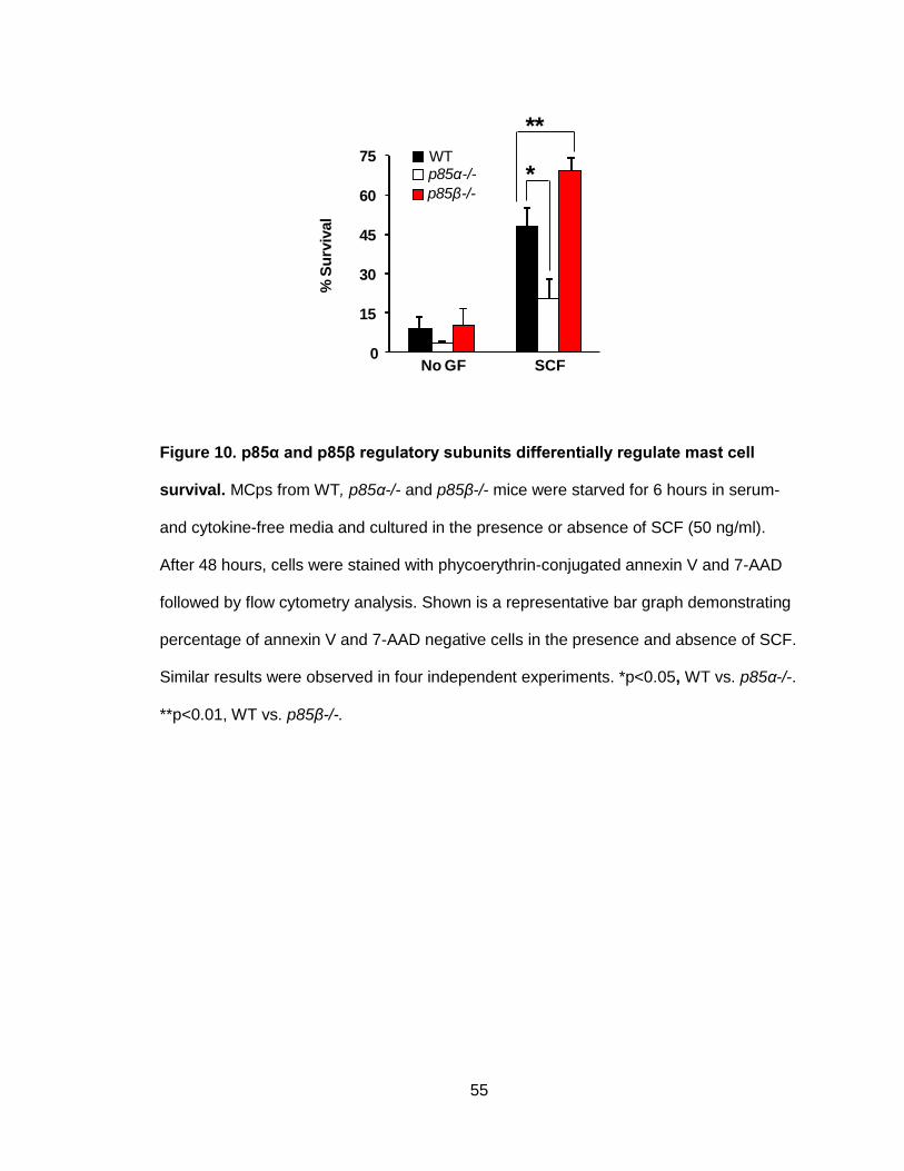
55
Figure 10. p85α and p85β regulatory subunits differentially regulate mast cell
survival. MCps from WT, p85α-/- and p85β-/- mice were starved for 6 hours in serum-
and cytokine-free media and cultured in the presence or absence of SCF (50 ng/ml).
After 48 hours, cells were stained with phycoerythrin-conjugated annexin V and 7-AAD
followed by flow cytometry analysis. Shown is a representative bar graph demonstrating
percentage of annexin V and 7-AAD negative cells in the presence and absence of SCF.
Similar results were observed in four independent experiments. *p<0.05, WT vs. p85α-/-.
**p<0.01, WT vs. p85β-/-.
p85α-/-
p85β-/-
WT
0
15
30
45
60
75
No GF SCF
% S
urv
ival
*
**

56
Figure 11. p85α and p85β regulatory subunits differentially regulate the cell cycle
of mast cells’. WT, p85α-/- and p85β-/- MCps were starved for 6 h and equal numbers of
cells were plated into individual well of a 24-well plate. Cells were either cultured in
cytokine-free culture medium (control) or in medium supplemented with SCF (100 ng/ml)
for 48 h. Cells were stained with propidium iodide, and then analyzed by flow cytometry.
The percentage of cells in S phase of the cell cycle is shown.
20.03% 5.85% 21.04%
Wt p85α-/- p85β-/-
No GF
SCF
0% 0% 0%
57
C. p85α and p85β differentially regulate maturation of MCps
While our aforementioned studies clearly demonstrate a functional dichotomy in cell
cycle progression between the two regulatory subunits in response to SCF, we wanted
to assess how the loss of p85α and p85β alters maturation in response to IL-3. Low-
density bone marrow cells from WT, p85α-/- and p85β-/- mice cells were harvested and
cultured in media supplemented with IL-3 (10 ng/ml). Maturation into mast cells was
evaluated based on expression of two mast cell surface markers: c-Kit and the IgE
receptor. Mast cell maturation was analyzed on a weekly basis by staining MCps with
antibodies to detect the expression of c-Kit and IgE receptors by flow cytometry. Figure
12 represents a dot blot profile for the maturation of these mast cells. While 91% of WT
MCps fully matured into c-Kit and IgE receptor double-positive cells, after four weeks,
only 20% of all p85α-deficient MCps had matured. By contrast, a p85β deficiency did not
impair maturation to mast cells, with 97% maturation to mast cells by the end of 4
weeks. These results demonstrate that p85α and p85β differentially regulate the
maturation of mast cells.
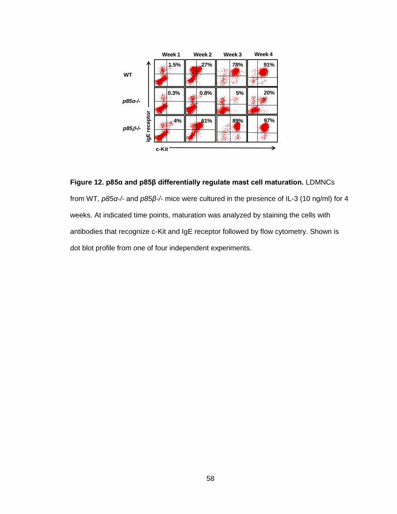
58
Figure 12. p85α and p85β differentially regulate mast cell maturation. LDMNCs
from WT, p85α-/- and p85β-/- mice were cultured in the presence of IL-3 (10 ng/ml) for 4
weeks. At indicated time points, maturation was analyzed by staining the cells with
antibodies that recognize c-Kit and IgE receptor followed by flow cytometry. Shown is
dot blot profile from one of four independent experiments.
Week 1
WT
p85α-/-
p85 -/-
c-KitIg
E r
ecep
tor
Week 2 Week 3 Week 4
1.5%
0.3%
4%
27% 78% 91%
0.8% 5% 20%
61% 89% 97%

59
D. The p85β regulatory subunit of PI-3K binds to c-Kit and becomes activated
upon SCF stimulation
To assess the molecular basis for differential roles of p58α or p85β regulatory subunits,
we first examined whether the regulatory subunits bind to c-Kit upon stimulation with
ligand SCF. The 32D cells that expressed c-Kit and p58α or p85β, were deprived of
serum and growth factors for 12 h. These cells were stimulated with 100 ng/ml of
recombinant rat SCF for 5 min, followed by lysis and immunoprecipitation using an anti-
c-Kit antibody. The immunoprecipitated proteins were then subjected to Western blot
analysis using anti-HA antibody. As seen in Figure 13, when c-Kit is activated by its
ligand SCF both p85α and p85β binds equally to c-Kit. We next examined whether these
HA-tagged regulatory subunits are activated to the same extend upon binding to
c-Kit. To address this question, we performed immunoprecipitations using the same
lysates and anti-HA antibody, and the immunocomplexes were subjected to Western blot
analyses using anti phospho-tyrosine antibody. As shown in Figure 13, both p85α and
p85β that were bound equally to c-Kit were activated upon SCF stimulation.

60
Figure 13. The p85β regulatory subunit of PI-3K binds to c-Kit and is activated
upon SCF stimulation. 32D cells expressing WT c-Kit and p85α or p85β were starved
for 8 hours and stimulated with SCF (100 ng/ml) for 5 min. Equal amount of cell lysates
(500 µg) were subjected to immunoprecipitation with an anti-c-Kit antibody followed by
western blot analysis with an anti-HA antibody or immunoprecipitated with an anti-HA
antibody followed by western blot analysis with an anti-c-Kit antibody or anti-phospho-
tyrosine antibody.
IP:c-Kit
WB: HA
0 5 0 5
p85α p85β
SCF:
IP:HA
WB: p-Tyr
IP:HA
WB: c-Kit

61
E. p85α and p85β differentially regulate c-Kit receptor-mediated signaling events
in MCps
The signaling molecules AKT, ERK and JNK MAP kinase play critical roles in c-Kit-
mediated proliferation and survival of mast cells (Rottapel, Reedijk et al. 1991; Duronio,
Welham et al. 1992; Chian, Young et al. 2001). In an effort to determine the mechanism
behind differential regulation of mast cell growth and survival by p85α and p85β, we
measured the phosphorylation of these regulatory subunits in WT, p85α-/- and p85β-/-
MCps in response to SCF. As seen in Figure 14, p85α-deficient MCps showed
significantly reduced activation of AKT, ERK and JNK MAP kinase in response to SCF
compared to WT controls. In contrast, enhanced activation of ERK and JNK MAP
kinase—but not AKT—was observed in p85β-deficient MCps compared to WT controls
in response to SCF stimulation. These results suggest that differential regulation of mast
cell growth and survival by p85α and p85β subunits are due to differential activation of
ERK and JNK MAP kinase downstream from c-Kit.
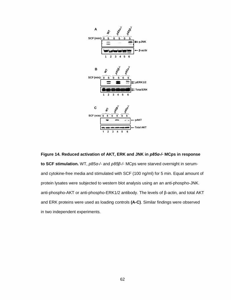
62
Figure 14. Reduced activation of AKT, ERK and JNK in p85α-/- MCps in response
to SCF stimulation. WT, p85α-/- and p85β-/- MCps were starved overnight in serum-
and cytokine-free media and stimulated with SCF (100 ng/ml) for 5 min. Equal amount of
protein lysates were subjected to western blot analysis using an an anti-phospho-JNK.
anti-phospho-AKT or anti-phospho-ERK1/2 antibody. The levels of β-actin, and total AKT
and ERK proteins were used as loading controls (A-C). Similar findings were observed
in two independent experiments.
0 5 0 5 0 5
1 2 3 4 5 6
β-actin
A
pJNK
SCF (min):
0 5 0 5 0 5
1 2 3 4 5 6
B
pERK1/2
Total ERK1/2
SCF (min):

63
V. p85β negatively regulates c-Kit receptor signaling by binding to phophorylated
E3 ubiquitin ligase, c-Cbl
A. p85β negatively regulates c-Kit receptor internalization and degradation
Activation of c-Kit receptor by its ligand SCF is followed by its internalization and
degradation (Levkowitz, Waterman et al. 1998; Lee, Wang et al. 1999; Miyake, Mullane-
Robinson et al. 1999; Taher, Tjin et al. 2002). To determine the mechanism behind
increased activation of signaling events downstream from the c-Kit receptor in p85β-
deficient cells, we examined the rate of internalization of the c-Kit receptor in response to
SCF in p85β-deficient MCps and also in murine myeloid 32D cells co-infected with c-Kit
and p85α or p85β. As seen in Figure 15, a deficiency of p85β in MCps inhibited the rate
of SCF-induced c-Kit receptor internalization compared to WT controls at all time points
examined.
We next performed studies to evaluate the levels of c-Kit receptor degradation in 32D
cells co-expressing c-Kit and p85α or p85β in response to SCF. After SCF stimulation,
there was significantly enhanced c-Kit receptor internalization and degradation in p85β
over-expressing cells compared to p85α over-expressing cells (Figures 16 and 17A).
These results were observed despite similar binding affinities of p85α and p85β to the c-
Kit receptor in response to SCF stimulation. Taken together, these results suggest that
p85β negatively regulates c-Kit receptor signaling in part by regulating its rate of
internalization and degradation.

64
Figure 15. Reduced c-Kit receptor internalization in p85β-deficient MCps
compared to WT in response to SCF. WT or p85β-/- MCps were starved overnight
and incubated with cyclohexamide (100 ng/ml) for 2 hours. After cyclohexamide
treatment, cells were stimulated with SCF (100 ng/ml) for indicated time points and c-Kit
receptor internalization was studied by staining the cells with PE-conjugated anti-c-Kit
receptor antibody followed by flow cytometric analysis. Shown is one of four independent
experiments performed in triplicate. *p<0.01, WT vs. p85β-/-.
0
25
50
75
100
125
0 10 20 30 45 60
% c
-Kit
exp
ressio
n
WTp85β-/-
Time (min)
* ** *

65
Figure 16. Enhanced. c-Kit receptor internalization in 32D cells over-expressing
p85β as compared to p85α subunit upon SCF stimulation. 32D cells co-infected with
c-Kit and p85α or p85β were starved for 8 hours and incubated with cyclohexamide (100
ng/ml) for 2 hours. After cyclohexamide treatment, cells were stimulated with SCF (100
ng/ml) for indicated time points and c-Kit receptor internalization was studied by staining
the cells with PE-conjugated anti-c-Kit receptor antibody followed by flow cytometric
analysis. Shown is one of six independent experiments performed in triplicate. *p<0.01,
p85α vs. p85β.
0
20
40
60
80
100
120
0 10 20 30 60
% c
-Kit
exp
ressio
n p85α
p85β
Time (min)
**
**
66
B. Cells over-expressing p85β demonstrate enhanced activation of E3 ubiquitin
ligase c-Cbl, compared to cells over-expressing p85α
Previous studies have shown that c-Kit internalization and degradation is regulated
largely by an E3 ubiquitin ligase c-Cbl (Levkowitz, Waterman et al. 1998; Lee, Wang et
al. 1999; Miyake, Mullane-Robinson et al. 1999; Taher, Tjin et al. 2002). We
hypothesized that p85α and p85β may differentially regulate the activation and binding of
c-Cbl in response to c-Kit activation. To test this, we first studied the activation of c-Cbl
in 32D cells over-expressing p85α or p85β. Significantly enhanced phosphorylation of c-
Cbl was found in 32D cells over-expressing p85β as compared to 32D cells over-
expressing p85α (Figure 19B).
C. p85β regulatory subunit of PI-3K shows enhanced binding to E3 ubiquitin
ligase, c-Cbl, as compared to p85α regulatory subunit of PI-3K
We next performed immunoprecipitation experiments in 32D cells expressing an HA-
tagged version of p85α or p85β. These transfected cells were stimulated with SCF,
followed by Western blot analysis using an anti-phospho-c-Cbl antibody. Significantly
enhanced binding of phospho-c-Cbl to p85β was observed compared to p85α in
response to SCF stimulation (Figure 17C).
D. 32D cells over-expressing p85β show enhanced c-Kit ubiquitination upon SCF
stimulation as compared to those over-expressing p85α
We then examined ubiquitination of the c-Kit receptor in 32D cells over-expressing p85α
or p85β. The 32D cells were starved of serum and growth factors, and then were treated
with cycloheximide, followed by SCF for upto 30 minutes at 37°C. Cell lysates were
incubated with anti-c-Kit antibody, immunoprecipitated and separated by SDS-PAGE,
and then analyzed by western blotting using anti-ubiquitin antibody. Enhanced

67
ubiquitination of c-Kit bands was detected in response to SCF stimulation in 32D cells
over-expressing p85β. Interestingly, there was enhanced binding of c-Cbl to p85β
coincident with the increased ubiquitination of c-Kit in cells over-expressing p85β. This
complex between c-Cbl to p85β was much reduced in cells over-expressing p85α.
Taken together, these results suggest that p85β negatively regulates c-Kit receptor
signaling in part by regulating its ubiquitination and degradation.

68
Figure 17. Enhanced c-Kit degradation and c-Cbl activation in 32D cells over-
expressing p85β regulatory subunit upon SCF stimulation. 32D cells co-infected
with c-Kit and p85α or p85β were starved and incubated with cyclohexamide (100
ng/ml), followed by stimulation with SCF. c-Kit expression in response to SCF
stimulation was evaluated by performing Western blot analysis using anti-c-Kit antibody
(A) and anti-phospho-c-Cbl antibody (B). (C) 32D cells co-infected with c-Kit and p85α or
p85β were starved for 8 h and stimulated with SCF (100 ng/ml) for 5 min. Equal amounts
of cell lysates (500 µg) were immunoprecipitated with an anti-HA antibody, followed by
Western blotting with a phospho-c-Cbl antibody. p85β preferentially binds to phospho c-
Cbl compared to p85α in response to SCF stimulation. (D) 32D cells co-infected with c-
0 5 0 5
1 2 3 4
p-c-cbl
IP: α-HA ab
WB: α-p-c-cbl ab
SCF (min):
B
0 5 15 30 0 5 15 30
p85α p85β
1 2 3 4 5 6 7 8
β-actin
p-c-cbl
SCF (min):
1 2 3 4 5 6 7 8
0 5 15 30 0 5 15 30
p85α p85β
β-actin
KIT
SCF (min):
A
1 2 3 4
IP: α-KIT ab
WB: α-Ub ab
0 30
p85αp85β
SCF (min):
KIT
60 0 30 60
5 6
C D

69
Kit and p85α or p85β regulatory subunits were starved and incubated with
cyclohexamide, followed by stimulation with SCF. To evaluate c-Kit ubiquitination in
response to SCF treatment, equal amounts of cell lysate were subjected to
immunoprecipitaion using c-Kit antibody, followed by Western blotting using an anti-
ubiquitin antibody. Shown is representative data from one of three independent
experiments.
Our study implies functional uniqueness for PI-3K regulatory subunits in mast cell
development. p85α deficiency impairs growth, maturation and survival of mast cells;
whereas p85β deficiency enhances c-Kit signaling inpart by inhibiting c-Kit
internalization, ubiquitination and degradation. Although p85α and p85β share 100%
protein domain homology, aminoacid comparison shows wide disparity in aminoacid
composition of SH3 and BH domains (Figure 18). Interestingly, p85α-/- MCps show
severe phenotypic defects, irrespective of the presence of shorter regulatory subunits
p50α and p55α. p50α and p55α are splice variants of p85α, but lack amino terminal
domains of SH3 and BH. Based on these facts, we hypothesized that the functional
uniqueness of p85α regulatory subunit might be in part imparted by its amino terminal
domains (SH3 and BH).

70
Figure 18. Sequence comparison between p85α and p85β regulatory subunits of
PI-3K. The sequences of the p85α and p85β regulatory subunits were compared using
the UniProt Multiple Sequence Alignment tool. Shaded areas represent identical amino
acid residues shared between the p85α and p85β. p85α and p85β regulatory subunits
share domain homology from the proteins’ amino to carboxy terminus. The
corresponding domain positions are enclosed in boxes: SH3 domain in blue, proline-rich
regions in red, BH domain in green, amino-SH2 in purple, and carboxy-SH2 in yellow. As
represented, the two regulatory subunits share lesser identity of amino acids at their
amino terminal domains of SH3 and BH. The carboxy terminal portion of the regulatory
subunits, which includes the amino-SH2, inter-SH2, and carboxy-SH2 regions, shares
more sequence identity.

71
VI. Cooperation between the SH3 and BH domains of p85α is required for mast cell
growth, but not maturation
To evaluate importance of SH3 and BH domains of p85α in mast cell functions, we used
two approaches (i) First, the collective contributions of the SH3 and BH domains of p85α
to mast cell biology were addressed by reconstituting and over-expressing p50α in p85α-
/- MCps. (ii) The second approach to analyze the individual contributions of the SH3 and
BH domains of p85α to mast cell functions involved expressing of different p85α mutants
in p85α-/- MCps that lack either SH3 or BH domain.
A. Over-expression of the p50α regulatory subunit in p85α-/- MCps corrects
maturation; and partially corrects growth in response to SCF stimulation
To assess the extent to which the shorter isoforms of p50α contribute to mast cell
maturation and proliferation, we cloned an HA-tagged p50α gene into a MIEG3
bicistronic retroviral vector that expresses EGFP via an internal ribosomal entry site.
Viral supernatants were generated and were used to infect WT and p85α-/- MCps.
Expression of HA-tagged p85α and p50α constructs were confirmed in EGFP-sorted
p85α-/- cells (Figure 19A). As expected, restoration of full-length p85α completely
corrected the observed defect in mast cell maturation associated with p85α-deficient
cells (Figure 19B). Over-expression of p50α in p85α-deficient cells also completely
corrected the defective mast cell maturation to WT levels in p85α-/- MCps. As seen in
Figure 19C the expression of full length p85α in p85α-/- MCps restores the SCF-induced
growth to WT levels. In contrast, there was only 50% rescue in SCF-induced proliferation
in p85α-deficient cells over-expressing the p50α subunit. These results suggest that the
shorter p50α do contributes to mast cell differentiation and proliferation. However, the
full-length form of p85α is indispensable for mast cell functions. These results suggest

72
that the amino terminal domains of SH3 and BH collectively play an important role in
mediating SCF-induced growth in MCps.
Figure 19.Over-expression of the PI-3kinase regulatory subunit of p50α restores
maturation and partially restores growth in response to SCF in p85α-/-MCps. (A)
Expression of p50α in p85α-/- MCps. MCps from WT and p85α-/- mice were transduced
with vector, HA tagged full length p85α or p50α and sorted to homogeneity. Expression
of p85α and p50α in sorted cells was analyzed by Western blotting using anti-HA and β-
actin antibodies. (B) Restoration of p50α corrects the defective maturation of p85α-
deficient BMMCs. BMMCs in (d) were collected at indicated time points. Mast cell
differentiation was analyzed by staining the cells with antibodies that recognize c-Kit and
IgE receptor followed by flow cytometric analysis. Shown is a representative dot blot
profile from five independent experiments. *p<0.05, WT vs. p85α-/-vector. (C) Over-
expression of p50α partially correct the defective growth of p85α-/- MCps. WT and
β-actin
p50α
p85α
1 2 3 4
Week 1 Week 2 Week 3
c-Kit
IgE
recep
tor
WT-vector
1% 20% 90%
p85α-/- -vector
0.4% 6% 3%
p85α-/- -p85α
1.3% 43% 97%
p85α-/- -p50α
2.5% 37% 97%
0
2000
4000
6000
8000
10000
12000
No GF SCF
Th
ym
idin
e in
co
rpo
rati
on
(CP
M)
*
A B
C

73
p85α-/- MCps expressing the indicated constructs were starved of growth factors and
stimulated with SCF. After 48 hours, proliferation was evaluated by [3H] thymidine
incorporation. Bars represent the mean [3H] thymidine incorporation (CPM ± SD). Shown
is data from one of many independent experiments. *p<0.05, p85α-/- -p85α vs. p85α-/- -
p50α. (Result observed in this thesis and Figure 19B polished by Raghuveer Mali)
We then evaluated the importance of the individual contributions of p85α’s SH3 and BH
domains to mast cell function.
B. Amino terminal mutants of p85α (p85αΔSH3 and p85αΔBH) rescue maturation
and Mitf expression in p85α-/- MCps
HA-tagged cDNA encoding p85α, p85αΔSH3 or p85αΔBH were cloned into a MIEG3
retrovirus (Figure 20A) and p85α-/- bone marrow cells were transduced with viral
supernatants expressing the indicated variants of the p85α regulatory subunit.These
cells were sorted to homogeneity based on EGFP expression. Expression of various HA-
tagged constructs in MCps were confirmed by Western blot analysis using an anti-HA
antibody (Figure 20B), and were used for studies described here. As seen in Figure 20C,
expression of p85α mutants p85αΔSH3 and p85αΔBH in the p85α-deficient cells
completely corrected the defect in mast cell maturation, as indicated by restoration of c-
Kit and IgE receptor double positive population. These results further confirm that the
amino terminal domains of p85α are not essential for mast cell maturation.
Maturation of mast cells is driven by a complex network of transcription and growth
factors. Microphtalmia-associated transcription factor (Mitf) is known to be a key
regulator of c-Kit expression and maturation of mast cells (Kasugai, Oguri et al. 1993;
Isozaki, Tsujimura et al. 1994; Kitamura, Morii et al. 2002). To determine how p85α and

74
p85α mutants p85αΔSH3 or p85αΔBH restore mast cell maturation in p85α-/- MCps, we
analyzed the expression of Mitf in p85α-deficient cells by transducing them with the full-
length form of p85α or one of the mutants p85αΔSH3 or p85αΔBH. As seen in Figure
20D, the p85α-deficient cells showed significant reduction in expression of Mitf
compared to WT controls cells. Expression of p85α or one of the p85α mutants (either
p85αΔSH3 or p85αΔBH) in p85α-deficient cells restored Mitf expression. These results
suggest that p85α regulates the expression of Mitf, which, in turn, controls mast cell
maturation.

75
Figure 20. Amino terminal mutants of p85α (p85αΔSH3 and p85αΔBH) rescue
maturation and Mitf expression in p85α-/- MCps. (A) Schematic of full length p85α
and p85α mutants lacking either the SH3 or the BH domain. Full length p85α or p85α
mutants lacking either the amino terminal SH3 domain (1-80 a.a) or the BH domain
(102-288 a.a) were cloned into a bi-cistronic retroviral vector MIEG3. The constructs
were HA tagged at the amino terminus to distinguish exogenous expression from
endogenous p85 protein. (B) Expression of full length p85α or p85α mutants. MCps from
p85α-/- mice were transduced with vector, full length p85α or p85α mutants lacking
either the SH3 domain (p85αΔSH3) or the BH domain (p85αΔBH) and sorted to
homogeneity. Cells were harvested and subjected to Western blot analysis using an
anti-HA antibody or β-actin antibody as indicated. Expression of various p85α mutants is
indicated in the upper panel. (C) Expression of p85α mutants into p85α-/- MCps corrects
p85α∆SH3
p85α
p85α∆BH
N -COOHSH3 PR BH PR SH2 iSH2 SH2-
N -COOHSH3 PR BH PR SH2 iSH2 SH2-
N -COOHSH3 PR BH PR SH2 iSH2 SH2-
HA
A
β-actin
p85α mutants
1 2 3
B
Week 1
Week 2
Week 3
c-Kit
IgE
recep
tor
vector p85αΔBHvector p85α p85αΔSH3
WT p85α-/-
0.9%
21%
90%
0.6%
5%
3%
1.5%
42%
97%
1.1%
27%
93%
1.3%
57%
95%
MITF
β-actin
D C

76
defective mast cell differentiation. At indicated time points, maturation was evaluated by
staining the cells with antibodies that recognize c-Kit and IgE receptor by flow cytometry.
Shown is a representative dot blot (C) from five independent experiments. *p<0.05, WT
vs. p85α-/-vector. (D) Sorted cells grown in the presence of IL-3 (10 ng/ml) were
harvested and subjected to Western blot analysis using an anti-Mitf antibody and a β-
actin antibody, as indicated. (Result observed in thesis and Figure 20C polished by
Baskar Ramdas)
C. The amino terminal domains of p85α (p85αΔSH3 and p85αΔBH) are critical for
growth of p85α-/- MCps
Because the growth defects in p85α-/- MCps could not completely restored in spite of the
over expression of p50α which lacks SH3 and BH domains of p85α, we hypothesized
that the amino terminal SH3 and BH domains of p85α must contribute significantly to the
growth of MCps in response to SCF stimulation. To test this idea, we performed
proliferation assays on sorted p85α-/- MCps transduced with full-length and mutant
versions of p85α which lacked either the SH3 or BH domains. As seen in Figure 21A, the
p85α-/- MCps transduced with only the empty vector showed significantly reduced mast
cell growth in response to SCF stimulation. This indicates that the p85α regulatory
subunit is important for mediating the signals downstream of c-Kit to induce mast cell
growth in response to SCF stimulation. Reconstitution of p85α-/- with full-length p85α
cDNA rescued the growth defect to WT levels. Interestingly, the reconstitution of mutant
constructs (p85αΔSH3 or p85αΔBH) could only marginally rescue the mast cell growth,
despite the fact that truncated regulatory subunits completely corrected the maturation
defect and c-Kit levels in p85α-/- MCps (Figure 20C). This demonstrates that the amino
terminal domains (SH3 and BH) work cooperatively to affect mast cell growth in
response to SCF stimulation, and signals mediated via the SH3 and BH domains of

77
p85α are critical for inducing c-Kit mediated growth in mast cells in response to SCF
stimulation.
D. The amino terminal domains of p85α (p85αΔSH3 and p85αΔBH) are critical for
survival of p85α-/- MCps
Although mutant p85α constructs (p85αΔSH3 and p85αΔBH) restored maturation of
p85α-/- MCps to WT levels (Figure 20C), SCF-induced growth could only be partially
restored in these cells (Figure 21A). We wished to determine whether the reduced
proliferation in the absence of p85α SH3 or BH domains is caused by reduced survival
potential of these cells in response to SCF stimulation. We transduced p85α-/- MCps
with empty vector, full-length p85α, p85αΔSH3 or p85αΔBH, and then starved these
cells, followed by treatment with SCF for 48 h. Apoptosis was assessed using Annexin V
and 7-AAD staining. As seen in Figure 21B, MCps lacking p85α failed to survive in
response to SCF stimulation. Expression of full-length p85α completely corrected the
survival defect in response to SCF stimulation of the transduced p85α-/- MCps. This
indicates that p85α plays a critical role in mast cell survival in response to SCF. By
contrast, p85α lacking either the SH3 (p85αΔSH3) or BH (p85αΔBH) domains showed
reduced survival. This result suggests that the amino terminal SH3 and BH domains of
the p85α regulatory subunit are critical for the survival of MCps in response to SCF
stimulation, possibly by mediating some downstream signals.
E. The amino terminal SH3 and BH domains of p85α are important for SCF-
induced AKT, ERK, and JNK1 activation, but not JNK2 activation
We then examined the activation of downstream c-Kit receptor signaling in response to
SCF stimulation when SH3 or BH domains were absent. As seen in Figure 21C,
activation of AKT, ERK, and JNK MAP kinase was restored to WT levels in p85α-/-

78
MCps expressing the full-length p85α; whereas only a parital correction in the the activity
of these molecules was observed in p85α-/- MCps expressing p85αΔSH3 or p85αΔBH.
These results suggest that cooperation between the SH3 and BH amino terminal
domains of full-length p85α is required for full activation of signaling downstream of the
c-Kit receptor.
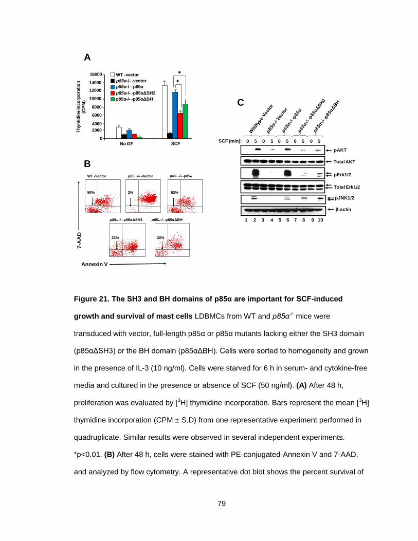
79
Figure 21. The SH3 and BH domains of p85α are important for SCF-induced
growth and survival of mast cells LDBMCs from WT and p85α-/- mice were
transduced with vector, full-length p85α or p85α mutants lacking either the SH3 domain
(p85αΔSH3) or the BH domain (p85αΔBH). Cells were sorted to homogeneity and grown
in the presence of IL-3 (10 ng/ml). Cells were starved for 6 h in serum- and cytokine-free
media and cultured in the presence or absence of SCF (50 ng/ml). (A) After 48 h,
proliferation was evaluated by [3H] thymidine incorporation. Bars represent the mean [3H]
thymidine incorporation (CPM ± S.D) from one representative experiment performed in
quadruplicate. Similar results were observed in several independent experiments.
*p<0.01. (B) After 48 h, cells were stained with PE-conjugated-Annexin V and 7-AAD,
and analyzed by flow cytometry. A representative dot blot shows the percent survival of
p85α-/- -vector
WT -vector
p85α-/- -p85α
p85α-/- -p85αΔSH3
p85α-/- -p85αΔBH
0
2000
4000
6000
8000
10000
12000
14000
16000
No GF SCF
Th
ym
idin
e in
co
rpo
rati
on
(CP
M)
*
*
50% 2%
p85 -/- -VectorWT -Vector p85 -/- -p85
52%
23% 20%
p85 -/- -p85 ΔBHp85 -/- -p85 ΔSH3
Annexin V
7-A
AD
A
B
pJNK1/2
SCF (min): 0 5 0 5 0 5 0 5 0 5
1 2 3 4 5 6 7 8 9 10
β-actin
pAKT
Total AKT
pErk1/2
Total Erk1/2
C

80
BMMCs (lower-left coordinate) as determined by the lack of staining of either Annexin V
and/or 7-AAD. (C) Transduced and sorted cells grown in the presence of IL-3 (10 ng/ml)
were starved overnight in serum- and cytokine-free media and stimulated with SCF (100
ng/ml) for 5 min. Cells were harvested and equal amounts of protein lysates were
subjected to Western blot analysis using an anti-phospho- AKT antibody, anti-phospho-
ERK1/2, or anti-phospho-JNK1/2, as indicated. The amount of total AKT and total ERK
proteins were also measured by western blotting. β-actin was used as a loading control
for the western analysis.
F. p85α mutant constructs bind to Gab1, Gab2, and Rac2 upon SCF stimulation
Although mutant p85α constructs (p85αΔSH3 and p85αΔBH) restored c-Kit expression
and bound to activated c-Kit upon SCF stimulation (Figure 22B), the absence of the SH3
or BH domains of p85α significantly reduced the growth and survival of mast cells, and
significantly reduced activation of AKT, ERK and JNK1 in response to SCF stimulation.
To understand the molecular basis for these signaling events, we used the 32D myeloid
cell line to evaluate full-length and mutant p85α constructs for their interactions with
other proteins of known importance to mast cell functions when stimulated with SCF.
Because 32D cells do not have endogenous c-Kit expression, we co-infected 32D cells
with c-Kit and the WT and mutant p85α constructs. Figure 22A shows the expression of
various constructs in 32D cells as assessed by Western blots, using an anti-HA antibody
and a p85α-specific antibody. Immunoprecipitation experiments were performed using
lysates prepared from the 32D cells expressing HA-tagged versions of p85α, p85αΔSH3
or p85αΔBH. The proteins in the immunocomplexes were then analyzed by Western
blot, using an antibodies recognizing Gab1, Gab2, Grb2 or Rac2. As seen in Figure 23,
both full-length and mutant p85α constructs bound to these molecules upon SCF
stimulation.

81
Figure 22. p85α mutant constructs bind to Gab1, Gab2, and Rac2 upon SCF
stimulation (A) Western blot analyses of lysates prepared from the 32D cells show
expression of p85α expression (top panel), and the various HA-tagged constructs using
anti-HA antibody (middle panel). The levels of protein in each lane were normalized by
the amount of β-actin (bottom panel). 32D cells co-infected with c-Kit and p85 constructs
were starved and stimulated with SCF (100 ng/ml) for 5 min. (B) Equal amounts of cell
lysates were subjected to immunoprecipitation with anti-c-Kit antibody, followed by
Western blot analysis with an anti-HA antibody. Equal amounts of cell lysates were
subjected to immunoprecipitation with anti-HA antibody, followed by Western blot
analysis with (C) anti-Gab1antibody, (D) an anti- Gab2 antibody, (E) an anti-Grb2
antibody or (F) anti-Rac2 antibody.
WB: HA
WB: β-actin
WB:p85α
1 2 3 4
A B C
D E F

82
VII. Deficiency of p85α alters the gene expression profile of cultured mast cells
Our results suggest that the p85α regulatory subunit of PI-3K plays critical role in
maturation, proliferation, survival and cell cycle of mast cells. To explain the underlying
molecular mechanism for the action of p85α, we performed microarray analyses to
compare the gene expression profile of bone-marrow–derived WT- and p85α-deficient
mast cells upon stimulation with SCF. Agilent whole mouse genome oligo microarrays (4
x 44K) were used to profile gene expression using RNA purified from the WT and
p85α-/- MCps.
Figure 23. Methodology followed for microarray analysis. Low-density bone marrow
cells collected from three independent biological samples of WT and p85α-/- mice were
cultured in the presence of IL-3 (10 ng/ml) for 1 week. FACS was used to sort the mast
cells that were double-positive for c-Kit and IgE expression. Double-positive cells were
3 independent
biological samples were sorted and stimulated with SCF for
48 hours
c-K
iT
IgE receptor
WT
1.7%
4.1%

83
then stimulated with 100 ng/ml SCF for 48 h. These samples were then sent for
microarray analysis for gene expression profiling.
The quality of the microarray results was assessed by performing a Principal Component
Analysis (PCA) using Partek software. PCA was applied to the expression data with WT
and p85α-/- genotypes as variables, and the gene expression measurements as
observations. Based on gene expression values, Eigen values and Eigen vectors were
calculated, and spatial distribution of each treatment group (genotype) was plotted on a
3D scatter plot. Statistically, we observed good spatial separation between the samples,
which indicates a low correlation between the two groups of samples (WT and p85α-/-).
Low variance within each group was indicated by close clustering of samples within a
group. The results suggested good-quality microarray output that could be analyzed for
alteration in gene expression. The data were used for further analysis to investigate
underlying molecular mechanism to explain the defective phenotype in p85α-/- MCps in
response to SCF stimulation.
To determine significantly altered genes in p85α-deficient mast cells as compared to
WT, we calculated the p value for gene expression pattern based on Student’s t-test
using Partek software with the following critieria: change in expression of at least 1.5-fold
and p<0.05. This analysis identified 151 unique genes exhibiting altered expression
between WT and p85α-/- cells (Table 1).
To address the potential role of these changes in gene expression in mast cell biology,
we collated these genes into functional categories using the DAVID bioinformatics tool,
and the Ingenuity Pathway Analysis (IPA) software. The DAVID tool relates altered

84
genes due to lack of p85α to transcription, cell cycle, cell adhesion, cell differentiation,
cell survival and signal transduction (Tables 2 and 3).
Most interesting genes from microarray data are - Rrm1 (Ribonucleotide Reductase M1),
which is found to be upregulated in p85α-/- MCps in response to SCF stimulation.
Upregulation of Rrm1 might be partly contributing to significantly reduced PI-3K activity
in p85α-/- MCps upon SCF stimulation, as this gene is report to induce the expression of
PI-3K negative regulator PTEN (Gautam, Li et al. 2003). Taok3 (Tao kinase 3) and Fbln5
(Fibulin5) which has been reported to activate ERK1/ERK2, and is down regulated in
p85α-/- MCps in response to SCF stimulation (Zhang, Chen et al. 2000). These genes
might be partly regulating the ERK activation downstream of p85α; the downregulation of
which results in significantly reduced growth in reponse to SCF stimulation in p85α -/-
MCps. Pim3 (Pim3 kinase) has been reported to positively regulate cell proliferation and
survival via activation of BIM (Macdonald, Campbell et al. 2006). Down regulation of
Pim3 might contribute to significantly increased apoptosis of p85α-deficient cells. These
information could be evaluated in future study in context of explaining the growth and
survival defects in p85α-/- MCps upon SCF stimulation.

85
Gene symbol Gene description Fold Change p-value
4930447A16Rik RIKEN cDNA 4930447A16 gene -7.69 0.02
Cdh8 Cadherin 8 -7.09 0.05
Atpbd4 ATP binding domain 4 -6.37 0.008
1810047C23Rik RIKEN cDNA 1810047C23 gene -6.12 0.03
Qsox1 Quiescin Q6 sulfhydryl oxidase 1 -5.58 0.03
A030006P16Rik Keratin associated protein 17-1 -5.21 0.024
NAP069078-1 NAP069078-1 -4.64 0.001
1700017D01Rik 1700017D01Rik -4.63 0.01
AK047500 AK047500 -4.5 0.007
Laptm4b Lysosomal-associated protein transmembrane 4B -4.13 0.005
9330155M09Rik RIKEN cDNA 9330155M09 gene -3.85 0.02
Txndc5 Thioredoxin domain containing 5 -3.72 0.001
Pim3 Proviral integration site 3 -3.64 0.000904
Tcf20 Transcription factor 20 -3.5 0.01
Taok3 TAO kinase 3 -3.41 0.02
TC1531727 TC1531727 -3.38 0.02
4922502D21Rik RIKEN cDNA 4922502D21 gene -3.33 0.02
Ubl4b Ubiquitin-like 4B -3.32 0.03
Slc1a1 Solute carrier family 1 -3.28 0.05
D330028D13Rik RIKEN cDNA D330028D13 gene -3.15 0.01
1700069L16Rik RIKEN cDNA 1700069L16 gene -3.09 0.02
Uimc1 Ubiquitin interaction motif containing 1 -3.06 0.02
Cbx5 Chromobox homolog 5 -2.95 0.04
Bpgm 2,3-bisphosphoglycerate mutase -2.95 0.03
1200009I06Rik RIKEN cDNA 1200009I06 gene -2.93 0.02
Dus1l Dihydrouridine synthase 1-like -2.86 0.02
A_52_P868798 A_52_P868798 -2.68 0.03
Pofut1 Protein O-fucosyltransferase 1 -2.67 0.04
D830039M14Rik RIKEN cDNA D830039M14 gene -2.58 0.04
BC049715 Hypothetical protein LOC100044248 -2.54 0.02
AB182283 AB182283 -2.48 0.05
2310040A13Rik WD repeat domain 20A -2.44 0.02
Nbn Nibrin -2.41 0.02
Olfr414 Olfactory receptor 414 -2.31 0.03
NAP123291-1 NAP123291-1 -2.28 0.04
ENSMUST00000101328 ENSMUST00000101328 -2.28 0.03
Sema3d Sema domain, immunoglobulin domain -2.26 0.04
Table 1. List of of genes altered in p85α-/- in response to SCF stimulation

86
Gene symbol Gene description FoldChange p-value
1700023L04Rik RIKEN cDNA 1700023L04 gene -2.21 0.04
Slc25a10 Solute carrier family 25,member 10 -2.21 0.03
Arhgef3 Rho guanine nucleotide exchange factor (GEF) 3 -2.19 0.05
1700021K19Rik RIKEN cDNA 1700021K19 gene -2.18 0.03
Dyrk1a Dual-specificity tyrosine-(Y)-phosphorylation -2.13 0.04
regulated kinase 1a
Nsd1 Nuclear receptor-binding SET-domain protein 1 -2.12 0.03
Abca12 ATP-binding cassette, sub-family A (ABC1) -2.11 0.02
Defb41 Defensin beta 41 -2.11 0.02
Deaf1 Deformed epidermal autoregulatory factor 1 -2.1 0.02
D930016D06Rik RIKEN cDNA D930016D06 gene -2.1 0.02
Mbnl1 Muscleblind-like 1 -2.04 0.009
AK087625 AK087625 -2.04 0.04
Stard3 START domain containing 3 -2.03 0.02
Zfp667 Zinc finger protein 667 -2.02 0.04
Gls Glutaminase -1.97 0.04
Caln1 Calneuron 1 -1.92 0.04
Fbxo3 F-box protein 3 -1.91 0.04
Pcgf2 Polycomb group ring finger 2 -1.9 0.02
F730031O20Rik Cas scaffolding protein family member 4 -1.88 0.02
ENSMUST00000061772 ENSMUST00000061772 -1.86 0.009
AK085452 Predicted gene 9973 -1.84 0.017
Nudt16l1 Nudix type motif 16-like 1 -1.77 0.02
Psme4 Proteasome activator subunit 4 -1.77 0.04
4933404M19Rik RIKEN cDNA 4933404M19 gene -1.77 0.04
Rp2h Retinitis pigmentosa 2 homolog -1.75 0.02
Rsad2 Radical S-adenosyl methionine domain containing 2 -1.74 0.02
Arntl2 Aryl hydrocarbon receptor nuclear translocator -1.73 0.01
LOC620807 LOC620807 -1.72 0.04
Mov10l1 Moloney leukemia virus 10-like 1 -1.7 0.04
Pex11c Peroxisomal biogenesis factor 11 gamma -1.7 0.03
Zdhhc5 Zinc finger, DHHC domain containing 5 -1.68 0.02
Olfr309 Olfactory receptor 309 -1.66 0.01
Zfp592 Zinc finger protein 592 -1.66 0.02
Btn1a1 Butyrophilin, subfamily 1, member A1 -1.65 0.005
Fbln5 Fibulin 5 -1.64 0.02
2310021P13Rik RIKEN cDNA 2310021P13 gene -1.64 0.02
Ubie Methyltransferase like 7A2 -1.63 0.04
P4ha3 Procollagen-proline, 2-oxoglutarate 4-dioxygenase -1.59 0.03
alpha polypeptide III

87
Gene symbol Gene description FoldChange p-value
Mobkl1a MOB1, Mps One Binder kinase activator-like 1A 1.77 0.01
Smgc Submandibular gland protein C 1.79 0.04
AA536717 AA536717 1.79 0.04
AK033099 AK033099 1.79 0.01
Phip Pleckstrin homology domain interacting protein 1.82 0.01
Ubr2 Ubiquitin protein ligase E3 component n-recognin 2 1.85 0.05
Trps1 Trichorhinophalangeal syndrome I 1.87 0.02
Phox2a Paired-like homeobox 2a 1.88 0.03
Sptlc1 Serine palmitoyltransferase, long chain base subunit 1 1.92 0.01
AK033617 AK033617 1.92 0.007
Klk6 Kallikrein related-peptidase 6 1.93 0.02
Prdm4 PR domain containing 4 1.95 0.01
Lyrm5 LYR motif containing 5 1.95 0.04
AK019527 AK019527 1.97 0.01
Bub3 Budding uninhibited by benzimidazoles 3 homolog 2.1 0.01
Sufu Suppressor of fused homolog 2.1 0.01
Fert2 Fer (fms/fps related) protein kinase, testis specific 2 2.25 0.006
2610020C11Rik Zinc finger protein 157 2.26 0.03
AK082639 AK082639 2.3 0.01
Agps Alkylglycerone phosphate synthase 2.3 0.04
Ift81 Intraflagellar transport 81 homolog 2.42 0.03
Ucp2 Uncoupling protein 2 2.43 0.02
AK047379 AK047379 2.45 0.003
A630007B06Rik RIKEN cDNA A630007B06 gene 2.45 0.03
D930043O14Rik RIKEN cDNA D930043O14 gene 2.51 0.04
Cetn1 Centrin 1 2.64 0.05
Hisppd2a Histidine acid phosphatase domain containing 2A 2.66 0.02
Asb5 Ankyrin repeat and SOCs box-containing 5 2.8 0.01
Kcnj14 Potassium inwardly-rectifying channel, subfamily 2.88 0.03
4930413O22Rik RIKEN cDNA 4930413O22 gene 3.34 0.006
AK083897 AK083897 3.48 0.01
Acad11 Acyl-Coenzyme A dehydrogenase family 3.9 0.02
Lin9 Lin-9 homolog 4.17 0.03
Zfp26 Zinc finger protein 26 4.3 0.006
Rnf150 Ring finger protein 150 4.4 0.01
NAP028759-1 NAP028759-1 4.43 0.005
2610016C23Rik Family with sequence similarity 54, member A 4.58 0.01
Ttc29 Tetratricopeptide repeat domain 29 4.65 0.002
1110032A13Rik RIKEN cDNA 1110032A13 gene 6.83 0.04
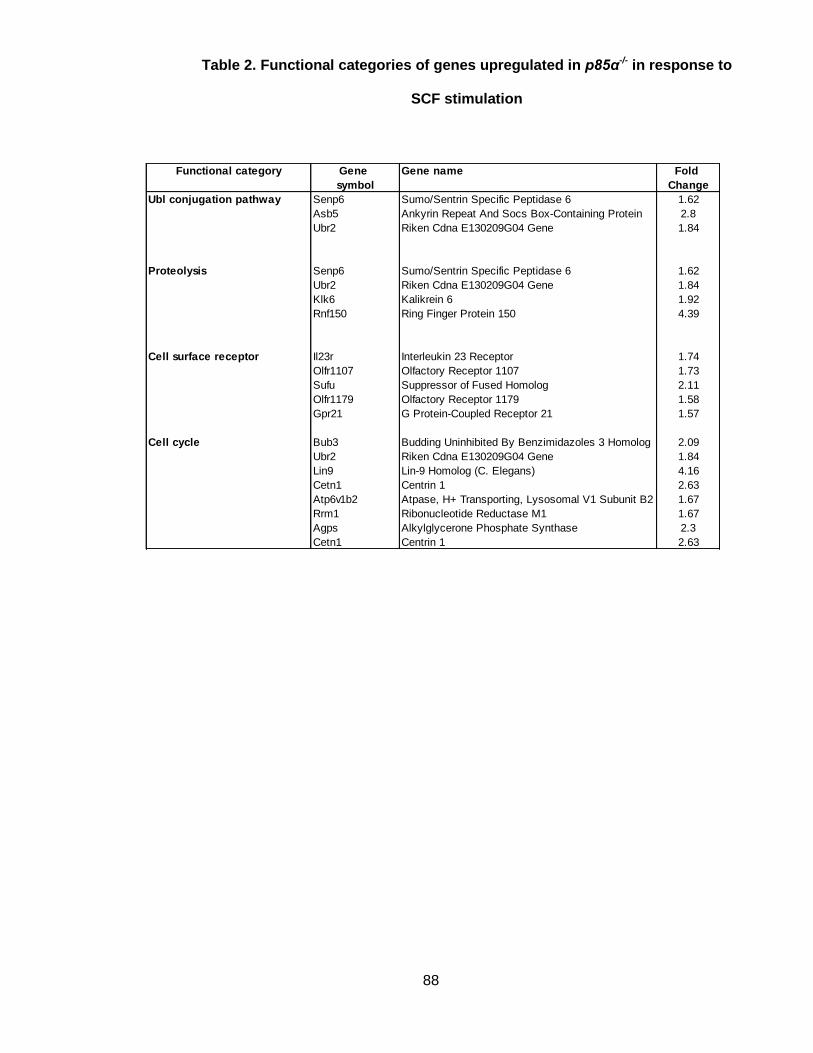
88
Functional category Gene Gene name Fold
symbol Change
Ubl conjugation pathway Senp6 Sumo/Sentrin Specific Peptidase 6 1.62
Asb5 Ankyrin Repeat And Socs Box-Containing Protein 2.8
Ubr2 Riken Cdna E130209G04 Gene 1.84
Proteolysis Senp6 Sumo/Sentrin Specific Peptidase 6 1.62
Ubr2 Riken Cdna E130209G04 Gene 1.84
Klk6 Kalikrein 6 1.92
Rnf150 Ring Finger Protein 150 4.39
Cell surface receptor Il23r Interleukin 23 Receptor 1.74
Olfr1107 Olfactory Receptor 1107 1.73
Sufu Suppressor of Fused Homolog 2.11
Olfr1179 Olfactory Receptor 1179 1.58
Gpr21 G Protein-Coupled Receptor 21 1.57
Cell cycle Bub3 Budding Uninhibited By Benzimidazoles 3 Homolog 2.09
Ubr2 Riken Cdna E130209G04 Gene 1.84
Lin9 Lin-9 Homolog (C. Elegans) 4.16
Cetn1 Centrin 1 2.63
Atp6v1b2 Atpase, H+ Transporting, Lysosomal V1 Subunit B2 1.67
Rrm1 Ribonucleotide Reductase M1 1.67
Agps Alkylglycerone Phosphate Synthase 2.3
Cetn1 Centrin 1 2.63
Table 2. Functional categories of genes upregulated in p85α-/- in response to
SCF stimulation

89
Functional category Gene Gene name Fold
symbol Change
Transcription regulation Nsd1 Nuclear receptor-binding set-domain protein 1 -2.11
Arntl2 Aryl hydrocarbon receptor nuclear translocator-like -1.72
Pcgf2 Polycomb group ring finger 2 -1.9
Tcf20 Transcription factor 20 -3.5
Pou4f3 Pou domain, class 4, transcription factor 3 -1.55
Uimc1 Retinoid x receptor interacting protein 110 -3.06
Protein kinase activity Pim3 Proviral integration site 3 -3.63
Taok3 Tao kinase 3 -3.41
Dyrk1a Dual-specificity tyrosine phosphorylation -2.13
regulated kinase 1a
Cell differentiation Sema3d Riken cDNA 4631426b19 gene -2.26
Psme4 Proteasome activator subunit 4 -1.77
Mbnl1 Muscleblind-like 1 (drosophila) -2.03
Pou4f3 Pou domain, class 4, transcription factor 3 -1.55
Cell adhesion Cdh8 Cadherin 8 -7.09
Fbln5 Fibulin 5 -1.64
Col2a1 Procollagen, type ii, alpha 1 -1.56
Signal transduction Pofut1 Protein o-fucosyl transferase 1 -2.66
Arntl2 Aryl hydrocarbon receptor nuclear translocator-like 2 -1.72
Taok3 Tao kinase 3 -3.41
Arhgef3 Rho guanine nucleotide exchage factor 3 -2.18
Rabl2a Rab, member of ras oncogene family-like 2a -1.58
Post-translational Nsd1 Nuclear receptor binding set domain protein 1 -2.11
protein modification Pim3 Proviral integration site 3 -3.63
Taok3 Tao kinase 3 -3.41
Pcgf2 Polycomb group ring finger 2 -1.9
Fbxo3 F-box only protein 3 -1.9
Dyrk1a Dual-specificity tyrosine phosphorylation -2.13
regulated kinase 1a
Table 3. Functional categories of genes downregulated in p85α-/- in response to
SCF stimulation

90
We performed Ingenuity Pathway Analysis (IPA) to investigate the associated biological
pathways which are altered in the absence of p85α in mast cells. IPA is a curated
database that provides information on biological functional networks, based on published
scientific literature. Figure 24 shows relevant networks altered in p85α-deficient cells,
which are applicable to this study.

91
A
B

92
C
D

93
Figure 24. IPA results on functional network of significantly altered genes in p85α-
/- cells in response to SCF stimulation. Altered genes in p85α-/- cells after SCF
stimulation were analyzed by Ingenuity Pathway Analysis (IPA) software to analyze their
closely associated gene network. Interestingly, significantly altered gene in p85α-/- were
associated with (A) cell survival, (B) cell growth, (C) cell migration and (D) the cell cycle.
Identification of these four principal networks is relevant to this study, as our functional
data suggest that mast cells lacking the p85α regulatory subunit display defective cell
cycles, altered survival and differentiation. The IPA network provides further information
on additional genes including Pdgfb, AKT, Hnf4A, Stat3, Tgfb, Myc, Brca1, Crebbp.
These genes identified by IPA tool, which are related to the altered genes in p85α-/- per
the microarray data, could also play roles in mast cell biological functions in response to
SCF stimulation.
VIII. PI-3K regulatory subunits p85α and p85β differentially regulate mast cell
development in vivo
To confirm our in vitro data, in vivo, we transplanted Wsh/sh recipient mice, with p85α-/-
stem cells transduced with various p85α constructs. p85α-deficient bone marrow cells
treated with 5-FU (5-flurouracil), was transduced with bicistronic MIEG3 expression
plasmids encoding p85α, p85β, p85αΔSH3 or p85αΔBH, cells were then sorted for
homogeneity based on EGFP expression and transplanted into Wsh/sh mice. We
hypothesized that because Wsh/sh mice lack mast cells, any mast cell population
generated in the transplanted mice should be due to the activity of the donor cells’. After
transplantation, we monitored the homing efficiency of the donor cells in the transplanted
mice on a monthly basis by evaluating EGFP expression in their peripheral blood. After 4
months, we sacrificed the Wsh/sh recipient mice and harvested tissues from each mouse.

94
These tissues, which included the ear, skin, lungs, spleen, stomach, duodenum, ileum,
jejunum and colon, were subsequently analyzed for mast cell reconstitution in various
tissues.
Representative spleen and stomach (Figure 25) sections of Wsh/sh mice transplanted with
p85α-deficient cells infected the WT or mutant p85 constructs are shown, with mast cells
indicated by arrows. Consistent with our in vitro studies, full-length p85α transduced into
p85α-/- BM cells fully reconstituted the mast cell population in Wsh/sh transplanted mice,
where as p85α mutants p85αΔSH3 and p85αΔBH partially reconstituted mast cells. In
contrast, p85β-expressing p85α-/- BM cells showed only minimal reconstitution of mast
cells (Figure 26). These results confirm the differential regulation of mast cell
development by PI-3K regulatory subunits p85α and p85β in vivo, and the importance of
the SH3 and BH domains of p85α in normal mast cell development.

95
Figure 25. The amino terminal SH3 and BH domains of p85α are critical for mast
cell development in vivo. p85α-/- mice were injected intraperitoneally with 5-FU (150
mg/kg body weight) and LDBMCs were harvested 72 h after injection. LDBMCs were
transduced with full-length p85α or p85α mutants (p85αΔSH3 or p85αΔBH) and sorted
to homogeneity according to EGFP expression. Sorted cells (1 x 10⁶ cells) were mixed
with recipient Wsh/sh splenocytes (0.1 x 10⁶ cells) and injected into mast-cell–deficient
Wsh/sh mice. Four months after transplantation, the mice were sacrificed; different tissues
were harvested and analyzed for mast cells by leader staining. Shown are
representative sections of spleen (A) and stomach (B).
p85α-/- /vector
p85α-/- /p85αΔBH
p85α-/- /p85α
p85α-/- /p85αΔSH3
p85α-/- /vector p85α-/- /p85α
p85α-/- /p85αΔBHp85α-/- /p85αΔSH3
A B
*
*

96
Figure. 26. PI-3K regulatory subunits p85α and p85β differentially regulates mast
cell development in vivo. Histological analysis of stomach and spleen showing the
reconstitution of mast cells in Wsh mice. p85α-/- mice were injected intraperitoneally with
5-flurouracil (150 mg/kg body weight) and BM cells were harvested after 72 hours of
injection. These cells were transduced with p85α or p85β and sorted to homogeneity on
the basis of EGFP expression. Sorted cells (1 x 10⁶) were mixed with recipient Wsh
splenocytes (0.1 x 10⁶ cells) and injected into mast cell deficient Wsh mice. After 4
months of transplantation, mice were sacrificed; different tissues were harvested and
analyzed for mast cells by leader staining. Shown are representative sections of spleen
and stomach from the indicated genotypes. Arrows indicate mast cells in various tissues.
Spleen
p85α-/- -vector p85α-/- -p85βp85α-/- -p85α
Stomach

97
DISCUSSION
Previous studies have demonstrated the crucial role of the c-Kit receptor and its ligand
SCF in mast cell development (Kitamura, Go et al. 1978; Galli and Kitamura 1987;
Chabot, Stephenson et al. 1988; Copeland, Gilbert et al. 1990; Huang, Nocka et al.
1990; Martin, Suggs et al. 1990; Matsui, Zsebo et al. 1990; Nocka, Buck et al. 1990;
Williams, Eisenman et al. 1990; Zsebo, Williams et al. 1990; Zsebo, Wypych et al. 1990;
Tsai, Shih et al. 1991). PI-3K, which is activated by binding to the 719Y residue of c-Kit,
is a critical mediator in transmitting signals downstream of c-Kit. (Shivakrupa, Bernstein
et al. 2003). PI-3K is also thought to be a critical mediator of abnormal c-Kit signaling,
due to c-Kit point mutation (c-Kit D816V in human and c-Kit D814V in mouse), which
leads to clinical symptoms, such as mastocytosis and mast cell leukemia (Nagata,
Worobec et al. 1995; Longley, Tyrrell et al. 1996; Longley, Metcalfe et al. 1999; Longley,
Reguera et al. 2001). Pharmacological inhibitors of PI-3K activity and dominant negative
inhibitory PI-3K proteins provide evidence for the crucial role of PI-3K in mediating
ligand-independent abnormal c-Kit signaling (Munugalavadla, Sims et al. 2007).
Class IA PI-3K is a complex of regulatory (p85) and catalytic subunit (p110), the latter of
which is activated upon binding to c-Kit via its regulatory subunit. Based on recent
studies demonstrating enhanced insulin sensitivity in both Pik3r1-/- and
Pik3r2-/- mice, it is generally believed that all regulatory subunits of class IA PI3K
behave in a functionally redundant manner with their primary function being to stabilize
and activate the different p110 catalytic subunits(Terauchi, Tsuji et al. 1999; Fruman,
Mauvais-Jarvis et al. 2000; Ueki, Yballe et al. 2002; Chen, Mauvais-Jarvis et al. 2004).
In this study, we evaluated the role of p85α regulatory subunit in mast cell growth,
maturation and survival. We showed that genetic disruption of p85α results in defective

98
IL-3 mediated mast cell differentiation and c-kit mediated growth and survival,
irrespective of the presence of other PI-3K regulatory subunits; p85β, p55α and p50α.
p85α being the most abundantly expressed regulatory subunit in mast cells, its genetic
disruption might significantly reduce the total amount and balance of PI-3K regulatory
subunits overall, which might be the reason for defective phenotype in p85α-/-. We
evaluated this possibility by performing reconstitution studies, and observed that the
reconstitution of p85α-/- MCps with p85α but not p85β completely corrects both IL-3-
mediated differentiation as well as c-Kit induced proliferation. This suggests that the
phenotypic defects observed in p85α-/- MCps are a result of specific loss of p85α (full-
length) and not due to quantitative reduction in the overall expression of regulatory
subunits.
Given the fact that p85β cannot substitute for the loss of p85α in MCps, we then
evaluated whether these regulatory subunits have differential roles in mast cell biological
functions in response to SCF and IL-3 stimulation. Our results in primary hematopoietic
cells using both genetic and biochemical approaches clearly demonstrate unique role(s)
for p85 regulatory subunits in mast cell growth, maturation and survival. We show that
genetic disruption of p85α (full-length) results in defective IL-3 mediated mast cell
differentiation and c-Kit mediated growth and survival; whereas genetic disruption of
p85β results in enhanced IL-3 mediated c-Kit expression; and enhanced c-Kit mediated
growth and survival. Significantly impaired response of p85α-/- MCps to SCF stimulation
might be due to reduced surface c-Kit expression in these cells, or might be due to an
intrinsic defect due to loss of p85α regulatory subunit. We evaluated the possibilities
performing thymidine incorporation assay on c-Kit sorted WT and p85α-/- MCps and
confirmed that the defective mast cell growth in response to SCF stimulation is infact
due to the loss of p85α regulatory subunit.

99
SCF stimulation of c-Kit signaling initiate activation of several ubiquitin ligases that
terminate c-Kit signals in normal cells (Masson, Heiss et al. 2006). Among those ligases
is c-Cbl; its direct binding contributes to c-Kit receptor ubiquitination, internalization and
degradation (Masson, Heiss et al. 2006). Ubiquitination and subsequent degradation of
c-Kit has been implicated as the key mechanism in regulating the duration and intensity
of downstream signaling events (Miyazawa, Toyama et al. 1994). Our results show
impaired c-Kit receptor internalization in p85β-/- MCps compared to WT controls.
Consistently, we show that overexpression of p85β enhances the rate of c-Kit
internalization and degradation compared to cells expressing p85α subunit. These
results suggest that the enhanced proliferation and survival of p85β-/- MCps in response
to SCF is likely due to reduced c-Kit receptor internalization leading to prolonged
activation of downstream signals including ERK and JNK MAP kinase. E3 ubiquitin
ligases including Cbl proteins are involved in binding and degradation of activated c-Kit
leading to down-regulation of c-Kit receptor signaling (Zeng, Xu et al. 2005). We
observed preferential binding of p85β to c-Cbl compared to p85α in c-Kit stimulated
cells, which was associated with enhanced c-Kit receptor ubiquitination and degradation.
Our data is in agreement with previous study done in T-cell, where p85β was reported to
preferentially bind to c-Cbl via its N-SH2 and SH3 domain in modulating T-cell functions
(Alcazar, Cortes et al. 2009). This function of p85β explains the reduced c-Kit
internalization, and enhanced c-Kit expression as well as enhanced signaling events
downstream of c-Kit in p85β-/- MCps. Thus, p85β through interaction with c-Cbl
regulates the internalization and degradation of c-Kit, deficiency of which is likely to
contribute to enhanced c-Kit induced growth, survival and activation of downstream
signals.

100
p85α and p85β regulatory subunits of class IA PI3-K share near identity in the known
functional domains in the carboxy terminus,including the amino-SH2 and the carboxy-
SH2 domains, but wide disparity in aminoacid composition exists in the amino terminal
domains of SH3 and BH. Interestingly p85α splice variants - p50α and p55α share the
domains in the carboxy terminus, but lack amino terminal domains of SH3 and BH.
Therefore the basis for functional uniqueness of p85α is likely to be derived from the
amino terminus SH3 and BH domains of this regulatory subunit. The notion of specificity
was further exemplified in studies demonstrating only a partial rescue in c-Kit induced
growth and survival in p85α-/- MCps reconstituted with p50α, p85αΔSH3 or p85αΔBH.
While our studies show high degree of specificity by various subunits in regulating c-Kit
induced functions; the role in IL-3 induced maturation of MCps is somewhat redundant.
Overexpression of p50α or p85αΔSH3 or p85αΔBH in p85α-/- MCps completely restores
maturation as well as the surface c-Kit expression to WT levels. Restoration of c-Kit
expression in these cells might be either due to restored expression of a transcription
factor–Mitf, which enhances c-Kit protein expression in these cells; or due to inability of
the p85αΔSH3 or p85αΔBH constructs to bind to ubiquiting ligase c-Cbl, which hinders
c-Kit internalization and degradation.
Mitf is a key regulator of mast cell maturation. Mitf regulates a number of proteases, cell
surface receptors, signaling molecules and transporters critical for mast cell function
(Shahlaee, Brandal et al. 2007). Although Mitf upregulation is essential during early
stages of mast cell maturation, the molecular mechanism regulating Mitf expression in
mast cell maturation is poorly understood. Here we observe a possible role of p85α
regulatory subunit of PI-3K in IL-3 mediated Mitf regulation in MCps, which in turn partly
affects c-Kit expression and maturation of mast cells. Mitf expression was abolished in
absence of p85α regulatory subunit in MCps. Restoration of full length p85α in p85α-/-

101
MCps restores the expression of Mitf to WT levels. The result suggests that Mitf
expression in mast cells in response to IL-3 stimulation is tightly regulated by p85α
regulatory subunit of PI-3K.
Earlier studies report the regulational role of Mitf on c-Kit expression; and a significant
down regulation of c-Kit expression in observed in mast cells from Mitf mutant mice (Ebi,
Kanakura et al. 1992; Isozaki, Tsujimura et al. 1994). Consistently in this study, the
down regulation of Mitf expression coincided with significant reduction in c-Kit
expression in p85α-/- MCps. Further the restoration of full length p85α in p85α-/- MCps
which restores the expression of Mitf, also restored c-Kit expression to WT levels; which
suggests involvement of p85α regulatory subunit in the process. Amino terminal SH3
and BH domains of p85α might not be involved in the process, as p50α or p85α mutants
which lack SH3 or BH domains restores the expression of Mitf and c-Kit receptor in
p85α-/- MCps to WT levels.
PI-3K regulatory subunit p85α might regulate Mitf expression at different levels, from
transcriptional to post-translational modifications. Mitf expression is regulated by multiple
signaling pathways which involve signaling molecules such as AKT and ERK (Wu,
Hemesath et al. 2000). Mitf is also regulated by a variety of transcription factors like
Pax3, CREB, Sox10, Lef1 and BRN-2 (Goding 2000). Our data suggests that AKT
activation is significantly reduced in response to SCF stimulation in MCps lacking p85α
regulatory subunit, the activation which was restored upon expression of full length
p85α. Consistently, Mitf expression which was significantly impaired in p85α-/- MCps,
was completely restored with full length p85α expression upon IL-3 stimulation.
Involvement of PI-3K and AKT in Mitf regulation could be further evaluated by
expressing p110CAAX and activated form of AKT in p85α-/- MCps.

102
Inter players mediating the AKT and Mitf activation is yet to be evaluated. PI-3K
regulated activation of AKT is reported to induce phosphorylation and activation of
CREB, which inturn regulates transcription of Mitf. PI-3K might also be involved in AKT
mediated inhibition of GSK-3β expression, which is involved in phosphorylation and
activation of Mitf (Takeda, Takemoto et al. 2000). Thus, p85α regulatory subunit of PI-3K
might be involved in transcriptional regulation of Mitf via AKT and CREB and/or GSK-3β.
SCF signaling pathway in melanocytes is reported to target Mitf simultaneously for
activation and proteolytic degradation, which is mediated via ERK activation (Wu,
Hemesath et al. 2000). Activation of ERK via c-Kit signaling is reported to increase Mitf
transactivation potential (Steingrimsson, Copeland et al. 2004). Phosphorylation and
activation of Mitf also leads to its ubiquitination and degradation via ubiquitination ligase
Ubc9. p85α regulatory subunit is critical for ERK activation in mast cells as indicated by
significant reduction in ERK activity in MCps lacking p85α regulatory subunit upon SCF
stimulation. ERK activity and Mitf expression was restored in p85α-/- MCps upon
restoration of full length p85α expression. Future experiments using ERK pathway
specific inhibitor PD98059 would confirm the involvement of ERK in p85α regulated Mitf
expression in MCps. Although this study evaluates AKT and ERK signaling downstream
of c-Kit upon SCF stimulation, Mitf and c-Kit expression was evaluated in in vitro MCps
cultured in presence of IL-3. Further evaluation is needed on specific effects of IL-3 and
SCF signaling in both Mitf expression and AKT and ERK activation.
Along with Mitf, other transcription factors GATA-1, GATA-2, PU.1 play essential roles in
mast cell maturation. Different stages of mast cell development are tightly regulated by a
molecular balance between different transcription factors. Although GATA-2 is critical for
maturation of embryonic stem cells into mast cells, results from our lab demonstrate that

103
over-expression of GATA-2 represses the Mitf expression and antagonizes the
maturation of adult bone marrow derived mast cells; the phenotypic defects similar to
p85α deficiency seen in MCps. Previous studies in adipose tissues report that insulin-
dependent phosphorylation of GATA-2 in a PI-3K/AKT dependent manner impairs
GATA-2 translocation to nucleus and its DNA binding activity. Taken together, these
observations suggest that p85α regulatory subunit of PI-3K might regulate Mitf
expression in mast cell by multiple signaling pathways which might involve multiple
signaling molecules and transcription factors.

104
Figure 27 . A model describing the possible mechanisms involved in Mitf
regulation by PI3K pathway. Expression of Mitf in MCps might be regulated by multiple
signaling pathways involving multiple transcription factors. The above figure represents
possible mechanism for Mitf regulation based on information from various previously
published literatures.
Altough carboxy terminal domains of p85α might be important in mast cell maturation;
the growth and survival of mast cells in response to SCF stimulation is critically
regulated by amino terminal domains of p85α. We observe that although c-Kit
expression was restored in p85α-/- MCps expressing p85αΔSH3 or p85αΔBH, signals
downstream of c-Kit is still impaired in these cells. These results suggest that the signals
emanating from the amino terminus domains of p85α are likely to play a critical role in c-
Kit induced mast cell biological functions. To evaluate this, we examined the interaction
CREB GSK-3β
Nucleus
AKT
Mast cell
GATA-2
p85α/ p110
ERK
Mitf
c-Kit
SCF IL-3
IL-R c-Kit
Mitf
P P

105
of HA-tagged full-length p85α and mutant p85α constructs with various proteins known
to regulate mast cell growth and survival. We performed immunoprecipitation studies on
the 32D mast cell line and examined the interactions of these constructs with proteins
including Gab2, Gab1, Rac2 and Grb2.
Gab2 is critical for mast cell proliferation, and its lack of results in severe mast cell
deficiency in various tissues (Nishida, Wang et al. 2002; Yu, Luo et al. 2006). Though
this adaptor protein is known to bind to the p85 regulatory subunit of PI-3K to bring about
this effect (Gu, Pratt et al. 1998; Nishida, Yoshida et al. 1999; Zhao, Yu et al. 1999),
here we evaluated how the absence of the SH3 or BH domain of p85α affects its
interaction with Gab2.
Gab1 is a member of Gab/Dos family, which is activated when SCF stimulates c-Kit by
SCF. This adaptor protein is linked to ERK kinase activation, which promotes cell
survival (Itoh, Yoshida et al. 2000). Even though p85 mutant constructs bound to Gab1
as much as full-length p85α did upon SCF stimulation, ERK activation was only partially
rescued in p85α-/- MCps expressing the p85αΔSH3 or p85αΔBH construct.
Grb2 is another scaffolding protein; which regulates cell growth in response to cytokine
stimulation-via-activation of Ras signaling (Cutler, Liu et al. 1993). Rac2 is another
member of Rho related small GTPase family, that was reported to specifically activate
JNK signaling cascade in mast cells (Gu, Byrne et al. 2002). Rac2 deficient mice
express severe mast cell deficiency and reduced mast cell survival in response to SCF
stimulation (Yang, Kapur et al. 2000). Here we evaluated how the absence of the SH3 or
BH domain of p85α affects its interaction with Grb2 and Rac2.
106
To determine whether the interactions of the aforementioned proteins were affected by
SCF stimulation in the absence of the SH3 or BH domains, we performed functional and
immunuprecipitation studies. Using full-length p85α and mutant p85αΔSH3 and
p85αΔBH, we noted that defective mast cell proliferation and survival in response to
SCF stimulation in the absence of an SH3 and/or BH domain is not due to defective
interactions with Gab1, Gab2, Grb2, or Rac2 proteins, although they are known to be
important effectors of mast cell proliferation and survival. Further, we performed
microarray and compared gene expression profiles of WT and p85α-/- MCps in response
to SCF stimulation. Objective was to find potential effectors of PI-3K which might
regulate mast cell biological functions upon SCF stimulation. Interesting genes for future
consideration in context of explaining the growth and survival defects in p85α-/- MCps
upon SCF stimulation regard are – Rrm1, Taok3, Pim3 and Fbln5.
Mastocytosis is a myeloid neoplasm characterized by abnormal accumulation and
frequent activation of mast cells (MCs) in various organs. The activating point mutation
D816V found in the c-Kit kinase domain occurs in 80-95% of systemic mastocysis (SM)
patients. Unfortunately, the prognosis in these patients remains poor and no effective
treatment is currently available for the treatment of SM and AML caused due to c-Kit
D816V mutations. Through the use of pharmacological inhibitors and genetic studies, PI-
3K has been shown to play indespinsible role in regulating normal c-Kit signaling, as well
as abnormal c-Kit signaling due to D816V mutations. This study was undertaken better
understand the role of p85 regulatory subunits and its domains in c-Kit signaling, the
information which might be important in designing strategies to regulate abnormal
signaling downstream c-Kit D816V.

107
A redundant functional role of p85α and p85β regulatory subunits has been reported by
several previous studies (Shepherd, Withers et al. 1998; Ueki, Yballe et al. 2002;
Brachmann, Yballe et al. 2005). This study is novel in that it invalidates preexisting
assumptions about the functional redundancy or similarity among the various PI-3K
regulatory subunits. Performing in vitro and in vivo studies, we demonstrated that the
p85α and p85β regulatory subunits of PI-3K play different roles in mast cell
development. p85α is a positive regulator of c-Kit signaling, and the SH3 and BH
domains of p85α were indispensible for the growth of mast cells in response to SCF
stimulation. Notably, p85β appears to be a negative regulator of c-Kit signaling; the
absence of p85β in MCps results in hyperproliferation in response to SCF stimulation,
but overexpression of this regulatory subunit in the same cell lines resulted in
significantly reduced proliferation and reduced c-Kit expression. Our findings have
important implications for the rational design of c-Kit inhibitors, and might be applicable
for treatment of malignancies involving abnormal c-Kit 816 mutations, and in tumors
involving normal c-Kit signaling.

108
REFERENCES
Abonia, J. P., K. F. Austen, et al. (2005). "Constitutive homing of mast cell progenitors to the intestine depends on autologous expression of the chemokine receptor CXCR2." Blood 105(11): 4308-13.
Abonia, J. P., J. Hallgren, et al. (2006). "Alpha-4 integrins and VCAM-1, but not MAdCAM-1, are essential for recruitment of mast cell progenitors to the inflamed lung." Blood 108(5): 1588-94.
Alcazar, I., I. Cortes, et al. (2009). "p85beta phosphoinositide 3-kinase regulates CD28 coreceptor function." Blood 113(14): 3198-208.
Alfredsson, J., H. Puthalakath, et al. (2005). "Proapoptotic Bcl-2 family member Bim is involved in the control of mast cell survival and is induced together with Bcl-XL upon IgE-receptor activation." Cell Death Differ 12(2): 136-44.
Arinobu, Y., H. Iwasaki, et al. (2005). "Developmental checkpoints of the basophil/mast cell lineages in adult murine hematopoiesis." Proc Natl Acad Sci U S A 102(50): 18105-10.
Baghestanian, M., J. H. Jordan, et al. (2002). "Activation of human mast cells through stem cell factor receptor (KIT) is associated with expression of bcl-2." Int Arch Allergy Immunol 129(3): 228-36.
Bandi, S. R., C. Brandts, et al. (2009). "E3 ligase-defective Cbl mutants lead to a generalized mastocytosis and myeloproliferative disease." Blood 114(19): 4197-208.
Barrett, K. E. and D. D. Metcalfe (1984). "Mast cell heterogeneity: evidence and implications." J Clin Immunol 4(4): 253-61.
Beghini, A., P. Peterlongo, et al. (2000). "C-kit mutations in core binding factor leukemias." Blood 95(2): 726-7.
Berlanga, O., N. Emambokus, et al. (2005). "GPIIb (CD41) integrin is expressed on mast cells and influences their adhesion properties." Exp Hematol 33(4): 403-12.
Berrozpe, G., I. Timokhina, et al. (1999). "The W(sh), W(57), and Ph Kit expression mutations define tissue-specific control elements located between -23 and -154 kb upstream of Kit." Blood 94(8): 2658-66.
Bienenstock, J., A. D. Befus, et al. (1983). "Mast cell heterogeneity." Monogr Allergy 18: 124-8.
Bienenstock, J., D. Befus, et al. (1985). "Comparative aspects of mast cell heterogeneity in different species and sites." International archives of allergy and applied immunology 77(1-2): 126-9.
Blechman, J. M., S. Lev, et al. (1995). "The fourth immunoglobulin domain of the stem cell factor receptor couples ligand binding to signal transduction." Cell 80(1): 103-13.
Blume-Jensen, P., R. Janknecht, et al. (1998). "The kit receptor promotes cell survival via activation of PI 3-kinase and subsequent Akt-mediated phosphorylation of Bad on Ser136." Curr Biol 8(13): 779-82.
Brachmann, S. M., C. M. Yballe, et al. (2005). "Role of phosphoinositide 3-kinase regulatory isoforms in development and actin rearrangement." Mol Cell Biol 25(7): 2593-606.
Bradding, P., Y. Okayama, et al. (1995). "Heterogeneity of human mast cells based on cytokine content." J Immunol 155(1): 297-307.
Cantor, A. B. and S. H. Orkin (2001). "Hematopoietic development: a balancing act." Curr Opin Genet Dev 11(5): 513-9.

109
Chabot, B., D. A. Stephenson, et al. (1988). "The proto-oncogene c-kit encoding a transmembrane tyrosine kinase receptor maps to the mouse W locus." Nature 335(6185): 88-9.
Chen, C. C., M. A. Grimbaldeston, et al. (2005). "Identification of mast cell progenitors in adult mice." Proc Natl Acad Sci U S A 102(32): 11408-13.
Chen, D., F. Mauvais-Jarvis, et al. (2004). "p50alpha/p55alpha phosphoinositide 3-kinase knockout mice exhibit enhanced insulin sensitivity." Mol Cell Biol 24(1): 320-9.
Chian, R., S. Young, et al. (2001). "Phosphatidylinositol 3 kinase contributes to the transformation of hematopoietic cells by the D816V c-Kit mutant." Blood 98(5): 1365-73.
Copeland, N. G., D. J. Gilbert, et al. (1990). "Mast cell growth factor maps near the steel locus on mouse chromosome 10 and is deleted in a number of steel alleles." Cell 63(1): 175-83.
Crapper, R. M. and J. W. Schrader (1983). "Frequency of mast cell precursors in normal tissues determined by an in vitro assay: antigen induces parallel increases in the frequency of P cell precursors and mast cells." J Immunol 131(2): 923-8.
Cutler, R. L., L. Liu, et al. (1993). "Multiple cytokines induce the tyrosine phosphorylation of Shc and its association with Grb2 in hemopoietic cells." J Biol Chem 268(29): 21463-5.
Da Silva, C. A., L. Reber, et al. (2006). "Stem cell factor expression, mast cells and inflammation in asthma." Fundam Clin Pharmacol 20(1): 21-39.
Dastych, J. and D. D. Metcalfe (1994). "Stem cell factor induces mast cell adhesion to fibronectin." J Immunol 152(1): 213-9.
Dastych, J., D. Taub, et al. (1998). "Tyrosine kinase-deficient Wv c-kit induces mast cell adhesion and chemotaxis." Am J Physiol 275(5 Pt 1): C1291-9.
Downward, J. (2004). "PI 3-kinase, Akt and cell survival." Semin Cell Dev Biol 15(2): 177-82.
Dubreuil, P., L. Forrester, et al. (1991). "The c-fms gene complements the mitogenic defect in mast cells derived from mutant W mice but not mi (microphthalmia) mice." Proc Natl Acad Sci U S A 88(6): 2341-5.
Duensing, A., M. C. Heinrich, et al. (2004). "Biology of gastrointestinal stromal tumors: KIT mutations and beyond." Cancer Invest 22(1): 106-16.
Duensing, A., F. Medeiros, et al. (2004). "Mechanisms of oncogenic KIT signal transduction in primary gastrointestinal stromal tumors (GISTs)." Oncogene 23(22): 3999-4006.
Duronio, V., M. J. Welham, et al. (1992). "p21ras activation via hemopoietin receptors and c-kit requires tyrosine kinase activity but not tyrosine phosphorylation of p21ras GTPase-activating protein." Proc Natl Acad Sci U S A 89(5): 1587-91.
Duttlinger, R., K. Manova, et al. (1993). "W-sash affects positive and negative elements controlling c-kit expression: ectopic c-kit expression at sites of kit-ligand expression affects melanogenesis." Development 118(3): 705-17.
Ebi, Y., Y. Kanakura, et al. (1992). "Low c-kit expression of cultured mast cells of mi/mi genotype may be involved in their defective responses to fibroblasts that express the ligand for c-kit." Blood 80(6): 1454-62.
Ehrlich, P. (1878). Beitrage zur Theorie und Praxis der Histologischen Farbung. German, Leipzig University.
Enerback, L. (1966). "Mast cells in rat gastrointestinal mucosa. 2. Dye-binding and metachromatic properties." Acta Pathol Microbiol Scand 66(3): 303-12.
Enerback, L. (1966). "Mast cells in rat gastrointestinal mucosa. I. Effects of fixation." Acta Pathol Microbiol Scand 66(3): 289-302.

110
Enerback, L. and G. B. Lowhagen (1979). "Long term increase of mucosal mast cells in the rat induced by administration of compound 48/80." Cell and tissue research 198(2): 209-15.
Feger, F., S. Varadaradjalou, et al. (2002). "The role of mast cells in host defense and their subversion by bacterial pathogens." Trends Immunol 23(3): 151-8.
Finotto, S., Y. A. Mekori, et al. (1997). "Glucocorticoids decrease tissue mast cell number by reducing the production of the c-kit ligand, stem cell factor, by resident cells: in vitro and in vivo evidence in murine systems." J Clin Invest 99(7): 1721-8.
Flanagan, J. G., D. C. Chan, et al. (1991). "Transmembrane form of the kit ligand growth factor is determined by alternative splicing and is missing in the Sld mutant." Cell 64(5): 1025-35.
Flanagan, J. G. and P. Leder (1990). "The kit ligand: a cell surface molecule altered in steel mutant fibroblasts." Cell 63(1): 185-94.
Fodinger, M., G. Fritsch, et al. (1994). "Origin of human mast cells: development from transplanted hematopoietic stem cells after allogeneic bone marrow transplantation." Blood 84(9): 2954-9.
Fruman, D. A., L. C. Cantley, et al. (1996). "Structural organization and alternative splicing of the murine phosphoinositide 3-kinase p85 alpha gene." Genomics 37(1): 113-21.
Fruman, D. A., F. Mauvais-Jarvis, et al. (2000). "Hypoglycaemia, liver necrosis and perinatal death in mice lacking all isoforms of phosphoinositide 3-kinase p85 alpha." Nat Genet 26(3): 379-82.
Fruman, D. A., R. E. Meyers, et al. (1998). "Phosphoinositide kinases." Annu Rev Biochem 67: 481-507.
Fukao, T., T. Yamada, et al. (2002). "Selective loss of gastrointestinal mast cells and impaired immunity in PI3K-deficient mice." Nat Immunol 3(3): 295-304.
Furitsu, T., T. Tsujimura, et al. (1993). "Identification of mutations in the coding sequence of the proto-oncogene c-kit in a human mast cell leukemia cell line causing ligand-independent activation of c-kit product." J Clin Invest 92(4): 1736-44.
Galli, S. J. (1990). "New insights into the riddle of the mast cells: microenvironmental regulation of mast cell development and phenotypic heterogeneity." Lab.Invest 62(1): 5-33.
Galli, S. J. (2000). "Mast cells and basophils." Curr Opin Hematol 7(1): 32-9. Galli, S. J., J. Kalesnikoff, et al. (2005). "Mast cells as "tunable" effector and
immunoregulatory cells: recent advances." Annual review of immunology 23: 749-86.
Galli, S. J. and Y. Kitamura (1987). "Genetically mast-cell-deficient W/Wv and Sl/Sld mice. Their value for the analysis of the roles of mast cells in biologic responses in vivo." Am J Pathol 127(1): 191-8.
Galli, S. J., M. Maurer, et al. (1999). "Mast cells as sentinels of innate immunity." Current Opinion in Immunology 11(1): 53-59.
Galli, S. J., S. Nakae, et al. (2005). "Mast cells in the development of adaptive immune responses." Nat Immunol 6(2): 135-42.
Galli, S. J., M. Tsai, et al. (1993). "The c-kit receptor, stem cell factor, and mast cells. What each is teaching us about the others." Am J Pathol 142(4): 965-74.
Galli, S. J., K. M. Zsebo, et al. (1994). "The kit ligand, stem cell factor." Adv Immunol 55: 1-96.
Gautam, A., Z. R. Li, et al. (2003). "RRM1-induced metastasis suppression through PTEN-regulated pathways." Oncogene 22(14): 2135-42.

111
Geissler, E. N., M. A. Ryan, et al. (1988). "The dominant-white spotting (W) locus of the mouse encodes the c-kit proto-oncogene." Cell 55(1): 185-92.
Goding, C. R. (2000). "Mitf from neural crest to melanoma: signal transduction and transcription in the melanocyte lineage." Genes Dev 14(14): 1712-28.
Gu, H., J. C. Pratt, et al. (1998). "Cloning of p97/Gab2, the major SHP2-binding protein in hematopoietic cells, reveals a novel pathway for cytokine-induced gene activation." Mol Cell 2(6): 729-40.
Gu, Y., M. C. Byrne, et al. (2002). "Rac2, a hematopoiesis-specific Rho GTPase, specifically regulates mast cell protease gene expression in bone marrow-derived mast cells." Mol Cell Biol 22(21): 7645-57.
Gurish, M. F., H. Tao, et al. (2001). "Intestinal mast cell progenitors require CD49dbeta7 (alpha4beta7 integrin) for tissue-specific homing." J Exp Med 194(9): 1243-52.
Guy-Grand, D., M. Dy, et al. (1984). "Gut mucosal mast cells. Origin, traffic, and differentiation." J Exp Med 160(1): 12-28.
Harigae, H., S. Takahashi, et al. (1998). "Differential roles of GATA-1 and GATA-2 in growth and differentiation of mast cells." Genes Cells 3(1): 39-50.
Hashimoto, K., I. Matsumura, et al. (2003). "Necessity of tyrosine 719 and phosphatidylinositol 3'-kinase-mediated signal pathway in constitutive activation and oncogenic potential of c-kit receptor tyrosine kinase with the Asp814Val mutation." Blood 101(3): 1094-102.
Hawkins, P. T., K. E. Anderson, et al. (2006). "Signalling through Class I PI3Ks in mammalian cells." Biochem Soc Trans 34(Pt 5): 647-62.
Hemesath, T. J., E. R. Price, et al. (1998). "MAP kinase links the transcription factor Microphthalmia to c-Kit signalling in melanocytes." Nature 391(6664): 298-301.
Henkel, G. and M. A. Brown (1994). "PU.1 and GATA: components of a mast cell-specific interleukin 4 intronic enhancer." Proc Natl Acad Sci U S A 91(16): 7737-41.
Huang, C., A. Sali, et al. (1998). "Regulation and function of mast cell proteases in inflammation." Journal of clinical immunology 18(3): 169-83.
Huang, E., K. Nocka, et al. (1990). "The hematopoietic growth factor KL is encoded by the Sl locus and is the ligand of the c-kit receptor, the gene product of the W locus." Cell 63(1): 225-33.
Humbles, A. A., B. Lu, et al. (2002). "The murine CCR3 receptor regulates both the role of eosinophils and mast cells in allergen-induced airway inflammation and hyperresponsiveness." Proc Natl Acad Sci U S A 99(3): 1479-84.
Iemura, A., M. Tsai, et al. (1994). "The c-kit ligand, stem cell factor, promotes mast cell survival by suppressing apoptosis." Am J Pathol 144(2): 321-8.
Inukai, K., M. Funaki, et al. (1997). "p85alpha gene generates three isoforms of regulatory subunit for phosphatidylinositol 3-kinase (PI 3-Kinase), p50alpha, p55alpha, and p85alpha, with different PI 3-kinase activity elevating responses to insulin." J Biol Chem 272(12): 7873-82.
Irani, A. A., N. M. Schechter, et al. (1986). "Two types of human mast cells that have distinct neutral protease compositions." Proc Natl Acad Sci U S A 83(12): 4464-8.
Irani, A. M., S. S. Craig, et al. (1987). "Deficiency of the tryptase-positive, chymase-negative mast cell type in gastrointestinal mucosa of patients with defective T lymphocyte function." J Immunol 138(12): 4381-6.
Isozaki, K., T. Tsujimura, et al. (1994). "Cell type-specific deficiency of c-kit gene expression in mutant mice of mi/mi genotype." Am J Pathol 145(4): 827-36.
Ito, A., E. Morii, et al. (1998). "Systematic method to obtain novel genes that are regulated by mi transcription factor: impaired expression of granzyme B and tryptophan hydroxylase in mi/mi cultured mast cells." Blood 91(9): 3210-21.

112
Itoh, M., Y. Yoshida, et al. (2000). "Role of Gab1 in heart, placenta, and skin development and growth factor- and cytokine-induced extracellular signal-regulated kinase mitogen-activated protein kinase activation." Mol Cell Biol 20(10): 3695-704.
Iwasaki, H., S. Mizuno, et al. (2006). "The order of expression of transcription factors directs hierarchical specification of hematopoietic lineages." Genes Dev 20(21): 3010-21.
Jamur, M. C., A. C. Grodzki, et al. (2005). "Identification and characterization of undifferentiated mast cells in mouse bone marrow." Blood 105(11): 4282-9.
Jippo, T., H. Mizuno, et al. (1996). "Abundant expression of transcription factor GATA-2 in proliferating but not in differentiated mast cells in tissues of mice: demonstration by in situ hybridization." Blood 87(3): 993-8.
Jippo, T., E. Morii, et al. (2003). "Effect of anatomical distribution of mast cells on their defense function against bacterial infections: demonstration using partially mast cell-deficient tg/tg mice." J Exp Med 197(11): 1417-25.
Kalesnikoff, J. and S. J. Galli (2008). "New developments in mast cell biology." Nat Immunol 9(11): 1215-23.
Kapur, R., M. Majumdar, et al. (1998). "Signaling through the interaction of membrane-restricted stem cell factor and c-kit receptor tyrosine kinase: genetic evidence for a differential role in erythropoiesis." Blood 91(3): 879-89.
Kasugai, T., K. Oguri, et al. (1993). "Deficient differentiation of mast cells in the skin of mi/mi mice. Usefulness of in situ hybridization for evaluation of mast cell phenotype." Am J Pathol 143(5): 1337-47.
Kasugai, T., H. Tei, et al. (1995). "Infection with Nippostrongylus brasiliensis induces invasion of mast cell precursors from peripheral blood to small intestine." Blood 85(5): 1334-40.
Katz, H. R., R. L. Stevens, et al. (1985). "Heterogeneity of mammalian mast cells differentiated in vivo and in vitro." J Allergy Clin Immunol 76(2 Pt 2): 250-9.
Kempuraj, D., H. Saito, et al. (1999). "Characterization of mast cell-committed progenitors present in human umbilical cord blood." Blood 93(10): 3338-46.
Khalil, R. M., A. Luz, et al. (1996). "Schistosoma mansoni infection in mice augments the capacity for interleukin 3 (IL-3) and IL-9 production and concurrently enlarges progenitor pools for mast cells and granulocytes-macrophages." Infect Immun 64(12): 4960-6.
Kinet, J. P. (1999). "The high-affinity IgE receptor (Fc epsilon RI): from physiology to pathology." Annu Rev Immunol 17: 931-72.
Kirshenbaum, A. S., J. P. Goff, et al. (1999). "Demonstration that human mast cells arise from a progenitor cell population that is CD34(+), c-kit(+), and expresses aminopeptidase N (CD13)." Blood 94(7): 2333-42.
Kirshenbaum, A. S., S. W. Kessler, et al. (1991). "Demonstration of the origin of human mast cells from CD34+ bone marrow progenitor cells." J Immunol 146(5): 1410-5.
Kitamura, Y. (1989). "Heterogeneity of mast cells and phenotypic change between subpopulations." Annu Rev Immunol 7: 59-76.
Kitamura, Y. and J. Fujita (1989). "Regulation of mast cell differentiation." Bioessays 10(6): 193-6.
Kitamura, Y. and S. Go (1979). "Decreased production of mast cells in S1/S1d anemic mice." Blood 53(3): 492-7.
Kitamura, Y., S. Go, et al. (1978). "Decrease of mast cells in W/Wv mice and their increase by bone marrow transplantation." Blood 52(2): 447-52.
Kitamura, Y., T. Kasugai, et al. (1993). "Development of mast cells and basophils: processes and regulation mechanisms." Am J Med Sci 306(3): 185-91.
113
Kitamura, Y., E. Morii, et al. (2002). "Regulation of mast cell phenotype by MITF." Int Arch Allergy Immunol 127(2): 106-9.
Kitamura, Y., K. Oboki, et al. (2006). "Molecular mechanisms of mast cell development." Immunol Allergy Clin North Am 26(3): 387-405; v.
Kitamura, Y., M. Shimada, et al. (1979). "Presence of mast cell precursors in fetal liver of mice." Dev Biol 70(2): 510-4.
Kitamura, Y., M. Shimada, et al. (1977). "Development of mast cells from grafted bone marrow cells in irradiated mice." Nature 268(5619): 442-3.
Kitamura, Y., M. Yokoyama, et al. (1981). "Spleen colony-forming cell as common precursor for tissue mast cells and granulocytes." Nature 291(5811): 159-60.
Kitayama, H., Y. Kanakura, et al. (1995). "Constitutively activating mutations of c-kit receptor tyrosine kinase confer factor-independent growth and tumorigenicity of factor-dependent hematopoietic cell lines." Blood 85(3): 790-8.
Krystal, G. W., G. Sulanke, et al. (2002). "Inhibition of phosphatidylinositol 3-kinase-Akt signaling blocks growth, promotes apoptosis, and enhances sensitivity of small cell lung cancer cells to chemotherapy." Mol Cancer Ther 1(11): 913-22.
Lee, D. M., D. S. Friend, et al. (2002). "Mast cells: a cellular link between autoantibodies and inflammatory arthritis." Science 297(5587): 1689-92.
Lee, P. S., Y. Wang, et al. (1999). "The Cbl protooncoprotein stimulates CSF-1 receptor multiubiquitination and endocytosis, and attenuates macrophage proliferation." EMBO J 18(13): 3616-28.
Lev, S., D. Givol, et al. (1992). "Interkinase domain of kit contains the binding site for phosphatidylinositol 3' kinase." Proc Natl Acad Sci U S A 89(2): 678-82.
Levkowitz, G., H. Waterman, et al. (1998). "c-Cbl/Sli-1 regulates endocytic sorting and ubiquitination of the epidermal growth factor receptor." Genes Dev 12(23): 3663-74.
Longley, B. J., Jr., D. D. Metcalfe, et al. (1999). "Activating and dominant inactivating c-KIT catalytic domain mutations in distinct clinical forms of human mastocytosis." Proc Natl Acad Sci U S A 96(4): 1609-14.
Longley, B. J., M. J. Reguera, et al. (2001). "Classes of c-KIT activating mutations: proposed mechanisms of action and implications for disease classification and therapy." Leuk Res 25(7): 571-6.
Longley, B. J., L. Tyrrell, et al. (1996). "Somatic c-KIT activating mutation in urticaria pigmentosa and aggressive mastocytosis: establishment of clonality in a human mast cell neoplasm." Nat Genet 12(3): 312-4.
Macdonald, A., D. G. Campbell, et al. (2006). "Pim kinases phosphorylate multiple sites on Bad and promote 14-3-3 binding and dissociation from Bcl-XL." BMC Cell Biol 7: 1.
Maeda, K., C. Nishiyama, et al. (2003). "Regulation of cell type-specific mouse Fc epsilon RI beta-chain gene expression by GATA-1 via four GATA motifs in the promoter." J Immunol 170(1): 334-40.
Martin, F. H., S. V. Suggs, et al. (1990). "Primary structure and functional expression of rat and human stem cell factor DNAs." Cell 63(1): 203-11.
Masson, K., E. Heiss, et al. (2006). "Direct binding of Cbl to Tyr568 and Tyr936 of the stem cell factor receptor/c-Kit is required for ligand-induced ubiquitination, internalization and degradation." Biochem J 399(1): 59-67.
Masuda, A., Y. Yoshikai, et al. (2004). "The interaction between GATA proteins and activator protein-1 promotes the transcription of IL-13 in mast cells." J Immunol 173(9): 5564-73.

114
Matsui, Y., K. M. Zsebo, et al. (1990). "Embryonic expression of a haematopoietic growth factor encoded by the Sl locus and the ligand for c-kit." Nature 347(6294): 667-9.
Meininger, C. J., H. Yano, et al. (1992). "The c-kit receptor ligand functions as a mast cell chemoattractant." Blood 79(4): 958-63.
Mekori, Y. A., A. M. Gilfillan, et al. (2001). "Human mast cell apoptosis is regulated through Bcl-2 and Bcl-XL." J Clin Immunol 21(3): 171-4.
Mekori, Y. A., C. K. Oh, et al. (1993). "IL-3-dependent murine mast cells undergo apoptosis on removal of IL-3. Prevention of apoptosis by c-kit ligand." J Immunol 151(7): 3775-84.
Metcalfe, D. D., D. Baram, et al. (1997). "Mast cells." Physiol Rev 77(4): 1033-79. Metcalfe, D. D., J. A. Mekori, et al. (1995). "Mast cell ontogeny and apoptosis." Exp
Dermatol 4(4 Pt 2): 227-30. Migliaccio, A. R., R. A. Rana, et al. (2003). "GATA-1 as a regulator of mast cell
differentiation revealed by the phenotype of the GATA-1low mouse mutant." J Exp Med 197(3): 281-96.
Miyake, S., K. P. Mullane-Robinson, et al. (1999). "Cbl-mediated negative regulation of platelet-derived growth factor receptor-dependent cell proliferation. A critical role for Cbl tyrosine kinase-binding domain." J Biol Chem 274(23): 16619-28.
Miyazawa, K., K. Toyama, et al. (1994). "Ligand-dependent polyubiquitination of c-kit gene product: a possible mechanism of receptor down modulation in M07e cells." Blood 83(1): 137-45.
Mol, C. D., K. B. Lim, et al. (2003). "Structure of a c-kit product complex reveals the basis for kinase transactivation." J Biol Chem 278(34): 31461-4.
Moore, K. J. (1995). "Insight into the microphthalmia gene." Trends Genet 11(11): 442-8. Morii, E., K. Oboki, et al. (2004). "Roles of MITF for development of mast cells in mice:
effects on both precursors and tissue environments." Blood 104(6): 1656-61. Morii, E., T. Tsujimura, et al. (1996). "Regulation of mouse mast cell protease 6 gene
expression by transcription factor encoded by the mi locus." Blood 88(7): 2488-94.
Munugalavadla, V., E. C. Sims, et al. (2007). "Genetic and pharmacologic evidence implicating the p85 alpha, but not p85 beta, regulatory subunit of PI3K and Rac2 GTPase in regulating oncogenic KIT-induced transformation in acute myeloid leukemia and systemic mastocytosis." Blood 110(5): 1612-20.
Nagata, H., A. S. Worobec, et al. (1995). "Identification of a point mutation in the catalytic domain of the protooncogene c-kit in peripheral blood mononuclear cells of patients who have mastocytosis with an associated hematologic disorder." Proc Natl Acad Sci U S A 92(23): 10560-4.
Nilsson, G., J. H. Butterfield, et al. (1994). "Stem cell factor is a chemotactic factor for human mast cells." J Immunol 153(8): 3717-23.
Nishida, K., L. Wang, et al. (2002). "Requirement of Gab2 for mast cell development and KitL/c-Kit signaling." Blood 99(5): 1866-9.
Nishida, K., Y. Yoshida, et al. (1999). "Gab-family adapter proteins act downstream of cytokine and growth factor receptors and T- and B-cell antigen receptors." Blood 93(6): 1809-16.
Nishiyama, C., M. Hasegawa, et al. (2002). "Regulation of human Fc epsilon RI alpha-chain gene expression by multiple transcription factors." J Immunol 168(9): 4546-52.
Nocka, K., J. Buck, et al. (1990). "Candidate ligand for the c-kit transmembrane kinase receptor: KL, a fibroblast derived growth factor stimulates mast cells and erythroid progenitors." EMBO J 9(10): 3287-94.

115
Nocka, K., J. C. Tan, et al. (1990). "Molecular bases of dominant negative and loss of function mutations at the murine c-kit/white spotting locus: W37, Wv, W41 and W." EMBO J 9(6): 1805-13.
Ochi, H., W. M. Hirani, et al. (1999). "T helper cell type 2 cytokine-mediated comitogenic responses and CCR3 expression during differentiation of human mast cells in vitro." J Exp Med 190(2): 267-80.
Okayama, Y. and T. Kawakami (2006). "Development, migration, and survival of mast cells." Immunol Res 34(2): 97-115.
Okkenhaug, K. and B. Vanhaesebroeck (2003). "PI3K-signalling in B- and T-cells: insights from gene-targeted mice." Biochem Soc Trans 31(Pt 1): 270-4.
Oku, Y., H. Itayama, et al. (1984). "Expulsion of Trichinella spiralis from the intestine of W/Wv mice reconstituted with haematopoietic and lymphopoietic cells and origin of mucosal mast cells." Immunology 53(2): 337-44.
Orkin, S. H. (2000). "Diversification of haematopoietic stem cells to specific lineages." Nat Rev Genet 1(1): 57-64.
Orkin, S. H. (2003). "Priming the hematopoietic pump." Immunity 19(5): 633-4. Otsu, M., I. Hiles, et al. (1991). "Characterization of two 85 kd proteins that associate
with receptor tyrosine kinases, middle-T/pp60c-src complexes, and PI3-kinase." Cell 65(1): 91-104.
Pawson, T. (1995). "Protein modules and signalling networks." Nature 373(6515): 573-80.
Pennock, J. L. and R. K. Grencis (2004). "In vivo exit of c-kit+/CD49d(hi)/beta7+ mucosal mast cell precursors from the bone marrow following infection with the intestinal nematode Trichinella spiralis." Blood 103(7): 2655-60.
Razin, E., J. N. Ihle, et al. (1984). "Interleukin 3: A differentiation and growth factor for the mouse mast cell that contains chondroitin sulfate E proteoglycan." J Immunol 132(3): 1479-86.
Rodewald, H. R., M. Dessing, et al. (1996). "Identification of a committed precursor for the mast cell lineage." Science 271(5250): 818-22.
Roskoski, R., Jr. (2005). "Structure and regulation of Kit protein-tyrosine kinase--the stem cell factor receptor." Biochem Biophys Res Commun 338(3): 1307-15.
Rottapel, R., M. Reedijk, et al. (1991). "The Steel/W transduction pathway: kit autophosphorylation and its association with a unique subset of cytoplasmic signaling proteins is induced by the Steel factor." Mol Cell Biol 11(6): 3043-51.
Rottem, M., T. Okada, et al. (1994). "Mast cells cultured from the peripheral blood of normal donors and patients with mastocytosis originate from a CD34+/Fc epsilon RI- cell population." Blood 84(8): 2489-96.
Ruitenberg, E. J. and A. Elgersma (1976). "Absence of intestinal mast cell response in congenitally athymic mice during Trichinella spiralis infection." Nature 264(5583): 258-60.
Sasaki, T., A. Suzuki, et al. (2002). "Phosphoinositide 3-kinases in immunity: lessons from knockout mice." J Biochem 131(4): 495-501.
Scheinfeld, N. (2006). "A comprehensive review of imatinib mesylate (Gleevec) for dermatological diseases." J Drugs Dermatol 5(2): 117-22.
Secor, V. H., W. E. Secor, et al. (2000). "Mast cells are essential for early onset and severe disease in a murine model of multiple sclerosis." J Exp Med 191(5): 813-22.
Serve, H., Y. C. Hsu, et al. (1994). "Tyrosine residue 719 of the c-kit receptor is essential for binding of the P85 subunit of phosphatidylinositol (PI) 3-kinase and for c-kit-associated PI 3-kinase activity in COS-1 cells." J Biol Chem 269(8): 6026-30.

116
Serve, H., N. S. Yee, et al. (1995). "Differential roles of PI3-kinase and Kit tyrosine 821 in Kit receptor-mediated proliferation, survival and cell adhesion in mast cells." EMBO J 14(3): 473-83.
Shahlaee, A. H., S. Brandal, et al. (2007). "Distinct and shared transcriptomes are regulated by microphthalmia-associated transcription factor isoforms in mast cells." J Immunol 178(1): 378-88.
Shattil, S. J. and P. J. Newman (2004). "Integrins: dynamic scaffolds for adhesion and signaling in platelets." Blood 104(6): 1606-15.
Shelburne, C. P., M. E. McCoy, et al. (2003). "Stat5 expression is critical for mast cell development and survival." Blood 102(4): 1290-7.
Shepherd, P. R., D. J. Withers, et al. (1998). "Phosphoinositide 3-kinase: the key switch mechanism in insulin signalling." Biochem J 333 ( Pt 3): 471-90.
Shivakrupa, R., A. Bernstein, et al. (2003). "Phosphatidylinositol 3'-kinase is required for growth of mast cells expressing the kit catalytic domain mutant." Cancer Res 63(15): 4412-9.
Sonoda, S., T. Sonoda, et al. (1986). "Development of mucosal mast cells after injection of a single connective tissue-type mast cell in the stomach mucosa of genetically mast cell-deficient W/Wv mice." J Immunol 137(4): 1319-22.
Sonoda, T., C. Hayashi, et al. (1983). "Presence of mast cell precursors in the yolk sac of mice." Dev Biol 97(1): 89-94.
Steingrimsson, E., N. G. Copeland, et al. (2004). "Melanocytes and the microphthalmia transcription factor network." Annu Rev Genet 38: 365-411.
Taher, T. E., E. P. Tjin, et al. (2002). "c-Cbl is involved in Met signaling in B cells and mediates hepatocyte growth factor-induced receptor ubiquitination." J Immunol 169(7): 3793-800.
Takeda, K., C. Takemoto, et al. (2000). "Ser298 of MITF, a mutation site in Waardenburg syndrome type 2, is a phosphorylation site with functional significance." Hum Mol Genet 9(1): 125-32.
Taylor, M. L. and D. D. Metcalfe (2000). "Kit signal transduction." Hematol Oncol Clin North Am 14(3): 517-35.
Terauchi, Y., Y. Tsuji, et al. (1999). "Increased insulin sensitivity and hypoglycaemia in mice lacking the p85 alpha subunit of phosphoinositide 3-kinase." Nat Genet 21(2): 230-5.
Thompson, H. L., D. D. Metcalfe, et al. (1990). "Early expression of high-affinity receptor for immunoglobulin E (Fc epsilon RI) during differentiation of mouse mast cells and human basophils." J Clin Invest 85(4): 1227-33.
Tian, Q., H. F. Frierson, Jr., et al. (1999). "Activating c-kit gene mutations in human germ cell tumors." Am J Pathol 154(6): 1643-7.
Tsai, F. Y., G. Keller, et al. (1994). "An early haematopoietic defect in mice lacking the transcription factor GATA-2." Nature 371(6494): 221-6.
Tsai, M., L. S. Shih, et al. (1991). "The rat c-kit ligand, stem cell factor, induces the development of connective tissue-type and mucosal mast cells in vivo. Analysis by anatomical distribution, histochemistry, and protease phenotype." J Exp Med 174(1): 125-31.
Tsai, M., T. Takeishi, et al. (1991). "Induction of mast cell proliferation, maturation, and heparin synthesis by the rat c-kit ligand, stem cell factor." Proc Natl Acad Sci U S A 88(14): 6382-6.
Tsujimura, T., T. Furitsu, et al. (1994). "Ligand-independent activation of c-kit receptor tyrosine kinase in a murine mastocytoma cell line P-815 generated by a point mutation." Blood 83(9): 2619-26.
117
Tsujimura, T., M. Morimoto, et al. (1996). "Constitutive activation of c-kit in FMA3 murine mastocytoma cells caused by deletion of seven amino acids at the juxtamembrane domain." Blood 87(1): 273-83.
Ueki, K., D. A. Fruman, et al. (2002). "Molecular balance between the regulatory and catalytic subunits of phosphoinositide 3-kinase regulates cell signaling and survival." Mol Cell Biol 22(3): 965-77.
Ueki, K., C. M. Yballe, et al. (2002). "Increased insulin sensitivity in mice lacking p85beta subunit of phosphoinositide 3-kinase." Proc Natl Acad Sci U S A 99(1): 419-24.
Valent, P., H. P. Horny, et al. (2001). "Diagnostic criteria and classification of mastocytosis: a consensus proposal." Leuk Res 25(7): 603-25.
Vemula, S., B. Ramdas, et al. "Essential role for focal adhesion kinase in regulating stress hematopoiesis." Blood 116(20): 4103-15.
Vendome, J., S. Letard, et al. (2005). "Molecular modeling of wild-type and D816V c-Kit inhibition based on ATP-competitive binding of ellipticine derivatives to tyrosine kinases." J Med Chem 48(20): 6194-201.
Vivanco, I. and C. L. Sawyers (2002). "The phosphatidylinositol 3-Kinase AKT pathway in human cancer." Nat Rev Cancer 2(7): 489-501.
Vosseller, K., G. Stella, et al. (1997). "c-kit receptor signaling through its phosphatidylinositide-3'-kinase-binding site and protein kinase C: role in mast cell enhancement of degranulation, adhesion, and membrane ruffling." Mol Biol Cell 8(5): 909-22.
Walker, E. H., O. Perisic, et al. (1999). "Structural insights into phosphoinositide 3-kinase catalysis and signalling." Nature 402(6759): 313-20.
Williams, D. E., J. Eisenman, et al. (1990). "Identification of a ligand for the c-kit proto-oncogene." Cell 63(1): 167-74.
Wright, H. V., D. Bailey, et al. (2006). "IL-3-mediated TNF production is necessary for mast cell development." J Immunol 176(4): 2114-21.
Wu, M., T. J. Hemesath, et al. (2000). "c-Kit triggers dual phosphorylations, which couple activation and degradation of the essential melanocyte factor Mi." Genes Dev 14(3): 301-12.
Wymann, M. P. and L. Pirola (1998). "Structure and function of phosphoinositide 3-kinases." Biochim Biophys Acta 1436(1-2): 127-50.
Yamazaki, M., T. Tsujimura, et al. (1994). "C-kit gene is expressed by skin mast cells in embryos but not in puppies of Wsh/Wsh mice: age-dependent abolishment of c-kit gene expression." Blood 83(12): 3509-16.
Yang, F. C., R. Kapur, et al. (2000). "Rac2 stimulates Akt activation affecting BAD/Bcl-XL expression while mediating survival and actin function in primary mast cells." Immunity 12(5): 557-68.
Yarden, Y., W. J. Kuang, et al. (1987). "Human proto-oncogene c-kit: a new cell surface receptor tyrosine kinase for an unidentified ligand." EMBO J 6(11): 3341-51.
Yee, N. S., C. W. Hsiau, et al. (1994). "Mechanism of down-regulation of c-kit receptor. Roles of receptor tyrosine kinase, phosphatidylinositol 3'-kinase, and protein kinase C." J Biol Chem 269(50): 31991-8.
Yong, L. C. (1997). "The mast cell: origin, morphology, distribution, and function." Exp Toxicol Pathol 49(6): 409-24.
Yu, M., J. Luo, et al. (2006). "The scaffolding adapter Gab2, via Shp-2, regulates kit-evoked mast cell proliferation by activating the Rac/JNK pathway." J Biol Chem 281(39): 28615-26.
Zeng, S., Z. Xu, et al. (2005). "Regulation of stem cell factor receptor signaling by Cbl family proteins (Cbl-b/c-Cbl)." Blood 105(1): 226-32.

118
Zhang, W., T. Chen, et al. (2000). "Cloning of DPK, a novel dendritic cell-derived protein kinase activating the ERK1/ERK2 and JNK/SAPK pathways." Biochem Biophys Res Commun 274(3): 872-9.
Zhang, Z., R. Zhang, et al. (2000). "Crystal structure of human stem cell factor: implication for stem cell factor receptor dimerization and activation." Proc Natl Acad Sci U S A 97(14): 7732-7.
Zhao, C., D. H. Yu, et al. (1999). "Gab2, a new pleckstrin homology domain-containing adapter protein, acts to uncouple signaling from ERK kinase to Elk-1." J Biol Chem 274(28): 19649-54.
Zon, L. I., M. F. Gurish, et al. (1991). "GATA-binding transcription factors in mast cells regulate the promoter of the mast cell carboxypeptidase A gene." J Biol Chem 266(34): 22948-53.
Zsebo, K. M., D. A. Williams, et al. (1990). "Stem cell factor is encoded at the Sl locus of the mouse and is the ligand for the c-kit tyrosine kinase receptor." Cell 63(1): 213-24.
Zsebo, K. M., J. Wypych, et al. (1990). "Identification, purification, and biological characterization of hematopoietic stem cell factor from buffalo rat liver--conditioned medium." Cell 63(1): 195-201.

CURRICULUM VITAE
Subha Krishnan
Education: Doctor of Philosophy, 08/2011 Indiana University, Indianapolis, IN
Major: Biochemistry and Molecular Biology Minor: Bioinformatics
Master of Science, 08/2004
Indiana University, Indianapolis, IN Major: Bioinformatics
Master of Science, 04/2001
Kerala Agricultural University Major: Plant Breeding and Genetics
Professional Background: Research Assistant, 2010-present Translational Genomics Research Institute, Phoenix, AZ Research Assistant, 2005-2011
Department of Pediatrics, Indiana University School of Medicine PI: Dr. Reuben Kapur
Summer Intern, Summer 2004 Monsanto, St. Louis, MO Research Assistant, 2001-2004
Department of Medicine PI: Dr. Michael J. Econs
Awards: 2007 Keystone Symposium Travel Award
Publications: Subha Krishnan, Raghuveer Singh Mali, Baskar Ramdas, Emily Sims, Peilin Ma, Veerendra Munugalavadla, Philip Hanneman, Joal D. Beane, and Reuben Kapur. p85β regulatory subunit of class IA PI3Kinase negatively regulates mast cell growth, maturation and leukemogenesis (Submitted) Peilin Ma*, Raghuveer Singh Mali*, Veerendra Munugalavadla*, Subha Krishnan*, Baskar Ramdas, Emily Sims, Holly Martin, Joydeep Ghosh, Shuo Li, Rebecca J. Chan, Gerald Krystal, Andrew W. Craig, Clifford Takemoto and Reuben Kapur. The Phosphatidylinositol-3-kinase Pathway via Microphthalmia Transcription Factor Drives the Maturation of Mast Cells (Accepted)

Subha Krishnan, Raghuveer Singh Mali, Baskar Ramdas, Peilin Ma and Reuben KapurClass IA PI3Kinase regulatory subunit, p85α mediates mast cell development through regulation of growth and survival related genes (Submitted)
Veerendra Munugalavadla, Sasidhar Vemula, Emily Catherine Sims, Subha Krishnan, Shi Chen, Jincheng Yan, Huijie Li, Paul J. Niziolek, Clifford Takemoto, Alexander G. Robling, Feng-Chun Yang, and Reuben Kapur. p85α subunit of class IA PI-3Kinase regulates the expression of multiple genes involved in osteoclast maturation and migration Mol. Cell. Biol. 2008 28 (23):7182-7198 Munro Peacock, Daniel L. Koller, Tonya Fishburn, Subha Krishnan, Dongbing Lai, Siu Hui, C. Conrad Johnston, Tatiana Foroud, and Michael J. Econs .Sex-Specific and Non-Sex-Specific Quantitative Trait Loci Contribute to Normal Variation in Bone Mineral Density in Men. J Clin Endocrinol Metab 2005 90: 3060-3066. K84/S4 Genome Consortium: Frank Arnold, Tom Burr, Sigrid Carle, Zijin Du, Adam Ewing, Stephen Farrand,Brad Goodner, Barry Goldman, Guixia Hao, Sara Heisel, Jinal Jhaveri, Subha Krishnan, Jing Lu, Nancy Miller,Eugene Nester, Gary Olsen, Dan Ondrusek, Nicole Pride, Joao Setubal, Steve Slater, Mark Vaudin, Lindsey Wilson & Derek Wood. * All first authors. Abstracts Differential role of p85α and p85β regulatory subunits of class IA PI-3Kinase in mast cell growth and differentiation. Keystone Symposium, Albuquerque, New Mexico, 2007 (Award winning abstract). Differential role of Class IA PI3-Kinase regulatory subunit in mast cell growth, maturation and survival‖. American Society of Hematology, New Orleans,2009. Differential role of class 1A PI 3-kinase regulatory subunits in mast cell growth, maturation and survival. The 8th Annual Midwest Blood Club Symposium, Indianapolis, 2010.