diagnosis of trypanosomiasis
DESCRIPTION
This PPT contains all diagnostic methods for Trypanosomiasis.TRANSCRIPT
Department of Veterinary Parasitology
DIAGNOSIS OF
TRYPANOSOMIASIS
Submitted by: Dr. Vijayata
M.V.Sc.(Scholar) Veterinary Parasitology

DISEASE CAUSED BY Trypanosoma spp.

Organism Vector DiseaseT. brucei Sleeping sickness in humans and
nagana in cattle
Trypansosma cruzi Triatomine bugs Chagas’ disease/American trypanosomiasisin humans
Trypanosoma evansi Surra
T. congolense Nagana in cattle, horses, and camels
Trypanosoma equinum, in South American horses
Tabanidae Mal-de-caderas (Horse)
T. equiperdum Dourine or Covering sickness in horses
T. vivax Nagana

DIAGNOSIS OF
AFRICAN TRYPANOSOMIASIS

INTRODUCTION
• Human African trypanosomiasis (HAT), or sleeping sickness, is a disease caused by infection with the protozoan Trypanosoma brucei gambiense & Trypanosoma brucei rhodesiene two morphologically identical subspecies of Trypanosoma brucei.
• The two forms of the disease are transmitted by tsetse flies of the genus Glossina (order Diptera) and are restricted to sub-Saharan Africa. Both are fatal if left untreated.

SAMPLE TO BE COLLECTED
Demonstration of trypanosomes in chancre fluid, lymph node aspirates, PB, BM or CSF
Tissue phase • Fluid aspirated from a chancre
Hemolymphatic phase
Detection of parasite in the bloodstream, lymph secretions and enlarged lymph node aspirate provides a definitive diagnosis in early (acute) stages.
• Lymph-node aspirate• Concentrated of the blood buffy coat-The parasite in blood can
be concentrated by centrifugation or by the use of anionic support media.
CNS phase
Cerebrospinal fluid must always be examined for organisms• Double centrifugation technique
LABORATORY DIAGNOSIS
Laboratory diagnosis of African trypanosomiasis is by:
1. Direct examination techniques:-
A. Blood examination:
a) Wet blood films- A wet preparation should be examined for the motile trypanosomes,
b) Thick blood films
c) Thin blood smear films
are made and stained with Giemsa (or Field), and examined.

B. Parasite concentration techniques:-
can be used prior to microscopic examination
a) Triple centrifugation technique
b) Microhaematocrit centrifugation technique (Woo method)
c) Dark-ground/phase-contrast buffy coat technique (Murray method)
d) Mini-anion-exchange centrifugation technique
2. Examination of aspirates:-
A. From enlarged lymph glands for the parasites :-
B. Examination of the CSF for the parasite, lymphocytes and other mononuclear cells. -
CSF only in later stages of disease. May see Morula cells (IgM production) in CSF.
CSF examination should be done as soon as possible. Trypanosoma are unable tosurvive for more than 15-20 minutes in CSF and rapidly lysed.
3.Isolation:-
a) In-vitro cultivation/Blood culture
b) Animal inoculation
4. Test to detect trypanosomal antigen
DNA amplification tests(PCR)
5. Serological tests:- ( Detection of trypanosomal antibodies in the serum )
a) Indirect fluorescent antibody test (IFT)
b) Antibody-detection enzyme-linked immunosorbent assay (ELISA)
c) CATT/T. b. gambiense.
d) LATEX/T. b. gambiense

1. DIRECT EXAMINATION TECHNIQUES
BLOOD EXAMINATION
Blood films
Thick blood smear
Thin blood smear
A. Examination of blood
1. DIRECT EXAMINATION TECHNIQUES
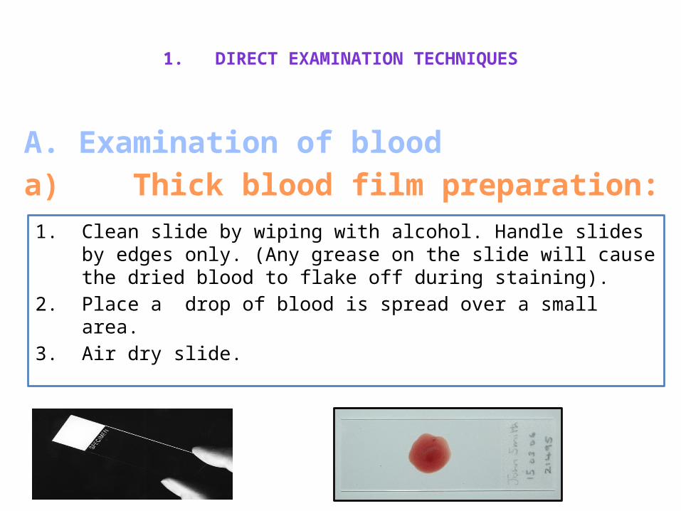
A. Examination of blooda) Thick blood film preparation:
1. Clean slide by wiping with alcohol. Handle slides by edges only. (Any grease on the slide will cause the dried blood to flake off during staining).
2. Place a drop of blood is spread over a small area.
3. Air dry slide.
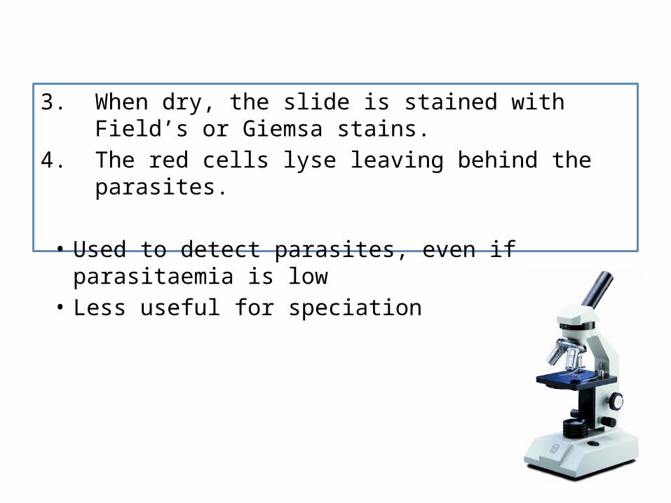
3. When dry, the slide is stained with Field’s or Giemsa stains.
4. The red cells lyse leaving behind the parasites.
• Used to detect parasites, even if parasitaemia is low• Less useful for speciation

Step 2: Place a clean slide on a flat rigid surface. Transfer a spot of the blood collected on the corner of the PUSHER slide to one end of this slide.
Step 3: Wipe the end of the PUSHER slide until clean and dry.
Step 4: Holding the PUSHER slide at a 30° angle draw it back along the specimen slide until the bottom edge contacts the drop of blood.
Step 5: After the blood spreads along the bottom edge of the PUSHER slide move it smoothly forward in one quick movement. This should spread an even bullet shaped film of blood onto the specimen slide.
b) Thin blood film preparation

Step 6: This shows the angle to hold, and the direction to move the PUSHER slide when making the blood smear on the specimen slide.
Fixed in methanol and stained with Giemsa stain.• Used for speciation• Does not detect low
parasitaemia

Trypansoma brucei sp. in thick blood smears stained with Giemsa.
Trypanosoma brucei sp. in a thin blood smear stained with Giemsa.

Trypanosoma brucei gambiense trypomastigotes
T. brucei rhodesiense trypomastigotes
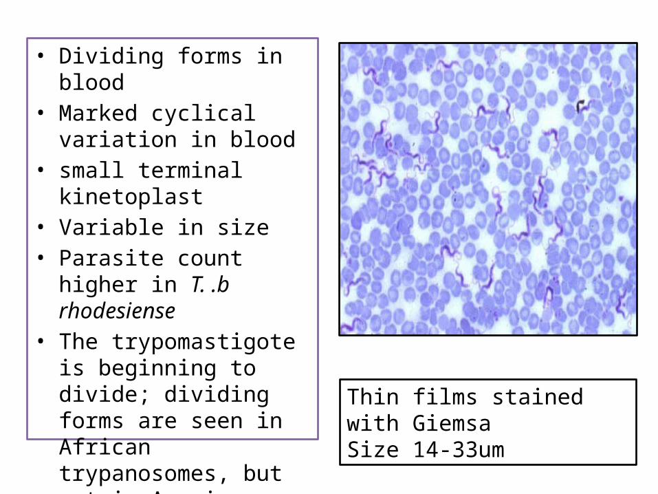
• Dividing forms in blood• Marked cyclical variation in
blood• small terminal kinetoplast• Variable in size• Parasite count higher in T. .b
rhodesiense• The trypomastigote is
beginning to divide; dividing forms are seen in African trypanosomes, but not in American trypanosomes. Thin films stained with Giemsa
Size 14-33um

1. DIRECT EXAMINATION TECHNIQUES
Parasite concentration
techniques
Triple centrifugation
technique
Microhaematocrit centrifugation technique (Woo
method)
Miniature anion-exchange
centrifugation technique
Buffy coat examination
B. Parasite concentration techniques:-

B. Parasite concentration techniques:- a) Triple centrifugation technique
5 to 10 ml of blood is centrifuged at 2000 rpm for 5 minutes to pack the red blood cells.
The plasma and white cell layer are removed by a Pasteur pipette and transferred to a clean centrifuge tube and centrifuged.
The supernatant fluid is removed by pipette to a clean tube and centrifuged at 5000 rpm for 10 minutes.
The supernatant fluid is removed with a pipette and discarded.
The deposit is examined microscopically for trypanosomes.

b) Microhaematocrit centrifugation technique (Woo method)
Principle• A capillary tube is filled with whole blood by capillary action
to within 1 to 2 cm of the end.• The unfilled end is sealed and the tube is centrifuged. • After centrifugation, the capillary tube is placed in a reading
device and the hematocrit value determined.
Procedure.
(1) Fill a plain capillary tube with aanticoagulated blood.If blood without anticoagulant is used, fill a heparinized capillary tube with the blood specimen.
- A heparinized capillary tube is identified by red line on the tube specimen.
- A heparinized capillary tube is identified by a red line on the tube.

(2) Allow blood to enter two capillary tubes until they are approximately 2/3 filled with blood. (Air bubbles denote poor technique, but do not affect the results of the test.
(3) Seal the unfilled ends of the tubes with clay.
(4) Place the two hematocrit tubes in the radial grooves of the centrifuge head exactly opposite each other, with the sealed end away from the center of the centrifuge.
(5) Screw the flat centrifuge head cover in place.
(6) Centrifuge at 10,000 rpm for 5 minutes.
(7) Remove the hematocrit tubes as soon as the centrifuge has stopped spinning.
(8) Determine the hematocrit values with the aid of a microhematocrit reader.
• The microhematocrit technique is advantageous because of speed, and because only a small quantity of blood is necessary for the determination.
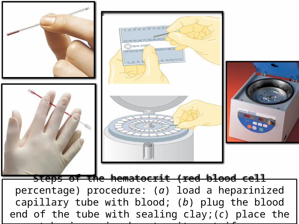
Steps of the hematocrit (red blood cell percentage) procedure: (a) load a heparinized capillary tube with blood; (b) plug the blood end of the
tube with sealing clay;(c) place the tube in a microhematocrit centrifuge.

c) Miniature anion-exchange centrifugation technique• The number of parasites in the blood of Trypanosoma brucei
rhodesiense patients is usually high enough for them to be seen by direct examination of blood under a light microscope.
• However, T. b. gambiense and cerebrospinal fluid (CSF) parasites exist in such low numbers that they have to be concentrated before viewing.
• The most sensitive method for detection of trypanosomes in blood that is commonly used is the mini Anion Exchange Centrifugation Technique (mAECT).
• The mAECT method is carried out in two stages, including chromatography then concentration and viewing.
• In the first step, the parasites are separated from venous blood in a gel column by anion exchange chromatography and collected in a sealed glass tube.

• Heparinised blood is passed through an anion exchange column.
• As the blood travels down the column the red cells are adsorbed while the less strongly charged trypanosomes are washed through with saline.
• The eluate is centrifuged and examined microscopically for motile trypanosomes.
• The glass tube is sealed by passing the narrow end of a Pasteur pipette over a flame in a procedure that is potentially dangerous.
• In a second step, the parasites are concentrated at the bottom of the sealed glass tube by low speed centrifugation. The tip of the glass tube is then examined for the presence of motile trypanosomes under low magnification microscopy.(Figure: 2A)
Limitations: • It is expensive, time consuming, not easy to standardize, and cannot
be used in most field situations where electricity is often lacking.

Figure 1A Figure 1B
Figure 1: (A) The old gel column used to filter trypanosomes from blood in the mAECT test. The gel is retained in the injector by two filters, a large upper one and a small lower one, seen here in yellow. (B) Filtration of blood using the new version of gel column. The gel is held in place by two white polyethylene filters of equal size (here shown just above and below the blood-stained gel), and mounted on a holding rack that allows easy collection of the filtrate in the collector tube below (Figure 2B).
Gel column
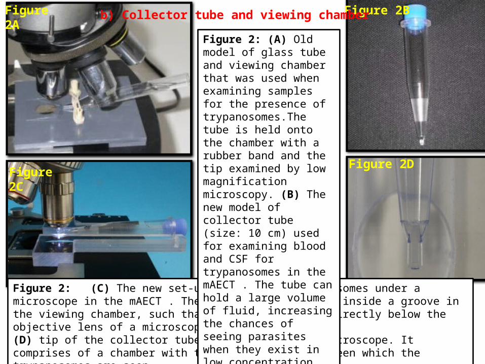
Figure 2: (C) The new set-up for viewing trypanosomes under a microscope in the mAECT . The collector tube rests inside a groove in the viewing chamber, such that its narrow end is directly below the objective lens of a microscope. (D) tip of the collector tube as seen under the microscope. It comprises of a chamber with two flat surfaces between which the trypanosomes are seen.
Figure 2A Figure 2B
Figure 2DFigure 2C
Figure 2: (A) Old model of glass tube and viewing chamber that was used when examining samples for the presence of trypanosomes.The tube is held onto the chamber with a rubber band and the tip examined by low magnification microscopy. (B) The new model of collector tube (size: 10 cm) used for examining blood and CSF for trypanosomes in the mAECT . The tube can hold a large volume of fluid, increasing the chances of seeing parasites when they exist in low concentration.
b) Collector tube and viewing chamber

This set-up is also used for viewing parasites in the modified single centrifugation (MSC) technique for examination of cerebrospinal fluid (CSF) during staging of HAT patients, and follow-up after treatment. Briefly, CSF is collected into the tube by gravity, directly from the lumbar puncture needle, then examined for the presence of trypanosomes under a microscope after low-speed centrifugation.

d) Buffy coat examination• Buffy coat ia s yellowish-white layer of leukocytes and platelets
that forms on top of the column of red blood cells upon centrifugation of whole blood.
• Trypanosomes are centrifuged in a microhaematoctit tube for 5 minutes.
• Parasites can be seen microscopically at the junction of the packed red cells and plasma.
• This technique is rapid and sensitive
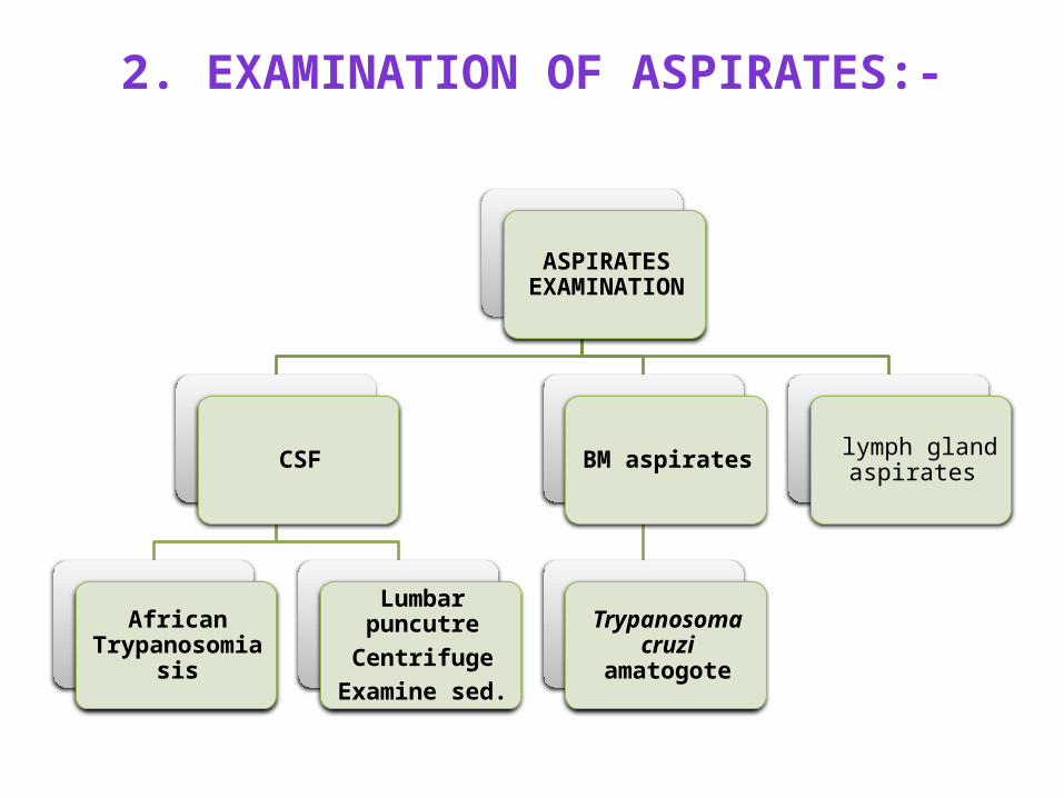
2. EXAMINATION OF ASPIRATES:-
ASPIRATES EXAMINATION
CSF
African Trypanosomiasis
Lumbar puncutreCentrifuge
Examine sed.
BM aspirates
Trypanosoma cruzi amatogote
lymph gland aspirates
A. Examination of lymph gland aspirates The aspirate can be examined microscopically by making a wet
preparation, or if there is not much material, it can be allowed to dry on a slide and then stained with either rapid Field’s stain or with Giemsa and examined microscopically.

B. Examination of CSF• In the late stages of African trypanosomiasis, trypanosomes may
be found in the CSF together with IgM - containing morula (Mott) cells, lymphocytes and other mononuclear cells.
• Once the CSF has been collected it must be examined as soon as possible. (The parasites are unable to survive for more than 15-20 minutes in CSF once it has been removed.The parasites become inactive, are rapidly lysed and will not therefore be detected.)
• The CSF should be examined wet and spun down in a sterile tube and a film made from the deposit.
• The film is then stained with rapid Field’s or Giemsa and examined microscopically.

C. Lumbar aspiration
Figure 1: Undergoing a painful lumbar puncture in a patient whose blood was positive for African Sleeping Sickness parasites (photo courtesy of WHO)
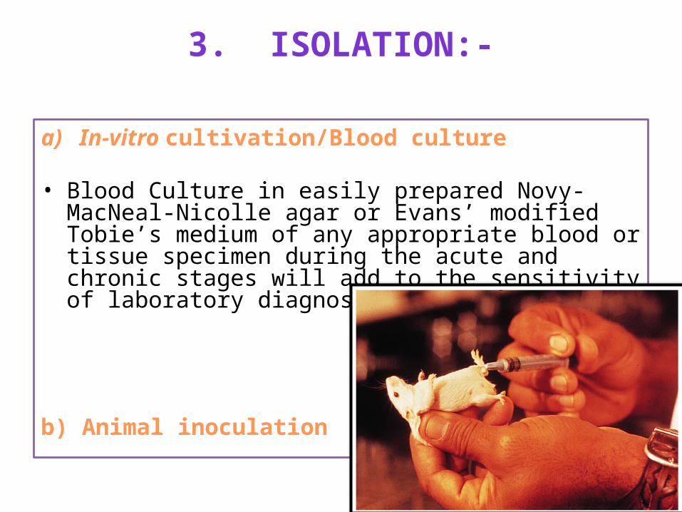
3. ISOLATION:-
a) In-vitro cultivation/Blood culture
• Blood Culture in easily prepared Novy-MacNeal-Nicolle agar or Evans’ modified Tobie’s medium of any appropriate blood or tissue specimen during the acute and chronic stages will add to the sensitivity of laboratory diagnosis.
b) Animal inoculation
4. TEST TO DETECT TRYPANOSOMAL ANTIGEN
DNA amplification tests (PCR):• An in vitro method for enzymatic synthesis of DNA
• Reaction uses two oligonucleotide primers that hybridize to opposite strands and flank the region of interest.
• A heat stable DNA polymerase catalyses the elongation of primers.
• Primers extension products serve as template in next cycle.
• Numbers of target copies double in each cycle.

PCR component mixture


Serological tests
Immuno
fuorescence test (IFT)
ELISACATT/T. b. gambiense.
LATEX/T. b.
gambiense

5. SEROLOGICAL TESTS:-
a) Immunofluorescence test(IFT)• Fluorescent dyes (fluorescein or rhodamine) attached to
known specific Abs, used to detect presence of Abs in serum or Ag (Parasites) in a sample– Direct fluorescent Ab test: used to detect Ag or Parasite– Indirect fluorescent Ab test: used to find a specific Ab in
the serum• In chronic phase when count is low• Uses IFA antigen slides prepared from a suspension of
epimastigotes
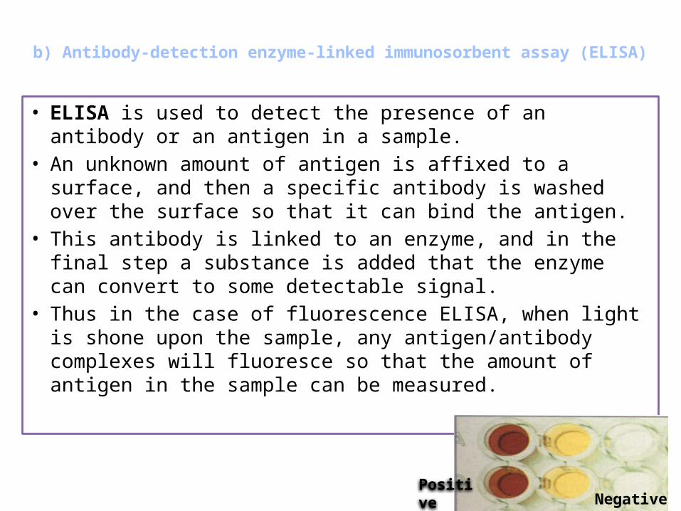
b) Antibody-detection enzyme-linked immunosorbent assay (ELISA)
• ELISA is used to detect the presence of an antibody or an antigen in a sample.
• An unknown amount of antigen is affixed to a surface, and then a specific antibody is washed over the surface so that it can bind the antigen.
• This antibody is linked to an enzyme, and in the final step a substance is added that the enzyme can convert to some detectable signal.
• Thus in the case of fluorescence ELISA, when light is shone upon the sample, any antigen/antibody complexes will fluoresce so that the amount of antigen in the sample can be measured.
PositiveNegative
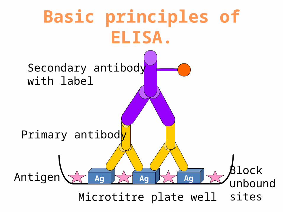
Basic principles of ELISA.
Ag Ag Ag
Microtitre plate well
Antigen Block unbound sites
Primary antibody
Secondary antibody with label

c) CATT/T. b. gambiense.
• Card Agglutination Test for Trypanosomiasis(CATT) is a Antibody based molecular diagnosis.
• Anti-trypanosomal IgM detected by simple / rapid CATT.• Serum samples are mixed on a plastic card with fixed and
stained trypanosomes as antigen and the test is positive when the antigen agglutinates.
• A titre can be determined by serial dilutions of the serum.• The great advantage of this test is that it is easy to carry out
even in the field.
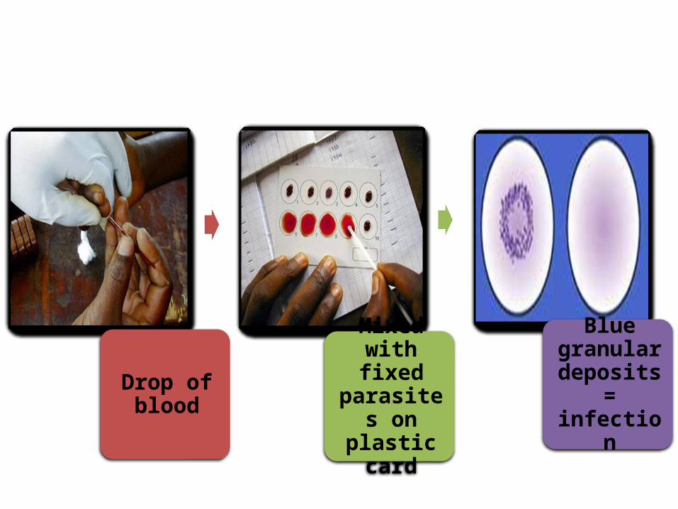
Drop of blood
Mixed with fixed
parasites on plastic
card
Blue granular
deposits = infection
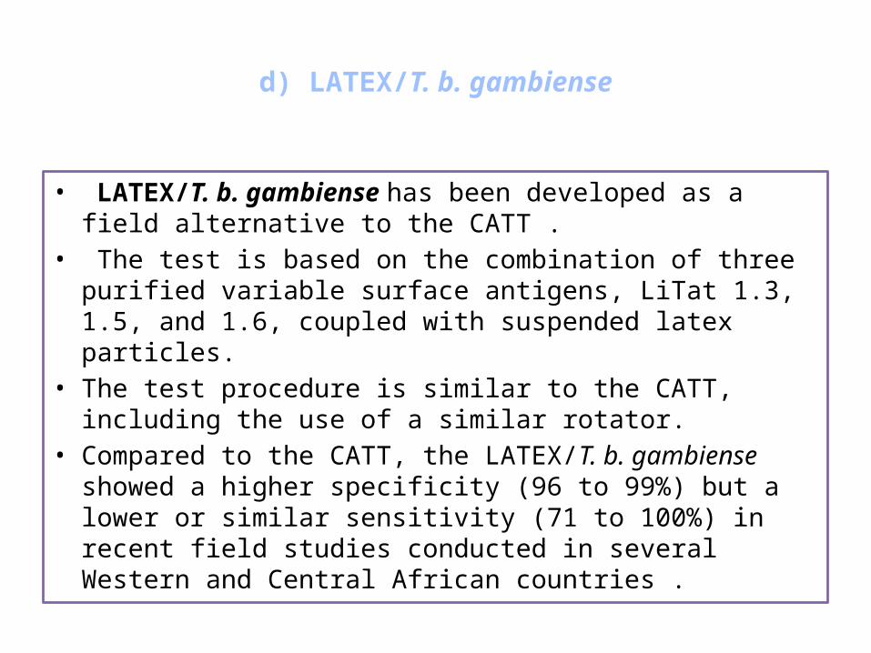
d) LATEX/T. b. gambiense
• LATEX/T. b. gambiense has been developed as a field alternative to the CATT .
• The test is based on the combination of three purified variable surface antigens, LiTat 1.3, 1.5, and 1.6, coupled with suspended latex particles.
• The test procedure is similar to the CATT, including the use of a similar rotator.
• Compared to the CATT, the LATEX/T. b. gambiense showed a higher specificity (96 to 99%) but a lower or similar sensitivity (71 to 100%) in recent field studies conducted in several Western and Central African countries .

e) Molecular Dipstick Test
• A dyed strip is dipped into a blood sample from a potential patient.
• The test picks up a signature "biomarker" molecule from a parasite, which causes the colour of the dipstick to change under ultra-violet light — indicating parasite presence.
• Dipstick test is used to detect african sleeping sickness, Chaga’s disease and Leishmaniasis.

• LAMP of DNA is a promising new molecular technique that shows high sensitivity and specificity.
• The test uses only one enzyme, and amplification occurs under isothermal conditions
• The test amplifies target DNA at a constant temperature, meaning that it can be carried out with minimal equipment.
• The parasitologic tests in use for diagnosis of human African trypanosomiasis (HAT), or sleeping sickness.
• LAMP Method for Rapid Detection of Trypanosoma brucei rhodesiense.
• Detection of trypanosomal DNA sequences from a patient’s blood, urine or saliva could be a significant improvement on parasitological examination.
f) LAMP: Loop-mediated isothermal amplification of DNA
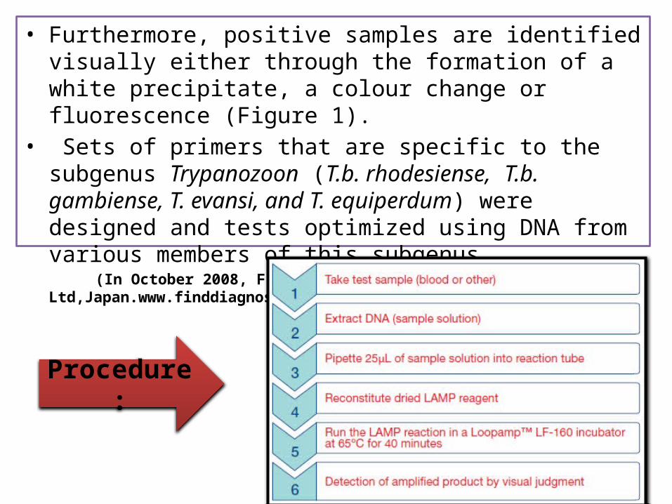
• Furthermore, positive samples are identified visually either through the formation of a white precipitate, a colour change or fluorescence (Figure 1).
• Sets of primers that are specific to the subgenus Trypanozoon (T.b. rhodesiense, T.b. gambiense, T. evansi, and T. equiperdum) were designed and tests optimized using DNA from various members of this subgenus.
(In October 2008, FIND and Eiken Chemical Company Ltd,Japan.www.finddiagnostics.org)
Procedure :
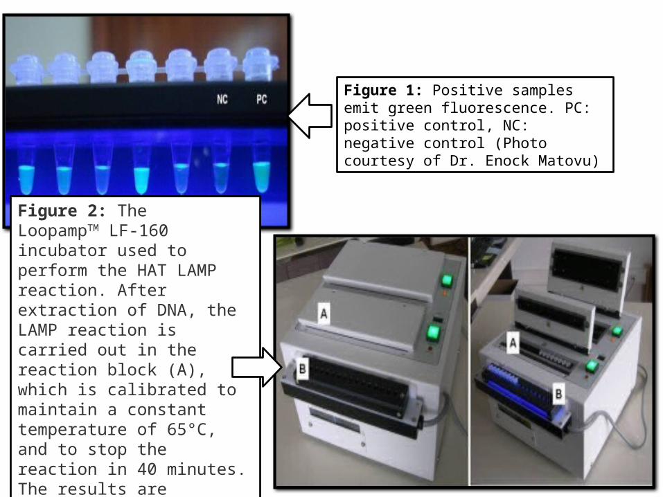
Figure 1: Positive samples emit green fluorescence. PC: positive control, NC: negative control (Photo courtesy of Dr. Enock Matovu)
Figure 2: The LoopampTM LF-160 incubator used to perform the HAT LAMP reaction. After extraction of DNA, the LAMP reaction is carried out in the reaction block (A), which is calibrated to maintain a constant temperature of 65°C, and to stop the reaction in 40 minutes. The results are visualized under LED light (B).

DIAGNOSIS OF
CHAGAS’ DISEASE
IDENTIFICATION
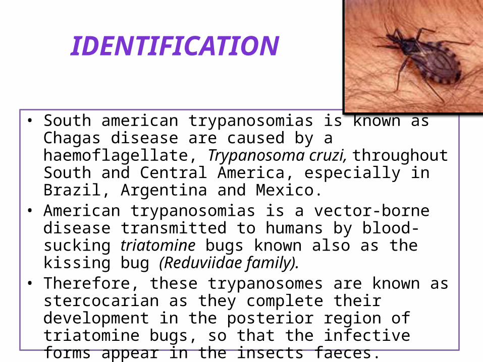
• South american trypanosomias is known as Chagas disease are caused by a haemoflagellate, Trypanosoma cruzi, throughout South and Central America, especially in Brazil, Argentina and Mexico.
• American trypanosomias is a vector-borne disease transmitted to humans by blood-sucking triatomine bugs known also as the kissing bug (Reduviidae family).
• Therefore, these trypanosomes are known as stercocarian as they complete their development in the posterior region of triatomine bugs, so that the infective forms appear in the insects faeces.
• Hosts are infected by the contaminative route.

CLINICAL DIAGNOSIS
is usually easy among children in endemic areas.
irregular fever and enlarged lymph nodes
Cardiac dilation, megacolon and megaesophagus in individuals from endemic areas indicate present or former infection.
Sample
Demonstration of trypanosomes in blood, lymph node aspirates, cerebral spinal fluid
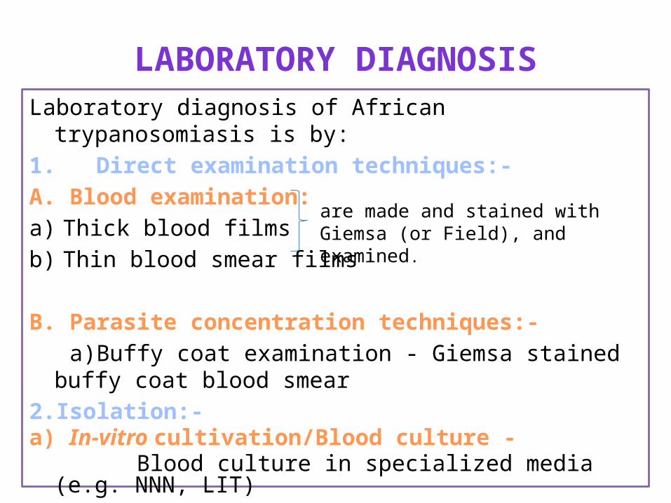
LABORATORY DIAGNOSISLaboratory diagnosis of African trypanosomiasis is by:
1. Direct examination techniques:-
A. Blood examination:
a) Thick blood films
b) Thin blood smear films
B. Parasite concentration techniques:-
a)Buffy coat examination - Giemsa stained buffy coat blood smear
2.Isolation:-a) In-vitro cultivation/Blood culture - Blood culture in specialized media (e.g. NNN, LIT)
are made and stained with Giemsa (or Field), and examined.

T. cruzi trypomastigote in cerebrospinal fluid (CSF) stained with Giemsa.
Trypanosoma cruzi in thin blood film (Leishmans stain)
showing developing tryptomastigotes that have a
free flagellum

Trypomastigotes show:
• large, subterminal or terminal kinetoplast,
• centrally located nucleus, • an undulating membrane,• flagellum leaving the body at
anterior end. • Trypanosomes measure about
20µm in length.• No dividing blood forms

b) Animal inoculation - inoculation into mice- Definitive diagnosis requires the demonstration of trypanosomes by biological tests (in the insect or mice).
c) Xenodiagnosis - where uninfected Reduviidae bugs are fed on the patient's blood, and their gut contents examined for parasites 4 weeks later
4. Test to detect trypanosomal antigen• DNA amplification tests(PCR)
5. Serological tests:- ( Detection of trypanosomal antibodies in the serum)
a) Indirect fluorescent antibody test (IFT)
b) Antibody-detection enzyme-linked immunosorbent assay (ELISA)
c) Complement fixation test (CFT)
d) Immunofluorescence assay (IFA)
e) Indirect hemagglutination
f) RIA• Raised ESR, marked lymphocytosis with atypical mononuclear
lymphocytes

Blood CultureBlood culture is as sensitive as xenodiagnosis but it requires sterile conditions.
Xenodiagnosis• A method of diagnosing certain diseases caused by insects, ticks,
or other vectors, by allowing uninfected vectors to feed on the patient and later examining them for infections
• Xenodiagnosis is useful in chronic and sub acute (low parasitaemia) disease.
• Sterile bugs are fed on patients by attaching a black bag containing the bugs to the arm of the patient and allowing them to feed for 30 minutes.
• 25 to 30 days later the bugs are dissected and the contents of the hindgut and rectum are examined microscopically for the presence of trypanosomes.
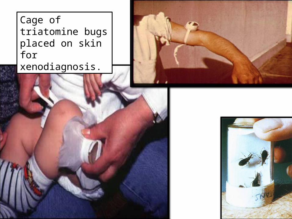
Cage of triatomine bugs placed on skin for xenodiagnosis.

5. SEROLOGICAL DIAGNOSIS
a) Complement fixation test (CFT) :
• The complement fixation test is to detect the presence of either specific antibody or specific antigen in a patient's serum.
Complement Fixation Test in Microtiter Plate.
Rows 1 and 2 exhibit complement fixation obtained with acute and convalescent phase serum specimens, respectively. (2-fold serum dilutions were used) The observed 4-fold increase is significant and indicates recent infection.

It uses sheep red blood cells (sRBC), anti sRBC antibody and complement, plus specific antigen (if looking for antibody in serum) or specific antibody (if looking for antigen in serum).
But if the antibody (or antigen) is not present, then the complement is not used up, so it binds anti-sRBC antibody, and the sRBCs are lysed.
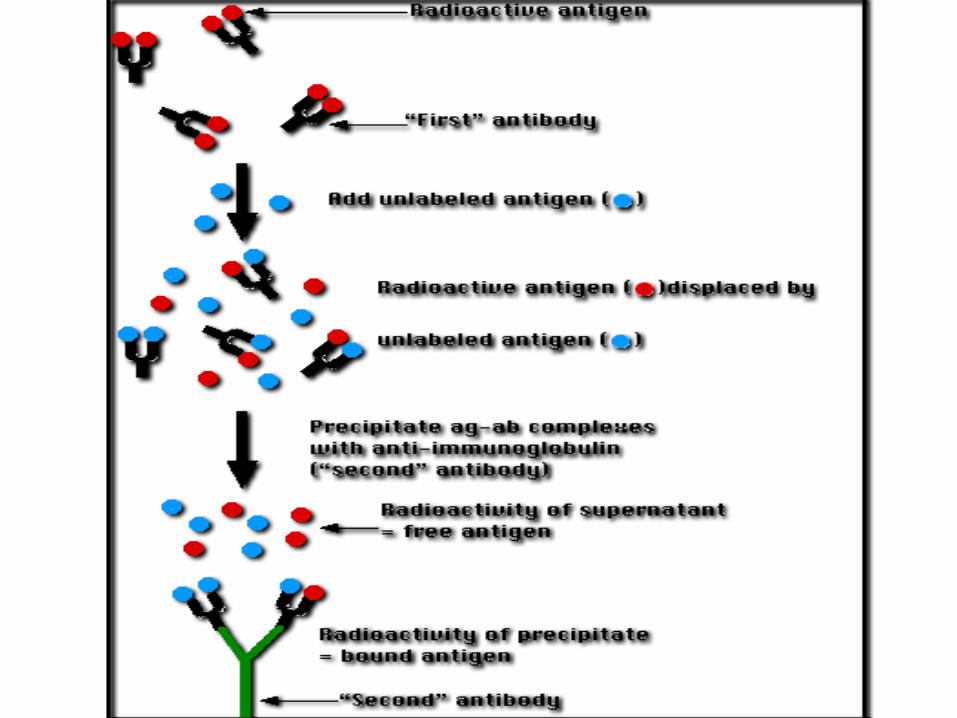
b) Radio immune assay (RIA)
• RIA involves the separation of a protein (from a mixture) using the specificity of antibody - antigen binding and quantitation using radioactivity.
• A mixture is prepared of – radioactive antigen – antibodies against that antigen.
• Known amounts of unlabeled ("cold") antigen are added to samples of the mixture.
• These compete for the binding sites of the antibodies.• At increasing concentrations of unlabeled antigen, an increasing
amount of radioactive antigen is displaced from the antibody molecules.
• The antibody-bound antigen is separated from the free antigen in the supernatant fluid, and the radioactivity of each is measured.

Example of antibody based molecular diagnosis
1. FATALA kit : measures T. cruzi antibodies in blood using 2 recombinant proteins
2. BIO CHAGAS kit: uses cocktail recombinant T. cruzi antigens.
Infection sera produces blue
precipitate on strip in 60 mins.
DIAGNOSIS OF
SURRA

Introduction
• Trypanosoma evansi causes a trypanosomosis known as ‘surra’.
• The principal host species such as camels, horses, buffalos and cattle are particularly affected, although other animals, including wildlife, are also susceptible.
• It is an arthropod-borne disease; several species of haematophagous flies, including Tabanids and Stomoxes, are implicated in transferring infection from host to host, acting as mechanical vectors.
• In South America, it causes murrina and can be transmitted mechanically by the bite of vampire bats.

LABORATORY DIAGNOSISLaboratory diagnosis of African trypanosomiasis is by:
1. Diagnosis Based on Clinical sign:-• The clinical signs of surra are indicative but are not sufficiently
pathognomonic, thus,diagnosis must be confirmed by laboratory methods
(Dia et al., 1997a)
2. Direct examination techniques:-
A. Blood examination:
a) Thick blood films
b) Thin blood smear filmsare made and stained with Giemsa (or Field), and examined.

Demonstration of parasites in blood films:
67
B. Parasite concentration techniques:-
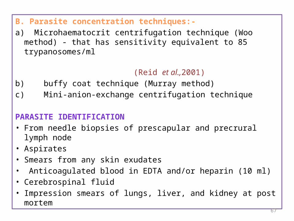
a) Microhaematocrit centrifugation technique (Woo method) - that has sensitivity equivalent to 85 trypanosomes/ml
(Reid et al.,2001)
b) buffy coat technique (Murray method)
c) Mini-anion-exchange centrifugation technique
PARASITE IDENTIFICATION• From needle biopsies of prescapular and precrural lymph node• Aspirates• Smears from any skin exudates• Anticoagulated blood in EDTA and/or heparin (10 ml)• Cerebrospinal fluid• Impression smears of lungs, liver, and kidney at post mortem

3.Chemical test
a) Mercuric chloride test
b) Formal gel test
c) Thymol turbidity test
d) Jone’s nitric acid test
e) Stilbamidine test
Mercuric chloride test:- Helpful in carrier animals’ examination- 99% accuracy- 1 drop serum + 1 cc merchuric perchloride- White precipitates with in few minutes = POSITIVE
69
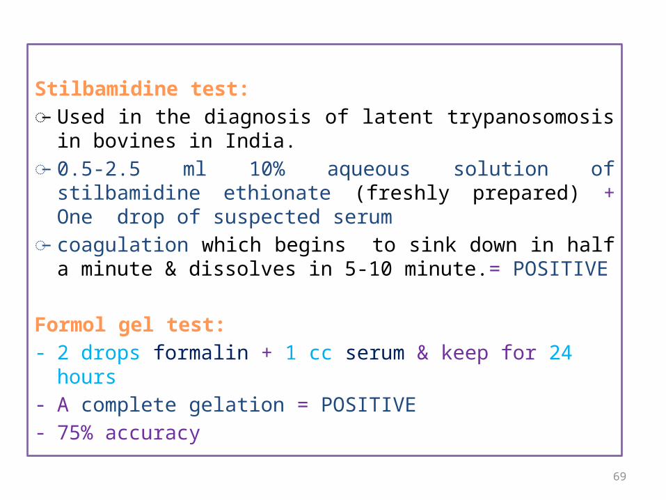
Stilbamidine test:̵R Used in the diagnosis of latent trypanosomosis in bovines in
India. ̵R 0.5-2.5 ml 10% aqueous solution of stilbamidine ethionate
(freshly prepared) + One drop of suspected serum̵R coagulation which begins to sink down in half a minute &
dissolves in 5-10 minute.= POSITIVE
Formol gel test:- 2 drops formalin + 1 cc serum & keep for 24 hours- A complete gelation = POSITIVE- 75% accuracy

4. Blood & serum analysis: - Low Glucose (Hyoglycaemia)- Increased protein levels (β &
γ globulin)5.Isolation:-
a) In-vitro cultivation/Blood culture
b) Animal inoculation
Mouse inoculation(MI) test:
is accepted as the most sensitive method to detect the animal trypanosomosis.
- Inoculate heparinised blood intraperitoneally into rats (1–2 ml) or mice (0.25–0.5 ml).
- Inoculate a minimum of two animals.- Bleed animals from the tail after every 48 hours to detect
parasitaemia. - Numerous parasites in blood after few days

4. Molecular tests
(Test to detect trypanosomal antigen)
a) DNA-probes (nucleic acid probes).
b) The polymerase chain reaction (PCR).
c) Loop-mediated Isothermal Amplification (LAMP) test for detection of Trypanosoma evansi strain B
• The principle of molecular tests is the demonstration of the occurrence of sequences of nucleotides, which are specific for a trypanosome subgenus, species or even type or strain.
• A positive result indicates active infection with the trypanosome for which the sequences are specific, as parasite DNA will not persist for long in the host after all live parasites have been eliminated.
• These tests are not only suitable for detecting parasites in the mammalian host, but also in the insect vector.

72
DNA-probes (nucleic acid probes).
• The sample to be examined is heated to separate the two strands of DNA (this is also called denaturing of DNA), and these are fixed to a membrane, so that they cannot recombine again on cooling.
• A probe is then added. A probe consists of a linear sequence of nucleotides of a certain length, which has been prepared to correspond with a similar sequence of nucleotides in one of the strands of the parasite which the test is meant to detect.
• The probe will link (hybridize) with that part of the parasite DNA strand which is the mirror image of the base sequence of the probe.
73
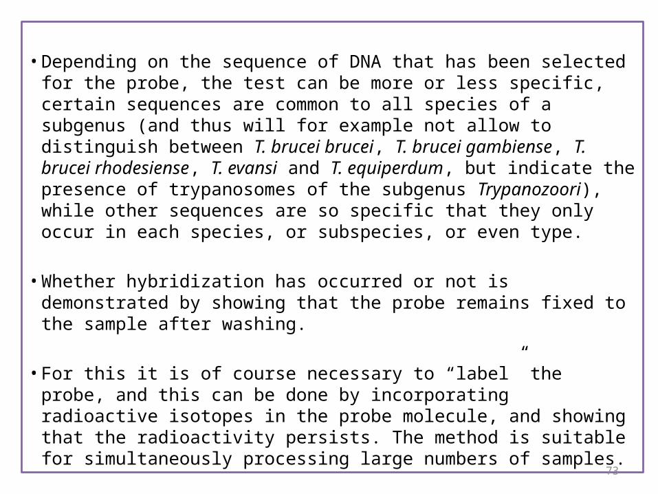
• Depending on the sequence of DNA that has been selected for the probe, the test can be more or less specific, certain sequences are common to all species of a subgenus (and thus will for example not allow to distinguish between T. brucei brucei, T. brucei gambiense, T. brucei rhodesiense, T. evansi and T. equiperdum, but indicate the presence of trypanosomes of the subgenus Trypanozoori), while other sequences are so specific that they only occur in each species, or subspecies, or even type.
• Whether hybridization has occurred or not is demonstrated by showing that the probe remains fixed to the sample after washing.
• For this it is of course necessary to “label” the probe, and this can be done by incorporating radioactive isotopes in the probe molecule, and showing that the radioactivity persists. The method is suitable for simultaneously processing large numbers of samples.
74
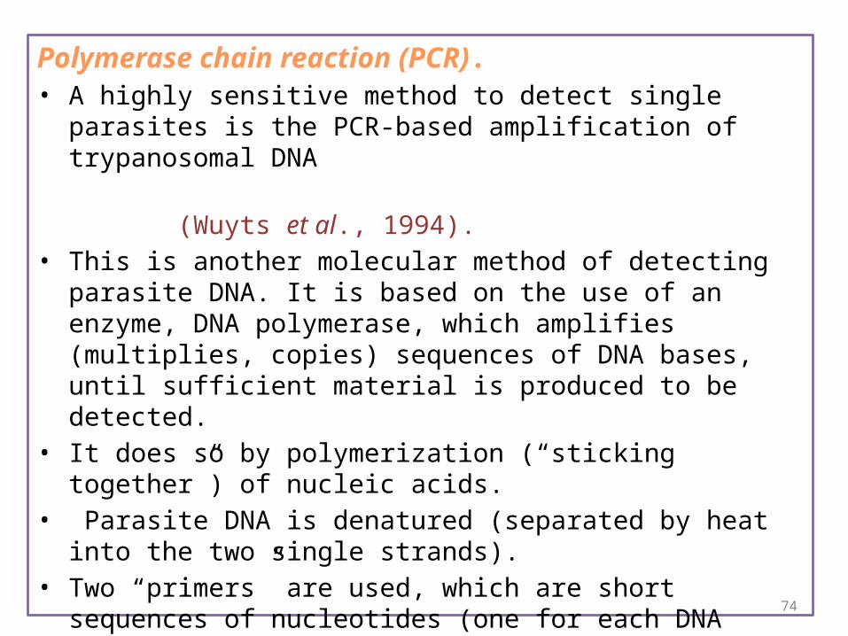
Polymerase chain reaction (PCR). • A highly sensitive method to detect single parasites is the PCR-based
amplification of trypanosomal DNA
(Wuyts et al., 1994).• This is another molecular method of detecting parasite DNA. It is
based on the use of an enzyme, DNA polymerase, which amplifies (multiplies, copies) sequences of DNA bases, until sufficient material is produced to be detected.
• It does so by polymerization (“sticking together”) of nucleic acids.• Parasite DNA is denatured (separated by heat into the two single
strands). • Two “primers” are used, which are short sequences of nucleotides (one
for each DNA strand), each constructed so as to be complementary to a specific site on one of the two single parasite DNA strands.
• The primers attach to the sites for which they are complementary and DNA polymerase then starts to reproduce the rest of each complementary sequence which follows from that primer.

75
• This occurs in opposite directions until the entire sequence of double-stranded DNA between the primers has been doubled (as a complementary strand is produced from each primer).
• The polymerase can of course only do its work when nucleic acids are added to the test material.
• The cycle is then repeated, the two double-stranded DNA sequences are again denatured, the primers attach again, the polymerase amplifies, etc.
• In the end, the PCR product is submitted to electrophoresis and the bands are detected by special staining.

76
Loop-mediated Isothermal Amplification (LAMP) test for detection of Trypanosoma evansi strain B.
• Trypanosoma evansi strains that express variable surface glycoprotein (VSG) RoTat 1.2. However, in Kenya a second causative strain that does not express RoTat 1.2 VSG (T. evansi type B) has been identified.
• This work reports the development of a sensitive and specific diagnostic assay capable of detecting T. evansi type B based on the strategy of Loop-mediated Isothermal Amplification (LAMP) of DNA.
• The test is rapid and amplification is achieved within 20-25min at 63C using a real time PCR machine.
• Restriction enzyme AluI digestion of the amplicon gave the predicted 83bp and 89bp sized bands and the LAMP product melt curves showed consistent melting temperature (T(m)) of approximately 89C.
• The assay analytical sensitivity is approximately 0.1tryps/ml while that of classical PCR test targeting the same gene is approximately 10tryps/ml.
( Zablon K Njiru et al.,2010)

77
5. Serological tests:-
( Detection of trypanosomal antibodies in the serum )
a) Latex agglutination test:• also makes use of T. evansi RoTat 1.2 clone
(World Organisation for Animal Health ,2008)
b) Enzyme linked immunosorbent assays : (Antibody detection ELISA)
• very useful for large-scale surveys• ELISA using variable surface glycoproteins from a T. evansi RoTat
1.2 clone
c) Card agglutination test: (CATT/T. evansi)• also makes use of T. evansi RoTat 1.2 clone• This has been developed from a commercial test for the diagnosis of
human sleeping sickness (the Testryp® CATT), into a commercial kit for T. evansi, CATT test T. evansi®. For the detection of antibodies to surra.

78
Immunofluorescence test (IFAT): • A smear of blood containing fixed trypanosomes constitutes the
antigen. • The bovine serum to be examined is put into contact with the
smear, and immunoglobulins (antibodies) against the trypanosomal antigens attach themselves to the trypanosomes in the smear, and remain stuck to the smear even when the serum is washed off.
• Rabbit anti- bovine immunoglobulins (In order to show that antibodies have reacted with the antigen, a commercial preparation containing antibodies (also immunoglobulins) raised in laboratory animals), is applied to the smear, and washed off after being allowed to react.

79
• If these rabbit antibodies remain stuck to the slide, the test is positive, as they indicate the presence of anti-trypanosome bovine immunoglobulins which have reacted with the antigen slide.
• In order to show the presence or absence of specific antibodies against trypanosomes in the bovine serum, the rabbit anti-bovine immunoglobulins in are conjugated with a fluorescent dye, (fluoresceine) which can be detected by ultraviolet light.
• The combination of immunoglobulins and fluoresceine is called a conjugate, and in this case it is a rabbit anti-bovine conjugate.
• If the microscopic examination shows the trypanosomes in the antigen smear to be fluorescent, the test result is positive.
• One can “titrate” the bovine serum (determine the level of specific anti-trypanosome immunoglobulins) by using serial dilutions of the serum and determining the end point, the highest dilution still giving a positive test result.

80
Figure : shows the principle of
the test.

Reference• Eastern meditterranean health journal vol.13 No. 7 2007• C H A P T E R 2 . 4 . 1 8 .TRYPANOSOMOSIS (tsetse-transmitted) OIE
Terrestrial Manual 2008• World Organization for Animal Health [OIE] . Manual of diagnostic tests
and vaccines for terrestrial animals [online]. Paris: OIE; 2008. Trypanosomosis (Tsetse-transmitted). Available at: http://www.oie.int/eng/normes/mmanual/2008/pdf/2.04.18_TRYPANOSOMOSIS.pdf. Accessed 27 Aug 2009.
• (Chapter 2.1.17. — Trypanosoma evansi infection (surra) 14 OIE Terrestrial Manual 2010)

THANK YOU