diagnosis dilemmas in vital pulp therapy: treatment for ... · pdf filebetween the pulp and...
TRANSCRIPT
DtIJ
ATdnoelpuatars(
KD
CC
j
vt0
t
Did
Pulp Symposium
S
iagnosis Dilemmas in Vital Pulp Therapy: Treatment forhe Toothache Is Changing, Especially in Young,mmature Teethoe H. Camp, DDS, MSD
Depmi
cMsheCc
Cnbtc
drrats
mticce
icadtmgds
o
bstracthe literature is almost devoid of scientific studies ofiagnosis of pulpal pathology in primary and perma-ent teeth with open apices. Most reports are empiricalr retrospective studies without adequate prior knowl-dge of preexisting conditions or histologic findingseading to the necessity of pulpal procedures. Appro-riate diagnostic tests and their effectiveness are doc-mented for both groups. This article reviews the avail-ble literature and current techniques of indirect pulpherapy, pulp capping, and pulpotomy for primary teethnd permanent teeth with open apex. T h e apical bar-ier with mineral trioxide aggregate followed by roottrengthening with bonded composite is reviewed.J Endod 2008;34:S6-S12)
ey Wordsiagnosis, pulp, pulp capping, pulpotomy
From private practice of endodontics, Charlotte, Northarolina, and Department of Endodontics, University of Northarolina, Chapel Hill, North Carolina.
Address requests for reprints to Dr Camp at E-mail address:[email protected].
Conflict of Interest: Joe H. Camp, DDS, MSD, is the Uni-ersity Clinical Consultant for Dentsply Tulsa Dental Special-ies, a part-time, paid position.099-2399/$0 - see front matter
Copyright © 2008 American Academy of Pediatric Den-istry and American Association of Endodontists.
This article is being published concurrently in Pediatricentistry, May/June 2008; Volume 30, Issue 3. The articles are
dentical. Either citation can be used when citing this article.oi:10.1016/j.joen.2008.03.020
s
6 Camp
iagnosis in primary and young, permanent, immature teeth varies greatly from thatin fully formed permanent teeth. Most of the diagnostic tests used in conventional
ndodontic therapy are of very little or no value in primary teeth and of limited value inermanent immature teeth. While admittedly poor for diagnosing the degree of inflam-ation in this group of teeth, diagnostic tests must be performed to obtain as much
nformation as possible before arriving at treatment options.Diagnostic literature based on scientific studies is almost nonexistent. Most out-
ome reports are supported by empirical treatment and anecdotal case reports (1).any outcome studies are conducted retrospectively on the basis of clinical signs and
ymptoms and make assumptions regarding the pulpal status before treatment withoutistologic or bacterial data to support the preoperative diagnosis. Without histologicxamination an accurate determination of the extent of inflammation is impossible (2).orrelation between clinical symptoms and histopathologic conditions is poor andomplicates diagnosis of pulpal health in exposed pulps of children (3).
Many of our treatments are based on our diagnosis of the root development stage.onsequently, to properly diagnose and treat primary and young permanent teeth, it isecessary to have a thorough knowledge of normal root formation and the differencesetween developing and fully formed teeth. The decision to render conservative vital
reatment to allow root formation completion or more radical treatment such as rootanal therapy might hinge on our diagnosis of root development.
According to Orban (4), the tooth root’s development begins after enamel andentin formation has reached the cementoenamel junction (CEJ). Hertwig’s epithelialoot sheath is formed by the epithelial dental organ, with one tube for each of the futureoots. As root formation proceeds apically, each root is wide open, diverging apicallynd limited by the epithelial diaphragm. Each root’s internal surface is lined by odon-oblasts. Once root length is established, the sheath disappears, whereas dentin depo-ition is continued until root formation is completed.
Depending on each root’s external anatomy, differentiation into multiple canalsight occur. During this formative stage, communication exists between the canals in
he form of isthmuses. As growth continues, the opposing walls meet and coalesce, andslands of dentin are formed, which eventually expand to divide the root into separateanals. Continued dentin deposition narrows the canals, and the apex is eventuallylosed with dentin and cementum, creating apical convergence. Isthmuses and finsxtending toward the root’s center might persist in fully formed teeth.
In permanent teeth, root formation is not completed until 1– 4 years after eruptionnto the oral cavity. Because of the shorter roots of primary teeth, root formation isompleted faster than for permanent teeth. Because the faciolingual width of most rootsnd canals is greater than the mesiodistal width, apical closure cannot usually beetermined radiographically. The x-ray beam is exposed in the faciolingual plane, but
he radiograph is read mesiodistally. Because of this anatomy, with the exception of theaxillary central and lateral incisors and some single canal lower premolars, radio-
raphs cannot determine apical closure. Therefore, the clinician must rely on time toetermine root closure in all other teeth to prevent treatment protocols that cannot beuccessfully completed without apical convergence (5).
During this formative period, treatments should be oriented toward maintenancef vitality to allow completion of root formation. Further deposition of dentin will
trengthen the roots’ thin dentinal walls and help diminish future root fracture.JOE — Volume 34, Number 7S, July 2008
macabmwtomfa
nstaoiar
pAdDotvrf
abrn
c
et
adawaadifo
ordscsc
smmcpmt
fm
o
eaaqctp
uWmatc
tru
cldtctF
Nr
Pulp Symposium
J
The root canals of primary teeth differ greatly from those of per-anent teeth, and treatment is complicated by apical resorption to
llow for eruption of the succeeding teeth. At the time of root lengthompletion, the root canals roughly correspond to the externalnatomy’s form and shape. At this time, resorption of the rootsegins and, combined with additional dentin deposition internally,ight significantly change the number, size, and shape of the canalsithin the primary roots. Continued physiologic, apical resorption of
he roots makes the teeth progressively shorter. In addition, resorptionn the roots’ internal surfaces adjacent to the forming permanent toothight open other communications with the periapical tissues. These
actors complicate establishment of working lengths if root canal ther-py is necessary.
In the primary anterior teeth (incisors and canines), the perma-ent tooth buds lie apically and lingually near the primary roots. Re-orption is initiated on the primary root’s lingual surface. This causeshe apical foramen to move coronally, resulting in a difference in thepical foramen and the anatomic apex and complicating determinationf root canal length. One study demonstrated that half of the primaryncisor’s root might be resorbed lingually before it becomes obvious onradiograph (6). Primary anterior teeth have one simple root canal andarely have lateral or accessory canals.
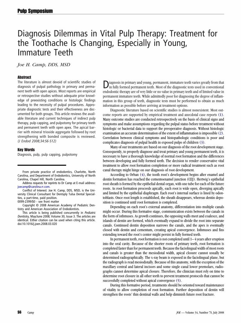
The primary molar teeth normally have the same number andosition of roots and root canals as the corresponding permanent teeth.t root length completion, most roots have only 1 canal, but continuedeposition of dentin might divide the root into 2 or more canals (7–9).uring this time, communication exists between the canals in the formf isthmuses or fins (Fig. 1). Secondary dentin deposition contributes tohis change in morphology (10, 11). Like the permanent molars, mostariations occur in the mesial roots of mandibular molars and facialoots of the maxillary molars. Also, these variations are usually in theacial to lingual plane and cannot be visualized on radiographs (7, 9).
Accessory and lateral canals and apical ramifications of the pulpre common in primary molars (8). In addition, other communicationsetween the pulp and the periapical tissues are formed by physiologicesorption of the internal surfaces of the roots adjacent to the perma-ent tooth buds.
Diagnosis of Pulpal Status in Primary TeethAs with any dental procedure, a thorough medical history must be
ompleted, and any implications related to treatment must be consid-
igure 1. Silicone models of the pulps of 2 primary mandibular second molars.ote the communication between the facial and lingual canals in the mesial
moots.
OE — Volume 34, Number 7S, July 2008
red. A child with systemic disease might necessitate different treatmenthan a healthy one.
The examination should begin with a thorough history and char-cteristics of any pain, because these are often important in helping toetermine pulpal status and eventual treatment. Whereas pain usuallyccompanies pulpal inflammation, extensive problems might ariseithout any history of pain. If possible, a distinction between provokednd spontaneous pain should be ascertained. Provoked pain that ceasesfter removal of the causative stimulation is usually reversible and in-icative of minor inflammatory changes. Stimuli include thermal, chem-
cal, and mechanical irritants and many times are due to deep caries,aulty restorations, soreness around a primary tooth nearing exfoliation,r an erupting permanent tooth.
Spontaneous pain is a constant or throbbing pain that occurs with-ut stimulation or continues long after the causative factor has beenemoved. In a well-controlled histologic study of primary teeth witheep carious lesions, Guthrie et al. (12) demonstrated that a history ofpontaneous toothache is usually associated with extensive degenerativehanges extending into the root canals. Primary teeth with a history ofpontaneous pain should not receive vital pulpal treatments and areandidates for pulpectomy or extraction.
The clinical examination might produce evidence of pulpal patho-is. Redness, swelling, fluctuance, severe dental decay, defective orissing restorations, and draining parulis might indicate pulpal involve-ent. Percussion sensitivity might be valuable to the diagnosis, but it is
omplicated by the reliability of the child’s response because of thesychological aspects involved. Tooth mobility might be present nor-ally because of physiologic resorption, and many pulpally involved
eeth have no mobility.Electric pulp tests are not valid in primary teeth (1). Laser Doppler
lowmetry might be of greater help in determining vitality, but this equip-ent has not been perfected, and the price is prohibitive (13).
Thermal tests are usually not conducted on primary teeth becausef their unreliability (1, 5).
After the clinical examination, radiographs of good quality aressential. Like permanent teeth, periapical radiolucencies appear at thepices in primary anterior teeth. In primary molars, pathologic changesre most often apparent in the bifurcation or trifurcation areas. Conse-uently, bite-wing radiographs are often best to observe pathologichanges in posterior primary teeth. Pathologic bone and root resorp-ions are signs of advanced pulpal pathosis that has spread into theeriapical tissues and is usually treatable only with extraction.
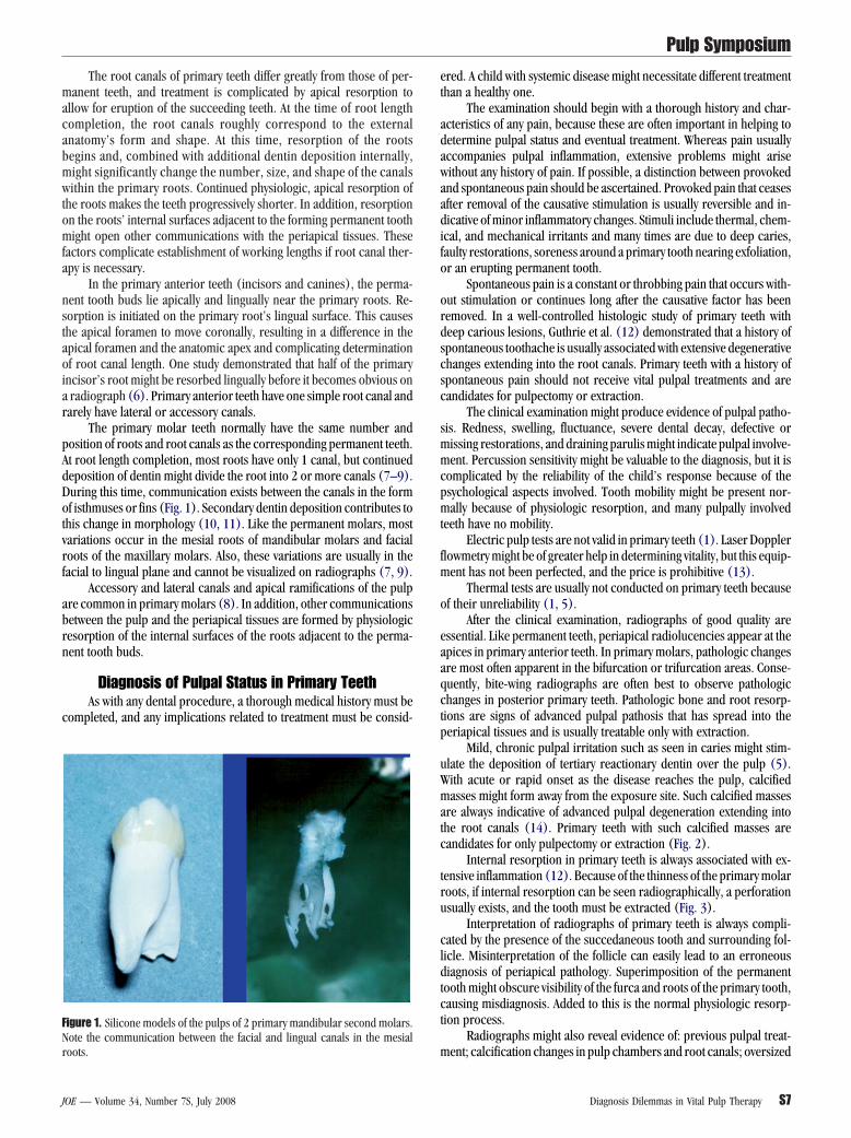
Mild, chronic pulpal irritation such as seen in caries might stim-late the deposition of tertiary reactionary dentin over the pulp (5).ith acute or rapid onset as the disease reaches the pulp, calcifiedasses might form away from the exposure site. Such calcified masses
re always indicative of advanced pulpal degeneration extending intohe root canals (14). Primary teeth with such calcified masses areandidates for only pulpectomy or extraction (Fig. 2).
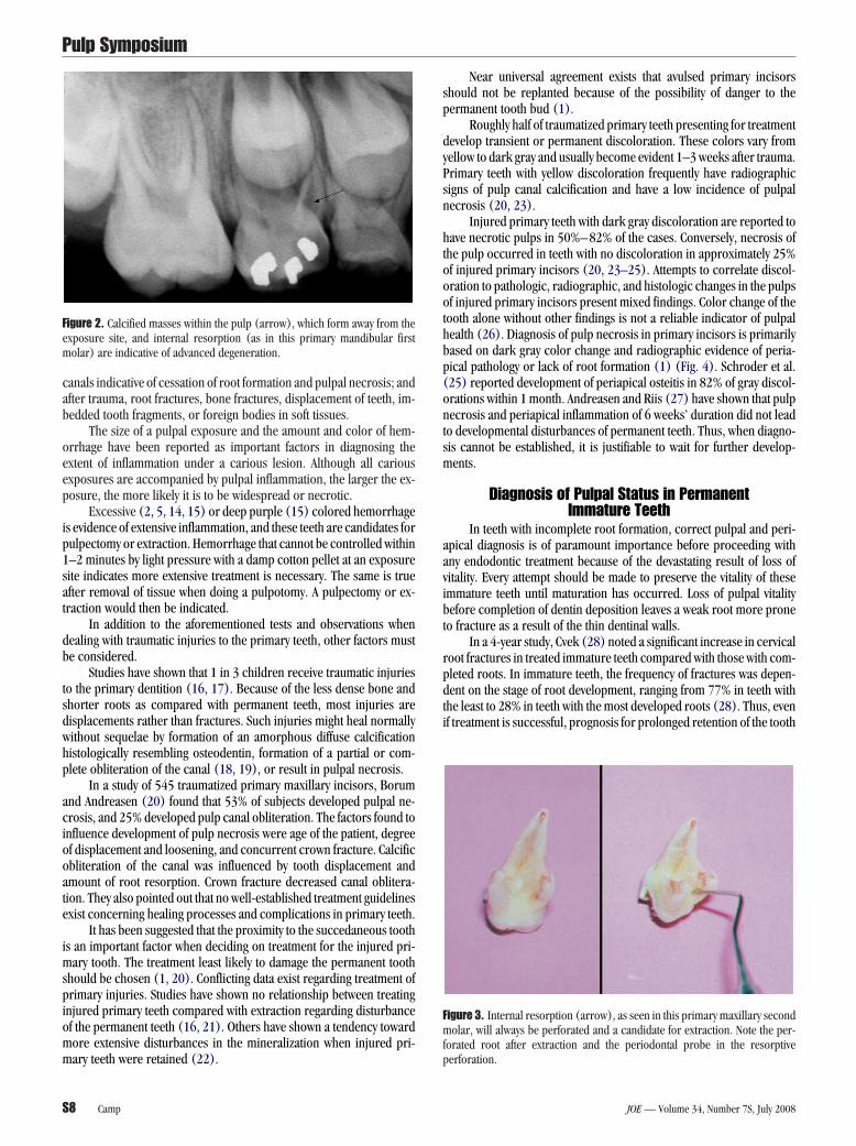
Internal resorption in primary teeth is always associated with ex-ensive inflammation (12). Because of the thinness of the primary molaroots, if internal resorption can be seen radiographically, a perforationsually exists, and the tooth must be extracted (Fig. 3).
Interpretation of radiographs of primary teeth is always compli-ated by the presence of the succedaneous tooth and surrounding fol-icle. Misinterpretation of the follicle can easily lead to an erroneousiagnosis of periapical pathology. Superimposition of the permanent
ooth might obscure visibility of the furca and roots of the primary tooth,ausing misdiagnosis. Added to this is the normal physiologic resorp-ion process.
Radiographs might also reveal evidence of: previous pulpal treat-
ent; calcification changes in pulp chambers and root canals; oversizedDiagnosis Dilemmas in Vital Pulp Therapy S7
cab
oeep
ip1sat
db
tsdwhp
aciooate
imspiomm
sp
dyPsn
htooothbp(ontsm
aavibt
rpdti
Fem
Fmf
Pulp Symposium
S
anals indicative of cessation of root formation and pulpal necrosis; andfter trauma, root fractures, bone fractures, displacement of teeth, im-edded tooth fragments, or foreign bodies in soft tissues.
The size of a pulpal exposure and the amount and color of hem-rrhage have been reported as important factors in diagnosing thextent of inflammation under a carious lesion. Although all cariousxposures are accompanied by pulpal inflammation, the larger the ex-osure, the more likely it is to be widespread or necrotic.
Excessive (2, 5, 14, 15) or deep purple (15) colored hemorrhages evidence of extensive inflammation, and these teeth are candidates forulpectomy or extraction. Hemorrhage that cannot be controlled within–2 minutes by light pressure with a damp cotton pellet at an exposureite indicates more extensive treatment is necessary. The same is truefter removal of tissue when doing a pulpotomy. A pulpectomy or ex-raction would then be indicated.
In addition to the aforementioned tests and observations whenealing with traumatic injuries to the primary teeth, other factors muste considered.
Studies have shown that 1 in 3 children receive traumatic injurieso the primary dentition (16, 17). Because of the less dense bone andhorter roots as compared with permanent teeth, most injuries areisplacements rather than fractures. Such injuries might heal normallyithout sequelae by formation of an amorphous diffuse calcificationistologically resembling osteodentin, formation of a partial or com-lete obliteration of the canal (18, 19), or result in pulpal necrosis.
In a study of 545 traumatized primary maxillary incisors, Borumnd Andreasen (20) found that 53% of subjects developed pulpal ne-rosis, and 25% developed pulp canal obliteration. The factors found tonfluence development of pulp necrosis were age of the patient, degreef displacement and loosening, and concurrent crown fracture. Calcificbliteration of the canal was influenced by tooth displacement andmount of root resorption. Crown fracture decreased canal oblitera-ion. They also pointed out that no well-established treatment guidelinesxist concerning healing processes and complications in primary teeth.
It has been suggested that the proximity to the succedaneous tooths an important factor when deciding on treatment for the injured pri-
ary tooth. The treatment least likely to damage the permanent toothhould be chosen (1, 20). Conflicting data exist regarding treatment ofrimary injuries. Studies have shown no relationship between treatingnjured primary teeth compared with extraction regarding disturbancef the permanent teeth (16, 21). Others have shown a tendency towardore extensive disturbances in the mineralization when injured pri-
igure 2. Calcified masses within the pulp (arrow), which form away from thexposure site, and internal resorption (as in this primary mandibular firstolar) are indicative of advanced degeneration.
ary teeth were retained (22). p
8 Camp
Near universal agreement exists that avulsed primary incisorshould not be replanted because of the possibility of danger to theermanent tooth bud (1).
Roughly half of traumatized primary teeth presenting for treatmentevelop transient or permanent discoloration. These colors vary fromellow to dark gray and usually become evident 1–3 weeks after trauma.rimary teeth with yellow discoloration frequently have radiographicigns of pulp canal calcification and have a low incidence of pulpalecrosis (20, 23).
Injured primary teeth with dark gray discoloration are reported toave necrotic pulps in 50%– 82% of the cases. Conversely, necrosis of
he pulp occurred in teeth with no discoloration in approximately 25%f injured primary incisors (20, 23–25). Attempts to correlate discol-ration to pathologic, radiographic, and histologic changes in the pulpsf injured primary incisors present mixed findings. Color change of the
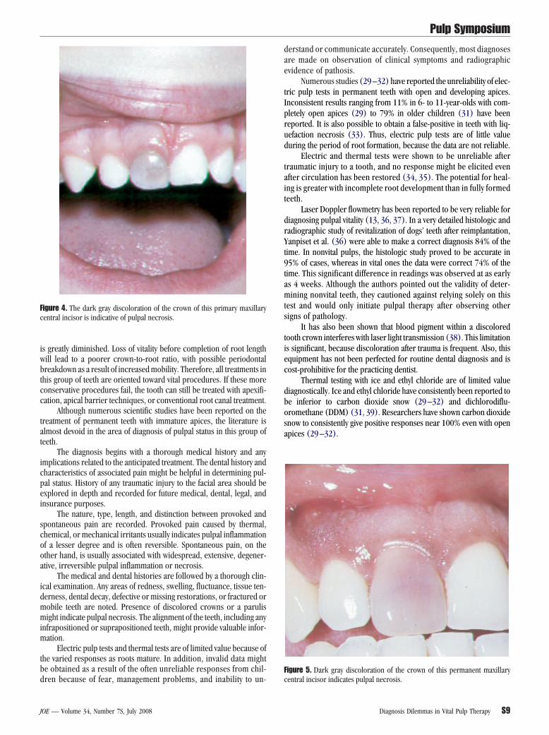
ooth alone without other findings is not a reliable indicator of pulpalealth (26). Diagnosis of pulp necrosis in primary incisors is primarilyased on dark gray color change and radiographic evidence of peria-ical pathology or lack of root formation (1) (Fig. 4). Schroder et al.25) reported development of periapical osteitis in 82% of gray discol-rations within 1 month. Andreasen and Riis (27) have shown that pulpecrosis and periapical inflammation of 6 weeks’ duration did not lead
o developmental disturbances of permanent teeth. Thus, when diagno-is cannot be established, it is justifiable to wait for further develop-ents.
Diagnosis of Pulpal Status in PermanentImmature Teeth
In teeth with incomplete root formation, correct pulpal and peri-pical diagnosis is of paramount importance before proceeding withny endodontic treatment because of the devastating result of loss ofitality. Every attempt should be made to preserve the vitality of thesemmature teeth until maturation has occurred. Loss of pulpal vitalityefore completion of dentin deposition leaves a weak root more prone
o fracture as a result of the thin dentinal walls.In a 4-year study, Cvek (28) noted a significant increase in cervical
oot fractures in treated immature teeth compared with those with com-leted roots. In immature teeth, the frequency of fractures was depen-ent on the stage of root development, ranging from 77% in teeth with
he least to 28% in teeth with the most developed roots (28). Thus, evenf treatment is successful, prognosis for prolonged retention of the tooth
igure 3. Internal resorption (arrow), as seen in this primary maxillary secondolar, will always be perforated and a candidate for extraction. Note the per-
orated root after extraction and the periodontal probe in the resorptive
erforation.JOE — Volume 34, Number 7S, July 2008
iwbtcc
tat
icpei
scooa
idmmim
tbd
dae
tIprud
tait
drYt9tamts
tiec
dbosa
Fc
F
Pulp Symposium
J
s greatly diminished. Loss of vitality before completion of root lengthill lead to a poorer crown-to-root ratio, with possible periodontalreakdown as a result of increased mobility. Therefore, all treatments inhis group of teeth are oriented toward vital procedures. If these moreonservative procedures fail, the tooth can still be treated with apexifi-ation, apical barrier techniques, or conventional root canal treatment.
Although numerous scientific studies have been reported on thereatment of permanent teeth with immature apices, the literature islmost devoid in the area of diagnosis of pulpal status in this group ofeeth.
The diagnosis begins with a thorough medical history and anymplications related to the anticipated treatment. The dental history andharacteristics of associated pain might be helpful in determining pul-al status. History of any traumatic injury to the facial area should bexplored in depth and recorded for future medical, dental, legal, andnsurance purposes.
The nature, type, length, and distinction between provoked andpontaneous pain are recorded. Provoked pain caused by thermal,hemical, or mechanical irritants usually indicates pulpal inflammationf a lesser degree and is often reversible. Spontaneous pain, on thether hand, is usually associated with widespread, extensive, degener-tive, irreversible pulpal inflammation or necrosis.
The medical and dental histories are followed by a thorough clin-cal examination. Any areas of redness, swelling, fluctuance, tissue ten-erness, dental decay, defective or missing restorations, or fractured orobile teeth are noted. Presence of discolored crowns or a parulisight indicate pulpal necrosis. The alignment of the teeth, including any
nfrapositioned or suprapositioned teeth, might provide valuable infor-ation.
Electric pulp tests and thermal tests are of limited value because ofhe varied responses as roots mature. In addition, invalid data mighte obtained as a result of the often unreliable responses from chil-
igure 4. The dark gray discoloration of the crown of this primary maxillaryentral incisor is indicative of pulpal necrosis.
ren because of fear, management problems, and inability to un- c
OE — Volume 34, Number 7S, July 2008
erstand or communicate accurately. Consequently, most diagnosesre made on observation of clinical symptoms and radiographicvidence of pathosis.
Numerous studies (29 –32) have reported the unreliability of elec-ric pulp tests in permanent teeth with open and developing apices.nconsistent results ranging from 11% in 6- to 11-year-olds with com-letely open apices (29) to 79% in older children (31) have beeneported. It is also possible to obtain a false-positive in teeth with liq-efaction necrosis (33). Thus, electric pulp tests are of little valueuring the period of root formation, because the data are not reliable.
Electric and thermal tests were shown to be unreliable afterraumatic injury to a tooth, and no response might be elicited evenfter circulation has been restored (34, 35). The potential for heal-ng is greater with incomplete root development than in fully formedeeth.
Laser Doppler flowmetry has been reported to be very reliable foriagnosing pulpal vitality (13, 36, 37). In a very detailed histologic andadiographic study of revitalization of dogs’ teeth after reimplantation,anpiset et al. (36) were able to make a correct diagnosis 84% of theime. In nonvital pulps, the histologic study proved to be accurate in5% of cases, whereas in vital ones the data were correct 74% of the
ime. This significant difference in readings was observed at as earlys 4 weeks. Although the authors pointed out the validity of deter-ining nonvital teeth, they cautioned against relying solely on this
est and would only initiate pulpal therapy after observing otherigns of pathology.
It has also been shown that blood pigment within a discoloredooth crown interferes with laser light transmission (38). This limitations significant, because discoloration after trauma is frequent. Also, thisquipment has not been perfected for routine dental diagnosis and isost-prohibitive for the practicing dentist.
Thermal testing with ice and ethyl chloride are of limited valueiagnostically. Ice and ethyl chloride have consistently been reported toe inferior to carbon dioxide snow (29 –32) and dichlorodiflu-romethane (DDM) (31, 39). Researchers have shown carbon dioxidenow to consistently give positive responses near 100% even with openpices (29 –32).
igure 5. Dark gray discoloration of the crown of this permanent maxillary
entral incisor indicates pulpal necrosis.Diagnosis Dilemmas in Vital Pulp Therapy S9
ar
dp
omt
dm
FT(
Pulp Symposium
S
Thermal tests with heat in permanent teeth with developing apicesre of limited value because of inconsistent responses (32) and arearely performed.
Radiographic examination and interpretation are key elements in theiagnosis of pulpal pathology in teeth with developing apices. Good qualityeriapical radiographs of any involved teeth are used to assess root devel-
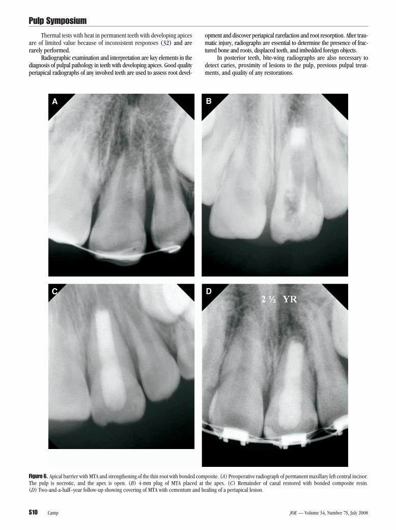
igure 6. Apical barrier with MTA and strengthening of the thin root with bondedhe pulp is necrotic, and the apex is open. (B) 4-mm plug of MTA place
D) Two-and-a-half–year follow-up showing covering of MTA with cementum and he10 Camp
pment and discover periapical rarefaction and root resorption. After trau-atic injury, radiographs are essential to determine the presence of frac-
ured bone and roots, displaced teeth, and imbedded foreign objects.In posterior teeth, bite-wing radiographs are also necessary to
etect caries, proximity of lesions to the pulp, previous pulpal treat-ents, and quality of any restorations.
osite. (A) Preoperative radiograph of permanent maxillary left central incisor.the apex. (C) Remainder of canal restored with bonded composite resin.
compd at
aling of a periapical lesion.
JOE — Volume 34, Number 7S, July 2008
aou
trphsl
apes(phe
pfonRdnotcr
of
drpntPidg
citttcha
map
toc
haati(
rarfTaCee
hpwaa
1
1
1
1
1
1
1
1
1
1
Pulp Symposium
J
Discoloration of a tooth crown after trauma is a common sequeland one of the foremost diagnostic indicators (40 – 42). Yellow discol-ration is usually indicative of pulp space calcification, and a gray colorsually signifies pulpal necrosis (40) (Fig. 5).
Transient coronal discoloration has been reported (42) in 4% ofeeth after luxation injuries as a result of vascular damage and hemor-hage immediately after injury. In these cases, it was speculated thatulpal healing depends on the bacterial status. With bacterial infection,ealing is unlikely. In this group of teeth, determination of bacterialtatus could not be ascertained on the basis of coronal discoloration,oss of pulpal sensibility, or periapical rarefaction (42).
Transient apical breakdown occurs after displacement injuriesnd might lead to misdiagnosis (42, 43). The development of transienteriapical radiolucency—together with coronal discoloration, negativelectric pulp test, and cold response up to 4 months—was shown toubsequently regain the original color and normal pulpal responses43). Transient apical breakdown apparently is linked to the repairrocess in the pulp and periapical tissues and returns to normal whenealing is complete (42). Bone loss, which produces the radiolucency,ventually heals with new bone.
Universal agreement exists that immature teeth have the greatestotential to heal after trauma or caries, particularly when the apicaloramen is wide open. This group of teeth also has the greatest chancef misdiagnosis and mistreatment. To avoid mistakes, treatment mustot be undertaken on the basis of negative responses to pulp testing.adiographic and symptomatic assessment is currently the principaliagnostic criterion. The following factors are key in making the diag-ostic determination: symptoms of irreversible pulpitis or apical peri-dontitis; clinical signs of periradicular infection including swelling,enderness to percussion, mobility, or parulis formation; radiographi-ally detectible bone loss; progressive root resorption; and arrestedoot development compared with other adjacent teeth (44).
If doubtful, do not start treatment. Keep the patient under closebservation and continue to reassess the diagnostic criteria until a de-initive diagnosis can be established.
The treatment of primary and young permanent teeth has changedramatically in recent years as new materials have been developed andesearched. The use of calcium hydroxide (for decades the standard forulp protection), pulp capping, and pulpotomy procedures in perma-ent teeth is being replaced with composite resins (45, 46) and mineralrioxide aggregate (MTA) (ProRoot; Dentsply Tulsa Dental, Tulsa, OK).ulp capping with resin composites in monkeys produced the lowestncidence of bacterial microleakage, pulpal inflammation, and inci-ence of pulpal necrosis when compared with calcium hydroxide andlass ionomer cement (46).
When compared with calcium hydroxide, MTA produced signifi-antly more dentinal bridging in a shorter time with significantly lessnflammation and less pulpal necrosis (47– 49). MTA has been showno be cementoconductive, with attachment of cementoblasts to the ma-erial (49). Sarkar et al. (50) studied the interactions of MTA, a syn-hetic tissue fluid, and dentin of extracted teeth. They concluded thatalcium from the MTA reacted with phosphate in tissue fluid, producingydroxyapatite. The sealing ability, biocompatibility, and dentinogenicctivity of the material occur because of these physiochemical reactions.
Once considered taboo, vital pulpal treatment of symptomatic per-anent teeth with MTA has been reported (5, 51, 52) to be successful,
llowing continued root development. Stronger roots with greatly im-roved prognosis for permanent retention are now possible.
Loss of pulpal vitality in open apex teeth in the past led to pro-racted treatment and often ended in early tooth loss caused by fracturef weak roots. Apical barrier techniques with MTA now allow timely
ompletion of apexification and have eliminated the use of calciumOE — Volume 34, Number 7S, July 2008
ydroxide, except as a temporary canal disinfectant. The use of MTA asn apical barrier has become the standard for treatment of the openpex pulpless tooth (Fig. 6). The development of bonded compositeechniques now allows strengthening of these weak roots to levels ofntact, fully formed roots and has virtually ended root fractures (53–56)Fig. 6C).
Revascularization of teeth with necrotic infected canals has beeneported by using combinations of antibiotics (57, 58). The canals areccessed and disinfected with copious irrigation of sodium hypochlo-ite. The canals are not instrumented. A paste of metronidazole, cipro-loxacin, and minocycline is placed in the canals and left for 1 month.he tooth is re-entered, and endodontic files are inserted through thepices to stimulate bleeding to produce a blood clot at the level of theEJ. After clotting, MTA is placed over the blood clot, and a permanentxternal seal is placed. The clot is then revascularized, producing thick-ning of the canal walls and apical closure.
Stem cell research holds great hope for the future, with the aim ofealing impaired dental tissues including dentin, pulp, cementum, anderiodontal tissues. By stimulating the body’s intrinsic capacities, weill be able to regenerate tissues, allowing further development of teethnd bone or possibly the formation of new teeth to replace those rav-ged by decay or lost to traumatic injuries.
References1. Flores MT, Holan G, Borum M, Andreasen JO. Injuries to the primary dentition. In:
Andreasen JO, Andreasen F, Andersson L, eds. Textbook and color atlas of traumaticinjuries to the teeth. 4th ed. Oxford, UK: Blackwell Munksgaard, 2007.
2. Starkey PE. Management of deep caries of pulpally involved teeth in children. In:Goldman HM, Forrest SP, Byrd DL, et al, eds. Current therapy in dentistry. vol 3. StLouis, MO: Mosby, 1968.
3. Mass E, Zilberman U, Fuks AB. Partial pulpotomy: another treatment option forcariously exposed permanent molars. J Dent Child 1995;62:342–5.
4. Orban BJ, ed. Oral histology and embryology. 4th ed. St Louis, MO: CV Mosby Co,1957.
5. Camp JH, Fuks AB. Pediatric endodontics: endodontic treatment for the primary andyoung, permanent dentition. In: Cohen S, Hargreaves K, eds: Pathways of the pulp. 9thed. St Louis, MO: Mosby, 2006:822– 82.
6. Kamijo Y. Studies on morphological change in the alveolar region of the jaw bonewith development of the permanent tooth from the standpoint of clinical anatomy.Bull Tokyo Dent Coll 1967;8:41.
7. Zurcher E. The anatomy of the root canals of the teeth of the deciduous dentition andof the first permanent molars. New York: William Wood & Co, 1925.
8. Hibbard E, Ireland RL. Morphology of the root canals of the primary molar teeth.J Dent Child 1957;24:250.
9. Finn SB. Morphology of the primary teeth. In: Finn SB, Caldwell RC, Cheraskin E,et al, eds. Clinical pedodontics. 3rd ed. Philadelphia: WB Saunders, 1967.
0. Ireland RL. Secondary dentin formation in deciduous teeth. J Am Dent Assoc1941;28:1626.
1. Bevelander G, Benzer D. Morphology and incidence in secondary dentin in humanteeth. J Am Dent Assoc 1943;30:1079.
2. Guthrie TJ, McDonald RE, Mitchell DF. Dental hemogram. J Dent Res 1965;44:678 – 82.
3. Evans D, Reid J, Strang R, Stirrups D. A comparison of laser Doppler flowmetry withother methods of assessing the vitality of traumatized anterior teeth. Endod DentTraumatol 1999;15:284 –90.
4. McDonald RE, Avery DR. Treatment of deep caries, vital pulp exposure, and pulplessteeth in children. In: McDonald RE, Avery DR, eds. Dentistry for the child and ado-lescent. 7th ed. St Louis, MO: Mosby, 1999.
5. Pinkham JR. Diagnosis. In: Pinkham JR, ed. Pediatric dentistry: infancy throughadolescence. Philadelphia: WB Saunders, 1988.
6. Andreasen JO, Ravn JJ. Epidemiology of traumatic dental injuries to primary andpermanent teeth in a Danish population sample. Int J Oral Surg 1972;1:235–9.
7. Glendor U. On dental trauma in children and adolescents: incidence, risk, treatment,time, and cost. Swed Dent J 2000;140(Suppl):1–52.
8. Robertson A, Lundgren T, Andreasen JO, Dietz W, Hoyer I, Noren JG. Pulp calcifica-tions in traumatized primary incisors: a morphologic and inductive analysis study.Eur J Oral Sci 1997;105:196 –206.
9. Holan G. Tube like mineralizations in the dental pulp of traumatized primary incisors.
Endod Dent Traumatol 1998;14:279 – 84.Diagnosis Dilemmas in Vital Pulp Therapy S11
2
2
2
2
2
2
2
2
2
2
3
3
3
3
3
3
3
33
3
4
4
4
4
4
4
4
4
4
4
5
5
5
5
5
5
5
5
5
Pulp Symposium
S
0. Borum MK, Andreasen JO. Sequelae of trauma to primary maxillary incisors:I— complications in the primary dentition. Endod Dent Traumatol 1998;14:31– 44.
1. Andreasen JO, Ravn JJ. The effect of traumatic injuries to primary teeth on theirpermanent successors: II—a clinical and radiographic follow-up study of 213 teeth.Scand J Dent Res 1971;79:284 –94.
2. Selliseth N. The significance of traumatized primary incisors on the development anderuption of permanent teeth. Eur Orthodont Soc Trans 1970;46:443.
3. Holan G, Fuks AB. The diagnosis value of coronal dark-gray discoloration in primaryteeth following traumatic injuries. Pediatr Dent 1996;18:224 –7.
4. Holan G. Development of clinical and radiographic signs associated with dark dis-colored primary incisors following traumatic injuries: a prospective controlled study.Dent Traumatol 2004;20:276 – 87.
5. Schröder U, Wennberg E, Granath LE, Moller H. Traumatized primary incisors: fol-low-up program based on frequency of periapical osteitis related to tooth color. SwedDent J 1977;1:95– 8.
6. Croll TP, Pascon EA, Langeland K. Traumatically injured primary incisors: a clinicaland histological study. J Dent Child 1987;54:401–22.
7. Andreasen JO, Riis I. Influence of pulp necrosis and periapical inflammation ofprimary teeth and their permanent successors: combined macroscopic and histo-logical study in monkeys. Int J Oral Surg 1978;7:178 – 87.
8. Cvek M. Prognosis of luxated non-vital maxillary incisors treated with calcium hy-droxide and filled with gutta-percha: a retrospective clinical study. Endod Dent Trau-matol 1992;8:45–55.
9. Klein H. Pulp responses to electric pulp stimulator in the developing permanentanterior dentition. J Dent Child 1978;45:199 –202.
0. Fulling HJ, Andreasen JO. Influence of maturation status and tooth type of permanentteeth upon electrometric and thermal pulp testing procedures. Scand J Dent Res1976;84:286 –90.
1. Fuss Z, Trowbridge H, Bender IB, Rickoff B. Assessment of reliability of electrical andthermal pulp testing agents. J Endod 1986;12:301–5.
2. Fulling HJ, Andreasen JO. Influence of splints and temporary crowns upon electricand thermal pulp-testing procedures. Scand J Dent Res 1976;84:291– 6.
3. Ehrmann EH. Pulp testers and pulp testing with particular reference to the use of dryice. Aust Dent J 1977;22:272–9.
4. Ohman A. Healing and sensitivity to pain in young replanted human teeth: an exper-imental clinical and histologic study. Odontol Tidskr 1965;73:166 –227.
5. Bhaskar SN, Rappaport HM. Dental vitality tests and pulp status. J Am Dent Assoc1973;86:409 –11.
6. Yanpiset K, Vongsavan N, Sigurdsson A, Trope M. Efficacy of laser Doppler flowmetryfor the diagnosis of revascularization of reimplanted immature dog teeth. Dent Trau-matol 2001;17:63–70.
7. Sigurdsson A. Pulpal diagnosis. Endod Top 2003;5:12–25.8. Heithersay GS, Hirsch RS. Tooth discoloration and resolution following a luxation
injury: Significance of blood pigment in dentin to laser Doppler flowmetry readings.Quintessence Int 1993;24:669 –76.
9. Karibe H, Ohide Y, Kohno H, et al. Study on thermal pulp testing of immature per-
manent teeth. Shigaku Odontol 1989;77:1006 –13.12 Camp
0. Bakland LK. Endodontic considerations in dental trauma. In: Ingle JI, BaklandLK, eds. Endodontics. 5th ed. Hamilton, Ontario, Canada: BC Decker Inc, 2002:795– 843.
1. Jacobsen I. Criteria for diagnosis of pulpal necrosis in traumatized permanent inci-sors. Scand J Dent Res 1980;88:306 –12.
2. Andreasen F. Transient apical breakdown and its relation to color and sensibilitychanges. Endod Dent Traumatol 1986;2:9 –19.
3. Cohenca N, Karni S, Rotstein I. Transient apical breakdown following tooth luxation.Dent Traumatol 2003;19:289 –91.
4. Mackie IC, Hill FJ. A clinical guide to the endodontic treatment of nonvital immaturepermanent teeth. Br Dent J 1999;186:54 – 8.
5. Cox CF, Suzuki S. Re-evaluating pulpal protection: calcium hydroxide liners vs cohe-sive hybridization. J Am Dent Assoc 1994;125:823–31.
6. Murray PE, Hafez AA, Smith AJ, Cox CF. Identification of hierarchical factors to guideclinical decision making for successful long-term pulp capping. Quintessence Int2003;34:61–70.
7. Junn DJ, McMillan P, Bakland LK, Torabinejad M. Quantitative assessment of dentinbridge formation following pulp-capping with mineral trioxide aggregate (MTA). JEndod 1998;24:278.
8. Pitt-Ford TR, Torabinejad M, Abedi HR, Bakland LK, Kariyawasam SP. Usingmineral trioxide aggregate as a pulp-capping material. J Am Dent Assoc1996;127:1491– 4.
9. Thomson TS, Berry JE, Somerman MJ, Kirkwood KL. Cementoblasts maintain expres-sion of osteocalcin in the presence of mineral trioxide aggregate. J Endod2003;29:407–12.
0. Sarkar NK, Caicedo R, Ritwik R, Moiseyeva R, Kawashima I. Physiochemical basis ofthe biologic properties of mineral trioxide aggregate. J Endod 2005;31:97–100.
1. Schmitt D, Lee J, Bogen G. Multifaceted use of Pro Root MTA root canal repairmaterial. Pediatr Dent 2001;23:326 –30.
2. Mejare I, Cvek M. Partial pulpotomy in young permanent teeth with deep cariouslesions. Endod Dent Traumatol 1993;9:238 – 42.
3. Hernandez R, Bader S, Boston D, Trope M. Resistance to fracture of endodonticallytreated premolars restored with new generation dentin bonding systems. Int EndodJ 1994;27:281– 4.
4. Katebzadeh N, Dalton BC, Trope M. Strengthening immature teeth during and afterapexification. J Endod 1998;24:256 –9.
5. Lawley GR, Schindler WG, Walker WA, Kolodrubetz D. Evaluation of ultrasonicallyplaced MTA and fracture resistance intracanal composite resin in a model of apexi-fication. J Endod 2004;30:167–72.
6. Goldberg F, Kaplan A, Roitman M, Manfre S, Picca M. Reinforcement effect of a resinglass ionomer in the restoration of immature roots in vitro. Dent Traumatol2002;18:70 –2.
7. Iwaya SI, Ikawa M, Kubota M. Revascularization of an immature permanent tooth withapical periodontitis and sinus tract. Dent Traumatol 2001;17:185–7.
8. Banchs F, Trope M. Revascularization of immature permanent teeth with apical
periodontitis: new technique protocol? J Endod 2004;30:196 –200.JOE — Volume 34, Number 7S, July 2008