diagnosis and therapeutic problems in endometrial cancer and therapeutic problems in... ·...
TRANSCRIPT

UNIVERSITY OF MEDICINE AND PHARMACY CRAIOVA
Dissertation Thesis Abstract
DIAGNOSIS AND THERAPEUTIC PROBLEMS IN
ENDOMETRIAL CANCER
Scientific Supervisor Prof. Nemeş Răducu Nicolae, PhD, MD
PhD Candidate Voinea Bogdan, MD
Craiova 2013
Introduction
While the early twentieth century uterus tumor pathology was dominated by
cervical cancer, endometrial cancer, with an increasing incidence in the last decades,
has become the most common cancer of the female genital tract, at least for the
socioeconomically developed countries; the causes which led to these changes are the
average lifespan increase in the female population (the high incidence of endometrial
cancer belongs to the 61-70 year-age group), and the existence of some risk factors
such as unopposed estrogen replacement therapy for menopausal women, increased
incidence of obesity, which is becoming "epidemic", diabetes and associated
hypertension, and the improvement of the investigative possibilities. In Romania,
endometrial cancer has been the second most common cancer, after cervical cancer.
Although it is the third most common cause of death among the female genital cancers,
after ovarian and cervical cancer, the endometrial cancer benefits from a complex
treatment, with a survival rate of 50%, even for the FIGO IIIB stage tumors, if the
diagnosis is established as early as possible and the treatment is immediately set up.
Material and method
62 endometrial cancer (30% of all uterine cancers with the mean incidence of
12.4 cases/year), hospitalized between 2007 and 2012 in the Obstetrics and
Gynecology Clinics of Hospital No. 2 Craiova, were analyzed. The age of the patients (a
mean of 62.24 years within the range of 45-80 years) showed us that most patients
(53.22%) belonged to the age groups 61-70 years or over 70 years (21 and 11 cases),
and that a significant part (28=45.16%) represented relatively young women (51-60
years), many of them in pre- or perimenopausal status; in 2 cases (3.23%) the disease
occurred even in women younger than 50 years.
The diagnosis was established on the basis of an algorithm including clinical
exam, imaging test, curettage biopsy and pathological examination of operative
specimen. The clinical diagnosis was a presumptive one, abnormal uterine bleeding
representing the first and most constant sign, present in all cases as postmenopausal
metrorrhagia (51 cases = 82.25%), quantitative and/or menstrual flow rate changes in
perimenopausal period (7 cases = 11.29%) and as breakthrough bleeding or abnormal

menstrual flow outside menopause in young women working in reproductive period (4
cases = 6.66%) [Table no. 1].
Clinical signs Cases %
Metrorrhagia
- Intermenstrum bleeding
- Perimenopauzal flux rate changing
- Postmenopauzal metrorrhagia
62
4
7
51
100
6.66
11.29
82.25
Leucorrhea 7 11.29
Pelvic pain 22 35.48
Dyspareunia 18 29.03
Urinary signs 8 12.90
Table Nr. 1 The endometrial cancer – clinical signs
The delay between the onset of the uterine bleeding and diagnosis establishment
varied between a few days and 14 months; so, only 24 (38.70%) from 54
postmenopausal patients have seen the doctor at the first metrorrhagia, the others
being diagnosed as follows: 14 cases in the first 3 months (24.50%), 10 cases between
3 and 6 months (19.20%) and 6 cases in more than 6 months (9.80%). In patients with
pre- or perimenopausal status, the diagnosis was established during the first 3 months
in 1 case, within the first 6 months in 2 cases and over 6 months in 5 cases. Besides
abnormal uterine bleeding, the clinical picture was completed by the presence of
leucorrhea (7 cases = 11.29%), pelvic pain (22 cases = 35.48%), dispareunia (18 cases
= 29.03%) and urinary signs (8 cases = 12.90%), especially in more advanced stages of
the disease, usually suggesting spread to cervical invasion or neighboring structures
(parameters, annexes, bladder, etc.).
The obesity was the most strongly objective sign, present in 32 cases (51.61%),
android type in 21 cases (65.62%) and ginoid type in 11 cases (34.28%). Acne, oily skin
(8 cases = 12.90%), skin pigmentation (6 cases = 9.67%) and hirsutism (4 cases =
6.41%) as an expression of hyperoestrogenia and anxiety, insomnia and irritability were
found in 7 cases (11.29%), more prominent in the patients with polycystic ovarian
syndrome (4 cases). Gynecological examination revealed blood in the vagina and/or red
blood leaking through the cervix in 51 cases (82.25%); the cervix was unchanged in 42
cases (67.74%), weakness in 18 cases (29.03%), lesional in 2 cases (3.22%) and
tumoral in one case (1.61%). The volume of the uterus, assessed by vaginal touch (39
cases = 62.90%) ranged from a few centimeters up to that of a 13 week-pregnancy; the
consistency of the uterus was of a hard fibroid type in 13 cases (20.96%) and uneven,
rough areas, alternating with other soft ones in 12 cases (19.5%).
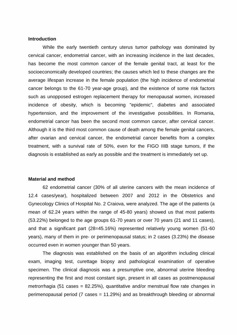
Abdominal ultrasound was performed in all cases (52=83.87% cases received
vaginal ultrasound examination), the data (table 2) being used both for positive
diagnosis and for pre-therapeutic staging.
Abdominal/vaginal ultrasound Cases %
Tumor (3-12 cm) 52 100
Inner half myometrial invasion 11 21,15
Complete or more then inner half myometrial
invasion
8 15,38
Extension to the cervix 10 19,23
Liver metastasis 3 5,76
Ascites 4 7,62
Table Nr. 2. Endometrial cancer – ultrasound date
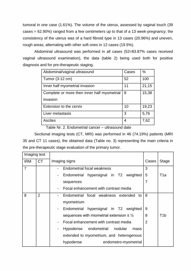
Sectional imaging tests (CT, MRI) was performed in 46 (74.19%) patients (MRI
35 and CT 11 cases), the obtained data (Table no. 3) representing the main criteria in
the pre-therapeutic stage evaluation of the primary tumor.
Imaging test
Imaging signs
Cases
Stage IRM CT
7 - Endometrial focal weakness
- Endometrial hypersignal in T2 weighted
sequences
- Focal enhancement with contrast media
3
5
7
T1a
8 2 - Endometrial focal weakness extended to
myometrium
- Endometrial hypersignal in T2 weighted
sequences with miometrial extension ≤ ½
- Focal enhancement with contrast media
- Hypodense endometrial nodular mass
extended to myometrium, and heterogenous
hypodense endometro-myometrial
8
9
8
2
T1b

postcontrast media aspect (T2b over staging)
6 2 - Endometrial focal weakness extended to the
myometrium
- Endometrial hypersignal in T2 weighted
sequences with miometrial extension ≤ ½
- Focal enhancement with contrast media
- Hypodense endometrial nodular mass
extended to myometrium, and heterogenous
hypodense endometro-myometrial
postcontrast media aspect < ½ - T2b in
staging
5
6
6
2
T1c
1 3 - Cervical invasion with focal enhancement with
contrast media - T2b over staging
4
T2a
9 - Cervical invasion with cervical signal
abnormalities and focal enhancement with
contrast media
9
T2b
3 3 - Serosal extension with ascites
- Lymph nodes invasion with normal size of
nodes
- Abdominal-pelvic lymph nodes hypertrofia
6
1
1
T3a
2 1 - Abdominal-pelvic structures invasion 3 T4
Table Nr. 3 Endometrial cancer – sectional imaging tests (CT< MRI) – the primary tumor
staging
An accurate diagnosis of endometrial cancer belonged to pathological
examination, which included two sequences, curettage biopsy and pathological
examination of operative specimen, each with well-defined objectives.
The curettage biopsy preceded any therapeutic gesture in all cases; the
histological examination confirmed the diagnosis and identified the primary tumor type
(endometrioid carcinoma in 51 cases = 82.25% and nonendometrioid carcinoma in 11
cases = 17.75%) and the tumoral grading (G1 33 cases = 53.22%, G2 19 cases =
30.64% and G3 10 cases=19.14%) while the immunohistochemical examination was
performed to establish the precise location of the tumor (endometrial or endocervical),
the differential diagnosis between the two tumor locations being particularly important,
as the treatment is different for each of these topographical forms.
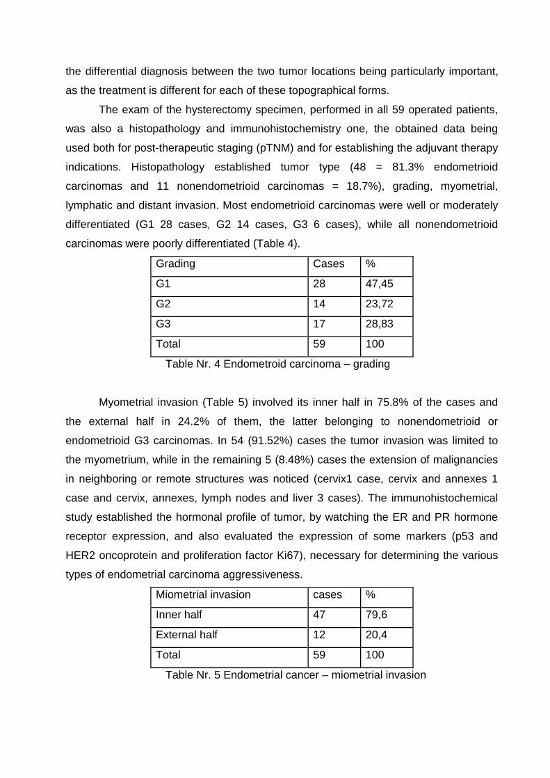
The exam of the hysterectomy specimen, performed in all 59 operated patients,
was also a histopathology and immunohistochemistry one, the obtained data being
used both for post-therapeutic staging (pTNM) and for establishing the adjuvant therapy
indications. Histopathology established tumor type (48 = 81.3% endometrioid
carcinomas and 11 nonendometrioid carcinomas = 18.7%), grading, myometrial,
lymphatic and distant invasion. Most endometrioid carcinomas were well or moderately
differentiated (G1 28 cases, G2 14 cases, G3 6 cases), while all nonendometrioid
carcinomas were poorly differentiated (Table 4).
Grading Cases %
G1 28 47,45
G2 14 23,72
G3 17 28,83
Total 59 100
Table Nr. 4 Endometroid carcinoma – grading
Myometrial invasion (Table 5) involved its inner half in 75.8% of the cases and
the external half in 24.2% of them, the latter belonging to nonendometrioid or
endometrioid G3 carcinomas. In 54 (91.52%) cases the tumor invasion was limited to
the myometrium, while in the remaining 5 (8.48%) cases the extension of malignancies
in neighboring or remote structures was noticed (cervix1 case, cervix and annexes 1
case and cervix, annexes, lymph nodes and liver 3 cases). The immunohistochemical
study established the hormonal profile of tumor, by watching the ER and PR hormone
receptor expression, and also evaluated the expression of some markers (p53 and
HER2 oncoprotein and proliferation factor Ki67), necessary for determining the various
types of endometrial carcinoma aggressiveness.
Miometrial invasion cases %
Inner half 47 79,6
External half 12 20,4
Total 59 100
Table Nr. 5 Endometrial cancer – miometrial invasion

There are two distinct moments for the tumor staging which is the main criterion
in establishing the therapeutic algorithm: the pre-therapeutic staging (TNM) based on
clinical examination data, imaging tests and curettage biopsy, useful to choose a
surgical procedure and to determine a neoadjuvant therapy, and the postoperative
surgical and anatomic staging (FIGO/pTNM) based on intraoperative exploration and
histopathology and immunohistochemical exam of the hysterectomy specimen, useful to
determine the indications and to choose the sequence of the adjuvant therapeutic
means. The comparative analysis of the pre-therapeutic (TNM) and postsurgical staging
(pTNM/FIGO) showed us the following (Chart 1):
- 59 cases, preoperatively staged, were classified in stage I 35 cases
(59.32%), stage II 13 cases (22.03%) and stage III 11 cases (18.64%), the
two basic morphological types of endometrial cancer (squamous
endometrioid and non-endometrioid carcinoma), being distributed in all
stages
- The operated cases (59=95,16%) and postoperatively staged ones
(pTNM/FIGO) were classified in stage IA, all of them being endometrioid
carcinomas;
- All cases assigned after surgery (pTNM / FIGO) in advanced stages (III and
IV) were non-endometrioid carcinomas.
Graphic Nr. 1 Endometrial cancer: TNM vs. pTNM/FIGO
The treatment of endometrial cancer is a complex one, the following therapeutic
means being available: surgery, radiotherapy, and chemotherapy and hormone therapy.
IA IB IC IIA IIB IIIA IIIC IV
TNM/FIGO 16 11 8 4 9 8 3 0
pTNM 47 7 0 1 0 1 1 2
05
101520253035404550
stage
Endometrial cancer - TNM vs. pTNM/FIGO staging

The type and sequences of the therapeutic means were established according to NCCN
Guidelines, the main criterion being the pre-therapeutic (TNM) and postsurgical staging
(pTNM/FIGO).
Surgery was the main therapeutic method, 59 (95%) patients being operated on
(Table no. 6); according to the pre-therapeutic staging (TNM) we performed two types of
surgery:
- total hysterectomy with bilateral anexectomy and pelvic lymphadenectomy,
performed in 38 (64.4%) cases: 35 stage I and 3 in stage III C
- lymphadenocolpohysterectomy with bilateral anexectomy, Wertheim type, in 21
(35.6%) cases in stage II and IIIA
TNM stage
Pelvic
radiotherapy 75-80 Gy
Vaginal
brahitherapy
Chemotherapy
Surgical procedure Nr Cazuri Total hysterectomy with
bilateral adnexectomy + pelvic
lymphadenectomy
Total lymphadenocolpo-hysterectomy + bilateral adnexectomy (Wertheim
type)
I 35 35
II 13 13 13
IIIA/IIIB 8 8 8 8 8
IIIC 3 3
IV 0 0 0 0 0
Total 21 8 8 38 21 59
Table Nr. 6 Endometrial cancer – treatment
Neoadjuvant therapy (Table no. 6) preceded surgery and consisted of pelvic
radiation (75-80Gy dose) for 13 patients classified in stage II and a complex
neoadjuvant treatment for 8 patients in stage IIIA (vaginal brachytherapy + external
radiotherapy + chemotherapy).
Adjuvant therapy was indicated, according to NCCN Guidelines
recommendations and taking into account the pTNM stage, grading and recurrence
rate. The most used therapeutic means were vaginal brachytherapy, external beam
radiation and chemotherapy with specific indications:
vaginal brachytherapy – 9 patients with pTNM stages IB and C with G3, II with
G3 and III with G1-3
external radiation – 11 patients with pTNM stages IB and C with G3 and II, III
and IV, regardless of G
chemotherapy (CAP formula: cyclophosphamide + anthracycline + taxane) in 4
patients with stages III and IV
hormone therapy (progestins, progestogens - megestrol 1tb/day for 2 years)
indicated in patients with positive expression of progesterone receptor (PR +),
with local or systemic advanced cancer or recurrence.
Results
48 (81.4%) operated patients had a fair evolution; we registered 11 postoperative
complications (postoperative morbidity rate of 18.6%): local complications in 6 cases (4
wound abscesses and 2 prolonged dynamic ileus) and general complications in 5 cases
(pulmonary infection 2 cases and deep vein thrombosis in 3 cases), all of them
managed by conservative methods.
We did not register any postoperative death (postoperative mortality rate: 0)
Regarding the late survival rate, we have to mention that, at the end of our
studied period, only 14 (22.58%) patients had more than 5 years from the diagnosis
establishment and the beginning of the treatment, so that the 5 year-survival rate could
not be evaluated for all patients. So, for 44 patients who remained in evidence at the
end of our study, we noticed the following survival rate: 82.78% for the patients in stage
I, 69.83% for the patients in stage II, and 54.54% for those in stage III.
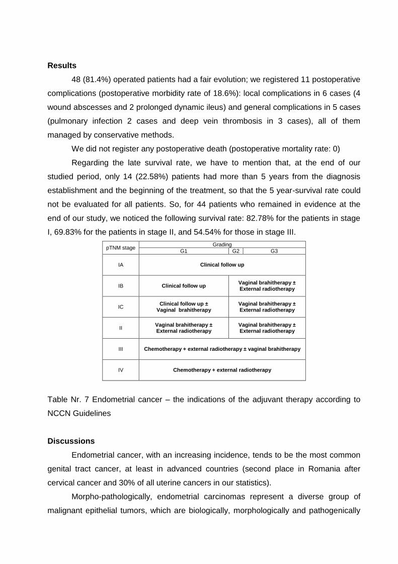
pTNM stage Grading
G1 G2 G3
IA Clinical follow up
IB Clinical follow up Vaginal brahitherapy ± External radiotherapy
IC Clinical follow up ±
Vaginal brahitherapy Vaginal brahitherapy ± External radiotherapy
II Vaginal brahitherapy ± External radiotherapy
Vaginal brahitherapy ± External radiotherapy
III Chemotherapy + external radiotherapy ± vaginal brahitherapy
IV Chemotherapy + external radiotherapy
Table Nr. 7 Endometrial cancer – the indications of the adjuvant therapy according to
NCCN Guidelines
Discussions
Endometrial cancer, with an increasing incidence, tends to be the most common
genital tract cancer, at least in advanced countries (second place in Romania after
cervical cancer and 30% of all uterine cancers in our statistics).
Morpho-pathologically, endometrial carcinomas represent a diverse group of
malignant epithelial tumors, which are biologically, morphologically and pathogenically
different, having two basic forms, type I (endometrioid type) and type II (non-
endometrioid type), including serous carcinomas and other aggressive endometrial
carcinomas, with different clinical, pathological, immunohistochemical and molecular
biology characters, suggesting the existence of two pathways of endometrial
carcinogenesis.
The diagnosis of endometrial cancer is a complex one, established on an
algorithm that includes clinical examination, imaging tests, curettage biopsy and
pathologic exam of the hysterectomy specimen and has several features to be taken
into account.
Endometrial cancer does not benefit from a screening test to detect early disease
and early clinical diagnosis is difficult, because of its nonspecific clinical picture and
because of the fact that the disease may progress asymptomatically for a long time
(several years) before becoming clinically manifested.
Abnormal uterine bleeding pre-, peri- or postmenopausal (82.25%), the first and
most constant sign of endometrial cancer, present in all our cases (over 90% in the
literature) without marking the onset of the disease, represents a warning sign, which
imposes a complex investigation involving clinical exam, imaging tests and pathological
exam, thus leading to diagnosis. Leucorrhea (11.29%), pelvic pain (35.48%),
dyspareunia (29.03%) and urinary signs (12.90%), suggesting spread to cervix or
invasion of neighboring structures (parameters, appendices, bladder, etc..) together with
obesity (51.61%) and signs of hyperoestrogenia (acne, oily skin pigmentation and
hirsutism) completed the clinical picture, while gynecological examination provided
useful data for evaluating the size, structure and mobility of the uterus, invasion of the
cervix, parameters and/or neighborhood viscera.
There is no biological test that can confirm the presence of endometrial cancer.
Imaging tests (abdominal and transvaginal ultrasound, CT and/or MRI) are
indispensable for the diagnosis of endometrial cancer, the provided data (size and
topography of the primary tumor, invasion of myometrium and/or neighborhood
structures, lymph node invasion and distant metastasis) can confirm the diagnosis and
can also be the main criteria for pre-therapeutic staging and therapeutic algorithm
choice. Abdominal and transvaginal ultrasound, a first-line imaging, precedes curettage
biopsy and is mandatory in all the patients with abnormal uterine bleeding in
postmenopausal, pre- and/or perimenopausal period. The main information provided by
ultrasound investigation in the study group was: evidence of heterogeneous endometrial

masses, (3-12 cm in size), imprecise bounded face of myometrium (52 cases),
myometrium invasion in 19 cases, extension to cervix in 10 cases, distant metastasis in
3 cases and ascites in 4 cases.
Sectional imaging tests (CT 11 cases, MRI 35 cases), although less currently
used for the initial diagnosis, are essential for the initial staging of endometrial
carcinoma, since the evaluation of lymphatic extension, abdomino-pelvic and distant
metastasis cannot be assessed clinically and, from this point of view, ultrasound is listed
with a significantly lower sensitivity and specificity. Preoperative imaging performed for
staging addresses to the cases with clinically difficult diagnosis, those with other
associated injuries and with aggressive tumor histological subtypes or advanced stages.
MRI was superior in revealing loco-regional extension to parameters and lymph node
invasion while CT is more commonly used to evaluate extrapelvine extension (better to
assess lung parenchyma) and radiotherapy planning.
The certainty of diagnosis belongs to the pathological exam (histopathology and
immunohistochemistry) and includes two distinct sequences: examination of the
pathological material taken by curettage biopsy and pathological exam of the
hysterectomy specimen.
Curettage biopsy, performed in all cases, preceded any therapeutic gesture, and
the pathological material collected was subjected to histological examination to confirm
the diagnosis of endometrial cancer and to determine the type and grading of the
primary tumor and to immunohistochemical examination necessary to establish the very
centre of the tumor (endometrial or endocervical), the differential diagnosis between the
two tumor locations being particularly important, since the treatment is different.
The pathological exam of the hysterectomy specimen, performed in 59 operated
patients, was also a histopathology and immunohistochemical one, the obtained data
being used both for post-therapeutic staging (pTNM) and for establishing the indications
of the adjuvant therapy.
Staging, using both classification systems (TNM and FIGO), is the most
important moment in the therapeutic decision; there are two distinct moments for the
tumor staging: the pre-therapeutic staging (TNM) based on clinical examination data,
imaging tests and curettage biopsy, useful to choose surgical procedure and determine
whether neoadjuvant therapy is necessary and the postoperative surgical and anatomic
staging (FIGO/pTNM) based on intraoperative exploration and histopathology and
immunohistochemical exam of the hysterectomy specimen, useful to determine the
indications, type and sequence of the adjuvant therapeutic means. Comparative
analysis of the case distribution according to pre-therapeutic (TNM/FIGO) and
postsurgical staging (pTNM) showed, on the one hand, that all the cases assigned after
surgery in advanced stages (III and IV pTNM/FIGO stage) were nonendometrioid
carcinomas and, on the other hand, we could notice the limits of the imaging tests
(ultrasound, CT, MRI) in assessing tumor invasion, so that the pre-therapeutic staging is
usually an overstated one.
Although it is the third most common cause of death in female genital cancers,
the endometrial cancer benefits from a complex treatment and has a survival rate of
50% including for IIIB FIGO stage tumors, if the diagnosis is early and treatment is
initiated immediately it is established. The treatment of the endometrial cancer is a
complex, sequential one with the following fundamental therapeutic means: surgery,
radiotherapy, chemotherapy and hormone therapy, the choice of the treatment methods
and their sequence being determined by pre- and post-surgical staging (pTNM/FIGO),
according to NCCN Guidelines recommendations.
Surgery was the main therapeutic method, 59 (95%) being operated on;
according to the pre-therapeutic staging (TNM) we had to choose between two surgical
procedures: total hysterectomy with anexectomy and bilateral pelvic lymphadenectomy
performed in 38 (64.4 %) patients (35 in stage I and 3 in stage I IIIC) and total
lymphadenocolpohysterectomy with bilateral anexectomy, type Wertheim, performed in
21 (35.6%) patients in stage II and IIIA.
Neoadjuvant therapy preceded surgery and consisted in pelvic radiation (75-
80Gy dose) as the only neoadjuvant method for the patients classified in stage II (13
cases), while the patients classified in stage IIIA (8 cases) needed a complex
neoadjuvant treatment: vaginal brachytherapy + external radiotherapy + chemotherapy.
Adjuvant therapy was indicated, according to NCCN Guidelines recommendations,
guided by the pTNM stage, grading and recurrence rate. Therapeutic means were
vaginal brachytherapy, external beam radiation and chemotherapy with specific
indications: vaginal brachytherapy for the patients classified in pTNM stages I B, C with
G3, II with G3, and III with G1-3 (9 cases), external radiation for the patients classified
in stages I B, C with G3 and II, III and IV, regardless of G (11 cases) and
chemotherapy (CAP formula: cyclophosphamide+anthracycline+taxane) for the patients
classified in stage III and IV (4 cases). We also used hormone therapy (progestins,
progestogens - megestrol 1tb/day for 2 years) in patients with positive expression of
progesterone receptor (PR +), with local or systemic advanced or recurrence cancer.
Conclusions.
1. Endometrial cancer, with maximum incidence in postmenopausal women belonging
to the 61-70 year-age group (53.22%), tends to be the most common genital tract
cancer, at least in the advanced countries (second place in Romania after cervical
cancer)
2. Abnormal uterine bleeding pre-, peri-or postmenopausal- the first and most constant
sign of endometrial cancer - imposes a complex investigation plan, involving clinical
exam, imaging tests and pathological exam, leading to diagnosis.
3. Curettage biopsy is mandatory, prior to any therapeutic gesture.
4. Staging (pre-therapeutic and postsurgical), using both classification systems (TNM
and FIGO), is the most important moment in the therapeutic decision.
5. Surgery, the main therapeutic method, according to the pre-therapeutic staging
(TNM), had to choose between two surgical procedures: total hysterectomy with
anexectomy and bilateral pelvic lymphadenectomy, and total
lymphadenocolpohysterectomy with bilateral anexectomy, type Wertheim.
6. Adjuvant and neoadjuvant therapy have specific indications, according to NCCN
Guidelines recommendations, guided by the tumoral type, grading, stage and
recurrence rate
7. Endometrial cancer has a good therapeutic response and a 5-year survival rate of
over 50%, even for tumors of stage III B, if the diagnosis is early established and the
treatment is immediately initiated.
References
Horner MJ, Ries LAG, Krapcho M, Neyman N, Aminou R, Howlader N, Altekruse
SF, Feuer EJ, Huang L, Mariotto A, Miller BA, Lewis DR, Eisner MP, SEER
Cancer Statistics Review, 1975-2006, National Cancer Institute, Bethesda, MD;
Centrul de Calcul, Statistică sanitara și Documentare medicală: Registrul
Național de Cancer, MSP, București, 2004;
NCCN Clinical Practice Guidelines in Oncology. Uterine neoplasms.V.I.2008. url:
http://www.nccn.org/professionals/physician_gls/PDF/uterine.pdf;
Ronnett BM, Kurman RJ - Precursors lesions of endometrial carcinoma. In:
Kurman RJ. Blaustein’s Pathology of the Female Genital Tract, 5th edition,
Springer-Verlag, New York, 467-500, 2002;
Bansal N, Yendluri V, Wenham RM – The molecular biology of endometrial
cancers and the implications for pathogenesis, classification and targeted
therapies, Cancer Control, 16(1):8-13, 2009;
Llobet D, Pallares J, Yeramian A, Santacana M, Eritja N, Velasco A, Dolcet X,
Matias-Guiu X – Molecular pathology of endometrial carcinoma: practical aspects
from the diagnostic and therapeutic viewpoints, J Clin Pathol, 62(9)777-85, 2009;
Lee, Joseph K. T.; Sagel, Stuart S.; Stanley, Robert J.; Heiken, Jay P. Computed
Body Tomography with MRI Correlation , 4th Edition, Lippincott Williams &
Wilkins, p1376-1415, 2006;
Mathias Prokop, Michael Galanski - Spiral and Multislice Computer tomography
of the Body, Georg Thieme Verlag,716-719, 2003;
Reid-Nicholson M, Iyengar P, Hummer AJ, Linkov I, Asher M, Soslow RA -
Immunophenotypic diversity of endometrial adenocarcinomas: implications for
differential diagnosis Modem Pathology ; 19, 1091-100, 2006;
Creasman W, Odicino F, Maisonneuve P, et al - FIGO 26th Annual Report on the
Results of Treatment in Gynecological Cancer, 5105, 2006;
Morrow, CP, Bundy, BN, Kurman, RJ, et al. Relationship between surgical-
pathological risk factors and outcome in clinical stage I and II carcinoma of the
endometrium: a Gynecologic Oncology Group study.Gynecol Oncol , 40:55,
1991;
Creasman, WT, Odicino, F, Maisonneuve, P, et al. Carcinoma of the corpus uteri.
J Epid Biostat , 6:45, 2001;
ASTEC – A Study in the Treatment of Endometrial Cancer: A Randomised Trial
of Lymphadenectomy in the Treatment of Endometrial Cancer, Abstract 45;
Chan JK, Wu H., Cheung MK et al. – The outcomes of 27,063 women with
unstaged endometroid uterine cancer, Gynecol Oncol, 106:282, 2007;
Rittenberg PV, Lotocki RJ, Heywood MS, et al - Stage II endometrial carcinoma:
Limiting post-operative radiotherapy to the vaginal vault in node-negative tumors.
Gynecol Oncol 98:434, 2005;

Ayhan A, Taskiran C, Celik C, et al - The long-term survival of women with
surgical stage II endometrioid type endometrial cancer. Gynecol Oncol 93:9,
2004;
Fung-Kee-Fung, M, Dodge, J, Elit, L, et al. - Follow-up after primary therapy for
endometrial cancer: A systematic review. Gynecol Oncol, 101:520, 2006.