diabetic patient with fungal renal infection and fungus ... · the kidney, development of...
TRANSCRIPT

Remedy Publications LLC.
Journal of Clinical Nephrology & Kidney Diseases
2017 | Volume 2 | Issue 1 | Article 10061
IntroductionPrimary urinary tract infection due to Candida is usually seen in patients with
immunocompromised risk factors such as diabetes mellitus, prior antibiotic therapy urinary tract drainage devices, urinary tract pathology and malignancy [1].
A European observational study found that Candida is the third most common organism isolated from urine in hospitalized patients [2], while its frequency is significantly increasing among patients in intensive care units (ICUs). Older age, presence of diabetes mellitus, prolonged hospitalization, ventilator support, protracted antibiotic treatment and parenteral nutrition are risk factors predisposing to candiduria [3].
Although most patients with candiduria remain asymptomatic, and the yeast may represent colonisation, many complications may arise in high risk patients; such as ascending infection of the kidney, development of candidemia or disseminated candidiasis. Disseminated candidiasis can lead to serious and even life threatening complications. Candiduria may be the first manifestation of candidemia. Therefore, patients with candiduria and predisposing risk factors as mentioned before should be carefully evaluated for systemic Candidiasis and should be treated aggressively. The kidneys are the most commonly involved organ [4]. The clinical characteristics of kidney infections depend on whether the disease presents secondary to candidemia or to ascending bladder infection. Renal function is rarely compromised but papillary necrosis can occur and it may be complicated by fungus ball formation.
Patients with diabetes and permanent Candiduria should be promptly evaluated for renal infection by ultrasound or computed tomography (CT) in order to reveal hydronephrosis, fungus balls and perinephric abscesses that are usually the result of ascending infection [1,5].
Fungus balls should be treated with a combination of surgical intervention and systemic
Diabetic Patient with Fungal Renal Infection and Fungus Balls: Case Study
OPEN ACCESS
*Correspondence:Maria Stangou, Department of
Nephrology, Aristotle University of Thessaloniki, Hippokration Hospital,
Thessaloniki, Greece, Tel: 0030 2310892603; Fax: 0030 2310892382;
E-mail: [email protected] Date: 23 Oct 2016Accepted Date: 04 Jan 2017Published Date: 06 Jan 2017
Citation: Sambani E, Toulkeridis G, Stangou M,
Skoularopoulou M, Ioanna-Theologia L, Antachopoulos C, et al. Diabetic Patient with Fungal Renal Infection and Fungus
Balls: Case Study. J Clin Nephrol Kidney Dis. 2017; 2(1): 1006.
Copyright © 2017 Stangou M. This is an open access article distributed under
the Creative Commons Attribution License, which permits unrestricted
use, distribution, and reproduction in any medium, provided the original work
is properly cited.
Case ReportPublished: 06 Jan, 2017
Sambani E1, Toulkeridis G1, Stangou M1*, Skoularopoulou M1, Ioanna-Theologia L1, Antachopoulos C2, Salpigidis G3, Papagianni A1 and Efstratiadis G1
1Department of Nephrology, Aristotle University of Thessaloniki, Greece
2Department of Pediatrics, Aristotle University of Thessaloniki, Greece
3Department of Urology, Hippokration Hospital, Greece
AbstractThe presence of diabetes predisposes to severe urinary infections, such as emphysematous pyelonephritis and cystitis, perinephric abscesses and renal candidiasis.
We present a case of severe fungal kidney infection, complicated by bilateral fungus balls in a 20-year-old diabetic female. The patient presented to the Emergency Department with high fever (38.50C), fatigue and back pain. She had poor control of diabetes, with a long history of multiple episodes of diabetic ketoasidosis. Clinical examination revealed positive Giordano sign and sensitivity in the right lateral abdominal area. Laboratory tests on admission showed high ESR, CRP, normal white cell counts impaired renal function and pyuria. Kidney ultrasound and abdominal CT scans revealed bilateral kidney hypertrophy, mild hydronephrosis of the right kidney and severe thickening of ureter and bladder wall. The patient was successfully treated with intravenous fluconazole and amphotericin B for almost three months, in combination with bilateral nephrostomy placements, local infusions of amphotericin B through the nephrostomies and removal of the fungus balls. After repeated negative blood and urine cultures, normal kidney ultrasounds and abdominal CT scans the nephrostomies were removed. The patient was discharged after 152 days of hospitalisation, afebrile, with stabilized renal function and negative blood and urine cultures.
Keywords: Urinary tract infection; Diabetes mellitus; Fungus balls
Stangou M, et al. Journal of Clinical Nephrology & Kidney Diseases
Remedy Publications LLC. 2017 | Volume 2 | Issue 1 | Article 10062
antifungal therapy [6]. We present a case of a 20 year old female with type I diabetes mellitus and upper tract urinary infection due to systematic candidemia
Case PresentationA 20-year-old diabetic, black female presented to the Emergency
Department with fever and back pain that had started about 24 hours before admission. She had a long-term history of type I Diabetes, diagnosed when she was five years old. The patient was not compliant with treatment regulations, diabetes was inadequately controlled, and thus, she had repeated admissions to the hospital due to diabetic ketoacidosis. She recently had an episode of candidemia and fungal pyelonephritis, presented with fever and bilateral hydronephrosis. During her previous hospitalisation she was treated with echinocandin for 12 days and kidney drainage with bilateral pigtail placement. Two weeks prior admission she was discharged, after secession of antibiotics and re-movement of pigtails.
Physical examination on admission showed the temperature of 38.4°C, the blood pressure 140/80 mmHg, the pulse 100 beats per minute while auscultation of the chest revealed no abnormal findings. Heart sounds were normal. The examination of the abdomen revealed sensitivity in the right lateral abdominal area and positive Giordano sign.
Laboratory tests on admission showed elevated CRP (171 mg/l) and ESR (140 mm/hr), normal white blood cell count (WBC 9520/mm) impaired renal function with a serum creatinine of 3.06mg/dl. Urine microscopy revealed 28-30 white cells, 3-4 red cells per high-power field, while urine cultures for common bacteria were negative.
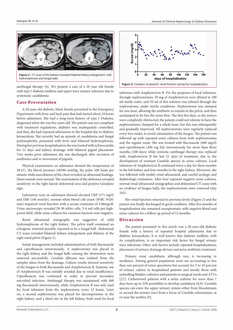
Renal ultrasound sonography was suggestive of mild hydronephrosis of the right kidney. The pelvis itself contained an echogenic material possibly reported to be a fungal ball. Abdominal CT scans revealed bilateral kidney enlargement and dilation of the right renal pelvis (Figure 1).
Initial management included administration of both fluconazole and ciprofloxacin intravenously. A nephrostomy was placed in the right kidney and the fungal balls causing the obstruction were removed successfully. Candida albicans was isolated from the samples taken from the drainage. Culture results showed sensitivity of the fungus to both fluconazole and Amphotericin B. Systemic use of Amphotericin B was initially avoided due to renal insufficiency. Ciprofloxacin was continued in order to prevent secondary microbial infection. Antifungal therapy was maintained with 400 mg fluconazole intravenously, while Amphotericin B was only used for local infusions from the nephrostomy every 12 hours. Later on, a second nephrostomy was placed for decompression in the right kidney, and a third one in the left kidney, both used for local
infusions with Amphotericin B. For the purposes of local infusions, through nephrostomies, 50 mg of Amphotericin were diluted in 100 ml sterile water, and 10 ml of this solution was infused through the nephrostomy, under sterile conditions. Nephrostomy was clamped for one hour, allowing the antibiotic to remain in the pelvis, and then unclamped to let free the urine flow. The first few days, as the ureters were completely obstructed, the patient could not tolerate to have the nephrostomies clumped for a whole hour, but this was subsequently and gradually improved. All nephrostomies were regularly replaced every two weeks, to avoid colonization of the fungus. The patient was followed up with repeated urine cultures from both nephrostomies and the regular route. She was treated with fluconazole (400 mg/d) and ciprofloxacin (200 mg bd) intravenously for more than three months (108 days) while systemic antifungal therapy was replaced with Amphotericin B the last 21 days of treatment, due to the development of resistant Candida species in urine cultures. Local infusions of Amphotericin B continued twice a day for three months in the left kidney and four months in the right kidney. Moreover, she was followed with weekly renal ultrasound, and careful urologic and diabetologic evaluation. After four repeated negative urine cultures, normal renal ultrasound sonographies and abdominal CT scans with no evidence of fungus balls, the nephrostomies were removed (day 165).
Her renal function returned to previous levels (Figure 2) and the patient was finally discharged in good condition. After five months of hospitalisation she remained asymptomatic with negative blood and urine cultures for a follow up period of 12 months.
Discussion The patient presented in this article was a 20-year-old diabetic
female with a history of repeated hospital admissions due to diabetic ketoacidosis. It is well known that diabetes mellitus, with its complications, is an important risk factor for fungal urinary tract infections. Other risk factors include repeated hospitalizations, placement of urinary drainage devices and prior antibiotic treatment.
Primary renal candidiasis, although rare, is increasing in incidence. Among general population yeast are accounting in less than one percent of urine specimens but account for 5 to 10 percent of urinary culture in hospitalized patients and mostly those with indwelling bladder catheters and patients in surgical wards and ATUs [2,7]. Catheterised patients with a urine catheter for more than 7 days have up to 25% possibility to develop candiduria [8,9]. Candida species can enter the upper urinary system either from bloodstream or ascend the urinary tract from a focus of Candida colonization, at or near the urethra [9].
Figure 1: CT scan of the kidneys revealed bilateral kidney enlargement, mild hydronephrosis and fungal balls.
Figure 2: Variation of patients’ renal function during her hospitalization.

Stangou M, et al. Journal of Clinical Nephrology & Kidney Diseases
Remedy Publications LLC. 2017 | Volume 2 | Issue 1 | Article 10063
On the current admission, patient presented with symptoms and sings suggestive of systemic infection including fatigue, fever, and abdominal sensitivity. Laboratory findings, including elevated CRP and ESR, confirmed systemic infection, while impaired renal function and pyuria indicated complicated upper urinary truck infection. Persistent candiduria in diabetics should always prompt radiologic imaging of the kidneys with ultrasound or CT scans, to assess possible renal involvement [1]. Candida spp is one of the most common microorganisms in urine among hospitalized patients and its presence is followed by increased mortality [1]. However, regarding the frequency of candidemia, the results are controversial, some investigators describe candidemia in <5% of patients in ICUs, while others increase this number to 37% [10,11]. Abdominal CT scans and renal ultrasounds in our patient revealed hydronephrosis and the presence of fungus balls in the right kidney. History of recent fungal infection and candidemia was indicative of a possible secondary kidney involvement due to systemic infection. At that previous episode, the patient had candidemia and candiduria, and although echinocandin was the appropriate treatment for candidemia, it was probably not sufficient to treat the urinary infection, as echinocandins have low concentrations in urine [12]. Fungus balls had probably already been developed during candidemia and pyelonephritis. According to the Infections Diseases Society of America (IDSA) guidelines for the treatment of candidiasis the presence of fungus balls should be managed with the combination of medical and surgical therapy. Removal or change in urine catheter may eradicate Candida in 20-40% of the cases [6]. Antifungal treatment should be applied only in cases with kidney involvement or systemic infection [9]. Fluconazole remains the treatment of choice [13]. The combination of Amphotericin B and Flu-cytosine is an alternative regimen [14]. Antibiotic therapy should be continued until a surgical or endoscopic procedure to remove the fungus balls has been accomplished, symptoms have resolved and urine cultures are negative. Fluconazole may be the mainstay of therapy, but if access to the renal pelvis by nephrostomy tubes is available, use of local treatment with Amphotericin B is a useful adjunctive therapy [6,15].
In the case presented above placement of nephrostomies and local infusions of Amphotericin B took place along with systematic treatment with Fluconazole. Due to the development of Candida resistance during treatment, the systemic antifungal treatment regiment was altered to Amphotericin B during the last 21 days of hospitalization.
Both local and systemic antifungal therapy was continued until the repeated urine cultures were negative, the sonographies, CT scans and laboratory tests were normal and the patient was afebrile without any symptoms of active infection.
In conclusion patients with predisposing risk factors for renal candidiasis such as diabetes mellitus should always be evaluated for systematic infection when symptomatic candiduria is present. Treatment regimen should be tailored according to the Candida species isolated from the urine or blood cultures and according to whether ascending or disseminated infection is present. Fluconazole resistant yeast can be treated with intravenous Amphotericin B. Flu-cytosine may be added to Amphotericin B when pyelonephritis or
fungus balls are present Fungus balls should be always managed with the combination of medical treatment and surgical intervention.
References1. Sadegi BJ, Patel BK, Wilbur AC, Khosla A, Shamim A. Primar Renal
Candidiasis: Importance of Imaging and Clinical History in Diagnosis and Management. J Ultrasound Med. 2009; 28: 507-514.
2. Bouza E, San Juan R, Muñoz P, Voss A, Kluytmans J, Co-operative Group of the European Study Group on Nosocomial Infections. A European perspective on nosocomial urinary tract infections I. Report on the microbiology workload, etiology and antimicrobial susceptibility (ESGNI-003 study). European Study Group on Nosocomial Infections. Clin Microbiol Infect. 2001; 7: 523-531.
3. Alvarez-Lerma F, Nolla-Salas J, León C, Palomar M, Jordá R, Carrasco N, et al. Candiduria in critically ill patients admitted to intensive care medical units. Intensive Care Med. 2003; 29: 1069-1076.
4. Lehner T. Systemic Candidiasis and Renal Involvement. Lancet. 1964; 1: 1414-1416.
5. Kauffman CA, Fisher JF, Sobel JD, Newman CA. Candidda urinary tract infection: Diagnosis. Clin Infect Dis. 2011; 52: S452-S456.
6. Pappas PD, Kauffman CA, Andes D, Benjamin DK, Calandra TF, Edwards JE, et al. Clinical practice guidelines for the management of candidiasis: 2009 update by the Infectious Diseases Society of America. Clin Infect Dis. 2009; 48: 503-535.
7. Maki DG, Tambyah PA. Engineering out the risk for infection with urinary catheters. Emerg Infect Dis. 2001; 7: 342-347.
8. Lundstrom T, Sobel J. Nosocomial candiduria: a review. Clin Infect Dis. 2001; 32: 1602-1607.
9. Kauffman CA, Vazquez JA, Sobel JD, Gallis HA, McKinsey DS, Karchmer AW, et al. Prospective multicenter surveillance study of funguria in hospitalized patients. The National Institute for Allergy and Infectious Diseases (NIAID) Mycoses Study Group. Clin Infect Dis. 2000; 30: 14-18.
10. Leroy O, Gangneux JP, Montravers P, Mira JP, Gouin F, Sollet JP, et al. AmarCand Study Group Epidemiology, management, and risk factors for death of invasive Candida infections in critical care: a multicenter, prospective, observational study in France (2005-2006). Crit Care Med. 2009; 37: 1612-1618.
11. Fisher JF, Sobel JD, Kauffman CA, Newman CA. Candida urinary tract infections: Treatment. Clin Infect Dis. 2011; 52: S452-S466.
12. Petraitis V, Petraitiene R, Groll AH, Roussillon K, Hemmings M, Lyman CA, et al. Comparative antifungal activities and plasma pharmacokinetics of micafungin (FK463) against disseminated candidiasis and invasive pulmonary aspergillosis in persistently neutropenic rabbits. Antimicrob Agents Chemother. 2002; 46: 1857-1869.
13. Sobel JD, Kauffman CA, McKinsey D, Zervos M, Vazquez JA, Karchmer AW, et al. Candiduria: a randomized, double-blind study of treatment with fluconazole and placebo. The National Institute of Allergy and Infectious Diseases (NIAID) Mycoses Study Group. Clin Infect Dis. 2000; 30:19-24.
14. Nolla-Salas J, Sitges-Serra A, León-Gil C, Martínez-González J, León-Regidor MA, Ibáñez-Lucía P, et al. Candidemia in non-neutropenic critically ill patients: analysis of prognostic factors and assessment of systemic antifungal therapy. Study Group of Fungal Infection in the ICU. Intensive Care Med. 1997; 23: 23-30.
15. Herbrecht R. Posaconazole: a potent, extended-spectrum triazole anti-fungal for the treatment of serious fungal infections. Int J Clin Pract. 2004; 58: 612-624.