dft calculation of nitrogen chemicat shifts in the active site of
TRANSCRIPT

Polish J. Chent.,80. I177-l183 (2006)
DFT Calculation of Nitrogen Chemicat Shiftsin the Active Site of Vitamin D Receptor.
by W. Sicińska--
Institute of organic Chemistry,, Polish 'Ącademłof Sciences, Kaspłzaka 44/52, 0]-224 Warsaw, Polclnd
(Received October l9rh. 2005: accepted Februan'3rd, 2006)
The complereC vilamin D receptor (VDR) is responsible for calcium homeostasis.Tqptophan is of special importance for the receptor's functions, as it appearsjust once in:::e YDR sequence and occupies the center ofthe ligand binding pocket. DFT calcula-:ions oi nirrogen chemical shifts for Trp-NH56 moiety, presented in this work forliganded and free receptor, agree with NMR studies on the VDR specifically labeled with[UL]
I5N2 Trp. Our calculations confirm orientation of the C(7):C(8) vitamin D bond un-der the tryptophan ring. We suggest that interactions with water molecules are responsi-ble for observed deshielding of indole Trp-nitrogen in unliganded VDR.
Key words: nuclear receptors, vitamin D receptor, protein ligand interactions, DFTcalculations of nitrogen chemical shifts
The vitamin D receptor is a member of a nuclear receptor (NR) superfamily com-prisrng receptors for the steroid, retinoid and thyroid hormones Il]. During the lastien \'ears. most of the steroid receptors u.ere crystallizęd in both apo (un1iganded) andholo lliganded) forms [2-6]. These crystallographic data allov'ed for the determina-tion of active sites in the ligand binding pockets (LBP) of the NR family. However,comparison of biological potency of wild type \rDR and its mutants revealed thatsome contact sites found in solid state do not exist ir vivo [1].It can therefore be antic-ipated that knowledge of detailed architecture of a LBP in a medium close to the phys-iological milieu is necessary to understand its intęraction with a ligand' LBP-VDR istoo large (31 .4 kDa) for full conformational identification by NMR rechniques [8].Ner-ertheless, employment of ęditęd NMR techniques developed for proteins withmolecular mass exceeding 15 kDa and selective isotope labeling permits partial de-termination of its structure. There is no doubt tlrat tryptophan is onę of the most im-portant amino acids in thę VDR sequence. Recently, NMR studies verified that inaqueous solutions, just like in the crystal state, the position of tryptophan inligand-VDR complexes is preserved, even in thę casę of vitamin D analogs, whichdrastically differ (Table l) in their biological activities [9-10]. comparison of
* Dedicated to Professor M. Witanowski on the occasion of his 70th birthday.Presented at 1st Symposium on Nuclear Magnetic Resonance in ChemisĘ, Biology and Medicine,September 8-10, 2005, Warsaw, Poland.
** E-mail: [email protected] (Dr' w. Sicińska).

I 178 W. Sicińska
'H[''N] HSQC spectra of apo and holo VDR ręvealed that theTrp282 N'l signalshifts downfield by 4 ppm (from 125.8 to 129.8 ppm) when the ligand is removed [9].It is worth mentioning that the structure of the free vitamin D receptor is still un-known, as only crystals of liganded VDR mutants have bęen obtained [5_6]' Proba-bly the presencę of a highly mobile domain (165-215 in human VDR) hampers
crystallization ęfforts of the full length LBD-VDR. Recently, this 50 amino acidnonhomologous receptor fragment, excised in crystallized protein mutants, wasmodeled by SICHO (Side-CHain Only), while full length LBD-VDR was created bycombined homology and lattice modeling [11].
Table l. Literature datau for biological activities.
VDR Amountbinding ratiob (pmol)
Compound(number)
ICA
none (control)
1cx,25-(OH):D: (1)
2MD (3)
none (control)
lcx,25-(OH)zD: (1)
2AM20R (4)
none (control)
1cx,2s-(OH)2D3 (1)
1
0.77
I
0.22
0
260
260
0
260
260
5.5 r 0.2
6.2 + 0.4
4.6 t 0.7
2.3 + 0.4
5.6 + 0,6
5.3 + 0.ó
5.1 + 0.2
1.2 + 0.5
14.4 + 0.6
3.9 + 0.1
6.1 + 0.2
5.8 + 0.3
9.3 + 0.4
10.6 + 0.1
0
0.Ż"
70"
aliteratue data are taken from [10] and forcompound 5 from [9]. ICA and BCM denote intestinal calciumabsorption and bond calcium mobilization, respectively. bThe binding capabiliry ofanalogs is expressed
as fraction ofhormone activity. "Dose level in pglkg.
In this work we calculate chęmical shifts of Trp and its nearest (3.5 R) neighbors(Ser 271, PheŻ7 5 and Gln 3 l3) in the free and liganded vitamin D receptor. our aim is
to sęarch for such VDR structurę in which the tryptophan residue is deshielded in apo
and shielded in holo form.
RESULTS AND DISCUSSION
Since VDR crystallizes only in the presence of ligands [5-6], structural compari-
son of its two forms can only be done through molęcular modęling. Among the amino
acids residing in the ligand binding pocket (LBP), tryptophan (Trp) is without any
doubt of crucial importance. Its natural mutation to arginine leads to sevęre herędi_
tary vitamin D-ręsistant rickęts [12], while the replacement of Trp withphenylalaninę, serine or alanine dęcreases binding receptor capabi1ities between 1 00
(Phe) and 1000 (Ser, Ala) times t l3-141. Several studies attempted to explain the role

t179l DFT calculation of nitrogen chemical shifts in the active...
of Trp in ligand binding. X-ray analyses of structures of liganded VDR revealed thatthe indole group of tryptophan is always stacked with the vitamin D C(6)-C(7):C(8)fragment, regardless of the biological potency of the ligand [6,10]. Since some con-tact sites found in solid state do not exist iłr vivo, a question arose whether this uniqueamino acid also conserves its orientation with respect to the anchored ligands in aque-ous buffers, a milieu closer to physiological conditions. Recently published studieson thę VDR specifically labeled with [UL] l5N2 Trp confirmęd that it is the case [9].Even though the analyzed ligands differed markedly in their biological activity (Ta-
ble 1), T.p Hul and Trp Nul chemical shifts in the complexes were virtually identical,with nitrogennuclęi inholo and apo VDR clustering around 125.8 and l29.8 ppm, re-spectively (Table 2). These results indicatę that the interaction betweęn the ligandsandTrp28Z is not responsible for variations in calcemic activity observed in vitaminD analogs. Rather, it appears that the indole ring of the tryptophan residue acts as a
common binding site for the intercyclic 5,7-diene moiety of vitamin D compoundsanchored in the VDR binding pocket. In this work we have focused our attention onDFT calculations of tryptophan chemical shifts and the amino acids: Ser271,Phe27 5,G1n3 13 nearest to Trp in holo VDR. Scheme I depicts structures of ligands occupyingthe VDR cavity. Tablę 2 contains experimental and computed indole nitrogen chemi-cal shifts of tryptophan. The gęometry of complexes for crystal structures was ob-tainęd from the PDB database, while for modeled complexes it was generated inBiopolymer (module of Sybyl 7.0). The size(ca.l70 atoms) of these protein systemsprecluded ab initio calculations of NMR parameters with a large set of basis func-tions due to extensive computational requirements. Intęręstingly, it appeared that cal-culations of nitrogen screening constants with a minimal 6-3 1G basis sęt using DFTwith the hybrid B3LYP (Becke, Lee, Yang, Parr) functionals and the GIAO method(Gauge Independent Atomic Orbitals) reproduced chemical shifts with satisfactoryqualitative (ca. 1 ppm) accuracy [ 1 5-20] . Since for thę nuclei ofthę sęcond row ofpe-riodic table variations in chemical shifts originate mostly from the paramagnetic part,the calculations were repeated using the larger 6-3 1G* basis set which includes po-Iarizatton functions. It turned out that nitrogen chemical shifts computęd in both ba_sis sets produced similar results, being close (^6 Ś l ppm) to the experimentalnitrogen shielding. Thęrefore thę addition of polarization functions did not improvethe results. The consistęncy of the calculated Trp N€l chemical shifts in crystal struc_tures (av. 125.7 ppm) with experimental NMR ręsults (av. l25.8 ppm) indicates thatthe position of Trp in the receptor cavity does not change between the solid and liquidstate' Taking into account the fact that in crystals thę distance between tryptophanrings and the intercyclic 5,7-diene System is rather |ong(ca.4 R), it would bę unręa-sonable to expect that sandwich interaction are strong enough to be responsible forshielding of nitrogen indolę nuclei in thęse complexed VDR. This possibility shouldbę rulęd out because a chęmical shift value of 80 ppm was calculatęd for l 5N1*-56 in asimulated complex in which this distance was shortened to 2 B. In docking experi-ments of C-2 modified vitamins to full-length LBD-VDR, it was observed that theC(5):C(6)-C(7):C(8) diene moiety can shift along the Trp aromatic rings [21].

I 180 W Sicińską
Table 2. Experimentala and calculatedb'c nitrogen chemical shifts of tryptophan indole moiety (in ppm)in liganded VDR.
LigandPDBCode
Res.(A)
15Ne I
exp.
l5NE I
calcd.b
l5N€l
calcd."
1o,25-(OH)2D3
1cr,25-(OH)zDr
20(.1)-I(l,2s-(OH)zD:2MD2AM2OR2MbisP
uExperimental data are taken from [9]. bComputing of the nitrogen shielding of Trp Nsl and reference NH3
was performed with a 6-3 1G basis set using DFT with the hybrid B3LYP functionals and the GIAO method
lI5-20,221. Geometry of NH3 was optimized with aug-cc-pVDZ basis set using DFT B3LYP method.
When the nitrogen shielding of Trp N'l was calculated using the GIA0/B3LYP/6-3lG approach and ręfer-
enced to NH3 taken from Gaussian 03 software package chemical shifts (in ppm) for: lDB l, I RK3, MC 1288,
1RJK, l RKH, 1RKG denoted l25.2, |27 .5,lŻ6.0,l28.3, |26.4 and 126.7, respectively. cComputing of the ni-trogen shielding of Trp Ntl and reference NH3 were performed with expanded 6-3lG* basis set.
1: 1o.,25-(OH)rD. 2: (20S)-1 o,25-(OH)2D3
3: 2MD 4: 2AM20R 5: 2Mbisp
Scheme 1. Chemical structure of (1) 1tx,25-(OH)2D3 and (2) its 20-epi analogue, (3) 2MD: (208-2-methylene-19-nor-1cr,,25-(OH)zDr, (4) 2AM20R: 2cr-methyl-19-nor-1c1,25-(OH)2D3 and
(5) 2MbisP: (20.S)-2methylene-19-nor-1cr-(OH)-bishomopregnacalcifero[.
1DB1
IRK3MCl288lRJKlRKHIRKG
2.2
2.Ż
1.5
r.99
2.28
1.90
1Ż5 '6
r25.9
125.8
123.7
126.0
t24.6
tŻ6.9125.0
rzs.3
r23.0
125.0
123.9
126.4
t24.4124.6
DFT calculation of nitrogen chemical shifts in the active.. ll81
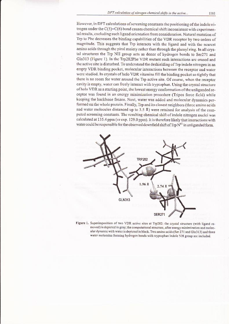
However, in DFT calculations of screening constants the positioning of the indole ni-trogen under the C(5):C(6) bond causes chemical shift inconsistent with experimen-tal results, excluding such ligand orientation from consideration. Natural mutation ofTrp to Phe decreases the binding capabilities of the VDR receptor by two ordęrs ofmagnitude. This suggests that Trp interacts with the ligand and with the nearęstamino acids through the pirol moiety rathęr than through the phenyl ring. In all crys-tal structuręs the Trp NH group acts aS donor of hydrogen bonds to Ser271 andGln3l3 (Figure l). In the TrpŻlZPhe VDR mutant such interactions are erased andthe active site is disturbed. To understand thę deshielding of Trp indole nitrogen in anempty VDR binding pocket, molecular interactions between the receptor and waterwęre studied. In crystals of holo VDR vitamins fill the binding pocket so tightly thatthere is no room for water around the Trp active site. of course, when the receptorcavity is empty, water can freely interact with tryptophan. Using the crystal structureof holo VDR as a starting point, the lowest energy conformation of the unliganded re-ceptor was found in an energy minimization procedure (Tripos force field) whilekeeping the backbone frozen. Next, water was added and molecular dynamics per-formed on the whole protein. Finally, Trp and its closest neighbors (three amino acidsand water molecules distanced up to 3.5 A) were retained for analysis of the com-puted screening constants. Thę resulting chemical shift of indole nitrogen nuclęi wascalculated at l3 5.4 ppm (vs exp. 129.0 ppm). It is therefore likely that interactions withwater could be responsible for the observed downfield shift of Trp N€
l in unliganded form.
Figure l. Superimposition of two VDR active sites at Trp282: the crystal structure (with ligand re-moved) is depicted in gray; the computational structure, after energy minimization and molec-ular dynamic with water is depicted in black. Two amino acids (Ser 271 and Gln313) and threewater molecules forming hydrogen bonds with tryptophan indole NH group are included.
&Lt{1ti

n82 W. Sicińską
CONCLUSIONS
Ab initio quantum mechanical calculations using GIAO/B3LYP method rępro-duce nitrogen chemical shifts ofN1.p_SCmoięty in a 170 atomprotein systemwith theaccuracy of ca. I ppm. Since discrepancies in nuclear screening constants calculatedwith the minimal 6-3 I G and the larger 6-3 I G* basis set do not exceed I pp*, the min-imal set of basis functions is sufficient for calculations of nitrogen shieldings in thevitamin D-VDR complexes. Computation of nitrogen chemical shifts in a large pro-tein system of known crystal structurę and subsequent comparison witlr chemicalshifts dętected by lsNMR in aqueous buffers could constitutę a useful tool for dętęct-ing differencęs in the Structure ofactive protein sites in the solid and liquid state.
COMPUTA|IONALDETAILS
The calculations were carried out using the GAUSSIAN 03 software package |Ż2).Tbe nitrogenshielding calculations were performed with ó-31G and 6_3lG* basis sets of wavefunctions using DFTwith the hybrid B3LYP functionals and the GIAO method (Gauge Idependent Atomic Orbitals) [ l5*20].The atom coordinates required for calculation ofTrp chemical shifts in liganded and the free receptorwere taken from PDB database (Table 2) and from computational models, respectively [23]. Docking sim-ulations were performed by FlexX software from the Biopolymer module, which uses a generic algorithmfor the search ofconformational space ofthe ligand with respect to the receptor's binding site. Severalsimulations of 100,000 steps each were performed with various initial positions of the ligand with respectto the binding pocket ofthe ręceptor. FoI final consideration, structures ofthe lowest conformational en-
ergy of the ligand-receptor complex were selected. Using the crystal structure (IRJK) as a guide , the bestmodel of the VDR binding cavity filled with water molecules was obtained in a few steps. After removingthe ligand, the structwe of the apo VDR was energy-minimized with frozen backbone using the Triposforce field.ThenaboxwithH2Omoleculeswascreatedandmoleculardynamics(runsat300"K, 10000it-erations) executed. only tryptophan with its nearest (3.5 A) amino acids and water moleculęs werę takenfor DFT calculations' To preserve the backbone nativę conformation ofexcised residues, we capped themat C terminal with amide group. Biodesigner, molecular modeling and visualization program, was usedfor finding the molecules at distances smaller than 3.5 R from Trp-NHs6 group [24].
Acknowledgments
W.S. thanks Professor A. Kolinski (Warsaw University) for helpful discussions and Dr. P. Rotkiewicz(University of Buffalo) for his valuable comments. W.S. is grateful for the generous amount of computer timegranted to her by Theory of Biopolymers Laboratory (Faculty of Chemistry, Warsaw University).
REFERENCES
L Evans R.M., Science,240, 889 (1988).
2. WagnerR.L., AprilettiJ.W, McGrathM.E., WestB.L., BaxterJ.D. andFletterickR.J.,Nature,378,690(lees).
3. Renaud J.P., Rochel N., RuffM., Vivat V., Chambon P., Gronemeyer H. and Moras D., Na ture,378,68l(1ee5).
4. Bourguet W., RuffM., Chambon P., Gronemeyer H. and Moras D., Nature,375,377 (1995).

DFT calculation of nitrogen chemical shifts in the active... 1183
5. Rochel N., Wurtz J.M., Mitschler A., Klaholz B. and Moras D., MoL CelI' 5, I73 (Ż000).
6. VanhookeJ.L., BenningM.M., BauerC.B., PikeJ.W. andDeLucaH.F., Biochemistry,43,4l0l (2004).
7. Yamamoto K', Masuno H., Choi M., Nakashima K., Taga T., ooizumi H., Umesono K., Sicińska W.,Vanhooke J., Deluca H.F. and Yamada S., PNIS, 97,1461 (2000).
8. Wider G. and Wuthrich K., Curr. Opin. Struct. Biol., 9, 594 (1999).9. Sicińska W., Westler WM. and Deluca H.F., Proteins, ól' 461 (2005).
10. Siciński R.R., Prahl J.M. and Smith C.M.' l Med' Chem.,41' 4662 (1998).l l. Siciński R.R', Rotkiewicz P.' Koliński A., Sicińska W., Prahl J.M., Smith C.M. and Deluca H.F.,
J. Med. Chem., 45, 3366 (2002).
12. Nguyen T.M., Adiceam P., Koftler M.L., Guillozo H., Rizk-Rabin M., Brouillard F., Lagier P., Palix C.,Gamier J.M' and Garabedian M., J. Bone. Miner Res,,17 , 17Ż8 (200Ż)'
13' Rotkiewicz P., Sicińska W., Koliński A. and Deluca H.F., Proteins,44' l88 (200l).14. Choi M., Yamamoto K., Itoh T., Makishima M., Mangelsdorf D.J., Moras D., Deluca H.F. and Yamada S.,
Chem. Biol., 10, 261 (2003).15. Becke A.D., Phys. Rev., A38,3098 (1988).
I6.Lee C, Yang W. and Parr R.G., Phys. Rev., B37, 785 (1988).
17. Miehlich B., Savin A., Stoll H. and Preuss H, Chem. Phys. Lett.,157, 200 (1989).18. Becke A.D., "/. Phys. Chem.,98, 5648 (1993).
19.London F.,J. Phys. Radium.,8,397 (1937).
20' Woliński K., Hinton J.F. and Pulay P., J' Am. Chem. Soc', ll2,825l (l990).2l ' Sicińska W., Rotkiewicz P. and Deluca H.F., Proceedings of the l2th Workshop on Vitamin D, in
J. Steroid Biochem. Mol. Biol.,89-90,10'l (2004).22. Gaussian 03. Revision B.04, M.J. Frisch, G.W. Trucks, H.B. Schlegel, G.E. Scuseria, M.A. Robb,
J.R. Cheeseman, J.A. Montgomery Jr., T. Vreven, K.N. Kudin, J.C. Burant, J.M. Millam, S.S. Iyengar,J. Tomasi, V. Barone, B. Mennucci, M. Cossi, G. Scalmani, N.A. Rega, H. Petersson, M. Nakatsuji,M. Hada, K. Ehara, R. Toyota, J. Fukuda, G. Hasegawa, M. Ishida, T. Nakajima, Y. Honda, O. Kitao,H. Nakai, M. Klene, X. Li, J.E. Knox, H.P. Hratchian, J.B. Cross, C. Adamo, J. Jaramillo, R. Gomperts,R.E. Stratmann,O.Yazyev, A.J. Austin, R. Cammi, C. Pomelli, J.W. Ochterski, P.Y. Ayala,K. Morokuma, G'A. Voth' P. Salvador, J.J. Dannenberg,Y'G. Za|łzewski, S. Dapprich, A'D. Daniels,M.C. Strain, O. Farkas, D.K. Malick, A.D. Rabuck, K. Raghavachari, J.B. Foresman, J.V. Ortiz, Q. Cui,A.G. Baboul, S. Cliftord, J. Cioslowski, B.B. Stefanov, G. Liu, A. Liashenko, P. Piskorz, I. Komaromi,R.L. Martin, D.J. Fox, T. Keith, M.A. Al-Laham, C.Y. Peng, A. Nanayakkara, M. Challacombe, P.M.W.Gill, B. Johnson, W. Chen, M.W. Wong, C. Gonzalez and J.A. Pople, Gaussian, Inc., Pittsburgh PA,(2003).
23. SYBYL Modeling Program, 7.0 ed. Tripos Inc., St. Louis, MO.24. Rotkiewicz P., Biodesigner, Molecular Modeling and Visualization Program http://www.pirx.com