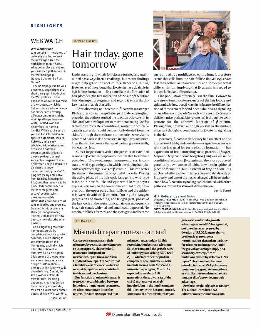
development: hair today, gone tomorrow

1
© 2001 Macmillan Magazines Ltd 490 | JULY 2001 | VOLUME 2 www.nature.com/reviews/molcellbio HIGHLIGHTS Wnt wonderland Wnt proteins — mediators of cell–cell signalling — are in the news again (see the Highlight on page 488) so what better place to expand your knowledge than to visit the Wnt homepage, launched and run by Roel Nusse? The homepage itself is well presented, beginning with a short paragraph introducing the Wnt proteins. This is positioned above an overview of the contents, which is further subdivided into colour- coded sections covering different components of the Wnt signalling pathway — Wnts, Frizzled, axin and dishevelled, to name a handful. Within each section you can find information on species alignments, links to PubMed and clearly tabulated information about expression patterns, chromosomal location. For those seeking structural satisfaction, regions of axin, dishevelled and β-catenin can be viewed in three dimensions using the Cn3D program (easily obtainable from NCBI by following the links). Another resource to be particularly commended is the ‘Wnt reagents and assays’ section, which provides invaluable information about sources of Wnt antibodies and proteins. Included in this section are strategies for generating antisera and advice on how best to make bioactive Wnt proteins. As no signalling molecule homepage would be complete without a signalling cascade, it is reassuring to see thumbnails on the homepage, each of which offers the option of an attractive full-size diagram. Click on one of the proteins and you instantly receive a deluge of information — perhaps even slightly visually overwhelming. Overall, the site provides extremely relevant links, including upcoming meetings (which are admirably up-to-date), reviews on Wnts and contact details of fellow Wnt workers. Katrin Bussell WEB WATCH Understanding how hair follicles are formed and main- tained has always been a challenge, but recent findings might help get to the root of this. Reporting in Cell, Huelsken et al. have found that β-catenin has a dual role in hair follicle formation — first it mediates the formation of hair placodes (the first indication of the site of the future hair) during embryogenesis, and second it acts in the dif- ferentiation of adult skin cells. After observing an increase in β-catenin messenger RNA expression in the epithelial part of developing hair placodes, the authors studied the function of β-catenin in skin and hair development in more detail using Cre/lox technology to create a conditional mutant in which β- catenin expression could be specifically deleted from the skin. Although the resultant mutant mice were viable, patches of hairless skin were seen in eight-day-old mice. Over the next two weeks, the rest of the hair grew normally, but was then lost. Closer inspection revealed the presence of extended regions of β-catenin-negative epithelium that lacked hair placodes in 15-day-old mutant mouse embryos, in con- trast to wild-type embryos, in which developing hair pla- codes expressed high levels of β-catenin. This implicates β-catenin in the formation of epithelial placodes. During the active phase of the hair cycle (anagen) in wild-type mice, both hair follicle and epidermal keratinocytes express β-catenin. In the conditional-mutant mice, how- ever, both the upper part of hair follicles and the epider- mis were devoid of β-catenin. During the catagen (regression and shortening) and telogen (rest) phases of the hair cycle in the mutant mice, hair was subsequently lost, hair canals widened and small cysts appeared. No new hair follicles formed, and the cysts grew and became surrounded by a multilayered epithelium. It therefore seems that cells from the hair-follicle-derived cysts have lost their follicular characteristics and show epidermal differentiation, implying that β-catenin is needed to induce follicular differentiation. One population of stem cells in the skin is known to give rise to keratinocyte precursors of the hair follicle and epidermis. So how does β-catenin influence the differentia- tion of these stem cells? And does it do this as a signalling or an adhesion molecule? In early embryos of β-catenin- deficient mice, plakoglobin (γ-catenin) is thought to com- pensate for the adhesive function of β-catenin. Plakoglobin, however, although present in the mutant mice, isn’t thought to compensate for β-catenin signalling in the skin. Moreover, β-catenin deficiency had no effect on the expression of tabby and downless — a ligand–receptor sys- tem that is crucial for early placode formation — but expression of bone morphogenetic protein 2 (bmp2), bmp4 and bmp7 and sonic hedgehog (shh) was lost in the conditional mutants. β-catenin can therefore be placed genetically downstream of tabby/downless in epithelial placode formation, but upstream of bmp and shh. It is unclear whether β-catenin targets bmp and shh directly or indirectly, and one of the next challenges will be to under- stand how β-catenin signalling is coordinated with other pathways involved in stem-cell differentiation. Katrin Bussell References and links ORIGINAL RESEARCH PAPER Huelsken, J. et al. β-catenin controls hair follicle morphogenesis and stem cell differentation in the skin. Cell 104, 533–545 (2001) FURTHER READING Oshima, H. et al. Morphogenesis and renewal of hair follicles from adult multipotent stem cells. Cell 105, 233–245 (2001) Hair today, gone tomorrow DEVELOPMENT Cancer cells can maintain their telomeres by reactivating telomerase or using a poorly characterized telomerase-independent mechanism. Aylin Rizki and Vicki Lundblad now report in Nature that a familiar cause of cancer — lack of mismatch repair — may contribute to this second mechanism. One function of mismatch repair is to prevent recombination between imperfectly homologous sequences. As telomeres contain imperfect repeats, the authors suspected that mismatch repair might inhibit recombination between telomeres. So, they compared the growth rates of budding yeast lacking EST2 (est2- ) — which encodes the protein component of telomerase — with mutants lacking both EST2 and a mismatch repair gene, MSH2. As expected, after about 100 generations the growth rate of the est2-mutants was severely impaired, but in the double mutants this phenotype was less pronounced. Mutations of other mismatch repair genes also conferred a growth advantage in an est2-background, but the effect was reversed by deletion of RAD52, a gene shown previously to promote a recombination-dependent pathway for telomere maintenance. Could the growth advantage simply be a secondary consequence of mutations caused by defective DNA repair? This is unlikely because introduction of a DNA polymerase mutation that generates mutations at a similar rate to mismatch repair mutants didn’t provide a growth advantage. Are these results relevant to cancer? The authors introduced two different missense mutations into Mismatch repair comes to an end TELOMERES

Upload: katrin

Post on 29-Jul-2016

222 views

Category:

Documents


2 download

TRANSCRIPT

© 2001 Macmillan Magazines Ltd490 | JULY 2001 | VOLUME 2 www.nature.com/reviews/molcellbio

H I G H L I G H T S

Wnt wonderlandWnt proteins — mediators ofcell–cell signalling — are inthe news again (see theHighlight on page 488) sowhat better place to expandyour knowledge than to visitthe Wnt homepage,launched and run by RoelNusse?

The homepage itself is wellpresented, beginning with ashort paragraph introducingthe Wnt proteins. This ispositioned above an overviewof the contents, which isfurther subdivided into colour-coded sections coveringdifferent components of theWnt signalling pathway —Wnts, Frizzled, axin anddishevelled, to name ahandful. Within each sectionyou can find information onspecies alignments, links toPubMed and clearlytabulated information aboutexpression patterns,chromosomal location. Forthose seeking structuralsatisfaction, regions of axin,dishevelled and β-catenin canbe viewed in threedimensions using the Cn3Dprogram (easily obtainablefrom NCBI by following thelinks). Another resource to beparticularly commended isthe ‘Wnt reagents andassays’ section, whichprovides invaluableinformation about sources ofWnt antibodies and proteins.Included in this section arestrategies for generatingantisera and advice on howbest to make bioactive Wntproteins.

As no signalling moleculehomepage would becomplete without a signallingcascade, it is reassuring tosee thumbnails on thehomepage, each of whichoffers the option of anattractive full-size diagram.Click on one of the proteinsand you instantly receive adeluge of information —perhaps even slightly visuallyoverwhelming. Overall, thesite provides extremelyrelevant links, includingupcoming meetings (whichare admirably up-to-date),reviews on Wnts and contactdetails of fellow Wnt workers.

Katrin Bussell

WEB WATCH

Understanding how hair follicles are formed and main-tained has always been a challenge, but recent findingsmight help get to the root of this. Reporting in Cell,Huelsken et al. have found that β-catenin has a dual role inhair follicle formation — first it mediates the formation ofhair placodes (the first indication of the site of the futurehair) during embryogenesis, and second it acts in the dif-ferentiation of adult skin cells.

After observing an increase in β-catenin messengerRNA expression in the epithelial part of developing hairplacodes, the authors studied the function of β-catenin inskin and hair development in more detail using Cre/loxtechnology to create a conditional mutant in which β-catenin expression could be specifically deleted from theskin. Although the resultant mutant mice were viable,patches of hairless skin were seen in eight-day-old mice.Over the next two weeks, the rest of the hair grew normally,but was then lost.

Closer inspection revealed the presence of extendedregions of β-catenin-negative epithelium that lacked hairplacodes in 15-day-old mutant mouse embryos, in con-trast to wild-type embryos, in which developing hair pla-codes expressed high levels of β-catenin. This implicatesβ-catenin in the formation of epithelial placodes. Duringthe active phase of the hair cycle (anagen) in wild-typemice, both hair follicle and epidermal keratinocytesexpress β-catenin. In the conditional-mutant mice, how-ever, both the upper part of hair follicles and the epider-mis were devoid of β-catenin. During the catagen(regression and shortening) and telogen (rest) phases ofthe hair cycle in the mutant mice, hair was subsequentlylost, hair canals widened and small cysts appeared. Nonew hair follicles formed, and the cysts grew and became

surrounded by a multilayered epithelium. It thereforeseems that cells from the hair-follicle-derived cysts havelost their follicular characteristics and show epidermaldifferentiation, implying that β-catenin is needed toinduce follicular differentiation.

One population of stem cells in the skin is known togive rise to keratinocyte precursors of the hair follicle andepidermis. So how does β-catenin influence the differentia-tion of these stem cells? And does it do this as a signallingor an adhesion molecule? In early embryos of β-catenin-deficient mice, plakoglobin (γ-catenin) is thought to com-pensate for the adhesive function of β-catenin.Plakoglobin, however, although present in the mutantmice, isn’t thought to compensate for β-catenin signallingin the skin.

Moreover, β-catenin deficiency had no effect on theexpression of tabby and downless — a ligand–receptor sys-tem that is crucial for early placode formation — butexpression of bone morphogenetic protein 2 (bmp2),bmp4 and bmp7 and sonic hedgehog (shh) was lost in theconditional mutants. β-catenin can therefore be placedgenetically downstream of tabby/downless in epithelialplacode formation, but upstream of bmp and shh. It isunclear whether β-catenin targets bmp and shh directly orindirectly, and one of the next challenges will be to under-stand how β-catenin signalling is coordinated with otherpathways involved in stem-cell differentiation.

Katrin Bussell

References and linksORIGINAL RESEARCH PAPER Huelsken, J. et al. β-catenin controls hairfollicle morphogenesis and stem cell differentation in the skin. Cell 104,533–545 (2001) FURTHER READING Oshima, H. et al. Morphogenesis and renewal of hairfollicles from adult multipotent stem cells. Cell 105, 233–245 (2001)

Hair today, gonetomorrow

D E V E LO P M E N T

Cancer cells can maintain theirtelomeres by reactivating telomeraseor using a poorly characterizedtelomerase-independentmechanism. Aylin Rizki and VickiLundblad now report in Nature thata familiar cause of cancer — lack ofmismatch repair — may contributeto this second mechanism.

One function of mismatch repair isto prevent recombination betweenimperfectly homologous sequences.As telomeres contain imperfectrepeats, the authors suspected that

mismatch repair might inhibitrecombination between telomeres.So, they compared the growth ratesof budding yeast lacking EST2 (est2-∆) — which encodes the proteincomponent of telomerase — withmutants lacking both EST2 and amismatch repair gene, MSH2. Asexpected, after about 100generations the growth rate of theest2-∆ mutants was severelyimpaired, but in the double mutantsthis phenotype was less pronounced.Mutations of other mismatch repair

genes also conferred a growthadvantage in an est2-∆ background,but the effect was reversed bydeletion of RAD52, a gene shownpreviously to promote arecombination-dependent pathwayfor telomere maintenance. Couldthe growth advantage simply be asecondary consequence ofmutations caused by defective DNArepair? This is unlikely becauseintroduction of a DNA polymerasemutation that generates mutationsat a similar rate to mismatch repairmutants didn’t provide a growthadvantage.

Are these results relevant to cancer?The authors introduced twodifferent missense mutations into

Mismatch repair comes to an end

T E LO M E R E S