development 139, 678-689 (2012) doi:10.1242/dev.074500...
TRANSCRIPT

RESEARCH ARTICLE DEVELOPMENT AND STEM CELLS678
Development 139, 678-689 (2012) doi:10.1242/dev.074500© 2012. Published by The Company of Biologists Ltd
INTRODUCTIONDuring nervous system development, progenitor cells often divideasymmetrically, renewing themselves and budding off daughtercells that typically have a more limited mitotic potential. Thedaughter cell may in turn display three alternative behaviors:directly differentiating into a neuron or glia; dividing once togenerate two neurons and/or glia; or dividing multiple times (i.e.acting as a transit-amplifying or intermediate neural precursor cell)before finally generating neurons and/or glia (Brand and Livesey,2011; Gotz and Huttner, 2005; Kriegstein et al., 2006; Rowitch andKriegstein, 2010). Adding to the complexity, recent studies inDrosophila melanogaster have revealed that some lineages displaya switch in daughter cell proliferation, from daughters dividingonce to daughters that directly differentiate (Baumgardt et al.,2009; Karcavich and Doe, 2005). As a result of these differentproliferative behaviors of the daughter cells, lineage topology (thebranching pattern of the lineage tree) may differ considerably. Inaddition to the complexity of different lineage topologies, it isbecoming increasingly clear that both vertebrate and invertebrateneural progenitor cells undergo programmed temporal changes intheir competence, as evidenced by the generation of different celltypes at different developmental time points (Jacob et al., 2008;Okano and Temple, 2009; Pearson and Doe, 2004). Importantly, astemporal competence changes occur simultaneously with changesin daughter cell proliferation, it results in an interplay that canregulate the precise numbers of each unique cell type. Even thoughthis basic principle of ‘topology-temporal interplay’ is likely to be
of fundamental importance during nervous system development, itis not well recognized, let alone well understood at the moleculargenetic level.
The Drosophila ventral nerve cord (VNC) is derived from a set ofneural progenitor cells, termed neuroblasts (NBs), generated in theearly embryo (Doe and Technau, 1993). Each neuroblast generates aunique lineage, progressing by multiple rounds of asymmetric celldivision, renewing itself and budding off daughter cells denotedganglion mother cells (GMCs). Each GMC in turn undergoes a finaldivision to generate two neurons and/or glia (Doe, 2008; Knoblich,2008; Skeath and Thor, 2003; Technau et al., 2006). In most, if notall, cases the GMC division has also been found to be asymmetric(Bardin et al., 2004; Doe, 2008; Knoblich, 2008), and the twopostmitotic sibling cells are often generically referred to as cell fate‘A’ and ‘B’ (Cau and Blader, 2009). Thus, Drosophila embryonicVNC lineages undergo repeated rounds of NBrGMCrA/Bdivisions, until each neuroblast enters quiescence or undergoesapoptosis (Maurange and Gould, 2005). The homeodomain proteinProspero (Pros) plays a crucial role in limiting GMC proliferation(Doe, 2008; Egger et al., 2008; Knoblich, 2008). Pros is expressedcontinuously by most, if not all, embryonic VNC lineages, but isblocked from acting in the neuroblast by being sequestered in thecytoplasm by its adaptor protein Miranda (Ikeshima-Kataoka et al.,1997; Shen et al., 1997). As the neuroblast buds off each GMC, Prosis asymmetrically distributed to the GMC, where it enters the nucleusand regulates gene expression (Choksi et al., 2006; Li and Vaessin,2000). The precise timing of these events allows each GMC to divideonce, but prevents its daughter cells from dividing. During larvalstages, neuroblasts exit quiescence and proliferate to generate theadult nervous system (Maurange and Gould, 2005). During thispostembryonic lineage progression, Pros again plays a role incontrolling daughter cell proliferation, and studies have found thatNotch signaling is also crucial for limiting daughter cell proliferationpostembryonically (Bello et al., 2006; Bowman et al., 2008; Lee et
Department of Clinical and Experimental Medicine, Linkoping University, SE-581 85,Linkoping, Sweden.
*Author for correspondence ([email protected])
Accepted 6 December 2011
SUMMARYDuring neural lineage progression, differences in daughter cell proliferation can generate different lineage topologies. This isapparent in the Drosophila neuroblast 5-6 lineage (NB5-6T), which undergoes a daughter cell proliferation switch fromgenerating daughter cells that divide once to generating neurons directly. Simultaneously, neural lineages, e.g. NB5-6T, undergotemporal changes in competence, as evidenced by the generation of different neural subtypes at distinct time points. Whendaughter proliferation is altered against a backdrop of temporal competence changes, it may create an integrative mechanismfor simultaneously controlling cell fate and number. Here, we identify two independent pathways, Prospero and Notch, which actin concert to control the different daughter cell proliferation modes in NB5-6T. Altering daughter cell proliferation and temporalprogression, individually and simultaneously, results in predictable changes in cell fate and number. This demonstrates thatdifferent daughter cell proliferation modes can be integrated with temporal competence changes, and suggests a novelmechanism for coordinately controlling neuronal subtype numbers.
KEY WORDS: Lineage topology, Daughter cell proliferation, Neural progenitor, Proliferation control, Temporal competence changes,Drosophila
Control of neuronal cell fate and number by integration ofdistinct daughter cell proliferation modes with temporalprogressionCarina Ulvklo, Ryan MacDonald, Caroline Bivik, Magnus Baumgardt, Daniel Karlsson and Stefan Thor*
DEVELO
PMENT
679RESEARCH ARTICLENotch and Pros control proliferation
al., 2006; Weng et al., 2010). However, despite this recent progress,it is unclear how distinct proliferation control mechanisms areintegrated to shape different lineage trees.
To begin addressing these issues, we are focusing on oneparticular Drosophila embryonic neuroblast, NB5-6. In the threethoracic (T) segments, NB5-6T generates a lineage of 20 cells(Schmid et al., 1999; Schmidt et al., 1997), including a thorax-specific set of four related neurons that express the Apterous (Ap)LIM-homeodomain protein: the Ap neurons (Fig. 1A,B) (Baumgardtet al., 2007). Ap neurons are born at the very end of the lineage, andNB5-6T undergoes a programmed switch in daughter cellproliferation prior to generating Ap neurons (Baumgardt et al., 2009).Thus, there is a switch from an NBrGMCrA/B to an NBrApneuron lineage progression, and the Ap neurons are born directlyfrom the neuroblast (Fig. 1B). Here, we identify two separatedaughter proliferation mechanisms acting within the NB5-6Tlineage. We find that Pros controls daughter cell (GMC) proliferationin the early lineage. In the late lineage, as Ap cells are generated, theactivation of the Notch pathway is superimposed upon Pros activity,acting to further limit daughter cell proliferation and resulting in theprogrammed proliferation switch. In contrast to their roles incontrolling daughter cell proliferation, neither pathway plays any rolein controlling the cell cycle exit of the neuroblast. Moreover, the Prosand Notch pathways do not regulate each other and therefore,whereas single mutants show limited overproliferation, doublemutants – lacking both of these daughter cell proliferation controls– display extensive overproliferation of the entire NB5-6T lineage,as well as of the entire VNC. Finally, the identification of thisdaughter cell proliferation switch allowed us to genetically assess theconcept of topology-temporal interplay. To this end, we generateddouble mutants for the Notch pathway and for nab, a subtemporalgene that affects Ap neuron specification (Baumgardt et al., 2009)(i.e. a topology-temporal perturbation). These mutants show thepredicted combined alteration of both cell fate and cell number, aphenotype not found in either mutant alone.
These results identify Notch signaling as the first bona fidedaughter proliferation switch and, moreover, demonstrate a novelrole for Notch in Drosophila neuroblasts. Furthermore, wedemonstrate that a neural lineage may depend upon twocomplementary regulatory mechanisms, involving Pros and Notch,for controlling daughter proliferation and thereby achieve a distinctlineage topology. Finally, we find that daughter cell proliferationcontrol is integrated with temporal progression in this lineage,thereby allowing for the generation of the proper numbers of eachunique cell type.
MATERIALS AND METHODSFly stockslbe(K)-EGFP reporter transgenic strains were generated by insertingfragment K (De Graeve et al., 2004) from the ladybird early gene into thepGreen H-Pelican vector (Barolo et al., 2000) and generating transgenesby standard techniques (BestGene, Chino Hills, CA, USA).
Other fly stocks used were: lbe(K)-lacZ [provided by K. Jagla (DeGraeve et al., 2004)]; lbe(K)-Gal4 (Baumgardt et al., 2009); elav-Gal4[provided by A. DiAntonio (DiAntonio et al., 2001)]; nabR52 [provided byF. Diaz-Benjumea (Terriente Felix et al., 2007)]; E(spl)m8-EGFP [providedby J. Posakony (Castro et al., 2005)]; E(spl)m8-lacZ [provided by F.Schweisguth (Lecourtois and Schweisguth, 1995)]; cas1 and cas3
(provided by W. Odenwald, NINDS, National Institutes of Health,Bethesda, MD, USA). UAS-Notch-RNAi stocks used were KK100002(VDRC) and BL7076 (Bloomington), which were combined in the samefly. To enhance the RNAi, elav-Gal4 was combined with UAS-dcr2(Baumgardt et al., 2007). kuze29-4; Dl6B; neur1; neurA101; 2xUAS-Tom;
spdoG104; UAS-MamDN; Df(3R)BSC751 (referred to as E(spl)-def); pros17;numb1 (all from the Bloomington Drosophila Stock Center;http://fly.bio.indiana.edu/). Mutants were maintained over GFP- or YFP-marked balancer chromosomes. As wild type, OregonR or w1118 was used.
For UAS-shmiR constructs, putative siRNA sequences targeting Su(H)were predicted (Haley et al., 2008), and the three highest-scoring siRNAswere selected. Oligonucleotides (72 bp) facilitating the transgenicexpression of each siRNA were inserted into the pNE2 vector (Haley et al.,2008). Multiple transgenes were generated for each of the three constructs.The construct expressing a shmiR targeting position 2056-2077 of theSu(H) transcript (mRNA transcript RA http://flybase.org/reports/FBgn0004837.html) (GCGACAGAACAATAACAATAA) was used.
ImmunohistochemistryAntibodies were generated in rat against Dpn expressed in E. coli from aplasmid provided by J. B. Skeath (Washington University School ofMedicine, St Louis, MO, USA). Immunohistochemistry was performed aspreviously described (Baumgardt et al., 2009). Primary antibodies were:guinea pig anti-Hey [1:500; provided by C. Deldiakis (Monastirioti et al.,2010)]; guinea pig anti-Deadpan (1:1000; provided by J. B. Skeath); rabbitanti-phospho-histone H3 Ser10 (pH3) (1:250; Upstate/Millipore, Billerica,MA, USA); rabbit anti--gal (1:5000; ICN-Cappel, Aurora, OH, USA);rabbit anti-cleaved Caspase 3 (1:100; Cell Signaling Technology, Danvers,MA, USA); chicken anti--gal (1:1000; Abcam, Cambridge, UK); rabbitanti-GFP (1:500; Molecular Probes, Eugene, OR, USA); guinea pig anti-Col (1:1000), guinea pig anti-Dimm (1:1000), chicken anti-proNplp1(1:1000) and rabbit anti-proFMRFa (1:1000) (Baumgardt et al., 2007); ratanti-Grh (1:1000) (Karlsson et al., 2010); rabbit anti-Nab [1:1000; providedby F. Díaz-Benjumea (Terriente Felix et al., 2007)]; rabbit anti-Cas [1:250;provided by W. Odenwald (Kambadur et al., 1998)]; rat mAb anti-GsbN[1:10; provided by R. Holmgren (Buenzow and Holmgren, 1995)]; mousemAb anti-Dac dac2-3 (1:25), mAb anti-Antp (1:10), mAb anti-Pros MR1A(1:10), mAb anti-Eya 10H6 (1:250) and mAb anti-NICD (1:10)(Developmental Studies Hybridoma Bank, Iowa City, IA, USA); rabbitanti-Inscutable [1:1000; provided by F. Yu and W. Hongyan, TemasekLifesciences Laboratory (TLL), National University of Singapore,Singapore)]; rat anti-Miranda (1:100; provided by F. Matsuzaki, Center forDevelopmental Biology, RIKEN, Kobe, Japan); and guinea pig anti-Numb(1:1000; provided by J. B. Skeath).
EdU labelingEmbryos at St11-13 were injected with 5-ethynyl-2�deoxyuridine (EdU)solution (0.2 mM EdU, 0.1 mM KCl) (Click-iT Alexa Fluor 488 ImagingKit, Invitrogen) by standard procedures and placed at 26°C overnight. At18 hAEL, the central nervous system was dissected, attached to poly-L-lysine-coated slides and fixed with 4% paraformaldehyde for 15 minutes.Slides were immunostained as previously described (Baumgardt et al.,2009). The Click-iT reaction was carried out according to themanufacturer’s instructions.
Chemical mutagenesisBriefly, the previously identified 450 bp FMRFa Tv enhancer (Schneideret al., 1993) was inserted into the pGreen H-Pelican vector (Barolo et al.,2000). A total of 127 FMRFa-EGFP transgenic lines were generated,yielding two robust reporter lines, one on the second and one on the thirdchromosome, in which Ap4/FMRFa neurons could be visualized in theliving late embryo and larvae (Fig. 1D). A total of 9781 mutant lines weregenerated in an F3 screen, mutated on the second or third chromosomewith ethyl methanesulfonate. Mutants were mapped by deletions andfinally to known alleles.
Confocal imaging and data acquisitionA Zeiss LSM 700 or a Zeiss META 510 confocal microscope was used forfluorescent images; confocal stacks were merged using LSM software orAdobe Photoshop. Statistical calculations were performed using GraphPadPrism software (v4.03). To address statistical significance, Student’s t-testor, in the case of non-Gaussian distribution of variables, a nonparametricMann-Whitney U test or Wilcoxon signed rank test, was used. Images andgraphs were compiled in Adobe Illustrator. D
EVELO
PMENT

680
RESULTSA genetic screen for extra Ap4/FMRFa neuronsidentifies the Notch pathwayNB5-6T and its lineage can be readily identified by reporter genesunder the control of an enhancer fragment from the ladybird early(lbe) gene [lbe(K)] (Baumgardt et al., 2007; De Graeve et al.,2004). NB5-6T delaminates at late stage (St) 8 and generates alineage of 20 cells until it exits the cell cycle at St15 and undergoesapoptosis at St16 (Baumgardt et al., 2007; De Graeve et al., 2004).The four Ap neurons, identifiable by Eyes absent (Eya) (Miguel-Aliaga et al., 2004), are born sequentially and directly from theneuroblast at the end of this lineage (Fig. 1A) (Baumgardt et al.,2009). In the T1 segment, a modified NB5-6T lineage is generated,displaying five Ap cells, and we have therefore focused on the T2-T3 segments. The Ap neurons can be subdivided into threesubtypes: the Ap1/Nplp1 and Ap4/FMRFa neurons, which expressthe Nplp1 and FMRFamide (FMRFa) neuropeptides, respectively,and the Ap2/Ap3 interneurons (Fig. 1A,B). Expression of Nplp1and FMRFa commences 18 hours after egg laying (hAEL).
RESEARCH ARTICLE Development 139 (4)
Drosophila embryonic neuroblasts express a sequential cascade oftranscription factors that act to determine competence; this temporalgene cascade (Brody and Odenwald, 2002; Jacob et al., 2008;Pearson and Doe, 2004) consists of Hunchback (Hb), Kruppel (Kr),Nubbin (also known as Pdm1) and Pdm2 (collectively denoted Pdmherein), Castor (Cas) and Grainy head (Grh), in the orderHbrKrrPdmrCasrGrh. NB5-6T displays the typical progressionof these factors, and Ap neurons are generated within a late Cas/Grhtemporal window, in which Cas in particular plays a key role incontrolling Ap neuron specification (Fig. 1C) (Baumgardt et al.,2009). In addition, the ‘subtemporal’ regulators Squeeze and Nab actdownstream of Cas to subdivide the Ap window into the three Apneuron subtypes (Fig. 1C) (Baumgardt et al., 2009).
In a genetic screen scoring for FMRFa-EGFP expression, weidentified a group of mutants with additional Ap4/FMRFa cells(Fig. 1D-F). Of these ‘double Ap4’ mutants, seven were geneticallymapped to the kuzbanian (kuz) gene and one to the neuralized(neur) gene, two positive regulators in the Notch signaltransduction pathway (reviewed by Kopan and Ilagan, 2009).
Fig. 1. A genetic screen for the Ap4/FMRFa neuron identifies ‘double Ap4’ mutants. (A)Expression of lbe(K)>nmEGFP reveals the NB5-6lineage in the embryonic Drosophila VNC. In the three thoracic segments, Ap clusters are generated at the end of the lineage and can be identifiedby Eya expression. The Ap1/Nplp1 and Ap4/FMRFa neurons are identified by the selective expression of each neuropeptide. (B)The NB5-6T lineage.The lineage initially progresses via typical rounds of NBrGMCrA/B divisions. At St12, there is a switch in daughter cell proliferation, and the lastneurons in the lineage are born directly from the neuroblast. The Ap4/FMRFa neuron is the last-born cell in the lineage. (C)The expression of thefive temporal factors and of the subtemporal factors Sqz and Nab in the NB5-6T lineage (Baumgardt et al., 2009). (D-F)Expression of FMRFa-EGFPin the late control embryo reveals the six Ap4/FMRFa neurons (D). In the kuz2C041 (E) and neur13P10 (F) mutants, Ap4/FMRFa neurons are frequentlydoubled. Genotypes: (A) lbe(K)-EGFP; (D) FMRFa-EGFP, UAS-mRFP; (E) kuz2C041, FMRFa-EGFP, UAS-mRFP; (F) neur13P10, FMRFa-EGFP, UAS-mRFP. D
EVELO
PMENT

Perturbation of the Notch pathway results inextra Ap neurons via two different mechanismsFurther characterization of kuz and neur, and of other Notchpathway perturbations, revealed not only extra Ap4/FMRFa cellsbut also other additional Ap neurons, as evident by Eya and Nplp1expression (i.e. an increase in the number of all late-born NB5-6Tcells) (supplementary material Fig. S1A-D,M). What is the originof these extra Ap cells? Notch signaling is well known for its earlyrole in controlling the generation of neuroblasts in the ectoderm –in Notch pathway mutants too many neuroblasts are generated(Artavanis-Tsakonas et al., 1983). Thus, we anticipated that earlyNotch pathway perturbation might indeed lead to the formation ofadditional NB5-6T neuroblasts, and hence entire NB5-6T lineages.This was evident in neur and Delta (Dl) alleles, where additionalAp neurons were indeed accompanied by an increase in the overallnumber of NB5-6T lineage cells (supplementary material Fig. S1A-C,E-G,M,N). This overall increase of NB5-6T cells indeedstemmed from formation of extra NB5-6T neuroblasts, as evidentby staining for Deadpan (Dpn), a basic-helix-loop-helix (bHLH)protein expressed in neuroblasts and transiently in GMCs (Bier etal., 1992) (supplementary material Fig. S1I-K,O). Thus, asanticipated, early perturbation of the Notch pathway can result inexcess Ap neurons simply as an effect of the generation of
681RESEARCH ARTICLENotch and Pros control proliferation
additional NB5-6T neuroblasts during neuroblast selection.However, kuz is expressed maternally (see www.FlyBase.org),thereby allowing for proper Notch signaling at early embryonicstages in kuz mutants. In line with this, in zygotic kuz mutants wefound that supernumerary Ap cells were generated withoutevidence of additional NB5-6T neuroblasts or of additionalcomplete NB 5-6T lineages (supplementary material Fig. S1D,H,L-O).
These findings reveal that supernumerary Ap cells are generatedby at least two mechanisms in Notch pathway mutants: additionalentire NB5-6T lineages and additional Ap neurons from within asingle NB5-6T lineage.
The Notch pathway is crucial for proper cellnumbers, but not for cell fate, in the late NB5-6TlineageTo further validate our findings that the Notch pathway controls Apcell numbers also within a single NB5-6T lineage, we analyzed anumber of other Notch pathway perturbations selected tospecifically enable study of the Notch pathway in this lineage afterneuroblast delamination (Fig. 2A). These late Notch pathwayperturbations all resulted in an increase in the number of Apneurons (Fig. 2B-M). However, there was no apparent effect upon
Fig. 2. Late Notch pathway perturbations reveal an increased number of Apterous neurons in NB5-6T. (A)The canonical Drosophila Notchpathway (Bray, 2006). (B)In the control, the four Ap neurons express Eya, and the Ap1 and Ap 4 neurons express Nplp1 and FMRFa, respectively.(C-J)Notch pathway perturbations result in extra Ap neurons in thoracic hemisegments. However, the number of Ap cluster cells never exceedseight per hemisegment, and the neuropeptide-expressing cells never exceed two. (K-M)Data are represented as mean number of Eya-, Nplp1- orFMRFa-expressing cells per thoracic T2/T3 Ap cluster (± s.e.m.; n≥30 clusters). Asterisks denote significant difference compared with control(P<0.05). Genotypes: (B) OregonR; (C) lbe(K)-Gal4/+; UAS-Tom, UAS-Tom/+; (D) elav-Gal4/UAS-Tom, UAS-Tom; (E) UAS-dcr2/UAS-N.dsRNABL7076,UAS-N.dsRNAkk100002; elav-Gal4/+; (F) kuze29-4; (G) sanpodoG104; (H) elav-Gal4/UAS-mamDN; (I) elav-Gal4/UAS-shmiR-Su(H)2056; (J) E(spl)-C/+. D
EVELO
PMENT

682
the differentiation of Ap neurons, evident by the expression of theNplp1 and FMRFa neuropeptides, as well as nine previouslyidentified Ap neuron determinants (Fig. 2B-M; supplementarymaterial Fig. S2) (Allan et al., 2005; Allan et al., 2003; Baumgardtet al., 2009; Baumgardt et al., 2007; Benveniste et al., 1998; Heweset al., 2003; Karlsson et al., 2010; Lundgren et al., 1995; Miguel-Aliaga et al., 2004). Hence, Notch signaling is not involved in thespecification of Ap neurons.
To address whether Notch also controls cell numbers during theearlier stages of NB5-6T lineage progression, we counted thenumber of cells arising early in the N5-6T lineage in kuz mutants,but found no evidence of additional cells at St13 (Fig. 3V).Moreover, in Dl mutants, which in all cases displayed additionalNB5-6T neuroblasts in each hemisegment, we did not observe anincrease in the numbers of NB5-6T lineage cells beyond thatanticipated from the presence of multiple lineages (supplementarymaterial Fig. S1C,K,M-O).
RESEARCH ARTICLE Development 139 (4)
These results demonstrate that the Notch pathway acts to controlcell numbers in the latter part of the NB5-6T lineage by preventingproliferation of Ap cells and furthermore indicate that the Notchpathway is not involved in daughter proliferation control in theearly lineage.
Notch signaling triggers the switch in daughterproliferation in the NB5-6T lineageNotch pathway perturbations that do not result in extra NB5-6Tneuroblasts still result in an excess of Ap neurons generated fromwithin a single lineage. We postulated three possible mechanismsunderlying this effect: (1) a premature switch in the temporalcascade, resulting in the premature specification of Ap neurons; (2)a failure of the neuroblast to exit the cell cycle, resulting in acontinuous birth of Ap neurons; or (3) a failure of the switch fromGMC to direct neuron (Fig. 3W). To distinguish between thesepossibilities, we determined the time points at which Ap neurons
Fig. 3. Notch controls the daughter cellproliferation switch. (A-H)In both controland kuz, expression of Col and Eya is absentat St13 (A,B) and commences at St14 (C,D) insimilar numbers of cells (E,F). However, atSt15 and St16 additional Ap cells areapparent in kuz (G,H). (I,J)At early St13, bothcontrol and kuz show expression of Cas butnot Grh in the neuroblast (Dpn+ cell).(K,L)Onset of Grh, at late St13, is observed inboth control and kuz. (M)In control, only onecell can be observed undergoing cell divisionin the late lineage at this stage (0% doubledivisions; n21 T2/T3 hemisegments). (N)Inkuz, additional dividing cells can be oftenobserved (37%; n19 T2/T3 hemisegments).(O,P)The NB5-6T neuroblast undergoestypical apoptosis in both genotypes. (Q-U)Incontrol (Q) and nab (R), Ap cells can beseparately labeled by EdU, in line with theirsequential generation, and with the cell fatechange in Ap2 neurons to an Ap1/Nplp1 fatein nab. By contrast, in kuz, elav>Notch-RNAiand elav>Tom embryos, the ectopic pairs ofFMRFa or Nplp1 cells cannot be separatelylabeled by EdU (see supplementary materialFig. S3 for details). (V)Quantification of thenumber (± s.e.m.) of lbe(K)-EGFP-expressingcells in control and kuz at St13 (n≥22 T2/T3hemisegments). Note that at this stage, priorto the switch, there is no sign of additionalcells generated in kuz when compared withcontrol. (W)The wild-type NB5-6T lineageand three possible mechanisms to explain theNotch pathway phenotypes observed. Thenormal temporal progression and appearanceof Ap cells, the ectopic cell divisions in thelate lineage, the death of the neuroblast atSt16, and the failure to distinguish the pairsof Ap1/Nplp1 and Ap4/FMRFa cells rule outmechanisms I and II. Genotypes:(A,C,E,G,I,K,M,O) lbe(K)-EGFP; (Q) OregonR;(B,D,F,H,J,L,N,P) kuze29-4; lbe(K)-EGFP; (R)nabR52; (S) kuze29-4; (T) UAS-dcr2/UAS-N.dsRNABL7076, UAS-N.dsRNAkk100002; elav-Gal4/+; (U) elav-Gal4/UAS-Tom, UAS-Tom.
DEVELO
PMENT

were generated and the temporal progression in the neuroblast. Wealso identified the mitotic events in this lineage and performedDNA labeling to determine sibling cell relationships.
We first addressed the timing of Ap neuron generation. In bothwild type and kuz, Ap neurons were identifiable at St14 by theonset of Collier (Col; Knot – FlyBase) and Eya expression (Fig.3A-D). In line with the late effects on the lineage in kuz mutants,we did not observe supernumerary Col and Eya cells until St15-16(Fig. 3E-H). Next, we addressed the temporal competenceprogression in NB5-6T. Analyzing the expression of Cas and Grhin wild type and kuz, we found that expression of these factors inthe NB5-6T neuroblast commences at their normal time points: lateSt11 (Cas) and late St13 (Grh) (Fig. 3I-L; not shown) (Baumgardtet al., 2009). Hence, Notch pathway perturbation does not lead tothe premature appearance of Ap neurons, nor to shifts in NB5-6Ttemporal progression.
Previous studies using phospho-histone H3 Ser10 (pH3)antibodies and DNA labeling (BrdU/EdU) of the NB5-6T lineagerevealed that Ap neurons are born sequentially at the end of thislineage (Baumgardt et al., 2009). We confirmed this in wild type,as demonstrated by the finding of only one mitotic cell, theneuroblast, in the latter part of the lineage (St14; Fig. 3M; 0% ofdouble divisions). By contrast, in kuz mutants we frequently foundevidence of abnormal cell divisions, including the presence of twomitotic cells in the Ap window, or of dividing Ap cells (Fig. 3N;37% of double divisions). However, pH3 analysis of the neuroblast(Dpn+ cell) at late St16 revealed no evidence of ectopic divisions(0% divisions; n42 neuroblasts). Moreover, the neuroblastunderwent its stereotyped apoptosis at late St16 (Fig. 3O,P). Thus,although we find clear evidence of ectopic cell divisions in the latelineage, we find no evidence for extended neuroblast divisions pastthe normal stage (St15).
Finally, to determine sibling cell relationships, we performedDNA labeling by injecting St11-13 embryos with EdU, allowingembryos to develop until 18 hAEL, and staining for EdU combinedwith Eya, Nplp1 and FMRFa. In wild type, Ap neurons aregenerated sequentially (Baumgardt et al., 2009) and can hence beseparately labeled by EdU (Fig. 3Q; supplementary material Fig.S3). Similarly, in mutants affecting the temporal progression of thelineage, such as nab, in which the Ap2 neuron is misspecified intoan Ap1/Nplp1 neuron without the generation of any additional Apneurons (Baumgardt et al., 2009), we found that the twoAp1/Nplp1 neurons could be distinguished by EdU labeling (Fig.3R; supplementary material Fig. S3). As outlined above, in Notchpathway mutants the temporal progression in NB5-6T is unaffected(Fig. 3A-L). As anticipated from these findings, the sequential birthof the three Ap neuron cell types (Ap1/Nplp1, Ap2/3 andAp4/FMRFa) was unaltered in Notch pathway mutants (Fig. 3S-U;supplementary material Fig. S3). However, the two Ap1/Nplp1 andAp4/FMRFa neurons observed in Notch pathway mutants couldnot be distinguished from each other by EdU staining (Fig. 3S-U;supplementary material Fig. S3). Thus, these cells must begenerated as consecutive sibling pairs. This presumably applies tothe Ap2 and Ap3 cells as well, but we are currently unable todetermine this owing to a lack of markers for distinguishingbetween these two cell types.
In summary, in Notch pathway mutants we find that the temporalcompetence progression (CasrGrh) is unaltered and that thetemporal birth order of the three types of Ap neurons is normal.The onset of Col and Eya expression is also unaltered. However,additional Ap neurons appear late during lineage progression, andthis is accompanied by ectopic mitotic events late in the lineage in
683RESEARCH ARTICLENotch and Pros control proliferation
Ap cells. In addition, double Ap1/Nplp1 and Ap4/FMRFa cellscannot be distinguished by DNA labeling, demonstrating that theyare generated as sibling pairs. Finally, the neuroblast exits the cellcycle at St15 and undergoes apoptosis at St16, as in wild type.These findings are in agreement with a model in whichperturbation of the Notch pathway results in a failure of theneuroblast to undergo the GMC-to-direct neuron switch, resultingin the aberrant division of Ap cells (Fig. 3W).
The NB5-6T lineage progresses via typicalasymmetric cell divisionsDuring Drosophila VNC development, Notch signaling is off indelaminating neuroblasts – a prerequisite for ectodermal cells toassume a neuroblast identity (reviewed by Skeath and Thor, 2003).Notch signaling is subsequently controlled in each lineage by theasymmetric distribution of Numb, a membrane-associated proteinthat blocks Notch signaling (reviewed by Fortini, 2009). Numbasymmetrically distributes from the neuroblast to the GMC, andfinally asymmetrically to one of the postmitotic daughter cells – the‘B’ cell – whereas Numb is absent from the ‘A’ cell. Thus, in theNBrGMCrA/B lineage progression only the ‘A’ cell lacks Numb,and a number of genetic analyses have demonstrated a role forNotch in specifying the ‘A’ cell fate (Fuerstenberg et al., 1998). Bycontrast, our analysis suggests that the Notch pathway is activatedin the NB5-6T neuroblast itself. This could indicate alterations inthe apical-basal asymmetric protein distribution machinery.
To address this we analyzed the expression and localization ofInscutable (Insc) and Miranda (two apical proteins) (Knoblich,2008), as well as Pros and Numb. We find that all four proteinsdisplay the typical apical-basal cellular distribution in the NB5-6Tneuroblast during late stages (supplementary material Fig. S4A-C).We furthermore find no evidence that Notch itself alters theasymmetric machinery, as evident by the normal expression anddistribution of the asymmetric proteins in kuz (supplementarymaterial Fig. S4D-I). To determine whether Numb is involved inmodulating Notch signaling in the later part of this lineage, weanalyzed numb. We found only a minor effect upon Ap neurongeneration, with a partial loss of Eya and FMRFa, but not ofNplp1, at 18 hAEL (supplementary material Fig. S5A,B,G).Analysis of neuroblast divisions using pH3 revealed that theneuroblast was undergoing divisions during St13-15, and prior toSt16 we did not observe any evidence of apoptosis (supplementarymaterial Fig. S5C-F; not shown). This indicates that all four Apcells are generated in numb and are only partially affected in theirterminal differentiation.
The distribution of Inscutable, Miranda, Pros and Numb revealsthat the NB5-6T lineage displays the typical apical-basalasymmetric lineage progression. The genetic analysis furtherreveals that numb only plays a minor role in the latter part of thislineage and acts to keep Notch off in the Ap neurons, therebyallowing them to terminally differentiate.
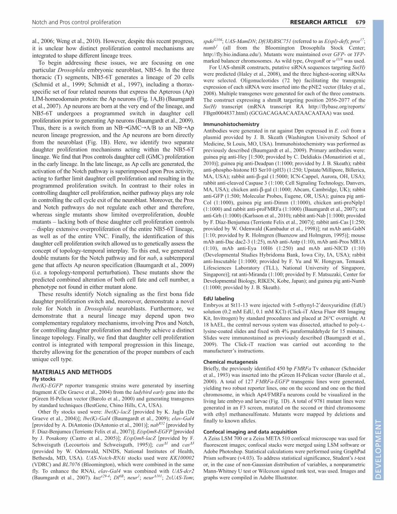
The Notch pathway is activated in the NB5-6Tneuroblast prior to the proliferation switchWhen and where is the Notch pathway activated in the NB5-6Tlineage? To address this we analyzed Notch pathway activationdirectly by staining with an antibody against the Notch intracellulardomain (NICD), aiming to track the cleavage and intracellularrelocalization of Notch. This revealed progressively stronger Notchactivation in the NB5-6T neuroblast as the lineage progressed (Fig.4A,B). In addition, we assayed Notch pathway activation in anindirect manner by analyzing the expression of well-known Notch D
EVELO
PMENT
684
target genes: members of the HES (Hairy, Enhancer of Split)family of bHLH transcription factors (reviewed by Bray, 2006), aswell as the HES-related Notch target Hey (Monastirioti et al.,2010). Hey has recently been found to be expressed in many ‘A’cells (Notch-activated sibling neurons and/or glia) and to dependupon Notch signaling for its expression (Monastirioti et al., 2010).In line with this, we found that postmitotic cells in the early part ofthe lineage express Hey (Fig. 4C). However, analysis of Heyexpression in the late NB5-6T lineage revealed expression neitherin Ap neurons nor in the neuroblast (Fig. 4D). Next, we analyzedthe expression of E(spl)m8-EGFP (Castro et al., 2005) andE(spl)m8-lacZ (Lecourtois and Schweisguth, 1995). Expressionwas weak and variable in the neuroblast at St10-11 but becamemore robust at St12, just prior to the daughter cell proliferationswitch (Fig. 4E-J). To validate the use of E(spl)m8 reporters as a
RESEARCH ARTICLE Development 139 (4)
bona fide readout of Notch activation specifically in the NB5-6Tlineage, we analyzed E(spl)m8-EGFP expression in kuz mutantsand found complete loss of reporter expression within the NB5-6Tneuroblast (Fig. 4K).
The expression analysis of Notch itself and of the Notch targetsamong the HES gene family indicates that during early stagesNB5-6T undergoes multiple rounds of the typical NBrGMCrA/Bprogression, with Notch activation in the ‘A’ cell and specificactivation of the HES factor Hey in ‘A’ cells as an effect thereof.At St10-11, expression of E(spl)m8-EGFP is consistent with thenotion that Notch is also activated in the NB5-6T neuroblast, priorto the GMC-to-direct neuron switch (Fig. 4L). This pattern ofNotch activation is also supported by the Notch pathway geneticanalysis. The expression of Hey in ‘A’ cells but not in theneuroblast, and the activation of E(spl) reporter gene expression in
Fig. 4. Notch signaling is activated in the NB5-6T neuroblast prior to the proliferation switch. (A,B)At St12, staining for the Notchintracellular domain reveals weak staining in the neuroblast (A), but expression increases toward St15 (B). Images A and B are from the same slideand processed using the same confocal settings. (C,D)At St13 (C) and St16 (D), Hey is expressed in a number of cells within the NB5-6T lineage,but not in the neuroblast (Dpn+) nor in Ap neurons. (E-J)Weak expression of the E(spl)m8-EGFP reporter was observed within the neuroblast atSt10-11 and became more robust at St12-15 in the neuroblast. (K)The expression of E(spl)m8-EGFP was lost in the neuroblast in kuz mutants (0%expression at St15; n28 hemisegments). (L)The expression of Hey and E(spl)m8-EGFP in the NB5-6T lineage. The expression of EGFP in theneuroblast was variable and was quantified in more than 60 lineages for each stage. Genotypes: (A-D) lbe(K)-EGFP; (E-J) E(spl)m8-EGFP; lbe(K)-lacZ;(K) E(spl)m8-EGFP; kuze29-4.
DEVELO
PMENT

the neuroblast, demonstrate that Notch signaling results in context-dependent HES gene activation within various parts and at variousstages of progression of this one VNC lineage.
Prospero controls daughter cell proliferationwithin early stages of NB5-6T lineagedevelopment but is not involved in theproliferation switchAs outlined above, Pros has also been shown to control daughtercell proliferation, i.e. of GMCs in the embryonic VNC. Weaddressed the possible role of Pros in the NB5-6T lineage. In linewith previous studies, we found that pros mutants displayed a clearincrease of NB5-6T cells in the early parts of the lineage (Fig.5A,B,J). In the latter part of the NB5-6T lineage, in the Apwindow, we also observed expression of Pros, with the typicalweaker, cytoplasmic staining in the neuroblast versus stronger,nuclear staining in the Ap neurons (supplementary material Fig.S6A,F). Strikingly, despite the typical expression and distributionof Pros, we found no evidence of additional Ap neurons beinggenerated in pros mutants (Fig. 5A,B,H,I). In addition, in prosmutants we found no evidence of continued division of theneuroblast past St15 (supplementary material Fig. S6D) andobserved the scheduled apoptosis of the neuroblast at late St16(supplementary material Fig. S6E).
685RESEARCH ARTICLENotch and Pros control proliferation
These results reveal that pros controls daughter cell (GMC)proliferation in the early NB5-6T lineage. However, although Prosis expressed in the late lineage, pros is involved neither in theproliferation switch nor in neuroblast cell cycle exit.
Prospero, the Notch pathway and the temporalfactor Castor do not regulate each otherTo address the possible regulatory connection between Pros and theNotch pathway, we analyzed Pros expression in kuz mutants and,conversely, E(spl)m8-GFP expression in pros mutants. Asanticipated, in neither case did we find any evidence for cross-regulation of one pathway upon the other (supplementary materialFig. S6A-C,L).
The cas temporal gene is crucial for cell specification in thelatter part of NB5-6T development and activates a number of genes(grh, dac, sqz, nab, col), thereby triggering a cascade of regulatoryevents that ultimately results in the proper specification of thedifferent Ap neurons (Baumgardt et al., 2009). Cas is expressedimmediately prior to the daughter proliferation switch in NB5-6T(Fig. 1C). We therefore addressed the regulatory interplay betweencas, pros and the Notch pathway. We did not observe any loss ofPros or E(spl)m8-EGFP in cas mutants, nor of Cas in pros mutants(supplementary material Fig. S6F-K). Above, we already noted thatCas is not affected in kuz mutants (Fig. 3).
Fig. 5. Prospero is crucial for controlling GMC and ‘Ap GMC’ divisions. (A)In the control, four Ap neurons are visible and the normal numberof cells is observed in the lineage. (B)In pros mutants, there are additional cells in the lineage, but Ap cell numbers are normal. (C)In kuz, there areadditional Ap cells, but no additional early-born cells. (D)In kuz;pros, there are additional early- and late-born cells. Col+ cells, Col/Eya+ cells andEya+ cells are visible. (E)kuz;pros mutants at St15 display a number of actively dividing cells within the NB5-6T lineage. (F,G)Expression of lbe(K)-EGFP, Col and Grh (F) or Dac (G) reveals that the majority of Col cells also express Grh and Dac. (H-J)Quantification of lbe(K)-EGFP-, Eya- or Col-expressing cells (± s.e.m.; n≥9 T2/T3 hemisegments) counted at St16 (Eya) or St15 [Col, lbe(K)-EGFP]. Asterisks denote significant differencecompared with control (P<0.05). (K)Model of the NB5-6T lineage, showing the activity of the Pros and Notch pathways, as well as the phenotypesin different genetic backgrounds. Genotypes: (A,C,F) lbe(K)-EGFP; (B,G) lbe(K)-EGFP;pros17; (J) E(spl)m8-GFP/+; pros17; (D,H) lbe(K)-EGFP, kuze29-4;(I,K,L) lbe(K)-EGFP, kuze29-4; pros17; (E) lbe(K)-EGFP; pros17/Df(3R)Exel7308. D
EVELO
PMENT

686
These results show that the two daughter cell proliferationmechanisms – Pros and the Notch pathway – and the key latetemporal factor Cas do not regulate each other.
Prospero limits the division of Apterous cells inNotch pathway mutantsIn Notch pathway perturbations we observe a maximum of eightAp cells per NB5-6T lineage, resulting from one aberrant divisionof each of the four Ap cells. We postulated that this was due to Apcells being converted to GMC-like cells as a result of Notchpathway perturbations (Fig. 5K,L). Because Pros, which is a crucialcontroller of GMC divisions in the early lineage (Fig. 5K,M), isexpressed in the latter part of the lineage, in both wild type and kuzmutants, the possibility was raised that Ap cells are prevented fromdividing more than once by the action of Pros. To test this idea, wegenerated kuz;pros double mutants.
kuz;pros double mutants were malformed, displaying extensiveovergrowth of the entire VNC, and did not develop past ~St16.Owing to developmental halt at St16, we did not detect expressionof the Nplp1 or FMRFa neuropeptides (not shown). However,using the lbe(K)-EGFP marker, we were able to identify the NB5-6T lineage and to address its development. As anticipated, thesedouble mutants displayed an increase in Ap cells beyond thatobserved in kuz, with an average of 16 Col-expressing cells in theNB5-6T lineage (Fig. 5A-D,I). To confirm the identity of theseCol-expressing cells as Ap neurons, we used Eya, Grh and Dac asadditional markers for Ap neurons, revealing the frequent co-expression of these markers (Fig. 5D,F,G).
In addition to the increase in Ap cells, double mutants displayedan extensive overproliferation of the entire NB5-6T lineage, withan average of 69 cells (Fig. 5D,J). As anticipated from thegeneration of such high numbers of cells within this relatively shorttime frame (late St8 to St16), we observed a number of mitoticcells within each NB5-6T lineage (Fig. 5E).
In kuz;pros double mutants both daughter proliferation controlsare absent, and we find an increase in the number of Ap neuronsgenerated when compared with kuz mutants alone (Fig. 5N). Thatwe observe an average of only 16 Ap cells is likely to reflect thefact that kuz;pros mutants do not develop past St16, and as Ap
RESEARCH ARTICLE Development 139 (4)
neurons are born between St13 and St15 this precludes moreextensive overproliferation. In addition, the entire NB5-6T isovergrown, and multiple cells throughout the lineage areundergoing continuous proliferation.
Simultaneous disruption of Notch signaling andtemporal coding reveals a topology-temporalinterplayMutations in the subtemporal gene nab result in a misspecificationof the Ap2 neuron into an Ap1/Nplp1 neuron, without thegeneration of any additional Ap neurons (Fig. 3R; supplementarymaterial Fig. S3) (Baumgardt et al., 2009). Conversely, latedisruption of the Notch pathway results in one division of each Apneuron, but without any alteration in temporal progression or Apcell fate (Figs 2, 3). Thus, both nab and kuz mutants display twoAp1/Nplp1 cells, but as a result of different mechanisms (Fig. 6A-C,F). Importantly, the Notch pathway does not regulate Nab, andthe crucial temporal gene cas – an activator of nab – does notregulate the Notch pathway (see above). These findings allowed usto genetically test the concept of topology-temporal interplay. Wereasoned that by simultaneously perturbing the daughter cellproliferation control and the temporal progression, we should beable to generate additional Ap1/Nplp1 neurons beyond thoseobserved when each system is affected individually. To this end,we generated kuz;nab double mutants and analyzed them for Eyaand Nplp1 expression at 18 hAEL. Strikingly, these double mutantsrevealed a combined effect, with extra Ap1/Nplp1 neurons beyondthose observed in kuz or nab alone. Specifically, whereas kuz ornab single mutants never displayed more than two Ap1/Nplp1neurons, double mutants displayed up to four Ap1/Nplp1 neurons(Fig. 6A-E). Thus, daughter cell proliferation and temporalprogression can be independently or combinatorially interferedwith, leading to predictable outcomes of cell fate and cell numbers(Fig. 6F).
DISCUSSIONWe find that the NB5-6T lineage utilizes two distinct mechanismsto control daughter cell proliferation. In the early part of thelineage, pros limits daughter cell (GMC) proliferation, whereas in
Fig. 6. kuz;nab doublemutants reveal a topology-temporal interplay. (A-D)Incontrol, four Ap neurons arepresent, with one Ap1/Nplp1neuron. In both kuz and nab,one additional Ap1/Nplp1neuron is evident. In kuz;nabdouble mutants, up to fourAp1/Nplp1 neurons are evident.(E)Quantification of Eya- orNplp1-expressing cells perthoracic T2/T3 hemisegment (± s.e.m.; n≥28 clusters). Cellswere counted at 18 hAEL. Datafor control and kuz were copiedfrom Fig. 2. (F)Summary of theobserved effects. Genotype: (A)OregonR; (B) kuze29-4; (C)nabR52; (D) kuze29-4;nabR52.
DEVELO
PMENT

the late part canonical Notch signaling in the neuroblast furtherrestricts daughter cell proliferation, resulting in a switch to thegeneration of neurons directly (Fig. 5K). The switch in daughtercell proliferation is integrated with temporal lineage progressionand enables the specification of different Ap neuron subtypes andthe control of their numbers.
A programmed switch in daughter cellproliferationOur data on Notch activation in the NB5-6T lineage, using bothantibodies and reporters, indicate progressive activation in theneuroblast: weak at St10-11 and more robust from St12 onward.Thus, Notch activity coincides with the proliferation mode switch.How is this gradual activation of Notch in the neuroblast controlled?NB5-6T undergoes the typical progression of the temporal genecascade, with Cas expression preceding strong Notch activation.Thus, one possible scenario is that the late temporal gene casactivates the Notch pathway. However, our analysis of the E(spl)m8-EGFP reporter shows that this Notch target is still activated at theproper stage in cas mutants. Although this does not rule out thepossibility that other, unknown, temporal factors might regulateNotch signaling, it rules out one obvious player, cas. Alternatively,as Notch signaling is off when neuroblasts are formed – aprerequisite for neuroblast selection – Notch activation in theneuroblast at later stages might simply reflect a gradual reactivationof the pathway. Although such a reactivation might at a first glanceappear too imprecise, it is possible that the specificity of thisparticular Notch output – proliferation control – might becombinatorially achieved by the intersection of Notch signaling withother, more tightly controlled, temporal changes.
687RESEARCH ARTICLENotch and Pros control proliferation
Overlapping daughter cell proliferation controls: acooperative tumor-forming mechanismPros and Notch control daughter proliferation in different parts ofNB5-6T and we find no evidence of cross-regulation between thesepathways. The limited overproliferation of the lineage when eachpathway is separately mutated results not from redundant functions,but rather stems from the biphasic nature of this lineage.Specifically, in pros mutants, Notch signaling is likely to be on inall ‘A’ type sibling daughter cells, as Numb continues to beasymmetrically distributed between daughter cells. Thus, Notchsignaling in ‘A’ cells may preclude each ‘A’ cell from dividing evenonce (Fig. 5M, blue circles). This notion is in line with recentstudies showing that postmitotic Notch activated cells (‘A’ cells)within the Drosophila bristle lineages are particularly resilient tooverexpression of cell cycle genes (Simon et al., 2009). Similarly,in Notch pathway mutants, as Ap cells now divide (in essencebecoming GMC-type cells), Pros will still play its normal role inthese ‘GMCs’ and limit their proliferation to a single extra celldivision. However, in kuz;pros double mutants, Ap cells arerelieved of both types of daughter cell proliferation control and canthus divide for many additional rounds (Fig. 5N). This notion alsoapplies to early parts of the NB5-6T lineage and probably to themajority of other VNC lineages, as indicated by the extensiveoverproliferation of the entire NB5-6T lineage, and to the generaloverproliferation of the VNC. However, based on our findings thatneither the Notch pathway nor pros controls neuroblast identity orits progression, we postulate that these large clones contain asingle, normally behaving NB5-6T neuroblast. In fact, theneuroblast is likely to exit the cell cycle and undergo apoptosis onschedule, as neither of these decisions depends upon pros or the
Fig. 7. Mechanisms for controlling neural subtypecell numbers. Based on previous studies, three modelscan be proposed to explain the generation of differentnumbers of distinct neural subtypes (small circles) froma common progenitor domain (large circles) within ageneric central nervous system. (A)The same number ofprogenitors generates the same number of differentneural subtypes, but programmed cell death (X)removes some cells of a certain subtype. (B)In aprogenitor domain a variable number of progenitors isactive at different stages. (C)The same number ofprogenitors is active at all stages, but temporalcompetence windows differ in length. (D)Resultspresented in this study suggest a novel fourthmechanism. Here, temporal competence changes areaccompanied by changes in daughter cell proliferation,resulting in the generation of different numbers ofdifferent neural subtypes. Red: progenitors in an earlytemporal competence window, generating early-bornneurons. Green: progenitors in a late temporalcompetence window, generating late-born neurons.White: inactive progenitors.
DEVELO
PMENT

688
Notch pathway. Of interest with respect to cancer biology is thatour findings point to a novel mechanism whereby mutation in twotumor suppressors (e.g. Pros and Notch) cooperate to generateextensive overproliferation: not by acting in the same progenitorcell at the same time, but by playing complementary rolescontrolling daughter cell proliferation.
The topology-temporal interplay: a noveldevelopmental intersectionAs an effect of alternate daughter cell proliferation patterns, bothvertebrates and invertebrates display variability in neural lineagetopology (Brand and Livesey, 2011; Gotz and Huttner, 2005;Kriegstein et al., 2006; Rowitch and Kriegstein, 2010). Similarly,progenitors in these systems undergo temporal changes incompetence, as evident by changes in the types of neurons and gliagenerated at different time points (Jacob et al., 2008; Okano andTemple, 2009; Pearson and Doe, 2004). Hence, the temporal-topology interplay described in this study is likely to be extensivelyused and to be conserved in mammals. As a proof of principle ofthis novel developmental intersection, we analyzed single anddouble mutants for kuz and nab, thereby independently versuscombinatorially affecting temporal progression and daughter cellproliferation. Strikingly, these mutants show the predictedcombined effect, with the appearance of additional Ap1/Nplp1neurons beyond those found in each individual mutant.
If programmed proliferation switches are conserved, how mightsuch a topology-temporal interplay become utilized in mammals?There are several examples in which different clusters/pools/nucleiof neurons of distinct cell fate are generated from the sameprogenitor domain in the developing mammalian nervous system.Such pools often contain different numbers of cells, but theunderlying mechanisms controlling the precise numbers of eachsubtype are poorly understood. Based on previous studies in anumber of models, at least three different mechanisms can beenvisioned (Fig. 7A-C). Based on our current study we propose anovel fourth mechanism, whereby alteration of daughter cellproliferation is integrated with temporal progression to controlsubtype cell numbers (Fig. 7D). These four mechanisms are notmutually exclusive, and given the complexity of the mammaliannervous system it is tempting to speculate that all four mechanismsare utilized during development.
AcknowledgementsWe thank C. Deldiakis, S. Cohen, F. Schweisguth, F. Díaz-Benjumea, R.Holmgren, W. Odenwald, M. Levine, J. B. Skeath, J. Posakony, theDevelopmental Studies Hybridoma Bank at the University of Iowa, theDrosophila Genetic Resource Center at Kyoto and The Bloomington StockCenter for sharing antibodies, fly lines and DNAs; T. Edlund, D. van Meyel, I.Miguel-Aliaga and J. B. Thomas for critically reading the manuscript; and H.Ekman, A. Angel and A. Starkenberg for technical assistance. FlyBase providedimportant information used in this work.
FundingThis work was supported by the Swedish Research Council, by the Knut andAlice Wallenberg foundation and by the Swedish Cancer Foundation to S.T.
Competing interests statementThe authors declare no competing financial interests.
Supplementary materialSupplementary material available online athttp://dev.biologists.org/lookup/suppl/doi:10.1242/dev.074500/-/DC1
ReferencesAllan, D. W., Pierre, S. E., Miguel-Aliaga, I. and Thor, S. (2003). Specification
of neuropeptide cell identity by the integration of retrograde bmp signaling anda combinatorial transcription factor code. Cell 113, 73-86.
RESEARCH ARTICLE Development 139 (4)
Allan, D. W., Park, D., St Pierre, S. E., Taghert, P. H. and Thor, S. (2005).Regulators acting in combinatorial codes also act independently in singledifferentiating neurons. Neuron 45, 689-700.
Artavanis-Tsakonas, S., Muskavitch, M. A. and Yedvobnick, B. (1983).Molecular cloning of Notch, a locus affecting neurogenesis in Drosophilamelanogaster. Proc. Natl. Acad. Sci. USA 80, 1977-1981.
Bardin, A. J., Le Borgne, R. and Schweisguth, F. (2004). Asymmetriclocalization and function of cell-fate determinants: a fly’s view. Curr. Opin.Neurobiol. 14, 6-14.
Barolo, S., Carver, L. A. and Posakony, J. W. (2000). GFP and beta-galactosidasetransformation vectors for promoter/enhancer analysis in Drosophila.Biotechniques 29, 726-732.
Baumgardt, M., Miguel-Aliaga, I., Karlsson, D., Ekman, H. and Thor, S.(2007). Specification of neuronal identities by feedforward combinatorialcoding. PLoS Biol. 5, 295-308.
Baumgardt, M., Karlsson, D., Terriente, J., Diaz-Benjumea, F. J. and Thor, S.(2009). Neuronal subtype specification within a lineage by opposing temporalfeed-forward loops. Cell 139, 969-982.
Bello, B., Reichert, H. and Hirth, F. (2006). The brain tumor gene negativelyregulates neural progenitor cell proliferation in the larval central brain ofDrosophila. Development 133, 2639-2648.
Benveniste, R. J., Thor, S., Thomas, J. B. and Taghert, P. H. (1998). Cell type-specific regulation of the Drosophila FMRF-NH2 neuropeptide gene by Apterous,a LIM homeodomain transcription factor. Development 125, 4757-4765.
Bier, E., Vaessin, H., Younger-Shepherd, S., Jan, L. Y. and Jan, Y. N. (1992).deadpan, an essential pan-neural gene in Drosophila, encodes a helix-loop-helixprotein similar to the hairy gene product. Genes Dev. 6, 2137-2151.
Bowman, S. K., Rolland, V., Betschinger, J., Kinsey, K. A., Emery, G. andKnoblich, J. A. (2008). The tumor suppressors Brat and Numb regulate transit-amplifying neuroblast lineages in Drosophila. Dev. Cell 14, 535-546.
Brand, A. H. and Livesey, F. J. (2011). Neural stem cell biology in vertebrates andinvertebrates: more alike than different? Neuron 70, 719-729.
Bray, S. J. (2006). Notch signalling: a simple pathway becomes complex. Nat. Rev.Mol. Cell Biol. 7, 678-689.
Brody, T. and Odenwald, W. F. (2002). Cellular diversity in the developingnervous system: a temporal view from Drosophila. Development 129, 3763-3770.
Buenzow, D. E. and Holmgren, R. (1995). Expression of the Drosophilagooseberry locus defines a subset of neuroblast lineages in the central nervoussystem. Dev. Biol. 170, 338-349.
Castro, B., Barolo, S., Bailey, A. M. and Posakony, J. W. (2005). Lateralinhibition in proneural clusters: cis-regulatory logic and default repression bySuppressor of Hairless. Development 132, 3333-3344.
Cau, E. and Blader, P. (2009). Notch activity in the nervous system: to switch ornot switch? Neural Dev. 4, 36.
Choksi, S. P., Southall, T. D., Bossing, T., Edoff, K., de Wit, E., Fischer, B. E.,van Steensel, B., Micklem, G. and Brand, A. H. (2006). Prospero acts as abinary switch between self-renewal and differentiation in Drosophila neural stemcells. Dev. Cell 11, 775-789.
De Graeve, F., Jagla, T., Daponte, J. P., Rickert, C., Dastugue, B., Urban, J.and Jagla, K. (2004). The ladybird homeobox genes are essential for thespecification of a subpopulation of neural cells. Dev. Biol. 270, 122-134.
DiAntonio, A., Haghighi, A. P., Portman, S. L., Lee, J. D., Amaranto, A. M.and Goodman, C. S. (2001). Ubiquitination-dependent mechanisms regulatesynaptic growth and function. Nature 412, 449-452.
Doe, C. Q. (2008). Neural stem cells: balancing self-renewal with differentiation.Development 135, 1575-1587.
Doe, C. Q. and Technau, G. M. (1993). Identification and cell lineage of individualneural precursors in the Drosophila CNS. Trends Neurosci. 16, 510-514.
Egger, B., Chell, J. M. and Brand, A. H. (2008). Insights into neural stem cellbiology from flies. Philos. Trans. R. Soc. Lond. B Biol. Sci. 363, 39-56.
Fortini, M. E. (2009). Notch signaling: the core pathway and its posttranslationalregulation. Dev. Cell 16, 633-647.
Fuerstenberg, S., Broadus, J. and Doe, C. Q. (1998). Asymmetry and cell fate inthe Drosophila embryonic CNS. Int. J. Dev. Biol. 42, 379-383.
Gotz, M. and Huttner, W. B. (2005). The cell biology of neurogenesis. Nat. Rev.Mol. Cell Biol. 6, 777-788.
Haley, B., Hendrix, D., Trang, V. and Levine, M. (2008). A simplified miRNA-based gene silencing method for Drosophila melanogaster. Dev. Biol. 321, 482-490.
Hewes, R. S., Park, D., Gauthier, S. A., Schaefer, A. M. and Taghert, P. H.(2003). The bHLH protein Dimmed controls neuroendocrine cell differentiation inDrosophila. Development 130, 1771-1781.
Ikeshima-Kataoka, H., Skeath, J. B., Nabeshima, Y., Doe, C. Q. andMatsuzaki, F. (1997). Miranda directs Prospero to a daughter cell duringDrosophila asymmetric divisions. Nature 390, 625-629.
Jacob, J., Maurange, C. and Gould, A. P. (2008). Temporal control of neuronaldiversity: common regulatory principles in insects and vertebrates? Development135, 3481-3489. D
EVELO
PMENT

689RESEARCH ARTICLENotch and Pros control proliferation
Kambadur, R., Koizumi, K., Stivers, C., Nagle, J., Poole, S. J. and Odenwald,W. F. (1998). Regulation of POU genes by castor and hunchback establisheslayered compartments in the Drosophila CNS. Genes Dev. 12, 246-260.
Karcavich, R. and Doe, C. Q. (2005). Drosophila neuroblast 7-3 cell lineage: amodel system for studying programmed cell death, Notch/Numb signaling, andsequential specification of ganglion mother cell identity. J. Comp. Neurol. 481,240-251.
Karlsson, D., Baumgardt, M. and Thor, S. (2010). Segment-specific neuronalsubtype specification by the integration of anteroposterior and temporal cues.PLoS Biol. 8, e1000368.
Knoblich, J. A. (2008). Mechanisms of asymmetric stem cell division. Cell 132,583-597.
Kopan, R. and Ilagan, M. X. (2009). The canonical Notch signaling pathway:unfolding the activation mechanism. Cell 137, 216-233.
Kriegstein, A., Noctor, S. and Martinez-Cerdeno, V. (2006). Patterns of neuralstem and progenitor cell division may underlie evolutionary cortical expansion.Nat. Rev. Neurosci. 7, 883-890.
Lecourtois, M. and Schweisguth, F. (1995). The neurogenic suppressor ofhairless DNA-binding protein mediates the transcriptional activation of theenhancer of split complex genes triggered by Notch signaling. Genes Dev. 9,2598-2608.
Lee, C. Y., Wilkinson, B. D., Siegrist, S. E., Wharton, R. P. and Doe, C. Q.(2006). Brat is a Miranda cargo protein that promotes neuronal differentiationand inhibits neuroblast self-renewal. Dev. Cell 10, 441-449.
Li, L. and Vaessin, H. (2000). Pan-neural Prospero terminates cell proliferationduring Drosophila neurogenesis. Genes Dev. 14, 147-151.
Lundgren, S. E., Callahan, C. A., Thor, S. and Thomas, J. B. (1995). Control ofneuronal pathway selection by the Drosophila LIM homeodomain geneapterous. Development 121, 1769-1773.
Maurange, C. and Gould, A. P. (2005). Brainy but not too brainy: starting andstopping neuroblast divisions in Drosophila. Trends Neurosci. 28, 30-36.
Miguel-Aliaga, I., Allan, D. W. and Thor, S. (2004). Independent roles of thedachshund and eyes absent genes in BMP signaling, axon pathfinding andneuronal specification. Development 131, 5837-5848.
Monastirioti, M., Giagtzoglou, N., Koumbanakis, K. A., Zacharioudaki, E.,Deligiannaki, M., Wech, I., Almeida, M., Preiss, A., Bray, S. and Delidakis,
C. (2010). Drosophila Hey is a target of Notch in asymmetric divisions duringembryonic and larval neurogenesis. Development 137, 191-201.
Okano, H. and Temple, S. (2009). Cell types to order: temporal specification ofCNS stem cells. Curr. Opin. Neurobiol. 19, 112-119.
Pearson, B. J. and Doe, C. Q. (2004). Specification of temporal identity in thedeveloping nervous system. Annu. Rev. Cell Dev. Biol. 20, 619-647.
Rowitch, D. H. and Kriegstein, A. R. (2010). Developmental genetics ofvertebrate glial-cell specification. Nature 468, 214-222.
Schmid, A., Chiba, A. and Doe, C. Q. (1999). Clonal analysis of Drosophilaembryonic neuroblasts: neural cell types, axon projections and muscle targets.Development 126, 4653-4689.
Schmidt, H., Rickert, C., Bossing, T., Vef, O., Urban, J. and Technau, G. M.(1997). The embryonic central nervous system lineages of Drosophilamelanogaster. II. Neuroblast lineages derived from the dorsal part of theneuroectoderm. Dev. Biol. 189, 186-204.
Schneider, L. E., Roberts, M. S. and Taghert, P. H. (1993). Cell type-specifictranscriptional regulation of the Drosophila FMRFamide neuropeptide gene.Neuron 10, 279-291.
Shen, C. P., Jan, L. Y. and Jan, Y. N. (1997). Miranda is required for theasymmetric localization of Prospero during mitosis in Drosophila. Cell 90, 449-458.
Simon, F., Fichelson, P., Gho, M. and Audibert, A. (2009). Notch and Prosperorepress proliferation following cyclin E overexpression in the Drosophila bristlelineage. PLoS Genet. 5, e1000594.
Skeath, J. B. and Thor, S. (2003). Genetic control of Drosophila nerve corddevelopment. Curr. Opin. Neurobiol. 13, 8-15.
Technau, G. M., Berger, C. and Urbach, R. (2006). Generation of cell diversityand segmental pattern in the embryonic central nervous system of Drosophila.Dev. Dyn. 235, 861-869.
Terriente Felix, J., Magarinos, M. and Diaz-Benjumea, F. J. (2007). Nabcontrols the activity of the zinc-finger transcription factors Squeeze and Rotundin Drosophila development. Development 134, 1845-1852.
Weng, M., Golden, K. L. and Lee, C. Y. (2010). dFezf/Earmuff maintains therestricted developmental potential of intermediate neural progenitors inDrosophila. Dev. Cell 18, 126-135.
DEVELO
PMENT