developing methods to detect and diagnose chronic
TRANSCRIPT
RESEARCH Open Access
Developing methods to detect anddiagnose chronic traumaticencephalopathy during life: rationale,design, and methodology for theDIAGNOSE CTE Research ProjectMichael L. Alosco1, Megan L. Mariani2, Charles H. Adler3, Laura J. Balcer4, Charles Bernick5,29, Rhoda Au6,30,31,Sarah J. Banks7, William B. Barr8, Sylvain Bouix9, Robert C. Cantu10, Michael J. Coleman11, David W. Dodick3,Lindsay A. Farrer12, Yonas E. Geda13, Douglas I. Katz14,32, Inga K. Koerte9,33, Neil W. Kowall10,34, Alexander P. Lin15,Daniel S. Marcus16, Kenneth L. Marek17, Michael D. McClean18, Ann C. McKee1,34, Jesse Mez19,Joseph N. Palmisano20, Elaine R. Peskind21, Yorghos Tripodis22, Robert W. Turner II23, Jennifer V. Wethe24,Jeffrey L. Cummings25, Eric M. Reiman26, Martha E. Shenton27, Robert A. Stern28* for the DIAGNOSE CTEResearch Project Investigators
Abstract
Background: Chronic traumatic encephalopathy (CTE) is a neurodegenerative disease that has beenneuropathologically diagnosed in brain donors exposed to repetitive head impacts, including boxers and Americanfootball, soccer, ice hockey, and rugby players. CTE cannot yet be diagnosed during life. In December 2015, theNational Institute of Neurological Disorders and Stroke awarded a seven-year grant (U01NS093334) to fund the“Diagnostics, Imaging, and Genetics Network for the Objective Study and Evaluation of Chronic TraumaticEncephalopathy (DIAGNOSE CTE) Research Project.” The objectives of this multicenter project are to: develop in vivofluid and neuroimaging biomarkers for CTE; characterize its clinical presentation; refine and validate clinical researchdiagnostic criteria (i.e., traumatic encephalopathy syndrome [TES]); examine repetitive head impact exposure,genetic, and other risk factors; and provide shared resources of anonymized data and biological samples to theresearch community. In this paper, we provide a detailed overview of the rationale, design, and methods for theDIAGNOSE CTE Research Project.
© The Author(s). 2021 Open Access This article is licensed under a Creative Commons Attribution 4.0 International License,which permits use, sharing, adaptation, distribution and reproduction in any medium or format, as long as you giveappropriate credit to the original author(s) and the source, provide a link to the Creative Commons licence, and indicate ifchanges were made. The images or other third party material in this article are included in the article's Creative Commonslicence, unless indicated otherwise in a credit line to the material. If material is not included in the article's Creative Commonslicence and your intended use is not permitted by statutory regulation or exceeds the permitted use, you will need to obtainpermission directly from the copyright holder. To view a copy of this licence, visit http://creativecommons.org/licenses/by/4.0/.The Creative Commons Public Domain Dedication waiver (http://creativecommons.org/publicdomain/zero/1.0/) applies to thedata made available in this article, unless otherwise stated in a credit line to the data.
* Correspondence: [email protected] University Alzheimer’s Disease Research Center, Boston UniversityCTE Center, Departments of Neurology, Neurosurgery, and Anatomy &Neurobiology, Boston University School of Medicine, Boston, MA, USAFull list of author information is available at the end of the article
Alosco et al. Alzheimer's Research & Therapy (2021) 13:136 https://doi.org/10.1186/s13195-021-00872-x
Methods: The targeted sample and sample size was 240 male participants, ages 45–74, including 120 formerprofessional football players, 60 former collegiate football players, and 60 asymptomatic participants without ahistory of head trauma or participation in organized contact sports. Participants were evaluated at one of four U.S.sites and underwent the following baseline procedures: neurological and neuropsychological examinations; tau andamyloid positron emission tomography; magnetic resonance imaging and spectroscopy; lumbar puncture; bloodand saliva collection; and standardized self-report measures of neuropsychiatric, cognitive, and daily functioning.Study partners completed similar informant-report measures. Follow-up evaluations were intended to be in-personand at 3 years post-baseline. Multidisciplinary diagnostic consensus conferences are held, and the reliability andvalidity of TES diagnostic criteria are examined.
Results: Participant enrollment and all baseline evaluations were completed in February 2020. Three-year follow-upevaluations began in October 2019. However, in-person evaluation ceased with the COVID-19 pandemic, andresumed as remote, 4-year follow-up evaluations (including telephone-, online-, and videoconference-basedcognitive, neuropsychiatric, and neurologic examinations, as well as in-home blood draw) in February 2021.
Conclusions: Findings from the DIAGNOSE CTE Research Project should facilitate detection and diagnosis of CTEduring life, and thereby accelerate research on risk factors, mechanisms, epidemiology, treatment, and prevention ofCTE.
Trial registration: NCT02798185
Keywords: Neurodegenerative disease, Biomarkers, Chronic traumatic encephalopathy, Cognitive function,Concussion, Football, Head trauma, MRI, MRS, National Football League, College football, Neuroimaging, Repetitivehead impacts, Traumatic brain injury, Subconcussion, Traumatic encephalopathy syndrome, Positron emissiontomography, Tau, Remote assessment
BackgroundChronic traumatic encephalopathy (CTE) is a neurode-generative disease that has been neuropathologically diag-nosed in former contact sport athletes, military combatveterans, and other individuals exposed to repetitive headimpacts [1–9]. Although CTE is often portrayed as a newdisease, its history dates back to the 1920s and 1930s, withdescriptions of “punch drunk” boxers [10] and “dementiapugilistica” [11], with the term “chronic traumatic enceph-alopathy” first used in publications in the 1940s [12, 13].CTE began to receive significant lay and scientific atten-tion in 2005, following a report of neuropathological evi-dence of hyperphosphorylated tau (p-tau) in an irregularpattern in a deceased former National Football League(NFL) player who had ante-mortem cognitive and neuro-psychiatric symptoms [5]. CTE has since been neuro-pathologically diagnosed in hundreds of deceasedAmerican football players [1–3], in addition to other con-tact and collision sport athletes (e.g., ice hockey, soccer,rugby) [2, 8, 14] and military veterans [4]. Given the mil-lions of active and former contact sport athletes, militaryservice members, and veterans, CTE is potentially a majorpublic health concern. However, disease incidence andprevalence remain unknown due to the inability to detectand diagnose CTE during life.
Neuropathology of CTEIn 2013, McKee et al. examined the immunocytochem-ical characteristics of 68 cases (including 50 American
football players) with autopsy-confirmed CTE and pro-posed neuropathological diagnostic criteria for CTE inaddition to a 4-stage classification scheme to grade thepathological severity of p-tau [2, 15]. As part of the Na-tional Institute of Neurological Disorders and Stroke(NINDS)-funded “Understanding Neurologic Injury inTraumatic Encephalopathy (UNITE)” study [16], twoconsensus meetings have been convened to define theneuropathological diagnostic criteria for CTE [17, 18].The pathognomonic lesion is now defined as p-tau ag-gregates as neurofibrillary tangles (NFT) in neurons,with or without p-tau in astrocytes, deposited aroundsmall blood vessels, in an irregular pattern at the depthsof the cortical sulci, with the focus in superficial corticallayers. The panels concluded that the pattern of p-tau inCTE is distinct from that of any other neurodegenerativedisease [17, 18]. The molecular structure of tau filamentsin CTE has also since been shown to be unique [19] [20][21]. Although CTE is a mixed three and fourmicrotubule-binding domain repeat (3R and 4R) tauopa-thy, similar to AD, recent research has shown that theCTE tau isoforms shift across disease severity, from 4Rto 3R, with 4R isoforms found primarily in astrocytes[22]. Unlike in AD, amyloid-beta protein (Aβ) depositsin CTE are not an early feature, they are not diagnostic,and they tend to accumulate with advancing age as a co-morbidity. When present in CTE, the Aβ plaques tendto be diffuse and not neuritic [23]. CTE is nonethelessfrequently co-morbid with other neurodegenerative (e.g.,
Alosco et al. Alzheimer's Research & Therapy (2021) 13:136 Page 2 of 23
Lewy body disease) and non-neurodegenerative condi-tions (e.g., white matter rarefaction, arteriolosclerosis)[3, 24–26].
Risk factors for CTEThe primary risk factor for CTE is exposure to repetitivehead impacts and the resulting repeated concussionsand subconcussive trauma (i.e., head impacts that resultin neuronal injury but do not cause immediate symp-toms) [3, 9, 24, 27–30]. Repetitive subconcussive headimpacts play a prominent role in the development of thisdisease [31, 32].The duration of American football playhas been identified as a strong predictor of the risk andseverity of CTE neuropathology [30]. Small autopsy caseseries provide suggestive links between soccer and rugbyplay and CTE [8, 14]. These associations between repeti-tive head impacts and neuropathology have been com-plemented by in vivo research studies that link exposureto repetitive head impacts with later-life cognitive andneuropsychiatric symptoms [24, 27, 33, 34]. However,not all individuals who are exposed to repetitive headimpacts will develop CTE or other long-term neuro-logical disorders. Risk modifiers may include severityand nature of repetitive head impact exposure (e.g., fre-quency [27, 35], type of contact sport played [36–38],younger age of first exposure [39–44]), lower cognitivereserve [45], cerebrovascular health [24, 46], and/or gen-etic make-up [47–49].
Clinical presentation of CTE and traumaticencephalopathy syndromeCTE is a neuropathological diagnosis and cannot cur-rently be diagnosed during life because its clinical pres-entation has been—until recently—ill-defined, andbecause validated in vivo biomarkers for the detection ofCTE neuropathology do not yet exist [50]. To date, theclinical presentation of CTE has been described primar-ily through the use of retrospective telephone interviewswith next-of-kin and other informants of brains donorswith autopsy-confirmed CTE. These studies have re-ported a constellation of progressively worsening andnon-specific cognitive impairments (particularly in ex-ecutive functions and episodic memory), poor regulationor control of emotions and/or behavior (including im-pulsivity, explosiveness, rage, and/or emotional lability),and, in some instances, parkinsonism and motor neurondisease [3, 47, 51]. Provisional clinical research diagnos-tic criteria for CTE have been proposed [52, 53], includ-ing the traumatic encephalopathy syndrome (TES)research criteria published in 2014 [54]. Since the timeof the original 2014 publication, the research diagnosticcriteria for TES have been used in several studies. Theirvalidity in predicting neuropathological CTE diagnoseshas recently been reported along with an item-level
analysis suggesting that cognitive symptoms—more thanneuropsychiatric features—are particularly valuable inpredicting CTE pathology [55]. These findings informednew NINDS Consensus Diagnostic Criteria for TES thathave recently been published [56].Consistent with the clinical diagnosis of other neuro-
degenerative diseases [57–60], an accurate diagnosis ofCTE during life will require validated biomarkers of theunderlying pathophysiology of the disease. There havebeen several preliminary studies that have examinedboth specific (e.g., those that measure regional p-taupathology) and non-specific (e.g., general neurodegenera-tion) biomarkers of CTE. Examples of potential support-ive or non-specific fluid biomarkers of CTE includecerebrospinal fluid (CSF) and plasma markers of neuro-degeneration (e.g., total tau [t-tau] [35, 61, 62], neurofila-ment light [NfL] chain protein [63]) and microglialactivation (e.g., soluble triggering receptor expressed onmyeloid cells 2 [sTREM2]) [61], as well as measures oftau in exosomes isolated from plasma and proteomicprofiling of plasma and CSF extracellular vesicles [64–66]. Candidate neuroimaging biomarkers for the detec-tion of non-specific neurodegenerative changes of CTEhave recently been reviewed [67, 68] and include the fol-lowing: cavum septum pellucidum (a common grossneuropathological finding in CTE [1, 2]) on structuralT1-weighted magnetic resonance imaging (MRI) [69–72]; frontotemporal and medial temporal lobe atrophyon structural T1-weighted MRI [73–79]; decreased cor-tical thickness on T1-weighted MRI [44]; white matteralterations on diffusion tensor imaging (DTI) [42, 74,80–84]; cerebral hypoperfusion and functional hypoac-tivity on single photon emission computerized tomog-raphy [85]; arterial spin labeling [86] and functional MRI(fMRI) [87]; and neurochemical alterations on magneticresonance spectroscopy (MRS) [88–90].Efforts are underway to identify specific biomarkers of
CTE. Amyloid positron emission tomography (PET) im-aging is a neuropathologically validated biomarker forthe early detection, tracking, and diagnosis of neuriticAβ deposition, one of the cardinal neuropathological fea-tures of AD [58, 91]. Although postmortem studies ofCTE have found diffuse Aβ plaques, evidence of neuriticplaques has primarily been found in late-stage disease [3,15, 23]. It would be unlikely for amyloid PET tracers todemonstrate significant binding in CTE, as they bindpreferentially to neuritic plaques [91]. More recently,PET imaging of paired helical filament tau has become aneuropathologically validated biomarker for the detec-tion, tracking, and diagnosis of AD-related tau tangle de-position. For instance, the PET radiotracer flortaucipir(AV1451; T807) demonstrates high affinity binding tothe mixed 3R/4R tau isoforms in AD [92]. Flortaucipir’sbinding to other tauopathies with primarily 4R isoforms
Alosco et al. Alzheimer's Research & Therapy (2021) 13:136 Page 3 of 23
has not been as promising [93, 94]. Although CTE andAD have similar (3R/4R) tau isoforms, there are differ-ences at the molecular level [19], and, as mentioned earl-ier, in CTE the ratio of 3R:4R differs across disease stageand between neuronal and glial tau [22].Stern et al. studied flortaucipir PET in 26 symptomatic
former NFL players (ages 40–69) and 31 same-ageasymptomatic men without a history of traumatic braininjury (TBI) [95]. At a group level, the former playershad significantly higher flortaucipir uptake in superiorfrontal, medial temporal, and parietal regions, comparedto the asymptomatic men without TBI; however, the up-take levels were not as high as seen in individuals withAD. Although the level of flortaucipir uptake in thethree brain regions was significantly associated with thenumber of years playing football, it was not significantlyassociated with cognitive and neuropsychiatric testscores. Importantly, the former players did not have ele-vated florbetapir levels compared to the unexposedgroup, and only one former NFL player had slightly ele-vated florbetapir standardized uptake value ratio(SUVR), indicating that the former NFL players’ cogni-tive impairment was not due to AD. Based on thesefindings and other in vivo and neuropathological studies[78, 96, 97], the utility and specificity of flortaucipir forthe detection of CTE paired helical filament tau isunclear.
The DIAGNOSE CTE Research ProjectIn December 2015, NINDS funded a 7-year, multicentergrant proposal, “Chronic Traumatic Encephalopathy:Detection, Diagnosis, Course, and Risk Factors,” submit-ted by our multidisciplinary team of investigators, underthe leadership of four co-principal investigators (co-PIs;RAS [contact PI], JLC, EMR, MES). This is now referredto as the “Diagnostics, Imaging, and Genetics Networkfor the Objective Study and Evaluation of Chronic Trau-matic Encephalopathy (DIAGNOSE CTE) Research Pro-ject.” The specific aims of the DIAGNOSE CTEResearch Project are listed in Table 1. The primary end-points of the study are to characterize the clinical pres-entation of CTE and to identify in vivo biomarkers that
can support a “probable CTE” diagnosis. There wereseveral guiding principles that were followed throughoutthe development of the grant proposal and study design,including (1) CTE is viewed as a neurodegenerative dis-ease; (2) although the necessary risk factor for the devel-opment of this tauopathy appears to be a history ofrepetitive head impact exposure, CTE itself should notbe confused with TBI or considered the aggregate effectof multiple symptomatic concussions; (3) to conduct astudy of the clinical presentation, diagnostic criteria, bio-markers, and risk factors of CTE requires expertiseacross many disciplines, including Neurology, Neuro-psychology, Psychiatry, Neuroimaging, Molecular Medi-cine, Neuropathology, Exposure Science, Genetics,Biostatistics, Epidemiology, Computer Science, and Soci-ology; and (4) a team science approach is required,breaking down typical academic and institutional “silos”to conduct the best research in the most efficientmanner.The objective of this paper is to describe the rationale,
methodology, and design of the DIAGNOSE CTE Re-search Project. We provide updates on the current statusof the project and conclude with methodological consid-erations, and discussion of the expected impact of theproject results, as well as the infrastructure and re-sources created to support further studies.
MethodsInfrastructure overviewThe DIAGNOSE CTE Research Project is a 7-year (nowexpected to be 8-year following COVID-19 pandemic-related modifications described below) study that in-volves more than 40 investigators from 12 research insti-tutions across the USA (Supplementary Table 1,Additional File 1). There is a 7-member External Advis-ory Board (Supplementary Table 2, Additional File 1)that meets annually with the co-PIs and the NINDS Sci-entific Program Official. The project is overseen by anExecutive Committee consisting of the co-PIs, Partici-pant Evaluation Site PIs, NINDS Scientific Program Offi-cial, Advisory Board Chair (ex officio member), andTeam Leaders for the seven multidisciplinary/multi-in-stitutional study teams: Data; Neuroimaging; Fluid Bio-markers; Clinical Outcomes; Diagnostic Criteria; RiskAssessment; and Diversity, Equity, and Inclusion (DEI).Investigator meetings are held annually.
Sample selectionThe targeted sample and sample size was 240 men, ages45–74, divided into three groups based on the extent ofexposure to repetitive head impacts: 120 former profes-sional football players (PRO); 60 former college footballplayers (COL); and a group of 60 participants with nohistory of participation in organized contact/collision
Table 1 Specific aims of the DIAGNOSE CTE Research Project
Aim 1: To collect and analyze neuroimaging and fluid biomarkers forthe in vivo detection of CTE.
Aim 2: To characterize the clinical presentation of CTE.
Aim 3: To examine the progression of CTE in a longitudinal study.
Aim 4: To refine diagnostic criteria for the clinical diagnosis of CTE.
Aim 5: To investigate genetic and head impact exposure risk factors forCTE.
Aim 6: To share project data with researchers across the country andabroad.
Alosco et al. Alzheimer's Research & Therapy (2021) 13:136 Page 4 of 23
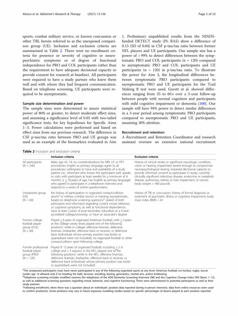
sports, combat military service, or known concussion orother TBI, herein referred to as the unexposed compari-son group (UE). Inclusion and exclusion criteria aresummarized in Table 2. There were no enrollment cri-teria for presence or severity of cognitive or neuro-psychiatric symptoms or of degree of functionalindependence for PRO and COL participants (other thanthe requirement to have adequate decisional capacity toprovide consent for research at baseline). All participantswere required to have a study partner who knew themwell and with whom they had frequent communication.Based on telephone screening, UE participants were re-quired to be asymptomatic.
Sample size determination and powerThe sample sizes were determined to assure statisticalpower of 80% or greater, to detect moderate effect sizes,and assuming a significance level of 0.05 with two-tailedsignificance tests, for key hypotheses for Specific Aims1–5. Power calculations were performed and based oneffect sizes from our previous research. The difference inCSF p-tau/tau ratio between PRO and UE groups wasused as an example of the biomarkers evaluated in Aim
1. Preliminary unpublished results from the NINDS-funded DETECT study (PI: RAS) show a difference of0.15 (SD of 0.04) in CSF p-tau/tau ratio between formerNFL players and UE participants. Our sample size has apower of > 99% to detect differences between the symp-tomatic PRO and COL participants (n = 120) comparedto asymptomatic PRO and COL participants and UEparticipants (n = 120) in p-tau/tau ratio. To illustratethe power for Aim 3, the longitudinal differences be-tween symptomatic PRO participants compared toasymptomatic PRO and UE participants for the TrailMaking B test were used. Gavett et al. showed differ-ences ranging from 35 to 60 s over a 3-year follow-upbetween people with normal cognition and participantswith mild cognitive impairment or dementia [100]. Oursample will have 94% power to detect similar differencesin a 3-year period among symptomatic PRO participantscompared to asymptomatic PRO and UE participants,assuming 30% attrition.
Recruitment and retentionA Recruitment and Retention Coordinator and researchassistant oversaw an extensive national recruitment
Table 2 Inclusion and exclusion criteria
Inclusion criteria Exclusion criteria
All participants(N = 240)
Male; age 45–74; no contraindications for MRI, LP, or PETprocedures; English as primary language; agree to allprocedures; willingness to have and availability of a studypartner (i.e., informant who knows the participant well, speaksor visits with participant at least weekly for a minimum of 6months, is > 18 years of age, has English as primary language)who agrees to participate in a telephone interview andrespond to a series of online questionnaires.
History of clinical stroke or significant neurologic condition;vision or hearing impairment severe enough to compromiseneuropsychological testing; impaired decisional capacity toprovide informed consent to participate in study; currentlyclinically significant infectious disease, endocrine or metabolicdisease, pulmonary, kidney or liver impairment, or cancer;body weight > 400 pounds
Unexposed group(UE)(N = 60)
No history of participation in organized contact/collisionsportsa or military combat service or training; asymptomatic,based on telephone screening questionsb (asked of bothparticipant and informant) regarding current mood, behavior,or cognitive symptoms, as well as functional dependence;have at least 2 years of post-secondary education at a 4-yearaccredited college/university, or have an associate’s degree
History of TBI or concussion; history of formal diagnosis ortreatment of psychiatric illness or cognitive impairment; bodymass index (BMI) < 24
Former collegefootball playergroup (COL)(N = 60)
Played > 6 years of organized American football, with > 3 yearsat the college varsity level; played one of the followingpositionsc while in college: offensive lineman, defensivelineman, linebacker, offensive back or receiver, or defensiveback (individuals whose primary position was kicker orquarterback were not included); no organized football or othercontact/collision sport following college
Former professionalfootball playergroup (PRO)(N = 120)
Played ≥ 12 years of organized football, including > 3 incollege and > 3 seasons in the NFL; played one of thefollowing positionsc while in the NFL: offensive lineman,defensive lineman, linebacker, offensive back or receiver, ordefensive back (individuals whose primary position was kickeror quarterback were not included)
aThe unexposed participants must have never participated in any of the following organized sports at any level: American football, ice hockey, rugby, soccer(under age 14 allowed only if no heading the ball), lacrosse, wrestling, boxing, gymnastics, martial arts, and/or kickboxingbTelephone screening includes modified versions (for telephone) of the AD8 Dementia Screening Interview [98] and the Cognitive Change Index [99] (Items 1–12),as well as additional screening questions regarding mood, behavior, and cognitive functioning. These were administered to potential participants as well as theirstudy partnerscFollowing enrollment, when there was a question about an individuals’ position data reported during in-person interview, data from online resources were usedto confirm position(s). Some positions may vary in future exposure modeling studies based on specific percentages of downs played at each position reported
Alosco et al. Alzheimer's Research & Therapy (2021) 13:136 Page 5 of 23
campaign (Supplementary Material, Additional File 1).Recruitment efforts were aimed at enrolling across acontinuum of symptom severity, from asymptomatic tomildly symptomatic to dementia, rather than to any spe-cific level of impairment. Interested potential partici-pants underwent a telephone screening interview byCoordinating Center staff, using a script approved by theInstitutional Review Board (IRB) at BU Medical Campus(BUMC). Structured and semi-structured questions wereasked about current mood, behavior, and cognitivesymptoms, as well as functional status. An additionaltelephone screening was conducted with an informant/study partner, using similar questions and assessments.The Recruitment and Retention Coordinator determinedthe Participant Evaluation Site most appropriate for theparticipant (based on balancing the number of partici-pants across sites, travel distance, and available sched-ules). To maximize sample retention over the follow-upperiod, participants are telephoned annually by projectstaff and sent birthday and holiday cards. A study-widenewsletter is published quarterly and distributed to allparticipants electronically. For participants with demen-tia, an additional annual call is made to the participantor (with permission) to a study partner/informant to as-sess the participant’s status and improve retention.
Study proceduresA centralized project Coordinating Center is located atBoston University (BU) School of Medicine (BUSM).There are four Participant Evaluation Sites: (1) Boston(BUSM, with MRI scans conducted at Brigham andWomen’s Hospital [BWH]); (2) Las Vegas (ClevelandClinic [CC] Lou Ruvo Center for Brain Health); (3) NewYork (New York University [NYU] Langone Health); and(4) Scottsdale/Phoenix (Mayo Clinic Arizona, with PETscans conducted at Banner Alzheimer’s Institute [BAI]in Phoenix). All participants received a baseline evalu-ation at one of the four Participant Evaluation Sites.Baseline evaluations included neurocognitive testing, as-sessment of functional status, neuropsychiatric question-naires, neurological assessment (including standardizedmotor examination, headache severity and sleep-relatedsymptoms measurement, and an olfaction test), MRI (in-cluding structural, diffusion, functional, and neurochem-ical), two PET scans (with florbetapir amyloid andflortaucipir tau tracers), lumbar puncture (LP; for CSFbanking and biomarkers), blood draws (for banking, bio-markers, and DNA extraction), and saliva samples (forbanking and biomarkers). In the original design, thePRO and UE groups would return for a 3-year, in-person follow-up evaluation. COL participants wouldnot be evaluated at follow-up because their inclusionwas for head impact exposure risk modeling at baseline(Aim 5) and to assure a large baseline sample size with
adequate variability of clinical presentation (Aims 1 &2).It was required that all participants have adequate de-
cisional capacity at the time of their baseline visit to par-ticipate. Because some participants have mild dementia,specific procedures were conducted to assure appropri-ate decisional capacity to consent to research participa-tion. Some participants who reported functionaldifficulties (n = 16) were accompanied by their studypartner or other care partner. All participants’ travel ex-penses (and that of a care partner if required) were paidby the study and each participant received $500 com-pensation for completion of the 3-day evaluation. Partic-ipants were informed during screening, at the time ofconsenting, and subsequent to their study visit, that theycould receive a summary of non-experimental study re-sults (including standardized neuropsychological testingand neuropsychiatric questionnaires, neurological exam-ination, clinical reads of the structural MRI, clinical la-boratory blood tests, electrocardiogram) and/or have theresults sent to their primary healthcare provider. If re-quested, they could discuss results of the non-experimental assessments with one of the co-PIs (RAS).At all times, participants were informed orally and inwritten reports that the results should not be used forclinical or medico-legal decision-making. At the time ofstudy initiation, and throughout the baseline evaluationperiod, flortaucipir (tau) PET imaging was in human tri-als and viewed as one of the experimental assessments.It received FDA approval 3 months following comple-tion of baseline examinations for the evaluation of AD,with specific package insert wording that it is not indi-cated for the evaluation of CTE. Florbetapir (amyloid)PET imaging received FDA approval prior to study initi-ation for patients being evaluated for AD and is consid-ered non-experimental for specific clinical purposes. Forthis study, the co-PIs, with input from external experts,chose not to disclose florbetapir results, though this doesnot necessarily represent the standard for the field mov-ing forward.
Data collectionClinical evaluationsEach baseline study visit was conducted over a 3-dayperiod and each follow-up visit was planned to takeplace over a two- or three-day period. Tables 3 and 4 listthe clinical exams and measures that were administered.Comprehensive semi-structured interviews for all partic-ipants were performed and supplemented by onlinequestionnaires in order to collect data on demographics(e.g., age, education, racial and ethnic identity); psycho-social and lifestyle history (e.g., exercise, occupationaland educational attainment, early childhood zip code orequivalent, parents’ educational attainment); medical,
Alosco et al. Alzheimer's Research & Therapy (2021) 13:136 Page 6 of 23
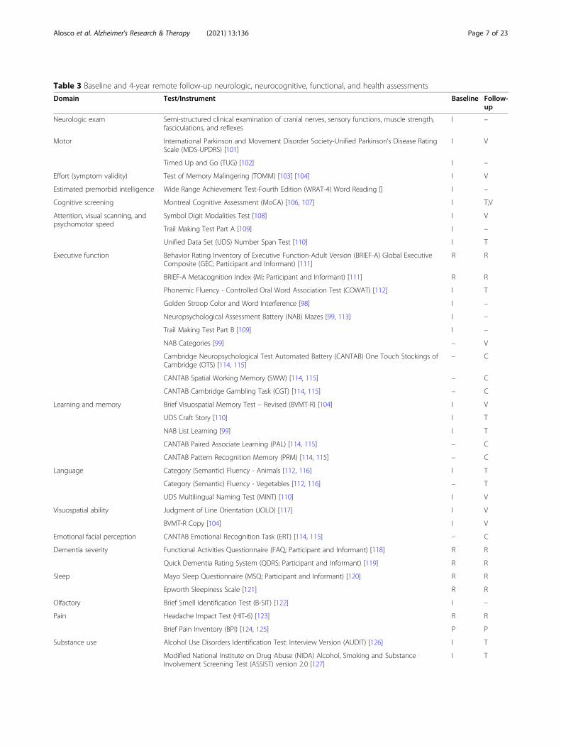
Table 3 Baseline and 4-year remote follow-up neurologic, neurocognitive, functional, and health assessments
Domain Test/Instrument Baseline Follow-up
Neurologic exam Semi-structured clinical examination of cranial nerves, sensory functions, muscle strength,fasciculations, and reflexes
I –
Motor International Parkinson and Movement Disorder Society-Unified Parkinson’s Disease RatingScale (MDS-UPDRS) [101]
I V
Timed Up and Go (TUG) [102] I –
Effort (symptom validity) Test of Memory Malingering (TOMM) [103] [104] I V
Estimated premorbid intelligence Wide Range Achievement Test-Fourth Edition (WRAT-4) Word Reading [] I –
Cognitive screening Montreal Cognitive Assessment (MoCA) [106, 107] I T,V
Attention, visual scanning, andpsychomotor speed
Symbol Digit Modalities Test [108] I V
Trail Making Test Part A [109] I –
Unified Data Set (UDS) Number Span Test [110] I T
Executive function Behavior Rating Inventory of Executive Function-Adult Version (BRIEF-A) Global ExecutiveComposite (GEC; Participant and Informant) [111]
R R
BRIEF-A Metacognition Index (MI; Participant and Informant) [111] R R
Phonemic Fluency - Controlled Oral Word Association Test (COWAT) [112] I T
Golden Stroop Color and Word Interference [98] I –
Neuropsychological Assessment Battery (NAB) Mazes [99, 113] I –
Trail Making Test Part B [109] I –
NAB Categories [99] – V
Cambridge Neuropsychological Test Automated Battery (CANTAB) One Touch Stockings ofCambridge (OTS) [114, 115]
– C
CANTAB Spatial Working Memory (SWW) [114, 115] – C
CANTAB Cambridge Gambling Task (CGT) [114, 115] – C
Learning and memory Brief Visuospatial Memory Test – Revised (BVMT-R) [104] I V
UDS Craft Story [110] I T
NAB List Learning [99] I T
CANTAB Paired Associate Learning (PAL) [114, 115] – C
CANTAB Pattern Recognition Memory (PRM) [114, 115] – C
Language Category (Semantic) Fluency - Animals [112, 116] I T
Category (Semantic) Fluency - Vegetables [112, 116] – T
UDS Multilingual Naming Test (MINT) [110] I V
Visuospatial ability Judgment of Line Orientation (JOLO) [117] I V
BVMT-R Copy [104] I V
Emotional facial perception CANTAB Emotional Recognition Task (ERT) [114, 115] – C
Dementia severity Functional Activities Questionnaire (FAQ; Participant and Informant) [118] R R
Quick Dementia Rating System (QDRS; Participant and Informant) [119] R R
Sleep Mayo Sleep Questionnaire (MSQ; Participant and Informant) [120] R R
Epworth Sleepiness Scale [121] R R
Olfactory Brief Smell Identification Test (B-SIT) [122] I –
Pain Headache Impact Test (HIT-6) [123] R R
Brief Pain Inventory (BPI) [124, 125] P P
Substance use Alcohol Use Disorders Identification Test: Interview Version (AUDIT) [126] I T
Modified National Institute on Drug Abuse (NIDA) Alcohol, Smoking and SubstanceInvolvement Screening Test (ASSIST) version 2.0 [127]
I T
Alosco et al. Alzheimer's Research & Therapy (2021) 13:136 Page 7 of 23
neurological, and psychiatric history (including sub-stance use and performance enhancing drug use); familyhistory of psychiatric and neurological conditions; ath-letic history (e.g., age of first exposure, level(s) and dur-ation of play, position(s) played, era of play); militaryhistory; and concussion and TBI history. For the COLand PRO participants, involvement in current or pend-ing litigation involving neurologic consequences of play-ing American football was also queried. Participants hadvital signs (e.g., blood pressure, pulse, height and weightmeasurement) assessed by a registered nurse. Safety pro-cedures (e.g., blood draw for platelet count and otherclotting tests, and ECG for abnormal heart rhythms and/or clinically significant cardiovascular disease) werereviewed by a qualified clinician to ensure participantswere eligible for the LP and flortaucipir PET scan, re-spectively. During the study visit, study partners wereemailed a survey link to a web-based Research ElectronicData Capture (REDCap) system to complete standard-ized measures and a self-report questionnaire on thepresence and onset of cognitive, behavior, and/or moodproblems, as well as an assessment of functional status.If the informant accompanied the participant to the
visits, she/he was asked to complete the online question-naires prior to returning home. All informants were alsointerviewed by telephone to provide additional historyand to clarify history provided by the participant.Clinical measures (see Tables 3 and 4) were selected,
in part, to assure harmonization with data sharing plat-forms, such as the Federal Interagency Traumatic BrainInjury Research (FITBIR) system and the NationalAlzheimer Coordinating Center (NACC). Many instru-ments and methodologies that overlap with the NINDSCommon Data Elements (CDE) and/or the NACCUniform Data Set (UDS) v.3.0 (the latter used by all ofthe National Institute on Aging-funded AD ResearchCenters) [110] were selected. Measures include thosethat assess clinical domains relevant to the featuresdescribed in neuropathologically confirmed cases ofCTE [2, 3, 47, 54] and were part of the 2014 TESresearch diagnostic criteria [54].To assure standardization of the administration and
scoring of clinical evaluations across sites and examiners,extensive training procedures were employed. Neurolo-gists administering the Movement Disorder Society(MDS)-Unified Parkinson’s Disease Rating Scale
Table 3 Baseline and 4-year remote follow-up neurologic, neurocognitive, functional, and health assessments (Continued)
Domain Test/Instrument Baseline Follow-up
Health status EQ-5D-5L Health Questionnaire [128] P P
National Health and Nutrition Examination Survey (NHANES) Physical Activity and PhysicalFitness Questionnaire (PAQ) [129]
I T
NHANES Weight History Questionnaire [130] I T
Note: I administered in-person; V Administered over HIPAA compliant Zoom Video conferencing (modified from original in-person method); T administered overtelephone; R administered over REDCap online data capture forms; P administered on paper form; C administered online using CANTAB Web-Based Testing platform
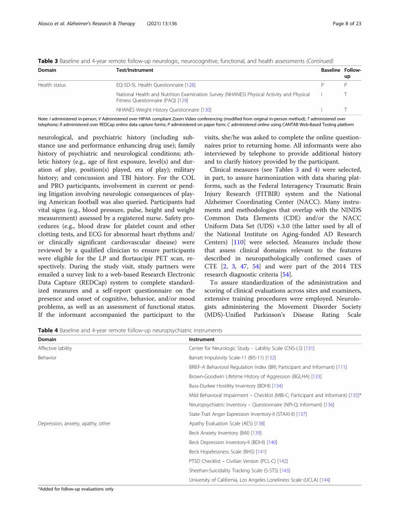
Table 4 Baseline and 4-year remote follow-up neuropsychiatric instruments
Domain Instrument
Affective lability Center for Neurologic Study – Lability Scale (CNS-LS) [131]
Behavior Barratt Impulsivity Scale-11 (BIS-11) [132]
BRIEF-A Behavioral Regulation Index (BRI; Participant and Informant) [111]
Brown-Goodwin Lifetime History of Aggression (BGLHA) [133]
Buss-Durkee Hostility Inventory (BDHI) [134]
Mild Behavioral Impairment – Checklist (MBI-C; Participant and Informant) [135]*
Neuropsychiatric Inventory – Questionnaire (NPI-Q; Informant) [136]
State-Trait Anger Expression Inventory-II (STAXI-II) [137]
Depression, anxiety, apathy, other Apathy Evaluation Scale (AES) [138]
Beck Anxiety Inventory (BAI) [139]
Beck Depression Inventory-II (BDI-II) [140]
Beck Hopelessness Scale (BHS) [141]
PTSD Checklist – Civilian Version (PCL-C) [142]
Sheehan-Suicidality Tracking Scale (S-STS) [143]
University of California, Los Angeles Loneliness Scale (UCLA) [144]
*Added for follow-up evaluations only
Alosco et al. Alzheimer's Research & Therapy (2021) 13:136 Page 8 of 23
(UPDRS) [101] completed formal online training offeredthrough MDS. A comprehensive neuropsychological testadministration and scoring manual was developed anddeployed to all sites, along with an accompanying train-ing video of a full test administration, including severaldemonstrations of how to respond to and score incor-rect or unusual responses. All staff administering theneurocognitive tests were certified (and re-certified an-nually) in test administration and scoring via mocktraining videos that were reviewed and certified by twolicensed clinical neuropsychologists at the CoordinatingCenter (MLA, RAS).
NeuroimagingNeuroimaging protocols include structural T1- and T2-weighted MRI, diffusion MRI (dMRI), resting-statefMRI, MRS, and molecular imaging with two PETtracers, florbetapir and flortaucipir. During the pre-enrollment period, BWH neuroimaging investigatorsand Invicro (a research-dedicated organization that col-laborates on large-scale diagnosis, progression, and dis-ease monitoring trials, providing molecular imagingservices, including florbetapir and flortaucipir) createdstudy-specific image acquisition sequences and technicaloperations manuals and developed and implementedtraining and setup procedures for the MR and PET cen-ters, respectively, at each of the Participant EvaluationSites. Details on the neuroimaging processing and ana-lysis are provided in the Supplementary Material (Add-itional File 1).
MRI MRIs across all four sites were conducted on WideBore 3 T scanners (Siemens Skyra, Erlangan, Germany;software version VE11) using a 20 channel head coil inorder to accommodate the wide range of participantsizes. The goal of the MRI sequence selection was to ob-tain the most advanced images consistent with otherlarge multi-site studies (e.g., Alzheimer’s Disease Neuro-imaging Initiative), and which could be acquired at eachsite within a reasonable time period to limit participantburden. The acquisition included sequences for anatom-ical images, as well as diffusion MRI (dMRI) and resting-state fMRI. High-resolution (1 × 1 × 1 mm3) 3D T1-weighted images using MPRAGE with an inversion timeof 1100ms were acquired, as were high-resolution (1 × 1× 1 mm3) 3D T2-weighted images and fluid attenuatedinversion recovery (FLAIR) sequences. The dMRI has amulti-shell design with 73 acquisitions spread over 5shells (4 b = 0, 3 b = 200, 6 b = 500, 30 b = 1000, and 30b = 2500 s/mm2). Images have a 2 × 2 × 2 mm3 reso-lution and 73 slices. The resting-state fMRI acquisitionwas an echo-planar imaging (EPI) acquisition with 3.5 ×3.5 × 3.5 mm3 resolution, with 37 slices, TR of 2.5 s, re-peated 149 times.
MRS 2D-chemical shift imaging (CSI) was acquiredusing the localized semi-adiabatic spin-echo refocusing(semi-LASER) with Gradient-Offset independent Adia-baticity Wurst modulation (GOIA-W) pulses and spiralencoding (853-ms duration, 12-kHz bandwidth, 90° flipangle, 160 mm field of view, 1.5-s repetition time, and40-ms echo time) [145]. Interleaved constant-densityspirals simultaneously encode one frequency and twospatial dimensions (16 × 16; 3 averages) for a resolutionof 1 × 1 × 1.5 cm3 and a scan time of 6 min. The 160 ×160 × 15 mm slab was placed across the corpus callosumparallel to the A/P plane. Single voxel spectroscopy(SVS) was acquired using point-resolved spectroscopy(PRESS; TE = 30ms, TR = 2 s, 2 × 2 × 2 cm3, 128 aver-ages; 16 average water reference) in the posterior cingu-late gyrus for a scan time of 5 min [146].
PET Participants underwent two PET imaging studies(florbetapir and flortaucipir) at baseline. Tracer doseswere requested through Avid Radiopharmaceuticals(Philadelphia, PA, USA) who then ordered the dosesfrom one of several contract manufacturing organiza-tions (usually the most proximate to a site) and coordi-nated dose shipping and delivery to the four PETcenters. The florbetapir protocol was as follows: immedi-ately after a 370MBq (10 mCi) bolus injection, the par-ticipant underwent brain scans consisting of 10 frames,each 1 min in length. Fifty minutes after injection, theparticipant completed a second 15-min brain scan con-sisting of three frames, each of which required 5 min.The use of flortaucipir in this study was carried out
through an Investigator Investigational New Drug (IND#131391) from the U.S. Food and Drug Administration.The flortaucipir protocol was as follows: 80min after a 370MBq (10mCi) bolus injection, the participant completed acontinuous dynamic 20-min brain scan (four frames, 5 mineach). Procedures described by Stern et al. [95] will serve asa guide for initial analyses of florbetapir and flortaucipir im-ages. However, additional analyses of PET amyloid and tauscans will be conducted, consistent with the most currentmethods and approaches [147–149].
Fluid biomarkersThe collection, tracking, banking, and distribution of allfluid biospecimens is done under the direction of theproject’s Fluid Biomarker Team leader (ERP) at VAPuget Sound. CSF, blood, and saliva collection and stor-age complies with the National Institute on Aging Bios-pecimen Task Force Guidelines and with NINDSRepository Biomarkers Discovery Samples Resource.Education and training were provided at each of the Par-ticipant Evaluation Sites (through in-person training,provision of a video DVD [150], and manuals) for thesafe, acceptable, and uniform methods for CSF, blood,
Alosco et al. Alzheimer's Research & Therapy (2021) 13:136 Page 9 of 23
and saliva collection. The Fluid Biomarker Team pro-vided all sites with prefabricated CSF, blood, and salivasample collection kits. Sample collection and sampleprocessing procedures are detailed in the SupplementaryMaterial (Additional File 1). An aliquot of whole bloodwas kept at room temperature and shipped to BUSM theday of collection for DNA extraction for genetic andgenomic analyses. All other saliva, blood, and CSF sam-ples were processed, aliquoted, and stored at − 80 °C atthe four Participant Evaluation Sites, and then batchshipped on dry ice overnight to VA Puget Sound, wherethey are stored in two − 70 °C freezers. Banked CSF,blood products, and saliva will be made available toqualified outside investigators.
Head impact exposure assessment and modelingA challenge of evaluating the long-term consequences ofrepetitive head impacts is that the outcomes are chronic,but the exposures are acute and, in this setting, remote.Each impact is of short duration, can be ambiguous, andrarely quantified. Task-based exposure assessmentmethods, such as job-exposure matrices, are often uti-lized to develop retrospective exposure metrics for inves-tigating exposure-disease relationships [151]. The samewill be applied to retrospectively estimate repetitive headimpact exposure in the COL and PRO groups. Aposition-exposure matrix (PEM) will be developed. Dif-ferent football positions (e.g., running back, offensivelineman) experience different impacts in terms of fre-quency, intensity, location, and type (linear or rotational)[152–154]. These measurements have been collected forover 1.8 million head impacts during games and prac-tices using the Head Impact Telemetry (HIT) System™[155–159]. Information from this extensive database willbe utilized to construct the PEM. The PEM will use themost current HIT data to summarize the variation ofimpacts by position and level of play. One limitation ofthis approach is that the NFL has not publicly released,nor have there been published reports of HIT System orother head impact sensor data from NFL players, thusresulting in the need to rely on college player HIT Sys-tem data in these PEMs. We will combine the PEM witheach participant’s football history (i.e., age of first expos-ure, level[s] and duration of play, position[s] played) todevelop participant-specific estimates of cumulative ex-posure to head impacts [27, 61, 160, 161]. Additionalmethods of estimating repetitive head impact exposurewill be included as they become available. Practical prox-ies of exposure to repetitive head impacts will be exam-ined, such as years of American football play and age offirst exposure to American football, among others. Dataon the participant’s self-reported number of concussions[162, 163] and number of episodes of loss of conscious-ness using the Ohio State University TBI Identification
Method-Interview Form [164] were collected as metricsof additional history. The participant-specific exposureestimates will be used to evaluate clinical and biomarkeroutcomes.
GeneticsWhole blood collected at the time of the blood drawwas shipped directly to the Molecular Genetics Core atBUSM where DNA was isolated, frozen, and used forapolipoprotein E (APOE) genotyping. We will conductgenome-wide genotyping using the Illumina GlobalScreening Array (Illumina, Inc., San Diego, CA, USA).
Multidisciplinary Diagnostic Consensus Conferences(MDCC)Each month there are two MDCCs held through video-conference and attended by a panel of 16 clinician-investigators, including 8 neurologists, 5 neuropsycholo-gists, 2 psychiatrists, and 1 neurosurgeon, from 7 institu-tions. Each MDCC is required to have a quorum of onepanelist from at least three of the four Participant Evalu-ation Sites, a minimum of two neurologists and two neu-ropsychologists, representation from at least two sitesoutside of BU, and a minimum of five panelists in at-tendance. At each MDCC, the history and findings fromapproximately 5–9 participants are presented. Followingpresentation of the history, course, and test score sum-maries (including measures of subjective cognitive com-plaints, functional independence, and sleep, as well asneurocognitive, neuropsychiatric, neurologic, and motorfunctioning), each MDCC member provides their inde-pendent diagnosis of TES, in addition to other clinicaldisorders due to neurodegenerative diseases using estab-lished diagnostic criteria (e.g., mild cognitive impairment[MCI] and AD dementia using the National Institute onAging – Alzheimer’s Association criteria [165]) and psy-chiatric disorders based on the Diagnostic and StatisticalManual of Mental Disorders, Fifth Edition (DSM-5)[166]. The MDCC members share and discuss their rat-ings and adjudicate a final consensus diagnosis (basedon majority).
Modified remote follow-up evaluationsAs a result of the COVID-19 pandemic, the project co-PIs, in collaboration with NINDS Program Officials, andwith input from the Executive Committee and ExternalAdvisory Board, decided that all follow-up evaluationswould be changed to entirely remote assessments tomaintain the safety of our participants and study staff,while also preserving the scientific integrity of the overallstudy. Remote assessments were required given that par-ticipants are flown from their homes to one of the 4 Par-ticipant Evaluation Sites, and the pandemic placedsevere restrictions on travel. All follow-up procedures
Alosco et al. Alzheimer's Research & Therapy (2021) 13:136 Page 10 of 23
were approved by the BUMC IRB. Each participant isassessed for decisional capacity to provide consent forresearch participation, using a modification of the Uni-versity of California, San Diego Brief Assessment of Cap-acity to Consent (UBACC) [167]. Informed consentforms are signed digitally and, in the case of participantswho are determined to lack decisional capacity, their re-search proxy or legally authorized representative digitallysigns the consent form. Each participant receives $325compensation for completion of all follow-up proce-dures. The modified remote follow-up evaluation is con-ducted using three separate platforms: (1) telephone, (2)online, and (3) video. Based on prior Baseline Evaluationexperience, all participants have access to a telephoneand most have access to an internet-connected desktopor laptop computer. Results from a survey conducted ofstudy participants in the spring of 2020 indicated that alarge majority have access to a desktop or laptop com-puter with a webcam for videoconferencing. Most of thetests included in the remote follow-up evaluation havebeen found to result in comparable performance whenadministered in-person or remotely via telephone, on-line, or videoconference platforms [107, 115, 168–171].All follow-up participants are interviewed over the
telephone to update any history and lifestyle informationand to conduct standardized interview-based assess-ments. In addition, all participants are administered atelephone-based neurocognitive evaluation which in-cludes the telephone modification of the NACC UDS 3.0cognitive assessment battery (T-Cog, including theNeuropsychological Assessment Battery (NAB) ListLearning Test) [99, 110] and the telephone version ofthe Montreal Cognitive Assessment (MoCA) (T-MoCA)[107]. Participants with access to an internet-connecteddesktop or laptop computer also complete a battery ofweb-based computerized cognitive tests from the Cam-bridge Neuropsychological Test Automated Battery(CANTAB) [114]. Those participants who have a web-cam (with proficiency in using these devices determinedduring a screening prior to follow-up) are administeredadditional video-based (using the Zoom videoconferenceplatform) neurocognitive tests and also undergo aneurological evaluation, including a modified MDS-UPDRS examination [172, 173], by a board-certified andMDS-UPDRS-trained neurologist who specializes inmovement disorders (CHA). Selection of the final bat-tery of neurocognitive measures was made by the fullteam of project neuropsychologist-investigators to assurethat all domains of interest were assessed. All partici-pants and study partners are asked to complete an on-line REDCap survey to assess cognitive, mood, andbehavior difficulties, as well as functional independence,using identical methods employed during baseline evalu-ations. Follow-up tests and questionnaires, including the
modality of assessment, are listed in Tables 3 and 4. TheAdverse Childhood Experiences (ACEs) questionnaire[174] was added to the follow-up evaluation to assesschildhood factors that may contribute to adult physicaland psychological health outcomes.All consenting participants have a fasting blood draw
at their home at the time of their follow-up evaluations.Blood collection and sample preparation is conducted byphlebotomists from ExamOne (a Quest DiagnosticsCompany, Lenexa, KS) who undergo study-specifictraining and who are provided with prefabricated bloodcollection and sample preparation supplies, along with amanual and infographic detailing all procedures, fromthe BU Coordinating Center. Whole blood, serum, andplasma samples are prepared and aliquoted, put on dryice within 90min of centrifugation, and shipped to VAPuget Sound, where they are banked for biomarker as-says and distribution to qualified investigators (see Sup-plementary Material, Additional File 1, for details).All participants will receive a follow-up diagnosis 4
years after their initial assessment using the NINDSConsensus Diagnostic Criteria for TES [56] through thesame MDCC process as baseline diagnoses.
Management and sharing of data and biospecimensThe Biostatistics and Epidemiology Data Analytics Cen-ter (BEDAC) at the BU School of Public Health providesdata management, database and web development, anddata analytics for the project (the latter in collaborationwith the project’s lead biostatistician and Data TeamLeader (YT)). Data are collected using web-based datacapture for assessments using REDCap, as well as cus-tomized forms for complex data. Common data ele-ments and study-specific data elements are uploaded toFITBIR on a regular basis to allow for data sharing inthe latter part of the project. Once baseline data collec-tion was completed, the Data Team developed a web-based data sharing platform, initially for use by projectinvestigators, with the plan for subsequent availability toall qualified researchers (i.e., in the latter part of the pro-ject). Based on the specific needs of an investigator, acustomized dataset is created using an automated sys-tem. Raw imaging data and fluid biosamples will bemade available to qualified investigators (see Supplemen-tary Material, Additional File 1).
ResultsParticipantsAll Participant Evaluation Site institutions (i.e., BU, CC,Mayo, NYU) and associated sites (BWH, BAI) receivedapproval by their governing IRB by January 2017. Allparticipants provided written informed consent duringtheir baseline visit. Enrollment began in September 2016and the last baseline evaluation was completed in
Alosco et al. Alzheimer's Research & Therapy (2021) 13:136 Page 11 of 23
February 2020. The final analytic sample includes 240men, ages 45–74, including 120 PRO, 60 COL, and 60UE participants. Table 5 summarizes sample demo-graphics. An additional 24 participants who underwentsome or all baseline evaluations are excluded from alldata analyses and subsequent evaluation for a variety ofreasons, including UE participants who reported a his-tory of concussion (n = 6) or who were found to haveextensive psychiatric history (n = 3) during in-personinterview, participants with incomplete biomarker data(n = 10), participants who self-withdrew (n = 2), or werewithdrawn by a PI for other reasons (n = 3). Three-yearfollow-up in-person evaluations (for PRO and UE partic-ipants) began in October 2019, with 11 completed byMarch 6, 2020. Due to the COVID-19 pandemic, all in-person study activities were ceased on March 16, 2020.Follow-up evaluations have shifted to fully remote andare being conducted on all participants, including theCOL participants (see below for details).
NeuroimagingNeuroimaging protocols completed include structuralT1- and T2-weighted MRI, diffusion MRI (dMRI),resting-state fMRI, MRS, as well as molecular imagingwith two PET tracers, florbetapir and flortaucipir. Im-aging calibration and quality control (QC) procedureswere completed for all sites prior to participant enroll-ment and throughout data acquisition. PET phantomsfrom Invicro were checked and each site was certified bythe Invicro team using their standard protocols. Add-itional MRI and MRS harmonization and QC procedureswere employed by the BWH Psychiatry NeuroimagingLaboratory (PNL) and Center for Clinical Spectroscopy(CCS) (see Supplementary Material, Additional File 1).The four MRI sequences were acquired in approximately40 min. Total MRS scan time was 15 min, includingshimming. Participants also completed florbetapir andflortaucipir PET scans and 214 participants completedboth PET scans. Florbetapir scans were typically con-ducted first (n = 157 [73%] of the 214 participants whohad both PET scans), with at least 12 h between the twoscans. PET protocol length is described above.
Fluid biomarkersAt the completion of all baseline evaluations, plasma,and CSF samples were shipped on dry ice overnightfrom VA Puget Sound to the University of Gothenburg,Sweden, where primary biomarker assays were con-ducted in batch. Primary fluid biomarkers includeplasma and CSF measures of p-tau181, p-tau217, p-tau231,total tau, abeta40, abeta42, abeta38, glial fibrillary acidicprotein (GFAP), NfL, soluble triggering receptorexpressed on myeloid cells 2 (sTREM2), and solubleplatelet-derived growth factor receptor beta
(sPDGFRbeta). Supplemental assays will be conducted atVA Puget Sound and University of Washington and willinclude the following: CSF and plasma CNS-derivedextracellular vesicle (EV) total tau and p-tau epitopes181, 231, and 396; vascular endothelial growth factor-A(VEGF-A); basic fibroblast growth factor (bFGF); inter-leukins (ILs) 1alpha and beta, 7, and 17a; tumor necrosisfactor (TNF)-a; monocyte chemotactic protein (MCP)-1;C-reactive protein (CRP); alpha-synuclein; CSF serum al-bumin ratio; and CSF catecholamines and indolaminesand their precursors and metabolites. Additional CSF,plasma, and saliva biomarkers will be examined as newdiscoveries direct.
Multidisciplinary Diagnostic Consensus Conferences(MDCC)MDCCs were completed for all participants followingtheir baseline evaluations using the provisional 2014TES criteria [54]. MDCCs are being repeated for allbaseline evaluations to derive new TES diagnoses usingthe recently published 2021 NINDS Consensus Diagnos-tic Criteria for TES [56], but without re-diagnosing otherconditions. Only these new TES diagnoses will be usedin baseline data analyses. MDCCs will be held followingremote follow-up evaluations to determine any changein TES diagnosis (using the 2021 criteria) or in otherneurodegenerative and/or psychiatric diagnoses.
First NINDS Consensus Workshop to define the diagnosticcriteria for TESRefinement and validation of the 2014 research diagnos-tic criteria for TES [54] is an aim of the DIAGNOSECTE Research Project. Since the time of the original2014 publication, the TES criteria have been used in sev-eral ongoing research studies, including the UNITEstudy [55] and for the initial baseline evaluation MDCCsin this DIAGNOSE CTE Research Project. In April2019, the First NINDS Consensus Workshop to Definethe Diagnostic Criteria for TES was held in Phoenix, Ari-zona. The goal of the workshop was to evaluate and up-date TES criteria based on the following: (1) inter-raterreliability data from research studies (including baselineMDCC data from this project); (2) predictive validitydata, both published [55] and unpublished, examiningthe relationship between the specific criteria and neuro-pathologically diagnosed CTE; (3) systematic review ofCTE literature to date; and (4) expert opinion. A Modi-fied Delphi approach was followed and included a firstround of anonymous voting that took place during theWorkshop, followed by three additional online votingrounds on revised criteria. Voting panelists included 20clinician-researchers across a variety of disciplines (e.g.,Neurology, Neuropsychology, Psychiatry, Physical Medi-cine and Rehabilitation, Neurosurgery), areas of
Alosco et al. Alzheimer's Research & Therapy (2021) 13:136 Page 12 of 23
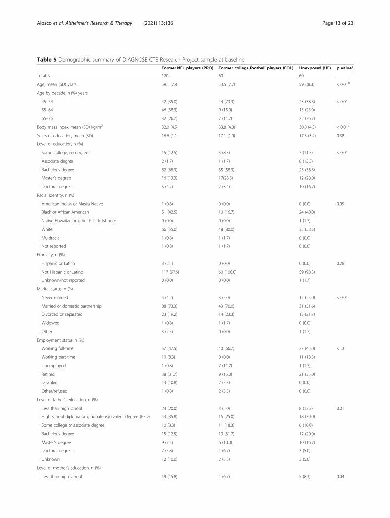
Table 5 Demographic summary of DIAGNOSE CTE Research Project sample at baselineFormer NFL players (PRO) Former college football players (COL) Unexposed (UE) p valuea
Total N 120 60 60 –
Age, mean (SD) years 59.1 (7.8) 53.5 (7.7) 59.3(8.3) < 0.01b
Age by decade, n (%) years
45–54 42 (35.0) 44 (73.3) 23 (38.3) < 0.01
55–64 46 (38.3) 9 (15.0) 15 (25.0)
65–75 32 (26.7) 7 (11.7) 22 (36.7)
Body mass index, mean (SD) kg/m2 32.0 (4.5) 33.8 (4.8) 30.8 (4.5) < 0.01c
Years of education, mean (SD) 16.6 (1.1) 17.1 (1.0) 17.3 (3.4) 0.38
Level of education, n (%)
Some college, no degree 15 (12.5) 5 (8.3) 7 (11.7) < 0.01
Associate degree 2 (1.7) 1 (1.7) 8 (13.3)
Bachelor’s degree 82 (68.3) 35 (58.3) 23 (38.3)
Master’s degree 16 (13.3) 17(28.3) 12 (20.0)
Doctoral degree 5 (4.2) 2 (3.4) 10 (16.7)
Racial Identity, n (%)
American Indian or Alaska Native 1 (0.8) 0 (0.0) 0 (0.0) 0.05
Black or African American 51 (42.5) 10 (16.7) 24 (40.0)
Native Hawaiian or other Pacific Islander 0 (0.0) 0 (0.0) 1 (1.7)
White 66 (55.0) 48 (80.0) 35 (58.3)
Multiracial 1 (0.8) 1 (1.7) 0 (0.0)
Not reported 1 (0.8) 1 (1.7) 0 (0.0)
Ethnicity, n (%)
Hispanic or Latino 3 (2.5) 0 (0.0) 0 (0.0) 0.28
Not Hispanic or Latino 117 (97.5) 60 (100.0) 59 (98.3)
Unknown/not reported 0 (0.0) 0 (0.0) 1 (1.7)
Marital status, n (%)
Never married 5 (4.2) 3 (5.0) 15 (25.0) < 0.01
Married or domestic partnership 88 (73.3) 43 (70.0) 31 (51.6)
Divorced or separated 23 (19.2) 14 (23.3) 13 (21.7)
Widowed 1 (0.8) 1 (1.7) 0 (0.0)
Other 3 (2.5) 0 (0.0) 1 (1.7)
Employment status, n (%)
Working full-time 57 (47.5) 40 (66.7) 27 (45.0) < .01
Working part-time 10 (8.3) 0 (0.0) 11 (18.3)
Unemployed 1 (0.8) 7 (11.7) 1 (1.7)
Retired 38 (31.7) 9 (15.0) 21 (35.0)
Disabled 13 (10.8) 2 (3.3) 0 (0.0)
Other/refused 1 (0.8) 2 (3.3) 0 (0.0)
Level of father’s education, n (%)
Less than high school 24 (20.0) 3 (5.0) 8 (13.3) 0.01
High school diploma or graduate equivalent degree (GED) 43 (35.8) 15 (25.0) 18 (30.0)
Some college or associate degree 10 (8.3) 11 (18.3) 6 (10.0)
Bachelor’s degree 15 (12.5) 19 (31.7) 12 (20.0)
Master’s degree 9 (7.5) 6 (10.0) 10 (16.7)
Doctoral degree 7 (5.8) 4 (6.7) 3 (5.0)
Unknown 12 (10.0) 2 (3.3) 3 (5.0)
Level of mother’s education, n (%)
Less than high school 19 (15.8) 4 (6.7) 5 (8.3) 0.04
Alosco et al. Alzheimer's Research & Therapy (2021) 13:136 Page 13 of 23
expertise (e.g., neurodegenerative disease, TBI), and aca-demic institutions (i.e., ten). The Delphi process wascompleted in January 2020 and a report on the newNINDS Consensus Diagnostic Criteria for TES was pub-lished in 2021 [56]. These new TES diagnostic criteriaare intended for research purposes and not for clinicaldiagnosis. It is expected that the criteria will be furtherupdated and revised through future NINDS Consensus
Workshops as research in this field and on the criteriaevolve and biomarker data become available.
DiscussionThis report provides a description of the methodologyfor the DIAGNOSE CTE Research Project, a multicen-ter, observational, cohort study designed to develop, re-fine, and validate in vivo biomarkers for CTE;
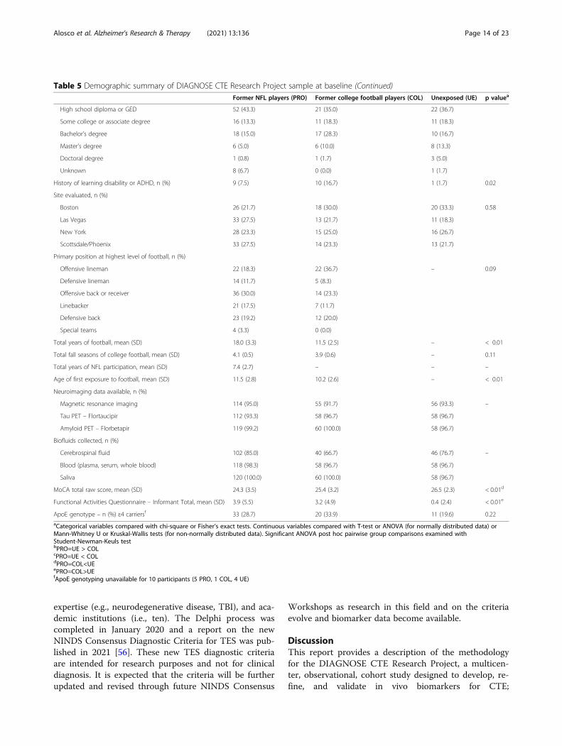
Table 5 Demographic summary of DIAGNOSE CTE Research Project sample at baseline (Continued)Former NFL players (PRO) Former college football players (COL) Unexposed (UE) p valuea
High school diploma or GED 52 (43.3) 21 (35.0) 22 (36.7)
Some college or associate degree 16 (13.3) 11 (18.3) 11 (18.3)
Bachelor’s degree 18 (15.0) 17 (28.3) 10 (16.7)
Master’s degree 6 (5.0) 6 (10.0) 8 (13.3)
Doctoral degree 1 (0.8) 1 (1.7) 3 (5.0)
Unknown 8 (6.7) 0 (0.0) 1 (1.7)
History of learning disability or ADHD, n (%) 9 (7.5) 10 (16.7) 1 (1.7) 0.02
Site evaluated, n (%)
Boston 26 (21.7) 18 (30.0) 20 (33.3) 0.58
Las Vegas 33 (27.5) 13 (21.7) 11 (18.3)
New York 28 (23.3) 15 (25.0) 16 (26.7)
Scottsdale/Phoenix 33 (27.5) 14 (23.3) 13 (21.7)
Primary position at highest level of football, n (%)
Offensive lineman 22 (18.3) 22 (36.7) – 0.09
Defensive lineman 14 (11.7) 5 (8.3)
Offensive back or receiver 36 (30.0) 14 (23.3)
Linebacker 21 (17.5) 7 (11.7)
Defensive back 23 (19.2) 12 (20.0)
Special teams 4 (3.3) 0 (0.0)
Total years of football, mean (SD) 18.0 (3.3) 11.5 (2.5) – < 0.01
Total fall seasons of college football, mean (SD) 4.1 (0.5) 3.9 (0.6) – 0.11
Total years of NFL participation, mean (SD) 7.4 (2.7) – – –
Age of first exposure to football, mean (SD) 11.5 (2.8) 10.2 (2.6) – < 0.01
Neuroimaging data available, n (%)
Magnetic resonance imaging 114 (95.0) 55 (91.7) 56 (93.3) –
Tau PET – Flortaucipir 112 (93.3) 58 (96.7) 58 (96.7)
Amyloid PET – Florbetapir 119 (99.2) 60 (100.0) 58 (96.7)
Biofluids collected, n (%)
Cerebrospinal fluid 102 (85.0) 40 (66.7) 46 (76.7) –
Blood (plasma, serum, whole blood) 118 (98.3) 58 (96.7) 58 (96.7)
Saliva 120 (100.0) 60 (100.0) 58 (96.7)
MoCA total raw score, mean (SD) 24.3 (3.5) 25.4 (3.2) 26.5 (2.3) < 0.01d
Functional Activities Questionnaire – Informant Total, mean (SD) 3.9 (5.5) 3.2 (4.9) 0.4 (2.4) < 0.01e
ApoE genotype – n (%) ε4 carriersf 33 (28.7) 20 (33.9) 11 (19.6) 0.22aCategorical variables compared with chi-square or Fisher’s exact tests. Continuous variables compared with T-test or ANOVA (for normally distributed data) orMann-Whitney U or Kruskal-Wallis tests (for non-normally distributed data). Significant ANOVA post hoc pairwise group comparisons examined withStudent-Newman-Keuls testbPRO=UE > COLcPRO=UE < COLdPRO=COL<UEePRO=COL>UEfApoE genotyping unavailable for 10 participants (5 PRO, 1 COL, 4 UE)
Alosco et al. Alzheimer's Research & Therapy (2021) 13:136 Page 14 of 23
characterize the clinical course and presentation of thistauopathy; identify potential risk factors; and refine andvalidate research diagnostic criteria for the clinical pres-entation associated with CTE (i.e., TES). This report alsoprovides a description of the demographics of the sam-ple, comprised of 120 former NFL players, 60 formercollege football players, and 60 unexposed same-ageasymptomatic men.Methodological decisions were made in designing the
DIAGNOSE CTE Research Project based on the over-arching goal of establishing clinical diagnostic criteriafor CTE with highly accurate in vivo biomarkers. Assuch, we decided to focus on a sample of former collegeand professional American football players to ensure asample at high risk for CTE [3] and to maximize powerfor hypothesis testing. Although the inclusion of a moreheterogeneous sample of contact and collision sport ath-letes (e.g., boxers, soccer players, ice hockey players, in-cluding women), as well as participants with othersources of repetitive head impact exposure, such as mili-tary combat veterans with blast-injuries, survivors of in-timate partner violence, and younger participants, mayincrease generalizability, it would be difficult to estimate“exposure” levels or achieve adequate statistical poweror assure that the sample was at high risk for CTE.Thus, at the time of initial development of in vivo CTEdiagnostics, homogeneity of the source of repetitive headimpacts was prioritized. An area of active investigationby our team includes other, non-football contact sportathletes (e.g., soccer, ice hockey, rugby), particularly fe-male former contact sport athletes. Some investigatorsof DIAGNOSE CTE are leading a new NIH-funded ini-tiative (PI: Stern), known as the Head Impact andTrauma Surveillance Study (HITSS), that will leveragethe online Brain Health Registry platform and recruit,enroll, and longitudinally follow female and male formersoccer players and male former American footballplayers (across all levels of play). This initiative willincrease our understanding of the long-term effects ofrepetitive head impacts across sports and in females,and lead to future investigations that are similar toDIAGNOSE CTE, allowing for rich clinical charac-terizations of female former contact sport athletes.The UE group was carefully selected. If the primary
goal was to study disease risk, then certain variablesshould have been well-controlled, e.g., cardiovascular/cerebrovascular risks, performance enhancing drug use,substance use, history of team sport involvement. How-ever, our primary goals were to examine possible bio-markers to detect CTE and the refinement of diagnosticcriteria for the clinical manifestations of CTE. Therefore,our comparison group included individuals who weresimilar to the former American football players in termsof age, sex, and BMI, but did not have repetitive head
impact exposure and were asymptomatic. This type ofdesign is appropriate for biomarker development andvalidation. Importantly, while the UE group will allow usto answer questions regarding biomarker developmentand validation, their inclusion in other types of analysesrequires careful consideration. For instance, it would beinappropriate to characterize the effect of repetitive headimpact exposure on clinical measures in both the formerplayer groups and UE group, given that the UE groupwas required to be asymptomatic (for neurological andpsychiatric conditions) at the time of recruitment andthe former players were not. Even among questions per-taining to biomarkers, it will be important to conductsensitivity analyses to determine if any potential groupdifferences are related to exposure to repetitive head im-pacts or to other factors. Lastly, other types of “control”groups were considered, but not incorporated into thedesign of the study. There have been efforts to recruitformer professional baseball players or body builders ascontrols for similar studies, because they have similarlifestyles and body habitus as former American footballplayers. Yet, there have been very few who had neverparticipated in organized contact sports or who, in thecase of baseball players, had not reported multiple con-cussions. Planned ancillary studies will also recruit par-ticipants with AD dementia and AD-related dementiasas disease comparison groups to the former professionAmerican football players.All participants were required to have a study part-
ner who knows them well to provide assessments oftheir perspective of the participant’s cognitive, behav-ioral, and functional status. In some cases, these re-ports may be inaccurate due to a variety of factors,including misattributions of symptoms to age-relatedchanges or stress; exaggeration of deficits for potentialsecondary gain (including financial compensationfrom disability or legal cases); and denial/unawarenessof deficits (including anosognosia) due to neurodegen-erative disease and other neurologic conditions. Thisrequirement may introduce some degree of selectionbias due to the potential for some participants withunderlying CTE to have neuropsychiatric features(e.g., rage, aggression, impulsivity) that result in theloss of close relationships and overall social isolation.Therefore, it is possible that potential participantswith more severe neuropsychiatric features were ex-cluded due to those features limiting the availabilityof a study partner.The primary method of validating biomarkers for the
detection of CTE pathology or to truly examine risk fac-tors for the development of CTE pathology is to com-pare data collected during life with postmortemneuropathology and diagnosis. The large majority offormer college and professional football players in the
Alosco et al. Alzheimer's Research & Therapy (2021) 13:136 Page 15 of 23
DIAGNOSE CTE cohort have agreed to brain donation.At the time of the current paper, five former players hadalready died, and their brain tissue will be examined forthese clinicopathological correlation and validationstudies.Our goal was to enroll a similar proportion of Black
participants across the three exposure groups (i.e., PRO,COL, UE), with the target of approximately 40% overall.Although the PRO and UE groups have a similar pro-portion of Black participants, with 42.5% and 40.0%, re-spectively, the COL group has a significantly smallerproportion of black participants (16.7%). Interactive ef-fects between levels of exposure to repetitive head im-pacts and Black racial identity on potentialneuroimaging and fluid biomarkers of CTE have beenreported [161]. Moreover, there are potential differencesbetween Black and White participants in the expressionof psychiatric symptoms and performance on cognitivetests [175, 176], as well as important racial disparities inlife-course social determinants of health, cognitive aging,and neurodegenerative disease [177–179]. For these rea-sons, interpretation of data analyses including the COLgroup will be done with these racial identity differencesin mind [180].
ConclusionsThe DIAGNOSE CTE Research Project will lead to arich dataset that will be used to further our under-standing of CTE in terms of its clinical presentation,in vivo biomarkers, clinical research diagnostic cri-teria, and risk and resiliency factors for the develop-ment of CTE. In addition to repetitive head impactexposure and genetic factors, project data will informon the role of demographic, lifestyle, medical, andpsychiatric risk and resilience variables, as well as onsocial determinants of health and racial disparities.Importantly, the data will provide the infrastructureand resources for opportunities to conduct ancillaryor add-on studies that target questions not being dir-ectly examined by the DIAGNOSE CTE ResearchProject. Ultimately, it is anticipated that findings fromthe DIAGNOSE CTE Research Project and associatedancillary studies will facilitate the ability to detect anddiagnose CTE during life and thereby accelerate re-search on risk factors, mechanisms, epidemiology,and, most importantly, treatment and prevention ofCTE.
Abbreviations3R: Three microtubule-binding domain repeat tau isoform; 4R: Fourmicrotubule-binding domain repeat tau isoform; Aβ: Beta-amyloid;AD: Alzheimer’s disease; AxD: Axial diffusivity; BAI: Banner Alzheimer’sInstitute; BMI: Body mass index; BU: Boston University; BUMC: BostonUniversity Medical Campus; BUSM: Boston University School of Medicine;BWH: Brigham and Women’s Hospital; CC: Cleveland Clinic; CDE: CommonData Elements; CSF: Cerebrospinal fluid; COL: Former college player group;
CTE: Chronic traumatic encephalopathy; DEI: Diversity, equity, and inclusion;DIAGNOSE CTE: Diagnostics, Imaging, and Genetics Network for theObjective Study and Evaluation of Chronic Traumatic Encephalopathy;dMRI: Diffusion MRI; DTI: Diffusion tensor imaging; FA: Fractional anisotropy;FITBIR: Federal Interagency Traumatic Brain Injury Research; FLAIR: Fluidattenuated inversion recovery; fMRI: Functional magnetic resonance imaging;GWAS: Genome-wide sequencing studies; IRB: Institutional Review Board;LP: Lumbar puncture; MD: Mean diffusivity; MDCC: Multidisciplinarydiagnostic consensus conference; MDS: Movement Disorder Society;MRI: Magnetic resonance imaging; MRS: Magnetic resonance spectroscopy;NFL: National Football League; NFT: Neurofibrillary tangle; NINDS: NationalInstitute for Neurological Disorders and Stroke; NYU: New York University;PET: Positron emission tomography; PI: Principal investigator; PNL: PsychiatryNeuroimaging Laboratory; PRO: Former profession (NFL) player group; p-tau: Hyperphosphorylated tau; QC: Quality control; RD: Radial diffusivity;SNP: Single-nucleotide polymorphism; TBI: Traumatic brain injury;TES: Traumatic encephalopathy syndrome; t-tau: Total tau; UDS: UniformData Set; UE: Unexposed group; UNITE: Understanding Neurologic Injury inTraumatic Encephalopathy
Supplementary InformationThe online version contains supplementary material available at https://doi.org/10.1186/s13195-021-00872-x.
Additional file 1.
AcknowledgementsWe thank Debra Babcock, M.D., Ph.D., Scientific Program Official of this U01grant at NINDS, for her support, guidance, and advice. We also thank ourExternal Advisory Board (David Knopman, M.D., [Chair], Col. [Ret.] Dallas Hack,M.D., Mike Haynes, Brian Hainline, M.D., Thomas McAllister, M.D., Arthur Toga,Ph.D., and Michael Weiner, M.D.) for sharing their time, effort, and expertise.Finally, we thank all of our participants and their study partners for makingthis study possible and for their tremendous contribution to ourunderstanding of chronic traumatic encephalopathy and other long-termconsequences of repetitive head impacts.The DIAGNOSE CTE Research Project Current and Former Investigatorsand Key PersonnelBanner Alzheimer’s InstituteInvestigatorsEric Reiman, M.D. (Co-PI)Yi Su, Ph.D.Kewei Chen, Ph.D.Hillary Protas, Ph.D.Non-InvestigatorsConnie Boker, M.B.A. (Director, Imaging Center Operations)Boston University School of MedicineInvestigatorsMichael L. Alosco, Ph.D.Rhoda Au, Ph.D.Robert C. Cantu, Ph.D.Lindsay Farrer, Ph.D.Robert Helm, M.D. *Douglas I. Katz, M.D.Neil Kowall, M.D. *Ann C. McKee, M.D.Jesse Mez, M.D.Gustavo Mercier, M.D., Ph.D. *James Otis, M.D. *Robert A. Stern, Ph.D. (Co-PI)Jason Weller, M.D.Non-InvestigatorsIrene Simkin, M.S. (Lab Manager, Molecular Genetics Core Facility)Boston University Project Coordinating Center StaffAlondra Andino, B.A. (Project Administrative Manager) *Shannon Conneely, B.A. (Site Coordinator) *Courtney Diamond, M.B.A. (Project Manager) *Tessa Fagle, B.A. (Research Assistant)Olivia Haller, B.A. (Recruitment Coordinator) *
Alosco et al. Alzheimer's Research & Therapy (2021) 13:136 Page 16 of 23
Tennyson Hunt, M.B.A. (Project Administrative Manager) *Nicole Gullotti, M.B.A. (Research Administrator) *Megan Mariani, B.S., B.A. (Project Manager)Brian Mayville, B.S. (Site Coordinator)Kathleen McLaughlin, B.A. (Research Assistant)Mary Nanna, B.A. (Retention Coordinator)Taylor Platt, M.P.H. (Recruitment Coordinator) *Surya Pulukuri, B.A. (Research Assistant)Fiona Rice, M.P.H. (Project Manager) *Madison Sestak, B.S. (Assistant Recruitment Coordinator) *Boston University School of Public HealthInvestigatorsMichael McClean, Sc.D.Yorghos Tripodis, Ph.D.Data Team StaffDouglas Annis, M.S. (Systems Analyst) *Christine Chaisson, M.P.H. (Leader of Data Management Sub-team) *Diane B. Dixon (Project Manager)Carolyn Finney, B.A. (Data Manager)Kerrin Gallagher, M.P.H. (Statistical Analyst) *Kaitlin Hartlage, M.P.H. (Statistical Analyst)Jun Lu, M.S. (Data Security and Technology Analyst)Brett Martin, M.S. (Statistical Manager)Emmanuel Ojo, M.P.H. (Statistical Analyst) *Joseph N. Palmisano, M.A., M.P.H. (Leader of Data Management Sub-team)Brittany Pine, B.A., B.S. (Statistical Analyst)Janani Ramachandran, M.S. (Data Manager)Brigham and Women’s HospitalInvestigatorsSylvain Bouix, Ph.D.Jennifer Fitzsimmons, M.D. *Alexander P. Lin, Ph.D.Inga K. Koerte, M.D., Ph.D.Ofer Pasternak, Ph.D.Martha E. Shenton, Ph.D. (Co-PI)Non-InvestigatorsHector Arciniega, Ph.D. (Postdoctoral Research Fellow)Tashrif Billah, M.S. (Software Engineer)Elena M. Bonke, M.S. (Ph.D. Student)Katherine Breedlove, Ph.D. (Postdoctoral Research Fellow)Eduardo Coello, Ph.D. (Postdoctoral Research Fellow)Michael J. Coleman, M.A. (Senior Scientist)Leonard B. Jung, (Ph.D. Student)Huijun Liao, B.S. (Study Coordinator)Maria Loy, M.B.A., M.P.H. (Senior Program Coordinator)Elizabeth Rizzoni, B.A. (Research Assistant)Vivian Schultz, M.D. (Postdoctoral Research Fellow)Annelise Silva, B.S. (Research Assistant) *Brynn Vessey, B.S. (Research Assistant)Tim L.T. Wiegand, (Ph.D. Student)Cleveland Clinic Lou Ruvo Center for Brain HealthInvestigatorsSarah Banks, Ph.D. (Now at University of California, San Diego)Charles Bernick, M.D. (Now at University of Washington)Jason Miller, Ph.D.Aaron Ritter, M.D.Marwan Sabbagh, M.D. *Non-InvestigatorsRaelynn de la Cruz, (Psychometrician)*Jan Durant, (Psychometrician)*Morgan Golceker (Site Coordinator)Nicolette Harmon, (Site Coordinator) *Kaeson Kaylegian, (Psychometrician)*Rachelle Long, (Site Coordinator) *Christin Nance, (Psychometrician)*Priscilla Sandoval (Site Coordinator) *George Washington University School of Medicine and Health SciencesInvestigatorRobert W. Turner, Ph.D.Invicro (formerly Molecular NeuroImaging)Investigator
Kenneth L. Marek, M.D.Non-InvestigatorAndrew Serrano, M.B.A.Mayo Clinic ArizonaInvestigatorsCharles H. Adler, M.D., Ph.D.,David W. Dodick, M.D.Yonas Geda, M.D., MSc (Now at Barrow Neurological Institute)Jennifer V. Wethe, Ph.D.Non-InvestigatorsBryce Falk, R.N.Amy Duffy, (Site Coordinator) *Marci Howard, (Psychometrician)*Michelle Montague, (Psychometrician)*Thomas Osgood, (Site Coordinator)National Institute of Neurological Disorders and Stroke (NINDS)Debra Babcock, M.D., Ph.D. (Scientific Program Official)Patrick Bellgowan, Ph.D. (Administrative Program Official)*New York University:InvestigatorsLaura Balcer, M.D., M.S.C.E.William Barr, PhD.Judith Goldberg, Sc.D.Thomas Wisniewski, M.D. *Ivan Kirov, Ph.D.Yvonne Lui, M.D.Charles Marmar, M.D.Non-InvestigatorsLisena Hasanaj (Site Coordinator)Liliana SerranoAlhassan Al-Kharafi (Psychometrician)*Allan George (Psychometrician)*Sammie Martin (Psychometrician)*Edward Riley (Psychometrician)*William Runge (Psychometrician)*University of Nevada, Las VegasJeffrey L. Cummings, M.D., ScD (Co-PI)University of Washington and VA Puget SoundInvestigatorElaine R. Peskind, M.D.Non-InvestigatorElizabeth Colasurdo (Lab Manager)Washington University (CNDA)InvestigatorsDaniel S. Marcus, Ph.D.Non-InvestigatorJenny Gurney, M.S.ConsultantsRichard Greenwald, Ph.D. (Simbex)*Keith A. Johnson, M.D. (Massachusetts General Hospital)*No longer involved in project.
Authors’ contributionsMLA and RAS drafted the manuscript. RAS, JLC, EMR, and MES designed andoversaw the study. MLM provided additional drafting of the manuscript andserved as project manager of the study. CHA, LJB, CB, and RAS were PIs ofthe baseline performance evaluation sites and oversaw data and samplecollection. RA and MDM oversaw risk assessment aspects of the study, withJM and LAF contributing to the genetics sections and RWT providingadditional contributions to sociodemographic data issues. MES, SB, MJC, IK,APL, and DSM designed and implemented the MRI and MRS data collectionand analytics. EMR and KLM designed and oversaw the PET data collectionand analytics. MLA, SJB, WBB, JVW, and RAS designed and implemented theneuropsychological battery and data collection. DIK, CB, DWD, JM, and RASdesigned and implemented the consensus diagnostic conferences. CHA andRCC designed the neurological and motor assessments. JLC, YEG, and RASdesigned the neuropsychiatric assessments. EP designed and oversaw fluidbiomarker collection and planned analyses. ACM provided input regardingthe neuropathology of CTE and NWK provided additional input regardingunderlying mechanisms. JNP oversaw data management systems. YT
Alosco et al. Alzheimer's Research & Therapy (2021) 13:136 Page 17 of 23
oversaw biostatistical and data analytic plans. All authors critically reviewedand revised the manuscript and approved its content before submission.
FundingThis work was supported by grants from the National Institutes of Health(U01NS093334; P30AG13846; R01NS078337; K23NS102399; P30AG019610;K01AG054762; R01NS100952; K00NS113419). This publication was supportedby the National Center for Advancing Translational Sciences, NationalInstitutes of Health, through BU-CTSI Grant Number 1UL1TR001430. The pri-mary funding source is the National Institute of Neurological Disorders andStroke (NINDS), through a U01 Research Project Cooperative Agreement(U01NS093334).
Availability of data and materialsThe datasets generated and analyzed during the current study will beavailable in the Federal Interagency Traumatic Brain Injury Research (FITBIR)repository, https://fitbir.nih.gov. Datasets will also be available through a datasharing portal for the DIAGNOSE CTE Research Project, http://diagnosecte.com. It is also anticipated that study datasets will be available in the GlobalAlzheimer’s Association Interactive Network (GAAIN) repository, http://www.gaain.org.
Declarations
Ethics approval and consent to participateAll Participant Evaluation Sites have received approval by their respectiveInstitutional Review Boards. All participants provide written informed consentduring their baseline and follow-up study visits.○ Boston University Medical Campus Institutional Review Board, #H-34799○ Cleveland Clinic Institutional Review Board, #16-1694○ Mayo Clinic Institutional Review Boards, #16-002662○ NYU School of Medicine Institutional Review Board, #i16-01032 CR3○ Partners Human Research Institutional Review Board, #2016P001359 and#2016P001328○ Western Copernicus Group (WCG) Institutional Review Board (BannerAlzheimer’s Institute), #1168489
Consent for publicationNot applicable.
Competing interestsMLA: None to report.MLM: None to report.CHA: None to report.LJB is Editor-in-Chief of the Journal of Neuro-Ophthalmology, and is a paidconsultant to Biogen (Cambridge, MA, USA).CB receives research support from the Ultimate Fighting Championship, TopRank promotions, Haymon Boxing, Las Vegas Raiders, and Professional BullRiders. He is a paid consultant for Aurora Concussion Therapy Systems, Inc.(St. Paul, MN).RA is a paid consultant to Biogen (Cambridge, MA, USA) and serves on theScientific Advisory Board of Signant Health (Blue Bell, PA).SJB: None to report.WBB provides expert witness testimony in legal cases involving concussionand CTE.SB: None to report.RCC is a Senior Advisor to the NFL Head Neck & Spine Committee; VicePresident, National Operating Committee on Standards for AthleticEquipment; and Chair, Scientific Advisory Committee, Co-Founder, and Med-ical Director, Concussion Legacy Foundation. He is a member of the MedicalScience Committee for the National Collegiate Athletic Association Student-Athlete Concussion Injury Litigation, and he receives royalties for publishedbooks from Houghton Mifflin Harcourt.MJC: None to report.DWD reports the following competing interests: Consulting: AEON, Amgen,Clexio, Cerecin, Cooltech, Ctrl M, Allergan, Alder, Biohaven, GSK, Linpharma,Lundbeck, Promius, Eli Lilly, eNeura, Novartis, Impel, Satsuma, Theranica, WLGore, Nocira, XoC, Zosano, Upjohn (Division of Pfizer), Pieris, Praxis, Revance,Equinox. Honoraria: Clinical Care Solutions, CME Outfitters, Curry RockefellerGroup, DeepBench, Global Access Meetings, KLJ Associates, Academy forContinued Healthcare Learning, Majallin LLC, Medlogix Communications,
MJH Lifesciences, Miller Medical Communications, Southern HeadacheSociety (MAHEC), WebMD Health/Medscape, Wolters Kluwer, OxfordUniversity Press, Cambridge University Press. Research Support: Departmentof Defense, National Institutes of Health, Henry Jackson Foundation, SperlingFoundation, American Migraine Foundation, Patient Centered OutcomesResearch Institute (PCORI). Stock Options/Shareholder/Patents/Board ofDirectors: Ctrl M (options), Aural analytics (options), ExSano (options), Palion(options), Healint (Options), Theranica (Options), Second Opinion/MobileHealth (Options), Epien (Options/Board), Nocira (options), Matterhorn (Shares/Board), Ontologics (Shares/Board), King-Devick Technologies (Options/Board),Precon Health (Options/Board). Patent 17189376.1-1466:vTitle: BotulinumToxin Dosage Regimen for Chronic Migraine Prophylaxis.LAF: None to report.YEG has received research funding from Roche Inc and has been a paidconsultant for Lundbeck.DIK receives royalties from Springer/Demos Publishing for a text book onbrain injury, serves as expert witness in legal cases involving brain injury andconcussion, receives a stipend from Encompass Health as program medicaldirector for brain injury and chair of the annual Neurorehabilitationconference; and has received honoraria for a keynote address for theHealthSouth annual Medical Directors meeting and some grand roundslectures involving TBI.IKK: None to report.NWK: None to report.APL is a paid consultant to Agios Pharmaceuticals (Cambridge, MA, USA),Biomarin Pharmaceuticals (Novato, CA, USA), and Moncton MRI (Moncton,Canada). He is the co-founder of BrainSpec, Inc.DSM has an equity interest in Radiologics, Inc., White Rabbit, Inc., and SoraNeuroscience, LLC.KLM is a consultant for the Michael J Fox Foundation, GE Healthcare, Roche,UCB, Lysosomal Therapeutic, Inc, Denali, Takeda, Samumed, Cerapsir, HANDL,Biohaven, Neuron23, Aprinoia, Hemacure, Genentech, and Invicro,MDM: None to report.ACM: None to report.JM: None to report.JNP: None to report.EP: None to report.YT: None to report.RWT: None to report.JVW: None to report.JLC has provided consultation to Acadia, Alkahest, AriBio, Avanir, Axsome,Behren Therapeutics, Biogen, Cassava, Cerecin, Cerevel, Cortexyme, EIPPharma, Eisai, GemVax, Genentech, Green Valley, Grifols, Janssen, Jazz, Karuna,Merck, Novo Nordisk, Otsuka, ReMYND, Resverlogix, Roche, Samumed, Samus,Signant Health, Sunovion, Suven, United Neuroscience, and Unlearn AIpharmaceutical and assessment companies. He owns the copyright of theNeuropsychiatric Inventory.EMR is a compensated scientific advisor for Alkahest, Alzheon, AuralAnalytics, Denali, Green Valley, Retromer Therapeutics, and Vaxxinity, and aco-founder of ALZPath.MES: None to report.RAS is a paid consultant to Biogen (Cambridge, MA, USA). He is a member ofthe Board of Directors of King-Devick Technologies, Inc. (Chicago, IL, USA),and he receives royalties for published neuropsychological tests from Psy-chological Assessment Resources, Inc. (Lutz, FL, USA). He is a member of theMedical Science Committee for the National Collegiate Athletic AssociationStudent-Athlete Concussion Injury Litigation.
Author details1Boston University Alzheimer’s Disease Research Center, Boston UniversityCTE Center, Department of Neurology, Boston University School of Medicine,Boston, MA, USA. 2Boston University CTE Center, Boston University School ofMedicine, Boston, MA, USA. 3Department of Neurology, Mayo Clinic Collegeof Medicine, Mayo Clinic Arizona, Scottsdale, AZ, USA. 4Departments ofNeurology, Population Health and Ophthalmology, NYU Grossman School ofMedicine, New York, NY, USA. 5Cleveland Clinic Lou Ruvo Center for BrainHealth, Las Vegas, NV, USA. 6Boston University Alzheimer’s Disease ResearchCenter, Boston University CTE Center, Framingham Heart Study, and SloneEpidemiology Center, Boston, MA, USA. 7Departments of Neuroscience andPsychiatry, University of California, San Diego, CA, USA. 8Department ofNeurology, NYU Grossman School of Medicine, New York, NY, USA.
Alosco et al. Alzheimer's Research & Therapy (2021) 13:136 Page 18 of 23
9Psychiatry Neuroimaging Laboratory, Department of Psychiatry, Brighamand Women’s Hospital, Harvard Medical School, Boston, MA, USA. 10BostonUniversity Alzheimer’s Disease Research Center, Departments of Neurologyand Neurosurgery, Boston University School of Medicine, Boston, MA, USA.11Psychiatry Neuroimaging Laboratory, Brigham and Women’s Hospital,Boston, MA, USA. 12Departments of Medicine (Biomedical Genetics),Neurology, Ophthalmology, Epidemiology, and Biostatistics, BU Schools ofMedicine and Public Health, Boston, MA, USA. 13Alzheimer’s Disease andMemory Disorders Program, Department of Neurology, Barrow NeurologicalInstitute, Phoenix, AZ, USA. 14Department of Neurology, Boston UniversitySchool of Medicine, Boston, MA, USA. 15Center for Clinical Spectroscopy,Department of Radiology, Psychiatry Neuroimaging Laboratory, Departmentof Psychiatry, Brigham and Women’s Hospital, Harvard Medical School,Boston, MA, USA. 16Department of Radiology, Washington University Schoolof Medicine, St. Louis, MO, USA. 17Institute for Neurodegenerative Disorders,Invicro, LLC, New Haven, CT, USA. 18Department of Environmental Health,Boston University School of Public Health, Boston, MA, USA. 19BostonUniversity Alzheimer’s Disease Research Center, Boston University CTE Center,Framingham Heart Study, Department of Neurology, Boston UniversitySchool of Medicine, Boston, MA, USA. 20Biostatistics and Epidemiology DataAnalytics Center (BEDAC), Boston University School of Public Health, Boston,MA, USA. 21VA Northwest Mental Illness Research, Education, and ClinicalCenter, VA Puget Sound Health Care System, Department of Psychiatry andBehavioral Sciences, University of Washington School of Medicine, Seattle,WA, USA. 22Department of Biostatistics, Boston University School of PublicHealth, Boston, MA, USA. 23Department of Clinical Research & Leadership,The George Washington University School of Medicine & Health Sciences,Washington, DC, USA. 24Department of Psychiatry and Psychology, MayoClinic School of Medicine, Mayo Clinic Arizona, Scottsdale, AZ, USA.25Chambers-Grundy Center for Transformative Neuroscience, Department ofBrain Health, School of Integrated Health Sciences, University of Nevada LasVegas, Las Vegas, NV, USA. 26Banner Alzheimer’s Institute, University ofArizona, Arizona State University, Translational Genomics Research Institute,and Arizona Alzheimer’s Consortium, Phoenix, AZ, USA. 27PsychiatryNeuroimaging Laboratory, Departments of Psychiatry and Radiology,Brigham and Women’s Hospital, Harvard Medical School, Boston, MA, USA.28Boston University Alzheimer’s Disease Research Center, Boston UniversityCTE Center, Departments of Neurology, Neurosurgery, and Anatomy &Neurobiology, Boston University School of Medicine, Boston, MA, USA.29Department of Neurology, University of Washington, Seattle, WA, USA.30Departments of Anatomy & Neurobiology and Neurology, BostonUniversity School of Medicine, Boston, MA, USA. 31Department ofEpidemiology, Boston University School of Public Health, Boston, MA, USA.32Encompass Health Braintree Rehabilitation Hospital, Braintree, MA, USA.33cBRAIN, Department of Child and Adolescent Psychiatry, Psychosomatics,and Psychotherapy, Ludwigs-Maximilians-Universität, Munich, Germany. 34VABoston Healthcare System, Boston, MA, USA.
Received: 6 May 2021 Accepted: 29 June 2021
References1. McKee AC, Cantu RC, Nowinski CJ, Hedley-Whyte ET, Gavett BE, Budson AE,
et al. Chronic traumatic encephalopathy in athletes: progressive tauopathyafter repetitive head injury. J Neuropath Exp Neurol. 2009;68(7):709–35.https://doi.org/10.1097/NEN.0b013e3181a9d503.
2. McKee AC, Stern RA, Nowinski CJ, Stein TD, Alvarez VE, Daneshvar DH, et al.The spectrum of disease in chronic traumatic encephalopathy. Brain. 2013;136(1):43–64. https://doi.org/10.1093/brain/aws307.
3. Mez J, Daneshvar DH, Kiernan PT, Abdolmohammadi B, Alvarez VE, HuberBR, et al. Clinicopathological evaluation of chronic traumaticencephalopathy in players of American Football. JAMA. 2017;318(4):360–70.https://doi.org/10.1001/jama.2017.8334.
4. Goldstein LE, Fisher AM, Tagge CA, Zhang XL, Velisek L, Sullivan JA, et al.Chronic traumatic encephalopathy in blast-exposed military veterans and ablast neurotrauma mouse model. Sci Transl Med. 2012;16(134):1–16.
5. Omalu BI, DeKosky ST, Minster RL, Kamboh MI, Hamilton RL, Wecht CH.Chronic traumatic encephalopathy in a National Football League player.Neurosurgery. 2005;57(1):128–34. https://doi.org/10.1227/01.NEU.0000163407.92769.ED.
6. Omalu BI, DeKosky ST, Hamilton RL, Minster RL, Kamboh MI, Shakir AM, et al.Chronic traumatic encephalopathy in a National Football League player:part II. Neurosurgery. 2006;59(5):1086–92. https://doi.org/10.1227/01.NEU.0000245601.69451.27.
7. Omalu BI, Fitzsimmons RP, Hammers J, Bailes J. Chronic traumaticencephalopathy in a professional American wrestler. J Forensic Nursing.2010;6(3):130–6. https://doi.org/10.1111/j.1939-3938.2010.01078.x.
8. Ling H, Morris HR, Neal JW, Lees AJ, Hardy J, Holton JL, et al. Mixedpathologies including chronic traumatic encephalopathy account fordementia in retired association football (soccer) players. Acta Neuropathol.2017;133(3):337–52. https://doi.org/10.1007/s00401-017-1680-3.
9. Bieniek KF, Ross OA, Cormier KA, Walton RL, Soto-Ortolaza A, Johnston AE,et al. Chronic traumatic encephalopathy pathology in a neurodegenerativedisorders brain bank. Acta Neuropathol. 2015;130(6):877–89. https://doi.org/10.1007/s00401-015-1502-4.
10. Martland HS. Punch drunk. JAMA. 1928;91(15):1103–7. https://doi.org/10.1001/jama.1928.02700150029009.
11. Millspaugh JA. Dementia pugilistica. US Naval Med Bull. 1937;35:297–303.12. Bowman KM, Blau A. Psychotic states following head and brain injury in
adults and children. Injuries of the skull, brain and spinal cord: Neuro-psychiatric, surgical, and medico-legal aspects. Baltimore: Williams & WilkinsCo; 1940.
13. Critchley M. Punch-drunk syndromes: the chronic traumatic encephalopathy ofboxers. In: Vincent C, editor. Neurochirurgie Hommage à Clovis Vincent. Paris:Maloine; 1949.
14. Buckland ME, Sy J, Szentmariay I, Kullen A, Lee M, Harding A, et al. Chronictraumatic encephalopathy in two former Australian National Rugby Leagueplayers. Acta Neuropathol Comm. 2019;7(1):97. https://doi.org/10.1186/s40478-019-0751-1.
15. Alosco ML, Cherry JD, Huber BR, Tripodis Y, Baucom ZH, Kowall NW, et al.Characterizing tau deposition in chronic traumatic encephalopathy (CTE):Utility of the McKee CTE Staging Scheme. Acta Neuropathol. 2020;140(4):495–512. https://doi.org/10.1007/s00401-020-02197-9.
16. Mez J, Solomon TM, Daneshvar DH, Murphy L, Kiernan PT, Montenigro PH,et al. Assessing clinicopathological correlation in chronic traumaticencephalopathy: rationale and methods for the UNITE study. Alzheimers ResTher. 2015;7(1):62. https://doi.org/10.1186/s13195-015-0148-8.
17. McKee AC, Cairns NJ, Dickson DW, Folkerth RD, Keene CD, Litvan I, et al. Thefirst NINDS/NIBIB consensus meeting to define neuropathological criteria forthe diagnosis of chronic traumatic encephalopathy. Acta Neuropathol. 2016;131(1):75–86. https://doi.org/10.1007/s00401-015-1515-z.
18. Bieniek KF, Cairns NJ, Crary JF, Dickson DW, Folkerth RD, Keene CD, et al.The second NINDS/NIBIB consensus meeting to define neuropathologicalcriteria for the diagnosis of chronic traumatic encephalopathy. J NeuropathExp Neurol. 2021;80(3):210–9. https://doi.org/10.1093/jnen/nlab001.
19. Falcon B, Zivanov J, Zhang W, Murzin AG, Garringer HJ, Vidal R, et al. Noveltau filament fold in chronic traumatic encephalopathy encloseshydrophobic molecules. Nature. 2019;568(7752):420–3. https://doi.org/10.1038/s41586-019-1026-5.
20. Falcon B, Zhang W, Murzin AG, Murshudov G, Garringer HJ, Vidal R, et al.Structures of filaments from Pick's disease reveal a novel tau protein fold.Nature. 2018;561(7721):137–40. https://doi.org/10.1038/s41586-018-0454-y.
21. Zhang W, Tarutani A, Newell KL, Murzin AG, Matsubara T, Falcon B, et al.Novel tau filament fold in corticobasal degeneration. Nature. 2020;580(7802):283–7. https://doi.org/10.1038/s41586-020-2043-0.
22. Cherry JD, Kim SH, Stein TD, Pothast MJ, Nicks R, Meng G, et al. Evolution ofneuronal and glial tau isoforms in chronic traumatic encephalopathy. BrainPathol. 2020;30(5):913–25. https://doi.org/10.1111/bpa.12867.
23. Stein TD, Montenigro PH, Alvarez VE, Xia W, Crary JF, Tripodis Y, et al. Beta-amyloid deposition in chronic traumatic encephalopathy. Acta Neuropathol.2015;130(1):21–34. https://doi.org/10.1007/s00401-015-1435-y.
24. Alosco ML, Stein TD, Tripodis Y, Chua AS, Kowall NW, Huber BR, et al.Association of white matter rarefaction, arteriolosclerosis, and tau withdementia in chronic traumatic encephalopathy. JAMA Neurol. 2019;76(11):1298–308. https://doi.org/10.1001/jamaneurol.2019.2244.
25. Adams JW, Alvarez VE, Mez J, Huber BR, Tripodis Y, Xia W, et al. Lewy bodypathology and chronic traumatic encephalopathy associated with contactsports. J Neuropathol Exp Neurol. 2018;77(9):757–68. https://doi.org/10.1093/jnen/nly065.
26. Standring OJ, Friedberg J, Tripodis Y, Chua AS, Cherry JD, Alvarez VE, et al.Contact sport participation and chronic traumatic encephalopathy are
Alosco et al. Alzheimer's Research & Therapy (2021) 13:136 Page 19 of 23
associated with altered severity and distribution of cerebral amyloidangiopathy. Acta Neuropathol. 2019;138(3):401–13. https://doi.org/10.1007/s00401-019-02031-x.
27. Montenigro PH, Alosco ML, Martin BM, Daneshvar DH, Mez J, Chaisson CE,et al. Cumulative head impact exposure predicts later-life depression,apathy, executive dysfunction, and cognitive impairment in former highschool and college football players. J Neurotrauma. 2017;34(2):328–40.https://doi.org/10.1089/neu.2016.4413.
28. Bieniek KF, Blessing MM, Heckman MG, Diehl NN, Serie AM, Paolini MA 2nd,et al. Association between contact sports participation and chronictraumatic encephalopathy: a retrospective cohort study. Brain Pathol. 2020;30(1):63–74. https://doi.org/10.1111/bpa.12757.
29. Cherry JD, Tripodis Y, Alvarez VE, Huber B, Kiernan PT, Daneshvar DH, et al.Microglial neuroinflammation contributes to tau accumulation in chronictraumatic encephalopathy. Acta neuropathol Commun. 2016;4(1):112.https://doi.org/10.1186/s40478-016-0382-8.
30. Mez J, Daneshvar DH, Abdolmohammadi B, Chua AS, Alosco ML, Kiernan PT,et al. Duration of American football play and chronic traumaticencephalopathy. Ann Neurol. 2020;87(1):116–31. https://doi.org/10.1002/ana.25611.
31. Morley WA. Environmental subconcussive injury, axonal injury, and chronictraumatic encephalopathy. Front Neurol. 2018;9:166. https://doi.org/10.3389/fneur.2018.00166.
32. Stein TD, Alvarez VE, McKee AC. Concussion in chronic traumaticencephalopathy. Curr Pain Headache Rep. 2015;19(10):47. https://doi.org/10.1007/s11916-015-0522-z.
33. Roberts AL, Pascual-Leone A, Speizer FE, Zafonte RD, Baggish AL, Taylor H Jr,et al. Exposure to American football and neuropsychiatric health in formerNational Football League players: findings from the football players healthstudy. Am J Sports Med. 2019;47(12):2871–80. https://doi.org/10.1177/0363546519868989.
34. Wright MJ, Woo E, Birath JB, Siders CA, Kelly DF, Wang C, et al. An indexpredictive of cognitive outcome in retired professional American Footballplayers with a history of sports concussion. J Clin Exp Neuropsychol. 2016;38(5):561–71. https://doi.org/10.1080/13803395.2016.1139057.
35. Kochsiek J, O'Donnell LJ, Zhang F, Bonke EM, Sollmann N, Tripodis Y, et al.Exposure to repetitive head impacts is associated with corpus callosummicrostructure and plasma total tau in former professional Americanfootball players. J Magnetic Resonance Imaging 202;1 Jun 16. doi: 10.1002/jmri.27774. Online ahead of print.
36. Esopenko C, Chow TW, Tartaglia MC, Bacopulos A, Kumar P, Binns MA, et al.Cognitive and psychosocial function in retired professional hockey players. JNeurol Neurosurg Psychiatry. 2017;88(6):512–9. https://doi.org/10.1136/jnnp-2016-315260.
37. McMillan TM, McSkimming P, Wainman-Lefley J, Maclean LM, Hay J,McConnachie A, et al. Long-term health outcomes after exposure torepeated concussion in elite level: rugby union players. J Neurol NeurosurgPsychiatry. 2017;88(6):505–11. https://doi.org/10.1136/jnnp-2016-314279.
38. Montenigro PH, Bernick C, Cantu RC. Clinical features of repetitive traumaticbrain injury and chronic traumatic encephalopathy. Brain Pathol. 2015;25(3):304–17. https://doi.org/10.1111/bpa.12250.
39. Alosco ML, Kasimis AB, Stamm JM, Chua AS, Baugh CM, Daneshvar DH,et al. Age of first exposure to American football and long-termneuropsychiatric and cognitive outcomes. Translational Psychiatry. 2017;7(9):e1236. https://doi.org/10.1038/tp.2017.197.
40. Stamm JM, Bourlas AP, Baugh CM, Fritts NG, Daneshvar DH, Martin BM,et al. Age of first exposure to football and later-life cognitive impairment informer NFL players. Neurology. 2015;84(11):1114–20. https://doi.org/10.1212/WNL.0000000000001358.
41. Schultz V, Stern RA, Tripodis Y, Stamm J, Wrobel P, Lepage C, et al. Age atfirst exposure to repetitive head impacts is associated with smaller thalamicvolumes in former professional American football players. J Neurotrauma.2018;35(2):278–85. https://doi.org/10.1089/neu.2017.5145.
42. Stamm JM, Koerte IK, Muehlmann M, Pasternak O, Bourlas AP, Baugh CM,et al. Age at first exposure to football is associated with altered corpuscallosum white matter microstructure in former professional footballplayers. J Neurotrauma. 2015;32(22):1768–76. https://doi.org/10.1089/neu.2014.3822.
43. Alosco ML, Mez J, Tripodis Y, Kiernan PT, Abdolmohammadi B, Murphy L,et al. Age of first exposure to tackle football and chronic traumatic
encephalopathy. Ann Neurology. 2018;83(5):886–901. https://doi.org/10.1002/ana.25245.
44. Kaufmann D, Sollmann N, Kaufmann E, Veggeberg R, Tripodis Y, Wrobel PP,et al. Age at first exposure to tackle football is associated with corticalthickness in former professional American football players. Cerebral Cortex.2021;31(7):3426–34. https://doi.org/10.1093/cercor/bhab021.
45. Alosco ML, Mez J, Kowall NW, Stein TD, Goldstein LE, Cantu RC, et al.Cognitive reserve as a modifier of clinical expression in chronic traumaticencephalopathy: a preliminary examination. J Neuropsychiat Clin Neurosci.2017;29(1):6–12. https://doi.org/10.1176/appi.neuropsych.16030043.
46. Alosco ML, Koerte IK, Tripodis Y, Mariani M, Chua AS, Jarnagin J, et al. Whitematter signal abnormalities in former National Football League players.Alzheimer's Dementia. 2018;10(1):56–65. https://doi.org/10.1016/j.dadm.2017.10.003.
47. Stern RA, Daneshvar DH, Baugh CM, Seichepine DR, Montenigro PH, Riley D,et al. Clinical presentation of chronic traumatic encephalopathy. Neurology.2013;81(13):1122–9. https://doi.org/10.1212/WNL.0b013e3182a55f7f.
48. Cherry JD, Mez J, Crary JF, Tripodis Y, Alvarez VE, Mahar I, et al. Variation inTMEM106B in chronic traumatic encephalopathy. Acta Neuropathol Comm.2018;6(1):115. https://doi.org/10.1186/s40478-018-0619-9.
49. Phelps A, Mez J, Stern RA, Alosco ML. Risk factors for chronic traumaticencephalopathy: a proposed framework. Sem Neurol. 2020;40(4):439–49.
50. Mariani M, Alosco ML, Mez J, Stern RA. Clinical presentation of chronictraumatic encephalopathy. Sem Neurol. 2020;40(4):370–83.
51. McKee AC, Gavett BE, Stern RA, Nowinski CJ, Cantu RC, Kowall NW, et al.TDP-43 proteinopathy and motor neuron disease in chronic traumaticencephalopathy. J Neuropath Exp Neurology. 2010;69(9):918–29. https://doi.org/10.1097/NEN.0b013e3181ee7d85.
52. Jordan BD. The clinical spectrum of sport-related traumatic brain injury. NatRev Neurol. 2013;9(4):222–30. https://doi.org/10.1038/nrneurol.2013.33.
53. Victoroff J. Traumatic encephalopathy: review and provisional researchdiagnostic criteria. NeuroRehabilitation. 2013;32(2):211–24. https://doi.org/10.3233/NRE-130839.
54. Montenigro PH, Baugh CM, Daneshvar DH, Mez J, Budson AE, Au R, et al.Clinical subtypes of chronic traumatic encephalopathy: literature review andproposed research diagnostic criteria for traumatic encephalopathysyndrome. Alzheimers Res Ther. 2014;6(5):68. https://doi.org/10.1186/s13195-014-0068-z.
55. Mez J, Alosco ML, Daneshvar DH, Saltiel N, Baucom Z, Abdolmohammadi B,et al. Validity of the 2014 traumatic encephalopathy syndrome criteria forCTE pathology. Alzheimer's Dementia. 2021. https://doi.org/10.1002/alz.12338 Online ahead of print.
56. Katz DI, Bernick C, Dodick DW, Mez J, Mariani ML, Adler CH, et al. NationalInstitute of Neurological Disorders and Stroke Consensus Diagnostic Criteriafor Traumatic Encephalopathy Syndrome. Neurology. 2021;96(18):848–63.https://doi.org/10.1212/WNL.0000000000011850.
57. Jack CR Jr, Bennett DA, Blennow K, Carrillo MC, Feldman HH, Frisoni GB,et al. A/T/N: An unbiased descriptive classification scheme for Alzheimerdisease biomarkers. Neurology. 2016;87(5):539–47. https://doi.org/10.1212/WNL.0000000000002923.
58. Jack CR Jr, Bennett DA, Blennow K, Carrillo MC, Dunn B, Haeberlein SB, et al.NIA-AA Research Framework: Toward a biological definition of Alzheimer'sdisease. Alzheimer's Dementia. 2018;14(4):535–62. https://doi.org/10.1016/j.jalz.2018.02.018.
59. McKeith IG, Boeve BF, Dickson DW, Halliday G, Taylor JP, Weintraub D, et al.Diagnosis and management of dementia with Lewy bodies: Fourthconsensus report of the DLB Consortium. Neurology. 2017;89(1):88–100.https://doi.org/10.1212/WNL.0000000000004058.
60. McKhann GM, Knopman DS, Chertkow H, Hyman BT, Jack CR Jr, Kawas CH,et al. The diagnosis of dementia due to Alzheimer's disease: recommendationsfrom the National Institute on Aging-Alzheimer's Association workgroups ondiagnostic guidelines for Alzheimer's disease. Alzheimer's Dementia. 2011;7(3):263–9. https://doi.org/10.1016/j.jalz.2011.03.005.
61. Alosco ML, Tripodis Y, Fritts NG, Heslegrave A, Baugh CM, Conneely S, et al.Cerebrospinal fluid tau, Abeta, and sTREM2 in former National FootballLeague players: modeling the relationship between repetitive head impacts,microglial activation, and neurodegeneration. Alzheimer's Dementia. 2018;4(9):1159–70.
62. Alosco ML, Tripodis Y, Jarnagin J, Baugh CM, Martin B, Chaisson CE, et al.Repetitive head impact exposure and later-life plasma total tau in former
Alosco et al. Alzheimer's Research & Therapy (2021) 13:136 Page 20 of 23
National Football League players. Alzheimer's Dementia. 2017;7(1):33–40.https://doi.org/10.1016/j.dadm.2016.11.003.
63. Dickstein DL, De Gasperi R, Gama Sosa MA, Perez-Garcia G, Short JA, Sosa H,et al. Brain and blood biomarkers of tauopathy and neuronal injury inhumans and rats with neurobehavioral syndromes following blast exposure.Mol Psychiatry. 2020. https://doi.org/10.1038/s41380-020-0674-z Onlineahead of print.
64. Muraoka S, Jedrychowski MP, Tatebe H, DeLeo AM, Ikezu S, Tokuda T, et al.Proteomic profiling of extracellular vesicles isolated from cerebrospinal fluidof former National Football League players at risk for chronic traumaticencephalopathy. Front Neurosci. 2019;13:1059. https://doi.org/10.3389/fnins.2019.01059.
65. Goetzl EJ, Ledreux A, Granholm AC, Elahi FM, Goetzl L, Hiramoto J, et al.Neuron-derived exosome proteins may contribute to progression fromrepetitive mild traumatic brain injuries to chronic traumatic encephalopathy.Front Neurosci. 2019;13:452. https://doi.org/10.3389/fnins.2019.00452.
66. Stern RA, Tripodis Y, Baugh CM, Fritts NG, Martin BM, Chaisson C, et al.Preliminary study of plasma exosomal tau as a potential biomarker forchronic traumatic encephalopathy. J Alzheimer's Dis. 2016;51(4):1099–109.https://doi.org/10.3233/JAD-151028.
67. Alosco ML, Culhane J, Mez J. Neuroimaging biomarkers of chronic traumaticencephalopathy: targets for the academic memory disorders clinic.Neurotherapeutics. 2021. https://doi.org/10.1007/s13311-021-01028-3 Onlineahead of print.
68. Lin A, Charney M, Shenton ME, Koerte IK. Chronic traumaticencephalopathy: neuroimaging biomarkers. Handbook Clinical Neurol. 2018;158:309–22. https://doi.org/10.1016/B978-0-444-63954-7.00029-X.
69. Gardner RC, Hess CP, Brus-Ramer M, Possin KL, Cohn-Sheehy BI, Kramer JH,et al. Cavum septum pellucidum in retired American pro-football players. JNeurotrauma. 2016;33(1):157–61. https://doi.org/10.1089/neu.2014.3805.
70. Gardner RC, Possin KL, Hess CP, Huang EJ, Grinberg LT, Nolan AL, et al.Evaluating and treating neurobehavioral symptoms in professionalAmerican football players: Lessons from a case series. Neurol Clin Pract.2015;5(4):285–95. https://doi.org/10.1212/CPJ.0000000000000157.
71. Koerte IK, Hufschmidt J, Muehlmann M, Tripodis Y, Stamm JM, Pasternak O, et al.Cavum septi pellucidi in symptomatic former professional football players. JNeurotrauma. 2016;33(4):346–53. https://doi.org/10.1089/neu.2015.3880.
72. Kuhn AW, Zuckerman SL, Solomon G, Casson I. Interrelationships amongneuroimaging biomarkers, neuropsychological test data, and symptomreporting in a cohort of retired National Football League players.Neurosurgery. 2016;63(Suppl 1):173.
73. Coughlin JM, Wang Y, Munro CA, Ma S, Yue C, Chen S, et al.Neuroinflammation and brain atrophy in former NFL players: An in vivomultimodal imaging pilot study. Neurobiol Dis. 2015;74:58–65. https://doi.org/10.1016/j.nbd.2014.10.019.
74. Goswami R, Dufort P, Tartaglia MC, Green RE, Crawley A, Tator CH, et al.Frontotemporal correlates of impulsivity and machine learning in retiredprofessional athletes with a history of multiple concussions. Brain StructFunct. 2016;221(4):1911–25. https://doi.org/10.1007/s00429-015-1012-0.
75. Adler CM, DelBello MP, Weber W, Williams M, Duran LR, Fleck D, et al. MRIevidence of neuropathic changes in former college football players. Clin JSport Med. 2018;28(2):100–5. https://doi.org/10.1097/JSM.0000000000000391.
76. Singh R, Meier TB, Kuplicki R, Savitz J, Mukai I, Cavanagh L, et al.Relationship of collegiate football experience and concussion withhippocampal volume and cognitive outcomes. JAMA. 2014;311(18):1883–8.https://doi.org/10.1001/jama.2014.3313.
77. Lesman-Segev OH, Edwards L, Rabinovici GD. Chronic traumaticencephalopathy: a comparison with Alzheimer's disease and frontotemporaldementia. Sem Neurology. 2020;40(4):394–410.
78. Mantyh WG, Spina S, Lee A, Iaccarino L, Soleimani-Meigooni D, Tsoy E, et al.Tau positron emission tomographic findings in a former US football playerwith pathologically confirmed chronic traumatic encephalopathy. JAMANeurol. 2020.
79. Lepage C, Muehlmann M, Tripodis Y, Hufschmidt J, Stamm J, Green K, et al.Limbic system structure volumes and associated neurocognitive functioningin former NFL players. Brain Imaging Behav. 2019;13(3):725–34. https://doi.org/10.1007/s11682-018-9895-z.
80. Strain J, Didehbani N, Cullum CM, Mansinghani S, Conover H, Kraut MA,et al. Depressive symptoms and white matter dysfunction in retired NFLplayers with concussion history. Neurology. 2013;81(1):25–32. https://doi.org/10.1212/WNL.0b013e318299ccf8.
81. Strain JF, Didehbani N, Spence J, Conover H, Bartz EK, Mansinghani S, et al.White matter changes and confrontation naming in retired aging NationalFootball League athletes. J Neurotrauma. 2016;34(2):372–9. https://doi.org/10.1089/neu.2016.4446.
82. Mishra VR, Zhuang X, Sreenivasan KR, Banks SJ, Yang Z, Bernick C, et al.Multimodal MR imaging signatures of cognitive impairment in activeprofessional fighters. Radiology. 2017;285(2):555–67. https://doi.org/10.1148/radiol.2017162403.
83. Bazarian JJ, Zhu T, Zhong J, Janigro D, Rozen E, Roberts A, et al. Persistent,long-term cerebral white matter changes after sports-related repetitivehead impacts. Plos One. 2014;9(4):e94734. https://doi.org/10.1371/journal.pone.0094734.
84. Koerte IK, Ertl-Wagner B, Reiser M, Zafonte R, Shenton ME. White matterintegrity in the brains of professional soccer players without a symptomaticconcussion. JAMA. 2012;308(18):1859–61. https://doi.org/10.1001/jama.2012.13735.
85. Amen DG, Willeumier K, Omalu B, Newberg A, Raghavendra C, Raji CA.Perfusion neuroimaging abnormalities alone distinguish National FootballLeague players from a healthy population. J Alzheimer's D. 2016;53(1):237–41. https://doi.org/10.3233/JAD-160207.
86. Hart J Jr, Kraut MA, Womack KB, Strain J, Didehbani N, Bartz E, et al.Neuroimaging of cognitive dysfunction and depression in aging retiredNational Football League players: a cross-sectional study. JAMA Neurology.2013;70(3):326–35. https://doi.org/10.1001/2013.jamaneurol.340.
87. Hampshire A, MacDonald A, Owen AM. Hypoconnectivity andhyperfrontality in retired American football players. Sci Rep. 2013;3(1):2972.https://doi.org/10.1038/srep02972.
88. Alosco ML, Tripodis Y, Rowland B, Chua AS, Liao H, Martin B, et al. Amagnetic resonance spectroscopy investigation in symptomatic former NFLplayers. Brain Imaging Behav. 2020;14(5):1419–29. https://doi.org/10.1007/s11682-019-00060-4.
89. Lin AP, Ramadan S, Stern RA, Box HC, Nowinski CJ, Ross BD, et al. Changesin the neurochemistry of athletes with repetitive brain trauma: preliminaryresults using localized correlated spectroscopy. Alzheimers Res Ther. 2015;7(1):13. https://doi.org/10.1186/s13195-015-0094-5.
90. Tremblay S, De Beaumont L, Henry LC, Boulanger Y, Evans AC, Bourgouin P,et al. Sports concussions and aging: a neuroimaging investigation. CerebralCortex. 2013;23(5):1159–66. https://doi.org/10.1093/cercor/bhs102.
91. Clark CM, Pontecorvo MJ, Beach TG, Bedell BJ, Coleman RE, Doraiswamy PM,et al. Cerebral PET with florbetapir compared with neuropathology atautopsy for detection of neuritic amyloid-β plaques: a prospective cohortstudy. Lancet Neurol. 2012;11(8):669–78. https://doi.org/10.1016/S1474-4422(12)70142-4.
92. Fleisher AS, Pontecorvo MJ, Devous MD Sr, Lu M, Arora AK, Truocchio SP,et al. Positron Emission Tomography Imaging With [18F]flortaucipir andPostmortem Assessment of Alzheimer Disease Neuropathologic Changes.JAMA Neurology. 2020;77(7):829–39. https://doi.org/10.1001/jamaneurol.2020.0528.
93. Tsai RM, Bejanin A, Lesman-Segev O, LaJoie R, Visani A, Bourakova V, et al.(18)F-flortaucipir (AV-1451) tau PET in frontotemporal dementia syndromes.Alzheimers Res Ther. 2019;11(1):13.
94. Soleimani-Meigooni DN, Iaccarino L, La Joie R, Baker S, Bourakova V, BoxerAL, et al. 18F-flortaucipir PET to autopsy comparisons in Alzheimer's diseaseand other neurodegenerative diseases. Brain. 2020;143(11):3477–94. https://doi.org/10.1093/brain/awaa276.
95. Stern RA, Adler CH, Chen K, Navitsky M, Luo J, Dodick DW, et al. Tau positron-emission tomography in former National Football League players. N Engl JMed. 2019;380(18):1716–25. https://doi.org/10.1056/NEJMoa1900757.
96. Marquie M, Aguero C, Amaral AC, Villarejo-Galende A, Ramanan P, Chong MST,et al. [(18)F]-AV-1451 binding profile in chronic traumatic encephalopathy: apostmortem case series. Acta Neuropath Comm. 2019;7(1):164.
97. Lesman-Segev OH, La Joie R, Stephens ML, Sonni I, Tsai R, Bourakova V,et al. Tau PET and multimodal brain imaging in patients at risk for chronictraumatic encephalopathy. Neuroimage Clin. 2019;24:102025. https://doi.org/10.1016/j.nicl.2019.102025.
98. Golden CJ, Freshwater SM. Stroop Color and Word Test. Torrance: WesternPsychological Services (WPS); 2002.
99. Stern RA, White T. Neuropsychological Assessment Battery. Lutz, FL:Psychological Assessment Resources, Inc.; 2003.
100. Gavett BE, Ozonoff A, Doktor V, Palmisano J, Nair AK, Green RC, et al.Predicting cognitive decline and conversion to Alzheimer's disease in older
Alosco et al. Alzheimer's Research & Therapy (2021) 13:136 Page 21 of 23
adults using the NAB List Learning test. J Int Neuropsychol Soc. 2010;16(4):651–60. https://doi.org/10.1017/S1355617710000421.
101. Goetz CG, Tilley BC, Shaftman SR, Stebbins GT, Fahn S, Martinez-Martin P,et al. Movement Disorder Society-sponsored revision of the UnifiedParkinson's Disease Rating Scale (MDS-UPDRS): scale presentation andclinimetric testing results. Mov Disord. 2008;23(15):2129–70. https://doi.org/10.1002/mds.22340.
102. Podsiadlo D, Richardson S. The timed “Up & Go”: a test of basic functionalmobility for frail elderly persons. J Amer Geriatrics Soc. 1991;39(2):142–8.https://doi.org/10.1111/j.1532-5415.1991.tb01616.x.
103. Tombaugh T. Test of Memory Malingering (TOMM). North Tonawanda:Multi-Health Systems; 1996.
104. Benedict RHB. Brief Visuospatial Memorty Test-Revised. Lutz: PsychologicalAssessment Resources, Inc.; 1997.
105. Wilkinson GS, Robertson GJ. Wide Range Achievement Test. Lutz: PsychologicalAssessment Resources, Inc.; 2006.
106. Nasreddine ZS, Phillips NA, Bédirian V, Charbonneau S, Whitehead V, Collin I,et al. The Montreal Cognitive Assessment, MoCA: a brief screening tool formild cognitive impairment. J Amer Geriatrics Soc. 2005;53(4):695–9. https://doi.org/10.1111/j.1532-5415.2005.53221.x.
107. Katz MJ, Wang C, Nester CO, Derby CA, Zimmerman ME, Lipton RB, et al. T-MoCA: A valid phone screen for cognitive impairment in diversecommunity samples. Alzheimers Dementia. 2021;13(1):e12144.
108. Smith A. Symbol digit modalities test. Los Angeles: Western PsychologicalServices; 1973.
109. Reitan RM. Trail making test: manual for administration and scoring. Tucson:Reitan Neuropsychological Laboratory; 1992.
110. Weintraub S, Besser L, Dodge HH, Teylan M, Ferris S, Goldstein FC, et al.Version 3 of the Alzheimer Disease Centers’ neuropsychological test batteryin the Uniform Data Set (UDS). Alzheimer Dis Assoc Dis. 2018;32(1):10–7.https://doi.org/10.1097/WAD.0000000000000223.
111. Roth RM, Isquith PK, Gioia GA. Behavior rating inventory of executive function-adult version (BRIEF-A). Lutz: Psychological Assessment Resources, Inc.; 2005.
112. Lezak MD. Neuropsychological Assessment. 5th ed. New York: OxfordUniversity Press; 2012.
113. Nuechterlein KH, Green MF, Kern RS, Baade LE, Barch DM, Cohen JD, et al.The MATRICS Consensus Cognitive Battery, part 1: test selection, reliability,and validity. Am J Psychiatry. 2008;165(2):203–13. https://doi.org/10.1176/appi.ajp.2007.07010042.
114. Cambridge-Cognition. CANTAB Web-Based Testing. Available from: https://www.cambridgecognition.com/products/cognitive-research/web-based-testing.Accessed 10 July 2021.
115. Backx R, Skirrow C, Dente P, Barnett JH, Cormack FK. Comparing web-basedand lab-based cognitive assessment using the CambridgeNeuropsychological Test Automated Battery: a within-subjectscounterbalanced study. J Med Internet Res. 2020;22(8):e16792. https://doi.org/10.2196/16792.
116. Morris JC, Heyman A, Mohs RC, Hughes JP, van Belle G, Fillenbaum G, et al.The Consortium to Establish a Registry for Alzheimer's Disease (CERAD). PartI. Clinical and neuropsychological assessment of Alzheimer's disease.Neurology. 1989;39(9):1159–65. https://doi.org/10.1212/wnl.39.9.1159.
117. Benton AL, Abigail B, Sivan AB, Kd H, Varney NR, Spreen O. Contributions toneuropsychological assessment: a clinical manual. USA: Oxford UniversityPress; 1994.
118. Pfeffer RI, Kurosaki TT, Harrah CH Jr, Chance JM, Filos S. Measurement offunctional activities in older adults in the community. J Gerontol. 1982;37(3):323–9. https://doi.org/10.1093/geronj/37.3.323.
119. Galvin JE. The Quick Dementia Rating System (QDRS): a rapid dementiastaging tool. Alzheimer's Dementia. 2015;1(2):249–59. https://doi.org/10.1016/j.dadm.2015.03.003.
120. Boeve BF, Molano JR, Ferman TJ, Smith GE, Lin SC, Bieniek K, et al. Validationof the Mayo Sleep Questionnaire to screen for REM sleep behavior disorderin an aging and dementia cohort. Sleep Med. 2011;12(5):445–53. https://doi.org/10.1016/j.sleep.2010.12.009.
121. Johns MW. A new method for measuring daytime sleepiness: the Epworthsleepiness scale. Sleep. 1991;14(6):540–5. https://doi.org/10.1093/sleep/14.6.540.
122. Doty RL. Brief Smell Identification Test (B-SIT): Administration Manual.Haddon Heights: Sensonics, Inc.; 2001.
123. Kosinski M, Bayliss MS, Bjorner JB, Ware JE, Garber WH, Batenhorst A, et al. Asix-item short-form survey for measuring headache impact: The HIT-6™. QualityLife Res. 2003;12(8):963–74. https://doi.org/10.1023/A:1026119331193.
124. Cleeland CS, Ryan KM. Pain assessment: global use of the Brief PainInventory. Ann Acad Med. 1994;23(2):129–38.
125. Keller S, Bann CM, Dodd SL, Schein J, Mendoza TR, Cleeland CS. Validity ofthe brief pain inventory for use in documenting the outcomes of patientswith noncancer pain. The Clin J Pain. 2004;20(5):309–18. https://doi.org/10.1097/00002508-200409000-00005.
126. Babor TF, De La Fuente J, Saunders J, Grant M. The Alcohol Use DisordersTest: guidelines of use in primary health care. Geneva: World HealthOrganization; 1989.
127. Group WAW. The alcohol, smoking and substance involvement screeningtest (ASSIST): development, reliability and feasibility. Addiction. 2002;97(9):1183–94. https://doi.org/10.1046/j.1360-0443.2002.00185.x.
128. Herdman M, Gudex C, Lloyd A, Janssen M, Kind P, Parkin D, et al.Development and preliminary testing of the new five-level version of EQ-5D(EQ-5D-5L). Quality Life Res. 2011;20(10):1727–36. https://doi.org/10.1007/s11136-011-9903-x.
129. NHANES Physical Activity and Physical Fitness Questionnaire (PAQ): Centersfor Disease Control and Prevention (CDC); 2013 Available from: https://www.cdc.gov/nchs/data/nhanes/nhanes_15_16/PAQ_I.pdf.
130. Prevention CfDCa. NHANES Weight History Questionnaire: Centers forDisease Control and Prevention; 2013. Available from: https://www-cdc-gov.ezproxy.bu.edu/nchs/nhanes.
131. Moore SR, Gresham LS, Bromberg MB, Kasarkis EJ, Smith RA. A self reportmeasure of affective lability. J Neurol Neurosurg Psychiatry. 1997;63(1):89–93. https://doi.org/10.1136/jnnp.63.1.89.
132. Barratt ES. Factor analysis of some psychometric measures of impulsivenessand anxiety. Psychol Rep. 1965;16(2):547–54. https://doi.org/10.2466/pr0.1965.16.2.547.
133. Brown GL, Goodwin FK, Ballenger JC, Goyer PF, Major LF. Aggression inhumans correlates with cerebrospinal fluid amine metabolites. PsychiatryRes. 1979;1(2):131–9. https://doi.org/10.1016/0165-1781(79)90053-2.
134. Buss AH, Durkee A. An inventory for assessing different kinds of hostility. JConsult Psychol. 1957;21(4):343–9. https://doi.org/10.1037/h0046900.
135. Ismail Z, Agüera-Ortiz L, Brodaty H, Cieslak A, Cummings J, Fischer CE, et al.The Mild Behavioral Impairment Checklist (MBI-C): a rating scale forneuropsychiatric symptoms in pre-dementia populations. J Alzheimer's Dis.2017;56(3):929–38. https://doi.org/10.3233/JAD-160979.
136. Kaufer DI, Cummings JL, Ketchel P, Smith V, MacMillan A, Shelley T, et al.Validation of the NPI-Q, a brief clinical form of the NeuropsychiatricInventory. J Neuropsychiatry Clin Neurosci. 2000;12(2):233–9. https://doi.org/10.1176/jnp.12.2.233.
137. Spielberger CD. State-Trait Anger Expression Inventory–2 (STAXI-2). Lutz:Psychological Assessment Resources, Inc.; 1999.
138. Marin R, Biedrzycki R, Firinciogullari S. Reliability and validity of the ApathyEvaluation Scale. Psychiatry Res. 1991;38(2):143–62. https://doi.org/10.1016/0165-1781(91)90040-V.
139. Beck A. Beck Anxiety Inventory. San Antonio: Psychological Corporation, Inc.;1993.
140. Beck A, Steer R, Brown G. Manual for the BDI-II. San Antonio: PsychologicalCorporation; 1977.
141. Beck A, Steer R. Manual for the Beck Hopelessness Scale. San Antonio:Psychological Corporation; 1988.
142. Weathers FW, Huska JA, Keane TM. PCL-C for DSM-IV. Boston: NationalCenter for PTSD – Behavioral Science Division; 1991.
143. Coric V, Stock EG, Pultz J, Marcus R, Sheehan DV. Sheehan SuicidalityTracking Scale (Sheehan-STS): Preliminary Results from a MulticenterClinical Trial in Generalized Anxiety Disorder. Psychiatry (Edgmont).2009;6(1):26–31.
144. Russell D, Peplau LA, Ferguson ML. Developing a measure of loneliness. JPersonality Assess. 1978;42(3):290–4. https://doi.org/10.1207/s15327752jpa4203_11.
145. Andronesi OC, Gagoski BA, Sorensen AG. Neurologic 3D MR spectroscopicimaging with low-power adiabatic pulses and fast spiral acquisition.Radiology. 2012;262(2):647–61. https://doi.org/10.1148/radiol.11110277.
146. Rowland B, L S, S J, Lin A. OpenMRSLab: an open-source software repositoryfor magnetic resonance spectroscopy data analysis tools. Proc Int Soc MagnReson Med. 2017;25:687-688.
147. Ghisays V, Goradia DD, Protas H, Bauer RJ 3rd, Devadas V, Tariot PN, et al.Brain imaging measurements of fibrillar amyloid-β burden, paired helicalfilament tau burden, and atrophy in cognitively unimpaired persons withtwo, one, and no copies of the APOE ε4 allele. Alzheimer's Dementia. 2020;16(4):598–609. https://doi.org/10.1016/j.jalz.2019.08.195.
Alosco et al. Alzheimer's Research & Therapy (2021) 13:136 Page 22 of 23
148. Whittington A, Gunn R. TauIQ - a canonical image based algorithm toquantify tau PET scans. J Nucl Med. 2021 ;jnumed.120.258962. doi: 10.2967/jnumed.120.258962. Online ahead of print.
149. Schoemaker D, Charidimou A, Zanon Zotin MC, Raposo N, Johnson KA,Sanchez JS, et al. Association of memory impairment with concomitant taupathology in patients with cerebral amyloid angiopathy. Neurology. 2021;96(15):e1975–86. https://doi.org/10.1212/WNL.0000000000011745.
150. Peskind ER. The lumbar puncture procedure (the Alzheimer's DiseaseNeuroimaging Initiative and the Alzheimer's Disease Cooperative Study, incollaboration with White Noise Productions); 2005.
151. Nieuwenhuijsen MJ. Exposure assessment in occupational & environmentalepidemiology: Oxford University Press; 2003, doi: https://doi.org/10.1093/acprof:oso/9780198528616.001.0001.
152. Crisco JJ, Fiore R, Beckwith JG, Chu JJ, Brolinson PG, Duma S, et al. Frequencyand location of head impact exposures in individual collegiate football players.J Athl Train. 2010;45(6):549–59. https://doi.org/10.4085/1062-6050-45.6.549.
153. Crisco JJ, Wilcox BJ, Beckwith JG, Chu JJ, Duhaime AC, Rowson S, et al.Head impact exposure in collegiate football players. J Biomech. 2011;44(15):2673–8. https://doi.org/10.1016/j.jbiomech.2011.08.003.
154. McAllister TW, Ford JC, Flashman LA, Maerlender A, Greenwald RM,Beckwith JG, et al. Effect of head impacts on diffusivity measures in a cohortof collegiate contact sport athletes. Neurology. 2014;82(1):63–9. https://doi.org/10.1212/01.wnl.0000438220.16190.42.
155. Beckwith JG, Greenwald RM, Chu JJ. Measuring head kinematics in football:correlation between the head impact telemetry system and Hybrid IIIheadform. Ann Biomed Eng. 2012;40(1):237–48. https://doi.org/10.1007/s10439-011-0422-2.
156. Crisco JJ, Chu JJ, Greenwald RM. An algorithm for estimating accelerationmagnitude and impact location using multiple nonorthogonal single-axisaccelerometers. J Biomech Eng. 2004;126(6):849–54. https://doi.org/10.1115/1.1824135.
157. Duma SM, Manoogian SJ, Bussone WR, Brolinson PG, Goforth MW,Donnenwerth JJ, et al. Analysis of real-time head accelerations in collegiatefootball players. Clin J Sport Med. 2005;15(1):3–8. https://doi.org/10.1097/00042752-200501000-00002.
158. Kelley ME, Kane JM, Espeland MA, Miller LE, Powers AK, Stitzel JD, et al.Head impact exposure measured in a single youth football team duringpractice drills. J Neurosurg Pediatr. 2017;20(5):489–97. https://doi.org/10.3171/2017.5.PEDS16627.
159. Stemper BD, Shah AS, Harezlak J, Rowson S, Duma S, Mihalik JP, et al.Repetitive head impact exposure in college football following an NCAA rulechange to eliminate two-a-day preseason practices: a study from the NCAA-DoD CARE Consortium. Ann Biomed Eng. 2019;47(10):2073–85. https://doi.org/10.1007/s10439-019-02335-9.
160. Kmush BL, Mackowski M, Ehrlich J, Walia B, Owora A, Sanders S. Associationof professional football cumulative head impact index scores with all-causemortality among National Football League players. JAMA Network Open.2020;3(5):e204442. https://doi.org/10.1001/jamanetworkopen.2020.4442.
161. Alosco ML, Tripodis Y, Koerte IK, Jackson JD, Chua AS, Mariani M, et al.Interactive effects of racial identity and repetitive head impacts on cognitivefunction, structural MRI-derived volumetric measures, and cerebrospinalfluid tau and Aβ. Front Hum Neurosci. 2019;13:440. https://doi.org/10.3389/fnhum.2019.00440.
162. Alosco ML, Jarnagin J, Tripodis Y, Martin B, Chaisson C, Baugh CM, et al.Utility of providing a concussion definition in the assessment of concussionhistory in former NFL players. Brain Inj. 2017;31(8):1116–23. https://doi.org/10.1080/02699052.2017.1294709.
163. Robbins CA, Daneshvar DH, Picano JD, Gavett BE, Baugh CM, Riley DO, et al.Self-reported concussion history: impact of providing a definition ofconcussion. Open Access J Sports Med. 2014;5:99–104.
164. Corrigan J, Bogner J. Initial reliability and validity of the Ohio StateUniversity TBI identification method. J Head Trauma Rehabil. 2007;22(6):318–29. https://doi.org/10.1097/01.HTR.0000300227.67748.77.
165. Jack CR Jr, Albert MS, Knopman DS, McKhann GM, Sperling RA, Carrillo MC,et al. Introduction to the recommendations from the National Institute onAging-Alzheimer's Association workgroups on diagnostic guidelines forAlzheimer's disease. Alzheimer's Dementia. 2011;7(3):257–62. https://doi.org/10.1016/j.jalz.2011.03.004.
166. Diagnostic and statistical manual of mental disorders: DSM-5™, 5th ed.American Psychiatric Association D-TF, editor. Arlington: AmericanPsychiatric Publishing, Inc.; 2013.
167. Jeste DV, Palmer BW, Appelbaum PS, Golshan S, Glorioso D, Dunn LB, et al.A new brief instrument for assessing decisional capacity for clinical research.Arch Gen Psychiat. 2007;64(8):966–74. https://doi.org/10.1001/archpsyc.64.8.966.
168. Tailby C, Collins AJ, Vaughan DN, Abbott DF, O'Shea M, Helmstaedter C,et al. Teleneuropsychology in the time of COVID-19: the experience of TheAustralian Epilepsy Project. Seizure. 2020;83:89–97. https://doi.org/10.1016/j.seizure.2020.10.005.
169. Carlew AR, Fatima H, Livingstone JR, Reese C, Lacritz L, Pendergrass C, et al.Cognitive assessment via telephone: a scoping review of instruments. ArchClin Neuropsychol. 2020;35(8):1215–33. https://doi.org/10.1093/arclin/acaa096.
170. Marra DE, Hamlet KM, Bauer RM, Bowers D. Validity of teleneuropsychologyfor older adults in response to COVID-19: A systematic and critical review.Clin Neuropsychol. 2020;34(7-8):1411–52. https://doi.org/10.1080/13854046.2020.1769192.
171. Parks AC, Davis J, Spresser CD, Stroescu I, Ecklund-Johnson E. Validity of in-home teleneuropsychological testing in the wake of COVID-19. Arch ClinNeuropsychol. 2021:acab002. https://doi.org/10.1093/arclin/acab002 Onlineahead of print.
172. Stillerova T, Liddle J, Gustafsson L, Lamont R, Silburn P. Remotely assessingsymptoms of Parkinson's disease using videoconferencing: a feasibilitystudy. Neurol Res Int. 2016;2016:4802570. https://doi.org/10.1155/2016/4802570.
173. Schneider RB, Myers TL, Tarolli CG, Amodeo K, Adams JL, Jensen-Roberts S,et al. Remote administration of the MDS-UPDRS in the time of COVID-19and beyond. J Parkinson's Dis. 2020;10(4):1379–82. https://doi.org/10.3233/JPD-202121.
174. Felitti VJ, Anda RF, Nordenberg D, Williamson DF, Spitz AM, Edwards V, et al.Relationship of childhood abuse and household dysfunction to many of theleading causes of death in adults. The Adverse Childhood Experiences (ACE)Study. Am J Prev Med. 1998;14(4):245–58. https://doi.org/10.1016/S0749-3797(98)00017-8.
175. Amariglio RE, Buckley RF, Rabin JS, Papp KV, Quiroz YT, Mormino EC, et al.Examining cognitive decline across Black and White participants in theHarvard Aging Brain Study. J Alzheimer's Dis. 2020;75(4):1437–46. https://doi.org/10.3233/JAD-191291.
176. Kurasz AM, Smith GE, McFarland MG, Armstrong MJ. Ethnoracial differencesin Lewy body diseases with cognitive impairment. J Alzheimer's Dis. 2020;77(1):165–74. https://doi.org/10.3233/JAD-200395.
177. Meeker KL, Wisch JK, Hudson D, Coble D, Xiong C, Babulal GM, et al.Socioeconomic status mediates racial differences seen using the AT(N)framework. Ann Neurol. 2021;89(2):254–65. https://doi.org/10.1002/ana.25948.
178. Caunca MR, Odden MC, Glymour MM, Elfassy T, Kershaw KN, Sidney S, et al.Association of racial residential segregation throughout young adulthoodand cognitive performance in middle-aged participants in the CARDIAStudy. JAMA Neurol. 2020;77(8):1000–7. https://doi.org/10.1001/jamaneurol.2020.0860.
179. Avila JF, Rentería MA, Jones RN, Vonk JMJ, Turney I, Sol K, et al. Educationdifferentially contributes to cognitive reserve across racial/ethnic groups.Alzheimer's Dementia. 2021;17(1):70–80. https://doi.org/10.1002/alz.12176.
180. Ioannidis JPA, Powe NR, Yancy C. Recalibrating the use of race in medicalresearch. JAMA. 2021;325(7):623–4. https://doi.org/10.1001/jama.2021.0003.
Publisher’s NoteSpringer Nature remains neutral with regard to jurisdictional claims inpublished maps and institutional affiliations.
Alosco et al. Alzheimer's Research & Therapy (2021) 13:136 Page 23 of 23