detecting the pigment network in dermoscopy images: a ... · detecting the pigment network in...
TRANSCRIPT
Detecting the Pigment Network in Dermoscopy Images:a Directional Approach
Catarina Barata Jorge S. Marques Jorge Rozeira
Abstract— Several algorithms have been recently proposedfor the analysis of dermoscopy images and the detection ofmelanomas. However, the pigment network is not consideredin most of these works, although this cue plays a major role inclinical diagnosis routines. This paper proposes an algorithmfor the detection of the pigment network. The algorithm isbased on a bank of directional filters (difference of Gaussians)and explores color, directionality and topological properties ofthe network.
I. I NTRODUCTION
Dermoscopy is a non-invasive diagnosis tool for the analy-sis of skin lesions, allowing an early detection of melanomas.The lesion is covered with gel and observed through anoptical system (dermatoscope) which magnifies the lesionby a factor of 6x to 80x [1]. This procedure significantlyimproves the diagnosis accuracy. Systematic studies haveproved that in melanoma diagnosis, an increase of sensitivityof 10 − 27% is observed when dermatoscopy images areavailable [2].
Several methods have been proposed by clinicians for theclassification of malignant lesions using dermoscopy images.Some notable examples are the Pattern Analysis [3], Menziesmethod [4], ABCD rule [5] and 7 point checklist [6]. Inall these methods the expert is asked to identify structures,colors and symmetry in the skin lesions. This requires a longtraining and the decision is subjective since similar cues maybe present in different types of lesions.
The importance of skin cancer detection and the complex-ity of the decision fostered the development of algorithms forthe analysis of skin lesions and melanoma detection. Most ofthese algorithms perform an automatic segmentation of thelesion and compute a large number of features: shape, colorand texture features [7], [8], [9]. Learning algorithms arethen used to select the most useful features and to providea binary decision (malignant or not). Very promising resultshave been obtained in some of these approaches and thereis currently one automatic system available on the net [8].
The above methods adopt a statistical approach and donot try to incorporate detailed medical knowledge. They arebased on the machine learning paradigm: if features are richenough and if we have a (very) large database of lesions,inference methods (SVM, neural networks, boosting) willbe able to learn the classification rule [10]. A differentapproach has been used by other authors who try to detect
This work was supported by FCT in the scope of project PTDC/SAU-BEB/103471/2008
C. Barata and J. S. Marques are with Institute for Systems andRobotics,Instituto Superior Tecnico, [email protected]
J. Rozeira is with the Hospital Pedro Hispano, Matosinhos, Portugal.
the medical features used by dermatologists (e.g., symmetry,specific colors, differential structures such as streaks, dots orvascular networks) [11], [12], [13], [14], [15].
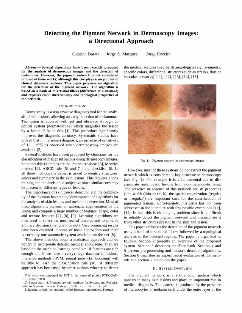
Fig. 1. Pigment network in dermoscopy images
However, most of these systems do not extract the pigmentnetwork which is considered a key structure in dermoscopy(see Fig. 1). For example it is a fundamental cue to dis-criminate melanocytic lesions from non-melanocytic ones.The presence or absence of this network and its properties(line width (thin or thick), the spatial organization (regularor irregular)) are important cues for the classification ofpigmented lesions. Unfortunately, this issue has not beenaddressed in the literature with few notable exceptions [11],[14]. In fact, this is challenging problem since it is difficultto reliably detect the pigment network and discriminate itfrom other structures present in the skin and lesion.
This paper addresses the detection of the pigment networkusing a bank of directional filters, followed by a topologicalanalysis of the detected regions. The paper is organized asfollows. Section 2 presents an overview of the proposedsystem. Section 3 describes the filter bank. Section 4 and5 present pre-processing and network detection algorithms.Section 6 describes an experimental evaluation of the meth-ods and section 7 concludes the paper.
II. SYSTEM OVERVIEW
The pigment network is a subtle color pattern whichappears in many skin lesions and plays an important role inmedical diagnosis. This pattern is produced by the presenceof melanocytes or melanin cells under the outer layer of the

skin and may exhibit different visual properties, namely dif-ferent color, width and spatial organization. Some examplesare shown in Fig. 1.
Despite this variability, some visual properties can be usedto detect the network and discriminate it from other structuressuch as blobs. Two properties will be considered:i) color:the network consists of a set of brown lines superimposedon a lighter background; andii) spatial organization: thelines form a connected net with holes of lighter color. Theholes tend to have an hexagonal shape and may exhibit aquasi-periodic distribution in space.
In practice, there are additional difficulties. dermoscopyimages often have multiple artifacts due to the presence ofhairs or reflections produced during the acquisition process.These artifacts have to be removed or attenuated before wetry to detect the network.
Fig. 2. Block diagram of the detection system
The detection system described in this paper consistsof three main blocks (see Fig. 2):i) pre-processing ii)network enhancement and iii) network detection. The firstblock detects artifacts and removes them from the image;the second block emphasizes the network structure takinginto account its color and spatial properties, while the thirdblock detects the presence of the pigment network using thisinformation.
III. D IRECTIONAL FILTERS
The pigment network and some of the artifacts (hairs)contain linear strokes. Therefore, we will adopt directionalfilters to enhance such structures. Since we do not know thestokes directions, we will use several directional filters eachof them tuned to a specific orientationθi ∈ [−π/2, π/2[,i = 1, . . . , N . The output of the directional filter is thencompared with the output of an omni directional filter. Wewill use Gaussian filters for this purpose.
Considering both contributions, the filter impulse responseis given by
hθ(x, y) = G1(x, y)−G2(x, y) (1)
whereGk(x, y) is a Gaussian filter
Gk(x, y) = Ck exp
{
−x′2
2σ2x,k
−y′
2
2σ2y,k
}
k = 1, 2 (2)
Ck is a normalization constant and(x′, y′) are related to(x, y) by a rotation of amplitudeθ
x′ = x cos θ + y sin θ (3)
y′ = −x sin θ + y cos θ (4)
The filter parametersσ2x,k, σ
2y,k are chosen in such a way
that the second filter is highly directional and the first is lessdirectional or even isotropic.
We filter a gray level imageI using all directional filters
Ii(x, y) = hθi(x, y) ∗ I(x, y) (5)
The outputs of the directional filtersIi are then combined asfollows. We select the maximum output at each pixel
J(x, y) = maxi
Ii(x, y) (6)
IV. PRE-PROCESSING
Let us describe the first processing step in this algorithm:pre-processing. This block performs two key operations:hair detection and reflection detection. First we convert thedermoscopic image (RGB image) into a gray scale image.We choose one of the color channels (the one with highestentropy). Then we apply the procedures described below.
Hair detection
This block tries to detect hair pixels in the gray levelimage. Hairs are long, linear and dark structures. We filterthe gray scale imageI using a bank of directional filters (1-6). If the filters responseJ(x, y) exceeds a thresholdTh thepixel (x, y) is classified as hair.
Fig. 3. Detection of hair (1st row) and reflections (2nd row).
Reflection detection
The reflection detection algorithm is very simple. Weclassify a pixel(x, y) as reflection if its intensity is high andif it is higher than the average intensityIav(x, y) computedin a neighborhood of the pixel. Therefore, the pixel shouldmeet the following condition
I(x, y) > Tr1 ∧ I(x, y)− Iav(x, y) > Tr2 (7)
whereTr1, Tr2 are thresholds andIav is the average intensity.Figure 3 shows examples of both pre-processing operations.
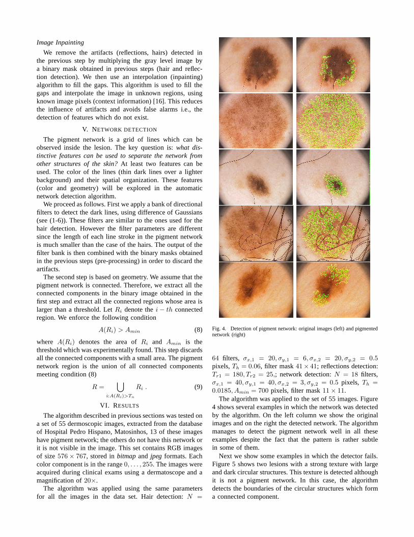
Image Inpainting
We remove the artifacts (reflections, hairs) detected inthe previous step by multiplying the gray level image bya binary mask obtained in previous steps (hair and reflec-tion detection). We then use an interpolation (inpainting)algorithm to fill the gaps. This algorithm is used to fill thegaps and interpolate the image in unknown regions, usingknown image pixels (context information) [16]. This reducesthe influence of artifacts and avoids false alarms i.e., thedetection of features which do not exist.
V. NETWORK DETECTION
The pigment network is a grid of lines which can beobserved inside the lesion. The key question is:what dis-tinctive features can be used to separate the network fromother structures of the skin?At least two features can beused. The color of the lines (thin dark lines over a lighterbackground) and their spatial organization. These features(color and geometry) will be explored in the automaticnetwork detection algorithm.
We proceed as follows. First we apply a bank of directionalfilters to detect the dark lines, using difference of Gaussians(see (1-6)). These filters are similar to the ones used for thehair detection. However the filter parameters are differentsince the length of each line stroke in the pigment networkis much smaller than the case of the hairs. The output of thefilter bank is then combined with the binary masks obtainedin the previous steps (pre-processing) in order to discard theartifacts.
The second step is based on geometry. We assume that thepigment network is connected. Therefore, we extract all theconnected components in the binary image obtained in thefirst step and extract all the connected regions whose area islarger than a threshold. LetRi denote thei− th connectedregion. We enforce the following condition
A(Ri) > Amin (8)
where A(Ri) denotes the area ofRi and Amin is thethreshold which was experimentally found. This step discardsall the connected components with a small area. The pigmentnetwork region is the union of all connected componentsmeeting condition (8)
R =⋃
i:A(Ri)>Tn
Ri . (9)
VI. RESULTS
The algorithm described in previous sections was tested ona set of 55 dermoscopic images, extracted from the databaseof Hospital Pedro Hispano, Matosinhos, 13 of these imageshave pigment network; the others do not have this network orit is not visible in the image. This set contains RGB imagesof size576× 767, stored inbitmapand jpeg formats. Eachcolor component is in the range0, . . . , 255. The images wereacquired during clinical exams using a dermatoscope and amagnification of20×.
The algorithm was applied using the same parametersfor all the images in the data set. Hair detection:N =
Fig. 4. Detection of pigment network: original images (left) and pigmentednetwork (right)
64 filters, σx,1 = 20, σy,1 = 6, σx,2 = 20, σy,2 = 0.5pixels,Th = 0.06, filter mask41× 41; reflections detection:Tr1 = 180, Tr2 = 25,; network detection:N = 18 filters,σx,1 = 40, σy,1 = 40, σx,2 = 3, σy,2 = 0.5 pixels, Th =0.0185, Amin = 700 pixels, filter mask11× 11.
The algorithm was applied to the set of 55 images. Figure4 shows several examples in which the network was detectedby the algorithm. On the left column we show the originalimages and on the right the detected network. The algorithmmanages to detect the pigment network well in all theseexamples despite the fact that the pattern is rather subtlein some of them.
Next we show some examples in which the detector fails.Figure 5 shows two lesions with a strong texture with largeand dark circular structures. This texture is detected althoughit is not a pigment network. In this case, the algorithmdetects the boundaries of the circular structures which forma connected component.

Fig. 5. Detection errors: original images (left) and pigmented network(right)
not detected detectedno network 67.5% 32.5%
pigment network 20, 0% 80.0%
TABLE I
STATISTICAL RESULTS
It is useful to perform a binary classification of theimages according to the presence of the pigment network.To improve the results, the network detected by the previousalgorithm should be verified to check if it has the expectedproperties, e.g., pigment color, background color, statisticsof holes and spatial organization of holes. An automaticclassifier can be trained from this data to learn the decisionand improve the results obtained by thresholding. In thispaper, we take a simpler approach. We compare the networkarea with a threshold and use this test to classify the imageinto one of two classes:with and without pigment network.The threshold is chosen as10% of the lesion area. Table 1shows the results obtained using this simple test in the dataset of 55 images.
The algorithm obtained a specificity SP=67.5% and asensibility SE=80.0% in this data set of clinical examples.
These statistics describe the output of the algorithm with-out any additional post-processing to verify if the detectednetworks are correct.
VII. C ONCLUSION
This paper presents an algorithm for the detection of thepigment network. The proposed algorithm explores the linecolor and geometry as well as the network topology usinga bank of directional filters. Experimental results in a set ofannotated images from Hospital Pedro Hispano show that thealgorithm achieves good detection scores and it is thereforean useful tool in a dermoscopy analysis system.
Future work should focus on a detailed evaluation of theproposed algorithm in a larger data base and the characteriza-tion of the detected network in order to discriminate typicalfrom atypical patterns which are important cues to detectmalign lesions.
ACKNOWLEDGMENT
We thank Profs. Teresa Mendonca, Andre Marcal and Ms.Barbara Amorim from the University of Porto, Drs. OfeliaMorais, Marta Pereira, Marta Teixeira and nurse Lina Santosfrom Hospital Pedro Hispano, for their support during thiswork.
REFERENCES
[1] Dermoscopy Tutorial, www.dermoscopy.org/atlas/base.htm[2] J. Mayer, Systematic review of the diagnostic accuracy of dermatoscopy
in detecting malignant melanoma, Medical Journal Australia, 206-210,1997.
[3] H. Pehamberger, A. Steiner, K. Wolff, In vivo epiluminescence mi-croscopy of pigmented skin lesions. I. Pattern analysis of pigmentedskin lesions. Journal of American Academy of Dermatololy, vol. 17,571-583 1987
[4] Scott W. Menzies, A Method for the diagnosis of primary cutaneousmelanoma using surface microscopy, Dermatologic Clinics Vol. 19,299-305, 2001.
[5] W. Stolz et al., ABCD rule of Dermatoscopy: a new practical methodfor early recognition of malignant melanoma, European Journal ofDermatology, vol. 4, 521-527, 1994.
[6] G. Argenziano et al., Epiluminescence microscopy for the diagnosis ofdoubtful melanocytic skin lesions. Comparison of the ABCD rule ofdermatoscopy and a new 7-point checklist based on pattern analysis,Archive of Dermatology, vol. 134, 1563-1570, 1998.
[7] P. Rubegni et al., Automated diagnosis of pigmented skinlesions,International Journal of Cancer, Vol. 101, 576-580, 2002.
[8] H. Iyatomi, et al., An Improved Internet-Based MelanomaScreeningSystem with Dermatologist-like Tumor Area Extraction Algorithm,Computerized Medical Imaging and Graphics, Vol. 32. 566-579, 2008.
[9] N. Situ et al., Malignant Melanoma Detection by Bag-of-FeaturesClassication, 3110-3113, EMBS 2008.
[10] R. Duda, P. Hart, D. Stork, Pattern Classification, Wiley, 2001.[11] G. Betta, et al., Dermoscopic image-analysis system: estimation of
atypical pigment network and atypical vascular pattern, IEEE Inter-national Workshop on Medical Measurement and Applications. 63-67,2006.
[12] M. E. Celebi, H. Iyatomi, W. V. Stoecker, R. H. Moss, H. S.Rabi-novitz, G. Argenziano, H. P. Soyer, Automatic Detection of Blue-WhiteVeil and Related Structures in Dermoscopy Images, ComputerizedMedical, Vol. 32. 670-677, 2008.
[13] G. Fabbrocini et al., Epiluminescence Image Processing forMelanocytic Skin Lesion Diagnosis Based on 7-Point Check-List, OpenDermatology Journal, 110-115, 2010.
[14] M. Sadeghi, M. Razmara, M. Ester, T. K. Lee, M. S. Atkins,Graph-based Pigment Network Detection in Skin Images, Proc. SPIE 7623,2010.
[15] N. Situ, X. Yuan, G. Zouridakis, Assisting Main Task Learningby Heterogeneous Auxiliary Tasks with Applications to SkinCancerScreening, International conference on artificial intelligence and statis-tics, April 2011.
[16] M. Bertalmio, G. Sapiro, V. Caselles, C. Ballester, Image Inpainting,SIGGRAPH, 417-424, 2000.