dermoscopy of eccrine acrospiroma masquerading...
TRANSCRIPT
23
C l i n i c i a n s r e p o r t s Dermoscopy of eccrine acrospiroma masquerading as nodular malignant melanoma
Acta Dermatoven APA Vol 19, 2010, No 4
Dermoscopy of eccrine acrospiroma masquerading as nodular
malignant melanomaA. Gatti, N. di Meo, and G. Trevisan
Eccrine acrospiroma, better known as eccrine poroma, is a benign adnexal neoplasm of the skin. Its clinical aspect can masquerade as some other nodular and cystic lesions. The current dermo-scopy literature offers very few case studies. Moreover, these very few examples present a totally different appearance pattern compared to the one we examined. Its homogeneous blue pattern suggested the better-known nodular malignant melanoma; in fact, this dermoscopic appearance was due to the Tyndall effect.
IntroductionEccrine acrospiroma is a benign sweat gland neo-
plasm that occurs as a single mass in the skin with a nodular or cystic structure. The color varies from that of the surrounding skin to red or reddish blue, and the covering skin may be smooth or thickened and ver-rucous. Clinically, the tumors lack diagnostic speci-ficity, but they should be included in the differential diagnosis of nodular and cystic lesions of the skin. In contrast, the cells and structure are histologically dis-tinctive (1). Dermoscopy is an effective and absolutely necessary non-invasive diagnostic technique for the study of pigmented lesions. Its use has substantially contributed to improving the early diagnosis of skin melanoma.
We report a case of eccrine acrospiroma located on the left leg, which caused an equivocal pigmented skin lesion both clinically and dermoscopically.
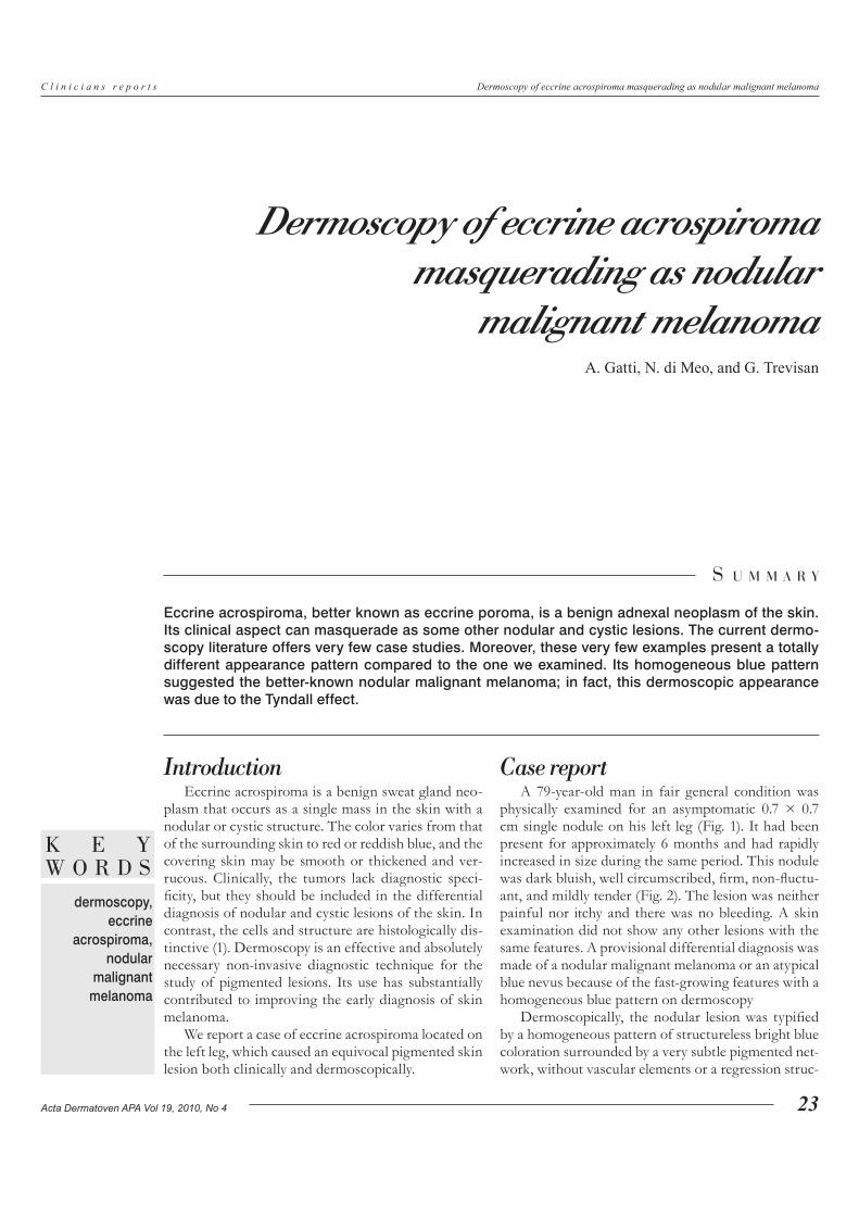
Case reportA 79-year-old man in fair general condition was
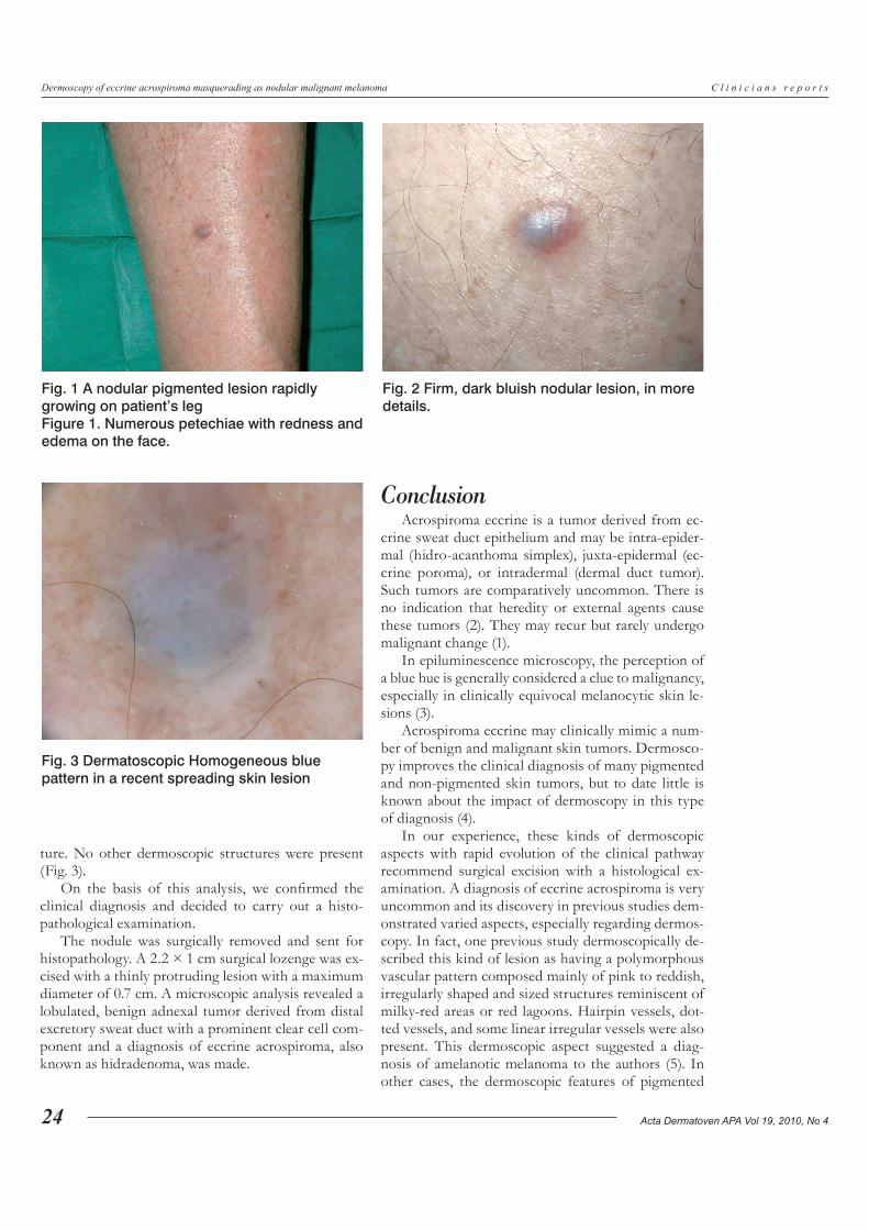
physically examined for an asymptomatic 0.7 × 0.7 cm single nodule on his left leg (Fig. 1). It had been present for approximately 6 months and had rapidly increased in size during the same period. This nodule was dark bluish, well circumscribed, firm, non-fluctu-ant, and mildly tender (Fig. 2). The lesion was neither painful nor itchy and there was no bleeding. A skin examination did not show any other lesions with the same features. A provisional differential diagnosis was made of a nodular malignant melanoma or an atypical blue nevus because of the fast-growing features with a homogeneous blue pattern on dermoscopy
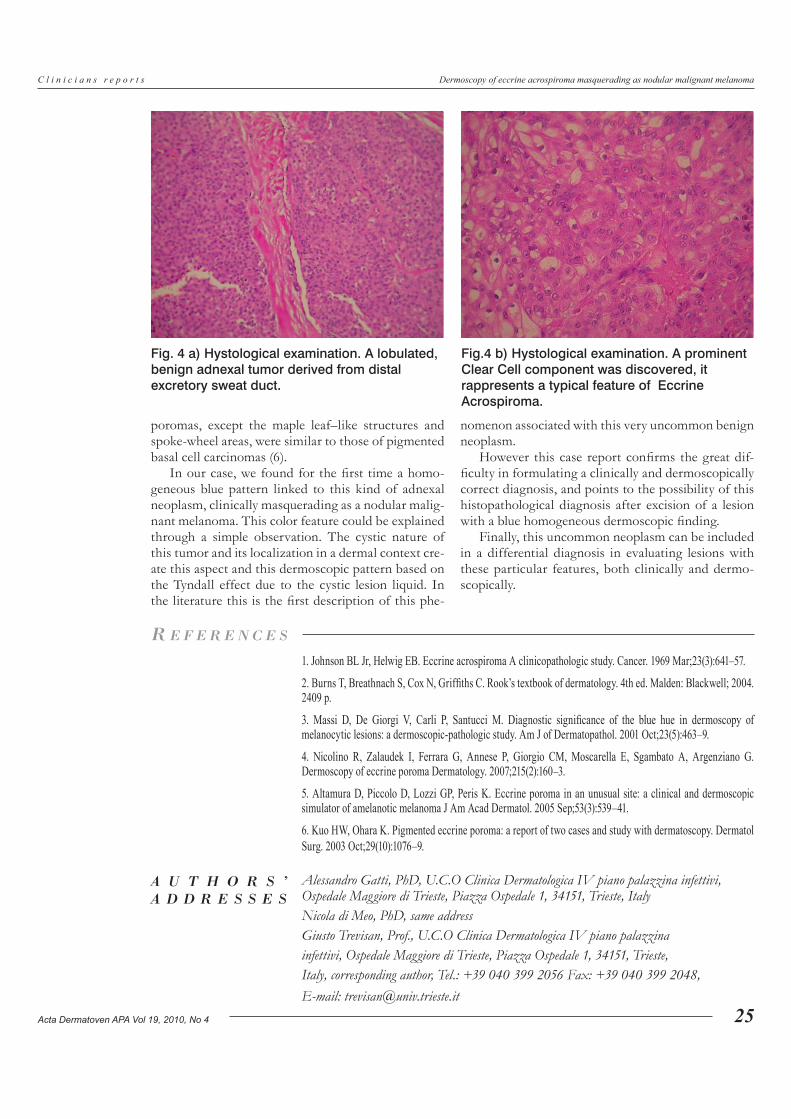
Dermoscopically, the nodular lesion was typified by a homogeneous pattern of structureless bright blue coloration surrounded by a very subtle pigmented net-work, without vascular elements or a regression struc-
K E YW O R D S
dermoscopy, eccrine
acrospiroma, nodular
malignant melanoma
S U M M A R Y
24
C l i n i c i a n s r e p o r t sDermoscopy of eccrine acrospiroma masquerading as nodular malignant melanoma
Acta Dermatoven APA Vol 19, 2010, No 4
ture. No other dermoscopic structures were present (Fig. 3).
On the basis of this analysis, we confirmed the clinical diagnosis and decided to carry out a histo-pathological examination.
The nodule was surgically removed and sent for histopathology. A 2.2 × 1 cm surgical lozenge was ex-cised with a thinly protruding lesion with a maximum diameter of 0.7 cm. A microscopic analysis revealed a lobulated, benign adnexal tumor derived from distal excretory sweat duct with a prominent clear cell com-ponent and a diagnosis of eccrine acrospiroma, also known as hidradenoma, was made.
ConclusionAcrospiroma eccrine is a tumor derived from ec-
crine sweat duct epithelium and may be intra-epider-mal (hidro-acanthoma simplex), juxta-epidermal (ec-crine poroma), or intradermal (dermal duct tumor). Such tumors are comparatively uncommon. There is no indication that heredity or external agents cause these tumors (2). They may recur but rarely undergo malignant change (1).
In epiluminescence microscopy, the perception of a blue hue is generally considered a clue to malignancy, especially in clinically equivocal melanocytic skin le-sions (3).
Acrospiroma eccrine may clinically mimic a num-ber of benign and malignant skin tumors. Dermosco-py improves the clinical diagnosis of many pigmented and non-pigmented skin tumors, but to date little is known about the impact of dermoscopy in this type of diagnosis (4).
In our experience, these kinds of dermoscopic aspects with rapid evolution of the clinical pathway recommend surgical excision with a histological ex-amination. A diagnosis of eccrine acrospiroma is very uncommon and its discovery in previous studies dem-onstrated varied aspects, especially regarding dermos-copy. In fact, one previous study dermoscopically de-scribed this kind of lesion as having a polymorphous vascular pattern composed mainly of pink to reddish, irregularly shaped and sized structures reminiscent of milky-red areas or red lagoons. Hairpin vessels, dot-ted vessels, and some linear irregular vessels were also present. This dermoscopic aspect suggested a diag-nosis of amelanotic melanoma to the authors (5). In other cases, the dermoscopic features of pigmented
Fig. 1 A nodular pigmented lesion rapidly growing on patient’s legFigure 1. Numerous petechiae with redness and edema on the face.
Fig. 2 Firm, dark bluish nodular lesion, in more details.
Fig. 3 Dermatoscopic Homogeneous blue pattern in a recent spreading skin lesion
25
C l i n i c i a n s r e p o r t s Dermoscopy of eccrine acrospiroma masquerading as nodular malignant melanoma
Acta Dermatoven APA Vol 19, 2010, No 4
poromas, except the maple leaf–like structures and spoke-wheel areas, were similar to those of pigmented basal cell carcinomas (6).
In our case, we found for the first time a homo-geneous blue pattern linked to this kind of adnexal neoplasm, clinically masquerading as a nodular malig-nant melanoma. This color feature could be explained through a simple observation. The cystic nature of this tumor and its localization in a dermal context cre-ate this aspect and this dermoscopic pattern based on the Tyndall effect due to the cystic lesion liquid. In the literature this is the first description of this phe-
nomenon associated with this very uncommon benign neoplasm.
However this case report confirms the great dif-ficulty in formulating a clinically and dermoscopically correct diagnosis, and points to the possibility of this histopathological diagnosis after excision of a lesion with a blue homogeneous dermoscopic finding.
Finally, this uncommon neoplasm can be included in a differential diagnosis in evaluating lesions with these particular features, both clinically and dermo-scopically.
Fig. 4 a) Hystological examination. A lobulated, benign adnexal tumor derived from distal excretory sweat duct.
Fig.4 b) Hystological examination. A prominent Clear Cell component was discovered, it rappresents a typical feature of Eccrine Acrospiroma.
R E F E R E N C E S
1. Johnson BL Jr, Helwig EB. Eccrine acrospiroma A clinicopathologic study. Cancer. 1969 Mar;23(3):641–57.
2. Burns T, Breathnach S, Cox N, Griffiths C. Rook’s textbook of dermatology. 4th ed. Malden: Blackwell; 2004. 2409 p.
3. Massi D, De Giorgi V, Carli P, Santucci M. Diagnostic significance of the blue hue in dermoscopy of melanocytic lesions: a dermoscopic-pathologic study. Am J of Dermatopathol. 2001 Oct;23(5):463–9.
4. Nicolino R, Zalaudek I, Ferrara G, Annese P, Giorgio CM, Moscarella E, Sgambato A, Argenziano G. Dermoscopy of eccrine poroma Dermatology. 2007;215(2):160–3.
5. Altamura D, Piccolo D, Lozzi GP, Peris K. Eccrine poroma in an unusual site: a clinical and dermoscopic simulator of amelanotic melanoma J Am Acad Dermatol. 2005 Sep;53(3):539–41.
6. Kuo HW, Ohara K. Pigmented eccrine poroma: a report of two cases and study with dermatoscopy. Dermatol Surg. 2003 Oct;29(10):1076–9.
A U T H O R S ’A D D R E S S E S
Alessandro Gatti, PhD, U.C.O Clinica Dermatologica IV piano palazzina infettivi, Ospedale Maggiore di Trieste, Piazza Ospedale 1, 34151, Trieste, ItalyNicola di Meo, PhD, same addressGiusto Trevisan, Prof., U.C.O Clinica Dermatologica IV piano palazzinainfettivi, Ospedale Maggiore di Trieste, Piazza Ospedale 1, 34151, Trieste,Italy, corresponding author, Tel.: +39 040 399 2056 Fax: +39 040 399 2048,E-mail: [email protected]