design and development of diphenhydramine hydrochloride topical
TRANSCRIPT
Research Article
DESIGN AND DEVELOPMENT OF DIPHENHYDRAMINE HYDROCHLORIDE TOPICAL LIPOSOMAL DRUG DELIVERY SYSTEM
SADHANA SHAHI*, UTTARA SONWANE, NITYANAND ZADBUKE, IMRAN TADWEE
Government College of Pharmacy, Department of Pharmaceutics, Aurangabad 431005, Maharashtra, India. Email: [email protected]
Received: 05 Apr 2013, Revised and Accepted: 28 May 2013
ABSTRACT
Liposomes are microscopic vesicles composed of a bilayer of phospholipids or any similar amphipathic lipids. Liposomal encapsulation of a drug can dramatically alter the pharmacokinetic properties of a drug, targeting the drug to particular organs and/or enhance the efficacy of the encapsulated drug.
Diphenhydramine hydrochloride (DPH HCl) is a first generation antihistamine used to treat a number of conditions including allergic symptoms and itchiness, the common cold, insomnia, motion sickness, and extrapyramidal symptoms.
DPH HCl distributes into the skin efficiently and sustains higher concentrations than in serum making it a potential treatment of skin disorders in which histamine is the mediator. Unfortunately DPH HCl has considerable CNS adverse effects, sedation, drying of mouth presents a problem for most of the patients, limiting its use orally.
DPH administration by topical liposomal formulations would yield greater and more persistent effects on the suppression of histamine-induced wheal formation over the 8 h period.
Objective: The main objective of the study was to design, develop, optimize and evaluate topical liposomal formulation of DPH HCl.
Methods: The effect of concentration of soya lecithin and cholesterol on drug entrapment (%) was studied.
Results: A stable and optimized thermo reversible topical liposomal gel of DPH HCl was prepared using Lutrol F-127 as a gelling agent by cold method. The prepared gel formulations were studied for their in vitro release characteristics through guinea pig skin and showed sustained release action for 8 h when compared with plain gel.
Conclusion: The stability study showed that liposomal suspension (L5) and liposomal gel as the stable formulation with respect to the parameters selected.
Keywords: Liposomal gel, DPH HCl, Cholesterol, Soya lecithin, Cold method, Lutrol F-127
INTRODUCTION
Liposomes are microscopic vesicles composed of a bilayer of phospholipids or any similar amphipathic lipids. They can encapsulate and effectively deliver both hydrophilic and lipophilic substance and [1-2] may be used as a non‐toxic vehicle for insoluble drugs [3]. Liposome as a microstructure consists of one or more concentric spheres of lipid bilayer separated by water or aqueous buffer compartments [4]. Liposomes have many of the requirements for good drug delivery systems as they are relatively non‐toxic and bio‐degradable [5]. They have been found to be useful carriers for both hydrophilic and hydrophobic drugs [6]. Liposomal encapsulation of a drug can dramatically alter the pharmacokinetic properties of a drug, targeting the drug to particular organs and/or enhance the efficacy of the encapsulated drug [7].
The incorporation of drugs into liposomes has several advantages as they protect their contents from interaction with plasma components, while favorably altering the pharmacokinetics and biodistribution of free compound since liposomes do not readily penetrate biologic membrane. They can be used for controlled release of drugs within body cavities such as pleural, peritoneal or intrathecal spaces [8-9].
The formulation of an appropriate liposomal system as a carrier for a given drug is dependent on the type of the lipid used and the method of preparation [10]. According to their size they are known as small unilamellar vesicles (SUV) or large unilamellar vesicles (LUV). If more bilayers are present they are referred to as multilamellar vesicles (MLV) [11-12].Depending on the composition liposomes can have a positive, negative, or neutral surface charge. Lecithin can provide liposomes with a neutral surface; stearylamine and phosphatidic acid components provide positive [13] and negative surface charge, respectively. Depending on the lipid
composition, methods of preparations and the nature of the encapsulated agents, many types of liposomal products can be formulated. The ideal drug candidates for liposomal encapsulation are those that have potent pharmacological activity and are either highly lipid or water soluble. If a drug is water soluble, it will be encapsulated within the aqueous compartment and its concentration in the liposomal product will depend on the volume of the entrapped water and the solubility of that drug in the encapsulated water.
The lipophilic drug is usually bound to the lipid bi‐layer or ‘dissolved’ in the lipid phase. A lipophilic drug is more likely to remain encapsulated during storage due to its partition coefficient. Since the lipophilic drug is associated with the lipid bi‐layers it will not leach out as readily to the ‘external’ water phase. Generally the encapsulation efficiency is higher for lipophilic drugs than hydrophilic drugs [14]. The applications of liposomes as nanoscale containers for drugs, vitamins [15-16], enzymes or genetic material [17-18] require control and prediction of the liposome dispersion stability [19].
Drug loading can be achieved either passively (i.e. the drug is encapsulated during liposome formulation) or actively (i.e. after liposome formation). Hydrophobic drugs, such as amphotericin B, taxol can be directly incorporated into liposomes during vesicle formation, and the extent of uptake and retention is governed by drug-lipid interactions. Trapping efficiencies of 100% are often achievable, but this is dependent on the solubility of the drug in the liposome membrane [20]. Drugs can be incorporated into liposomes using three primary mechanisms as Encapsulation, Partitioning and Reverse loading.
Diphenhydramine hydrochloride (DPH HCl) is a first generation antihistamine used to treat a number of conditions including allergic
International Journal of Pharmacy and Pharmaceutical Sciences
ISSN- 0975-1491 Vol 5, Issue 3, 2013
AAccaaddeemmiicc SScciieenncceess
Shahi et al. Int J Pharm Pharm Sci, Vol 5, Issue 3, 534-542
535
symptoms and itchiness, the common cold, insomnia, motion sickness, and extrapyramidal symptoms [21].
DPH HCl distributes into the skin efficiently and sustains higher concentrations than in serum making it a potential treatment of skin disorders in which histamine is the mediator. Unfortunately DPH HCl has considerable CNS adverse effects, sedation, drying of mouth presents a problem for most of the patients, limiting its use orally.
The main objective of the study was to design, develop, optimize and evaluate topical liposomal formulation of DPH HCl. The present study hypothesized that DPH HCl administration by topical liposomal formulations would yield greater and more persistent effects on the suppression of histamine-induced wheal formation over the 8 h period and the liposome formulations would result in lower serum concentrations after topical application compared to the traditional non-liposome cream formulations. This method of administration of DPH HCl would result in a better treatment of allergic skin diseases and also avoid the central nervous system side effects, particularly sedation & drying of mouth.
MATERIALS AND METHODS
Diphenhydramine hydrochloride was kind gift sample obtained from Wockhardt Ltd. Aurangabad, India. Cholesterol was purchased from Qualigens Fine Chemicals, Mumbai. Soya lecithins were purchased from Lipoid Germany. Lutrol F-127 were kind gift samples from Encube Ethicals, Mumbai. The laboratory grade chemicals used for the work were chloroform, acetone, methanol, stearic acid, propylene glycol and sodium chloride procured from S.D.Fine Chemicals. Potassium dihydrogen ortho phosphate was purchased from Loba Chemie.
Preformulation study
Procedure for the Preparation of Calibration Curve by UV
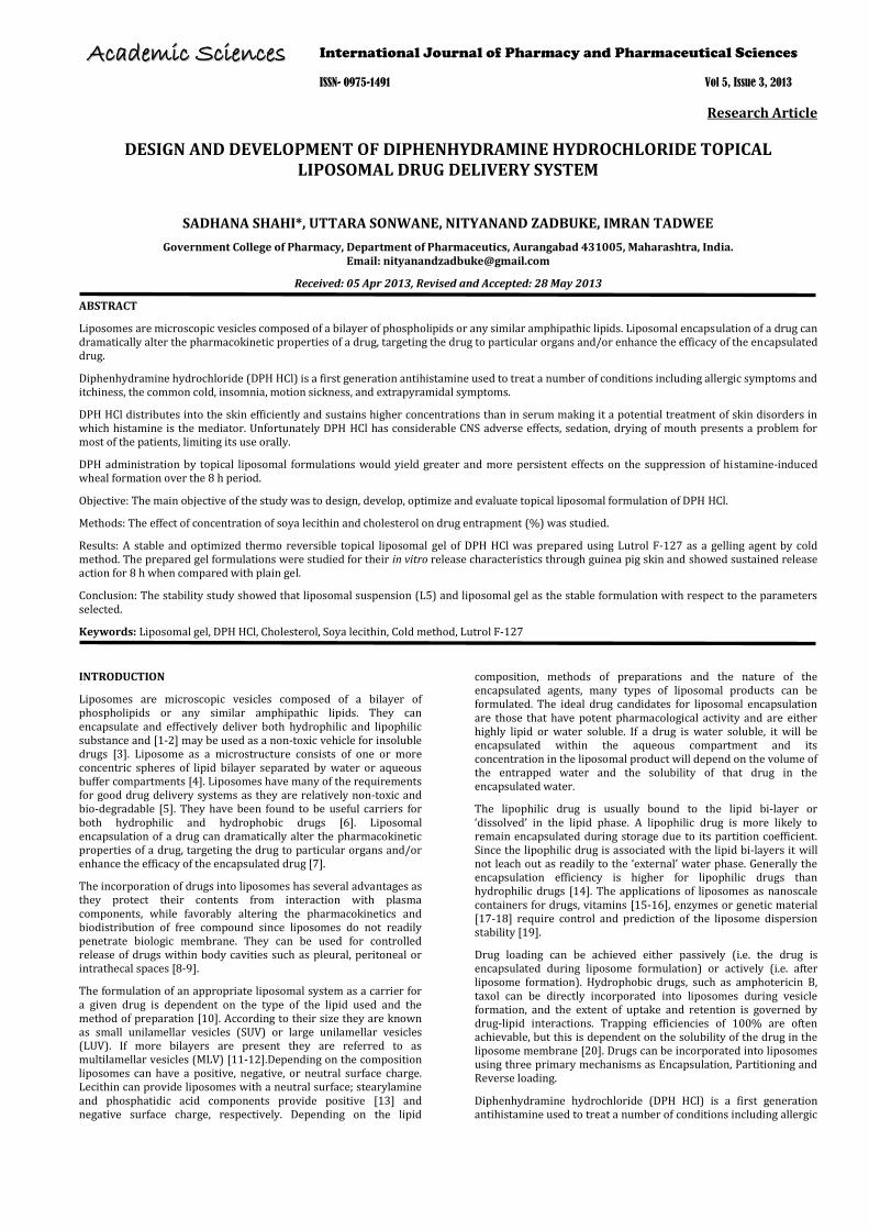
Standard calibration curve of DPH HCl was developed using pH 7.4 buffer solution by series of dilution in the range of 100-600 µg/ml and estimated by UV-Visible Spectrophotometer (UV 1700 PC Shimadzu, Japan) at λmax 258nm. Absorbance values were plotted against respective concentrations to obtain standard calibration curve (Figure 1).
Fig. 1: Standard calibration curve of DPH HCl at λmax 258nm
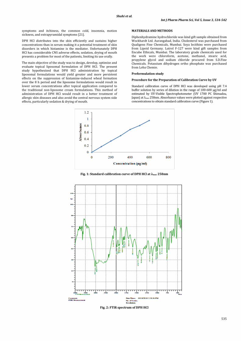
Fig. 2: FTIR spectrum of DPH HCl
Shahi et al. Int J Pharm Pharm Sci, Vol 5, Issue 3, 534-542
536
Vibrational Spectroscopic study
The FTIR spectrum of DPH HCl was recorded using Prestige-21 (SHIMADZU) with IR resolution software. The drug sample was scanned over the range of 400 to 4000 cm-1(Figure 2).
Preparation of DPH HCl Liposomes
In this study liposomes were prepared by lipid thin hydration method. The most widely used method for the preparation of MLV.
The different ratio of phospholipids: cholesterol and stearic acid were weighed and dissolved in 3 ml of chloroform: methanol (2:1) in 250 ml round bottom flask. A thin film was formed on the inner side of round bottom flask by evaporating organic solvent under vacuum in rotary evaporator at 45-50°C. The dry lipid film was hydrated with 10 ml phosphate buffer solution (pH 7.4) containing DPH HCl at a temperature of 60±2°C. The dispersion was left undisturbed at room temperature for 15 min to allow complete swelling of the lipid film. The vesicular suspension obtained was stored at 4°C.
Preliminary batches were prepared to study the effect of concentration of soya lecithin and cholesterol on drug entrapment.
Effect of soya lecithin on drug entrapment
Soya lecithin proportion was varied from 90 mg to 300 mg keeping all the other excipients constant (Table 1). The prepared liposomes were stored in refrigerator at 4°C. Entrapment of drug in vesicles was determined spectrophotometrically. The 200 mg concentration of soya lecithin gave greater entrapment efficacy and was selected for further studies.
Effect of cholesterol on drug entrapment
Cholesterol proportion was varied from 10 mg to 50 mg, keeping all other excipients constant (Table 2). Entrapment of drug in vesicles was determined spectrophotometrically. Entrapment efficiency decreases with the increase in concentration. The 30 mg concentration of cholesterol gave greater entrapment efficacy and was selected for further studies.
Table 1: Effect of soya lecithin on drug entrapment
DPH HCl (mg) Lecithin (mg) Cholesterol (mg) Stearic acid (mg) Entrapment (%) 50 90 30 30 40.364 50 100 30 30 60.07 50 200 30 30 85.235 50 300 30 30 74.368
Table 2: Effect of cholesterol on drug entrapment
DPH HCl (mg) Lecithin (mg) Cholesterol (mg) Stearic acid (mg) Entrapment (%) 50 200 10 30 54.895 50 200 20 30 71.860 50 200 30 30 84.687 50 200 40 30 60.256
The composition and proportion of lecithin and cholesterol for 32 factorial designed liposomal formulations were mentioned in Table 3.
Table 3: Composition of 32 factorial designed liposomal formulations of DPH HCL
Formulation code DPH HCl (mg)
Soya lecithin (mg)
Cholesterol (mg)
Stearic acid (mg)
L1 50 150 25 30 L2 50 200 25 30 L3 50 250 25 30 L4 50 150 30 30 L5 50 200 30 30 L6 50 250 30 30 L7 50 150 35 30 L8 50 200 35 30 L9 50 250 35 30
Entrapment efficiency determination
The prepared liposomes were kept at 4°C and ultra centrifuged (Megafuge 1.0 R, Hanau, Germany) for 1 h at 11000 rpm. Then 1-2 ml supernatant was withdrawn and centrifuged at 15000 rpm for 1h.The free (un entrapped) DPH HCl concentration was determined in supernatant spectrophotometrically at 258 nm (Shimadzu UV- 1601 PC Double Beam, Japan).
The DPH HCl entrapment efficiency was calculated by equation (1),
EE = ((Qt-Qs)/Qt)*100 ……………………………………………… (1)
Where, EE is the entrapment efficacy,
Qt is the theoretical amount of DPH HCl that was added, and Qs is the amount of DPH HCl detected only in the supernatant.
The liposomal suspension (L5) with maximum entrapment was used for the formulation of liposomal gel for further studies (Table 4).
Table 4: Entrapment efficiency determination
Formulation code Entrapment (%) Flux (µg/cm/h) Permeability coefficient (cm/h) L1 62.02 276.60±1.30 0.02766±1.30 L2 66.74 239.39±0.56 0.02393±0.56 L3 68.89 183.33±0.71 0.01833±0.74 L4 69.49 183.33±0.77 0.01861±0.78 L5 85.08 144.85±0.73 0.01440±0.73 L6 84.75 166.37±0.70 0.01663±0.70 L7 78.03 160.47±0.70 0.01604±0.70 L8 75.94 155.60±1.40 0.01556±1.40 L9 74.72 152.58±1.30 0.01525±1.30
Shahi et al. Int J Pharm Pharm Sci, Vol 5, Issue 3, 534-542
537
In vitro evaluation of permeation rates of 32 factorial designed liposomal formulations of DPH HCl through guinea pig skin [22]
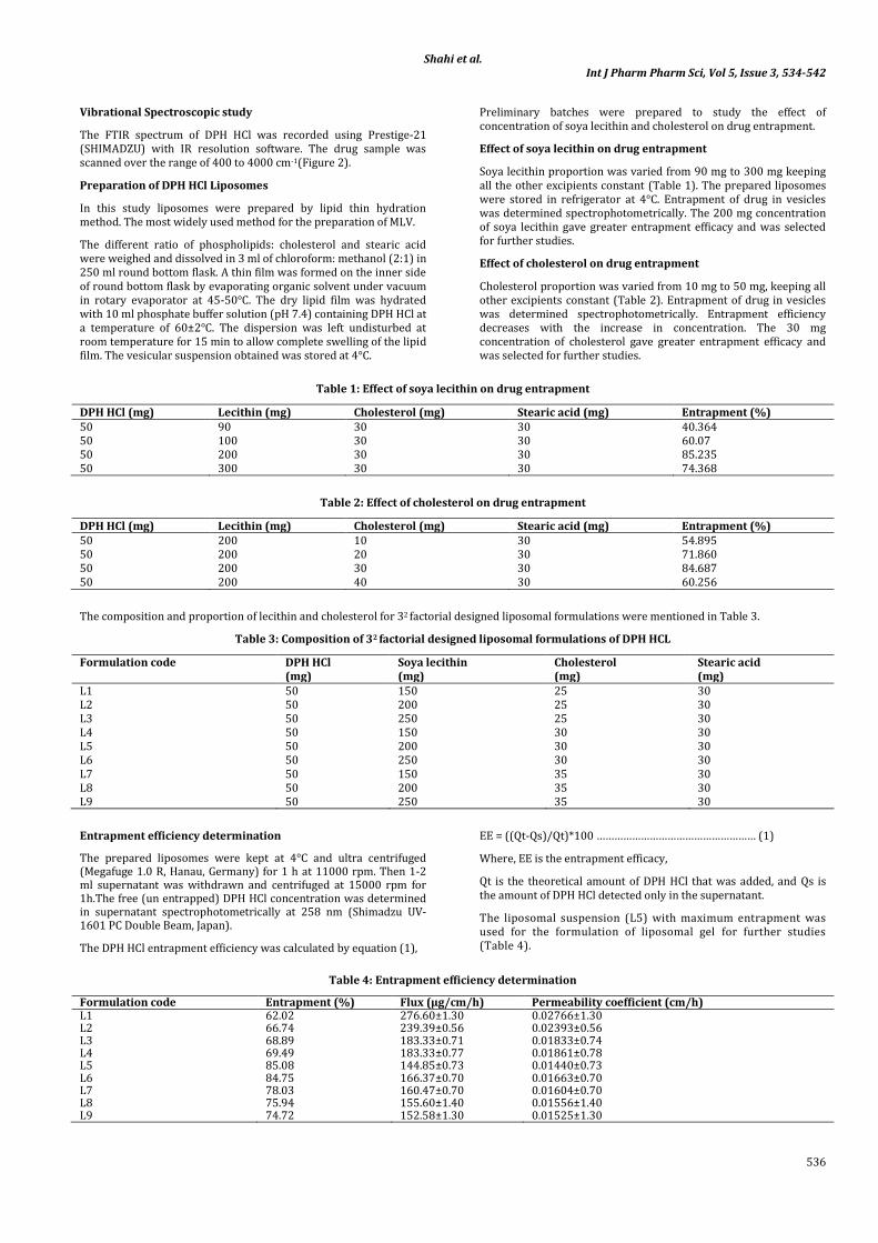
The modified glass diffusion cell (K-C cell) was used for permeation studies (Figure3). Freshly excised full thickness guinea pig skin was mounted between the donor and receptor compartment and compartment. Available skin surface area for permeation was approximately 2.4 cm2. Excessive skin at the sides was trimmed off to minimize lateral diffusion. The receptor compartment was filled with receptor media of composition pH 7.4 buffer solutions. Skin was allowed to equilibrate with receptor fluid for 2 h.
The receptor solution was stirred by a star- head magnetic bar (size 10 x 10 mm) (Himedia) rotating at a constant speed of 600 rpm by motor less magnetic stirrer (Whirlmatic-Mega, Spectra lab). The temperature was maintained at 37 ± 1ºC at using constant temperature water circulating bath (Deep Engineering), which circulates water through the water jacket surrounding the receptor compartment. At the predetermined sampling intervals, aliquots of 1 ml were withdrawn periodically and replaced with the same volume of fresh receptor fluid. The drugs concentrations were measured by UV spectrophotometer at λmax 258 nm.
Fig. 2: Assembly for in vitro permeation studies
The skin permeation studies were carried out for 8 h with drug solution and liposomal suspension (10 mg /ml) respectively.
Protocol for animal study was approved by Institutional Animal Ethical Committee (IAEC) of Government College of Pharmacy, Aurangabad (Proposal No: GCPA/IAEC//2011/235).
Characterization of liposomal suspension (L5)
The liposomal suspension (L5) was characterized for in vitro permeation study, viscosity, pH, vesicle size and size distribution, drug content and stability study.
Preparation and evaluation of topical liposomal gel of DPH HCl
A stable and optimized thermo reversible topical liposomal gel of DPH HCl was prepared using Lutrol F-127 as a gelling agent by cold method.
Liposomal suspension (L5) containing DPH HCl was incorporated into Lutrol F-127 20% w/w with constant stirring
using a Teflon-coated magnetic bead. The resultant mixture was stored at 4°C for 24 h to form sol. After 24 h the sol was removed and kept at room temperature to form a transparent liposomal gel.
The liposomal gel formulation of DPH HCl was evaluated for pH, viscosity, clarity, drug content, in vitro drug permeation study and stability study in comparison with plain gel of DPH HCl with 20% w/w Lutrol F-127 as a gel base.
RESULTS AND DISCUSSION
Kinetics of drug release
To analyze the mechanism of drug release in vitro release data were fitted to zero oder, first order Higuchi release model, Hixson Crowell model, Kosmeyer and peppas model by using PCP disso version 2.08 software, and model with the higher correlation coefficient was considered to be the best model (Table 5).
Table 5: Kinetics of Drug Release
Formulation Code R2 n k Zero Order 1st order Matrix Peppas Hixson
Crowell L1 0.6823 0.9698 0.9682 0.9786 0.9346 0.2962 48.2323 L2 0.6162 0.9565 0.9541 0.9758 0.9045 0.2661 50.2890 L3 0.5873 0.9118 0.9482 0.9728 0.8378 0.2551 45.8807 L4 0.4438 0.7970 0.9236 0.9870 0.7059 0.2227 44.1417 L5 0.5081 0.8325 0.9328 0.9766 0.7503 0.2346 43.1122 L6 0.5285 0.8200 0.9373 0.9767 0.7430 0.2386 40.3056 L7 0.6020 0.8453 0.9553 0.9936 0.7799 0.2748 36.8612 L8 0.5084 0.7981 0.9341 0.9806 0.7205 0.2348 39.5437 L9 0.7088 0.7406 09697 0.9705 0.7303 0.3028 0.3028 [
The results showed that the 32 factorial designed formulations followed Korsmeyer Peppas kinetics. The R2 values of Korsmeyer Peppas model was found close to one.
Shahi et al. Int J Pharm Pharm Sci, Vol 5, Issue 3, 534-542
538
Statistical analysis by Design Expert Software
The 32 factorial design was selected to study the effect of independent variables soya lecithin (X1) and cholesterol (X2) on dependent variables drug entrapment (%) and flux. A statistical model incorporating interactive and polynomial terms was utilized to evaluate the responses (Eq 2).
Y=b0+b1 X1+b2 X2+b12 X1X2+b11X12+b22X22………………………………… (2)
Where, Y is the dependent variable, b0 is the arithmetic mean response of the nine runs and bi(b1, b2, b12, b11 and b22) is the estimated coefficient for the corresponding factor Xi (X1, X2, X12, X11
and X22), which represents the average results of changing one factor at a time from its low to high value. The interaction term (X1X2) depicts the changes in the response when two factors are simultaneously changed. The polynomial terms (X12 and X22) are included to investigate nonlinearity.
The drug entrapment (%) and flux for the nine formulations of design showed a wide variation. The responses of the formulations prepared by 32 factorial designs were observed. The responses clearly indicate that the drug entrapment (%) and flux values are strongly dependent on the selected independent variables. The fitted regression equations relating the responses % entrapment and flux are shown in the following equations, respectively (Eqs. 3‑ 6).
Final equation for drug entrapment (%) in terms of coded factors:
Drug entrapment (%) = 81.73111 + 3.136667 X1 + 5.173333 X2 - 2.545 X1X2 - 2.93667 X12 - 8.71667 X22…………………………………… (3)
(r2=0.826946)
Final equation for drug entrapment (%) in terms of actual factors:
Drug entrapment (%) = 81.73111 + 3.136667*Soya lecithin + 5.173333*Cholesterol - 2.545*Soya lecithin*Cholesterol - 2.93667 *Soya lecithin2 - 8.71667*Cholesterol2…………. (4)
Final equation for flux in terms of coded factors:
Flux= 160.5392 -20.1487 X1 -38.443 X2 - 21.34325 X1X2 - 7.718667 X12 + 28.97867 X22…...(5)
(r2= 0.941566)
Final equation for flux in terms of actual factors:
Flux= 160.5392 - 20.1487 *Soya lecithin -38.443 *Cholesterol - 21.34325 *Soya lecithin*Cholesterol - 7.718667 *Soya lecithin2 + 28.97867 *Cholesterol2…………………. (6)
The regression coefficient values are used to validates the model fitting. The regression coefficient was high indicating the adequate fitting of model for response drug entrapment (%) and flux.
The polynomial equations can also be used to draw conclusion considering the magnitude of co-efficient and mathematical sign it carries; i.e. positive or negative.
In the present study negatively coefficient of independent variables soya lecithin (X1) and cholesterol (X2) showed that it contributes negatively to flux and retard the drug release of the drug release at all response points and it is a significant variable in drug release. However, the positive coefficient of soya lecithin (X1) and cholesterol (X2) of equations of entrapment showed that it contributes positively to drug entrapment (%) and leads to enhancement in drug entrapment.
ANOVA study
The analysis of variance study of the data also showed results revealing the cholesterol (X2) as significant variable (p value < 0.05) at flux as response point and insignificant at entrapment response, while soya lecithin (X1) is insignificant at all response point. ANOVA and Multiple regression analysis were done using Stat‑ Ease Design Expert 7.1.4 software.
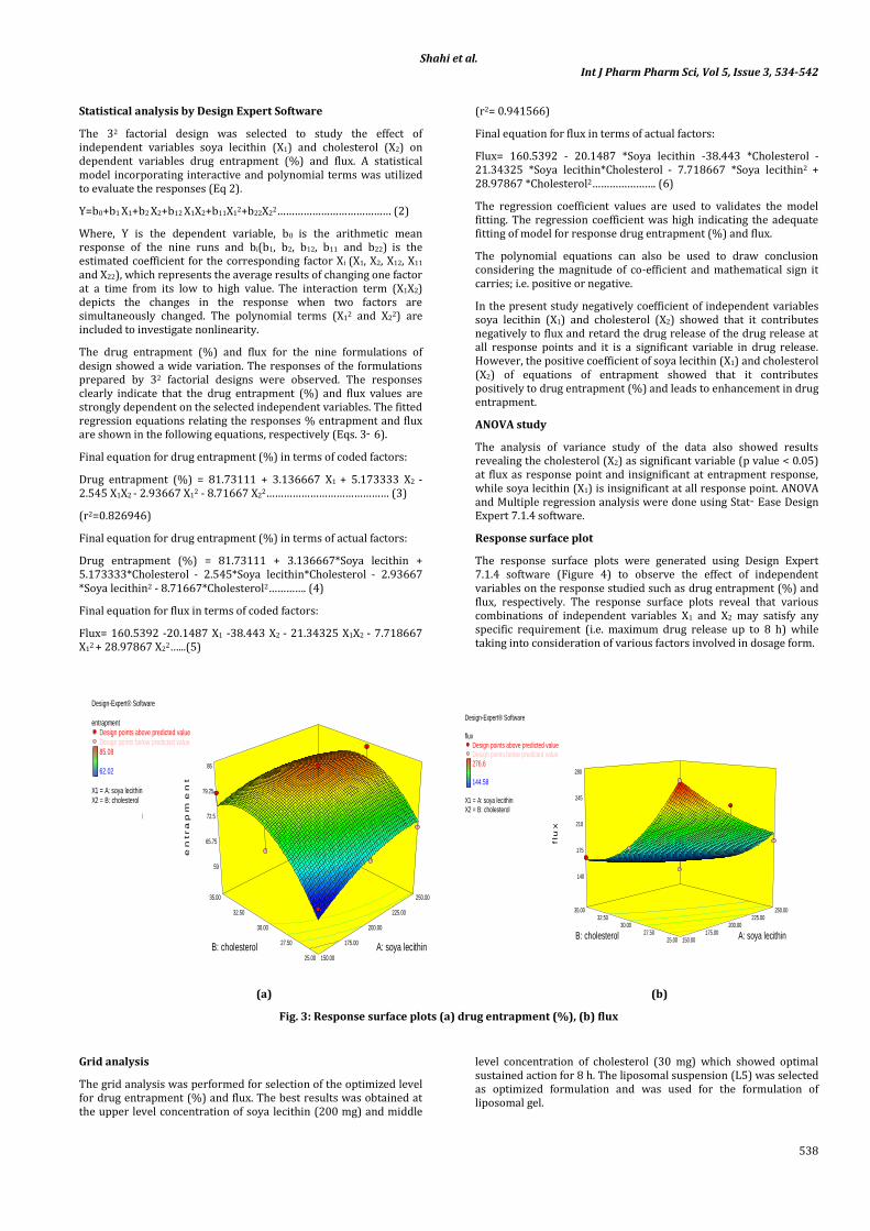
Response surface plot
The response surface plots were generated using Design Expert 7.1.4 software (Figure 4) to observe the effect of independent variables on the response studied such as drug entrapment (%) and flux, respectively. The response surface plots reveal that various combinations of independent variables X1 and X2 may satisfy any specific requirement (i.e. maximum drug release up to 8 h) while taking into consideration of various factors involved in dosage form.
(a) (b)
Fig. 3: Response surface plots (a) drug entrapment (%), (b) flux
Grid analysis
The grid analysis was performed for selection of the optimized level for drug entrapment (%) and flux. The best results was obtained at the upper level concentration of soya lecithin (200 mg) and middle
level concentration of cholesterol (30 mg) which showed optimal sustained action for 8 h. The liposomal suspension (L5) was selected as optimized formulation and was used for the formulation of liposomal gel.
Design-Expert® Software
entrapmentDesign points above predicted valueDesign points below predicted value85.08
62.02
X1 = A: soya lecithinX2 = B: cholesterol
150.00
175.00
200.00
225.00
250.00
25.00
27.50
30.00
32.50
35.00
59
65.75
72.5
79.25
86
e
ntra
pm
en
t
A: soya lecithin B: cholesterol
Design-Expert® Software
fluxDesign points above predicted valueDesign points below predicted value276.6
144.58
X1 = A: soya lecithinX2 = B: cholesterol
150.00 175.00
200.00 225.00
250.00
25.00 27.50
30.00 32.50
35.00
140
175
210
245
280
flu
x
A: soya lecithin B: cholesterol
Shahi et al. Int J Pharm Pharm Sci, Vol 5, Issue 3, 534-542
539
Characterization of liposomal suspension (L5)
In vitro evaluation of permeation rates of DPH HCl from liposomal suspension (L5) and plain drug solution through guinea pig skin
This study revealed that cumulative drug release and flux of liposomal suspension (L5) was less than plain drug solution containing same drug concentration when studied for 8 h (Table 6).
Presence of cholesterol high concentration decreased the drug release. In plain DPH HCl solution, drug is less saturated in skin as compared to liposomal suspension (L5).
Vesicle size and size distribution
Vesicle size (Table 7) and size distribution (Figure 5) of liposomal suspension L5 was determined using Zeta sizer (Beckman coulter counter C).
Table 6: In vitro permeation rate profiles of liposomal suspension L5 and plain drug solution
Formulation Cumulative drug release (µg/cm2/ml)
Flux (µg/cm2/h)
Permeability coefficient (cm/h)
Skin drug content (mg/cm2)
Plain drug solution 4028.331 315.6982484 0.031569825 2.75 Liposomal suspension (L5)
3151.035 264.8541178 0.014485412 4.1025
Table 7: Vesicle size analysis of liposomal suspension L5
Parameter Result Diameter 1374.5nm Polydispersity index (P.I) 0.066 Diffusion Cont. (D) 3.579e-009 cm2/sec
Fig. 4: Vesicle size distribution of liposomal suspension (L5)
Table 8: Stability study
Storage condition
4°C At Room temperature Color Vesicle settlement Entrapment (%) Color Vesicle settlement Entrapment (%)
Initial White No settlement 85.05 White Settlement 85.08 1 month White No settlement 85.02 White Settlement 84.86 2 months White No settlement 85.00 White Settlement 79.68 3 months White No settlement 85.00 White Settlement 78.90
Stability study
The stability study was conducted as per ICH guidelines at 4°C and room temperature for a period of three months. The liposomal suspension (L5) was evaluated for drug entrapment, physical appearances and settling of vesicles (Table 8).
The stability study revealed that the liposomal suspension (L5) is stable at 4°C with minimum leakage.
Evaluation of of topical liposomal gel of DPH HCl
The pH of the gel formulations was measured according to the method described by Nadia et al. [23] by pH meter. The pH of both gel was within specified limits of transdermal preparation i.e. around 6-7.
The viscosity of gel formulations was determined by using Brookfield Viscometer (CAP 2000). The gel containing drug in

Shahi et al. Int J Pharm Pharm Sci, Vol 5, Issue 3, 534-542
540
liposome suspension (L5) and 20% w/v of Lutrol F-127 was found to have more viscosity than viscosity of the plain gel of DPH HCl. The increase in viscosity may be attributed to the presence of drug in vesicular form. Plain gel of DPH HCl was found to have more clear and transparent. Liposomal gel was whitish in color. Drug content
uniformity in gel formulations were determined by UV spectroscopy and were found to be 109.866 ± 3.3993 and110.66 ±4.350 for plain gel and liposomal gel of DPH HCl, respectively. Evaluation parameters viscosity, clarity, and pH for prepared gels are summarized in Table 9.
Table 9: Evaluation of gel formulations
Formulation code pH Viscosity (cps) Clarity Plain gel of DPH HCl 6.7±0.21 84948.54 +++ Liposomal gel of DPH HCl 7.04±0.11 251292.27 ++
+: Poor, ++: Good, +++: Excellent
The prepared gel formulations were studied for their in vitro release characteristics through guinea pig skin for about 8 h. It was found that cumulative release of liposomal gel sustained up to 8 h and was less as compared with plain gel. In gel formulation vesicles act as
drug carrier. In plain gel, drug is less saturated in skin as compared to liposomal gel thus more skin drug content is found in case of liposomal gel than plain gel with the decrease in drug release (Table 10).
Table 10: In vitro permeation rate profiles of liposomal gel and plain gel
Formulation code
Cumulative drug release (µg/cm2/ml)
Flux (µg/cm2/h) Permeability coefficient (cm/h)
Skin content (mg/cm2)
Plain gel of DPH HCL 3122.007 262.843 0.021838035 2.9 Liposomal gel of DPH HCL 2887.522 71.82976 0.010139087 4.6
The comparative drug release study of plain drug solution, liposomal suspension (L5), plain gel and liposomal gel revealed that prolonged release of liposomal suspension as compared to plain drug solution for 8 h of study. However, liposomal gel showed less cumulative release than the plain gel as gel acts as barrier for drug penetration due to more viscosity than liposomal suspension and drug solutions. Thus, cumulative drug release and flux was found to increase in the
following order plain drug solution > liposomal suspension (L5) > plain gel > liposomal gel.
The liposomal gel formulation was kept for stability study as per ICH guidelines at 4°C and room temperature for a period of three months and was evaluated for color, viscosity, drug content uniformity (Table 11).
Table 11: Stability study of liposomal gel
Storage condition
4°C At Room temperature Color Viscosity
(cps) Drug content uniformity (%) Color Viscosity
(cps) Drug content uniformity (%)
Initial White Liquid 110.66 ±4.350 White 251292.27 110.66 ±4.350 1 month White Liquid 110.56 White 251284.15 110.51 2 month White Liquid 110.48 White 251278.56 110.35 3 month White Liquid 110.57 White 251278.42 110.19
The study revealed that there is no significant change in color, viscosity and content uniformity, therefore the liposomal formulation is considered to be stable.
CONCLUSION
The topical administration in conventional gel, creams and ointment preparations results in variation in action. The present study reveals successfully application of 32 factorial design and optimization technique for the development of topical liposomal drug delivery.
The liposomal suspension (L5) showed the sustained release action as compared to drug solution. Drug entrapment increased with the increase in the concentration of soya lecithin and cholesterol up to 200 mg and 30 mg, respectively. The grid analysis revealed liposomal suspension (L5) as the optimized formulation showing promising results for the responses, entrapment and flux.
A stable and optimized thermo reversible topical liposomal gel of DPH HCl is prepared using Lutrol F-127 as a gelling agent by cold method. Liposomal gel showed sustained release action for 8 h when compared with plain gel. The stability study showed that liposomal suspension (L5) and liposomal gel as the stable formulation with respect to the parameters selected.
Thus, a sustained release liposomal drug system for DPH HCl has been successfully developed to avoid the systemic side effect of DPH HCl i.e. drying of mouth and sedation. The better performance of liposomal suspension (L5) and liposomal gel necessitates its IVIVC studies for commercialization.
REFERENCES
1. Fielding MR. Liposomal drug delivery: advantages and limitations from a clinical pharmacokinetics and therapeutic perspective. Clin Pharmacokinetics 1991; 21: 155-164.
2. Akbarieh M, Besner JG, Galal, Tawashi R. Liposomal delivery system for the targeting and controlled release of praziquantel. Drug Dev Ind Pharm 1992; 18: 303-317.
3. Lidgate DM, Felgner PL, Fleitman JS, Whatley J, Fu RC. In-vitro and in-vivo studies evaluating a liposome system for drug solubilisation. Pharm Res 1988; 5: 759-764.
4. Weiner N, Martin F, Riox M. Liposomes as drug delivery system. Drug Dev Ind Pharm 1989; 15(10): 1523-1554.
5. Lopez-Berestein G, Fidler IJ. Liposomes in the Therapy of infectious Diseases and Cancer. Alan R. Liss; New York: 1989.
6. Yimei Jia, Helene Joly, Abdelwahab Omri. Liposomes as a carrier for gentamicin delivery: Development and evaluation of
Shahi et al. Int J Pharm Pharm Sci, Vol 5, Issue 3, 534-542
541
the physicochemical properties. International Journal of Pharmaceutics 2008; 359: 254-263.
7. Hwang KJ. In Liposomes: from Biophysics to Therapeutics. Marcel Dekker Inc. New York: 1987; 109-110.
8. Kirby CJ, Gregoriadis G. Preparation of liposomes containing factor VIII for oral treatment of haemophilia. J Microencapsul 1984; 1(1): 33-45.
9. Chrai SS, Murari R, Imran A. Liposomes a Review, Part One: Manufacturing Issues. BioPharm 2001; 10-14.
10. Taylor KMG, Taylor G, Kellaway IW, Stevens J. Drug entrapment and release from multilamellar and reverse-phase evaporation liposomes. Int J Pharm 1990; 58: 49-55.
11. Sharma S, Mishra L, Grover I, Gupta A, Kaur K. Liposomes: Vesicular System An Overview. Int J Pharm Pharm Sci 2010; 2 (4):11-17.
12. Kumar A, Badde S, Kamble R, Pokharkar V. Development And Characterization of Liposomal Drug Delivery System For Nimesulide. Int J Pharm Pharm Sci 2010; 2(4): 87-89.
13. Stebelska K. The Effect of PS Content on the Ability of Natural Membranes to Fuse with Positively Charged Liposomes and Lipoplexes. J Membrane Biol 2005; 206: 203-214.
14. Gregoriadis G. Liposomes as Drug Carriers: Recent Trends and Progress. John Wiley and Sons. New York: 1988; 663-677.
15. Lian T, Ho RJY. Trends and developments in liposome drug delivery systems. J Pharm Sci 2001; 90: 667-680.
16. Mahesh N, Varsha Pokharkar. Development of vitamin loaded topical liposomal formulation using factorial design approach: Drug deposition and stability. International Journal of Pharmaceutics 2006; 320: 37-44.
17. Gomez-Hens, Analytical methods for the control of liposomal delivery systems. Trends in Analytical Chemistry 2006; 25: 2.
18. Maitani Y, Igarashi S. Cationic liposome DC Chol/DOPE = 1:2 and a modified ethanol injection method to prepare liposomes, increased gene expression. International Journal of Pharmaceutics 2007; 342: 33-39.
19. Sabin J. Size and stability of liposomes: A possible role of hydration and osmotic forces. Eur Phys J E 2006; 20: 401-408.
20. Dorsky DI, Crumpacker CS. Drugs five years later:acyclovir, Ann. Intern. Med. 107 (987); 859–874.
21. Diphenhydramine Hydrochloride Monograph. Drugs.com. The American Society of Health-System Pharmacists. Available from: http://www.drugs.com/monograph/diphenhydramine-hydrochloride.html.
22. El Maghraby GM, Barry BW, Williams AC. Liposomes and skin: From drug delivery to model membranes. Eur J Pharm Sci 2008; 34: 203–222.
23. Nadia AB. Antimicrobial activity of four different dental gel four different dental gel formulas on cariogenic bacteria evaluated using the linear regression method”. Brazil J Pharm Sci 2005, 41 (3): 326.

Shahi et al. Int J Pharm Pharm Sci, Vol 5, Issue 3, 534-542
542
1. Fielding MR. Liposomal drug delivery: advantages and limitations from a clinical pharmacokinetics and therapeutic perspective. Clin
Pharmacokinetics 1991; 21: 155-164. 2. Akbarieh M, Besner JG, Galal, Tawashi R. Liposomal delivery system for the targeting and controlled release of praziquantel. Drug Dev Ind
Pharm 1992; 18: 303-317. 3. Lidgate DM, Felgner PL, Fleitman JS, Whatley J, Fu RC. In-vitro and in-vivo studies evaluating a liposome system for drug solubilisation. Pharm
Res 1988; 5: 759-764. 4. Weiner N, Martin F, Riox M. Liposomes as drug delivery system. Drug Dev Ind Pharm 1989; 15(10): 1523-1554. 5. Lopez-Berestein G, Fidler IJ. Liposomes in the Therapy of infectious Diseases and Cancer. Alan R. Liss; New York: 1989. 6. Yimei Jia, Helene Joly, Abdelwahab Omri. Liposomes as a carrier for gentamicin delivery: Development and evaluation of the physicochemical
properties. International Journal of Pharmaceutics 2008; 359: 254-263. 7. Hwang KJ. In Liposomes: from Biophysics to Therapeutics. Marcel Dekker Inc. New York: 1987; 109-110. 8. Kirby CJ, Gregoriadis G. Preparation of liposomes containing factor VIII for oral treatment of haemophilia. J Microencapsul 1984; 1(1): 33-45. 9. Chrai SS, Murari R, Imran A. Liposomes a Review, Part One: Manufacturing Issues. BioPharm 2001; 10-14. 10. Taylor KMG, Taylor G, Kellaway IW, Stevens J. Drug entrapment and release from multilamellar and reverse-phase evaporation liposomes. Int J
Pharm 1990; 58: 49-55. 11. Sharma S, Mishra L, Grover I, Gupta A, Kaur K. Liposomes: Vesicular System An Overview. Int J Pharm Pharm Sci 2010; 2 (4):11-17. 12. Kumar A, Badde S, Kamble R, Pokharkar V. Development And Characterization of Liposomal Drug Delivery System For Nimesulide. Int J Pharm
Pharm Sci 2010; 2(4): 87-89. 13. Stebelska K. The Effect of PS Content on the Ability of Natural Membranes to Fuse with Positively Charged Liposomes and Lipoplexes. J
Membrane Biol 2005; 206: 203-214. 14. Gregoriadis G. Liposomes as Drug Carriers: Recent Trends and Progress. John Wiley and Sons. New York: 1988; 663-677. 15. Lian T, Ho RJY. Trends and developments in liposome drug delivery systems. J Pharm Sci 2001; 90: 667-680. 16. Mahesh N, Varsha Pokharkar. Development of vitamin loaded topical liposomal formulation using factorial design approach: Drug deposition
and stability. International Journal of Pharmaceutics 2006; 320: 37-44. 17. Gomez-Hens, Analytical methods for the control of liposomal delivery systems. Trends in Analytical Chemistry 2006; 25: 2. 18. Maitani Y, Igarashi S. Cationic liposome DC Chol/DOPE = 1:2 and a modified ethanol injection method to prepare liposomes, increased gene
expression. International Journal of Pharmaceutics 2007; 342: 33-39. 19. Sabin J. Size and stability of liposomes: A possible role of hydration and osmotic forces. Eur Phys J E 2006; 20: 401-408. 20. Dorsky DI, Crumpacker CS. Drugs five years later:acyclovir, Ann. Intern. Med. 107 (987); 859–874. 21. Diphenhydramine Hydrochloride Monograph. Drugs.com. The American Society of Health-System Pharmacists. Available from:
http://www.drugs.com/monograph/diphenhydramine-hydrochloride.html. 22. El Maghraby GM, Barry BW, Williams AC. Liposomes and skin: From drug delivery to model membranes. Eur J Pharm Sci 2008; 34: 203–222. 23. Nadia AB. Antimicrobial activity of four different dental gel four different dental gel formulas on cariogenic bacteria evaluated using the linear
regression method”. Brazil J Pharm Sci 2005, 41 (3): 326.