description of larvae and juveniles of bairdiella ronchus ... · introduction the sciaenid genus...
TRANSCRIPT

INTRODUCTION
The sciaenid genus Bairdiella includes at least 7species inhabiting tropical and subtropical coastal
waters of the western Atlantic and eastern Pacific(Froese and Pauly, 2006). Bairdiella ronchus is theonly species known on the southeast coast of Brazil(Menezes et al., 2003). In the Caribbean, the north-
SCIENTIA MARINA 71(2)June 2007, 249-257, Barcelona (Spain)
ISSN: 0214-8358
Description of larvae and juveniles of Bairdiella ronchus (Sciaenidae: Teleostei) in southeastern Brazil
MICHAEL KENGO ITAGAKI, MÁRCIO HIDEKAZU OHKAWARA, JUNE FERRAZ DIASand MARIO KATSURAGAWA
Depto de Oceanografia Biológica, Instituto Oceanográfico, Universidade de São Paulo, 05508-120 São Paulo, Brazil. E-mail: [email protected]
SUMMARY: Developmental stages from flexion larvae to early juvenile of Bairdiella ronchus were described and illus-trated from specimens collected along the margins of tidal creeks in the southern zone of the Cananéia-Iguape System on thesoutheastern coast of Brazil. The B. ronchus larvae were identified working backwards from the juvenile using characteris-tics common to successively earlier ontogenetic stages. The number of myomeres was 25 (11+14). The flexion of notochordwas completed by 5.5 mm SL. The fin formation began in the following sequence: caudal, second dorsal and anal, pelvicfins, first dorsal and pectoral. It was fully completed in this sequence: principal caudal, second dorsal, anal, first dorsal, pelvicand pectoral fins. Squamation began between 10.0 mm SL and 11.1 mm SL, and was entirely completed by 35.0 mm SL.The major head spines included the posterior preocular, supraocular and post-temporal. The larval morphology and pig-mentation, mainly the swath pigmentation pattern, were very similar to those described for B. chrysoura in the South AtlanticBight of the United States. Among sciaenid larvae co-occurring in the study area, Stellifer rastrifer shows larval character-istics more similar to those of B. ronchus, mainly in the preflexion and flexion stages, and in the contraction of the swathpigmentation. They can be differentiated by the fact that in S. rastrifer there is a post-anal lateral pigmentation or caudal pig-ment on the ventral midline radiating dorsally, which is absent in B. ronchus.
Keywords: larval description, Bairdiella ronchus, southeastern Brazil.
RESUMEN: DESCRIPCIÓN DE LARVAS Y JUVENILES DE BAIRDIELLA RONCHUS (SCIAENIDAE: TELEOSTEI) EN EL SUDESTE DEBRASIL. – El desarrollo larvario de Bairdiella ronchus desde estadios de flexión hasta juvenil temprano de fueron des-critos e ilustrados a partir de especimenes recogidos a lo largo de los canales de marea de la parte sur del Sistema deCananéia-Iguape en la costa sudeste de Brasil. Las larvas de B. ronchus fueron identificadas a partir de los juveniles usan-do características comunes a sucesivos estadios tempranos ontogenéticos. El número de miómeros es de 25 (11+14). Laflexión de la notocorda se completa a los 5.5 mm SL. La formación de las aletas se inicia según la siguiente secuencia:caudal, segunda dorsal, anal, pélvicas, primera dorsal y pectoral, y alcanza la dotación final en este orden: caudal prin-cipal, segunda dorsal, anal, primera dorsal, pélvicas y pectoral. La escamación comienza entre 10.0 mm SL y 11.1 mmSL, y se completa a los 35.0 mm SL. Las principales espinas de la cabeza incluyen las preoculares anterior y posterior,supraocular y pos-temporal. La morfología y pigmentación larvaria, principalmente el patrón de pigmentación “swath”,fue muy similar al descrito para B. chrysoura del “South Atlantic Bight” de Estados Unidos. Entre las larvas sciaenidosencontradas también en el área de estudio, Stellifer rastrifer es la que muestra características más similares a B. ronchus,principalmente en los estadios de preflexión y flexión, y por la contracción de la pigmentación “swath”. Las larvas de S.rastrifer pueden ser identificadas por la presencia de una pigmentación pos-anal lateral o pigmentos caudales en la líneacentral ventral que radian dorsalmente, ausentes en B. ronchus.
Palabras clave: descripción larval, Bairdiella ronchus, sudeste de Brasil.
ern limit of its distribution, B. ronchus reaches alarger size than in Brazil and is of commercialimportance. Probably because of the small size ofadults, it is not exploited by local fisheries in Brazil(Chaves, 1995). However, this species is one of themost abundant demersal fishes, and may play animportant role in the nearshore and estuarine com-munities as prey items for larger fish.
A large number of early developmental studies ofsciaenids in the western Atlantic have been carriedout, mainly on the coast of the United States (e.g.,Hildebrand and Cable, 1934; Powles and Stender,1978; Powles, 1980; Fahay, 1983; Ditty, 1989; Dittyet al., 2006). Sinque (1980) described the larvae andjuveniles of Cynoscion leiarchus, Isopisthus parvip-innis, Macrodon ancylodon, Menticirrhus ameri-canus, Micropogonias furnieri and Stellifer rastrifercollected in the same area as that of the present study.
The embryology and larval development ofBairdiella chrysoura were described by Kuntz(1914), Powles (1980), Ditty (1989) and Ditty et al.(2006), but the early development of B. ronchus isdescribed for the first time in the present paper.Some studies of the adult of B. ronchus have beenmade, especially related to its biology and autecolo-gy, including reproduction (Chaves, 1995; TorresCastro et al., 1999), feeding (Vendel and Chaves,1998) and growth (Louis, 1985). Betancourt andGonzáles-Sansón (1990) studied the feeding behav-iour of B. ronchus juveniles from the coast of Cuba.Peralta (1993) studied the larval B. ronchus from theCiénaga Grande de Santa Marta (Colombia), butonly distribution data were published. The presentstudy describes the morphological development oflarval and juvenile Bairdiella ronchus in order toprovide useful information for identification of thisspecies during its early life stages.
MATERIAL AND METHODS
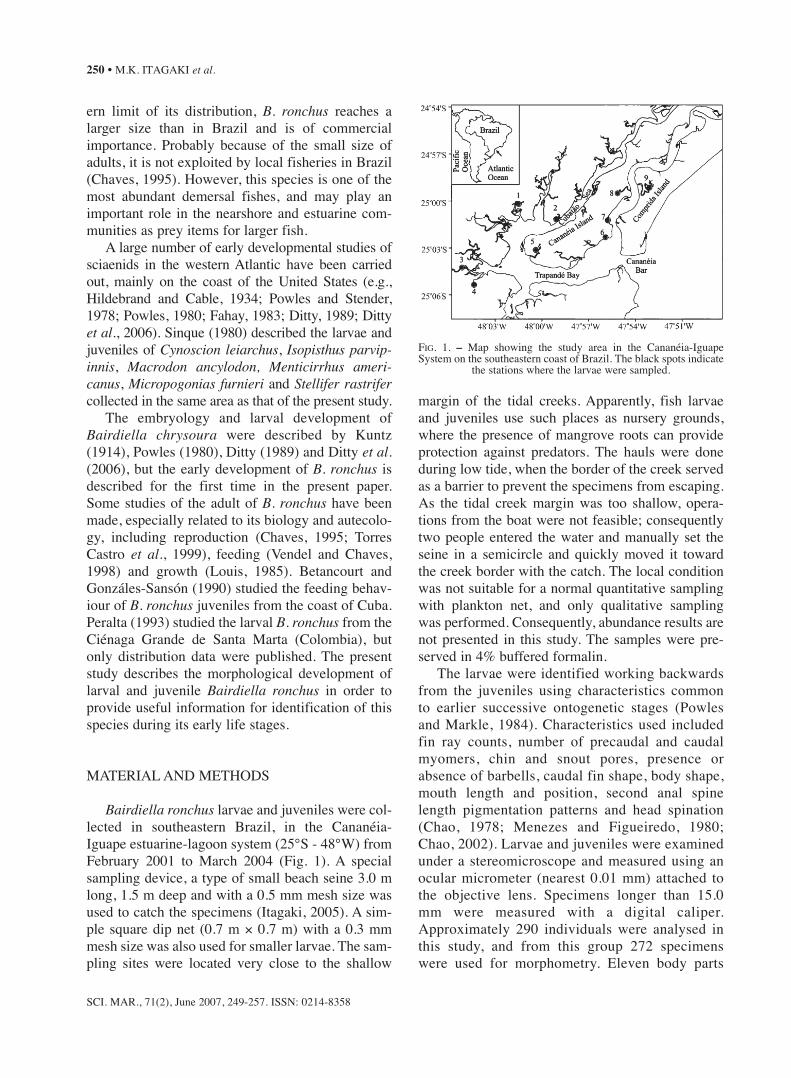
Bairdiella ronchus larvae and juveniles were col-lected in southeastern Brazil, in the Cananéia-Iguape estuarine-lagoon system (25°S - 48°W) fromFebruary 2001 to March 2004 (Fig. 1). A specialsampling device, a type of small beach seine 3.0 mlong, 1.5 m deep and with a 0.5 mm mesh size wasused to catch the specimens (Itagaki, 2005). A sim-ple square dip net (0.7 m × 0.7 m) with a 0.3 mmmesh size was also used for smaller larvae. The sam-pling sites were located very close to the shallow
margin of the tidal creeks. Apparently, fish larvaeand juveniles use such places as nursery grounds,where the presence of mangrove roots can provideprotection against predators. The hauls were doneduring low tide, when the border of the creek servedas a barrier to prevent the specimens from escaping.As the tidal creek margin was too shallow, opera-tions from the boat were not feasible; consequentlytwo people entered the water and manually set theseine in a semicircle and quickly moved it towardthe creek border with the catch. The local conditionwas not suitable for a normal quantitative samplingwith plankton net, and only qualitative samplingwas performed. Consequently, abundance results arenot presented in this study. The samples were pre-served in 4% buffered formalin.
The larvae were identified working backwardsfrom the juveniles using characteristics commonto earlier successive ontogenetic stages (Powlesand Markle, 1984). Characteristics used includedfin ray counts, number of precaudal and caudalmyomers, chin and snout pores, presence orabsence of barbells, caudal fin shape, body shape,mouth length and position, second anal spinelength pigmentation patterns and head spination(Chao, 1978; Menezes and Figueiredo, 1980;Chao, 2002). Larvae and juveniles were examinedunder a stereomicroscope and measured using anocular micrometer (nearest 0.01 mm) attached tothe objective lens. Specimens longer than 15.0mm were measured with a digital caliper.Approximately 290 individuals were analysed inthis study, and from this group 272 specimenswere used for morphometry. Eleven body parts
SCI. MAR., 71(2), June 2007, 249-257. ISSN: 0214-8358
250 • M.K. ITAGAKI et al.
FIG. 1. – Map showing the study area in the Cananéia-IguapeSystem on the southeastern coast of Brazil. The black spots indicate
the stations where the larvae were sampled.
were measured—body depth (BD), snout length(SnL), head length (HL), snout to anus (Sn-A),snout to anal fin origin (Sn-AO), caudal peduncledepth (CPD), eye diameter (ED), upper jaw length(UJL), anus to anal fin gap (GAP) and pre-dorsalfin length (PdL)—using the terminology andmethodology of Ditty (1989), Powles (1980),Sinque (1980), Leis and Trnski (1989) and Moser(1996). The flexion larvae measurements, fromthe tip of the snout to the end of the notochord,were expressed as notochord length (NL), where-as the post-flexion larvae and juvenile measure-ments, from the tip of snout to the posterior edgeof the developing hypurals, were expressed asstandard length (SL). The mean and standarddeviation of each measurement were estimated foreach developmental stage.
Larvae were categorised as flexion or postflexionstage, according to the state of notochord flexion(Ahlstrom et al., 1976). Larvae at the pre-flexionstage were not collected. The juvenile was definedgenerally as a pre-reproductive individual that ismorphologically similar to the adult with completefin ray elements and squamation (Moser, 1996). Thepresence of the rostral fold and shin and snout poreswas classified as upper, marginal and mental,according to Chao (1978) and head spination wasclassified according to (Moser, 1996 and Neira etal., 1998). Larvae and juveniles were illustratedwith a camera lucida. Some specimens were stainedwith Rose Bengal to highlight the details of mor-phological structures.
The specimens used in this study were deposited atthe Ichthyoplankton Laboratory of the OceanographicInstitute of the University of São Paulo.
RESULTS
Morphology
From the flexion to juvenile stage, B. ronchushad large heads and moderate body depth. Therewere 25 myomeres, including the urostyle (11 pre-caudal and 14 caudal). Except for the ratio of thedistance from anus to anal fin gap (GAP) vs bodylength (NL or SL) and the ratio of pre-dorsal finlength (PdL) vs body length, all other body propor-tions tended to increase from flexion to postflexionstage (Table 1). These increases were greater forhead length (HL), snout-to-anus distance (Sn-A),
SCI. MAR., 71(2), June 2007, 249-257. ISSN: 0214-8358
DESCRIPTION OF LARVAE AND JUVENILES OF BAIRDIELLA RONCHUS • 251
TA
BL
E1.
– B
ody
prop
ortio
ns (
%)
in r
elat
ion
to B
L (
n: n
umbe
r of
spe
cim
ens,
SD
: sta
ndar
d de
viat
ion)
of
Bai
rdie
lla
ronc
hus.
Mea
sure
men
t abb
revi
atio
ns e
xpla
ined
in M
ater
ial a
nd M
etho
ds.
Stag
eE
DSn
LU
JLH
LB
DPd
LSn
-ASn
-AO
GA
PC
PDn
Flex
ion
Mea
n ±S
D
12.2
±0.5
8.1±
0.7
15.8
±1.0
33.5
±2.5
34.4
±1.3
41.6
±7.5
47.8
±2.4
60.6
±2.2
12.8
±2.3
8.7±
0.9
n=30
Max
. – M
in.
13.1
-11.
39.
2-6.
517
.9-1
3.6
37.5
-25.
536
.4-3
0.9
60.1
-30.
952
.0-4
3.2
64.3
-56.
617
.0-8
.310
.3-6
.7nE
D=
29
n=10
4Po
sfle
xion
Mea
n ±S
D12
.6±0
.88.
8±1.
018
.6±1
.338
.0±2
.135
.0±1
.438
.8±2
.553
.6±3
.563
.8±1
.710
.4±3
.310
.1±0
.5nE
D=
103
Max
. – M
in.
14.3
-10.
710
.7-6
.220
.7-1
5.6
44.0
-32.
738
.3-3
0.1
54.2
-32.
759
.1-3
3.6
67.1
-57.
428
.2-5
.211
.1-8
.8nS
n-A
O=
102
nCPD
=10
3
n=96
Tra
nsfo
rmat
ion
Mea
n ±
SD11
.0±0
.58.
1±1.
017
.3±1
.038
.5±1
.833
.4±1
.437
.8±2
.158
.8±2
.165
.2±1
.56.
5±1.
310
.5±0
.5nU
JL=
95M
ax. –
Min
.12
.3-9
.911
.1-6
.620
.0-1
3.8
44.6
-34.
437
.5-3
0.1
43.1
-29.
863
.1-5
2.7
68.8
-59.
710
.6-2
.311
.5-9
.6nB
D=
94nG
ap=
95
Juve
nile
Mea
n ±
SD
10.8
±0.6
8.7±
0.7
16.1
±1.2
38.6
±2.0
32.6
±1.4
35.9
±1.9
60.3
±1.8
67.7
±2.3
7.4±
1.5
10.8
±0.3
n=42
Max
. – M
in.
12.2
-9.8
10.0
-7.1
18.6
-13.
843
.7-3
5.4
35.0
-29.
641
.3-3
3.5
63.
6-5
6.3
72.2
-63.
710
.4-4
.411
.3-1
0.3
distance from snout to anal fin origin (Sn-AO), andcaudal peduncle depth (CPD). In all these cases, theincreases were also continuous until the juvenilestage. HL increased rapidly from 33.5% NL (sd =2.5) in the flexion stage to 38.0% SL (sd = 2.1) inthe postflexion stage, and then slowly through thetransformation, reaching the maximum of 38.6%SL (sd = 2.0) in the juvenile stage. The Sn-A ratioincreased from 47.8% NL (sd = 2.4) in the flexionstage to 53.6% SL (sd = 3.5) in the postflexionstage, and to the maximum of 60.3% SL (sd = 1.8)in the juvenile stage. The Sn-AO ratio increasedconstantly from 60.6% NL (sd = 2.2) in the flexionstage to 63.8% SL (sd = 1.7) in the postflexionstage, and then to 67.7% SL (sd = 2.3) in the juve-nile stage. The CPD ratio increased from 8.7% NL(sd = 0 .9) in the flexion stage to 10.1% SL (sd =0.5) in the postflexion stage, and to 10.8% SL (sd=0.3) in the juvenile stage.
The relationship of body depth (BD) to bodylength ranged between a minimum of 29.6% esti-mated in the juvenile stage and a maximum of38.3% estimated in the postflexion larvae. Thesmaller specimens tended to be slightly deeper than
the larger ones, as the BD ratio increased from34.4% NL (sd = 1.3) in the flexion stage to 35.0%SL (sd = 1.4) in the postflexion stage, but thendecreased to 33.4% SL (sd = 1.4) in the transfor-mation stage, and to 32.6% SL (sd= 1.4) in thejuvenile stage. The SnL ratio increased from 8.1%NL (sd = 0.7) in the flexion stage to 8.8% NL (sd= 1.0) in flexion larvae and to 8.8% SL (sd = 1.0)in postflexion larvae. Thereafter, the ratiodecreased to 8.1% SL (sd = 1.0) in the transforma-tion stage, and then increased slightly to 8.7% SL(sd = 0.7) in the juvenile stage.
The ratio of the distance from anus to anal fingap (GAP) decreased from the maximum of 12.8%NL (sd = 2.3) observed in the flexion stage to 10.4%SL (sd = 3.3) in the postflexion stage, then declinedto 6.5% SL (sd = 1.3) in the transformation stage andthereafter tended to increase, reaching 7.4% SL (sd= 1.5) in the juvenile stage (Table 1). The visceralmass was almost triangular and reached the mid-body at about the transformation size. The ratio ofpre-dorsal fin length (PdL) decreased continuouslyfrom flexion larvae (41.6% NL, sd = 7.5) to thejuvenile stage (35.9% SL, sd = 1.9).
SCI. MAR., 71(2), June 2007, 249-257. ISSN: 0214-8358
252 • M.K. ITAGAKI et al.
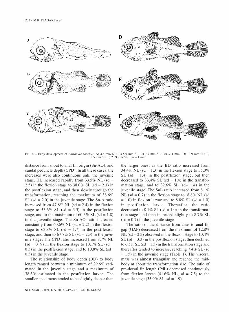
FIG. 2. – Early development of Bairdiella ronchus: A) 4.6 mm NL; B) 5.9 mm SL; C) 7.9 mm SL. Bar = 1 mm.; D) 13.9 mm SL; E) 18.5 mm SL; F) 23.9 mm SL. Bar = 1 mm

Head spination
The major head spines became visible early inthe larval stages (Fig. 2). By 4.4 mm NL, threeposterior preopercular spines, as well as a smallsupraocular ridge, were present. The number ofposterior preopercular spines had increased to 5 by8.0 mm SL (Table. 2), and by 18.5 mm SL a seriesof 6-7 spines had extended as a serrate preopercu-lar margin with strong spines at its angle, similarto those of adults (Fig. 2E-F). The supraocularridge enlarged and became slightly serrated in lar-vae larger than 6.5 mm SL (Fig. 2), but tended todisappear by the juvenile stages. The anterior pre-opercular spines were first observed only in larvaelarger than 5.0 mm NL and most frequently num-bered 1-2 spines. During development, the numberof anterior preopercular spines increased to 5 at8.0 mm SL, occasionally attaining 6 spines duringthe transformation and juvenile stages (Table 2).At 5.0 mm NL a single small post-temporal spinewas first observed, but the numbers of these spinesincreased to 2 in larvae larger than 10.0 mm SL,then to 4-5 in individuals larger than 13.5 mm SL.
Fin development
In the smallest larvae examined (4.4 mm NL),the pectoral fin buds were present and the pectoralfin rays were first observed by 6.2 mm SL. By 8.5mm SL, all specimens possessed the full comple-ment of 16-17 rays (Table 2). The caudal fin wasin process of forming by 4.4 mm NL, when theanlagen of hypurals and 7 principal fin rays were
first observed; from 5.5 mm SL on, all specimenspossessed the full complement of principal caudalrays (9+8) (Table 2); procurrent caudal fin raysbegan to form by 5.0 mm NL, and by 10.1 mm SLall individuals showed the 9+7 pattern. The cau-dal fin was initially rounded and then becameincreasingly truncated during the transformationand juvenile stages (Fig. 2D-F). The anlagen ofthe second dorsal fin and anal fin were visible by4.4 mm SL; development of the first rays of thesecond dorsal and anal fins began simultaneouslyby 4.6 mm NL; from 5.9 mm SL on, the full com-plement of anal fin rays (2 spines and 7-8 rays)was attained, and all elements of the second dor-sal fin (21-24 rays) were observed in individualsfrom 5.7 mm SL on. The first dorsal fin began toform by 5.1 mm NL, when the first spines becamevisible, and all specimens from 7.0 mm SLonward showed the full complement (XI). Thepelvic fins were observed as anlagen by 4.8 mmNL, but development of the pelvic fin rays onlybegan by 6.4 mm SL, and the complete count of 1spine and 5-6 rays was present in all specimensfrom 7.4 mm SL onward. The sequence of findevelopment was: caudal, second dorsal and anal,pelvic, first dorsal and pectoral. However, the finswere completely formed in a different order: prin-cipal caudal, second dorsal, anal, first dorsal,pelvic and pectoral. The second anal-fin spine,which was nearly the same size as the first anal-fin spine by 5.9 mm SL, increased substantiallyduring the development, reaching nearly 80% ofthe size of the first anal fin ray by 12.5–12.5 mmSL (mean = 79.2%; sd = 5.5).
SCI. MAR., 71(2), June 2007, 249-257. ISSN: 0214-8358
DESCRIPTION OF LARVAE AND JUVENILES OF BAIRDIELLA RONCHUS • 253
TABLE 2. – Number of fin spines, soft rays and head spines of Bairdiella ronchus larvae. Roman numeral indicates the number of fin spines. Dashed line indicates the limit between notochord flexion (above) and postflexion (below) stages.
BL(mm) Anal First Second Caudal Pectoral Pelvic Anterior Posterior Post- MyomeresFin Dorsal Fin Dorsal Fin Fin Fin Fin Preopercular Preopercular temporal
4.4 + + (1 + 6) + 3 11+14=254.6 5 8 (8 + 8) + + 3 11+14=254.8 8 17 (8 + 8) + + + 3 11+14=255.1 I + 8 II 20 (8 + 8)1 + + + 3 1 11+14=255.5 I + 7 III 20 (9 + 8)1 + + 3 4 1 11+14=256.2 II + 8 VI 22 (9 + 8)1 2 + 2 4 1 11+14=256.4 II + 7 XI 22 4(9 + 8)4 6 I + 3 4 4 1 11+14=256.5 II + 8 XI 23 4(9 + 8)3 13 I + 2 4 4 1 11+14=257.0 II + 8 XI 22 3(9 + 8)3 12 I + 4 3 4 1 11+14=257.9 II + 8 XI 23 6(9 + 8)5 13 I + 5 4 4 1 11+14=258.0 II + 8 XI 23 7(9 + 8)6 16 I + 5 5 5 1 11+14=258.8 II + 8 XI 23 7(9 + 8)5 16 I + 5 5 5 1 11+14=259.2 II + 8 XI 22 8(9 + 8)6 16 I + 5 4 4 1 11+14=259.6 II + 8 X 24 8(9 + 8)7 16 I + 5 4 5 1 11+14=2510.1 II + 8 XI 22 9(9 + 8)7 16 I + 5 5 5 2 11+14=25
Pigmentation
Larvae of B. ronchus were moderately pigment-ed during the flexion stage, but their pigmentationincreased markedly during the transition from post-flexion to juvenile (Fig. 2). A characteristic “swath”of wide, heavy pigmentation extended from the napeto the cleithral symphysis, covering most of the lat-eral part of the trunk, anterior to the anus (Figs. 2A,B). The swath was especially dark on the surface ofthe visceral mass. A branch of this pigmentationextended laterally as a strip behind the eye from theorbit margin (Fig. 2B). Occasionally, during the lateflexion stage, this pigmentation became less distinctdue to melanophore contractions or thickening ofthe body wall. During the postflexion stage (by 7.9mm SL) the swath almost disappeared, and themelanophores spread throughout the body, mainlyon the dorsal surface of the trunk and almost to thefirst half of the body (Fig. 2C).
Two melanophores, one at the angle of the lowerjaw and the other on the posterior surface of the den-tary, were observed from the early flexion (Fig. 2A),but later the pigmentation expanded to the premaxil-la and throughout the lower jaw (Fig. 2C-F). Severalmelanophores on the anterior surface of the midbrainand at the junction of the mid- and hindbrain began toappear by the early post flexion stage (Fig. 2B), butincreased thereafter covering the entire dorsal portionof the head by the transformation stage (Fig. 2D).During flexion and postflexion, two (occasionallythree) melanophores formed, independently of theswath, along the ventral midline between the cleithralsymphysis and the anus (Fig. 2).
At flexion stages a series of melanophores werepresent along the ventral midline of the tail, two ante-rior to the anal fin base, one at the anal fin base ter-minus (occasionally expanding to occupy a large por-tion of the ventral tail area), and three posterior to theanal fin base (Fig. 2A). Melanophores were absentalong the dorsal midline of the tail during the flexionstage, except for a rare melanophore located on thedorsal fin base (Fig. 2A), but a series of melanophoreswas present along the second dorsal fin base and cau-dal peduncle during the postflexion stage (Fig. 2C)and pigmentation along the lateral midline becameevident during the postflexion stage (Figs 2C, D).
The caudal fin pigmentation began with one tinymelanophore present laterally on the hypural lobe by4.4 mm NL (Fig. 2A), increasing in number of pig-ments and spreading along the hypural margins in
older specimens (Fig. 2D, F). Pigmentation on thefirst dorsal fin was first visible only during the post-flexion stage (Fig. 2C), when several melanophoresspread over the fin surface, being especially patchyin the superior region. Pigmentation on the anal fin,as well as on the second dorsal fin began to form lateduring the transformation and juvenile stages; a sin-gle melanophore on the second spine of the anal finwas visible by 18.5 mm SL (Fig. 2E), and only by23.9 mm SL (Fig. 2F) was a series of pigmentsobserved on the second dorsal fin.
DISCUSSION
The larval and juvenile B. ronchus were identifiedby a set of characteristics: the presence of swath pig-mentation; meristic counting including number ofprecaudal and caudal myomeres (11+14) and fins(first dorsal= XI-XII; second dorsal= 23 – 24; anal =II, 8; pectoral = 17); and morphological characteristicincluding compressed oblong body with triangularand compact visceral mass, rounded to rhomboid cau-dal fin, stout second anal spine, oblique and nearlysub-terminal mouth, and shin with pores in post-flex-ion stages but lacking barbells in early juveniles.
Flexion and early postflexion larvae heredescribed are clearly separated from otherSciaenidae larvae occurring in the area by the pres-ence of swath pigmentation. The swath pigmenta-tion pattern is very similar to that described in B.chrysoura by Powles and Stender (1978) andPowles (1980). We think that the presence of swathpigmentation is a useful diagnostic characteristic fordistinguishing Bairdiella larvae from other sciaenidspecies, and as B. ronchus is the only species of thisgenus inhabiting the area, the presence of this pig-mentation allows these larvae to be identified asbelonging to this species.
Postflexion and early juvenile B. ronchus can bedistinguished from other sciaenids inhabiting south-eastern Brazil by the number of precaudal and cau-dal myomeres and counts of fin elements (Table 3),except for Cynoscion microlepidotus and Umbrinacanosai. Although there is no description of earlystages of C. microlepidotus and U. canosai, a seriesof characteristics given by Matsuura and Nakatani(1979), Sinque (1980), and Ditty et al. (2006) indi-cate that Cynoscion postflexion larvae and juvenileshave a more elongated body than B. ronchus (exceptC. leiarchus and C. nothus); in general they have
SCI. MAR., 71(2), June 2007, 249-257. ISSN: 0214-8358
254 • M.K. ITAGAKI et al.

12-14 + 13-11 myomeres (except C. nothus), mod-erate to large postemporal spines (small in B.ronchus), a large mouth with a prominent lower jaw,moderate to large teeth and a chin without pores.The presence of a rigid barbell on the chin and amoderate second spine of the anal fin separate thejuvenile Umbrina from B. ronchus. In addition, B.ronchus specimens from transforming stage to earlyjuveniles (>14 mm SL) already have the followingadult morphological characteristics: compressedoblong body; sub-terminal and moderate obliquemouth, chin without barbells, pores in the snout, anda rostral fold; preopercle with two series of spines,one of small spines along the anterior margin, andone of small to moderate spines along the posteriormargin with the largest spine at its angle (>20.0 mmSL); stout second anal spine with approximately thesame length as the first soft ray, basal halves of thesoft dorsal and anal fin scaled at 28.0 mm SL; cau-dal fin slightly rounded to rhomboid; and a lateralline extending to the middle of the caudal fin at 20.0mm SL (Chao, 2002; Menezes and Figueiredo,1980). These adult characteristics and the meristicdata obtained from the analysed specimens provideenough information for correctly identifying thelarger B. ronchus of the present study.
Though all morphological characteristics of thetransformation stage and early juveniles are similarto those described for adults (Chao, 2002), thelargest individual (24.0 mm SL), nonetheless, differsfrom them. For instance, the adults have a morepointed head, and the anal fin base is much less thanhalf of the dorsal fin base. In body proportions, theratios in relative body length tend to decrease andthen increase gradually, except in a few cases. Thistrend suggests that the body shape of the juvenile(<24.0 mm SL) is different from that of the adult.Among some adult characteristics, only the rostralfold with the marginal pore inside (>6.0 mm SL) andthe dorsal base longer than the anal base (>8.0 mmSL) can be seen in the larval stage.
In southeastern Brazil, larvae of Stellifer ras-trifer, Cynoscion leiarchus, Isopisthus parvipinnis,Macrodon ancylodon, Menticirrhus americanus,and Micropogonias furnieri have been previouslyidentified (Pearson, 1929; Sinque, 1980; Ditty,1994) and are known to co-occur with larvae of B.ronchus in the Cananéia-Iguape estuarine-lagoonsystem region (Sinque, 1980). Larvae of Pogoniascromis (Itagaki, umpubl. data) can also be observedin the study area. Among these sciaenids, the larvalB. ronchus is more similar to S. rastrifer during the
SCI. MAR., 71(2), June 2007, 249-257. ISSN: 0214-8358
DESCRIPTION OF LARVAE AND JUVENILES OF BAIRDIELLA RONCHUS • 255
TABLE 3. – Summary of selected meristic characteristics and early life stage information for 27 Sciaenidae species of southeastern Brazilian coastal waters.
Species First Dorsal Second Dorsal Anal Pectoral Vertebrae Egg Larvve Juvenile
Bairiella ronchus XI-XII 23-24(21-25) II, 8(7-9) 17(16-18) 11+14 = 25 unknown x xCtenosciaena gracilicirrhus XI 21-23(20-24) II, 7- 8(9) 15-16 10+15 = 25 unknown unknown unknownCynoscion acoupa XI 18-20(17-23) II, 8(7-9) 17-18 12-13+13-12 = 25 unknown x unknownCynoscion jamaicensis XI 23-25(23-27) II, 9(8-10) 17 13+12 = 25 unknown unknown unknownCynoscion leiarchus X-XI 21-23(20-24) II, 11(10-12) 18 (17-19) 25 unknown x xCynoscion microlepidotus XI 23-24(22-25) II, 9(8-10) 20(18-21) 25 unknown unknown unknownCynoscion striatus XI 18-21 II, 8-9 16-18 unknown unknown unknown unknownCymoscion virescens XI 27-31 II, 8(7-9) 17 14+11 = 25 unknown unknown unknownEquetus lanceolatus XIII-XIV 46-50(44-55) II, 6(5-7) 15-16 10+15 = 25 unknown unknown unknownIsopisthus parvipinnis VIII-IX 18-20(21-22) II, 18 - 20(16-17) unknown 11+14 = 25 unknown x unknownLarimus breviceps X-XI 26-28(24-29) II,6-7 unknown 11+14 = 25 unknown unknown unknownMacrodon ancylodon XI 27-30 II, 8-9(10) 16 13+12 = 25 unknown x xMenticirrhus americanus XI 20-21(22-26) I, 7(6-8) ≤20(18-24) 10+15 = 25 unknown x xMenticirrhus littoralis XI 22-25(21-26) I, 7(6-8) ≤19(18-21) 10+15 = 25 unknown unknown xMicropogonias furnieri XI 26-28(26-30) II, 7-8 17-19 10+15 = 25 unknown x xNebris microps IX 31-33 II, 9-10 unknown unknown unknown unknown unknownOdontoscion dentex XII-XIII 22-27 II, 8-10 13-15 12+13 = 25 unknown unknown unknownOphioscion punctatissimus XI 23-24 II, 6-7(8) 18 10+15 = 25 unknown unknown unknownParalonchurus brasiliensis XI 28-31 II, 8(7-9) unknown 11+18 = 28 unknown unknown unknownPareques acuminatus X-XI 36-41 II, 7-8(6) 16-17 10+15 = 25 unknown unknown unknownPogonias cromis XI 21-23(19-23) II, 6(5-7) 18 10+14 = 24 x x xStellifer sp.B XIII-XIV 20-21 II, 8-9 unknown 10+15 = 25 unknown unknown unknownStellifer brasiliensis XI 21-22 II, 9 18-19 10+15 = 25 unknown unknown unknownStellifer rastrifer XI-XIII 21-23 II, 9(8) 18-20 10+15 = 25 unknown x xStellifer stellifer XII 18-20 II, 8 unknown 10+15 = 25 unknown unknown unknownUmbrina coroides XI 27-29(26-31) II, 6 17(16-18) 11+14 = 25 unknown x xUmbrina canosai XI 21-25 II, 7-8 unknown unknown unknown unknown unknown
Menezes and Figueiredo, 1980; Ditty and Shaw,1994; Ditty et al., 2006

early larval stages (< 6.0 mm SL), mainly when theswath pigmentation is contracted. The same wasobserved in the northern Gulf of Mexico and on theAtlantic coast of the USA, where B. chrysoura lar-vae can be confused with Stellifer lanceolatus larvae(Chao, 2002). Smaller larvae of B. ronchus can alsobe distinguished from S. rastrifer by the presence ofinternal pigmentation in the otic region and the pig-mentation embedded in the musculature of the nape(Table 4). Another difference is that in the larval S.rastrifer the post-anal lateral pigments and caudalpigments on the ventral and dorsal midline radiateinternally and externally towards the lateral midline,reaching the lateral pigmentation. In B. ronchussuch radiation was observed only externally, andpost-anal lateral pigments were absent.
B. chrysoura at 4.0 mm NL can be distinguishedfrom S. lanceolatus by eye diameter (Powles, 1980;Ditty, 1989): the proportion observed in B. chrysourais usually above 10% body length and that in S.lanceolatus under 10% body length. The same fea-ture could be observed between B. ronchus, whoseeye diameter proportion ranged from 10.7 to 14.3%,and S. rastrifer whose eye diameter proportionranged from 7.4 to 9.3% (Itagaki, unpublished data).
The compact triangular visceral mass proved tobe a useful character for distinguishing B. ronchusfrom other sciaenids, except for larval S. rastrifer.Ditty (1989) reported that B. chrysoura smallerthan 5 mm SL could be distinguished from mostother sciaenid species by their preanal length(<46% SL), except for S. lanceolatus. It was diffi-cult to analyse this relationship because of the smallnumber of B. ronchus smaller than 5.0 mm NL.Development of larvae smaller than 4.0 mm NL(preflexion) was not described in the present study.
In addition to its main diagnostics, B. ronchuslarvae can be easily differentiated from the co-occurring sciaenid larvae, distinct from S. ras-trifer, by a series of diagnostic characteristics pre-sented in Powles and Stender, 1978, Sinque (1980)and Ditty et al. (2006) (Tables 3 and 4). Flexionand postflexion larvae of Cynoscion leiarchus canbe distinguished from those of B. ronchus by alarger number of head spines, and by characteris-tic internal and external melanophores around theposterior surface of the visceral mass. Isopisthusparvipinnis posflexion larvae have a supra ocularspine, while those of B. ronchus have a slightlysupraocular ridge; in addition, Isopisthus parvip-innis can be distinguished from other sciaenids bya very high anal ray count and low dorsal raycount. Larvae of Macrodon ancylodon can be dis-tinguished from B. ronchus and other Scianidaefrom early stages by a prominent supraocciptalcrest. Flexion stages of Menticirrhus americanuscan be differentiated from those of B. ronchus andother sciaenids by characteristic rows of ventraland lateral midline pigmentation, multiple napesurface pigmentation, and lack of pigment anteriorto cleithral symphysis. In the posflexion,Menticirrhus americanus larvae became heavilypigmented over the head and trunk. LarvaeMicropogonias furnieri can be distinguished fromB. ronchus and other sciaenid genera by their lackof pigmentation on the anterior margin of visceralmass. Finally, the preflexion and flexion Pogoniascromis larvae can be distinguished from B.ronchus by their enlarged melanophores along thedorsal and ventral midlines and stream-lined bodyshape, and postflexion larvae can be distinguishedby their low anal element count.
SCI. MAR., 71(2), June 2007, 249-257. ISSN: 0214-8358
256 • M.K. ITAGAKI et al.
TABLE 4. – Pigmentation in 9 Sciaenidae species during the flexion stage: (D) dentary; (ALJ) angle lower jaw; (LBE) strip laterally behind theeye; (IUH) internal under hindbrain; (AVM) anterior visceral mass; (OGB) over gas bladder; (SNAPE) nape surface; (NAPE) embedded in themusculature of nape; (PMV) posterior visceral mass; (I) isthmus; (CS) anterior to cleithral symphysis; (LM) lateral midline; (DML) dorsal mid-line; (VG) ventral on gut, (CVM) caudal ventral midline; (+) present; (-) absent; (±) some.° this study; *Ditty, J. 1989; **Sinque, C.
1980; ***Ditty, J. 1994.
Species D ALJ Swath PMV I CS LM DML VG CVMLBE IUH AVM OGB SNAPAE NAPE
Bairdiella ronchus º + + + + + + + + + - + - ± ± ±Bairdiella chrysoura * + + + + + + + + - - + - ± ± ±Stellifer rastrifer ** - + - + + + + - + + ± ± ± ±Isopisthus parvipinnis **Macrodon ancylodon ** - - + - - + - ± ±Cynoscion leiarchus ** - + - + - + + + + + - ± -Menticirrhus americanus ** + + - + + + + + + ± ± +Pogonias cromis *** - + - - + + - + + - + ± +Micropogonias furnieri ** + + - + + + + ± + - ± ± ±
ACKNOWLEDGEMENTS
The authors would like to dedicate this paper to DrYasunobu Matsuura, who passed away in May 2003.We wish to thank Dr J.J.B. Alba for his help translat-ing the summary to Spanish, and the two anonymousreferees and the editor for their constructive criticismand suggestions. Financial support for this study camefrom the Fundação de Amparo à Pesquisa do Estado deSão Paulo (FAPESP).
REFERENCES
Aguilar-Betancourt, C. and G.G. Gonzáles-Sansón. – 1990.Alimentación natural de juveniles de Bairdiella ronchus(Cuvier) en un área de la plataforma noroccidental de Cuba.Rev. Invest. Mar., 11(1): 35-39.
Ahlstrom, E.H., J.L. Butler and B.Y. Sumida. – 1976. Pelagic stro-mateoid fishes (Pisces, Perciformes) of the eastern Pacific:kinds, distributions and early life histories and observations onfive of these from the Northwest Atlantic. Bull. Mar. Sci., 26:285-402.
Chao, L.N. – 1978. A basis for classifying western AtlanticSciaenidae (Teleostei: Perciformes). NOAA Tech. Rep., NMFSCirc., 415:1-64.
Chao, L.N. – 2002. Sciaenidae. Croakers (drums). In: K.E.Carpenter (ed.). The Living Marine Resources of the WesternCentral Atlantic. FAO Species Identification Guide for FisheryPurposes and American Society of Ichthyologists andHerpetologists. Spec Publs FAO, 5(3): 1583-1653.
Chaves, P.T.C. – 1995. Atividade reprodutiva de Bairdiella ronchus(Cuvier) (Pisces, Sciaenidae) na baía Guaratuba, Paraná, Brasil.Rev. Bras. Zool. 12(4): 759-766.
Ditty, J.G. – 1989. Separating early larvae of Sciaenidae from theWestern North Atlantic: a review and comparison of larvae offLouisiana and the Atlantic coast of the U.S. Bull. Mar. Sci.,44(3): 1083-1105.
Ditty, J.G. and R.F. Shaw. – 1994. Preliminary guide to the identi-fication of the early life history stages of Sciaenid fishes fromthe Western Central Atlantic. NOAA Tech. Memo. NMFS-SEFSC-349: 1-118.
Ditty, J.G.; R.F. Shaw and T. Farooqi. – 2006. Sciaenidae. In: W.J.Richards (ed.). Early stages of Atlantic Fishes: An identifica-tion guide of the Western Central North Atlantic. 2: 1669-1723.CRC Taylor and Francis. Boca Raton.
Fahay, M.P. – 1983. Guide of early stages of marine fishes occur-ring in the western North Atlantic Ocean, Cape Hatteras to thesouthern Scotian shelf. J. NW Atl. Fish. Sci., 4: 1-423.
Froese, R. and D. Pauly (eds.). – 2006. FishBase. World Wide Webelectronic publication. www.fishbase.org, accessed in 14August 2006.
Hildebrand, S.F. and L.E. Cable. – 1934. Reproduction and devel-opment of whitings or kingfishes, drums, spot, croaker, and
weakfishes or sea trouts, Family Sciaenidae, of the Atlanticcoast of the United States. Bull. US Bur. Fish., 48: 41-117.
Itagaki, M.K. – 2005. Potencial de recrutamento das larvas e juve-nis de robalo-peva, Centropomus paralleus (Teleostei:Centropomidae), no Sistema Cananéia-Iguape, São Paulo,Brasil. Ph.D. thesis, Instituto Oceanográfico, Univ. São Paulo.
Kuntz, A. – 1914. The embryology and larval development ofBairdiella chrysoura and Anchoa mitchilli. Bull. US Bur. Fish.,33: 1-19.
Leis, J.M. and T. Trnski. – 1989. The larvae of Indo-Pacific shore-fishes. University of Hawaii Press. Hawaii.
Louis, M. – 1985. Reproducion et croissance de Bairdiella ronchus(Poisson Sciaenidae) dans les mangroves de Guadeloupe(Antilles françaises). Rev. Hydrobiol. Trop., 18(1) 1985: 61-72.
Matsuura, Y. and K. Nakatani – 1979. Ocorrência de larvas e jovensde peixes na Ilha Anchieta (SP) com algumas anotações sobremorfologia de castanha, Umbrina coroides. Bol. Inst.Oceanogr., São Paulo, 28: 165-183.
Menezes, N.A. and J.L. Figueiredo. – 1980. Manual de peixes marin-hos do sudeste do Brasil. Museu de Zoologia, Universidade deSão Paulo, São Paulo/CNPq. IV. Teleostei (3): 42-59.
Menezes, N.A.; P.A. Buckup; J.L. Figueiredo and L.M. de Moura.– 2003. Catálogo das espécies de peixes marinhos do Brasil.Museu de Zoologia, Universidade de São Paulo.
Moser, H.G. – 1996. Introduction. In: H.G. Moser, ed. The earlystages of fishes in the California Current region. CalCOFI.Allen Press, Lawrence. Atlas 33: 1-72.
Neira, F.J.; A.G. Miskiewicz and T. Trnski. – 1998. Larvae of tem-perate Australian fishes. University of Western Australia Press.Nedlands.
Pearson, J.C. – 1929. Natural history and conservation of red fishand other commercial sciaenids of the Texas coast. Bull. USBur. Fish. 44: 129-214.
Peralta, R.H.L. – 1993. Estudio preliminar del ictioplâncton de laCiénaga Grande de Santa Rita (Caribe colombiano). Bol. Inst.Oceanogr. Venezuela, Universidad. Oriente, 32(1/2): 79-90.
Powles, H. – 1980. Descriptions of larval silver perch, Bairdiellachrysoura, banded drum, Larimus fasciatus, and star drum,Stellifer lanceolatus (Sciaenidae). Fish. Bull., U.S., 50: 79-102.
Powles, H. and B.W. Stender. – 1978. Taxonomic data on the earlylife history stages of Sciaenidae of the South Atlantic Bight ofthe United States. S. C. Mar. Resour. Center Tech. Rep., 31.
Powles, H. and D.F. Markle. – 1984. Identification of Larvae. In:H.G. Moser; W.J. Richards; D.M. Choen; M.P. Fahay; A.W.Kendall Jr. and S.L. Richardson (eds). Ontogeny and systemat-ics of fishes, pp. 31-33. Spec. Pub. Nº. 1. Allen Press.Lawrence, Kansas.
Sinque, C. – 1980. Larvas de Sciaenidae (Teleostei) identificadas naregião estuarino-lagunar de Cananéia. Bol. Zool., Univ. S.Paulo, 5: 39-77.
Torres Castro, L.; A.S. Santos Martínez and A. Acero P. – 1999.Reproducción de Bairdiella ronchus (Pisces: Sciaenidae) en laCiénaga Grande de Santa Marta, Caribe Colombiano. Rev. Biol.Trop., 47(3): 553-560.
Vendel, A.L. and P.T.C. Chaves. – 1998. Alimentação de Bairdiellaronchus (Cuvier) (Perciformes, Sciaenidae) na baía deGuanabara, Paraná, Brasil. Rev. Bras. Zool., 15(2): 297-305.
Scient. ed.: M.P. Olivar.Received May 2, 2006. Accepted January 2, 2007.Published online May 21, 2007.
SCI. MAR., 71(2), June 2007, 249-257. ISSN: 0214-8358
DESCRIPTION OF LARVAE AND JUVENILES OF BAIRDIELLA RONCHUS • 257
