derivation of endothelial colony forming cells from … · kapur, and jason meyer, for there...
TRANSCRIPT
DERIVATION OF ENDOTHELIAL COLONY FORMING CELLS FROM
HUMAN CORD BLOOD AND EMBRYONIC STEM CELLS
J. Luke Meador
Submitted to the faculty of the University Graduate School in partial fulfillment of the requirements
for the degree Master of Science
in the Department of Biochemistry and Molecular Biology, Indiana University
August 2013

ii
Accepted by the Faculty Indiana University, in partial fulfillment of the requirements for the degree of Master of Science.
_______________________________ Mervin C. Yoder MD, Chair
_______________________________ Reuben Kapur PhD
Master’s Thesis Committee
_______________________________ Timothy W. Corson PhD
_______________________________ Jason S. Meyer PhD

iii
Dedication
To my wife Kaitlin

iv
Acknowledgements
I would like to express my gratitude to my mentor Dr. Mervin C. Yoder for his
commitment to my education. He not only provided me with expert advice and guidance,
but always managed to do so in a positive way which helped me to excel in the program.
His commitment to graduate student education is unparalleled due to his commitment to
the balance of work and life.
I would like to thank Monica Henry and everyone in the Interdisciplinary
Biomedical Gateway program for their assistance in finding the right laboratory during
our first year in the program and setting me up for success in the subsequent years.
I would like to thank the members of my advisory committee: Tim Corson, Reuben
Kapur, and Jason Meyer, for there insight and guidance as I formulated my project and
learned the skills necessary to be successful.
I would also like to thank my colleagues in the IBMG program and the
Biochemistry Department for their friendship and willingness to study late into the night.
I have formed many lifelong friendships during my time at IUSM.
I am continually grateful to my parents, Ramona Meador and Bruce Meador, who
helped shape me into person I am today. I would like to thank my in-laws, Samuel Vogt
and Adina Vogt, who have helped my wife and me in every aspect as we started our life
together. And most importantly, I would like to thank the love of my life, Kate Meador,
who has done everything for me and who I am eternally grateful for.
v
J. Luke Meador
DERIVATION OF ENDOTHELIAL COLONY FORMING CELLS FROM HUMAN
CORD BLOOD AND EMBRYONIC STEM CELLS
Endothelial Colony Forming Cells (ECFCs) are highly proliferative endothelial
progenitor cells with clonal proliferative potential and in vivo vessel forming ability.
While endothelial cells have been derived from human induced pluripotent stem cells
(hiPS) or human embryonic stem cells (hES), they are not highly proliferative and require
ectopic expression of a TGFβ inhibitor to restrict plasticity. Neuropilin-1 (NRP-1) has
been reported to identify the emergence of endothelial precursor cells from human and
mouse ES cells undergoing endothelial differentiation. However, the protocol used in that
study was not well defined, used uncharacterized neuronal induction reagents in the
culture medium, and failed to fully characterize the endothelial cells derived. We
hypothesize that NRP-1 expression is critical for the emergence of stable endothelial cells
with ECFC properties from hES cells. We developed a novel serum and feeder free
defined endothelial differentiation protocol to induce stable endothelial cells possessing
cells with cord blood ECFC-like properties from hES cells. We have shown that Day 12
hES cell-derived endothelial cells express the endothelial markers CD31+ NRP-1+,
exhibit high proliferative potential at a single cell level, and display robust in vivo vessel
forming ability similar to that of cord blood-derived ECFCs. The efficient production of
the ECFCs from hES cells is 6 logs higher with this protocol than any previously
published method. These results demonstrate progress towards differentiating ECFC from

vi
hES and may provide patients with stable autologous cells capable of repairing injured,
dysfunctional, or senescent vasculature if these findings can be repeated with hiPS.
Mervin C. Yoder MD, Chair
vii
Table of Contents
List of Tables ................................................................................................................... viii
List of Figures .................................................................................................................... ix
Abbreviations ..................................................................................................................... xi
Introduction ........................................................................................................................1
Chapter I: Phenotypic and Functional Characterization of Endothelial Colony
Forming Cells Derived from Human Umbilical Cord Blood .........................................7
A. Abstract ....................................................................................................................8
B. Reagents and Solutions ..........................................................................................10
C. Protocol Text ..........................................................................................................13
D. Figures....................................................................................................................22
E. Discussion ..............................................................................................................26
Chapter II: Comparison of Derived Endothelial Cells from
Pluripotent Stem Cells .....................................................................................................27
A. Introduction ............................................................................................................28
B. Materials and Methods ...........................................................................................31
C. Results ....................................................................................................................36
D. Figures....................................................................................................................45
E. Discussion ..............................................................................................................58
References .........................................................................................................................61
Curriculum Vitae

viii
List of Tables
Table 1. Reagent List .........................................................................................................12
ix
List of Figures
Figure 1. Isolation of mononuclear cells (MNCs) from cord blood (CB)
and outgrowth of endothelial colony forming cells (ECFCs) from
cultured MNCs ...................................................................................................................22
Figure 2. Representative in vitro phenotypic assessment of endothelial
and hematopoietic cell surface antigen expression ............................................................23
Figure 3. Representative in vitro quantitation of the clonogenic and
proliferative potential of CB derived ECFCs .....................................................................24
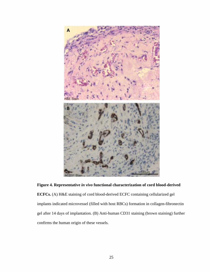
Figure 4. Representative in vivo functional characterization of
CB-derived ECFC ..............................................................................................................25
Figure 5. Characterization of EB derived hES-endothelial cells .......................................45
Figure 6. EB derived hES-endothelial cells are largely non-dividing and
LPP .....................................................................................................................................47
Figure 7. CD31 and NRP-1 expression in EB differentiation of hES-
endothelial cells and their proliferative potential ...............................................................48
Figure 8. EB derived NRP-1+ CD31+ hES-EC exhibit characteristic
CB-ECFC morphology in vitro and capillary-like network forming
ability on Matrigel coated plates ........................................................................................49
Figure 9. Optimization of hES differentiation into ECFC .................................................50
Figure 10. Kinetic Analysis of H9 directed Differentiation ..............................................51
Figure 11. Efficiency of expansion of hES-ECFCs ...........................................................52
x
Figure 12. CD31 NRP-1 double positive isolated cells do not express
α-SMA and replicate CB-ECFC proliferative potential ....................................................53
Figure 13. CD31 NRP-1 double positive isolated cells form in vitro
networks and vessels in vivo ..............................................................................................54
Figure 14. H9 hESC derived ECFC in vitro analysis of phenotype and
network formation ..............................................................................................................55
Figure 15. H9 hESC derived ECFC immunohistochemical kinetic
analysis of endothelial antigen expression .........................................................................56
Figure 16. Late Passage Analysis of hESC derived ECFCs ..............................................57
xi
Abbreviations
APC ............................................................................................................ Allophycocyanin
BMP ......................................................................................... Bone Morphogenetic Factor
BMP4 .................................................................................... Bone Morphogenetic Factor 4
CD31 ........................................................... Platelet Endothelial Cell Adhesion Molecule-1
CD144 .................................................................................. Vascular Endothelial Cadherin
CD146 .......................................................................... Melanoma Cell Adhesion Molecule
CLI ................................................................................................... Critical Limb Ischemia
EB ................................................................................................................ Embryoid Body
EC ............................................................................................................... Endothelial Cell
ECFC................................................................................ Endothelial Colony Forming Cell
EMT .......................................................................... Epithelial to Mesenchymal Transition
ES ........................................................................................................ Embryonic Stem Cell
FACS........................................................................... Fluorescence Activated Cell Sorting
FBS ........................................................................................................Fetal Bovine Serum
FITC ............................................................................................Fluorescein Isothiocyanate
FGF ............................................................................................... Fibroblast Growth Factor
hES ........................................................................................Human Embryonic Stem Cells
hPS ........................................................................................Human Pluripotent Stem Cells
IHC ................................................................................................... Immunohistochemistry
IPS ......................................................................................... Induced Pluripotent Stem Cell
MHC .......................................................................Major Histocompatability Cell Antigen

xii
NRP-1 ............................................................................................................... Neuropilin-1
PAD............................................................................................ Peripheral Arterial Disease
PE .................................................................................................................... Phycoerythrin
Pen/Strep ..........................................................................................Penicillin/Streptomycin
SMAD ............................................................... Portmanteau for genes (MAD) and (SMA)
TGFβ ............................................................................... Transforming Growth Factor Beta
VEGF .......................................................................... Vascular Endothelial Growth Factor

1
INTRODUCTION
2
Endothelial colony forming cells (ECFCs) are rare circulating endothelial cells
with robust clonal proliferative potential that display intrinsic in vivo vessel forming
ability[1-7]. ECFCs, also called blood outgrowth endothelial cells (BOEC)[8], have been
shown to be directly transplantable in sex-mismatched human bone marrow transplant
patients with the most proliferative circulating BOEC displaying genetic marking of the
donor marrow[8, 9]. While this study undoubtedly proves the direct transplantable nature
of these cells (without any intervening period of in vitro culture), it does not fully explain
the cell of origin within the transplanted donor marrow that gives rise to the BOEC, the
engraftment site of the BOEC in the host, or the mechanisms with which the cells are
mobilized into the systemic circulation. Given the known residence of ECFC within the
vascular endothelial intima[3], it is plausible that blood vessel endothelial cells may be
the resident niche for the circulating ECFC. ECFC rapidly mobilize into the blood stream
following an experimental myocardial ischemic event[10] and in human subjects, the
concentration of circulating ECFC correlates with the severity of coronary artery
occlusion and chronic myocardial ischemia[11, 12]. When cultured ECFCs are injected
intravenously into rodent vascular injury models, they are rapidly recruited into the site of
vascular injury or tissue ischemia to orchestrate the initiation of a vasculogenic
response[13-17]. In numerous animal models of human disease, ECFCs have been
reported to enhance vascular repair and improve blood flow following myocardial
infarction[18, 19], stroke[13], ischemic retinopathy[20, 21], ischemic limb injury[14-17],
and to engraft and re-endothelialize denuded vascular segments or implanted grafts. The
ability to increase the microvascular density within these regenerating tissues heralds
ECFC as one of the most promising cell-based strategies for therapeutic

3
revascularization. While cultured and implanted ECFCs form durable and functional
human blood vessels in vivo, they are rare in number in peripheral blood (1/107-108
peripheral blood mononuclear cells)[1, 2, 8, 12, 22, 23] and their number tends to decline
with age and disease in human[11, 24, 25] and non-human primates[26]. In elderly
patients and subjects with peripheral arterial disease (PAD) and critical limb ischemia
(CLI), circulating or resident ECFC may become prone to replicative senescence lacking
proliferative potential, thus, rendering them impotent for autologous vascular repair.
Thus, it is necessary to find an alternate source of healthy ECFCs that may be used for
vascular repair in these subjects.
Human pluripotent stem cells (human embryonic stem [hES] and induced
pluripotent stem [hiPS] cells) display virtually unlimited self-renewal capacity and ability
to differentiate into any cell types in our body[27-29] and these cells offer the opportunity
to derive ECFC for vascular repair. Both hES and hiPS cells have been reported to
differentiate into cells of the endothelial lineage[30-42], however the derived endothelial
cells are in some cases, unstable and are reported to drift to various non-endothelial
phenotypes[34, 43] or exhibit low proliferative potential with a proclivity to reach
replicative senescence within 5-7 passages[43-45]. Indeed, there is no published evidence
for derivation of ECFC that display the properties of high clonal proliferative potential
with in vivo vessel forming capacity from hES or hiPS cells.
Here we describe a novel hES cell differentiation protocol that can reproducibly
generate a homogenous and stable population of endothelial cells with ECFC properties
that are similar to cord blood ECFC. With our simple one step 2D serum- and stromal
cell-free differentiation protocol, we were able to consistently generate over a trillion
4
endothelial cells in less than 3 months of culture; an efficiency that has not been
approached by any of the previously published endothelial differentiation protocols. This
method involves initiation of endothelial lineage differentiation by growing hPS cells in
serum-free endothelial differentiation media supplemented with growth factors for 12
days on Matrigel coated dishes. On day 12, cells were harvested and sorted using
antibodies that recognize several well-established endothelial antigens. Only the NRP-
1+CD31+ subset of cells exhibited ECFC properties, including robust in vivo vessel
formation when implanted in immune-deficient mice. These ECFCs were not
transformed and could be implanted for >3 months without evidence of teratoma
formation. We have also determined that KDR activation via addition of a NRP-1-Fc
dimer and VEGF165 is required for the derivation and emergence of ECFCs from hES
cells. Overall, our studies reveal a novel paradigm for derivation of endothelial cells with
ECFC properties from hES cells via activation of a specific molecular pathway. Our
innovative strategy to produce clinically relevant numbers of ECFC with in vivo vessel
forming cell potential should be tested in hiPS cells and if successful, may soon permit
trials to regenerate the deficient microvasculature that may underlie the most prominent
deficit for vascular repair in patients with cardiovascular disease.
Novel Endothelial Colony Forming Cell Marker
Vascular endothelial cadherin (CD144) was at one time thought to be a specific
marker for vascular endothelial cells. However it has since been discovered that CD144 is
present on other cell types as well, such as hematopoietic cells[46]. Previous publications
on the derivation of endothelial cells employed the use of this marker to isolate the
5
endothelial cell types. Possibly extraneous cell types may have been isolated using this
process and restricted the ability of cells to reinforce cellular signaling and fully commit
the endothelial lineage. Because of their use of this marker inhibitors were necessary to
maintain the desired phenotype. Thus we set out to use a novel marker for the isolation of
ECFCs.
Neuropilin1 (NRP-1) is a VEGF coreceptor which is able to synergistically
upregulate the downstream signaling of some VEGF receptors[47]. NRP-1 homozygous
deficient mice demonstrate greatly diminished yolk sac vasculature, disorganized somatic
blood vessels, and this defect results in embryonic lethality by E12.5-13.5. In 2009
Cimato et al reported that NRP-1 was an early marker for endothelial cells predating
CD144 expression during endothelial cell differentiation from hES cells[33]. It has also
been shown that NRP-1 is crucial for the development of vascular endothelial cells. We
set out to use NRP-1 antibodies in concert with other known endothelial antigens to
develop a system to specifically isolate endothelial cells. While we show NRP-1 to be
essential for the specification of ECFCs it does not appear to be necessary for the
maintenance of the ECFC lineage and is not correlated with proliferative capacity in later
passages.
Aim of this Study
Our primary goal was to obtain highly proliferative endothelial cells with all of
the ECFC hallmarks. This goal required that I verify the techniques in which ECFCs are
obtained and characterized from human umbilical cord blood. I was fortunate to be
involved with writing a review protocol on the isolation, expansion, and characterization
6
of ECFCs during my first few months in the lab. This review was published in the
Journal of Visualized Experiments. This publication allowed me to perfect the techniques
necessary to perform my work on our current project, which will be submitted soon.
Chapter I discusses the isolation, derivation, and characterization of cord blood
derived ECFCs. We report optimized procedures for extracting ECFCs from Cord blood,
methods to culture and purify a homogenous population of ECFCs, and provide protocols
to characterize ECFC functions in vitro and in vivo. This work provides the foundation
and basis of comparison for all subsequent work on deriving ECFCs from hPS cells.
Chapter II discusses the major focus of my work on developing and optimizing a
novel protocol for isolating, expanding, and characterizing hES derived ECFCs. By
modifying several existing protocols we developed a novel robust protocol to efficiently
isolate ECFCs with stable phenotypes through many passages without loss of functions
including in vivo vessel forming potential. To date this is the only protocol for deriving
stable highly proliferative ECFCs from pluripotent stem cells.
7
Chapter I: Phenotypic and Functional Characterization of Endothelial Colony
Forming Cells Derived from Human Umbilical Cord Blood
8
Abstract
Longstanding views of new blood vessel formation via angiogenesis,
vasculogenesis, and arteriogenesis have been recently reviewed[48]. The presence of
circulating endothelial progenitor cells (EPCs) were first identified in adult human
peripheral blood by Asahara et al. in 1997[49] bringing an infusion of new hypotheses
and strategies for vascular regeneration and repair. EPCs are rare but normal components
of circulating blood that serve as sites of blood vessel formation or vascular remodeling,
and facilitate either postnatal vasculogeneis, angiogenesis, or arteriogenesis largely via
paracrine stimulation of existing vessel wall derived cells[50]. No specific marker to
identify an EPC has been identified, and at present the focus of the field is to understand
that numerous cell types including proangiogenic hematopoietic stem and progenitor
cells, circulating angiogenic cells, Tie2+ monocytes, myeloid progenitor cells, tumor
associated macrophages, and M2 activated macrophages participate in stimulating the
angiogenic process in a variety of preclinical animal model systems and in human
subjects in numerous disease states[51, 52]. Endothelial colony forming cells (ECFCs)
are rare, circulating viable endothelial cells characterized by robust clonal proliferative
potential, secondary and tertiary colony forming ability upon replating, the ability to form
intrinsic in vivo vessels upon transplantation into immunodeficient mice[1-3]. While
ECFCs have been successfully isolated from the peripheral blood of healthy adult
subjects, umbilical cord blood (CB) of healthy newborn infants, and vessel wall of
numerous human arterial and venous vessels [1-3, 53], CB possesses the highest
frequency of ECFCs[1] that display the most robust clonal proliferative potential and
form durable and functional blood vessels in vivo[2, 4-7]. While the derivation of ECFCs

9
from adult peripheral blood has been presented[8, 54], we describe the methodologies for
the derivation, cloning, expansion, and in vitro as well as in vivo characterization of
ECFCs from human umbilical CB.
10
Reagents and Solutions
EGM-2 media (Lonza, Cat. No. cc-3162 containing EBM-2 basal medium and EGM-
2 SingleQuot kit Supplements, and growth factors)
EBM-2 (Lonza, Cat. No. cc-3156) supplemented with the entire singlequot
kit supplements and growth factors (Lonza, Cat. No. cc-4176), 10% (v/v) fetal
bovine serum (FBS) and 1% (v/v) penicillin (10,000 U/ml)/streptomycin (10,000
µg/ml)/amphotericin (25 µg/ml). Store up to 1 month at 4˚C. Recommended
EGM-2 volumes to use for ECFC culture are 500 µl/well for 24-well plates, 2
ml/well for 6-well plates, 5 ml/25-cm2 flasks, and 10 ml/75-cm2 flasks, unless
otherwise specified in the protocol.
Collagen I solution
Dilute 0.575 ml of glacial acetic acid (17.4 N) in 495 ml of sterile distilled
water (0.02 N final concentration). Sterile filter the dilute acetic acid with a 0.22-
µm vacuum filtration system. Add 25 mg rat tail collagen I to the dilute acetic
acid to a final concentration of 50 µg/ml. The amount of collagen added will vary
depending on the collagen stock concentration. Store up to a month at 4˚C.
Preparation of collagen I-coated tissue culture surfaces
Place 1 ml of collagen I solution in each well of a 6-well tissue culture
plate (use 300 µl/well for 24-well plates, 3 ml/25-cm2 flasks, and 8 ml/75-cm2
flasks). Incubate 1 hour to overnight at 37˚C. Remove the collagen I solution and

11
wash surface two times, each time with PBS (use 500 µl/well for 24-well plates, 2
ml/well for 6-well plates, 5 ml/25-cm2 flasks, and 10 ml/75-cm2 flasks). Use
plates immediately for cell cultures.
FACS staining buffer
Phosphate-buffered saline (PBS) supplemented with 2% (v/v) fetal bovine
serum (FBS). Store up to 2 weeks at 4˚C.
12
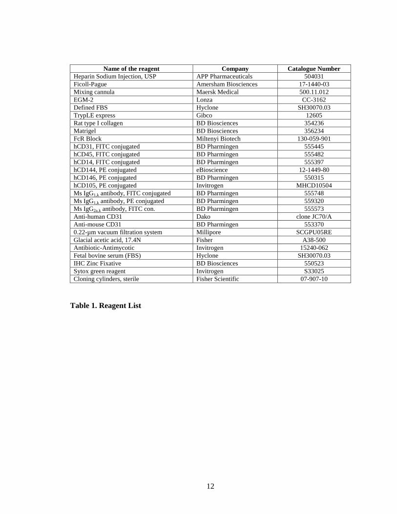
Name of the reagent Company Catalogue Number Heparin Sodium Injection, USP APP Pharmaceuticals 504031 Ficoll-Pague Amersham Biosciences 17-1440-03 Mixing cannula Maersk Medical 500.11.012 EGM-2 Lonza CC-3162 Defined FBS Hyclone SH30070.03 TrypLE express Gibco 12605 Rat type I collagen BD Biosciences 354236 Matrigel BD Biosciences 356234 FcR Block Miltenyi Biotech 130-059-901 hCD31, FITC conjugated BD Pharmingen 555445 hCD45, FITC conjugated BD Pharmingen 555482 hCD14, FITC conjugated BD Pharmingen 555397 hCD144, PE conjugated eBioscience 12-1449-80 hCD146, PE conjugated BD Pharmingen 550315 hCD105, PE conjugated Invitrogen MHCD10504 Ms IgG1,k antibody, FITC conjugated BD Pharmingen 555748 Ms IgG1,k antibody, PE conjugated BD Pharmingen 559320 Ms IgG2a,k antibody, FITC con. BD Pharmingen 555573 Anti-human CD31 Dako clone JC70/A Anti-mouse CD31 BD Pharmingen 553370 0.22-µm vacuum filtration system Millipore SCGPU05RE Glacial acetic acid, 17.4N Fisher A38-500 Antibiotic-Antimycotic Invitrogen 15240-062 Fetal bovine serum (FBS) Hyclone SH30070.03 IHC Zinc Fixative BD Biosciences 550523 Sytox green reagent Invitrogen S33025 Cloning cylinders, sterile Fisher Scientific 07-907-10
Table 1. Reagent List
13
Protocol Text
A.) ECFC Outgrowth, Cloning and Expansion
A.1.1) Collect CB with heparin (10 U heparin/ml blood) or EDTA as an anticoagulant
and transport to the laboratory at room temperature and immediately (within 2 hours of
infant’s delivery) process the sample for mononuclear cell (MNC) isolation.
A.1.2) Aliquot 15 ml of CB into each 50-ml conical centrifuge tube and add 20 ml of
PBS to each tube of CB and pipet several times to mix. Draw up 15 ml Ficoll-Paque into
a 20-ml syringe and attach a 20G needle or mixing cannula. Place the tip of the mixing
cannula at the bottom of the tube of diluted blood and carefully underlay 15 ml Ficoll-
Paque. Centrifuge the tubes 30 min at 740 x g, at room temperature, without the
deceleration brake setting.
A.1.3) Using a transfer pipet, remove the hazy layer of low-density MNCs located at the
interface between the Ficoll-Paque and diluted plasma. Dispense the MNCs into a 50-ml
conical tube containing 10 ml EGM-2 medium. Centrifuge the MNCs 10 min at 515 x g,
at room temperature, with a high deceleration brake setting.
A.1.4) Carefully aspirate and discard the supernatant. Wash the pelleted cells with EGM-
2 twice (10 min at 515 x g) and re-suspend the MNCs in EGM-2 at 1.25 x 10⁷ cells/ml.
Prepare collagen I coated 6-well tissue culture plates by adding 1 ml rat-tail collagen type
I (50 µg/ml) per well and incubating the plate overnight. Pipet 4 ml of this MNC
suspension into each well and place the cells in 37˚C, 5% CO2 humidified incubator.
A.1.5) After 24 hours (day 1), slowly remove the spent medium (do not disturb the
loosely adherent cells) from the well with a pipet, slowly add 4 ml EGM-2 to the well,
14
and return plate to the incubator. On day 2, refresh the medium by slowly removing the
medium (do not disturb the loosely adherent cells) from the well with a pipet and adding
4 ml EGM-2 to each well. Repeat the medium change daily until day 7 and every other
day thereafter until ECFC colonies appear for cloning. Typically colonies appear between
day 5 and day 14[1] (Fig. 1).
A.2.1) Visualize typical ECFC colonies with cobblestone morphology using inverted
microscope (magnification 10x) and mark the colony boundaries with a marker on the
underside of the well to indicate the position of each colony.
A.2.2) On day 14, wash these primary colonies 2 times with PBS and harvest individual
colonies using cloning cylinders and just enough TrypLE express to cover the cells inside
the cylinders. After aspirating the final wash of PBS, place a sterile gel coated cloning
cylinder around each colony and press firmly against the plate using sterile forceps. Add
2-3 drops of warm TrypLE express into each cloning cylinder and incubate for 3-5 min
until cells within the cylinder begin to detach. Add approximately 250 µl EGM-2
medium into the center of the cylinder and pipet up and down to generate single cell
suspension. Transfer the single cell suspension from each cloning cylinder separately into
an individual micro-centrifuge tube.
A.2.3)Wash the area within the cylinder 2-3 times with approximately 250 µl EGM-2
medium to collect all remaining cells and transfer the wash from each cylinder into the
respective micro-centrifuge tube. Centrifuge the cells at high speed (≤300 x g) on
tabletop centrifuge for 5 min.
A.2.4) Remove the supernatant and re-suspend the cell pellet in 1.5 ml fresh EGM-2
medium. Transfer the cell suspension (containing all the cells from individual colonies)

15
from each tube into one well of a 24-well tissue culture plate pre-coated with 500 µL rat-
tail collagen I (50 µg/ml).
A.2.5) Place inside the incubator for expansion with media change performed every other
day.
A.3.1) When cells approach 80-90% confluency, remove the spent medium then wash the
cells 2 times with PBS and add 500 µl TrypLE express medium to each well of 24-well
tissue culture plate. Place the plate inside the incubator for 3-5 min until cells begin to
round up and detach. Add 1 ml of EGM-2 (with 10% defined FBS) to each well to collect
the cells by washing and transfer them to a 15 ml tube. Wash the well with 1 ml EGM-2
medium to collect all remaining cells and transfer the wash from each well into respective
15 ml tube. Centrifuge the cells at 300 x g for 5 min.
A.3.2) Remove the medium and re-suspend the cell pellet in 1 ml fresh EGM-2 medium
(with 10% defined FBS). Obtain the viable cell count of an aliquot using a
hemacytometer and trypan blue exclusion.
A.3.3) Expand the cells by seeding about 5000 cells/cm2 onto a collagen I pre-coated
tissue culture surfaces in EGM-2 media with media change every other day until cells
approach 80-90% confluency before sub-culturing again.
B.) In vitro phenotypic characterization of ECFCs: Endothelial cell surface antigen
expression and single cell assay for clonal proliferative potential
B.1.1) For endothelial cell surface antigen expression, detach cells with TrypLE express
to collect cells by using methods described for ECFC cloning and expansion.
16
B.1.2) Re-suspend cells in FACS staining buffer (10 X 106 cells/ 1 ml FACS staining
buffer) then add FcR blocking reagent (20 µl/ 10 X 106 cells), mix gently and place on
ice for 10 min.
B.1.3) Aliquot 100 µl of this cell suspension in micro-centrifuge tube for each endothelial
surface antigen and isotype controls. Add appropriate amount of fluorochrome labeled
human monoclonal antibodies that recognize endothelial antigens (CD31, CD144,
CD146, and CD105) or hematopoietic antigens (CD45, and CD14) and leave on ice for
30 min protected from light. Caution: 1 X 10⁶ cells are not required for each test. The
authors typically prepare 0.5 to 1 X 10⁵ cells/tube to obtain 2 X 10⁴ analyzed events.
Also, follow the manufacturer’s recommendation for the amount of antibody to be used
for each test. Some antibodies may require titrating to determine the optimal antibody
concentration. All staining that the authors have performed are single color staining.
B.1.4) After 30 min of incubation on ice, centrifuge each tube at high speed on a tabletop
centrifuge for 5 min then remove the supernatant and re-suspend the cell pellet in FACS
buffer.
B.1.5) Analyze the samples on a flow cytometer to determine the percentage of cells that
stain positively or negatively for each antigen. ECFCs stain uniformly positive for CD31,
CD144, and CD146, but negative for CD45 and CD14 compared to the corresponding
isotype controls.
B.2.1) For single cell assay to examine the clonal proliferative potential, detach early
passage (2-3) cells with TrypLE express to collect cells using methods similar to that was
described for ECFC cloning and expansion.
17
B.2.2) Infect these cells with enhanced green fluorescent protein (EGFP) expressing
lentiviruses[55] and collect cells expressing EGFP by fluorescence cytometry. Caution:
Working with lentvirus is considered a biohazard and all bio-safety precautions should
be obeyed.
B.2.3) Culture them as described for culturing ECFC in the section for ECFC expansion.
B.2.4) Prepare Collagen I coated 96-well plates by adding 50 µl rat-tail collagen type I
(50 µg/ml) per well and incubate the plate for overnight. Collect the EGFP+ ECFCs by
using TrypLE express and resuspend them in EGM-2 medium with final concentration
~106 cells /ml.
B.2.5) Use FACS Vantage (Becton Dickson) or other comparable sorter with low flow
rate of 20 cells/second to sort one single EGFP+ ECFC per well of a 96-well plate and
adjust the final volume to 200 µl per well with EGM-2. Use an inverted fluorescence
microscope to ensure that each well received only one cell. Alternatively, single cells can
be sorted based on FSC and SSC and Sytox nuclear dye staining the colonies can be used
for cell scoring and quantification.
B.2.6) Incubate the plate over a period of two weeks with two media changes (200 µl
EGM-2 using multichannel pipetter) performed on day 5 and day 10. On day 14 of
culture, wash each well with 100 µl PBS before fixing the cells with 100 µl of 4%
paraformaldehyde for 30 min. For ECFCs that are not expressing EGFP, add Sytox
reagent, a green fluorescent nuclear dye, following paraformaldehyde fixation and
incubate at 4 ˚C overnight.
B.2.7) Use a fluorescence microscope to examine each well to quantitate the number of
endothelial cells that expanded from a single cell cultured for 14 days. For quantitative
18
analysis, score wells with 2 or more endothelial cells as positive (for proliferation) and
examine them further for total endothelial cell count by visual counting with the
fluorescence microscope.
B.2.8) To display the obtained data, wells with an endothelial cell number of: 2 to 50, 51-
2000, and 2001 or more, are considered as: endothelial cell clusters, low proliferative
potential (LPP)-ECFCs, and high proliferative potential (HPP)-ECFCs respectively.
ECFCs exhibit complete hierarchy of clonal proliferative potential by giving rise to
endothelial clusters, LPPs, and HPPs when cultured at a single cell level for 14 days[1].
C.) In vivo functional characterization of ECFCs: in vivo vessel formation assay to
examine the ECFC’s potential for vasculogenesis
C.1.1) Prepare cellularized gel implants by calculating the total volume (ml) of each of
the following gel materials (with the final concentration in the parenthesis) needed to cast
1-ml gel (which will be later bisected to generate 2 implants): FBS (10%), EBM-2 10:1
(adjust the final volume to 1 ml), sodium bicarbonate (1.5 mg/ml), NaOH (adjust pH to
7.4), HEPES (25 mM), fibronectin (100 µg/ml), and collagen I (1.5 mg/ml).
C.1.2) After gel material calculation, detach cells with TrypLE express to collect cells
using methods similar to that was described for ECFC cloning and expansion. Obtain a
viable cell count of an aliquot using a hemacytometer and trypan blue. Transfer 2.4
million viable cells into a 50 ml-conical tube and pellet the cells by centrifugation at 515
x g, room temperature. Simultaneously, prepare gel matrix solution by adding calculated
volumes of HEPES, sodium bicarbonate, EBM-2 10:1, FBS, fibronectin, and collagen to
19
an ice-cold 50-ml conical tube (in the same order as they are listed). Mix thoroughly and
adjust the pH to 7.4 with NaOH while keeping solution on ice.
C.1.3) Discard the supernatant from the cells after centrifugation and re-suspend the
pellet in 360 µl warm EGM-2 and to it add pH adjusted 840 µl of gel matrix solution,
making the final volume of gel implant 1 ml. Mix the cells into gel matrix solution slowly
until cells are thoroughly suspended in the gel solution.
C.1.4) Transfer this 1 ml cellularized gel suspension to one well of a 12-well tissue
culture plate and incubate for 20-30 min until the gel polymerizes. Gently cover the gel
with 2 ml warm EGM-2 and incubate overnight.
C.1.5) Immediately before the implantation, bisect the overnight incubated gel into two
equal pieces using iris scissors and return the gel pieces to the same culture well
containing EGM-2 medium.
C.1.6) Use the animal surgical facility to sedate the animal (5-6 week old NOD/SCID
mice) by administrating isoflurane anesthesia. Shave the lower part of abdomen and
thoroughly clean the surgical site with alcohol pads.
C.1.7) Using sterile sharp iris scissors to make an approximately 5 mm incision in the
lower quadrant of the abdomen, exposing the subcutaneous space between the skin and
abdominal muscle. Perform blunt-dissection through the dermal layer from the abdominal
muscle to create a 15-20 mm wide pocket leading superior into the upper abdominal
quadrant. Repeat similar procedure to create similar open pocket in another side of the
same mouse abdomen.

20
C.1.8) Insert one half piece of gel into one side of abdominal pocket and another half
piece of gel into another side of the abdominal pocket by lifting the dermal layer just
caudal to the incision.
C.1.9) After insertion of gels, close each incision with 2-3 stitches using 5-0
polypropylene suture on a cutting needle. Appropriately label the cage cards and perform
post-surgical monitoring and analgesia as per the institutional and protocol requirements
and allow 14 days for cells in these implanted gels to generate de novo vasculature.
C.1.10) Finally, harvesting of gel implants is typically performed 14 days after
implantation after euthanizing the mouse.
C.1.11) Swab the abdominal area with alcohol pads and cut the abdominal skin caudal to
the original incision line. Carefully, dissect out the implant by excising a flap of skin
caudal to the probable location of the gel. Excise the implant by cutting circumferentially
around the gel then place in zinc fixative (BD Biosciences, follow manufacturer’s
recommendation for optimal results) and allow them to fix for 1-2 hours at room
temperature.
C.1.12) Prepare the paraffin-embedded gels using standard histochemical protocols and
prepare 5 µm sections on glass slides to perform staining with hematoxylin and eosin,
anti-human CD31 or anti-mouse CD31 to visualize the vasculature within the gel. ECFCs
undergo de novo vasculogenesis to generate humanized vessels in cellularized gel
implants inserted into immunodeficient mice for 14 days[2, 5, 51].
Representative Results
21
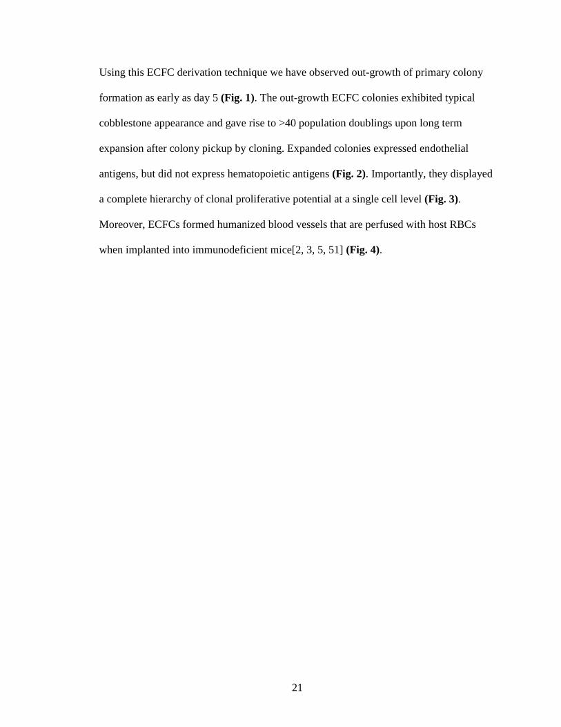
Using this ECFC derivation technique we have observed out-growth of primary colony
formation as early as day 5 (Fig. 1). The out-growth ECFC colonies exhibited typical
cobblestone appearance and gave rise to >40 population doublings upon long term
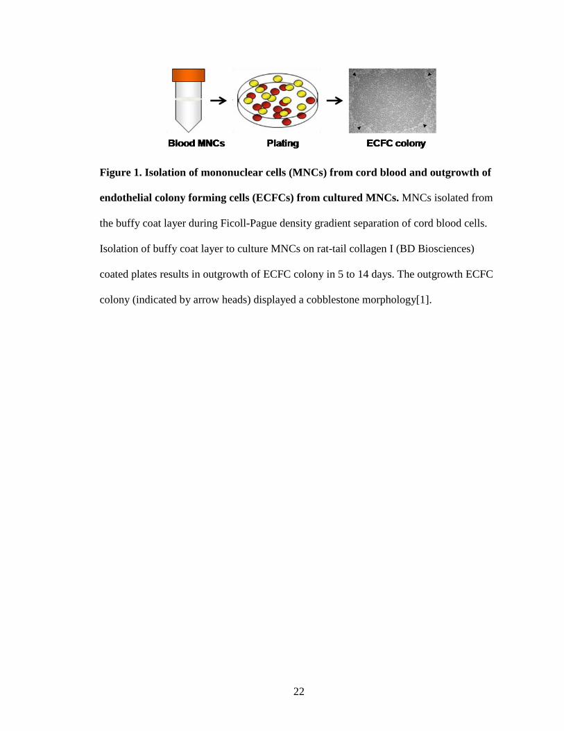
expansion after colony pickup by cloning. Expanded colonies expressed endothelial
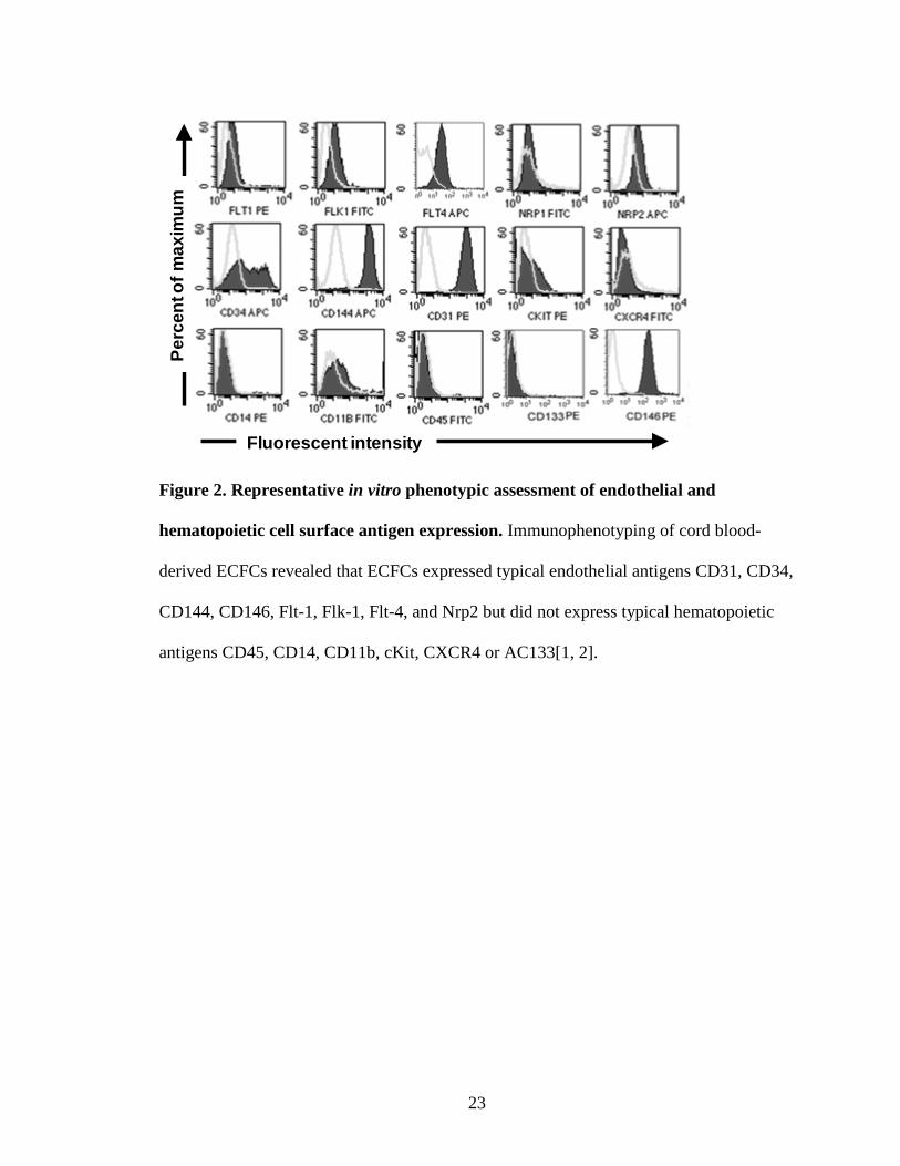
antigens, but did not express hematopoietic antigens (Fig. 2). Importantly, they displayed
a complete hierarchy of clonal proliferative potential at a single cell level (Fig. 3).
Moreover, ECFCs formed humanized blood vessels that are perfused with host RBCs
when implanted into immunodeficient mice[2, 3, 5, 51] (Fig. 4).
22
Figure 1. Isolation of mononuclear cells (MNCs) from cord blood and outgrowth of
endothelial colony forming cells (ECFCs) from cultured MNCs. MNCs isolated from
the buffy coat layer during Ficoll-Pague density gradient separation of cord blood cells.
Isolation of buffy coat layer to culture MNCs on rat-tail collagen I (BD Biosciences)
coated plates results in outgrowth of ECFC colony in 5 to 14 days. The outgrowth ECFC
colony (indicated by arrow heads) displayed a cobblestone morphology[1].
23
Figure 2. Representative in vitro phenotypic assessment of endothelial and
hematopoietic cell surface antigen expression. Immunophenotyping of cord blood-
derived ECFCs revealed that ECFCs expressed typical endothelial antigens CD31, CD34,
CD144, CD146, Flt-1, Flk-1, Flt-4, and Nrp2 but did not express typical hematopoietic
antigens CD45, CD14, CD11b, cKit, CXCR4 or AC133[1, 2].
Fluorescent intensity
Perc
ento
fmax
imum
24
Figure 3. Representative in vitro quantitation of the clonogenic and proliferative
potential of CB derived ECFCs. (A) cord blood-derived ECFCs display clonal
proliferative potential with a hierarchy of colonies ranging from clusters of 2-50 cells up
to colonies of >2001. (B) Micrographs of hierarchy of colonies (colonies stained with,
Sytox, a green fluorescent nuclear dye) obtained after cord blood-derived ECFCs were
cultured at a single cell level for 14 days. Scale bar represents 100µm[1, 2].
25
Figure 4. Representative in vivo functional characterization of cord blood-derived
ECFCs. (A) H&E staining of cord blood-derived ECFC containing cellularized gel
implants indicated microvessel (filled with host RBCs) formation in collagen-fibronectin
gel after 14 days of implantation. (B) Anti-human CD31 staining (brown staining) further
confirms the human origin of these vessels.
A
B

26
Discussion:
Phenotypic and functional characterization of putative endothelial progenitor cells is
important to identify the bona fide ECFCs that are capable of clonally and serially re-
plating in culture and give rise to durable and functional implantable blood vessels in
vivo. Human umbilical cord blood is enriched with ECFCs and the concentration of these
circulating cells declines with aging or disease. Recent studies suggest that ECFC may
play important roles in vascular repair or regeneration in situations of vascular injury,
myocardial infarction, or retinopathy[18, 21, 56].
Here, we have described simple and efficient methodologies for the derivation, cloning,
expansion, and in vitro as well as in vivo characterization of ECFCs from human
umbilical CB. These approaches enable researchers to identify and isolate bona fide
EPCs from CB which possess clonal proliferative potential and in vivo vessel forming
ability.

27
Chapter II: Derivation of Endothelial Colony Forming Cells from Human
Embryonic Stem Cells
28
Introduction
Endothelial dysfunction with resulting atherosclerosis is a fundamental cause of
peripheral arterial disease (PAD) in a large percentage of elderly individuals.
Development of critical limb ischemia (CLI) in patients with PAD is associated with
adverse events such as stroke and myocardial infarction[57-60]. Circulating endothelial
progenitor cells (EPCs) are rare but normal components of circulating blood[49] that are
implicated in the repair of damaged blood vessels such as those found in patients with
PAD. Endothelial colony forming cells (ECFCs) represent those rare circulating EPCs
with robust clonal proliferative potential that display intrinsic in vivo vessel forming
ability[1-3]. While ECFCs form durable and functional blood vessels in vivo, they are
rare in peripheral blood (1/107-108 peripheral blood mononuclear cells) and their number
tends to decline with age and disease[1]. In elderly patients and subjects with PAD, these
cells may become prone to replicative senescence lacking proliferative potential, thus,
rendering them impotent for vascular repair. Developing a protocol for the efficient
derivation and expansion of ECFCs is crucial for their implementation in regenerative
medicine.
While human umbilical cord blood is enriched with ECFCs, use of these cells in
patients with PAD as a cell therapy to repair endothelial dysfunction would constitute an
allogeneic transplant. This predisposes ECFCs for immune-rejection by the host. Human
embryonic stem (hES) cell differentiation into the endothelial lineage has been reported
to generate endothelial cells with mature endothelial phenotypes but low proliferative
potential[44]; indeed there is no published evidence for derivation of ECFC (clonal
proliferative endothelium) from hES cells. While human ES derived cells would also be
29
predisposed for host immune-rejection if not matched for MHC compatibility, identifying
a differentiation pathway for ECFC from the well-studied hES lines is an appropriate
starting point for future application to hiPS cells. While directed differentiation of hES
cells into the endothelial lineage has been reported to generate endothelial cells with a
mature endothelial phenotype possessing low proliferative potential, derivation of
endothelial cells with cord blood ECFC-like properties (progenitor cells with clonal
proliferative potential and in vivo vessel forming ability) from hES cells has not yet been
reported. Papers have even reported that hES and IPS cells may be incapable of
generating proliferative vascular cells[38, 61, 62].
In 2010, Rafii et al. reported that ECs could be derived from hES cells using a
protocol which first involved the formation of EB bodies and isolation by CD144
expression[62]. While this has been the gold standard protocol to date, reports also
indicate that the TGFβ inhibitor had to be present throughout the culture period in order
to restrict the EC from expressing alpha-smooth muscle actin (a protein more commonly
seen in mesenchymal cells). Since CD144 has been reported to be expressed by
mesoderm precursors, hematopoietic cells, and some cultured mesenchymal cells, we
questioned whether the use of CD144 as a marker to enrich for hES-derived EC may lead
to isolation of cells of many lineages.
In 2009 a report by Cimato et al. reported that NRP-1 may be useful in identifying
the emergence of endothelial cells during development from hES[33]. NRP1 is important
in endothelial cell development and acts as a co-receptor for VEGF which helps to
increase downstream signaling and enrich the commitment of cells to endothelial cell
30
fates[63]. Thus, we hypothesized that ECFCs may emerge from mesoderm precursors co-
expressing NRP-1 and CD31.
Here we report that by modifying earlier reported protocols, with addition of a
novel cell surface antigen selection, we have isolated endothelial cells with stable cord
blood ECFC-like properties from hES. Compared to other sub-sets of sorted cells, only
NRP-1+CD31+ cells exhibited ECFC properties. Our results show that NRP-1+CD31+
isolated cells formed a homogenous cell monolayer with characteristic cobblestone
morphology, exhibited clonal proliferative potential, generated capillary like structures
when cultured on Matrigel, and formed robust in vivo vessels when in implanted in
immune deficient mice. We speculate that this method of hES differentiation may also be
effective in isolating ECFC from hiPS cells.
31
Materials and Methods
Culturing of hES and IPS cells
H9 human embryonic stem cells were kindly provided by the Dr. Hal Broxmeyer
lab (IUSM) and were maintained as previously published [64]. Cells were maintained in
mTeSR1 complete media (Stem Cell Technologies, Vancouver, Canada) on Matrigel (BD
Biosciences) in 10 cm2 tissue culture dishes at 37˚C and 5% CO2. After the plating of
cells, media was changed on days 2, 3, and 4. Cells were routinely passaged on day 5.
Passaging of hES and IPS cells
Prior to passaging the hES cells, 10 cm2 tissue culture dishes were pre-coated with
Matrigel for 1 hr. Tissue culture media from the hES cultures was aspirated and 4-5 mL
of 2 mg/mL Dispase (Gibco, Grand Island, New York) was added to the plate. Cells were
then incubated at 37˚C for 3-5 minutes until the edges of the colonies had lifted from the
plate. The Dispase was aspirated from the plate and cells were gently washed with
DMEM-F12 (Gibco) 3 times to remove any lingering Dispase. Fresh media was then
used to harvest the cells from the plate using a forceful wash and scraping with a 5mL
pipette taking care to avoid generation of excessive bubbles. Collected colonies were then
centrifuged at 300xG for 5 min. During centrifugation, 7 mL of mTeSR1 complete media
was added to Matrigel coated dishes to serve as culture medium for the pelleted colonies.
The media was aspirated off of the centrifuged cells and then cells were resuspended in
10 mL of mTeSR1 complete media and added to the Matrigel coated dishes. Cultures
were checked for hES colony morphology quality on day 2.

32
Directed Differentiation
After 2 days of culture in mTeSR1 media, hES cell cultures were directed toward
the mesodermal lineage by pulsing with Activin A (10 ng/mL) under constant signaling
of FGF-2 (Stemgent, Cambridge, Massachusetts), VEGF-165 (R&D, Minneapolis,
Minnesota) and BMP4 (R&D) (10 ng/mL each). On day 1 Activin A containing media
was removed and replaced with 8mL of Stemline II Media (Sigma, San Antonio, Texas)
containing only FGF-2 and BMP4. Media was subsequently changed on days 3, 5, 7, and
8 with 8 mL of differentiation media. On day 9 and thereafter media was changed with 10
mL of differentiation media. This system was interrupted at any time for removal of cells
for FACS analysis.
Flow Cytometry
To release the cells from the plate and the secreted ECM, TryplE (Life
Technologies, Inc., Grand Island, New York) was added until the cells were just covered
(5mL). The plates were then incubated at 37˚C for 5 minutes. In the presence of TryplE,
cells were triturated using a 5 mL pipette and then deposited through a 0.4 micron filter
into a 50 mL conical tube containing 10 mL of EGM2 (Lonza, Walkersville, Maryland).
Tubes were then centrifuged at 300xG for 5 minutes. The supernatant was aspirated
leaving behind the cell pellet. The pellet of cells was resuspended in 1 mL of EGM2.
The cells were then counted and the appropriate amount of FCR block (Miltenyi
Biotech, Auburn, California) was then added to the tube and incubated for 15 min as per
manufacturer guidelines. A portion of the cell mixture was then withheld for an unstained
control while another portion was stained with the appropriate antibodies as per their
33
recommended protocol. Antibodies used included anti-human CD31 (CD31-FITC, clone
WM59 from BD Pharmigen, San Jose, California, CAT # 555445), CD144 (CD144-PE,
clone 16B1 from ebioscience, San Diego, California, CAT# 12-1449-82), and NRP-1
(NRP1-APC, clone AD5176 from Miltenyi Biotech, CAT # 130-090-900). The cells and
antibody were centrifuged at high speed for 30 sec. and the supernatant was removed.
The pelleted cells were resuspended in EGM2 containing propidium iodide (3 ng/mL).
The sample would then be immediately ready for analysis and remain on ice until
analyzed. Samples were analyzed using a FACS Vantage (Becton Dixon) flow cytometer
at a flow rate of 1/min. Compensation was set using cord blood derived ECFC color
controls. Samples were analyzed using flow minus one (FMO) control analysis and the
FloJo analysis software.
Cell Culture of Sorted Cells
Sorted cells were centrifuged at 300xG for 5 minutes then resuspended in 50%
EGM2 and 50% complete Stemline II differentiation media. A 12 well plate was then
coated with rat tail type I collagen (BD Biosciences) and plated with 2500 cells/well.
After 2 days the media was changed to 75% EGM2 and 25% differentiation media. Then
again after 2 days the media was changed to 100% EGM2. After approximately 1 week,
ECFC colonies appear as tightly adherent cells with cobblestone morphology. Cloning of
endothelial cell clusters was performed to isolate pure populations of highly proliferative
endothelial cells. Cells were then split and replated at about 80% confluency and plated at
10,000 cells/ cm2. Cells were then maintained in an endothelial culture system of cEGM-
2 media on collagen coated plates with media changes every other day.
34
In Vitro Network Formation Assay
Matrigel coated 96 well plates (50 µl of Matrigel to each well) were prepared in
triplicate. 10,000 ECFCs were prepared in 50 µl of EGM2 per well. 50 µl of EGM2
containing cells were placed on top of the Matrigel coated surface. Next the plate was
incubated overnight in 37˚C 5% CO2 (12-16 hours). On the next day the plate was
removed and the network formation could be observed under phase contrast microscope.
Immunohistochemistry and Western Blot Analysis
Twelve well plates with sterile glass coverslips on the bottom of the well were
prepared and coated with type 1 rat tail collagen (BD Biosciences). 50,000 ECFC were
added and cultured overnight. When cells formed a subconfluent monolayer they were
processed for fixation using 4% (w/v) paraformaldehyde for 30 min and permeabilized
with 0.1% (v/v) TritonX-100 in PBS for 5 min. After blocking with 10% (v/v) goat serum
for 30 min, cells were incubated with primary antibodies; anti-CD31 (Santa Cruz), anti-
CD144 (ebioscience), anti-NRP-1 (Santa Cruz) and anti-a-SMA, (Chemicon) overnight at
4˚C. Cells were washed with PBS, then incubated with secondary antibodies conjugated
with Alexa-488 or Alexa-565 (Molecular Probe) and visualized by confocal microscopy
after counterstaining with 2 g/ml DAPI (Sigma-Aldrich). The confocal images were
viewed with Olympus FV1000 mpE confocal microscope using as objective 2-photon
and Olympus uplanSapo 60xW/1.2NA/eus. All the images were taken at room
temperature.
35
In Vivo Vessel Formation Assay
Type 1 pig skin collagen master mix for gel implant was prepared by dissolving
lyophilized collagens in 0.01 NHCL and neutralized with 10x PBS and 0.1 N sodium
hydroxide to achieve neutral pH (7.4) and final collagen concentrations ranging from 0.5
to 2.75 mg/ml[65]. The master mix was then mixed with the desired cell number
(500,000 cells/ 250 microliter gel) in a 48 well tissue culture plate. The gel was allowed
to solidify for 30 minutes at 37˚C and 5% CO2 and then 500µl of EGM2 was added. The
cells then incubated overnight in 37˚C 5% CO2 (16-18 hours). The plate was taken the
next day for implantation into NOD/SCID mice flanks. Detailed information on
preparing, implanting, and removing gels after 14 days can be found in JOVE [66]. After
removing gel after 14 days the gel was fixed with 4% paraformaldehyde and samples
were processed for immunohistochemistry by a pathology laboratory for H&E staining
and rat antihuman-CD31 monoclonal staining antibodies which do not cross react with
host murine cells.
Biostatistics
All experiments were performed 3 times in triplicate and data are represented as mean +
standard deviation for statistical comparison. Significance of differences was assessed by
an unpaired t-test at p <0.05.
36
Results
Endothelial cells derived from hES cells using existing published protocols exhibit
low proliferative potential and lack properties of cord blood ECFC
Human endothelial cells have been derived from hPS cells using a variety of
methods. One approach is to promote differentiation of hPS through co-culture with OP9
stromal cells[32, 37-39, 41] or mouse embryonic fibroblasts[61]. Another more recent
approach seeks differentiation of hPS through embryoid body (EB) formation[30, 31, 33-
36, 40, 42, 44, 45, 67, 68] followed by application of various growth factors and/or
receptor signaling pathway inhibitors to promote endothelial cell differentiation. While
each method provides emergence of endothelial cells from the differentiated hPS cells,
many of these studies failed to examine or discuss the proliferative potential of the
endothelial cells produced or if mentioned, the endothelial cells were reported to possess
very low proliferative potential[34, 43, 44] and to undergo cell senescence after only a
few passages[45]. Indeed, none of the prior reports for the differentiation of endothelial
cells from hPS described cells that appeared to display the clonal proliferative potential,
replating capacity, or in vivo vessel forming ability of human cord blood ECFCs.
Therefore, we sought to determine whether endothelial cells derived via the EB formation
method of hES differentiation produces endothelial cells that possess cord blood ECFC
properties. After 7 days of differentiation through EB formation, KDR+NRP-1+ cells were
sorted and cultured in endothelial media as described[33]. When sorted cells were
expanded, we found early passage cells (P.1) gave rise to a heterogeneous population of
cells (Fig. 5), where only a portion of cells appeared to display an endothelial cell
morphology. When these cells were further expanded (p.4), the cultures became
37
predominantly comprised of cells with a fibroblastic-like appearance and little endothelial
cobblestone formation (Fig. 5). When these cells were characterized for endothelial
surface antigen expression using flow cytometric analysis, a heterogeneous pattern of
CD31, CD144, and CD146 expression was exhibited (Fig. 5) with only a portion of the
cells expressing each of these antigens. Similarly when these cells were analyzed for
ability to form vascular like networks on Matrigel they formed vascular like networks
with numerous smaller incomplete branches that were sprout-like in appearance (Fig. 5).
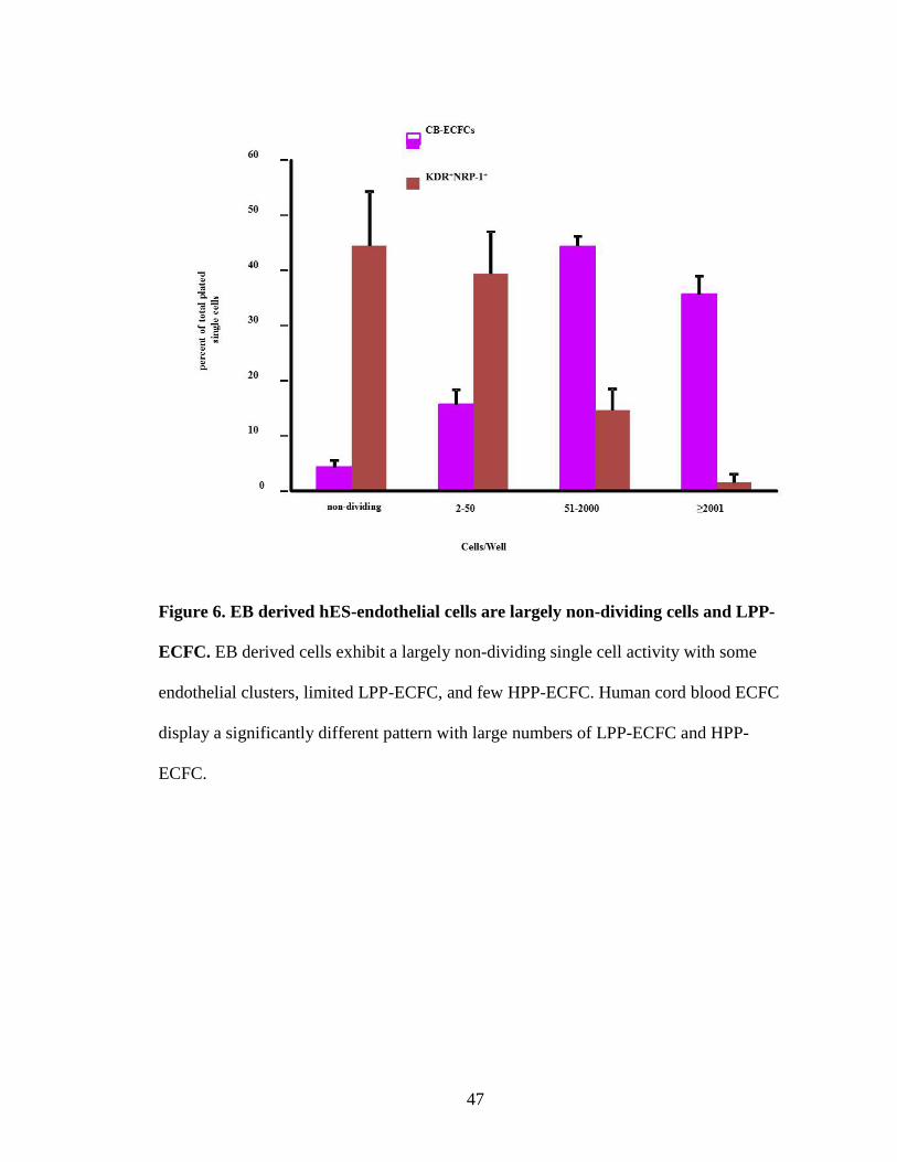
When we analyzed these cells (passage 3 to 4) for clonal proliferative potential we scored
the outcomes as single cells that did not divide or divided to form colonies of 2-50
(ECFC clusters), 51-500 (low proliferative potential ECFC; LPP-ECFC), 501-2000 (LPP-
ECFC), or >2000 cells (high proliferative potential-ECFC; HPP-ECFC). Less than 2% of
the EB-derived cells gave rise to HPP-ECFC (Fig. 6). More of the EB-derived
endothelial cells gave rise to LPP-ECFC (Fig. 6). Most of the EB-derived endothelial
cells did not divide or give rise to ECFC clusters (Fig. 6). These patterns of ECFC colony
formation were significantly different from the pattern displayed by single endothelial
cells derived from CB-ECFC colonies (Fig. 6). We were unable to expand the EB-
derived endothelial cells beyond passage 7 without them becoming senescent or drifting
into a non-endothelial fibroblastic phenotype and the endothelial cells at passage 5 failed
to give rise to human blood vessels in vivo upon implantation (Fig. 5).
Endothelial lineage differentiation of human embryonic stem (hES) cells results in
generation of subsets of cells that differ in phenotype and proliferative potential
38
Using a more recently published 2 step endothelial differentiation protocol that
involves initial EB formation and then 2D adherent cell culture (with added growth
factors)[66], we next sought to determine whether endothelial lineage differentiation of
hES cells would give rise to different subsets of endothelial cells, including a subset
displaying ECFC properties. Based upon the known importance of vascular endothelial
growth factor (VEGF) signaling pathway in the emergence of endothelial cells both
during development[69-71] and during endothelial lineage differentiation of hES
cells[33], we utilized neuropilin-1 (NRP-1) as one of the endothelial markers for
identifying emergence of ECFCs from hES cells. NRP-1 is a VEGF co-receptor and
Semaphorin 3A binding multifunctional protein expressed in various tissues including
endothelial cells, vascular smooth muscle cells and lymphocytes[72]. While the role of
NRP-1 in vasculogenesis is unknown, a double knock out of NRP-1 and NRP-2 in mice
leads to an embryonic lethal phenotype similar to that of the VEGFR-2 knockout[73, 74].
NRP-1 and NRP-2 double knockout mice die at day 8.5 and display an avascular yolk
sac[73]. Conversely, exogenous over-expression of NRP-1 has previously been shown to
form excess capillaries and hemorrhages in the developing cardiovascular, brain, and
limb regions of the chimeric mouse embryos[75]. Since NRP-1 has been suggested as a
potential early marker for the endothelial lineage during hES cell differentiation[33], we
hypothesized that NRP-1 expression early in hES differentiating down the endothelial
lineage may identify the emergence of ECFCs. We generated hES (H9 line) EBs in
suspension culture for 4 days, and seeded them on Matrigel coated dishes for 10 days as
described[34]. Cells were harvested and analyzed using antibodies that recognize typical
endothelial antigens (CD31, CD144, and CD146) and NRP-1 on days 0, 3, 6, 9 and 14, to
39
determine the kinetics of endothelial antigen expression during the differentiation
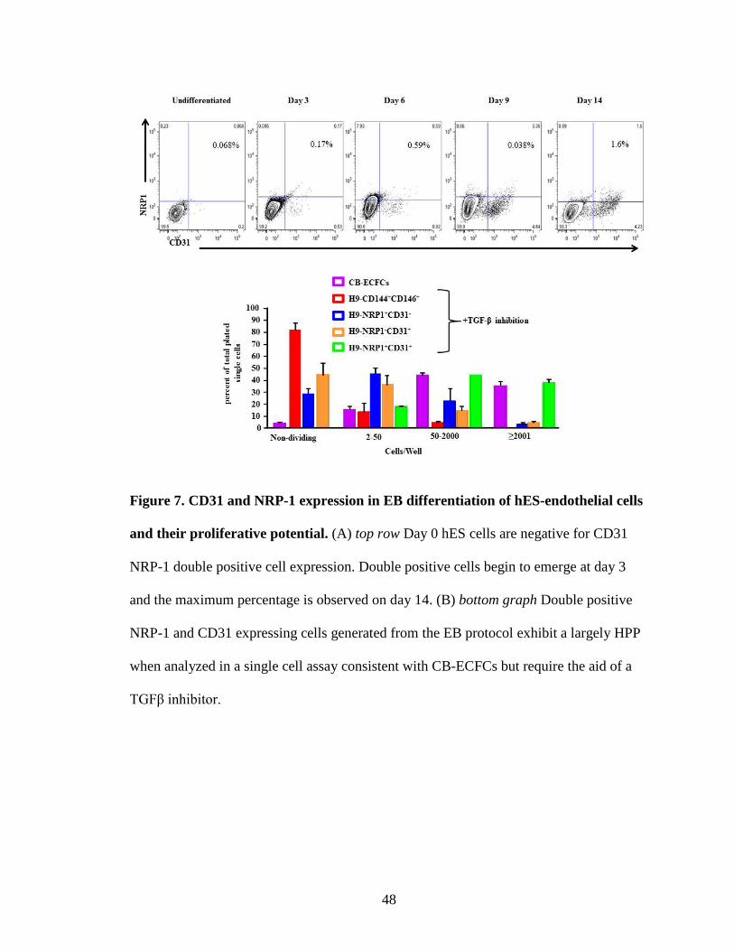
protocol (Fig. 7). Cells co-expressing NRP-1 and CD31 (NRP-1+CD31+ cells) appeared
on day 3 (0.17%) and increased overtime to peak at day 14 (1.6%) (Fig. 7). The different
subsets of day 14 sorted cells were cultured in endothelial growth (EGM-2) media
supplemented with TGFβ inhibitor (10 μM SB431542) for 2 weeks, as TGFβ inhibition
has been reported to promote endothelial lineage differentiation from hES cells and to
prevent the cells from transitioning to a mesenchymal fate[34]. All of these subsets
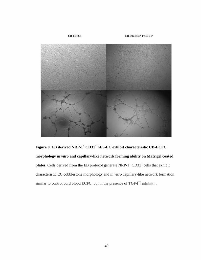
survived in endothelial growth media and formed confluent monolayers of cells. While
most hES-derived subsets formed incomplete capillary-like networks upon plating in
Matrigel, NRP-1+CD31+ cells formed complete structures similar to those exhibited by
CB derived ECFCs, (Fig. 8). When the endothelial subsets were seeded at a clonal level,
more than 55% of NRP-1+CD31- cells and more than 70% of NRP-1-CD31+ (Fig. 7) cells
divided and displayed a near complete hierarchy of clonal proliferative potential.
However, few clones from each of the sorted hES derived endothelial subsets gave rise to
HPP-ECFC (the most proliferative population derived from a single input cell). In
contrast, all of the NRP-1+CD31+ plated cells divided and many clones (37%) formed
HPP-ECFC, indicating that the NRP-1+CD31+ cell subset possesses ECFC properties
with the highest clonal proliferative potential (Fig. 7). Thus co-expression of NRP-1 and
CD31 in hES-derived cells undergoing endothelial differentiation (EB plus 2D protocol)
identifies a progenitor subset that gives rise to endothelial cells with high clonal
proliferative potential and angiogenic activity, but only if cultured in the continual
presence of TGF-β inhibition (upon removal of the TGF-β inhibitor, cells ceased to
proliferate, changed morphology from cobblestone to spindle elongated shapes, and
40
upregulated alpha-smooth muscle actin [α-SMA] a marker of mesenchymal
differentiation similar to previously published data)[66].
Novel ECFC protocol generates NRP-1+CD31+ cells from hES cells at higher
frequency and does not require TGFβ inhibition
While a two-step, EB plus 2 dimensional endothelial differentiation protocol was
able to give rise to NRP-1+CD31+ cells with clonal high proliferative potential, this was
an inefficient process that generated heterogeneous EBs containing cells from all germ
layers, required persistent presence of TGF-β inhibition to maintain the endothelial fate,
and yielded a low percentage of NRP-1+CD31+ cells. Therefore, we sought to develop an
endothelial lineage differentiation protocol that did not require EB formation and TGF-β
inhibition but ensured early preferential differentiation of hES cells towards the
mesodermal lineage to give rise to an increased yield of the NRP-1+CD31+ cells
possessing ECFC properties. Human PS cells were cultured on Matrigel coated plates in
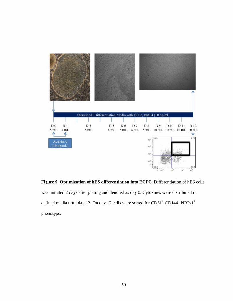
mTeSR1 media for two days as described[76]. To induce endothelial lineage
differentiation, mTeSR1 media was replaced with Stemline II media supplemented with
10 ng/mL Activin-A, BMP4, VEGF165 and FGF2 on day 0 of differentiation. The tissue
culture media was replaced the following day with fresh Stemline II media supplemented
with 10 ng/mL BMP4, VEGF165 and FGF2. Cells were harvested at day12 and analyzed
for cells co-expressing CD31, CD144 and NRP-1 antigens (Fig. 9). Using this protocol,
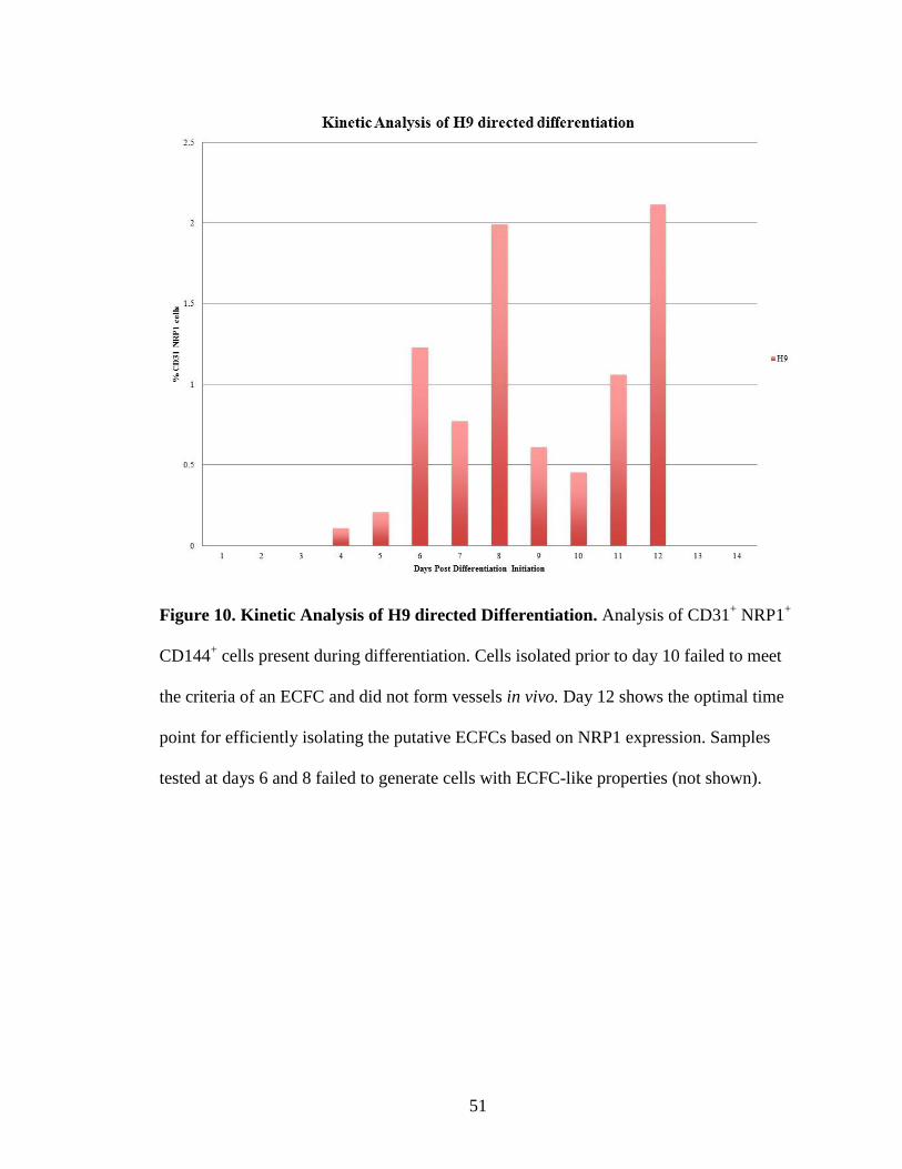
we were able to harvest an average 2% NRP-1+CD31+ cells from hES cells (Fig. 10). We
directly compared NRP-1+CD31+ and NRP-1-CD31+ cells for their ability to give rise to
endothelial colonies and total cell expansion after 7 days of plating in an endothelial
41
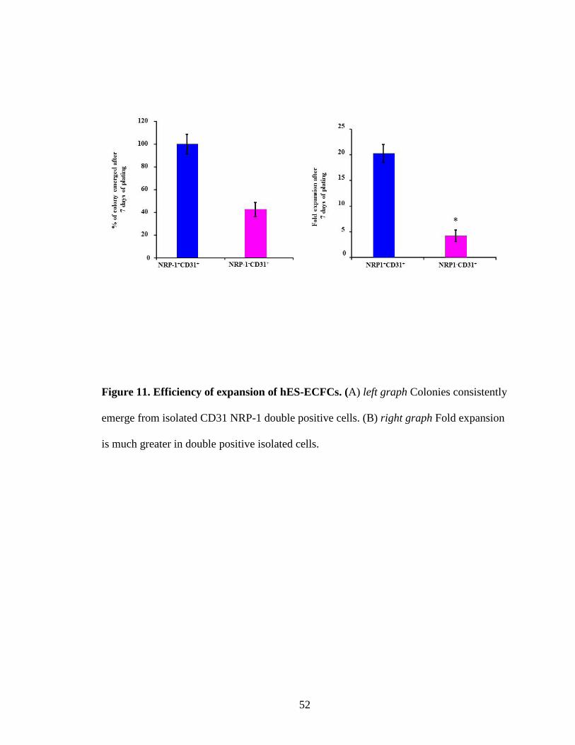
transitioning media. When we plated 2500 NRP-1+CD31+ and NRP-CD31+ sorted cells in
a well of a 12 well collagen coated plate, NRP-1+CD31- cells gave rise to significantly
fewer endothelial colonies compared to colonies generated from plated NRP-1+CD31+
cells (Fig. 11). Also, the morphology of NRP-1+CD31+ progeny was homogenous and
cobblestone in appearance (Fig. 12; top right panel), whereas, we found a heterogeneous
cell population within the colonies obtained from NRP-1-CD31+ cells. Finally, we
counted the total number of cells in these colonies after 7 days of culture and found a
significant (15 fold) increase in cultures initiated with NRP-1+CD31+ cells compared to
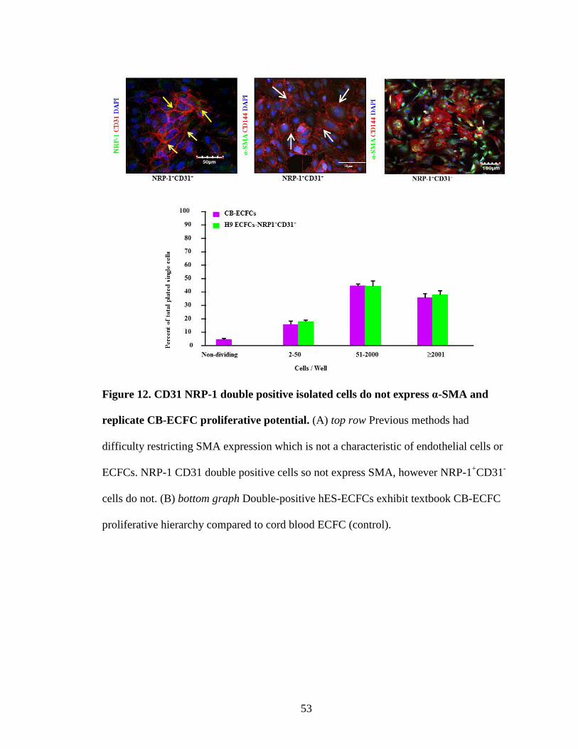
CD31+NRP- cells (Fig. 11). Further we performed immunocytochemistry analysis of both
NRP-1+CD31+ and NRP-CD31+ sorted cells using antibodies recognizing the endothelial
antigens CD31, CD144 and NRP-1 and the non-endothelial antigen α-SMA. Cells grown
from the NRP-1+CD31+ sorted fraction exhibited surface co-expression of CD31 and
NRP-1 (yellow arrows, Fig. 12; top left panel) and uniform expression of CD144 but
completely lacked expression of α-SMA (white arrows, Fig. 12; top middle panel).
However, in cells produced from the CD31+NRP- sorted fraction, few cells expressed
CD144 and almost the entire cell subset expressed α-SMA (arrows, Fig. 12; top right
panel), indicating a drift away from an endothelial phenotype. Finally we examined hES
derived NRP-1+CD31+ and NRP-CD31+ cells to determine the clonal proliferative
behavior of the cells and found that both cell subsets displayed some evidence of clonal
proliferative potential (Fig. 12). While more than 27% of cells derived from the
CD31+NRP- subset failed to divide, 45% formed endothelial clusters, 25% formed LPP-
ECFC and a mere 1% formed HPP-ECFC colonies. In contrast, only 2% of the NRP-
1+CD31+ cell subset failed to divide, 4% formed endothelial clusters, 45% formed LPP-
42
ECFC, and 48% formed HPP-ECFC (Fig. 12). Furthermore, NRP-1+CD31+ cells formed
highly branching capillary-tube like structures (Fig. 13), whereas, CD31+NRP- cells
formed incomplete capillary-like networks when plated on Matrigel (Fig. 13). Finally,
hES derived NRP-1+CD31+ cells produced endothelial cells with robust in vivo vessel
forming ability that inosculated with the host murine vessels (Fig. 13) similar to that of
CB-ECFCs previously described[2, 5]. Importantly, NRP-1+CD31+ cells did not induce
teratoma formation after more than 3 months of implantation into immunodeficient mice
in more than 24 animals (data not shown). However, CD31+NRP- cells failed to generate
functional human blood vessels (documented by the intraluminal presence of murine
RBCs), indicating lack of de novo vessel forming and/or inosculating ability (Fig. 13).
Collectively, ECFC protocol day12 derived NRP-1+CD31+ cells exhibited homogenous
cobblestone morphology, expressed typical endothelial antigens, formed capillary-like
networks on Matrigel in vitro, exhibited high clonal proliferative potential, and produced
robust in vivo vessels filled with host RBCs. Therefore, the day 12 hES derived NRP-
1+CD31+ cell fraction produces endothelial cells that possess properties similar to cord
blood ECFC.
NRP-1+CD31+ cells emerge as clusters of cells lacking a-SMA expression and upon
isolation and culture undergo extensive expansion, maintain stable endothelial
phenotype, and ultimately become senescent after long-term culture expansion
While hESD12 differentiated cells possess complete ECFC properties, the earliest
emergence of cells co-expressing CD31 and NRP-1 during hES differentiation had not
been determined. Cimato et al. reported that NRP-1 is not expressed in undifferentiated

43
hES cells. Our data also revealed that day 0 undifferentiated hES cells did not co-express
CD31 and NRP-1 (Fig. 10). We determined that NRP-1+CD31+ cells start to appear at
day 4 in hES cells undergoing ECFC differentiation (Fig. 10). The percentage of NRP-
1+CD31+ cells progressively increased over time and reached the highest levels at day 12
of differentiation(Fig. 10). Morphological analysis of hES cells undergoing ECFC
differentiation at day 6, day 9, and day 12 revealed emergence of cobblestone like
morphology at day 9 and day 12 (Fig. 14). Also, IHC revealed that NRP-1+CD31+ cells
emerge as a cluster of cells within the mass of differentiating cells (Fig. 15). Further
examination revealed that while hES-derived NRP-1+CD31+ cells emerged as early as
day 3 of differentiation, the endothelial cells derived at day 12 gave rise to ECFC that
exhibited the highest frequency of co-expression of the typical endothelial antigens
without expression of the mesenchymal antigen α-SMA (Fig. 14,15).
Next, we sought to examine the ability of hES-ECFCs to undergo long term
expansion by continually culturing them in extended endothelial culture until they began
to exhibit evidence of replicative senescence. Primary cells do not proliferate indefinitely
but instead undergo senescence after long term in vitro culture[77]. In extended
endothelial culture, we were able to expand both hES-ECFCs and CB-ECFCs up to 18
passages (Fig. 16). While the majority of hES-ECFCs and CB-ECFCs at passage 18 were
senescent and were exhibiting characteristics of mortal primary cells[77], they still
maintained an endothelial cobblestone morphology. We also detected continued
homogenous expression of endothelial CD31, NRP-1 and CD144 antigen expression in
these cells at passage 18 (Fig. 16). Collectively, this suggests that hES-ECFCs maintain a

44
stable endothelial phenotype throughout long term expansion culture but like any primary
cell type, eventually exhibit replicative senescence.
45
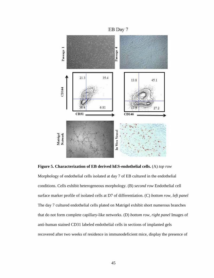
Figure 5. Characterization of EB derived hES-endothelial cells. (A) top row
Morphology of endothelial cells isolated at day 7 of EB cultured in the endothelial
conditions. Cells exhibit heterogeneous morphology. (B) second row Endothelial cell
surface marker profile of isolated cells at D7 of differentiation. (C) bottom row, left panel
The day 7 cultured endothelial cells plated on Matrigel exhibit short numerous branches
that do not form complete capillary-like networks. (D) bottom row, right panel Images of
anti-human stained CD31 labeled endothelial cells in sections of implanted gels
recovered after two weeks of residence in immunodeficient mice, display the presence of

46
human endothelial cells but lack vascular lumen formation (no presence of murine red
blood cells to indicate inosculation of the human vessels to the mouse circulation).
47
Figure 6. EB derived hES-endothelial cells are largely non-dividing cells and LPP-
ECFC. EB derived cells exhibit a largely non-dividing single cell activity with some
endothelial clusters, limited LPP-ECFC, and few HPP-ECFC. Human cord blood ECFC
display a significantly different pattern with large numbers of LPP-ECFC and HPP-
ECFC.
48
Figure 7. CD31 and NRP-1 expression in EB differentiation of hES-endothelial cells
and their proliferative potential. (A) top row Day 0 hES cells are negative for CD31
NRP-1 double positive cell expression. Double positive cells begin to emerge at day 3
and the maximum percentage is observed on day 14. (B) bottom graph Double positive
NRP-1 and CD31 expressing cells generated from the EB protocol exhibit a largely HPP
when analyzed in a single cell assay consistent with CB-ECFCs but require the aid of a
TGFβ inhibitor.
49
Figure 8. EB derived NRP-1+ CD31+ hES-EC exhibit characteristic CB-ECFC
morphology in vitro and capillary-like network forming ability on Matrigel coated
plates. Cells derived from the EB protocol generate NRP-1+ CD31+ cells that exhibit
characteristic EC cobblestone morphology and in vitro capillary-like network formation
similar to control cord blood ECFC, but in the presence of TGF- inhibitor.
50
Figure 9. Optimization of hES differentiation into ECFC. Differentiation of hES cells
was initiated 2 days after plating and denoted as day 0. Cytokines were distributed in
defined media until day 12. On day 12 cells were sorted for CD31+ CD144+ NRP-1+
phenotype.
51
Figure 10. Kinetic Analysis of H9 directed Differentiation. Analysis of CD31+ NRP1+
CD144+ cells present during differentiation. Cells isolated prior to day 10 failed to meet
the criteria of an ECFC and did not form vessels in vivo. Day 12 shows the optimal time
point for efficiently isolating the putative ECFCs based on NRP1 expression. Samples
tested at days 6 and 8 failed to generate cells with ECFC-like properties (not shown).
52
Figure 11. Efficiency of expansion of hES-ECFCs. (A) left graph Colonies consistently
emerge from isolated CD31 NRP-1 double positive cells. (B) right graph Fold expansion
is much greater in double positive isolated cells.
53
Figure 12. CD31 NRP-1 double positive isolated cells do not express α-SMA and
replicate CB-ECFC proliferative potential. (A) top row Previous methods had
difficulty restricting SMA expression which is not a characteristic of endothelial cells or
ECFCs. NRP-1 CD31 double positive cells so not express SMA, however NRP-1+CD31-
cells do not. (B) bottom graph Double-positive hES-ECFCs exhibit textbook CB-ECFC
proliferative hierarchy compared to cord blood ECFC (control).

54
Figure 13. CD31 NRP-1 double positive isolated cells form in vitro networks and
vessels in vivo. (A) While in vitro network formation of the Nrp-1+CD31+ cells is
robust, NRP-1+CD31- cells fail to form complete networks. (B) Murine red blood cell
perfused vessels stained with anti-human CD31 coupled with H&E staining shows the
ability of Nrp-1+CD31+ but not NRP-1+CD31- hES-ECFCs to integrate into the host
vasculature system
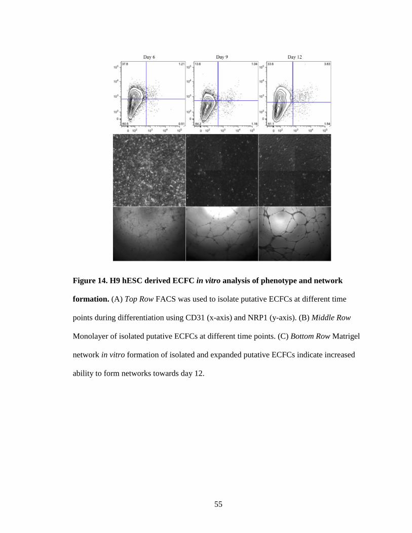
55
Figure 14. H9 hESC derived ECFC in vitro analysis of phenotype and network
formation. (A) Top Row FACS was used to isolate putative ECFCs at different time
points during differentiation using CD31 (x-axis) and NRP1 (y-axis). (B) Middle Row
Monolayer of isolated putative ECFCs at different time points. (C) Bottom Row Matrigel
network in vitro formation of isolated and expanded putative ECFCs indicate increased
ability to form networks towards day 12.

56
Figure 15. H9 hESC derived ECFC immunohistochemical kinetic analysis of
endothelial antigen expression. Differentiation was initiated in 12 well tissue culture
dishes and fixed at varying time points. The wells were then stained for endothelial
antigens and α-SMA. (A) top row unstained control (B) middle Row NRP1 staining
indicates the increase in expression towards day 12. CD31 staining indicates the increase
in expression towards day 12. (C) bottom Row αSMA staining indicates the absence of
expression at day 12. CD144 staining indicates the increase in expression at day 12 in a
pattern reminiscent of capillary-like structures.
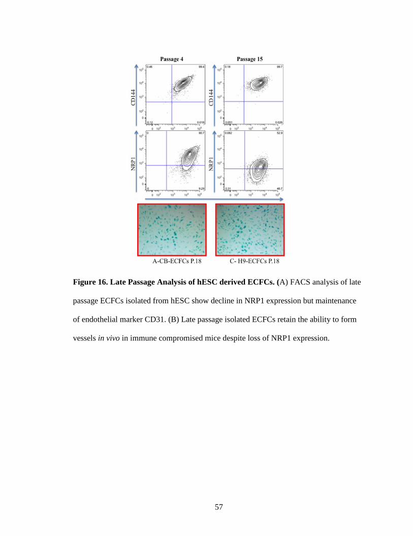
57
Figure 16. Late Passage Analysis of hESC derived ECFCs. (A) FACS analysis of late
passage ECFCs isolated from hESC show decline in NRP1 expression but maintenance
of endothelial marker CD31. (B) Late passage isolated ECFCs retain the ability to form
vessels in vivo in immune compromised mice despite loss of NRP1 expression.

58
Discussion
We have established a method to reproducibly derive and isolate a homogenous
and stable population of endothelial cells possessing umbilical cord blood ECFC-like
properties. NRP-1+CD31+ cells formed a homogenous monolayer with characteristic
cobblestone appearance, exhibited high clonal proliferative potential, demonstrated
angiogenic behavior by forming complete capillary like structures when cultured on
Matrigel, and formed robust in vivo inosculated vessels when implanted in immune
deficient mice. These hES-derived ECFCs were stable and did not transition to non-
endothelial cells over prolonged culture (18 passages) and could expand to over a trillion
endothelial cells in less than 3 months of culture.
Timing is essential in the derivation of cell lineages from hES cells. Because of
this we developed a detailed protocol and optimized the timing of growth factor delivery
to most efficiently isolate ECFCs using the novel marker NRP-1. We found that by
optimizing the timing of media alone we were able to substantially increase the efficiency
of isolation using this marker. The stepwise differentiation protocol and the use of the
double positive expression of NRP-1 and CD31 permitted isolation of cells with ECFC
properties for the first time. While endothelial cells have been isolated from human
pluripotent cells on many occasions previously, the cells derived failed to display
persistent high proliferative potential and were noted to reach senescence after relatively
few passages. Some previously reported protocols also required TGF-β inhibition both
for the derivation and maintenance of a stable endothelial cell phenotype from
differentiated hPS cells[34]. ECFCs generated from our methods exhibit a stable
endothelial phenotype and display a complete hierarchy of clonal high proliferative
59
potential and form robust in vivo functional human vessels that inosculate with host
blood vascular system in the complete absence of TGF-β inhibition. Our ECFC
differentiation protocol yields one NRP-1+CD31+ ECFC per each input hESc after 12
days of differentiation. More importantly, upon expansion, these NRP-1+CD31+ cells
generate stable highly proliferative potential cells that can undergo long term expansion
reaching a ratio of hES/ECFC: 1 to 1.1x10⁸ in less than 3 months of culture. This highly
efficient output of functional endothelial cells contrasts with reported yields of 0.6[31],
7.4[34] and 11.6[78] endothelial cells from differentiated hPS cells using other published
protocols. Thus, our method is highly scalable for potential clinical application and can
generate cells sufficient for permitting future clinical trials to regenerate the deficient
microvasculature in patients with cardiovascular diseases such as PAD and CLI.
Another key factor in the feasibility of implementation in clinical trials is not only
efficacy of the treatments but the potential adverse effects associated with them. The
ECFC derived in our protocol are not transformed which is shown by their normal
behavior in the characterization of their ECFC qualities. In addition, the cells senesce at
comparable passages as CB-ECFCs which indicates that telomere and telomerase activity
have not been altered in the directed differentiation of hES-ECFCs. Thus, our culture
derived cells are mortal in behavior in vitro and in vivo.
In no instance did these differentiated cells form a teratoma, a benign growth
which displays differentiated cell types from all 3 germ layers, when implanted in
immunocompromised mice. This proves that these cells are no longer undifferentiated
and have assumed a specified state (ECFC state) since all hES cells form teratomas upon

60
injection as pluripotent cells. This is a key factor which helps to ensure that this cell type
when administered to patients would be only positive in nature.

61
References

62
1. Ingram, D.A., et al., Identification of a novel hierarchy of endothelial progenitor cells using human peripheral and umbilical cord blood. Blood, 2004. 104(9): p. 2752-60.
2. Yoder, M.C., et al., Redefining endothelial progenitor cells via clonal analysis and hematopoietic stem/progenitor cell principals. Blood, 2007. 109(5): p. 1801-9.
3. Ingram, D.A., et al., Vessel wall-derived endothelial cells rapidly proliferate because they contain a complete hierarchy of endothelial progenitor cells. Blood, 2005. 105(7): p. 2783-6.
4. Au, P., et al., Differential in vivo potential of endothelial progenitor cells from human umbilical cord blood and adult peripheral blood to form functional long-lasting vessels. Blood, 2008. 111(3): p. 1302-5.
5. Critser, P.J., et al., Collagen matrix physical properties modulate endothelial colony forming cell-derived vessels in vivo. Microvasc Res. 80(1): p. 23-30.
6. Melero-Martin, J.M., et al., Engineering robust and functional vascular networks in vivo with human adult and cord blood-derived progenitor cells. Circ Res, 2008. 103(2): p. 194-202.
7. Melero-Martin, J.M., et al., In vivo vasculogenic potential of human blood-derived endothelial progenitor cells. Blood, 2007. 109(11): p. 4761-8.
8. Lin, Y., et al., Origins of circulating endothelial cells and endothelial outgrowth from blood. J Clin Invest, 2000. 105(1): p. 71-7.
9. Ikpeazu, C., et al., Donor origin of circulating endothelial progenitors after allogeneic bone marrow transplantation. Biol Blood Marrow Transplant, 2000. 6(3A): p. 301-8.
10. Huang, L., et al., Acute myocardial infarction in swine rapidly and selectively releases highly proliferative endothelial colony forming cells (ECFCs) into circulation. Cell Transplant, 2007. 16(9): p. 887-97.
11. Meneveau, N., et al., Presence of endothelial colony-forming cells is associated with reduced microvascular obstruction limiting infarct size and left ventricular remodelling in patients with acute myocardial infarction. Basic Res Cardiol. 106(6): p. 1397-410.
12. Massa, M., et al., Rapid and large increase of the frequency of circulating endothelial colony-forming cells (ECFCs) generating late outgrowth endothelial cells in patients with acute myocardial infarction. Exp Hematol, 2009. 37(1): p. 8-9.
13. Moubarik, C., et al., Transplanted late outgrowth endothelial progenitor cells as cell therapy product for stroke. Stem Cell Rev. 7(1): p. 208-20.
14. Lee, J.H., et al., CD34 Hybrid Cells Promote Endothelial Colony-Forming Cell Bioactivity and Therapeutic Potential for Ischemic Diseases. Arterioscler Thromb Vasc Biol.
15. Schwarz, T.M., et al., Vascular incorporation of endothelial colony-forming cells is essential for functional recovery of murine ischemic tissue following cell therapy. Arterioscler Thromb Vasc Biol. 32(2): p. e13-21.
16. Bouvard, C., et al., alpha6-integrin subunit plays a major role in the proangiogenic properties of endothelial progenitor cells. Arterioscler Thromb Vasc Biol. 30(8): p. 1569-75.
63
17. Saif, J., et al., Combination of injectable multiple growth factor-releasing scaffolds and cell therapy as an advanced modality to enhance tissue neovascularization. Arterioscler Thromb Vasc Biol. 30(10): p. 1897-904.
18. Dubois, C., et al., Differential effects of progenitor cell populations on left ventricular remodeling and myocardial neovascularization after myocardial infarction. J Am Coll Cardiol. 55(20): p. 2232-43.
19. Schuh, A., et al., Transplantation of endothelial progenitor cells improves neovascularization and left ventricular function after myocardial infarction in a rat model. Basic Res Cardiol, 2008. 103(1): p. 69-77.
20. Stitt, A.W., et al., Vascular stem cells and ischaemic retinopathies. Prog Retin Eye Res. 30(3): p. 149-66.
21. Medina, R.J., et al., Outgrowth endothelial cells: characterization and their potential for reversing ischemic retinopathy. Invest Ophthalmol Vis Sci. 51(11): p. 5906-13.
22. Solovey, A., et al., Circulating activated endothelial cells in sickle cell anemia. N Engl J Med, 1997. 337(22): p. 1584-90.
23. Reinisch, A., et al., Humanized large-scale expanded endothelial colony-forming cells function in vitro and in vivo. Blood, 2009. 113(26): p. 6716-25.
24. Heiss, C., et al., Impaired progenitor cell activity in age-related endothelial dysfunction. J Am Coll Cardiol, 2005. 45(9): p. 1441-8.
25. Wang, C.H., et al., Factors associated with purity, biological function, and activation potential of endothelial colony-forming cells. Am J Physiol Regul Integr Comp Physiol. 300(3): p. R586-94.
26. Shelley, W.C., et al., Changes in the frequency and in vivo vessel-forming ability of rhesus monkey circulating endothelial colony-forming cells across the lifespan (birth to aged). Pediatr Res. 71(2): p. 156-61.
27. Robbins, R.D., et al., Inducible pluripotent stem cells: not quite ready for prime time? Curr Opin Organ Transplant. 15(1): p. 61-7.
28. Broxmeyer, H.E., et al., Hematopoietic stem/progenitor cells, generation of induced pluripotent stem cells, and isolation of endothelial progenitors from 21- to 23.5-year cryopreserved cord blood. Blood. 117(18): p. 4773-7.
29. Lee, M.R., et al., Epigenetic regulation of NANOG by miR-302 cluster-MBD2 completes induced pluripotent stem cell reprogramming. Stem Cells. 31(4): p. 666-81.
30. Wang, L., et al., Endothelial and hematopoietic cell fate of human embryonic stem cells originates from primitive endothelium with hemangioblastic properties. Immunity, 2004. 21(1): p. 31-41.
31. Cho, S.W., et al., Improvement of postnatal neovascularization by human embryonic stem cell derived endothelial-like cell transplantation in a mouse model of hindlimb ischemia. Circulation, 2007. 116(21): p. 2409-19.
32. Choi, K.D., et al., Hematopoietic and endothelial differentiation of human induced pluripotent stem cells. Stem Cells, 2009. 27(3): p. 559-67.
33. Cimato, T., et al., Neuropilin-1 identifies endothelial precursors in human and murine embryonic stem cells before CD34 expression. Circulation, 2009. 119(16): p. 2170-8.

64
34. James, D., et al., Expansion and maintenance of human embryonic stem cell-derived endothelial cells by TGFbeta inhibition is Id1 dependent. Nat Biotechnol.
35. Levenberg, S., et al., Endothelial cells derived from human embryonic stem cells. Proc Natl Acad Sci U S A, 2002. 99(7): p. 4391-6.
36. Nakahara, M., et al., High-efficiency production of subculturable vascular endothelial cells from feeder-free human embryonic stem cells without cell-sorting technique. Cloning Stem Cells, 2009. 11(4): p. 509-22.
37. Vodyanik, M.A., et al., Human embryonic stem cell-derived CD34+ cells: efficient production in the coculture with OP9 stromal cells and analysis of lymphohematopoietic potential. Blood, 2005. 105(2): p. 617-26.
38. Sone, M., et al., Pathway for differentiation of human embryonic stem cells to vascular cell components and their potential for vascular regeneration. Arterioscler Thromb Vasc Biol, 2007. 27(10): p. 2127-34.
39. Taura, D., et al., Induction and isolation of vascular cells from human induced pluripotent stem cells--brief report. Arterioscler Thromb Vasc Biol, 2009. 29(7): p. 1100-3.
40. Rufaihah, A.J., et al., Endothelial cells derived from human iPSCS increase capillary density and improve perfusion in a mouse model of peripheral arterial disease. Arterioscler Thromb Vasc Biol. 31(11): p. e72-9.
41. Yamahara, K., et al., Augmentation of neovascularization [corrected] in hindlimb ischemia by combined transplantation of human embryonic stem cells-derived endothelial and mural cells. PLoS One, 2008. 3(2): p. e1666.
42. Lai, W.H., et al., Attenuation of hind-limb ischemia in mice with endothelial-like cells derived from different sources of human stem cells. PLoS One. 8(3): p. e57876.
43. Ginsberg, M., et al., Efficient direct reprogramming of mature amniotic cells into endothelial cells by ETS factors and TGFbeta suppression. Cell. 151(3): p. 559-75.
44. Goldman, O., et al., A boost of BMP4 accelerates the commitment of human embryonic stem cells to the endothelial lineage. Stem Cells, 2009. 27(8): p. 1750-9.
45. Feng, Q., et al., Hemangioblastic derivatives from human induced pluripotent stem cells exhibit limited expansion and early senescence. Stem Cells. 28(4): p. 704-12.
46. Nishikawa, S.I., et al., In vitro generation of lymphohematopoietic cells from endothelial cells purified from murine embryos. Immunity, 1998. 8(6): p. 761-9.
47. Rossignol, M., J. Pouyssegur, and M. Klagsbrun, Characterization of the neuropilin-1 promoter; gene expression is mediated by the transcription factor Sp1. J Cell Biochem, 2003. 88(4): p. 744-57.
48. Carmeliet, P. and R.K. Jain, Molecular mechanisms and clinical applications of angiogenesis. Nature. 473(7347): p. 298-307.
49. Asahara, T., et al., Isolation of putative progenitor endothelial cells for angiogenesis. Science, 1997. 275(5302): p. 964-7.
50. Urbich, C. and S. Dimmeler, Endothelial progenitor cells: characterization and role in vascular biology. Circ Res, 2004. 95(4): p. 343-53.
65
51. Critser, P.J., S.L. Voytik-Harbin, and M.C. Yoder, Isolating and defining cells to engineer human blood vessels. Cell Prolif. 44 Suppl 1: p. 15-21.
52. Matthias, M., N. David, and N. Josef, From bench to bedside: what physicians need to know about endothelial progenitor cells. Am J Med. 124(6): p. 489-97.
53. Reinisch, A. and D. Strunk, Isolation and animal serum free expansion of human umbilical cord derived mesenchymal stromal cells (MSCs) and endothelial colony forming progenitor cells (ECFCs). J Vis Exp, 2009(32).
54. Hofmann, N.A., A. Reinisch, and D. Strunk, Isolation and large scale expansion of adult human endothelial colony forming progenitor cells. J Vis Exp, 2009(32).
55. Witting, S.R., et al., Efficient Large Volume Lentiviral Vector Production Using Flow Electroporation. Hum Gene Ther.
56. Yoon, C.H., et al., Synergistic neovascularization by mixed transplantation of early endothelial progenitor cells and late outgrowth endothelial cells: the role of angiogenic cytokines and matrix metalloproteinases. Circulation, 2005. 112(11): p. 1618-27.
57. Norgren, L., et al., Inter-Society Consensus for the Management of Peripheral Arterial Disease (TASC II). Eur J Vasc Endovasc Surg, 2007. 33 Suppl 1: p. S1-75.
58. Brevetti, G., V. Schiano, and M. Chiariello, Endothelial dysfunction: a key to the pathophysiology and natural history of peripheral arterial disease? Atherosclerosis, 2008. 197(1): p. 1-11.
59. Eberhardt, R.T. and J.D. Coffman, Cardiovascular morbidity and mortality in peripheral arterial disease. Curr Drug Targets Cardiovasc Haematol Disord, 2004. 4(3): p. 209-17.
60. Hirsch, A.T., et al., Peripheral arterial disease detection, awareness, and treatment in primary care. JAMA, 2001. 286(11): p. 1317-24.
61. Wang, Z.Z., et al., Endothelial cells derived from human embryonic stem cells form durable blood vessels in vivo. Nat Biotechnol, 2007. 25(3): p. 317-8.
62. James, D., et al., Expansion and maintenance of human embryonic stem cell-derived endothelial cells by TGFbeta inhibition is Id1 dependent. Nat Biotechnol, 2010. 28(2): p. 161-6.
63. Djordjevic, S. and P.C. Driscoll, Targeting VEGF signalling via the neuropilin co-receptor. Drug Discov Today, 2012.
64. Liu, Y., et al., Tip110 maintains expression of pluripotent factors in and pluripotency of human embryonic stem cells. Stem Cells Dev, 2012. 21(6): p. 829-33.
65. Bailey, J.L., et al., Collagen oligomers modulate physical and biological properties of three-dimensional self-assembled matrices. Biopolymers, 2011. 95(2): p. 77-93.
66. Prasain, N., J.L. Meador, and M.C. Yoder, Phenotypic and functional characterization of endothelial colony forming cells derived from human umbilical cord blood. J Vis Exp, 2012(62).
67. Nourse, M.B., et al., VEGF induces differentiation of functional endothelium from human embryonic stem cells: implications for tissue engineering. Arterioscler Thromb Vasc Biol. 30(1): p. 80-9.
66
68. Lu, S.J., et al., Generation of functional hemangioblasts from human embryonic stem cells. Nat Methods, 2007. 4(6): p. 501-9.
69. Carmeliet, P., et al., Abnormal blood vessel development and lethality in embryos lacking a single VEGF allele. Nature, 1996. 380(6573): p. 435-9.
70. Ferrara, N. and T. Davis-Smyth, The biology of vascular endothelial growth factor. Endocr Rev, 1997. 18(1): p. 4-25.
71. Gerber, H.P., et al., VEGF is required for growth and survival in neonatal mice. Development, 1999. 126(6): p. 1149-59.
72. Staton, C.A., et al., Neuropilins in physiological and pathological angiogenesis. J Pathol, 2007. 212(3): p. 237-48.
73. Takashima, S., et al., Targeting of both mouse neuropilin-1 and neuropilin-2 genes severely impairs developmental yolk sac and embryonic angiogenesis. Proc Natl Acad Sci U S A, 2002. 99(6): p. 3657-62.
74. Shalaby, F., et al., Failure of blood-island formation and vasculogenesis in Flk-1-deficient mice. Nature, 1995. 376(6535): p. 62-6.
75. Kitsukawa, T., et al., Overexpression of a membrane protein, neuropilin, in chimeric mice causes anomalies in the cardiovascular system, nervous system and limbs. Development, 1995. 121(12): p. 4309-18.
76. Evseenko, D., et al., Mapping the first stages of mesoderm commitment during differentiation of human embryonic stem cells. Proc Natl Acad Sci U S A. 107(31): p. 13742-7.
77. Kuilman, T., et al., The essence of senescence. Genes Dev. 24(22): p. 2463-79. 78. Lippmann, E.S., et al., Derivation of blood-brain barrier endothelial cells from
human pluripotent stem cells. Nat Biotechnol. 30(8): p. 783-91.

Curriculum Vitae

J. Luke Meador
Education Indiana University Southeast, New Albany, IN
• Bachelor of Science in Chemistry ACS certified; Degree Awarded 2010 Specialization: Biochemistry Grade Point Average: 3.59 on 4.0 scale
• Bachelor of Arts in Chemistry and Business Management; Degree Awarded 2009 Specialization: Analytical Chemistry and Business Management Grade Point Average: 3.56 on 4.0 scale
Awards Indiana University School of Medicine, Indianapolis, IN
• T32 Hematopoiesis training fellowship; 2011-2012
Indiana University Southeast, New Albany, IN • IUS Paul Ogle Scholarship; 2005-2010
$5000/yr; Awarded for Outstanding High School G.P.A. and SAT scores
• IUS Chemistry Malcholm Kochert Scholarship; 2005-2009 $1000/yr increasing; Awarded for outstanding High School achievement for the pursuit of a degree in Chemistry
• Outstanding Chemistry Student; 2009 1 of 2 students selected for excellence in academics
• Dean’s List; 2005-2010 Awarded for obtaining a G.P.A. of 3.5or higher
• Who’s Who Among Students in American Universities and Colleges 1 of 2 Nominated by faculty for excellence in Academics in Chemistry
Teaching Experience Indiana University Southeast, New Albany, IN
• Teaching Assistant; Summer 2008 Teaching assistant for Introduction to Chemistry
• Supplemental Instructor; 2007-2010 Conducted outside tutor sessions at the request of faculty Taught: Principles of Economics, Principles of Chemistry, College Algebra
• Math Lab Tutor; 2009-2010 Provided a friendly and welcoming atmosphere to students in need of help with mathematics.
Activities and Community Service Floyd Central High School, Floyds Knobs, IN
• Head Diving Coach and Assistant Swimming Coach; 2005-2010 Indiana University Southeast, New Albany, IN
• Chemistry Club Member; 2006-2010 Avon High School, Avon, IN
• Diving Coach; 2011-2012 Indiana University School of Medicine
• Tour the Life Blog: Go Big or Go Home Publications Prasain N., Meador J. L., Yoder M. C., Phenotypic and Functional Characterization of Endothelial Colony Forming Cells Derived from Human Umbilical Cord Blood, Journal of Visualized Experiments, 2011. Nutan Prasain*, Man Ryul Lee*, Sasidhar Vemula, Jonathan Luke Meador, Momoko Yoshimoto, Michael J. Ferkowicz, Alexa Fett, Manav Gupta, Michael Ginsberg, Brian M. Rapp, Reza Saadatzadeh, Younghee Lee, Michael P. Murphy, Sherry L. Voytik-Harbin, Shahin Rafii, Hal E. Broxmeyer, and Mervin C. Yoder. Novel protocol to derive Endothelial Colony Forming Cells from human pluripotent stem cells that display high clonal proliferative potential, extensive replicative capacity, and in vivo vessel forming potential. In preparation for submission to Nature Medicine, 2013.